Preprint
Article

This version is not peer-reviewed.

Strain-Dependent In Vitro Susceptibility of Leptospira spp. to Isoquinoline Alkaloids - Chelerythrine and Sanguinarine

Submitted:

03 September 2026

Posted:

07 September 2026

You are already at the latest version

Abstract

Isoquinoline alkaloids such as sanguinarine and chelerythrine have demonstrated antimicrobial activity against a range of microorganisms; however, their effects on Leptospira spp. remain poorly characterized. In this study, we evaluated the in vitro susceptibility of multiple Leptospira strains to sanguinarine chloride, chelerythrine chloride, and a combined sanguinarine–chelerythrine fraction. Bacterial responses were assessed using flow cytometry, metabolic activity assays, and optical density measurements. The tested com-pounds exhibited limited and strain-dependent effects. The most pronounced response was observed in Leptospira interrogans serovar Australis (strain Balico), where chelerythrine chloride consistently reduced viability and metabolic activity under the conditions tested. In contrast, other strains showed minimal or inconsistent responses. The combined fraction did not demonstrate enhanced activity compared to individual compounds. These findings indicate that isoquinoline alkaloids exert variable and generally modest effects on Leptospira spp., highlighting substantial inter-strain heterogeneity in susceptibility. While the observed activity is limited, this work provides a preliminary assessment of these compounds in Leptospira and underscores the need for further studies to clarify their mechanisms of action and potential relevance.

Keywords: 
;  ;  ;  ;  

1. Introduction

As pathogens become increasingly resistant to antibiotics, active plant-derived compounds are emerging as a promising area of research to solve this problem [1,2,3,4,5,6,7]. Compounds with such potential are isoquinoline alkaloids, particularly sanguinarine and chelerythrine, which are prominent bioactive compounds biosynthesized in various plants of the Papaveraceae family, including Chelidonium majus, Sanguinaria canadensis, and Coptis chinensis [8,9,10]. These alkaloids are recognized for their broad spectrum of pharmacological activities, such as antimicrobial, an-ti-inflammatory, antifungal, and antitumor effects [8,11,12]. Sanguinarine and chelerythrine are most abundantly found in the roots and aerial parts of these plants, with sanguinarine generally present at higher concentrations than chelerythrine [8].
Both sanguinarine and chelerythrine have been reported to exhibit antimicrobial activity against a variety of mi-croorganisms [8,9,11,12,13]. However, most studies have focused on a limited number of model organisms, and to date, there are no published reports investigating their effects on Leptospira bacteria. Sanguinarine, in particular, has been shown to be more active than chelerythrine in several antimicrobial and anti-inflammatory assays, alt-hough exceptions exist [8,13].The antimicrobial properties of these alkaloids are attributed to their ability to inhibit the multiplication of microorganisms and disrupt cellular processes such as membrane integrity and cell prolifera-tion [13,14]. For example, sanguinarine has been reported to cause cell membrane damage and inhibit spore germi-nation in plant pathogenic fungi [14]. The biosynthesis of sanguinarine and chelerythrine in Papaveraceae plants involves conserved enzymatic pathways, starting from tyrosine derivatives and proceeding through a series of plant-specific enzymes [10]. These alkaloids are often co-extracted as natural fractions from medicinal plants such as Coptis chinensis, which is known to contain both sanguinarine and chelerythrine along with other isoquinoline alkaloids [10].
Although isoquinoline alkaloids, such as sanguinarine and chelerythrine, have demonstrated antimicrobial activity against selected microorganisms, their activity against Leptospira spp. remains poorly characterized. Importantly, the activity of such compounds cannot be directly extrapolated to Leptospira spp., as these bacteria exhibit distinct structural and physiological features that may influence their interaction with exogenous agents.
Leptospira spp. are spirochete bacteria responsible for leptospirosis, a globally distributed zoonotic disease main-tained in animal reservoirs and transmitted through environmental exposure [15,16]. Pathogenic leptospires estab-lish infection through rapid dissemination in the host and subsequent colonisation of target tissues, particularly the kidneys, where they may persist and be shed into the environment [16,17]. The genus Leptospira exhibits substantial genetic and serological diversity. In addition to genomic species classification, strains are grouped into numerous serovars (hereafter abbreviated as sv.) based on antigenic differences associated with lipopolysaccharide (LPS) structure. Importantly, serovar classification does not correspond directly to phylogeny, and closely related strains may differ in phenotypic traits, host adaptation, and environmental survival [16,18,19]. A characteristic feature of pathogenic leptospires is the structure of the outer membrane, which contains atypical LPS and a large number of surface-exposed lipoproteins. This outer membrane represents a dynamic interface involved in host interaction, adaptation, and survival under changing environmental conditions [15,16,20,21]. Its composition may vary be-tween strains and is subject to regulation in response to external stimuli. Given these features, the interaction of Leptospira with exogenous compounds cannot be assumed to follow patterns observed in other bacteria. The per-meability of the outer membrane, together with strain-specific differences in its composition, may influence the observed effects of bioactive compounds [15,16,20]. Therefore, responses to such compounds may be variable and dependent on the strain studied. The aim of this study was to evaluate the in vitro effects of selected isoquinoline alkaloids on different Leptospira strains, with particular attention to potential strain-dependent variability in the response under defined conditions. To our knowledge, this is one of the first systematic evaluations of isoquinoline alkaloids in Leptospira spp.

2. Results

2.1. Effects of Isoquinoline Alkaloids on Leptospira Growth and Viability

The effects of sanguinarine chloride, chelerythrine chloride, and their natural fraction on selected *Leptospira* strains were evaluated using complementary approaches, including flow cytometry (assessment of membrane integrity), total cell enumeration, and optical density measurements (Table 1, Table 2 and Table 3). For most tested strains—namely *L. interrogans* sv. Copenhageni (strain M20), *L. kirschneri* sv. Grippotyphosa (strain Moskwa V), *L. interrogans* sv. Pomona (strain Pomona), and *L. borgpetersenii* sv. Hardjo (strain KR39)—exposure to the tested compounds resulted in limited and variable changes in the analysed parameters. Total bacterial counts remained similar to control samples, and only minor variations in the proportion of live and dead cells were observed. Optical density values also did not show a consistent dose- or time-dependent trend, indicating that under the applied experimental conditions, the tested compounds did not induce a clear or reproducible inhibitory effect in these strains. In contrast, a distinct response was observed for *L. interrogans* sv. Australis (strain Balico), where increasing concentrations of the tested compounds led to reductions in total bacterial counts, the proportion of live cells, and optical density values, with effects more pronounced at higher concentrations and later time points. For sanguinarine chloride, moderate effects were observed in strain Balico at 5 and 10 µM, with decreases in cell counts and viability, particularly after 48–72 h, although some variability between time points was noted (Table 1). Chelerythrine chloride produced a more consistent effect in this strain, with reductions in bacterial counts and viability across all tested concentrations, and stronger effects at higher concentrations and longer incubation times (Table 2). The sanguinarine-chelerythrine fraction exhibited a weaker or intermediate profile, with reductions in bacterial counts and viability at higher concentrations, but the magnitude of the effect was generally lower than that observed for chelerythrine chloride and comparable to or slightly weaker than that observed for sanguinarine alone (Table 3).

2.1. Metabolic Activity (Alamar Blue Assay)

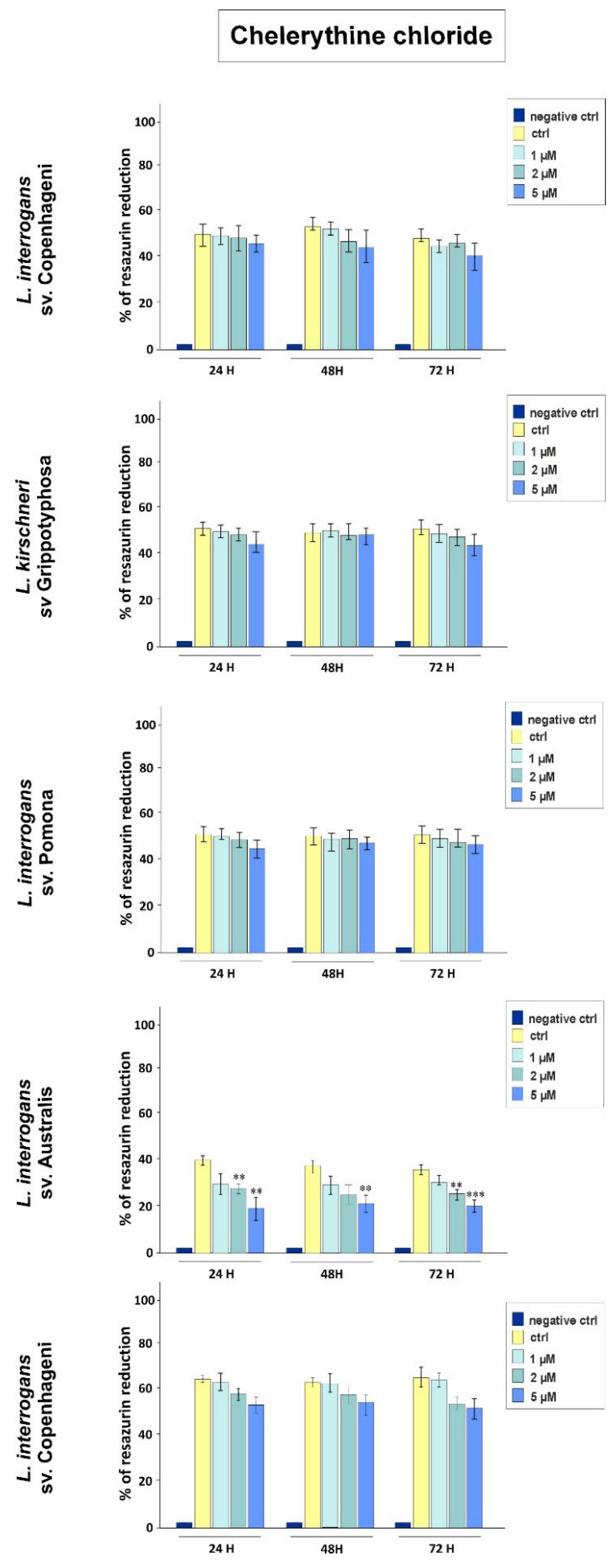
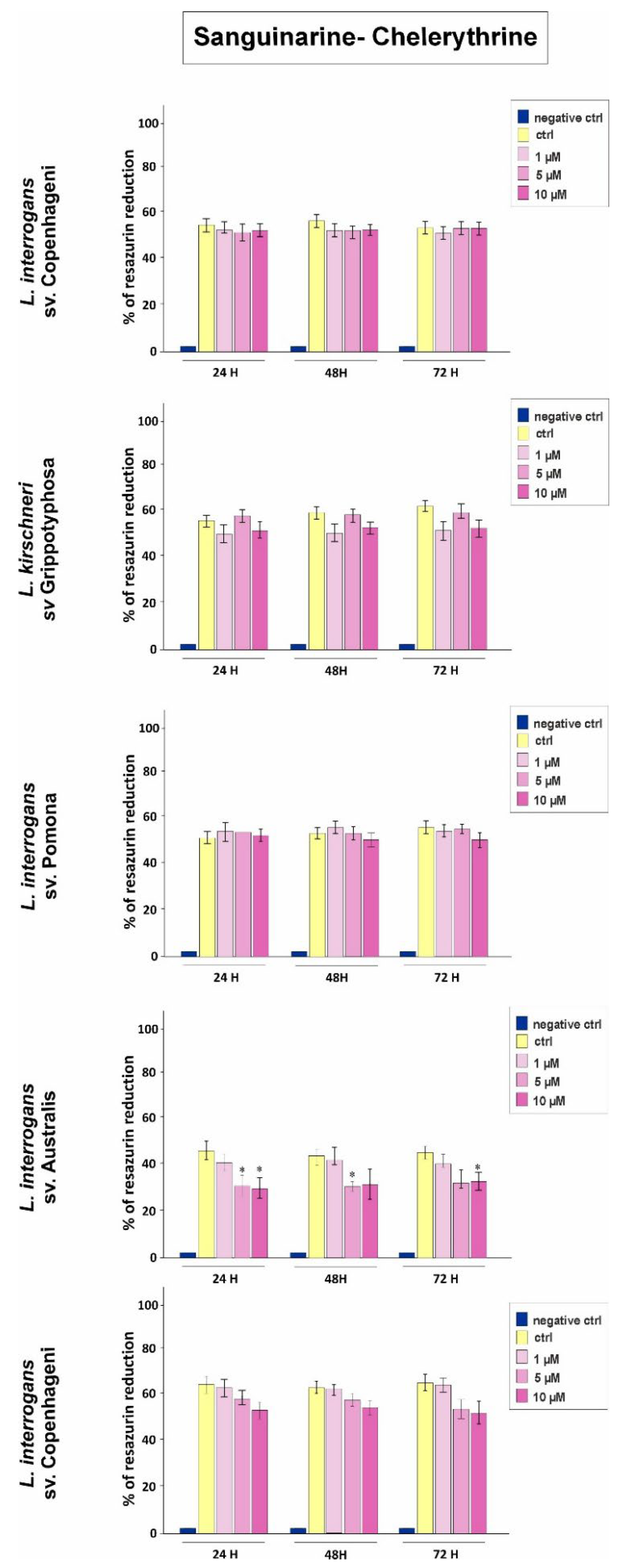
The Alamar Blue assay showed a pattern broadly consistent with the observations obtained using cytometric and growth-based methods. For most tested strains, metabolic activity remained relatively stable across the tested concentration range, and no consistent dose-dependent effect was observed. In contrast, in L. interrogans sv. Australis, strain Balico, a concentration-dependent decrease in metabolic activity was detected for all tested compounds. Statistical analysis was performed for all strains; however, statistically significant differences were observed only for this strain. Sanguinarine chloride induced a moderate reduction in metabolic activity, with statistically significant effects observed at 5 and 10 µM (p < 0.05), particularly at later time points. No consistent or significant effect was observed at 1 µM (Figure 1). Chelerythrine chloride produced a more pronounced response, with significant reductions in metabolic activity observed across the tested concentrations and time points (p < 0.01). The strongest effect was observed at the highest concentration and after prolonged incubation (72 h) (Figure 2). The sanguinarine–chelerythrine fraction induced a weaker effect compared to the individual compounds. Significant reductions in metabolic activity were observed primarily at higher concentrations (5 and 10 µM), while no significant effect was detected at 1 µM (Figure 3).

3. Discussion

The present study provides an in vitro evaluation of the effects of selected isoquinoline alkaloids on selected Lepto-spira strains. Using a combination of microscopic, cytometric, and metabolic assays, we observed that the effects of the tested compounds were limited and strongly strain-dependent. The most pronounced responses were detected in strain Balico, particularly at higher concentrations.

Effects of Sanguinarine Chloride

Among the tested compounds, sanguinarine chloride induced measurable changes in selected parameters, but these effects were not consistent across all strains and were primarily observed in strain Balico at higher concentrations and after prolonged incubation. Lower concentrations produced minimal or no detectable effects across most strains under the applied experimental conditions.
Previous studies have reported antimicrobial activity of sanguinarine against a range of microorganisms, however, direct comparison with Leptospira is limited due to major biological differences between these organisms [8,14,22,23,24]. Mechanistically, sanguinarine has been shown to disrupt membrane integrity, interfere with ion transport, inhibit key enzymes, and induce oxidative stress, leading to impaired cellular metabolism and eventual cell death, however, their relevance to Leptospira remains unclear [14,25,26,27]. The observed decrease in metabolic activity and membrane integrity in selected conditions suggests that the compounds may affect various aspects of leptospiral physiology; however, the underlying mechanism cannot be determined based on the present data.

Effect of Chelerythrine Chloride

Chelerythrine chloride showed a strain-dependent effect, with the most pronounced response observed in strain Balico. In most strains, the observed changes were limited, whereas in strain Balico more pronounced decrease in metabolic activity and viability was detected, even at lower concentrations.
Although chelerythrine is structurally related to sanguinarine and has demonstrated biological activity in other systems, differences observed between compounds in this study may reflect strain-specific properties of Leptospira as well as differences in compound interaction with the leptospiral cell envelop [12,28]. The outer membrane ar-chitecture of Leptospira may influence the interaction of exogenous compounds with the cell, potentially contrib-uting to strain-dependent differences in the observed response. Leptospira differs substantially from classical Gram-negative bacteria in terms of cell envelope organization and physiology, which may influence their interac-tion with exogenous compounds and limit direct comparison with data obtained for other bacterial species [15,16,20]. The atypical structure and variability of leptospiral lipopolysaccharide (LPS) and outer membrane components have been shown to play a key role in environmental adaptation and host interaction, which may also influence the response to external compounds [15,16,19,20].

Effects of the Sanguinarine-Chelerythrine Fraction

The sanguinarine–chelerythrine fraction did not show a stronger effect than the individual compounds. At higher concentrations, the fraction was associated with a decrease in metabolic activity and bacterial counts, however, the overall effect remained weaker than that observed for the individual compounds. Under the tested conditions, the combined fraction did not show a stronger effect than the individual compounds. This observation highlights the importance of experimentally evaluating the effects of compound mixtures rather than assuming enhanced activity based on individual components. However, antagonistic or neutral interactions among alkaloids have been report-ed previously [10,29,30,31], highlighting the importance of experimentally validating the activity of compound mix-tures rather than inferring efficacy from individual components.

Strain-Dependent Responses of Leptospira

A key finding of this study is the variability in response among different Leptospira strains. Most tested strains showed limited or no consistent changes across the applied conditions, whereas a more pronounced response was observed only in strain Balico. Taken together, the results of this study indicate that the effects of the tested iso-quinoline alkaloids on Leptospira are limited and strongly strain-dependent, and therefore should be interpreted as preliminary observations rather than evidence of consistent antimicrobial activity.

Implications and Limitations

The observed effects were limited to in vitro conditions and were most evident at relatively high concentrations, which restricts broader interpretation of their biological relevance. [28]. The present results indicate that the tested compounds may affect selected Leptospira strains under defined experimental conditions, however, they do not support conclusions regarding potential therapeutic applications at this stage [32]. This is particularly important in the context of leptospirosis, where pathogenic Leptospira rapidly disseminate within the host and may colonize tis-sues such as the kidneys. Any potential therapeutic compound would therefore require adequate systemic activity and tissue penetration, which were not assessed in the present study. The biological relevance of the observed ef-fects in vivo remains unknown.

4. Materials and Methods

4.1. Bacterial Strains and Culture Conditions

Five reference strains of Leptospira were used in this study: Leptospira interrogans serovar Copenhageni (strain M20), Leptospira kirschneri serovar Grippotyphosa (strain Moskwa V), Leptospira interrogans serovar Pomona (strain Pomona), Leptospira interrogans serovar Australis (strain Balico), and Leptospira borgpetersenii serovar Hardjo (strain KR39). All strains were obtained from reference collections as indicated above. Leptospires were cultivated in Ellinghausen–McCullough–Johnson–Harris (EMJH) liquid medium under standard laboratory conditions at 28 °C without agitation. Cultures were maintained in exponential growth phase and adjusted to an initial density of approximately 1 × 10⁷ cells/mL prior to each experiment. Following treatment, cultures were incubated under identical conditions and sampled after 24, 48, and 72 h. All analyses were performed at each time point.

4.2. Compounds and Treatment Conditions

Sanguinarine chloride and chelerythrine chloride (Extrasynthese, Genay, France; HPLC purity ≥ 90–95%) were used as model isoquinoline alkaloids. Compounds were dissolved in dimethyl sulfoxide (DMSO) to obtain stock solutions at a concentration of 1 mM. A natural fraction containing sanguinarine and chelerythrine, previously described, was also included in the analysis [13]. Working solutions were prepared by diluting stock solutions in EMJH medium immediately prior to use. The final concentration of DMSO in all experimental conditions did not exceed 1% (v/v).
Sanguinarine chloride was tested at final concentrations of 1, 5, and 10 µM. The selection of concentrations was based on previously published studies [23] as well as preliminary experiments. Initial testing included lower concentrations (e.g., 0.5–2 µM), corresponding to ranges reported in the literature; however, under the applied experimental conditions for Leptospira spp., these concentrations did not result in detectable changes in the applied assays (data not shown). Therefore, the concentration range was extended to include higher doses to enable the assessment of potential biological effects.
Chelerythrine chloride was tested at final concentrations of 1, 5, and 10 µM. Concentration ranges were selected based on previously published data [33] and preliminary experiments. As in the case of sanguinarine, lower concentrations corresponding to literature reports did not result in detectable changes under the applied experimental conditions (data not shown), and therefore higher concentrations were included in further analyses.
The sanguinarine–chelerythrine fraction was tested at final concentrations of 1, 5, and 10 µM (expressed as total alkaloid content). The selection of concentrations followed the same rationale as for individual compounds, including preliminary testing of lower doses that did not produce measurable effects (data not shown).
For each strain and each tested compound, the following experimental groups were included: a negative control containing only EMJH medium and Alamar Blue reagent, an untreated control containing bacterial culture without compound addition, and three treatment groups corresponding to the selected concentrations of the tested compound. All experimental conditions were maintained under identical incubation parameters. Samples were collected after 24, 48, and 72 h for further analyses. Each experiment was performed independently in triplicate (n = 3 biological replicates), and all assays were conducted under the same experimental conditions to ensure comparability between strains and treatments.

4.3. Assessment of Metabolic Activity Using Alamar Blue Assay

Metabolic activity of leptospiral cells was evaluated using the Alamar Blue® assay (Thermo Fisher Scientific), as described previously [32] with modifications adapted for Leptospira. Briefly, 180 µL of bacterial culture was transferred into wells of a 96-well plate, followed by the addition of 20 µL of Alamar Blue reagent. Plates were incubated at 28–30 °C under standard conditions until a measurable color change from blue (oxidized resazurin) to pink (reduced resorufin) was observed. Quantitative measurements were performed by recording absorbance at 570 nm and 600 nm using a microplate reader. Wells containing medium without bacteria were used as negative controls. The percentage reduction of resazurin was calculated according to established equations based on molar extinction coefficients, as described by [32], and used as an indicator of cellular metabolic activity.

4.4. Flow Cytometry Analysis and Viability Assessment

Leptospira cell counts and viability were assessed using flow cytometry with the LIVE/DEAD BacLight™ Bacterial Viability Kit (Thermo Fisher Scientific), as previously described [21,33,34]. Samples were stained with SYTO9 and propidium iodide (PI) at a ratio of 1:1000, gently mixed, and incubated for 3 minutes in the dark at room temperature. Immediately prior to analysis, samples were vortexed briefly to ensure homogeneity. Flow cytometric analysis was performed using a BD Accuri™ C6 flow cytometer (BD Biosciences). SYTO9 fluorescence (live cells) was detected in the FL1 channel, while fluorescence (cells with compromised membrane integrity) was detected in the FL3 channel. Instrument settings included a threshold of 2000 on the FL1 channel. Control samples consisting of untreated live bacteria and heat-inactivated bacteria (90 °C) were used to establish gating parameters and to distinguish between intact and membrane-compromised cells. For each sample, 10,000–50,000 events were recorded. Data were analyzed using BD Accuri C6 software and FlowJo v10 (BD Biosciences).

4.5. Cell Enumeration

Total leptospiral cell counts were determined using flow cytometry and additionally verified by direct counting in a Petroff–Hausser counting chamber under dark-field microscopy, as previously described. This approach allowed cross-validation of quantitative data obtained from cytometric analysis.

4.6. Optical Density Measurements

Bacterial growth was additionally monitored by measuring optical density (OD) using a spectrophotometer at (420nm). Measurements were performed for all samples at each time point.

4.7. Dark-Field Microscopy

Bacterial morphology and motility were evaluated qualitatively using dark-field microscopy, as described in previous studies [18]. Observations were performed directly from culture samples without fixation to assess potential changes in cell morphology and motility under different treatment conditions.

4.8. Statistical Analysis

Statistical analysis was performed for the Alamar Blue assay data for all tested strains. Data are presented as mean values ± standard deviation (SD) of three independent experiments (n = 3 biological replicates). Statistical significance between treated samples and the corresponding untreated control was assessed using one-way analysis of variance (ANOVA), followed by an appropriate post hoc test. A p-value < 0.05 was considered statistically significant.
Although statistical comparisons were performed for all strains, statistically significant differences were observed only in one strain, while in the remaining strains no significant differences were detected.

5. Conclusions

The present study demonstrates that the effects of selected isoquinoline alkaloids on Leptospira are limited and strongly strain-dependent under the applied in vitro conditions. Across most tested strains, no consistent or pronounced changes in bacterial viability, abundance, or metabolic activity were observed. A measurable response was detected primarily in) L. interrogans sv. Australis (strain Balico), particularly at higher concentrations. Among the tested compounds, chelerythrine chloride showed the most consistent effect in this strain, whereas sanguinarine chloride and the sanguinarine–chelerythrine fraction exhibited weaker or less consistent responses. The combined fraction did not demonstrate a stronger effect than the individual compounds under the applied conditions. These findings indicate that the response of Leptospira to the tested compounds cannot be generalized and may vary substantially between strains. Overall, the data suggest that selected isoquinoline alkaloids may influence Leptospira under defined in vitro conditions, however, their effects are restricted, strain-dependent, and observed mainly at higher concentrations. Further studies, taking into account standard antimicrobial parameters such as MIC, and dedicated mechanistic analyses, are necessary to explain the mechanism of action of these compounds on individual Leptospira strains, and to determine their biological relevance.

Author Contributions

Conceptualization, S.Z., L.P. and B,P.; methodology, L.P., I.S., K.D., M.W.; software, L.P.; validation, L.P., K.D. and Z.A.; formal analysis, L.P., I.S., K.D.; investigation, S.Z., L.P., Z.A.; resources, S.Z., Z.A.; data curation, L.P., S.Z.; writing—original draft prep-aration, S.Z., L.P.; writing—review and editing, S.Z., L.P., B.P., Z.A.; visualization, L.P.; supervision, L.P., Z.A.; project admin-istration, S.Z., L.P..; funding acquisition, S.Z., Z.A. All authors have read and agreed to the published version of the manuscript.

Funding

This research was funded by the National Science Centre Poland (NCN): Sonata 15 No. 2019/35/D/NZ7/00266 and OPUS 17 2019/33/B/NZ9/02159.

Data Availability Statement

The data are available on request from the corresponding authors.

Conflicts of Interest

The authors declare no conflicts of interest.

References

  1. Álvarez-Martínez, F.J.; Barrajón-Catalán, E.; Herranz-López, M.; Micol V. Antibacterial plant compounds, extracts and essential oils: An updated review on their effects and putative mechanisms of action. Phytomedicine 2021, 90, 153626.
  2. Jubair, N.; Rajagopal, M.; Chinnappan, S.; Abdullah N.B.; Fatima A. Review on the antibacterial mechanism of plant-derived compounds against multidrug-resistant bacteria (MDR). BioMed Research International 2021 3663315.
  3. Angelini, P. Plant-derived antimicrobials and their crucial role in combating antimicrobial resistance. Antibiotics 2024, 13: 746.
  4. Woo, S.; Marquez, L.; Crandall, W.; Risener, C.; Quave, C.L. Recent advances in the discovery of plant-derived antimicrobial natural products to combat antimicrobial resistant pathogens: insights from 2018–2022. Natural Product Reports 2023, 40, 1271–1290.
  5. Wójciak, M.; Feldo, M.; Stolarczyk, P.; Płachno, B.J. Carnivorous Plants from Nepenthaceae and Droseraceae as a Source of Secondary Metabolites. Molecules 2023, 28, 2155. [CrossRef]
  6. Wójciak, M., Feldo, M., Stolarczyk, P., Płachno, B. J. Biological potential of carnivorous plants from Nepenthales. Molecules 2023 a, 28(8), 3639.
  7. Miclea, I. Secondary Metabolites with Biomedical Applications from Plants of the Sarraceniaceae Family. Int. J. Mol. Sci. 2022, 23, 9877.
  8. Bussabong, P.; Rairat, T.; Chuchird, N.; Keetanon, A.; Phansawat, P.; Cherdkeattipol, K.; Pichitkul, P.; Kraitavin, W. Effects of isoquinoline alkaloids from Macleaya cordata on growth performance, survival, immune response, and resistance to Vibrio parahaemolyticus infection of Pacific white shrimp (Litopenaeus vannamei). PLoS One 2021; [CrossRef]
  9. Jursky, F.; Baliova, M.; Mihalikova, A. Molecular basis for differential glycine transporters sensitivity to sanguinarine. Toxicology Letters 2012, 212, 3, 262-267.
  10. Zielińska, S.; Jezierska-Domaradzka, A.; Wójciak-Kosior, M.; Sowa, I.; Junka, A.; Matkowski, A.M. Greater Celandine’s ups and downs−21 centuries of medicinal uses of Chelidonium majus from the viewpoint of today’s pharmacology. Front. Pharmacol. 2018, 9, 299.
  11. Leyva-Peralta, M.A.; Robles-Zepeda, R. E.; Garibay-Escobar, A.; Ruiz-Bustos, Eduardo.; Alvarez-Berber, L.P.; Gálvez-Ruiz, J.C. In vitro anti-proliferative activity of Argemone gracilenta and identification of some active components. BMC Complementary and Alternative Medicine 2015, 15:13. doi 10.1186/s12906-015-0532-8.
  12. Kaminskyy, V.; Kulachkovskyy, O.; Stoika, R. Adecisive role of mitochondria in defining rate and intensity of apoptosis induction by different alkaloids. Toxicology Letters 2008, 177, 168–181.
  13. Zielińska, S.; Wójciak-Kosior, M.; Dziągwa-Becker, M.; Gleńsk, M.; Sowa, I.; Fijałkowski, K.; Rurańska-Smutnicka, D.; Matkowski, A.; Junka, A. The Activity of Isoquinoline Alkaloids and Extracts from Chelidonium majus against Pathogenic Bacteria and Candida sp. Toxins 2019, 11, 406; [CrossRef]
  14. Zhao, Z.; Shang, X.; Lawoe, R.K.; Liu, Y.; Zhou, R.; Sun, Y.; Yan, Y.; Li, J.; Yang, G.; Yang, C. Anti-phytopathogenic activity and the possible mechanisms of action of isoquinoline alkaloid sanguinarine. Pesticide Biochemistry and Physiology 2019, 159 51–58.
  15. Adler, B.; de la Peña Moctezuma A. Leptospira and leptospirosis. Vet Microbiol. 2010, 140, 287–296. [CrossRef]
  16. Picardeau, M. Virulence of the zoonotic agent of leptospirosis: still terra incognita? Nat Rev Microbiol. 2017 a, 15: 297–307. [CrossRef]
  17. Costa, F.; Hagan, JE.; Calcagno, J.; Kane, M.; Torgerson, P.; Martinez-Silveira, M.S.; Stein, C.; Abela-Ridder, B.; Ko, AI. Global Morbidity and Mortality of Leptospirosis: A Systematic Review. PLoS Negl Trop Dis. 2015 17, 9(9):e0003898. [CrossRef]
  18. Arent, Z.; Pardyak, L.; Dubniewicz, K.; Płachno, B.; Kotula-Balak, M. Leptospira taxonomy: then and now. Med Weter. 2022, 78: 489–496.
  19. Lewicka, A.J.; Lyczakowski, J.J.; Pardyak, L.; Dubniewicz, K.; Latowski, D.; Arent Z. Beyond serology: saccharide profiling enables identification of antigenically similar Leptospira and prompts re-evaluation of bacterial lipopolysaccharide evolution. Front Mol Biosci. 2025, 17, 12:1581587. [CrossRef]
  20. Picardeau, M. Toolbox of Molecular Techniques for Studying Leptospira Spp.. In: Adler, B. (eds) Spirochete Biology: The Post Genomic Era. Current Topics in Microbiology and Immunology 2017b, vol. 415. Springer, Cham. [CrossRef]
  21. Dubniewicz, K.; Pardyak, L.; Gurgul, A.; Jasielczuk, I.; Szmatoła, T.; Arent, ZJ. Serovar-specific genomic features of Leptospira interrogans Hardjo: implications for host adaptation. Front Mol Biosci. 2025, 12, 1648097. [CrossRef]
  22. Ahsan, Haseeb.;Reagan-Shaw, Shannon.; Eggert, D.M.; Tan, Thomas C.; Afaq, Farrukh.; Mukhtar, Hasan.; Ahmad, N. Protective Effect of Sanguinarine on Ultraviolet B-mediated Damages in SKH-1 Hairless Mouse Skin: Implications for Prevention of Skin Cancer. Photochemistry and Photobiology 2007, 83: 986–993.
  23. Yan, S.; Lin, S.; Chen, K.; Yin, S.; Peng, H.; Cai, N.; Ma, W.; Songyang, Z.; Huang, Y. Natural Product Library Screens Identify Sanguinarine Chloride as a Potent Inhibitor of Telomerase Expression and Activity. Cells 2022, 11(9), 1485. [CrossRef]
  24. Li, J.Y.; Huang, H.; Pan, T.; Wang, N.; Shi, C.; Zhang, Bo.; Wang, C.; Yang G. Sanguinarine induces apoptosis in Eimeria tenella sporozoites via the generation of reactive oxygen species. Poultry Science 2022. 101, 101771, . [CrossRef]
  25. Jiao, X.; Fu, X.; Li, Q.; Bu, J.; Liu, X.; Savolainen, O.; Huang, L.; Guo, J.; Nielsen, J.; Chen, Y. De novo production of protoberberine and benzophenanthridine alkaloids through metabolic engineering of yeast. Nat Commun 2024, 15, 8759. [CrossRef]
  26. Huang, L.J.; Lan, J.X.; Wang, J.H.; Huang, H.; Lu, K.; Zhou, Z.N.; Xin, S.Y.; Zhang, Z.Y.; Wang, J.Y.; Dai, P.; Chen, X.M.; Hou, W. Bioactivity and mechanism of action of sanguinarine and its derivatives in the past 10 years. Biomed Pharmacother. 2024, 173, 116406. [CrossRef]
  27. Petakh, P.; Isevych, V.; Kamyshnyi, A.; Oksenych, V. Weil’s Disease—Immunopathogenesis, Multiple Organ Failure, and Potential Role of Gut Microbiota. Biomolecules 2022, 12, 1830. [CrossRef]
  28. Chen, N.; Qi, Y.; Ma, X.; Xiao, X.; Liu, Q.; Xia, T.; Xiang, J.; Zeng, J.; Tang, J. Rediscovery of Traditional Plant Medicine: An Underestimated Anticancer Drug of Chelerythrine. Front. Pharmacol., Sec. Ethnopharmacology 2022, 13 - 2022 | . [CrossRef]
  29. Danielewski, M.; Zielińska, S.; Matuszewska, A.; Słupski, W.; Włodarczyk, M.; Jęśkowiak, I.; Wiatrak, B.; Kowalski, K.; Jezierska-Domaradzka, A.; Ziółkowski, P.; Szeląg, A.; Nowak, B. Sanguinarine-Chelerythrine Fraction of Coptis chinensis Exerts Anti-inflammatory Activity in Carrageenan Paw Oedema Test in Rats and Reveals Reduced Gastrotoxicity. Oxidative Medicine and Cellular Longevity. 2022, 1504929, . [CrossRef]
  30. Danielewski, M.; Zielińska, S.; Merwid-Ląd, A.; Szandruk-Bender, M.; Słupski, W.; Włodarczyk, M.; Sozański, T.; Ziółkowski, P.; Szeląg, A.; Nowak, B. Sanguinarine–Chelerythrine from Coptis chinensis Offers Analgesic and Anti-Inflammatory Effects Without Gastrotoxicity. Pharmaceutics 2025, 17, 323.
  31. Dzięgielewska, M.; Zielińska, S.; Dudek, B.; Brożyna, M.; Kozłowska, W.; Matkowski, A.; Bartoszewicz, M.; Yanfang S.; Junka, A. Isoquinoline Alkaloids from Traditional Medicinal Plants of the Papaveraceae as a Third Line of Defense Against Bacterial Ocular Infections, bioRxiv the preprint server for Biology 2025, 11.21.689706; [CrossRef]
  32. Koizumi, N.; Picardeau, M. Leptospira Spp: Methods and Protocols in Humana 2020, ISBN 1071604600, 9781071604601.
  33. Heng, W.S.; Cheah, S.-C. Chelerythrine Chloride Downregulates β-Catenin and Inhibits Stem Cell Properties of Non-Small Cell Lung Carcinoma. Molecules 2020, 25, 224. [CrossRef]
  34. Fontana, C.; Crussard, S.; Simon-Dufay, N.; Pialot, D.; Bomchil, N.; Reyes, J. Use of flow cytometry for rapid and accurate enumeration of live pathogenic Leptospira strains. J.Microbiological Methods 2017, 132, 34-40. [CrossRef]
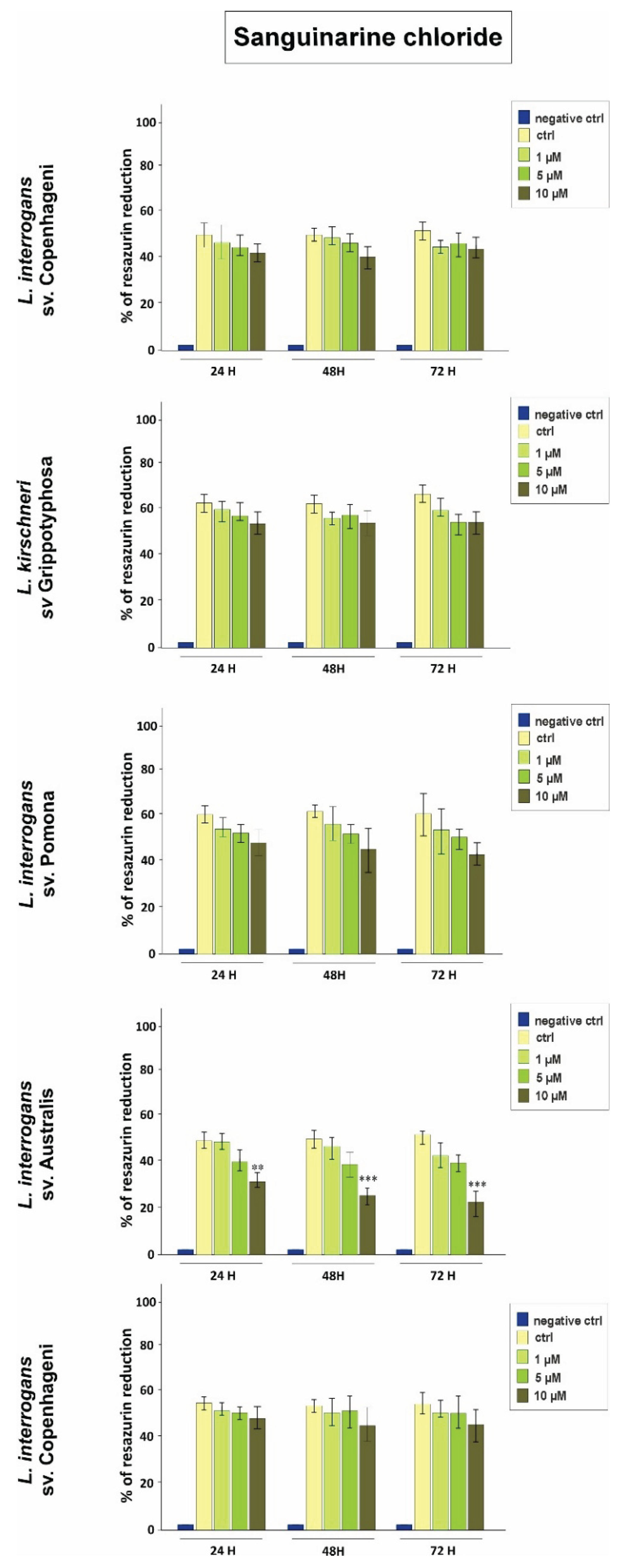
Figure 1. Effect of sanguinarine chloride on metabolic activity of selected Leptospira strains assessed using the Alamar Blue assay. Bacteria were exposed to 1, 5, and 10 µM of the compound for 24, 48, and 72 h. Metabolic activity is expressed as percentage of resazurin reduction. Data are presented as mean ± SD of three independent experiments (n = 3). Statistically significant differences compared to the untreated control are indicated (*p < 0.05, **p < 0.01). Significant effects were observed only in L. interrogans sv. Australis strain Balico.
Preprints 231565 g001
Figure 2. Effect of chelerythrine chloride on metabolic activity of selected Leptospira strains assessed using the Alamar Blue assay. Bacteria were exposed to 1, 2, and 5 µM of the compound for 24, 48, and 72 h. Metabolic activity is expressed as percentage of resazurin reduction. Data are presented as mean ± SD of three independent experiments (n = 3). Statistically significant differences compared to the untreated control are indicated (*p < 0.05, **p < 0.01, ***p < 0.001). Significant effects were observed only in L. interrogans sv. Australis strain Balico.
Preprints 231565 g002
Figure 3. Effect of the sanguinarine–chelerythrine fraction on metabolic activity of selected Leptospira strains assessed using the Alamar Blue assay. Bacteria were exposed to 1, 5, and 10 µM of the fraction for 24, 48, and 72 h. Metabolic activity is expressed as percentage of resazurin reduction. Data are presented as mean ± SD of three independent experiments (n = 3). Statistically significant differences compared to the untreated control are indicated (*p < 0.05). Significant effects were observed only in L. interrogans sv. Australis strain Balico.
Preprints 231565 g003
Table 1. Effect of sanguinarine chloride (1–10 µM) on the growth and viability of selected Leptospira strains, expressed as total bacterial count (cells/mL), percentage of live and dead cells, and optical density at 420 nm (OD420₂₀) after 24, 48, and 72 h of incubation.
Strain Dose Incubation (hour) Total bacteria count (cells/ml) Live (%) Dead (%) OD (420 nm)

Leptospira interrogans
sv. Copenhageni,
strain M20
Control 0 4.5×10⁷ 90 10 0.632

1 µM
24 4.6×10⁷ 91 9 0.688
48 4.8×10⁷ 89 11 0.646
72 4.9×10⁷ 88 12 0.627

5 µM
24 3.8×10⁷ 87 13 0.638
48 3.7×10⁷ 88 12 0.659
72 3.4×10⁷ 86 14 0.667

10 µM
24 3.9×10⁷ 81 18 0.613
48 4.0×10⁷ 82 17 0.612
72 3.8×10⁷ 80 20 0.620

Leptospira kirschneri sv. Grippotyphosa,
strain Moskwa V
Control 0 2.85×10⁷ 95 5 0.598

1 µM
24 2.7×10⁷ 92 8 0.578
48 2.4×10⁷ 91 9 0.601
72 2.9×10⁷ 90 10 0.597

5 µM
24 1.8×10⁷ 91 9 0.599
48 2.7×10⁷ 89 11 0.600
72 2.1×10⁷ 87 13 0.604

10 µM
24 2.9×10⁷ 87 13 0.597
48 2.2×10⁷ 85 15 0.556
72 1.8×10⁷ 84 16 0.569
Leptospira interrogans
sv. Pomona,
strain Pomona
Control 0 3.8×10⁷ 92 8 0.650

1 µM
24 3.4×10⁷ 90 10 0.640
48 2.4×10⁷ 91 9 0.630
72 2.9×10⁷ 92 8 0.635

5 µM
24 3.32×10⁷ 93 7 0.601
48 2.98×10⁷ 92 8 0.611
72 2.6×10⁷ 90 10 0.603

10 µM
24 2.4×10⁷ 89 11 0.610
48 1.98×10⁷ 88 12 0.598
72 2.01×10⁷ 87 13 0.590

Leptospira interrogans
sv. Australis,
strain Balico
Control 0 1.5×10⁷ 89 11 0.546

1 µM
24 1.2×10⁷ 85 15 0.545
48 1.15×10⁷ 84 16 0.536
72 1.7×10⁷ 82 18 0.546

5 µM
24 1.3×10⁷ 83 17 0.564
48 1.0×10⁵ 75 25 0.460
72 0.8×10⁵ 72 18 0.401

10 µM
24 1.1×10³ 65 35 0.312
48 0.5×10³ 45 55 0.299
72 0.6×10² 42 58 0.125

Leptospira borgpetersenii sv. Hardjo,
strain KR39
Control 0 4.5×10⁷ 94 6 0.645

1 µM
24 4.3×10⁷ 92 8 0.630
48 4.25×10⁷ 91 9 0.625
72 4.15×10⁷ 92 8 0.626

5 µM
24 4.3×10⁷ 89 11 0.564
48 4.0×10⁵ 85 15 0.560
72 3.8×10⁵ 82 18 0.581

10 µM
24 3.4×10⁷ 86 14 0.522
48 3.3×10⁷ 84 16 0.569
72 3.2×10⁷ 81 19 0.545
Table 2. Effect of chelerythrine chloride (1–5 µM) on the growth and viability of selected Leptospira strains, expressed as total bacterial count (cells/mL), percentage of live and dead cells, and optical density at 420 nm (OD420₂₀) after 24, 48, and 72 h of incubation.
Strain Dose Incubation (hour) Total bacteria count (cells/ml) Live (%) Dead (%) OD (420 nm)

Leptospira interrogans
sv. Copenhageni
strain M20
Control 0 4.5×10⁷ 90 10 0.632

1 µM
24 4.6×10⁷ 91 9 0.688
48 4.8×10⁷ 89 11 0.646
72 4.9×10⁷ 88 12 0.627

2 µM
24 3.8×10⁷ 87 13 0.638
48 3.7×10⁷ 88 12 0.659
72 3.4×10⁷ 86 14 0.667

5 µM
24 3.9×10⁷ 81 18 0.613
48 4.0×10⁷ 82 17 0.612
72 3.8×10⁷ 80 20 0.620

Leptospira kirschneri sv. Grippotyphosa
strain Moskwa V
Control 0 4.4×10⁷ 94 6 0.65
1 µM 24 4.2×10⁷ 93 7 0.64
48 4.0×10⁷ 92 8 0.63
72 2.9×10⁷ 91 9 0.635
2 µM 24 3.7×10⁷ 87 13 0.601
48 3.9×10⁷ 88 12 0.611
72 3.6×10⁷ 86 14 0.603
5 µM 24 4.1×10⁷ 88 12 0.610
48 3.9×10⁷ 82 18 0.598
72 3.6×10⁷ 80 20 0.59
Leptospira interrogans
sv. Pomona
strain Pomona
Control 0 2.5×10⁷ 96 4 0.65
1 µM 24 2.2×10⁷ 94 6 0.64
48 2.4×10⁷ 95 5 0.63
72 2.9×10⁷ 93 7 0.635
2 µM 24 2.7×10⁷ 93 7 0.601
48 1.9×10⁷ 92 8 0.611
72 2.6×10⁷ 96 4 0.603
5 µM 24 2.7×10⁷ 92 8 0.610
48 1.8×10⁷ 90 10 0.67
72 2.3×10⁷ 91 9 0.65

Leptospira interrogans
sv. Australis
strain Balico
Control 0 3.5×10⁷ 96 4 0.60
1 µM 24 3.2×10⁷ 94 6 0.61
48 3.5×10⁷ 95 5 0.62
72 3.6×10⁷ 93 7 0.66
2 µM 24 3.7×10⁷ 93 7 0.65
48 3.5×10⁷ 92 8 0.59
72 3.6×10⁷ 96 4 0.64
5 µM 24 3.4×10⁷ 93 7 0.64
48 3.5×10⁷ 94 6 0.61
72 3.9×10⁷ 93 7 0.59

Leptospira borgpetersenii sv. Hardjo
strain KR39
Control 0 1.6×10⁷ 96 4 0.62
1 µM 24 1.5×10⁷ 89 11 0.61
48 1.4×10⁷ 85 15 0.62
72 1.2×10⁷ 86 14 0.66
2 µM 24 1.6×10⁶ 83 17 0.65
48 1.5×10⁶ 82 18 0.59
72 2.6×10⁵ 80 20 0.64
5 µM 24 4.4×10⁴ 75 25 0.40
48 2.5×10⁴ 64 36 0.31
72 3.1×10⁴ 53 47 0.36
Table 3. Effect of the sanguinarine–chelerythrine fraction (1–5 µM) on the growth and viability of selected Leptospira strains, expressed as total bacterial count (cells/mL), percentage of live and dead cells, and optical density at 420 nm (OD420₂₀) after 24, 48, and 72 h of incubation.
Strain Dose Incubation Time Total bacteria count (cells/ml) Live (%) Dead (%) OD (420 nm)

Leptospira interrogans
sv. Copenhageni
strain M20
Control 0 3.4×10⁷ 89 11 0.56
1 µM 24 2.9×10⁷ 90 10 0.59
48 3.0×10⁷ 91 9 0.58
72 2.9×10⁷ 90 10 0.57
2 µM 24 3.1×10⁷ 85 15 0.57
48 3.2×10⁷ 83 17 0.59
72 3.0×10⁷ 86 14 0.58
5 µM 24 3.1×10⁷ 87 13 0.56
48 3.0×10⁷ 87 13 0.55
72 3.0×10⁷ 86 14 0.53

Leptospira kirschneri sv. Grippotyphosa
strain Moskwa V
Control 0 5.4×10⁷ 90 10 0.56
1 µM 24 4.9×10⁷ 91 9 0.59
48 4.6×10⁷ 89 11 0.58
72 4.7×10⁷ 86 14 0.57
2 µM 24 4.0×10⁷ 86 14 0.57
48 4.2×10⁷ 85 15 0.59
72 4.0×10⁷ 86 14 0.58
5 µM 24 4.6×10⁷ 89 11 0.57
48 4.5×10⁷ 88 12 0.57
72 4.2×10⁷ 85 15 0.56
Leptospira interrogans
sv. Pomona
strain Pomona
Control 0 2.8×10⁷ 93 7 0.58
1 µM 24 2.6×10⁷ 92 8 0.57
48 2.4×10⁷ 91 9 0.58
72 2.5×10⁷ 91 9 0.56
2 µM 24 2.1×10⁷ 90 10 0.54
48 2.2×10⁷ 89 11 0.56
72 2.4×10⁷ 88 12 0.54
5 µM 24 1.9×10⁷ 90 10 0.57
48 1.8×10⁷ 89 11 0.56
72 4.5×10⁶ 87 13 0.53

Leptospira interrogans
sv. Australis
strain Balico
Control 0 1.8×10⁷ 93 7 0.60
1 µM 24 2.5×10⁶ 92 8 0.58
48 1.8×10⁶ 91 9 0.57
72 1.5×10⁶ 91 9 0.56
2 µM 24 1.6×10⁶ 90 10 0.57
48 1.4×10⁶ 89 11 0.56
72 3.4×10⁵ 88 12 0.55
5 µM 24 1.7×10⁵ 86 14 0.53
48 1.8×10⁵ 82 18 0.52
72 1.5×10⁵ 79 21 0.49

Leptospira borgpetersenii sv. Hardjo
strain KR39
Control 0 3.4×10⁷ 92 8 0.61
1 µM 24 2.9×10⁷ 91 9 0.60
48 2.6×10⁷ 89 11 0.59
72 2.3×10⁷ 88 12 0.58
2 µM 24 3.0×10⁷ 89 11 0.59
48 2.7×10⁷ 86 14 0.59
72 2.4×10⁷ 85 15 0.56
5 µM 24 2.7×10⁷ 88 12 0.53
48 2.5×10⁷ 87 13 0.50
72 2.0×10⁷ 86 14 0.49
Disclaimer/Publisher’s Note: The statements, opinions and data contained in all publications are solely those of the individual author(s) and contributor(s) and not of MDPI and/or the editor(s). MDPI and/or the editor(s) disclaim responsibility for any injury to people or property resulting from any ideas, methods, instructions or products referred to in the content.
Copyright: This open access article is published under a Creative Commons CC BY 4.0 license, which permit the free download, distribution, and reuse, provided that the author and preprint are cited in any reuse.