

Submitted:
03 September 2026
Posted:
04 September 2026
You are already at the latest version
Abstract
Background: Behçet’s syndrome is a chronic, relapsing, multisystem inflammatory disorder characterised by recurrent mucosal ulceration and variable ocular, vascular, neurological, gastrointestinal, articular, and cutaneous involvement. Over the past two decades, the field has moved from syndrome-based description and empiric immunosuppression towards mechanism-informed phenotyping and targeted treatment. Objectives: To provide a contemporary narrative review of Behçet’s syndrome from 2000 to the present, with particular emphasis on search methods, pathophysiological foundations, clinical expression, diagnostic and classification frameworks, and the evidence base for conventional and targeted therapies. Methods: A structured narrative review was undertaken using major bibliographic databases and guideline repositories. The search strategy combined Medical Subject Headings, Emtree terms, and free-text keywords relating to Behçet’s syndrome, pathophysiology, diagnostic criteria, and treatment, with particular attention to randomised trials, prospective cohorts, systematic reviews, and international recommendations. Results: Current evidence supports Behçet’s syndrome as a complex immunovascular disease at the interface between autoinflammation, adaptive immune dysregulation, endothelial injury, and thromboinflammation. HLA-B51 remains the strongest genetic association, while ERAP1, IL10, IL23R-IL12RB2 and related pathways broaden the mechanistic framework. Neutrophil hyper-reactivity, NETosis, Th1/Th17 polarisation, endothelial activation, and cytokine circuits involving TNF, IL-1, IL-6, IL-17 and IL-23 underpin much of the clinical spectrum. The International Criteria for Behçet’s Disease improved sensitivity over the earlier International Study Group criteria, but diagnosis remains clinical. Therapeutically, colchicine, glucocorticoids and conventional immunosuppressants retain important roles, yet anti-TNF therapy has transformed outcomes in severe ocular, vascular, neurological and intestinal disease. Apremilast is now an evidence-based option for recurrent oral ulcers, while IL-1 blockade, IL-6 inhibition, IL-17 blockade, IL-12/23 inhibition and Janus kinase inhibition remain promising but incompletely defined strategies. Conclusions: Behçet’s syndrome requires rapid organ-based risk stratification and early suppression of major organ inflammation. The contemporary therapeutic landscape is considerably broader than it was at the turn of the century, but important gaps remain in comparative trials, biomarkers, treatment sequencing, and precision phenotyping.
Keywords:
Behçet’s syndrome
; Behçet disease
; vasculitis
; uveitis
; thrombosis
; anti-TNF
; apremilast
; autoinflammation
; NETosis
; review
1. Introduction
Behçet’s syndrome remains one of the most intriguing conditions in systemic inflammatory medicine. It is at once ancient in clinical description and strikingly modern in its immunological complexity. The disorder classically presents with recurrent oral ulceration, genital ulceration, and ocular inflammation, yet this familiar triad captures only part of the syndrome. Behçet’s syndrome is now more accurately regarded as a multisystem immunovascular disorder capable of involving the skin, joints, veins, arteries, central nervous system, gastrointestinal tract, and, more rarely, other organ systems [1,2,3].
The contemporary period has seen four major advances. First, the pathobiology of Behçet’s syndrome has been reframed around genetic susceptibility, barrier and microbial factors, dysregulated innate immunity, T-cell polarisation, endothelial activation, and thromboinflammation [2,4,5,6]. Secondly, classification has evolved from the International Study Group criteria towards the International Criteria for Behçet’s Disease, improving sensitivity for incomplete and early disease [7]. Thirdly, clinical management has become more phenotype-driven. Fourthly, targeted therapy has materially altered outcomes, particularly through anti-TNF treatment and apremilast [8,9].
This shift has not been merely semantic. The older vocabulary of a variable vasculitis of unknown cause has gradually given way to a more integrated concept of an immune-mediated vascular syndrome in which mucosal inflammation, endothelial dysfunction, cytokine amplification, and thrombotic propensity are mechanistically intertwined. Such a reframing has practical consequences: it encourages earlier identification of organ-threatening patterns, strengthens the rationale for mechanism-based therapy, and helps explain why responses differ across phenotypes [2,10,11].
Another important contemporary development has been the move away from a purely descriptive catalogue of manifestations towards a phenotype-oriented approach. Clinicians now routinely distinguish between predominantly mucocutaneous-articular disease and major organ phenotypes, including ocular, neurological, vascular, and intestinal involvement. This is not a taxonomic luxury but a clinical necessity, because prognosis, urgency, and therapeutic thresholds differ substantially between these groups [2,8,9].
The need for a comprehensive review also reflects the persistent mismatch between the rarity of Behçet’s syndrome in many Western settings and the complexity of decisions it generates. In low-prevalence countries, delayed recognition remains common, particularly when the presenting syndrome is vascular or neurological rather than classically mucocutaneous. Conversely, in high-prevalence regions, earlier recognition has not eliminated the challenge of preventing irreversible damage. A contemporary review must therefore bridge epidemiology, translational science, and organ-based therapeutics [2,10,12,13].
2. Methods of Literature Search
This structured narrative review was based on searches of MEDLINE/PubMed, Embase, Scopus, Web of Science Core Collection, the Cochrane Library, Google Scholar for citation tracking, and professional-society guideline repositories. Searches were updated to 31 August 2026.
The search strategy combined controlled vocabulary and free-text terms. Core Medical Subject Headings included ‘Behçet Syndrome’, ‘Vasculitis’, ‘Uveitis’, ‘Thrombosis’, ‘Mouth Ulcer’, ‘Biological Products’, ‘Tumor Necrosis Factor Inhibitors’, ‘Janus Kinase Inhibitors’, ‘Neutrophils’, ‘Extracellular Traps’, ‘Microbiota’, and related Emtree terms. Free-text terms included ‘Behçet disease’, ‘Behçet syndrome’, ‘Adamantiades-Behçet’, ‘neuro-Behçet’, ‘intestinal Behçet’, ‘HLA-B51’, ‘ERAP1’, ‘NETosis’, ‘anti-TNF’, ‘apremilast’, ‘tocilizumab’, ‘secukinumab’, and ‘JAK inhibitors’.
Priority was given to international classification criteria, EULAR recommendations, randomised controlled trials, prospective observational cohorts, systematic reviews, influential genetics studies, and organ-specific reviews with clear bearing on current practice.
The search was intentionally broad because Behçet’s syndrome spans several disciplines and no single bibliographic domain captures its full literature. For epidemiology and broad reviews, emphasis was placed on rheumatology, internal medicine, and ophthalmology databases. For drug-specific evidence, additional attention was paid to subspecialty literature in gastroenterology, neurology, dermatology, and ocular immunology. Citation chasing from pivotal reviews and guideline documents was undertaken to ensure that older but still practice-defining studies, such as seminal trials of azathioprine, ciclosporin, interferon-alpha, thalidomide, and etanercept, were not overlooked simply because more recent reviews dominate modern search ranking.
Search filters were preferentially applied to human studies and adult populations, but paediatric and transition-relevant literature was also considered when it informed disease mechanisms or classification. English-language articles were prioritised for consistency of synthesis, although globally influential international collaborative studies were considered regardless of the country of origin. Because Behçet’s syndrome remains a rare and heterogeneous disorder, the hierarchy of evidence was interpreted pragmatically: randomised controlled trials and formal recommendations were weighted most heavily, followed by prospective cohorts, multicentre retrospective studies, systematic reviews, and high-quality mechanistic reviews.
No formal meta-analytic pooling was attempted in this manuscript. Instead, the purpose was to assemble a clinically useful and intellectually coherent map of the field, giving particular prominence to studies that changed diagnostic thinking, refined phenotypic interpretation, or modified therapeutic standards. For emerging agents, especially IL-1 blockade, IL-6 blockade, IL-17 inhibition, IL-12/23 inhibition, and JAK inhibition, observational signals were included when they materially inform expert practice, but the distinction between established evidence and exploratory evidence is maintained throughout the text.
3. Epidemiology and Disease Burden
Behçet’s syndrome displays one of the most distinctive geographic distributions in inflammatory medicine, with greatest prevalence across populations historically aligned with the Silk Road, particularly Turkey, parts of the Middle East, and East Asia. Incidence and prevalence are markedly lower in northern Europe and the Americas [2,10,12].
The disease usually begins in early adult life, and men, especially young men, often experience more severe disease, with greater likelihood of ocular, vascular, and neurological involvement [2,12,13]. The burden of disease extends far beyond prevalence statistics because recurrent oral ulceration, uveitis, thrombosis, neurological disease, and intestinal involvement all exact major functional and quality-of-life costs [8,10,12].
Temporal trends are equally informative. Several cohorts suggest that the prognosis of Behçet’s syndrome has improved over recent decades, particularly with regard to severe ocular outcomes and mortality related to uncontrolled vascular disease. These gains likely reflect earlier recognition, better access to multidisciplinary care, and the adoption of immunosuppressive and biologic strategies capable of modifying the trajectory of major organ inflammation. Nonetheless, improvement has been uneven across regions, and the burden of chronic mucocutaneous disease, steroid toxicity, and irreversible damage remains substantial [2,8,9,13,14,15].
The epidemiology of disease expression also matters. Not all patients who meet diagnostic or classification criteria share the same risks. Men with early-onset disease appear more likely to develop severe ocular or vascular complications, whereas other patients may experience long-standing mucocutaneous-articular disease with comparatively limited life-threatening involvement. This variability has fuelled the modern emphasis on clinical subsets and supports the idea that Behçet’s syndrome should be approached as a family of related inflammatory phenotypes clustered under a common syndrome label [2,13].
From a health-services perspective, Behçet’s syndrome imposes a burden that is easy to underestimate. Rare-disease status can lead to fragmentation of care, repeated consultations across specialties, and delayed organ-specific intervention. Furthermore, the recurrent and sometimes socially stigmatising nature of oral and genital ulceration imposes a psychosocial cost that is not well captured by routine inflammatory markers or by crude outcome measures focused solely on mortality and hospitalisation. This reinforces the importance of patient-reported outcomes and quality-of-life assessment in both trials and daily practice [8,9,10,16].
4. Pathophysiological Foundations
The pathophysiology of Behçet’s syndrome is best understood as a convergence disorder positioned at the interface between autoinflammation, adaptive immune dysregulation, endothelial pathology, and thrombosis. This framework explains the broad clinical heterogeneity and the disorder’s distinctive capacity to involve both arteries and veins of any calibre [2,4,5,6,10,11,17].
A useful way to conceptualise Behçet’s syndrome is to imagine four partially overlapping biological layers. The first is inherited susceptibility, dominated by HLA class I biology but not reducible to it. The second is barrier-level interaction with microbial and environmental stimuli, especially at the oral and intestinal mucosa. The third is excessive activation of innate and adaptive inflammatory pathways. The fourth is tissue-specific amplification, particularly within the endothelium, vessel wall, retina, central nervous system, and bowel. Clinical disease results when these layers reinforce one another over time [2,4,5,10,11].
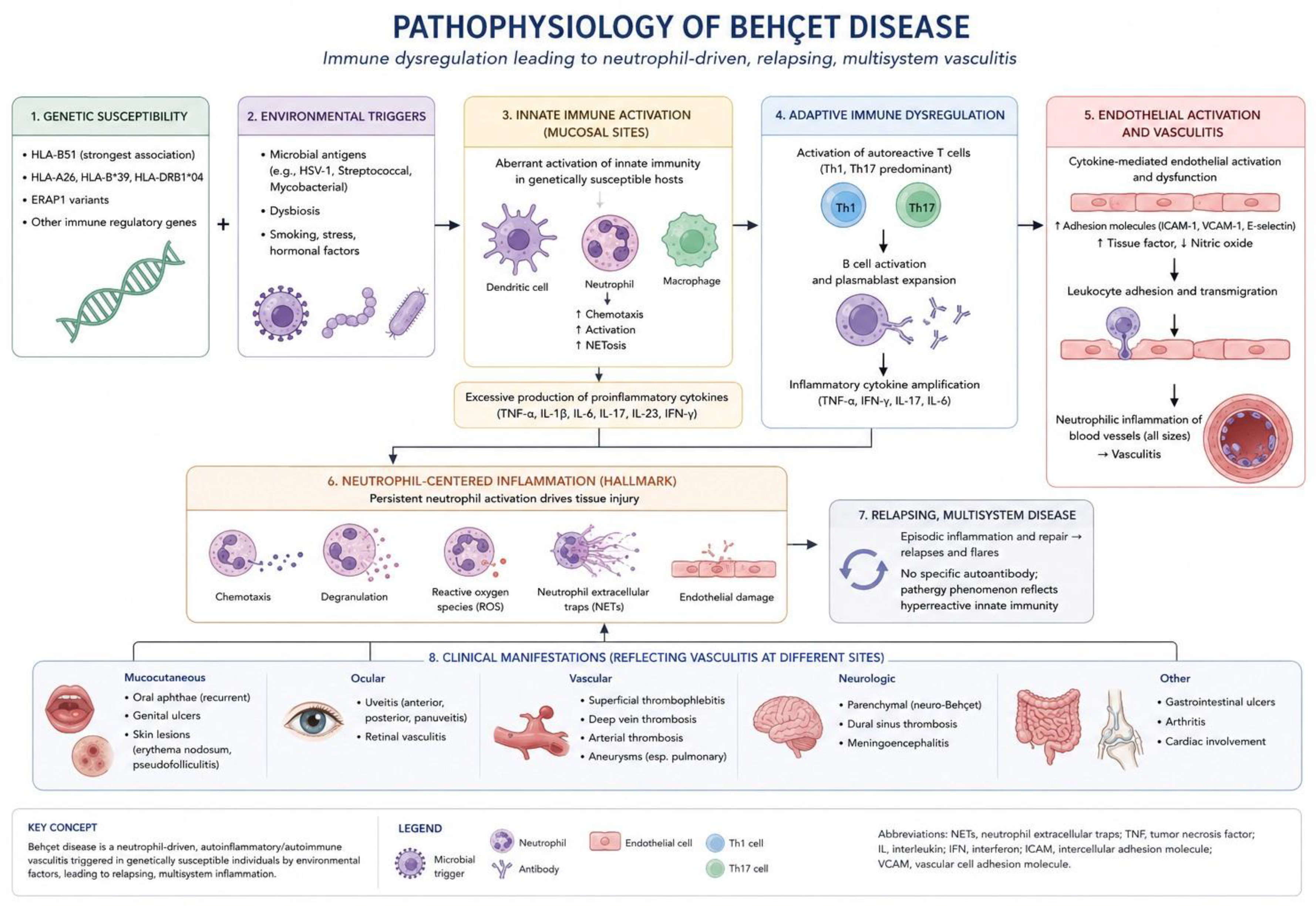
Figure 1.
Integrated pathophysiological model of Behçet’s syndrome. Genetic susceptibility and environmental or microbial triggers promote dysregulated innate and adaptive immune responses, with neutrophil hyper-reactivity, NETosis, pro-inflammatory cytokine amplification, and endothelial activation. The resulting neutrophil-centred vascular inflammation and tissue injury provide a common mechanistic basis for the relapsing mucocutaneous, ocular, vascular, neurological, gastrointestinal, articular, and cardiac manifestations of the disease. NETs, neutrophil extracellular traps; ROS, reactive oxygen species; TNF, tumour necrosis factor; IL, interleukin; IFN, interferon; ICAM, intercellular adhesion molecule; VCAM, vascular cell adhesion molecule.
Figure 1.
Integrated pathophysiological model of Behçet’s syndrome. Genetic susceptibility and environmental or microbial triggers promote dysregulated innate and adaptive immune responses, with neutrophil hyper-reactivity, NETosis, pro-inflammatory cytokine amplification, and endothelial activation. The resulting neutrophil-centred vascular inflammation and tissue injury provide a common mechanistic basis for the relapsing mucocutaneous, ocular, vascular, neurological, gastrointestinal, articular, and cardiac manifestations of the disease. NETs, neutrophil extracellular traps; ROS, reactive oxygen species; TNF, tumour necrosis factor; IL, interleukin; IFN, interferon; ICAM, intercellular adhesion molecule; VCAM, vascular cell adhesion molecule.
4.1. Genetics
The strongest genetic association is HLA-B51, a marker that is robustly associated with disease susceptibility across many populations but is neither necessary nor sufficient for disease development [5,6,18]. Genome-wide association studies have expanded this picture by implicating ERAP1, IL10, IL23R-IL12RB2 and related immune-regulatory loci, thereby linking Behçet’s syndrome with HLA class I-associated inflammatory biology and Th17-related pathways [4,5,6,11,19,20].
Other susceptibility loci reinforce the idea that Behçet’s syndrome sits within a broader immunological family of HLA class I-associated disorders. IL10-related variants suggest impaired anti-inflammatory restraint, whereas IL23R-IL12RB2 implicates pathways involved in Th17 maintenance and Th1 polarisation. These genetic observations do not by themselves explain organ selectivity, but they map convincingly onto later downstream findings in blood, tissue, and therapeutic response [4,5,10,11].
4.2. Innate Immunity, Neutrophils, and NETosis
Neutrophil hyper-reactivity is a central feature of Behçet’s syndrome. Increased chemotaxis, oxidative burst, degranulation, and heightened endothelial injury have long been recognised, and more recent work has highlighted the importance of neutrophil extracellular traps as a bridge between inflammation, endothelial dysfunction, and thrombosis [4,10,11].
This neutrophil-dominant signature helps explain several otherwise disparate clinical observations. Oral ulcers contain intense neutrophilic inflammation; skin lesions are often pustular or folliculitic; pathergy represents exaggerated local inflammatory reactivity; and venous thrombosis may develop in the absence of conventional thrombophilia. NET formation, by exposing DNA, histones, and granular proteins, can activate coagulation pathways, injure endothelium, and further recruit inflammatory cells. In that sense, NETosis is a plausible bridge between mucosal inflammation and vascular pathology [4,10,11,21,22].
Monocytes and macrophages also contribute substantially. They participate in cytokine amplification, tissue recruitment, and innate pattern recognition, thereby sustaining a milieu rich in TNF, IL-1, IL-6, and chemokines. The inflammasome has attracted growing interest in this context, because exaggerated innate sensing may help account for the intermittently explosive yet relapsing character of the disease. Although the exact hierarchy of innate triggers remains uncertain, the cumulative evidence strongly supports the view that Behçet’s syndrome is not principally a passive autoimmune process but an active autoinflammatory-immune vascular state [4,8,11].
4.3. Adaptive Immunity and Cytokine Networks
A dominant contemporary theme is the coexistence of Th1 and Th17 polarisation, with increased activity of TNF, IFN-γ, IL-1, IL-6, IL-17, IL-21, and IL-23 pathways. At the same time, counter-regulatory circuits, including regulatory T-cell function and IL-10-mediated control, appear insufficient or dysregulated [2,4,5,10,11].
This adaptive profile matters therapeutically because it offers a biological explanation for why TNF inhibition is so effective in severe disease and why IL-1, IL-6, IL-17, IL-12/23, and JAK-targeted strategies remain plausible even when evidence is still emerging. The coexistence of Th1 and Th17 signatures also helps explain overlap with other inflammatory conditions, including spondyloimmune disorders and inflammatory bowel disease, while preserving the clinical uniqueness of Behçet’s syndrome [4,8,9,10,11,16,23,24,25].
4.4. Endothelial Dysfunction and Thromboinflammation
Endothelial activation is pivotal because Behçet’s syndrome is not merely inflammatory but intrinsically vascular. Activated endothelium amplifies leukocyte adhesion, coagulation, and vessel wall inflammation. This helps explain why thrombosis in Behçet’s syndrome is fundamentally inflammation-driven rather than analogous to ordinary venous thromboembolism [2,11,21,22].
Unlike many other vasculitides, Behçet’s syndrome may involve arteries and veins of any size. This distribution suggests that the endothelial compartment is a primary terrain of disease expression rather than a secondary casualty. Histopathological and clinical evidence support a state in which vessel wall inflammation, neutrophil-mediated injury, and local coagulation are intertwined. The thrombus is often tightly adherent to the vessel wall, which is consistent with local inflammatory thrombosis rather than a purely embolic process. This helps explain both the clinical behaviour of venous disease and the centrality of immunosuppression in treatment [2,11,13,21,22].
The concept of thromboinflammation also clarifies why conventional thrombophilia testing is often disappointing in Behçet’s syndrome. Patients may develop extensive thrombosis despite lacking factor V Leiden, prothrombin mutations, or antiphospholipid antibodies. The dominant pathogenic force is local inflammatory activation of the vessel wall and clotting cascade. This does not mean anticoagulation is never appropriate, but it does mean that vascular Behçet’s syndrome cannot be understood or treated adequately if inflammation is not addressed first [8,9,21,22].
4.5. Microbiome and Barrier Immunity
The prominence of oral aphthosis and intestinal disease has focused attention on mucosal immunity and the microbiome. Although no single pathogen has been established as causal, the prevailing model is one of aberrant immune dialogue between a genetically susceptible host and mucosal microbial ecosystems [2,4,10,11].
Several candidate infectious or microbial triggers have been proposed historically, but the field has shifted away from a single-agent hypothesis towards a more nuanced view of barrier dysfunction and dysbiosis. Oral and gut microbiota may shape immune education, inflammatory set points, and relapsing lesion behaviour. In a genetically susceptible host with altered innate sensing and Th17-prone immunity, otherwise tolerated microbial patterns may become inflammatory triggers [4,10,11,26].
4.6. Why the Disease Is Clinically Heterogeneous
5. Diagnostic and Classification Frameworks
Diagnosis remains clinical. No laboratory test, imaging finding, or histopathological lesion is pathognomonic. The diagnosis depends on recurrent compatible manifestations, exclusion of reasonable mimics, and accumulation of a recognisable syndrome over time [1,2,7].
The International Study Group criteria were highly specific but insufficiently sensitive for early, incomplete, or non-classical disease. The International Criteria for Behçet’s Disease improved sensitivity by weighting manifestations and incorporating neurological and vascular features more explicitly [1,7]. Pathergy and HLA-B51 may support diagnostic reasoning but do not establish the diagnosis in isolation [5,6,18].
In practice, diagnosis is often delayed because manifestations appear sequentially rather than simultaneously. A young patient may present initially with recurrent oral ulceration and only later develop genital ulceration, pseudofolliculitis, thrombosis, or uveitis. The clinician therefore has to think longitudinally. Behçet’s syndrome is often recognised not because any single episode is pathognomonic, but because the accumulation of episodes across organs and over time becomes difficult to explain otherwise. This temporal dimension remains one of the reasons why expert judgement outperforms rigid criteria when treatment decisions must be made early [2,7].
The role of classification criteria should therefore be framed carefully. Classification criteria are designed to standardise research populations, not to dictate care at the bedside. A patient with posterior uveitis, recurrent oral ulceration, and compatible vascular or cutaneous disease may require urgent immunosuppression before fulfilling every formal threshold in a strictly classificatory sense. Conversely, isolated recurrent oral ulceration without other typical features should not be overcalled as Behçet’s syndrome merely because the clinician is aware of the disease [1,2,7,8].
Differential diagnosis is correspondingly broad and changes according to the presenting phenotype. Mucosal disease may overlap with recurrent aphthosis, herpes simplex, inflammatory bowel disease, or drug-related ulceration. Ocular disease may be confused with other causes of retinal vasculitis or posterior uveitis. Neurological disease overlaps with multiple sclerosis, primary CNS vasculitis, infection, and other inflammatory encephalitides. Vascular presentations require distinction from thrombophilia, antiphospholipid syndrome, infection-related thrombosis, and other vasculitides. The discipline of diagnosing Behçet’s syndrome therefore lies not in memorising a triad, but in integrating patterns across specialties [2,7,14,21,22,26,27,28].
Ancillary investigations are useful chiefly because they define organ involvement and exclude mimics. Ophthalmic angiography, optical coherence tomography, magnetic resonance imaging, cerebrospinal fluid examination, endoscopy, and vascular imaging all have important roles, but none serves as a diagnostic shortcut. The same is true of inflammatory markers: elevated acute phase reactants may support systemic activity, particularly in vascular or neurological disease, but normal results do not exclude clinically significant ocular or mucocutaneous inflammation [2,8,14,21,26,28,29,30].

Table 1.
Principal clinical expressions of Behçet’s syndrome.
| Domain | Typical manifestations | Clinical significance | Key practical points |
|---|---|---|---|
| Mucosal | Recurrent oral aphthae, genital ulceration | Very common; major quality-of-life burden | Often earliest manifestations; genital ulcers are more specific |
| Cutaneous | Papulopustular lesions, pseudofolliculitis, erythema nodosum-like lesions, pathergy | Common | Useful diagnostically in combination with mucosal disease |
| Articular | Arthralgia, recurrent non-erosive oligoarthritis | Common | Usually non-destructive but functionally relevant |
| Ocular | Anterior uveitis, posterior uveitis, panuveitis, retinal vasculitis | High morbidity; risk of visual loss | Urgent systemic treatment required |
| Venous vascular | DVT, vena cava thrombosis, Budd-Chiari, cerebral venous sinus thrombosis | Major organ disease | Inflammation-driven thrombosis |
| Arterial vascular | Pulmonary artery aneurysm, pseudoaneurysm, arterial occlusion | Life-threatening potential | Requires aggressive immunosuppression |
| Neurological | Parenchymal CNS disease, cerebral venous sinus thrombosis | Major morbidity | Differentiate parenchymal from venous disease |
| Gastrointestinal | Ileocaecal ulceration, bleeding, pain, perforation | Potentially severe | Overlap with Crohn’s disease may be challenging |
6. Organ-Based Clinical Expression
Mucocutaneous disease remains the commonest and often earliest manifestation. Oral ulcers are nearly universal and may precede diagnosis by years, while genital ulcers are more specific and often scar. Articular disease is usually a non-erosive recurrent oligoarthritis [2,10,31,32,33].
Ocular disease, particularly posterior uveitis, panuveitis, and retinal vasculitis, remains one of the most feared manifestations because recurrent flares cause cumulative damage and irreversible visual loss [2,8,14,34,35]. Ocular inflammation in Behçet’s syndrome is notable for its explosiveness and recurrence. Vitritis, retinal infiltrates, vasculitis, macular oedema, and optic disc involvement may coexist, and visual outcome depends heavily on how quickly inflammation is suppressed and how effectively relapse is prevented. The history of Behçet’s syndrome is, in many respects, also the history of progress in preserving vision [2,8,14,34,35,36].
Vascular disease, including venous thrombosis and arterial aneurysmal disease, is a defining feature of Behçet’s syndrome and a major determinant of mortality [2,13,21,22]. Venous manifestations include deep vein thrombosis of the limbs, vena cava thrombosis, Budd-Chiari syndrome, and cerebral venous sinus thrombosis. Arterial disease, though less common, is potentially catastrophic and includes pulmonary artery aneurysms, aortic or peripheral pseudoaneurysms, and occlusive lesions. What distinguishes these manifestations from more common vascular disorders is not only the age at presentation but the inflammatory nature of the vessel-wall process [2,13,21,22].
Neuro-Behçet includes parenchymal and non-parenchymal forms, the latter often represented by cerebral venous sinus thrombosis [27,28]. Parenchymal disease frequently targets the brainstem, diencephalon, or deep hemispheric structures and may present with pyramidal signs, cranial nerve dysfunction, ataxia, cognitive change, or meningoencephalitic features. The non-parenchymal form is pathophysiologically related to venous disease elsewhere in the syndrome and therefore sits at the intersection of neurology and vascular inflammation. The distinction is not merely descriptive; it strongly influences treatment and prognosis [27,28,37].
Intestinal Behçet’s syndrome most often affects the ileocaecal region and may mimic Crohn’s disease [26]. Deep, punched-out ulcers, abdominal pain, diarrhoea, bleeding, fever, and perforation in severe cases define the phenotype. In some patients, the gastrointestinal component dominates the clinical picture, whereas in others it coexists with classic mucocutaneous or vascular disease. The overlap with inflammatory bowel disease is conceptually important because it highlights shared pathways in mucosal immunity while also underscoring the distinctive systemic vascular and mucosal features of Behçet’s syndrome [26].
A final practical lesson is that phenotypes are not mutually exclusive. Patients may migrate over time from one dominant pattern to another, and the accumulation of manifestations may be separated by years. This is why longitudinal follow-up matters so much in Behçet’s syndrome: the disease seen at the first consultation is not always the disease that defines long-term prognosis [2,10,13].
Table 2.
Diagnostic and classification frameworks in Behçet’s syndrome.
| Framework | Core features | Strengths | Limitations | Contemporary role |
|---|---|---|---|---|
| Clinical diagnosis | Recurrent compatible multisystem disease with exclusion of mimics | Reflects real-world practice | Requires expertise; no single confirmatory test | Gold standard in practice |
| ISG criteria (1990) | Oral ulceration mandatory plus additional typical features | High specificity | Lower sensitivity for early/incomplete disease | Historical and comparative |
| ICBD | Weighted scoring of oral, genital, ocular, skin, neurological, vascular lesions and pathergy | Better sensitivity and broader phenotype capture | Still classification rather than diagnosis | Preferred classification framework |
| Ancillary tests | Pathergy, HLA-B51, ophthalmic imaging, MRI, endoscopy | Supportive for organ assessment | None diagnostic in isolation | Adjunctive only |
7. Therapeutic Principles
The principal contemporary rule is to treat according to the affected organ, inflammatory activity, prognostic risk, and the need to prevent irreversible damage rather than to apply a uniform syndrome-level regimen. Major-organ disease requires prompt suppression of inflammation, active minimisation of glucocorticoid exposure, and early multidisciplinary care. The formally published 2025 EULAR update, released in 2026, reinforces phenotype-specific escalation and incorporates the expanding roles of anti-TNF therapy, apremilast, and other targeted agents while acknowledging the scarcity of head-to-head trials [8,9,38].
The updated recommendations sharpen several clinically important distinctions. Recurrent mucocutaneous disease should be treated according to its effect on pain, oral intake, sexual health, and quality of life, whereas posterior uveitis, retinal vasculitis, parenchymal neurological disease, arterial aneurysm, and severe intestinal involvement demand rapid systemic treatment. In venous thrombosis, control of vascular inflammation remains central; anticoagulation is considered selectively after pulmonary arterial aneurysm and other major bleeding risks have been excluded. These principles favour shared decision-making and explicit treatment targets—suppression of active inflammation, prevention of relapse and damage, and reduction of treatment toxicity—rather than escalation based solely on laboratory markers [9,21,22,38].
A second overarching principle is glucocorticoid stewardship. Corticosteroids remain indispensable for rapid control of acute inflammation, but the modern standard is to use them as bridging agents while introducing therapies capable of sustaining remission or reducing relapse frequency. This is particularly important in a lifelong disease in which cumulative steroid toxicity may ultimately rival inflammatory damage itself [8,9,10,39].
A third principle is that management should be phenotype-sensitive rather than merely drug-centric. The same agent may be appropriate in one phenotype and suboptimal in another. For example, apremilast is highly relevant to recurrent oral ulceration but has no established place as primary therapy for arterial or parenchymal neurological disease; ciclosporin is useful in uveitis but is generally avoided in neuro-Behçet; and anti-TNF agents have particularly strong practical value in ocular, intestinal, vascular, and selected neurological phenotypes [8,9,14,16,23,24,26,34,35,36].
8. Conventional and Immunomodulatory Therapies: Drug-Specific Evidence
8.1. Colchicine and Symptomatic Control
Colchicine remains emblematic of the difference between disease burden and organ threat. In many patients it does not alter the dramatic complications that define severe Behçet’s syndrome, yet it reduces the frequency and intensity of recurrent mucosal and articular symptoms sufficiently to remain indispensable in routine practice. Older controlled studies and longstanding cohort experience have consistently supported its value in recurrent oral and genital ulceration, erythema nodosum-like lesions, and arthritis, even if the magnitude of effect varies between subgroups [9,31].
8.2. Azathioprine and Conventional Steroid-Sparing Therapy
Azathioprine remains one of the most consequential conventional drugs in Behçet’s syndrome because it helped establish the concept of sustained disease modification. It has maintained relevance across decades because it addresses a central practical problem: how to control recurrent inflammatory disease without leaving the patient exposed either to unrestrained relapse or to indefinite glucocorticoid toxicity. Evidence supporting azathioprine spans ocular, mucocutaneous, and systemic phenotypes, and it continues to occupy a central place in EULAR-informed treatment pathways [8,9,10,40,41].
In present-day practice, azathioprine is particularly useful in patients with ocular disease requiring maintenance after induction, in those with recurrent mucocutaneous disease not adequately controlled by colchicine, and in selected patients in whom biologic access is limited or biologic risk is considered unfavourable. The drug’s weakness is not lack of relevance but lack of glamour: it is less dramatic than anti-TNF therapy and less novel than newer targeted agents, yet still often clinically appropriate [8,9,14,40].
8.3. Interferon-Alpha: The Partially Forgotten Bridge to Targeted Treatment
Interferon-alpha occupies a distinctive historical place in Behçet therapeutics. Before biologics became embedded in routine ocular practice, interferon demonstrated that immune-directed treatment could produce meaningful, sometimes durable control of uveitis and mucocutaneous activity. Long-term experiences from specialist centres showed that some patients achieved major steroid sparing and prolonged remission, making interferon one of the earliest therapies to hint at true disease modification beyond conventional immunosuppression [14,31,34,42,43].
8.4. Ciclosporin and Ocular Disease
Ciclosporin once represented a major therapeutic advance in Behçet uveitis, and its efficacy should not be retrospectively diminished simply because newer therapies have since emerged. In the appropriate ocular phenotype, ciclosporin can suppress inflammation effectively and reduce relapse burden. Its decline in relative prominence has more to do with toxicity concerns and the rise of faster, often more durable biologic strategies than with any loss of intrinsic activity [14,34].
The caution surrounding ciclosporin in neuro-Behçet is one of the most practically important phenotype-specific caveats in the disease. Behçet’s syndrome repeatedly teaches the same lesson: a drug useful in one organ domain may be problematic in another. This reinforces the need for organ-informed treatment rather than indiscriminate syndrome-level prescribing [8,9,14,28,34].
8.5. Cyclophosphamide and the High-Risk Patient
Cyclophosphamide retains a role because not every severe Behçet phenotype can wait for slower escalation or tolerate therapeutic ambiguity. In rapidly progressive arterial disease, life-threatening pulmonary vascular disease, or selected severe neurological involvement, clinicians may still choose cyclophosphamide because its historical track record and immunosuppressive intensity are well established. It is a drug that belongs to the emergency grammar of Behçet’s syndrome rather than to its routine maintenance vocabulary [8,9,21,28].
Its toxicities, of course, ensure that it cannot be used casually. Fertility implications, cytopenias, infection risk, and long-term malignancy concerns mean that treatment intent must be explicit and justified by organ risk. In modern practice, the key question is often whether cyclophosphamide or anti-TNF induction best fits the severity, tempo, and anatomical pattern of disease. The answer is not uniform across all severe phenotypes [8,9,21,28].
8.6. Thalidomide and the Price of Efficacy
Thalidomide demonstrated that severe mucocutaneous Behçet disease could be brought under control when other options were limited, but it also exemplifies the cost at which efficacy may be purchased. Its ability to reduce oral and genital ulceration has long been recognised, yet teratogenicity, neuropathy, somnolence, and thrombotic concern sharply restrict its role in current practice. For most clinicians it is now a salvage or historical comparator rather than a mainstream option [9,10,44].
8.7. How to Interpret Observational Evidence in Behçet’s Syndrome
This does not mean the field should be satisfied with lower evidential ambition. Rather, it means that clinicians must learn to read Behçet’s literature contextually. A large, consistent ocular cohort showing rapid suppression of retinal vasculitis with infliximab may be more actionable than a trial diluted by inclusion of multiple non-comparable phenotypes. The challenge is not simply evidence generation, but evidence architecture that respects organ-specific disease biology [9,14,16,23,27,35,36].
9. Targeted Therapies: Clinical Evidence Overview
9.1. TNF Inhibitors
TNF inhibition represents the major therapeutic advance of the modern era. Etanercept demonstrated efficacy in mucocutaneous and articular disease in a placebo-controlled trial, but monoclonal anti-TNF agents have had the greatest impact in major organ disease [45]. Infliximab rapidly became a reference treatment for sight-threatening uveitis, supported first by dramatic clinical series and then by substantial real-world experience [23,35,36]. Adalimumab extended this strategy across ocular and intestinal phenotypes and has accumulated convincing observational support [14,26]. Across severe or refractory disease, anti-TNF therapy has transformed the outlook for many patients, especially those with ocular, vascular, intestinal, and selected neurological involvement [2,8,9,23,35,46,47].
Infliximab changed clinical behaviour because the therapeutic response in severe uveitis could be both rapid and dramatic. In patients with recurrent panuveitis or retinal vasculitis, the ability to suppress inflammation promptly and reduce the frequency of blinding relapses altered the perceived urgency of biologic therapy. Over time, this experience extended beyond ocular disease to severe vascular, intestinal, and neurological phenotypes, where infliximab often became the drug used when the clinician felt that conventional escalation was too slow or too uncertain [9,14,23,27,35,36].
Adalimumab broadened access to TNF inhibition by offering subcutaneous administration and a growing real-world evidence base across multiple organ manifestations. In intestinal Behçet’s syndrome, its role has been particularly noteworthy because this phenotype often behaves as chronic destructive mucosal inflammation requiring sustained suppression rather than intermittent rescue. The practical distinction between infliximab and adalimumab is often less about efficacy in the abstract than about route, speed, prior treatment history, and local experience [9,14,26].
Despite their success, anti-TNF agents do not answer every therapeutic question. Duration of therapy, de-escalation strategies, combination with conventional immunosuppressants, and sequencing after partial response remain incompletely standardised. Moreover, the evidence base for vascular and neurological Behçet still depends heavily on observational data. Nevertheless, few areas of Behçet therapeutics are as practice-changing or as consistently supported across organ-threatening phenotypes as TNF blockade [8,9,23,27,35].
9.2. Apremilast
Apremilast provided one of the clearest pieces of modern randomised-trial evidence in Behçet’s syndrome. In the pivotal phase 3 trial, apremilast significantly reduced the number and pain of oral ulcers and improved quality-of-life outcomes compared with placebo [16]. Its role is best defined in recurrent oral ulceration and mucocutaneous-predominant disease rather than in organ-threatening vascular, ocular, or neurological phenotypes [8,9,16].
The importance of the apremilast trial exceeds the drug itself. It validated the proposition that recurrent oral ulceration in Behçet’s syndrome is an outcome worthy of rigorous phase 3 investigation and not merely a nuisance symptom beneath the threshold of serious therapeutics. The trial also provided a model for how phenotype-focused development programmes might succeed in rare diseases where one therapy is unlikely to fit all organ manifestations [9,16].
Clinically, apremilast is most useful when the burden of disease is dominated by painful, frequent oral ulceration despite conventional approaches such as colchicine, topical therapy, or short steroid courses. It offers an oral, non-biologic, targeted option with a clearer evidence base than many treatments used historically for this purpose. Its limitations should also be kept in view: the drug does not replace systemic immunosuppression for major organ disease, and tolerability issues such as gastrointestinal upset, headache, weight loss, and mood symptoms may constrain its use in some patients [8,9,16].
9.3. Interleukin-1 Blockade
Anakinra and canakinumab have shown activity in refractory Behçet’s syndrome, particularly in mucocutaneous and systemic phenotypes, though the evidence base remains largely observational [8]. The biological rationale is strong, especially in a disease with marked innate immune activation, but robust phenotype-specific trial data are still lacking [4,8,48,49].
9.4. Interleukin-6 Blockade
Tocilizumab has emerged as a plausible option for refractory ocular, neurological, vascular, and systemic disease, particularly after anti-TNF failure or intolerance. Published evidence includes pooled reviews and multicentre observational experience rather than definitive randomised trials [24]. The available literature suggests that IL-6 blockade may be more useful for major organ disease than for pure mucocutaneous involvement [24,50,51,52].
9.5. Interleukin-17, Interleukin-12/23, and JAK Inhibition
Secukinumab, ustekinumab, and JAK inhibitors have a credible mechanistic rationale, but their evidence remains emerging and phenotype-dependent. The published signal for secukinumab is mixed, whereas ustekinumab appears promising in selected refractory mucocutaneous or intestinal disease. A 2026 systematic review confirms that the JAK-inhibitor literature remains dominated by small observational series: potential multisystem efficacy is offset by uncertainty about comparative effectiveness and by the need for careful surveillance of infection, cytopenia, cardiovascular risk, and thrombosis [9,25].
These newer agents illustrate the current frontier of Behçet therapeutics: biologically plausible, increasingly reported in real-world refractory cohorts, but not yet ordered by robust comparative evidence. JAK inhibition is particularly intriguing because it offers simultaneous modulation of multiple cytokine pathways. However, in a disease already predisposed to thrombosis, enthusiasm must be balanced against mechanistic caution and rigorous pharmacovigilance [9,11,25].
9.6. Anticoagulation in Vascular Behçet’s Syndrome
One of the field’s enduring controversies concerns the role of anticoagulation in venous thrombosis. The prevailing expert view is that thrombosis in Behçet’s syndrome is primarily inflammation-driven and therefore mandates immunosuppression as the therapeutic cornerstone. Anticoagulation may still be appropriate in selected patients, but decisions must be individualised and made with particular caution when pulmonary arterial disease is possible [8,9,21,22].
Table 3.
Therapeutic evidence, principal uses, and important adverse effects.
| Therapy | Principal Behçet phenotype(s) | Approximate evidence level | Important adverse effects / cautions |
|---|---|---|---|
| Colchicine | Mucocutaneous and articular disease | Moderate | Gastrointestinal intolerance, diarrhoea, myotoxicity, cytopenia |
| Glucocorticoids | Acute flares across phenotypes | High for short-term control | Infection, diabetes, osteoporosis, cataract, mood effects |
| Azathioprine | Ocular, mucocutaneous, articular, maintenance | Moderate | Myelosuppression, hepatotoxicity, infection |
| Ciclosporin | Ocular disease | Moderate | Nephrotoxicity, hypertension, neurotoxicity; caution in neuro-Behçet |
| Cyclophosphamide | Severe arterial or selected neurological disease | Moderate | Cytopenia, infection, infertility, cystitis, malignancy risk |
| Interferon-alpha | Ocular and mucocutaneous disease | Moderate observational | Flu-like symptoms, depression, cytopenia |
| Thalidomide | Refractory mucocutaneous disease | Moderate but toxicity-limited | Teratogenicity, neuropathy, sedation, thrombosis risk |
| Infliximab | Ocular, vascular, intestinal, neurological, severe systemic disease | Moderate-to-high real-world | Serious infection, infusion reactions, TB/hepatitis reactivation |
| Adalimumab | Ocular, intestinal, vascular, systemic disease | Moderate real-world | Serious infection, TB/hepatitis reactivation, injection reactions |
| Etanercept | Selected mucocutaneous and articular disease | Moderate | Infection; less favoured in major organ disease |
| Apremilast | Recurrent oral ulcers | High for oral ulcers | Diarrhoea, nausea, weight loss, headache, mood symptoms |
| Anakinra / canakinumab | Refractory mucocutaneous/systemic disease | Low-to-moderate | Infection, injection reactions, neutropenia |
| Tocilizumab | Refractory ocular, vascular, neurological, systemic disease | Low-to-moderate | Infection, neutropenia, transaminitis, lipid elevation |
| Secukinumab | Selected refractory disease | Low-to-moderate, mixed | Infection, candidiasis, possible intestinal concerns |
| Ustekinumab | Refractory mucocutaneous or intestinal disease | Low-to-moderate | Infection, injection reactions |
| JAK inhibitors | Refractory multisystem disease | Low, emerging | Infection, herpes zoster, cytopenia, thrombotic caution |
10. Special Situations
The literature on pregnancy remains relatively limited compared with ocular or vascular disease, yet several principles are clear. Disease activity should ideally be stabilised before conception, vascular history should be reviewed carefully, and treatment plans should be individualised according to prior major organ involvement. Colchicine, azathioprine, and some biologic strategies may be continued when clinically justified, whereas clearly teratogenic agents must be avoided. The central point is that pregnancy management in Behçet’s syndrome requires anticipatory planning rather than reactive improvisation [9,53].
Long-term follow-up also requires a distinction between activity and damage. This is especially important in ocular and neurological disease, where structural sequelae may persist despite suppression of inflammation. Treating scarred retinal damage or fixed neurological deficit as though it were ongoing inflammatory activity risks both overtreatment and misunderstanding of prognosis. Conversely, underestimating smouldering disease because inflammatory markers are modest may allow preventable relapses to accumulate. Skilled longitudinal assessment therefore remains one of the central arts of Behçet care [8,13,14,28].
11. Management by Phenotype: Practical Synthesis
For the practising clinician, an organ-based synthesis is often more useful than any drug-by-drug catalogue. In predominantly mucocutaneous-articular disease, colchicine remains a logical first-line therapy, with topical measures, short glucocorticoid courses, and escalation to apremilast or other steroid-sparing agents depending on burden and persistence. In that phenotype, therapeutic goals include pain reduction, ulcer frequency reduction, restoration of social and nutritional function, and avoidance of chronic corticosteroid exposure [9,16,31].
In ocular disease, the threshold for escalation is much lower because time matters. Posterior uveitis, panuveitis, and retinal vasculitis should prompt rapid systemic therapy, often with high-dose corticosteroids as bridging treatment and early introduction of azathioprine, anti-TNF therapy, or other specialist-directed strategies according to severity and prior course. The essential point is that repeated relapses are not benign; every delay may contribute to cumulative retinal injury [8,9,14,23,34,35,36].
In vascular disease, one must first identify whether the dominant problem is venous thrombosis, arterial aneurysm, or mixed vascular inflammation. Venous disease generally requires prompt immunosuppression and selective consideration of anticoagulation, whereas arterial or pulmonary vascular disease frequently demands more aggressive therapy and closer procedural collaboration. The practical mistake to avoid is treating all vascular Behçet as if it were ordinary thrombosis medicine [8,9,13,21,22].
In neuro-Behçet, the distinction between parenchymal and non-parenchymal disease should be made early because it shapes differential diagnosis, imaging priorities, and treatment framing. High-dose corticosteroids and immunosuppressive escalation are standard in parenchymal disease, with anti-TNF strategies increasingly relevant in refractory or severe cases. Here too, therapeutic delay is hazardous because fixed neurological disability may persist after inflammatory control has been achieved [27,28].
In intestinal Behçet’s syndrome, management overlaps partly with inflammatory bowel disease practice but should remain alert to the broader Behçet context. Corticosteroids, azathioprine, and anti-TNF therapy all have roles, and phenotype interpretation matters because intestinal disease may coexist with vascular or mucocutaneous activity that influences therapeutic choice. The bowel should not be treated as an isolated organ detached from the syndrome [26].
12. Expert Opinion
The modern management of Behçet’s syndrome is more sophisticated than the trial architecture supporting it. Nevertheless, several practical conclusions are secure. Major-organ disease must be recognised and treated rapidly; every delay in ocular, arterial, neurological, or severe intestinal inflammation risks irreversible damage. Vascular disease should be conceptualised primarily as inflammation with thrombosis rather than thrombosis with incidental inflammation. The 2026 EULAR publication and the contemporary treat-to-target framework move the field towards explicit, organ-specific goals, but neither substitutes for clinical judgement where evidence is observational or phenotypes overlap. The next major advance will probably arise from biologically informed stratification, validated biomarkers, and rational sequencing rather than from the indiscriminate addition of new agents [2,9,11,21,38].
From an expert standpoint, one of the persistent weaknesses of current practice is the temptation to think in terms of drugs before phenotypes. Behçet’s syndrome resists that habit. The most useful clinical question is seldom ‘Which drug is best for Behçet’s syndrome?’ but rather ‘Which dominant inflammatory pattern is threatening this patient now, how fast must it be controlled, and which mechanism is most likely to achieve that control safely?’. This mindset better reflects both the biology of the syndrome and the reality of the evidence base [2,8,9,10].
A second practical concern is over-reliance on acute phase reactants as proxies for disease control. In some patients with severe mucocutaneous or ocular disease, C-reactive protein and erythrocyte sedimentation rate may be unremarkable, whereas clinically meaningful inflammation persists. Conversely, systemic inflammatory markers may improve before durable organ stability is achieved. This disconnect reinforces the primacy of direct organ assessment, repeated clinical review, and close collaboration with subspecialists [8,9,14,28].
Finally, the field should resist a false dichotomy between conventional immunosuppression and targeted therapy. The future of Behçet management is unlikely to consist of replacing all older drugs with newer ones. Rather, it will involve better sequencing, rational combination in selected cases, and clearer identification of which patients need immediate biologic therapy and which can be controlled safely with established conventional agents. Precision medicine in Behçet’s syndrome will not emerge from novelty alone, but from disciplined matching of mechanism, organ risk, and timing [8,9,14,16,23,24,25,26,34,35,36,40,42].
13. Future Directions
Several priorities emerge for the next phase of Behçet research. The first is biomarker development. Clinicians still lack validated tools able to predict which patients with apparently limited mucocutaneous disease will later develop ocular, neurological, vascular, or intestinal complications. Genetic associations provide background susceptibility, but they do not yet function as useful prognostic instruments in individual patients. Integrating genetics with transcriptomics, proteomics, cellular profiling, and imaging may ultimately yield composite biomarkers that are more relevant to treatment decisions than single laboratory tests [4,5,9,10,11].
The second priority is trial design. Traditional broad-entry randomised trials are difficult in Behçet’s syndrome because organ phenotypes differ markedly in urgency, background therapy, and outcome measurement. A more promising strategy may be phenotype-specific adaptive trial platforms, in which mucocutaneous, ocular, vascular, neurological, and intestinal disease are studied separately but under a harmonised methodological umbrella. Such an approach would better reflect biological heterogeneity and provide more actionable sequencing data [8,9,16,27].
The third priority is long-term outcome definition. Many therapeutic studies report short- or medium-term control of relapses, but fewer capture accumulated damage, work disability, visual trajectory, neurocognitive outcomes, fertility concerns, and the burden of chronic treatment toxicity. For a disease with onset in early adult life, these are not secondary considerations. They are central to meaningful outcome assessment. Future cohorts should therefore integrate organ-specific damage indices and patient-reported outcomes more systematically than has often been the case historically [8,9,13,16].
A fourth area deserving greater scrutiny is the microbiome-barrier axis. If Behçet’s syndrome partly reflects maladaptive dialogue between mucosal ecosystems and an immunologically primed host, then therapeutic opportunities may eventually extend beyond cytokine blockade alone. Whether those opportunities will involve dietary intervention, microbial modulation, barrier-focused strategies, or merely improved mechanistic understanding remains uncertain, but the direction is conceptually compelling [4,10,11,26].
Lastly, the field needs a more explicit discussion of treatment withdrawal and de-escalation. As biologic therapy becomes more widely used, clinicians increasingly confront the question of whether a patient in prolonged remission should continue indefinite therapy, taper dose or interval, switch to maintenance immunosuppression, or attempt withdrawal. At present, these decisions are guided more by phenotype severity, prior relapse history, and local experience than by formal evidence. Generating robust data on de-escalation may prove as important as testing new induction therapies [8,9,14,16,23,26,35].
14. Conclusions
Behçet’s syndrome has evolved from a clinically fascinating but therapeutically limited syndrome into a model of immune-mediated vascular inflammation in which pathophysiological insight increasingly shapes practice. Over the past two decades, advances in genetics, immunobiology, phenotypic stratification, and targeted therapy have changed how the disease is conceptualised and treated. HLA-B51 and related pathways clarified inherited susceptibility; neutrophil biology, Th1/Th17 polarisation, and endothelial dysfunction illuminated mechanism; and modern organ-based management has improved the outlook for patients with severe ocular, vascular, neurological, and intestinal involvement [2,4,5,8,9,10,11].
Yet the disorder remains challenging precisely because it is heterogeneous. Behçet’s syndrome is not one disease behaving the same way in all patients, but a syndrome in which related inflammatory pathways express themselves differently across tissues, individuals, sexes, and populations. This heterogeneity complicates diagnosis, blurs the boundary between classification and care, and makes trial design difficult. It also explains why management still relies heavily on expert synthesis of imperfect evidence rather than on rigid algorithms [2,7,9,10].
The strongest practical lessons are therefore straightforward. Diagnose early, especially when major organ disease is suspected. Treat according to phenotype and organ threat rather than according to habit. Use glucocorticoids strategically but not passively. Recognise that vascular disease is inflammation-driven. Escalate rapidly when vision, the CNS, major vessels, or the bowel are at risk. At the same time, take mucocutaneous disease seriously because it dominates daily suffering for many patients [8,9,14,16,21,22,23,26,27,28,35,36].
Anti-TNF therapy has become a cornerstone of severe disease management, and apremilast has set a valuable standard for mucocutaneous trial design. Emerging therapies directed against IL-1, IL-6, IL-17, IL-12/23, and JAK signalling offer realistic hope for patients with refractory disease, but their precise place in therapeutic sequencing remains to be established. The next decisive gains in Behçet’s syndrome will probably come from better biological stratification, more intelligent trial architecture, and a sustained commitment to integrating patient-reported outcomes with hard organ-based endpoints [8,9,16,24,25,27].
15. Key Messages for Clinical Practice
- Behçet’s syndrome is an immunovascular disease rather than simple recurrent aphthosis.
- Diagnosis is clinical; classification criteria support but do not replace expert judgement.
- ICBD are more sensitive than the older ISG criteria for broader clinical phenotypes.
- Neutrophil hyper-reactivity, NETosis, endothelial activation, and Th1/Th17 polarisation are central to current pathophysiological models.
- HLA-B51 is the strongest genetic association but is neither diagnostic nor sufficient for disease development.
- Major organ disease should be treated early and aggressively, particularly ocular, neurological, arterial, and severe intestinal involvement.
- Vascular thrombosis in Behçet’s syndrome is primarily inflammation-driven, so immunosuppression is usually more important than anticoagulation alone.
- Anti-TNF therapy has transformed severe Behçet management, especially in uveitis and other organ-threatening disease.
- Apremilast has high-quality trial evidence for recurrent oral ulcers.
- The future of the field lies in biomarker-driven phenotyping and better treatment sequencing.
Funding
No specific funding was received for the preparation of this review.
Institutional Review Board Statement
Ethics approval was not required for this narrative review because it is based exclusively on published literature.
Informed Consent Statement
Not applicable.
Data Availability Statement
No new datasets were generated or analysed for this review article.
Acknowledgments
The author acknowledges the contribution of investigators and clinicians whose work has advanced the contemporary understanding of Behçet’s syndrome.
Conflicts of Interest
José Luis Patier de la Peña declares that he has no conflicts of interest relevant to this work.
Abbreviations
CNS, central nervous system; DNA, deoxyribonucleic acid; DVT, deep-vein thrombosis; ERAP1, endoplasmic reticulum aminopeptidase 1; EULAR, European Alliance of Associations for Rheumatology; HLA, human leucocyte antigen; ICBD, International Criteria for Behçet’s Disease; IFN, interferon; IL, interleukin; ISG, International Study Group; JAK, Janus kinase; MRI, magnetic resonance imaging; NET, neutrophil extracellular trap; NETosis, neutrophil extracellular trap formation; ROS, reactive oxygen species; TB, tuberculosis; Th1/Th17, T-helper 1/T-helper 17; TNF, tumour necrosis factor; Treg, regulatory T cell.
References
- International Study Group for Behçet’s Disease. Criteria for diagnosis of Behçet’s disease. Lancet 1990, 335(8697), 1078–1080. [Google Scholar] [CrossRef] [PubMed]
- Yazici, H.; Seyahi, E.; Hatemi, G.; Yazici, Y. Behçet syndrome: a contemporary view. Nat. Rev. Rheumatol. 2018, 14(2), 107–119. [Google Scholar] [CrossRef] [PubMed]
- Sakane, T.; Takeno, M.; Suzuki, N.; Inaba, G. Behçet’s disease. N Engl. J. Med. 1999, 341(17), 1284–1291. [Google Scholar] [CrossRef] [PubMed]
- Tong, B.; Liu, X.; Xiao, J.; Su, G. Immunopathogenesis of Behçet’s disease. Front Immunol. 2019, 10, 665. [Google Scholar] [CrossRef] [PubMed]
- Takeuchi, M.; Mizuki, N. Genetics of Behçet’s disease: lessons learned from genomewide association studies. Curr. Opin. Rheumatol. 2014, 26(1), 56–63. [Google Scholar] [CrossRef] [PubMed]
- de Menthon, M.; Lavalley, M.P.; Maldini, C.; Guillevin, L.; Mahr, A. HLA-B51/B5 and the risk of Behçet’s disease: a systematic review and meta-analysis of case-control genetic association studies. Arthritis Rheum. 2009, 61(10), 1287–1296. [Google Scholar] [CrossRef] [PubMed]
- International Team for the Revision of the International Criteria for Behçet’s Disease. The International Criteria for Behçet’s Disease (ICBD): a collaborative study of 27 countries on the sensitivity and specificity of the new criteria. J. Eur. Acad. Dermatol. Venereol. 2014, 28(3), 338–347. [Google Scholar] [CrossRef] [PubMed]
- Hatemi, G.; Christensen, R.; Bang, D.; et al. 2018 update of the EULAR recommendations for the management of Behçet’s syndrome. Ann. Rheum. Dis. 2018, 77(6), 808–818. [Google Scholar] [CrossRef] [PubMed]
- Hatemi, G.; Ramiro, S.; Ozguler, Y.; et al. EULAR recommendations for the management of Behçet’s syndrome: 2025 update. Ann. Rheum. Dis. 2026, 85(6), 1010–1025. [Google Scholar] [CrossRef] [PubMed]
- Greco, A.; De Virgilio, A.; Ralli, M.; et al. Behçet’s disease: new insights into pathophysiology, clinical features and treatment options. Autoimmun. Rev. 2018, 17(6), 567–575. [Google Scholar] [CrossRef] [PubMed]
- Mattioli, I.; Bettiol, A.; Saruhan-Direskeneli, G.; Direskeneli, H.; Emmi, G. Pathogenesis of Behçet’s syndrome: genetic, environmental and immunological factors. Front Med. 2021, 8, 713052. [Google Scholar] [CrossRef] [PubMed]
- Khairallah, M.; Accorinti, M.; Muccioli, C.; Kahloun, R.; Kempen, J.H. Epidemiology of Behçet disease. Ocul. Immunol. Inflamm. 2012, 20(5), 324–335. [Google Scholar] [CrossRef] [PubMed]
- Saadoun, D.; Wechsler, B.; Desseaux, K.; et al. Mortality in Behçet’s disease. Arthritis Rheum. 2010, 62(9), 2806–2812. [Google Scholar] [CrossRef] [PubMed]
- Tugal-Tutkun, I. Uveitis in Behçet disease: an update. Curr. Opin. Rheumatol. 2023, 35(1), 55–62. [Google Scholar] [CrossRef] [PubMed]
- Yazici, H.; Başaran, G.; Hamuryudan, V.; et al. The ten-year mortality in Behçet’s syndrome. Br. J. Rheumatol. 1996, 35(2), 139–141. [Google Scholar] [CrossRef] [PubMed]
- Hatemi, G.; Mahr, A.; Ishigatsubo, Y.; et al. Trial of apremilast for oral ulcers in Behçet’s syndrome. N Engl. J. Med. 2019, 381(20), 1918–1928. [Google Scholar] [CrossRef] [PubMed]
- Leccese, P.; Alpsoy, E. Behçet’s Disease: An Overview of Etiopathogenesis. Front Immunol. 2019, 10, 1067. [Google Scholar] [CrossRef] [PubMed]
- Gül, A.; Ohno, S. The association of Behçet’s syndrome with HLA-B51 as understood in 2021. Curr. Opin. Rheumatol. 2022, 34(1), 4–9. [Google Scholar] [CrossRef] [PubMed]
- Remmers, E.F.; Cosan, F.; Kirino, Y.; et al. Genome-wide association study identifies variants in the MHC class I, IL10, and IL23R-IL12RB2 regions associated with Behçet’s disease. Nat. Genet 2010, 42(8), 698–702. [Google Scholar] [CrossRef] [PubMed]
- Mizuki, N.; Meguro, A.; Ota, M.; et al. Genome-wide association studies identify IL23R-IL12RB2 and IL10 as Behçet’s disease susceptibility loci. Nat. Genet 2010, 42(8), 703–706. [Google Scholar] [CrossRef] [PubMed]
- Seyahi, E. Behçet’s disease: how to diagnose and treat vascular involvement. Best Pract. Res. Clin. Rheumatol. 2016, 30(2), 279–295. [Google Scholar] [CrossRef] [PubMed]
- Calamia, K.T.; Schirmer, M.; Melikoglu, M. Major vessel involvement in Behçet disease. Curr. Opin. Rheumatol. 2005, 17(1), 1–8. [Google Scholar] [CrossRef] [PubMed]
- Vallet, H.; Riviere, S.; Sanna, A.; et al. Efficacy of anti-TNF-α in severe and/or refractory Behçet’s disease: multicenter study of 124 patients. J. Autoimmun. 2015, 62, 67–74. [Google Scholar] [CrossRef] [PubMed]
- Arida, A.; Saadoun, D.; Sfikakis, P.P. IL-6 blockade for Behçet’s disease: review on 31 anti-TNF-naïve and 45 anti-TNF-experienced patients. Clin. Exp. Rheumatol. 2022, 40(8), 1575–1583. [Google Scholar] [CrossRef] [PubMed]
- Sulu, B.; Esatoglu, S.N.; Kurtulus-Erozkan, G.; Hatemi, I.; Celik, A.F.; Hatemi, G. Efficacy and safety of JAK inhibitors in Behçet syndrome: systematic literature review. Semin Arthritis Rheum. 2026, 80, 153025. [Google Scholar] [CrossRef] [PubMed]
- Tanida, S.; Inoue, N.; Kobayashi, K.; et al. Long-term safety and efficacy of adalimumab for intestinal Behçet’s disease in the open-label study following a phase 3 clinical trial. Intest Res. 2017, 15(3), 395–401. [Google Scholar] [CrossRef] [PubMed]
- Diwo, E.; et al. Biologics, colchicine, corticosteroids, immunosuppressants and interferon-alpha for Neuro-Behçet’s Syndrome. Cochrane Database Syst. Rev. 2014, (12), CD010729. [Google Scholar] [CrossRef] [PubMed]
- Al-Araji, A.; Kidd, D.P. Neuro-Behçet’s disease: epidemiology, clinical characteristics, and management. Lancet Neurol. 2009, 8(2), 192–204. [Google Scholar] [CrossRef] [PubMed]
- Bhakta, B.B.; Brennan, P.; James, T.E.; Chamberlain, M.A.; Noble, B.A.; Silman, A.J. Behçet’s disease: evaluation of a new instrument to measure clinical activity. Rheumatology 1999, 38(8), 728–733. [Google Scholar] [CrossRef] [PubMed]
- Lawton, G.; Bhakta, B.B.; Chamberlain, M.A.; Tennant, A. The Behçet’s disease activity index. Rheumatology 2004, 43(1), 73–78. [Google Scholar] [CrossRef] [PubMed]
- Yurdakul, S.; Mat, C.; Tüzün, Y.; et al. A double-blind trial of colchicine in Behçet’s syndrome. Arthritis Rheum. 2001, 44(11), 2686–2692. [Google Scholar] [CrossRef]
- Gürler, A.; Boyvat, A.; Türsen, U. Clinical manifestations of Behçet’s disease: an analysis of 2147 patients. Yonsei Med. J. 1997, 38(6), 423–427. [Google Scholar] [CrossRef] [PubMed]
- Alpsoy, E.; Zouboulis, C.C.; Ehrlich, G.E. Mucocutaneous lesions of Behçet’s disease. Yonsei Med. J. 2007, 48(4), 573–585. [Google Scholar] [CrossRef] [PubMed]
- Deuter, C.M.E.; Kötter, I.; Wallace, G.R.; Murray, P.I.; Stübiger, N.; Zierhut, M. Behçet’s disease: ocular effects and treatment. Prog. Retin Eye Res. 2008, 27(1), 111–136. [Google Scholar] [CrossRef] [PubMed]
- Tugal-Tutkun, I.; Mudun, A.; Urgancioglu, M.; et al. Efficacy of infliximab in the treatment of uveitis that is resistant to treatment with the combination of azathioprine, ciclosporin, and corticosteroids in Behçet’s disease: an open-label trial. Arthritis Rheum. 2005, 52(8), 2478–2484. [Google Scholar] [CrossRef] [PubMed]
- Sfikakis, P.P.; Theodossiadis, P.G.; Katsiari, C.G.; Kaklamanis, P.; Markomichelakis, N.N. Effect of infliximab on sight-threatening panuveitis in Behçet’s disease. Lancet 2001, 358(9278), 295–296. [Google Scholar] [CrossRef] [PubMed]
- Akman-Demir, G.; Serdaroglu, P.; Tasçi, B. Clinical patterns of neurological involvement in Behçet’s disease: evaluation of 200 patients. Brain 1999, 122 Pt 11, 2171–2182. [Google Scholar] [CrossRef] [PubMed]
- Bettiol, A.; Bertsias, G.; Bodaghi, B.; et al. Treat-to-target in Behçet syndrome. Nat. Rev. Rheumatol. 2026, 22(7), 459–470. [Google Scholar] [CrossRef] [PubMed]
- Mat, C.; Yurdakul, S.; Uysal, S.; et al. A double-blind trial of depot corticosteroids in Behçet’s syndrome. Rheumatology 2006, 45(3), 348–352. [Google Scholar] [CrossRef] [PubMed]
- Yazici, H.; Pazarli, H.; Barnes, C.G.; et al. A controlled trial of azathioprine in Behçet’s syndrome. N Engl. J. Med. 1990, 322(5), 281–285. [Google Scholar] [CrossRef] [PubMed]
- Hamuryudan, V.; Ozyazgan, Y.; Hizli, N.; et al. Azathioprine in Behçet’s syndrome: effects on long-term prognosis. Arthritis Rheum. 1997, 40(4), 769–774. [Google Scholar] [CrossRef] [PubMed]
- Kötter, I.; Zierhut, M.; Eckstein, A.K.; et al. Human recombinant interferon alfa-2a for the treatment of Behçet’s disease with sight-threatening posterior or panuveitis. Br. J. Ophthalmol. 2003, 87(4), 423–431. [Google Scholar] [CrossRef] [PubMed]
- Kötter, I.; Günaydin, I.; Zierhut, M.; Stübiger, N. The use of interferon alpha in Behçet disease: review of the literature. Semin Arthritis Rheum. 2004, 33(5), 320–335. [Google Scholar] [CrossRef] [PubMed]
- Hamuryudan, V.; Mat, C.; Saip, S.; et al. Thalidomide in the treatment of the mucocutaneous lesions of the Behçet syndrome. Ann. Intern Med. 1998, 128(6), 443–450. [Google Scholar] [CrossRef] [PubMed]
- Melikoglu, M.; Fresko, I.; Mat, C.; et al. Short-term trial of etanercept in Behçet’s disease: a double blind, placebo controlled study. J. Rheumatol. 2005, 32(1), 98–105. [Google Scholar] [PubMed]
- Martín-Varillas, J.L.; Atienza-Mateo, B.; Calvo-Río, V.; et al. Long-term Follow-up and Optimization of Infliximab in Refractory Uveitis Due to Behçet Disease: National Study of 103 White Patients. J. Rheumatol. 2021, 48(5), 741–750. [Google Scholar] [CrossRef] [PubMed]
- Atienza-Mateo, B.; Martín-Varillas, J.L.; Calvo-Río, V.; et al. Comparative Study of Infliximab Versus Adalimumab in Refractory Uveitis due to Behçet’s Disease: National Multicenter Study of 177 Cases. Arthritis Rheumatol. 2019, 71(12), 2081–2089. [Google Scholar] [CrossRef] [PubMed]
- Fabiani, C.; Vitale, A.; Emmi, G.; et al. Interleukin (IL)-1 inhibition with anakinra and canakinumab in Behçet’s disease-related uveitis: a multicenter retrospective observational study. Clin. Rheumatol. 2017, 36(1), 191–197. [Google Scholar] [CrossRef] [PubMed]
- Vitale, A.; Rigante, D.; Caso, F.; et al. Inhibition of interleukin-1 by canakinumab as a successful mono-drug strategy for refractory Behçet’s disease: a case series. Dermatology 2014, 228(3), 211–214. [Google Scholar] [CrossRef] [PubMed]
- Deroux, A.; Chiquet, C.; Bouillet, L. Tocilizumab in severe and refractory Behçet’s disease: four cases and literature review. Semin Arthritis Rheum. 2016, 45(6), 733–737. [Google Scholar] [CrossRef] [PubMed]
- Atienza-Mateo, B.; Calvo-Río, V.; Beltrán, E.; et al. Anti-interleukin 6 receptor tocilizumab in refractory uveitis associated with Behçet’s disease: multicentre retrospective study. Rheumatology 2018, 57(5), 856–864. [Google Scholar] [CrossRef] [PubMed]
- Barroso-García, N.; Atienza-Mateo, B.; Ferraz-Amaro, I.; et al. Anti-TNF vs tocilizumab in refractory uveitic cystoid macular edema due to Behçet’s disease: multicenter study of 49 patients. Semin Arthritis Rheum. 2023, 58, 152153. [Google Scholar] [CrossRef] [PubMed]
- Arbrile, M.; Radin, M.; Rossi, D.; Menegatti, E.; Roccatello, D.; Sciascia, S. Pregnancy outcomes in Behçet’s disease: results from a monocentric cohort. J. Reprod. Immunol. 2022, 153, 103682. [Google Scholar] [CrossRef] [PubMed]
Disclaimer/Publisher’s Note: The statements, opinions and data contained in all publications are solely those of the individual author(s) and contributor(s) and not of MDPI and/or the editor(s). MDPI and/or the editor(s) disclaim responsibility for any injury to people or property resulting from any ideas, methods, instructions or products referred to in the content. |
© 2026 by the author. Licensee MDPI, Basel, Switzerland. This article is an open access article distributed under the terms and conditions of the Creative Commons Attribution (CC BY) license.
Copyright: This open access article is published under a Creative Commons CC BY 4.0 license, which permit the free download, distribution, and reuse, provided that the author and preprint are cited in any reuse.