

Submitted:
03 September 2026
Posted:
04 September 2026
You are already at the latest version
Abstract
Obesity is increasingly recognized as a disorder of bidirectional communication among the gut microbiota, immune system, and neural circuits controlling energy balance and feeding behavior. This review examines how obesity-associated dysbiosis promotes chronic low-grade inflammation and disrupts appetite regulation through interconnected peripheral and central mechanisms. We discuss how impaired intestinal barrier integrity and microbial translocation drive metabolic endotoxemia, adipose and hepatic inflammation, and the release of circulating inflammatory mediators that reach the brain. Within the central nervous system, these signals promote microglial and astrocytic activation, impair leptin and insulin signaling, and disrupt hypothalamic POMC and AgRP/NPY neuronal networks. We also examine how microbial metabolites and structural components influence appetite through enteroendocrine hormones, vagal afferents, blood–brain barrier integrity, and direct actions on neural and glial cells. Particular emphasis is placed on short-chain fatty acids, tryptophan-derived metabolites, bile acids, and lipopolysaccharide, as well as their effects on GLP-1, PYY, serotonergic signaling, and mesolimbic reward pathways. Together, these mechanisms create a self-reinforcing cycle in which obesogenic diets alter the microbiota, dysbiosis amplifies inflammation, and neuroimmune dysfunction promotes hyperphagia and preference for energy-dense foods. Clarifying these microbiota–immune–brain interactions may identify new targets for restoring appetite control and interrupting obesity progression.
Keywords:
gut microbiota
; obesity
; neuroinflammation
; appetite regulation
; microbiota–gut–brain axis
1. Introduction
Obesity is a multifactorial disease characterized not only by an imbalance between energy intake and expenditure but also by profound alterations in immune and neuroendocrine pathways [1]. In recent years, the gut microbiota has emerged as a key regulator of host metabolism, contributing to the development of obesity and its associated comorbidities, including insulin resistance, dyslipidemia, and non-alcoholic fatty liver disease (NAFLD) [2].
A hallmark of obesity is chronic low-grade inflammation, which affects multiple tissues and is characterized by macrophage infiltration and increased production of pro-inflammatory cytokines through key signaling pathways, including nuclear factor kappa B (NF-κB), c-Jun N-terminal kinase (JNK), and the inflammasome in peripheral tissues [3,4]. Importantly, this inflammatory state also extends to the central nervous system, particularly the hypothalamus, resulting in a condition referred to as hypothalamic inflammation. This phenomenon has emerged as a key contributor to impaired glucose metabolism and insulin resistance [5,6] in both humans [7,8,9,10] and rodent models [8,9,11].
Hypothalamic inflammation may disrupt the function of agouti-related peptide (AgRP) and pro-opiomelanocortin (POMC) neurons, two key first-order neuronal populations involved in the regulation of hunger and satiety [12]. Under physiological conditions, these neuronal populations maintain energy homeostasis through tightly regulated reciprocal signaling within hypothalamic feeding circuits. However, obesity-induced hypothalamic inflammation may disturb this balance, contributing to dysregulated food intake and impaired metabolic control [13,14,15].
At the same time, obesity is associated with significant alterations in gut microbiota composition, characterized by the expansion of microbial populations capable of promoting local and systemic pro-inflammatory responses through structural components and the production of metabolites. These changes are accompanied by the loss of beneficial microorganisms that help maintain metabolic and immunological homeostasis [16,17]. Beyond these compositional alterations, the gut microbiota plays a central role in regulating host physiology through its effects on nutrient metabolism, gut barrier integrity, and immune system activation [18].
In parallel, growing evidence supports the existence of a bidirectional microbiota–gut–brain axis through which gut-derived signals influence brain physiology and behavior [19]. In particular, the gut microbiota has been shown to modulate the hypothalamic–pituitary–adrenal axis [20], central mechanisms controlling appetite and feeding behavior, and metabolic outcomes through the production of microbial metabolites and the activation of immune signaling pathways [21]. Conversely, alterations in central metabolic regulation may also affect gut microbial composition and function [22].
Taken together, these findings support the concept of obesity as a condition in which gut microbial dysbiosis, chronic inflammation, and hypothalamic dysfunction interact within a self-reinforcing cycle that promotes disease progression. This review explores how obesity-associated alterations in the gut microbiota contribute to inflammatory processes and appetite dysregulation, with a particular focus on the mechanisms linking microbiota-derived signals, hypothalamic inflammation, and metabolic dysfunction.
2. Gut Microbiota in Obesity
2.1. Alterations in Microbial Composition
Obesity is associated with significant alterations in gut microbiota composition, although findings across studies have not been entirely consistent [23] (Table 1). Early reports suggested an increased Firmicutes-to-Bacteroidetes ratio in individuals with obesity [24]; however, this pattern has not been consistently replicated in human cohorts, highlighting the limitations of high-level taxonomic analyses [25,26].
Despite this variability, several recurring trends have been identified. Obesity is frequently associated with a reduction in beneficial taxa such as Akkermansia muciniphila [27,28,34] and butyrate-producing bacteria [29], alongside an expansion of opportunistic and pro-inflammatory microorganisms, including members of the Proteobacteria phylum [28,30,31]. These compositional changes often precede detectable metabolic alterations, suggesting a potential causal role for the microbiota in disease development [30].
Recent studies further indicate that obesity-associated dysbiosis varies according to metabolic status and population background. In a Korean cohort, reduced abundances of Bacteroides, Shigella, Sutterella, and Prevotella were observed in metabolically healthy obese individuals with normal glucose tolerance, whereas metabolically unhealthy obese subjects exhibited higher levels of these taxa [35]. Similarly, metabolically healthy obesity in other cohorts is generally characterized by a higher abundance of SCFA-producing bacteria, including members of the Lachnospiraceae family and Faecalibacterium prausnitzii, whereas insulin-resistant obesity is associated with an enrichment of pro-inflammatory taxa such as Alistipes and Escherichia-Shigella [36]. These observations suggest that microbial composition may contribute not only to obesity itself but also to the heterogeneity of metabolic outcomes observed among obese individuals.
Supporting a causal relationship, supplementation with beneficial bacterial strains such as Lactobacillus bulgaricus, Lactobacillus rhamnosus, and Bifidobacterium longum has been shown to attenuate weight gain and reduce the expression of inflammatory markers in brown adipose tissue (BAT) in mice fed a high-fat diet [37].
2.2. Functional Shifts in the Obese Microbiome
While taxonomic changes provide valuable insights, functional alterations in the gut microbiome appear to be more consistently associated with obesity. These include changes in microbial metabolic activity, the production of bioactive compounds, and interactions with host immune pathways. The gut microbiota of insulin-resistant obese individuals has been reported to be enriched in pathways related to carbohydrate metabolism, whereas metabolically healthy obese subjects exhibit a greater abundance of pathways involved in amino acid and fatty acid metabolism [36]. These findings suggest that microbial metabolic capacity may contribute to the heterogeneity of metabolic outcomes observed among obese individuals.
In addition to broad metabolic alterations, functional shifts in the gut microbiome may arise from strain-specific host–microbiota interactions. For instance, distinct strains of A. muciniphila have been shown to exert differential metabolomic effects in a NAFLD model, with strain GP01 demonstrating greater efficacy in improving hyperlipidemia, hepatic steatosis, and glucose tolerance than strain GP25, despite both strains enhancing intestinal barrier integrity [38]. Notably, only GP01 reduced metabolic endotoxemia, highlighting functional divergence likely driven by strain-specific genetic and metabolic capacities.
2.2.1. Microbial Metabolites
Gut microbiota-derived metabolites play a central role in host metabolic regulation. Short-chain fatty acids (SCFAs), including acetate, propionate, and butyrate, are key products of microbial fermentation that influence energy homeostasis, immune responses, and gut barrier function [39]. In obesity, alterations in SCFA production have been reported, with potential consequences for epithelial integrity [40] and systemic inflammation [41,42]. Moreover, SCFAs have recently been shown to modulate systemic glucose tolerance through the activation of AMP-activated protein kinase (AMPK) [43].
In addition, microbial metabolism of dietary components generates bioactive molecules such as secondary bile acids and tryptophan-derived metabolites, which can modulate host metabolic pathways and neuroendocrine signaling. These metabolites are increasingly recognized as critical mediators of gut–brain communication.
2.2.2. Structural Components and Microbe-Associated Molecular Patterns (MAMPs)
In addition to metabolites, structural components of bacteria—collectively referred to as microbe-associated molecular patterns (MAMPs)—play a crucial role in host–microbiota interactions. Among these, lipopolysaccharide (LPS), derived from Gram-negative bacteria, has been extensively implicated in obesity-associated inflammation [44].
Disruption of the gut barrier facilitates the translocation of LPS into the systemic circulation, leading to a condition known as metabolic endotoxemia [45]. This process triggers the activation of innate immune pathways and contributes to chronic low-grade inflammation [46].
Several studies have demonstrated that structural and secreted components of A. muciniphila can ameliorate diet-induced obesity. Among these, the outer membrane protein Amuc_1100 has been shown to reduce fat mass accumulation, insulin resistance, and dyslipidemia while improving gut barrier integrity in HFD-fed mice through interaction with TLR2 [47]. In addition to membrane-associated factors, the A. muciniphila secretome, including extracellular vesicles (AmEVs), has been reported to decrease body weight gain, enhance glucose tolerance, and reduce intestinal permeability in HFD-fed mice [48,49]. Notably, AmEVs also reduced food intake and alleviated inflammation in adipose tissue and the colon [50]. Furthermore, the secreted protein P9 has been shown to stimulate GLP-1 secretion in both HFD-fed mice and enteroendocrine L cells through interaction with ICAM-1 and induction of IL-6 signaling [51]. Collectively, these findings highlight multiple mechanisms through which A. muciniphila exerts beneficial effects on diet-induced obesity.
3. Microbiota-Driven Inflammatory Mechanisms in Obesity
3.1. Intestinal Barrier Dysfunction and Endotoxemia
One of the earliest events linking gut microbiota alterations to obesity is the disruption of intestinal barrier integrity [52], leading to obesity-associated intestinal barrier dysfunction, which is reported by a reduced expression of genes encoding junctional proteins, including ZO-1, occluding, E-cadherin and several claudin isoforms [53,54]. These alterations have been validated through tissue immunofluorescence analyses and in vitro functional assays, further supporting their role in promoting increased intestinal permeability.
A major consequence of intestinal barrier disruption is the translocation of microbial components into the bloodstream, a process that contributes to metabolic endotoxemia. Once in the systemic circulation, these molecules promote widespread activation of inflammatory pathways and contribute to chronic low-grade inflammation in multiple tissues.
Among the microbial products implicated in endotoxemia, lipopolysaccharide (LPS), a structural component of the outer membrane of Gram-negative bacteria, has been the most extensively studied. Seminal work by Cani and colleagues demonstrated that chronic elevation of circulating LPS levels is sufficient to induce weight gain, adipose tissue inflammation, and insulin resistance in mice, thereby establishing the concept of metabolic endotoxemia [45,55]. Subsequent studies from the same group further demonstrated that this increase in circulating LPS arises from obesity-associated alterations in gut microbiota composition and intestinal permeability [45].
Mechanistically, circulating LPS activates innate immune signaling pathways, primarily through Toll-like receptor 4 (TLR4), thereby inducing the production of pro-inflammatory cytokines and contributing to insulin resistance and other features of metabolic syndrome. Consequently, several therapeutic strategies have focused on reducing obesity-associated endotoxemia. For example, administration of the phytochemical phlorizin (PHZ) has been shown to increase SCFA production, improve intestinal barrier integrity, and reduce circulating LPS levels, ultimately attenuating insulin resistance in high-fat diet (HFD)-fed mice [56]. Similarly, supplementation with Lactobacillus acidophilus decreases the abundance of Gram-negative bacteria and improves gut barrier function, resulting in lower circulating LPS concentrations and improved metabolic outcomes [57].
The clinical relevance of endotoxemia extends beyond obesity, as elevated circulating endotoxin levels have also been reported in patients with heart failure. This observation highlights the broader pathological consequences of impaired gut barrier function and microbial translocation in chronic inflammatory diseases [58].
Collectively, these findings identify intestinal barrier dysfunction and metabolic endotoxemia as central mechanisms linking obesity-associated gut dysbiosis to chronic systemic inflammation. By facilitating the translocation of microbial products into the circulation, disruption of the gut barrier promotes immune activation in key metabolically active tissues, as discussed below, thereby contributing to the development and progression of metabolic disease.
3.2. Adipose Tissue Inflammation
During metabolic endotoxemia, circulating LPS can accumulate in adipose tissue, where it promotes immune-cell recruitment and activation and may favor the polarization of macrophages toward a pro-inflammatory M1-like phenotype. This inflammatory response increases the production of cytokines such as TNF-α and IL-6, which can impair insulin signaling and contribute to insulin resistance [59,60].
Experimental evidence supports a causal role for the gut microbiota in adipose tissue expansion and inflammation. Germ-free mice are protected against diet-induced adiposity and insulin resistance, highlighting the importance of microbial signals in the development of metabolic disease [61]. Similarly, transplantation of gut microbiota from obese individuals is sufficient to induce weight gain and promote adipose tissue inflammation in recipient mice [62].
In addition to structural bacterial components, microbial metabolites also influence adipose tissue homeostasis. Tryptophan-derived metabolites regulate adipocyte function through the miR-181 pathway, thereby affecting energy expenditure, insulin sensitivity, and white adipose tissue inflammation [63]. Likewise, microbiota-derived leucine has been implicated in adipose tissue inflammation through IL-6 trans-signaling, potentially limiting the metabolic benefits of exercise in some individuals [64].
The gut microbiota also shapes the immune landscape of adipose tissue. Microbial depletion reduces regulatory T-cells populations in visceral adipose tissue, whereas microbiota-derived butyrate promotes the expansion of these cells and supports an anti-inflammatory environment [65]. Conversely, obesity-associated microbiota has been linked to increased neutrophil infiltration in visceral adipose tissue, further contributing to local inflammation [66].
Collectively, these findings indicate that the gut microbiota contributes to adipose tissue inflammation through multiple mechanisms, including metabolic endotoxemia, microbial-derived metabolites, and modulation of immune cell. These microbiota-driven interactions sustain the chronic low-grade inflammatory state that characterizes obesity and promotes the development of metabolic complications.
3.3. Hepatic Inflammation
The gut-liver axis plays a central role in obesity-associated liver disease. Disruption of the intestinal epithelial barrier and gut vascular barrier (GVB) allows microbial products and metabolites to reach the liver through the portal circulation [67], where they can activate immune cells and promote inflammation. This mechanism has been implicated in the development of metabolic dysfunction-associated steatotic liver disease (MASLD), including cases driven by microbial ethanol production by species such as Klebsiella pneumoniae [33].
Several microbiota-derived molecules have been identified as mediators of hepatic inflammation. In addition to LPS itself, recent evidence indicates that bacterial extracellular vesicles (bEVs) can cross the intestinal barrier and accumulate in the liver. In diet-induced obese mice, increased portal delivery of LPS-containing bEVs was associated with enhanced hepatic expression of TLR4, macrophage infiltration markers and inflammatory cytokines, suggesting that these vesicles may act as important carriers of pro-inflammatory signals between the gut and the liver [68]. Similarly, gut microbiota-derived D-lactate has emerged as a metabolically relevant microbial metabolite. In obese mice with MAFLD, intestinal trapping of D-lactate reduced hepatic inflammation and fibrosis, supporting a causal role for this microbial metabolite in metabolic and liver dysfunction [69].
Hepatocytes express a wide range of Toll-like receptors (TLRs), enabling them to directly sense microbial-derived signals that reach the liver through the portal circulation [70]. Among these receptors, TLR9 recognizes unmethylated CpG motifs present in bacterial DNA. Notably, elevated levels of bacterial DNA have been detected in both the blood and feces of patients with MASLD [71,72], suggesting increased hepatic exposure to microbiota-derived nucleic acids. Experimental studies have further demonstrated that TLR9 activation promotes hepatic inflammation and steatohepatitis in vivo [73]. However, despite the growing evidence linking bacterial DNA and TLR9 signaling to liver disease, it remains unclear whether microbiota-derived bacterial DNA directly drives hepatic inflammation during obesity.
In addition to microbe-associated molecular patterns, microbiota-derived metabolites also influence hepatic inflammation and insulin sensitivity. Alterations in microbial tryptophan metabolism have been associated with obesity and liver disease, partly through modulation of the aryl hydrocarbon receptor (AhR). Notably, the tryptophan-derived microbial metabolite 5-hydroxyindoleacetic acid (5-HIAA) improves hepatic insulin signaling and glucose tolerance through AhR-dependent inhibition of mTORC1 signaling in hepatocytes [74]. Additionally, gut microorganisms convert primary bile acids into secondary bile acids, modulating signaling pathways mediated by the farnesoid X receptor (FXR) and Takeda G protein-coupled receptor 5 (TGR5) [75]. These receptors regulate several processes, including inflammatory response during obesity [75,76]. Consequently, alterations in microbiota composition may contribute to obesity-associated changes in bile acid signaling, in addition to classical endotoxemia-mediated mechanisms.
3.4. Neuroinflammation and Hypothalamic Dysfunction
Emerging evidence indicates that microbiota-driven inflammation extends to the central nervous system [77]. Alterations in gut microbiota composition modify the production of microbial metabolites and inflammatory signals that can reach the brain through circulation, thereby affecting glial cell function and neuronal activity. Among these mediators, short-chain fatty acids (SCFAs) are of particular interest, as they can cross the blood-brain barrier and modulate neuroimmune responses [78,79,80]. Moreover, the gut microbiota has been shown to preserve blood-brain barrier integrity [81], whereas obesity disrupts this barrier and promotes neuroinflammation [82].
In this context, consumption of diets rich in saturated fatty acids promotes microglial activation, leading to the production of pro-inflammatory cytokines and alterations in neuronal function within hypothalamic circuits involved in energy homeostasis [83]. Consistent with a role for the gut microbiota in regulating these responses, microbiota depletion impairs microglial maturation and function, whereas restoration of microbial-derived metabolites rescues these alterations [78], highlighting the importance of gut-derived signals in maintaining central immune homeostasis.
Recent studies suggest that SCFAs may exert direct anti-inflammatory effects within the brain. Akkermansia muciniphila has been reported to attenuate neuroinflammation through SCFA-dependent inhibition of NF-κB and NLRP3 inflammasome signaling, leading to reduced expression of IL-1β and IL-6. Notably, these effects depend on GPR43 activation, supporting a role for the SCFA–GPR43 axis in regulating neuroinflammatory processes [84]. Furthermore, butyrate has been shown to regulate energy balance through primary cilia in hypothalamic AgRP neurons, revealing an additional mechanism by which microbiota-derived metabolites can influence central metabolic control independently of their peripheral actions [85].
Although the mechanisms by which SCFAs act within the hypothalamus remain incompletely understood, recent evidence suggests that GPR43 may contribute to central responses to microbial metabolites. GPR43 expression has been detected in astrocytes and neurons, appearing to be influenced by metabolic status [84,86,87]. Together, these findings support the hypothesis that obesity-associated alterations in gut microbiota composition and metabolite production contribute to hypothalamic inflammation, thereby impairing insulin and leptin signaling and promoting dysregulation of energy homeostasis.
In other pathological contexts, dysbiosis may promote neuroinflammation through multiple complementary mechanisms, including systemic dissemination of pro-inflammatory cytokines, alterations in blood-brain barrier integrity, recruitment of peripheral immune cells, modulation of vagal signaling, and changes in microbial-derived metabolites such as tryptophan derivatives [88]. However, these mechanisms have not yet been studied in models of obesity. Figure 1 summarizes the above-mentioned mechanisms by which diet-induced gut dysbiosis promotes inflammation in both peripheral organs and central nervous system.
4. Microbiota-Mediated Regulation of Appetite
4.1. Enteroendocrine Signaling
The gut microbiota plays a critical role in regulating the secretion of hormones involved in appetite control, including glucagon-like peptide-1 (GLP-1) and peptide YY (PYY); two key hypoglycemic hormones [89]. These hormones are secreted by enteroendocrine L-cells in response to nutrient and microbial-derived metabolites and act on both the central nervous system (CNS) and peripheral tissues to promote satiety and reduce food intake [90]. In this context, prebiotic supplementation with inulin and oligofructose has been shown to increase GLP-1 and PPY secretion while suppressing circulating ghrelin levels in both obese and healthy individuals, thereby reducing food intake [91,92]. Furtehrmore, in humans, circulating fasting ghrelin levels were positively correlated with the abundance of Bacteroides, Bifidobacterium longum, and Parabacteroides distasonis, whereas negative correlations were observed with Prevotella and Prevotellaceae [93], suggesting that gut microbial composition contributes to the regulation of hunger signaling. In addition, a growing body of evidence indicates that microbial metabolites, particularly short-chain fatty acids (SCFAs), are key regulators of enteroendocrine function. Specifically, SCFAs stimulate GLP-1 secretion through activation of GPR43 [94], providing a direct mechanistic link between microbial fermentation products and satiety signaling.
Beyond peripheral hormone secretion, the gut microbiota also modulates central pathways involved in appetite control by influencing leptin sensitivity. Lower gut bacterial richness has been associated with higher circulating leptin concentrations in both obese and non-obese individuals, suggesting a potential association between gut microbial diversity and leptin regulation [95]. Furthermore, leptin administration suppresses the hypothalamic expression of the orexigenic neuropeptides neuropeptide Y (NPY) and agouti-related peptide (AgRP) in germ-free mice, but not in conventionally colonized animals, highlighting a critical role for the gut microbiota in mediating leptin signaling [96]. Likewise, microbiota depletion or dysbiosis has been associated with reduced expression of anorexigenic neuropeptides, including proglucagon (Gcg) and brain-derived neurotrophic factor (Bdnf), suggesting that microbial signals extend beyond the regulation of enteroendocrine hormone secretion to directly influence hypothalamic and cortical circuits involved in energy homeostasis [96]. Collectively, these findings support the concept that the gut microbiota regulates appetite by integrating enteroendocrine hormone secretion with central neural circuits governing satiety and energy balance.
4.2. Vagal Sensing
In addition to endocrine communication, microbiota-derived signals can influence appetite by activating the vagus nerve, a major neural pathway connecting the gastrointestinal tract to the brain. Vagal afferent neurons continuously monitor mechanical, nutritional and hormonal signals originating in the gut and relay this information to the nucleus tractus solitarius (NTS) in the brainstem. These signals are subsequently integrated and transmitted to the paraventricular (PVN) and arcuate nucleus (ARC) of the hypothalamus, two key regions involved in appetite regulation [90,97,98].
Rather than directly sampling the intestinal lumen, vagal afferents rely on specialized intestinal sensory cells that translate luminal stimuli into neural signals. One of the primary mechanisms by which the gut microbiota modulates vagal activity involves enteroendocrine cells. As discussed above, SCFAs stimulate enteroendocrine L-cells to release the satiety hormones GLP-1 and PYY, which activate vagal afferent terminals and convey satiety signals to the brainstem [99]. Likewise, gastrointestinal mediators such as cholecystokinin (CCK) and serotonin (5-HT), released following nutrient ingestion, further promote vagal activation and contribute to the regulation of feeding behavior [97]. Beyond hormone secretion, recent evidence indicates that enteroendocrine cells can form direct glutamatergic synapses with vagal afferent neurons, thereby enabling rapid gut-to-brain communication in response to luminal nutrients [100].
In addition to producing microbial metabolites, several gut microorganisms can synthesize neurotransmitters, including gamma-aminobutyric acid (GABA), dopamine, acetylcholine. Although these molecules generally do not cross the intestinal epithelium or the barrier or the gut-brain-barrier, they can modulate intestinal epithelial cells, enteroendocrine cells and enteric neurons, thereby indirectly influencing vagal afferent activity [97]. SCFAs may also act directly on vagal neurons through GPR41 and GPR43, providing an additional mechanism by which microbial fermentation products modulate gut-brain communication [101,102]. However, despite the well-established role of SCFAs in appetite regulation [103], whether their effects depend on vagal nerve activation remains unclear.
Recent evidence has identified a dedicated gut-brain sensory circuit in which microbial flagellin activates TLR5-expressing colonic neuropod cells, thereby inducing PYY release onto vagal afferent neurons and ultimately suppressing food intake. This process, termed neurobiotic sensing, demonstrates that the host can rapidly detect microbial signals through specialized neuroepithelial cells, independently of systemic metabolic responses [104].
4.3. Direct Central Actions of Microbial Metabolites
Beyond endocrine and neural communication pathways, the composition of the gut microbiota and its metabolites may directly influence CNS functions involved in appetite regulation [105]. SCFAs exert many of their effects through specific receptors, primarily GPR41 and GPR43, which are expressed in intestinal and immune cells [106] and have recently been reported in several brain populations [84,87,107,108]. In this context, acetate can cross the blood-brain barrier and reach the CNS. Once in the brain, acetate has been shown to reduce food intake by promoting hypothalamic neuronal activation and altering neuroglial metabolism, including enhanced glutamate–glutamine and GABA cycling, as well as increased lactate production in the hypothalamus [109]. Similarly, intracerebroventricular administration of butyrate has been shown to reduce the expression of neuropeptide Y (NPY) and AgRP, inhibit the excitability of AgRP neurons, and ultimately decreasing food intake [85]. Collectively, these findings support a direct role for microbiota-derived SCFA in the central regulation of appetite. However, further studies are needed to determine the concentrations of these metabolites in the brain and cerebrospinal fluid and to elucidate the mechanisms underlying their anorexigenic effects.
Emerging evidence has identified GPR43 expression in cortical neurons and astrocytes, with receptor abundance appearing to vary according to the host’s metabolic status [86,87,110]. In addition, da Silva and colleagues demonstrated that GPR43 is expressed in hypothalamic pro-opiomelanocortin (POMC)-expressing neurons and that its expression is reduced in mice fed a high fat diet [110]. These findings raise the possibility that microbiota-derived SCFAs directly modulate neuronal and glial pathways involved in energy balance and feeding behavior. However, the role of GPR43 and other SCFAs receptors within the CNS remain largely unexplored.
As discussed above, gut microbiota-derived GABA may modulate appetite through mechanisms involving the gut–brain axis. In rabbits, increased microbial GABA production was associated with higher food intake and reduced secretion of the anorexigenic hormones CCK, GLP-1, and PYY [111]. However, the underlying mechanisms remain to be identified. Figure 2 illustrates the interaction bridges between gut microbiota metabolites and hypothalamic circuits commanding appetite regulation.
Another important microbiota-dependent mechanism involves tryptophan metabolism and activation of the aryl hydrocarbon receptor (AhR). Gut microorganisms metabolize dietary tryptophan into a broad range of bioactive compounds capable of activating AhR signaling in both peripheral and central tissues. Emerging evidence suggests that alterations in microbiota-dependent tryptophan metabolism may influence body weight regulation and overall metabolic homeostasis [112].
5. Crosstalk Between Inflammation and Appetite: A Vicious Cycle
5.1. Inflammation-Induced Disruption of Appetite Regulation
Chronic low-grade inflammation is now recognized as a major mechanism underlying obesity-associated dysregulation of appetite. As discussed previously, elevated circulating levels of LPS, pro-inflammatory cytokines, and saturated fatty acids activate inflammatory pathways in the hypothalamus, promoting microglial and astrocytic activation and inducing the local production of TNF-α, IL-1β, and IL-6 [9,113]. These inflammatory signals impair leptin and insulin receptor signaling through activation of the SOCS3, NF-κB, and JNK pathways, resulting in central leptin and insulin resistance [113]. Consequently, anorexigenic POMC neurons become less responsive to peripheral satiety signals, whereas orexigenic AgRP/NPY neurons remain active, ultimately promoting hyperphagia and a positive energy balance [114,115]. Thus, obesity-associated neuroinflammation shifts physiological appetite regulation toward a pathological state characterized by persistent hunger despite elevated levels of anorexigenic hormones.
5.2. Diet, Microbiota and Reinforcement of Inflammatory States
The inflammatory alterations associated with obesity are continuously reinforced by dietary habits. Long-term consumption of Western-style diets rich in saturated fatty acids and refined carbohydrates profoundly alters the composition of the gut microbiota, reducing beneficial SCFA-producing bacteria while increasing the abundance of Gram-negative bacteria capable of releasing LPS [45,55]. These changes reduce the secretion of SCFA-dependent satiety hormones, such as GLP-1 and PYY, while promoting a systemic pro-inflammatory state.
At the same time, disruption of intestinal barrier integrity facilitates the translocation of microbial products into the circulation, resulting in metabolic endotoxemia and chronic activation of immune pathways [55]. In parallel, reduced SCFA production limits their anti-inflammatory effects on immune cells and decreases signaling through GPR41 and GPR43, further amplifying inflammatory responses [116].
Because the same microbial metabolites that normally promote satiety also contribute to immune homeostasis, dysbiosis simultaneously impairs appetite regulation and enhances inflammation. The resulting increase in food intake further prolongs exposure to obesogenic diets, exacerbating microbial dysbiosis and sustaining a self-reinforcing cycle of inflammation.
5.3. Feeding Behavior and Hedonic Eating
Beyond homeostatic regulation, chronic inflammation may also affect neural circuits involved in food reward by altering dopaminergic neurotransmission within the mesolimbic reward system, potentially contributing to dysregulated food-reward processing [117,118]. Experimental studies further suggest that microbiota-derived metabolites, vagal signaling, and neuroimmune interactions modulate these circuits, thereby influencing preferences for highly palatable, energy-dense foods [119]. These foods may promote inflammation not only by altering the gut microbiota, but also through their high content of saturated fatty acids, which can activate pro-inflammatory signaling pathways and perturb cellular lipid homeostasis, including TLR-associated signaling, lipid raft organization, and oxidative stress [120,121].
Another important mechanism linking the gut microbiota to feeding behavior involves tryptophan metabolism. Gut microbiota can regulate host serotonin biosynthesis through microbial metabolites that promote serotonin production by colonic enterochromaffin cells, thereby influencing circulating serotonin levels [122]. Because serotonin plays essential roles in mood regulation, satiety, and emotional processing, dysbiosis-induced alterations in serotonergic signaling may contribute to anxiety, depressive symptoms, and emotional eating behaviors [119]. Together with disruptions in reward pathways and homeostatic appetite regulation, these mechanisms promote the consumption of highly palatable foods, which further alters gut microbial composition and sustains chronic inflammation. Thus, microbiota-dependent modulation of serotonin represents an additional mechanism through which gut dysbiosis reinforces the vicious cycle linking inflammation, altered feeding behavior, and obesity progression. Figure 3 illustrates a reinforcing loop in which diet-induced gut dysbiosis promotes inflammation, and this state alters neurological pathways involved in feeding behavior.
6. Conclusions
Obesity arises from a complex interplay among diet, gut microbial dysbiosis, chronic inflammation, and disrupted neural control of appetite. Alterations in microbial composition and function impair intestinal barrier integrity, promote metabolic endotoxemia, and sustain inflammatory responses in metabolically active tissues and the central nervous system. These processes interfere with leptin and insulin signaling, alter hypothalamic and mesolimbic circuits, and disrupt enteroendocrine, vagal, and metabolite-mediated pathways that normally regulate hunger, satiety, and food reward. In turn, increased consumption of highly palatable, energy-dense foods further aggravates dysbiosis and inflammation, establishing a self-reinforcing cycle that favors obesity progression. Although short-chain fatty acids, tryptophan-derived metabolites, bile acids, and microbial structural components have emerged as key mediators of microbiota–immune–brain communication, their tissue-specific effects, relevant concentrations, and causal contributions in humans remain incompletely defined. A better mechanistic understanding of these interactions may enable the development of microbiota-targeted, anti-inflammatory, and neuroendocrine interventions capable of restoring appetite regulation and improving metabolic health.
Author Contributions
Conceptualization, C.S. and R.P.; validation, C.S. and R.P.; formal analysis, C.S.; investigation, C.S.; data curation, R.P.; writing-original draft preparation, C.S.; writing-review and editing, R.P.; supervision, R.P.; project administration, R.P.; funding acquisition, C.S. and R.P. All authors have read and agreed to the published version of the manuscript.
Funding
This work was supported by the grants from Agencia Nacional de Investigación y Desarrollo (ANID): “Financiamiento Basal para Centros Científicos y Tecnológicos de Excelencia” Centro Ciencia & Vida [FB210008] (to Fundación Ciencia & Vida), and FONDECYT [1250021] (to R.P.). C.S-V. holds a scholarship for PhD studies from ANID (21251360).
Institutional Review Board Statement
Not applicable.
Informed Consent Statement
Not applicable.
Data Availability Statement
No new data were created or analyzed in this study. Data sharing is not applicable to this article.
Conflicts of Interest
The authors declare that they have no conflicts of interest.
References
- Romani-Perez, M.; Liebana-Garcia, R.; Flor-Duro, A.; Bonillo-Jimenez, D.; Bullich-Vilarrubias, C.; Olivares, M.; Sanz, Y. Obesity and the gut microbiota: implications of neuroendocrine and immune signaling. FEBS J 2025, 292, 1397–1420. [CrossRef]
- Lau, E.; Carvalho, D.; Freitas, P. Gut Microbiota: Association with NAFLD and Metabolic Disturbances. Biomed Res Int 2015, 2015, 979515. [CrossRef]
- Nandipati, K.C.; Subramanian, S.; Agrawal, D.K. Protein kinases: mechanisms and downstream targets in inflammation-mediated obesity and insulin resistance. Mol Cell Biochem 2017, 426, 27–45. [CrossRef]
- Sharma, B.R.; Kanneganti, T.D. NLRP3 inflammasome in cancer and metabolic diseases. Nat Immunol 2021, 22, 550–559. [CrossRef]
- Argente, J.; Farooqi, I.S.; Chowen, J.A.; Kuhnen, P.; Lopez, M.; Morselli, E.; Gan, H.W.; Spoudeas, H.A.; Wabitsch, M.; Tena-Sempere, M. Hypothalamic obesity: from basic mechanisms to clinical perspectives. Lancet Diabetes Endocrinol 2025, 13, 57–68. [CrossRef]
- Suren Garg, S.; Kushwaha, K.; Dubey, R.; Gupta, J. Association between obesity, inflammation and insulin resistance: Insights into signaling pathways and therapeutic interventions. Diabetes Res Clin Pract 2023, 200, 110691. [CrossRef]
- Ahrendsen, J.T.; Nong, Y.; Huo, Y.; Steele, J.; Anderson, M.P. CD8 cytotoxic T-cell infiltrates and cellular damage in the hypothalamus in human obesity. Acta neuropathologica communications 2023, 11, 163. [CrossRef]
- Bhusal, A.; Rahman, M.H.; Suk, K. Hypothalamic inflammation in metabolic disorders and aging. Cell Mol Life Sci 2021, 79, 32. [CrossRef]
- Thaler, J.P.; Yi, C.X.; Schur, E.A.; Guyenet, S.J.; Hwang, B.H.; Dietrich, M.O.; Zhao, X.; Sarruf, D.A.; Izgur, V.; Maravilla, K.R., et al. Obesity is associated with hypothalamic injury in rodents and humans. J Clin Invest 2012, 122, 153–162. [CrossRef]
- van de Sande-Lee, S.; Melhorn, S.J.; Rachid, B.; Rodovalho, S.; De-Lima-Junior, J.C.; Campos, B.M.; Pedro, T.; Beltramini, G.C.; Chaim, E.A.; Pareja, J.C., et al. Radiologic evidence that hypothalamic gliosis is improved after bariatric surgery in obese women with type 2 diabetes. Int J Obes (Lond) 2020, 44, 178–185. [CrossRef]
- De Souza, C.T.; Araujo, E.P.; Bordin, S.; Ashimine, R.; Zollner, R.L.; Boschero, A.C.; Saad, M.J.; Velloso, L.A. Consumption of a fat-rich diet activates a proinflammatory response and induces insulin resistance in the hypothalamus. Endocrinology 2005, 146, 4192–4199. [CrossRef]
- Zhu, W.; Libert, C.; Vanderhaeghen, T. Hypothalamic regulation of sepsis-associated anorexia: cytokine and hormonal signalling through AgRP/POMC circuits. EMBO molecular medicine 2026, 18, 1508–1529. [CrossRef]
- Le Thuc, O.; Stobbe, K.; Cansell, C.; Nahon, J.L.; Blondeau, N.; Rovere, C. Hypothalamic Inflammation and Energy Balance Disruptions: Spotlight on Chemokines. Frontiers in endocrinology 2017, 8, 197. [CrossRef]
- Yi, C.X.; Walter, M.; Gao, Y.; Pitra, S.; Legutko, B.; Kalin, S.; Layritz, C.; Garcia-Caceres, C.; Bielohuby, M.; Bidlingmaier, M., et al. TNFalpha drives mitochondrial stress in POMC neurons in obesity. Nature communications 2017, 8, 15143. [CrossRef]
- Mendes, N.F.; Kim, Y.B.; Velloso, L.A.; Araujo, E.P. Hypothalamic Microglial Activation in Obesity: A Mini-Review. Front Neurosci 2018, 12, 846. [CrossRef]
- Arellano-Garcia, L.; Portillo, M.P.; Hadjihambi, A.; Martinez, J.A.; Milton-Laskibar, I. The Gut-Brain Axis in Obesity: Mechanisms, Development, and Therapeutic Perspectives. Curr Nutr Rep 2026, 15. [CrossRef]
- Torres-Mayo, A.; Liebana-Garcia, R.; Olivares, M.; Pellon, A.; Anguita, J.; Sanz, Y. Gut microbiota and immunometabolism in obesity. Gut Microbes 2026, 18, 2667610. [CrossRef]
- Nunna Sai Venkata, L.; Mishra, A.K.; Mohanta, Y.K.; Rustagi, S.; Bahuguna, A.; Tomar, A.; Baek, K.H.; Mishra, B. The Gut Gambit: A Review of How Microbial Imbalance Fuels Metabolic Mayhem. Nutrients 2026, 18. [CrossRef]
- Xu, J.; Lu, Y. The microbiota-gut-brain axis and central nervous system diseases: from mechanisms of pathogenesis to therapeutic strategies. Front Microbiol 2025, 16, 1583562. [CrossRef]
- Rusch, J.A.; Layden, B.T.; Dugas, L.R. Signalling cognition: the gut microbiota and hypothalamic-pituitary-adrenal axis. Frontiers in endocrinology 2023, 14, 1130689. [CrossRef]
- de Freitas Queiroz Barros, H.D.; Casagrande, B.P.; Araujo, D.D.; Mutran, T.A.J.; Telles, M.M.; Estadella, D.; Pisani, L.P. From diet to hypothalamic dysfunction: Neuroanatomical and hormonal integration of the microbiota-hypothalamus-adipose tissue axis. Rev Endocr Metab Disord 2026, 27, 625–643. [CrossRef]
- Toledo, M.; Martinez-Martinez, S.; Van Hul, M.; Laudo, B.; Eyre, E.; Pelicaen, R.; Puel, A.; Altirriba, J.; Gomez-Valades, A.G.; Inderhees, J., et al. Rapid modulation of gut microbiota composition by hypothalamic circuits in mice. Nat Metab 2025, 7, 1123–1135. [CrossRef]
- Magne, F.; Gotteland, M.; Gauthier, L.; Zazueta, A.; Pesoa, S.; Navarrete, P.; Balamurugan, R. The Firmicutes/Bacteroidetes Ratio: A Relevant Marker of Gut Dysbiosis in Obese Patients? Nutrients 2020, 12. [CrossRef]
- Ley, R.E.; Backhed, F.; Turnbaugh, P.; Lozupone, C.A.; Knight, R.D.; Gordon, J.I. Obesity alters gut microbial ecology. Proc Natl Acad Sci U S A 2005, 102, 11070–11075. [CrossRef]
- Park, J.M.; Choi, J.E.; Kwon, Y.J.; Lee, J.W.; Hong, K.W. Firmicutes/Bacteroidetes ratio of the gut microbiota and its association with abdominal obesity and insulin resistance (METS-IR) in Korean adults. Scientific reports 2026, 16. [CrossRef]
- Nez-Castro, A.T.; Gonzalez-Olivares, L.G.; Olvera-Rosales, L.B.; Barron-Calva, E.G.; Chavez-Mejia, E.A.; Omana-Covarrubias, A.; Franco-Abuin, C.M.; Mondragon-Portocarrero, A.D.C. Firmicutes/Bacteroidetes Ratio as an Insufficient Indicator of Metabolic Status in Mexican Young Adults. Nutrients 2026, 18. [CrossRef]
- Thingholm, L.B.; Ruhlemann, M.C.; Koch, M.; Fuqua, B.; Laucke, G.; Boehm, R.; Bang, C.; Franzosa, E.A.; Hubenthal, M.; Rahnavard, A., et al. Obese Individuals with and without Type 2 Diabetes Show Different Gut Microbial Functional Capacity and Composition. Cell Host Microbe 2019, 26, 252–264 e210. [CrossRef]
- Zhou, W.; Xu, H.; Zhan, L.; Lu, X.; Zhang, L. Dynamic Development of Fecal Microbiome During the Progression of Diabetes Mellitus in Zucker Diabetic Fatty Rats. Front Microbiol 2019, 10, 232. [CrossRef]
- Qin, J.; Li, Y.; Cai, Z.; Li, S.; Zhu, J.; Zhang, F.; Liang, S.; Zhang, W.; Guan, Y.; Shen, D., et al. A metagenome-wide association study of gut microbiota in type 2 diabetes. Nature 2012, 490, 55–60. [CrossRef]
- Tomas, J.; Mulet, C.; Saffarian, A.; Cavin, J.B.; Ducroc, R.; Regnault, B.; Kun Tan, C.; Duszka, K.; Burcelin, R.; Wahli, W., et al. High-fat diet modifies the PPAR-gamma pathway leading to disruption of microbial and physiological ecosystem in murine small intestine. Proc Natl Acad Sci U S A 2016, 113, E5934–E5943. [CrossRef]
- Do, M.H.; Lee, E.; Oh, M.J.; Kim, Y.; Park, H.Y. High-Glucose or -Fructose Diet Cause Changes of the Gut Microbiota and Metabolic Disorders in Mice without Body Weight Change. Nutrients 2018, 10. [CrossRef]
- Fei, N.; Zhao, L. An opportunistic pathogen isolated from the gut of an obese human causes obesity in germfree mice. ISME J 2013, 7, 880–884. [CrossRef]
- Li, N.N.; Li, W.; Feng, J.X.; Zhang, W.W.; Zhang, R.; Du, S.H.; Liu, S.Y.; Xue, G.H.; Yan, C.; Cui, J.H., et al. High alcohol-producing Klebsiella pneumoniae causes fatty liver disease through 2,3-butanediol fermentation pathway in vivo. Gut Microbes 2021, 13, 1979883. [CrossRef]
- Yang, M.; Bose, S.; Lim, S.; Seo, J.; Shin, J.; Lee, D.; Chung, W.H.; Song, E.J.; Nam, Y.D.; Kim, H. Beneficial Effects of Newly Isolated Akkermansia muciniphila Strains from the Human Gut on Obesity and Metabolic Dysregulation. Microorganisms 2020, 8. [CrossRef]
- Wang, X.; Guo, Q.; Liu, Z.; Wang, Y.; Cao, C.; Jin, L.; Li, C.; Xiao, J.; Zhao, W. Alterations in the Gut Microbiota Composition in Obesity with and without Type 2 Diabetes: A Pilot Study. Diabetes Metab Syndr Obes 2024, 17, 3965–3974. [CrossRef]
- Pramono, A.; Kusuma, R.J.; Tyas, T.A.; Maharani, N.; Rustanti, N.; Noer, E.R.; Lestari, E.S.; Ardiaria, M. Gut microbiota alterations in adult individuals with obesity and insulin resistance: Its association with metabolic parameters and body compositions. Human Nutrition & Metabolism 2026, 44, 200362. [CrossRef]
- Hong, K.; Xue, H.; Kang, X.; Lin, Y.; Gao, Y.; Tang, Y.; Liu, X.; Liu, J.; Huang, W.; Zhan, J., et al. Probiotic supplementation alleviates diet-induced obesity via improving gut immune homeostasis and enhancing immunoglobulin A secretion. Food Res Int 2026, 233, 119021. [CrossRef]
- Deng, L.; Ou, Z.; Huang, D.; Li, C.; Lu, Z.; Liu, W.; Wu, F.; Nong, C.; Gao, J.; Peng, Y. Diverse effects of different Akkermansia muciniphila genotypes on Brown adipose tissue inflammation and whitening in a high-fat-diet murine model. Microb Pathog 2020, 147, 104353. [CrossRef]
- Rodríguez-Cortés, L.; Pacheco, R. A short-chain fatty acid triad in pain regulation. Exploration of Neuroprotective Therapy 2026, 6. [CrossRef]
- You, H.; Tan, Y.; Yu, D.; Qiu, S.; Bai, Y.; He, J.; Cao, H.; Che, Q.; Guo, J.; Su, Z. The Therapeutic Effect of SCFA-Mediated Regulation of the Intestinal Environment on Obesity. Front Nutr 2022, 9, 886902. [CrossRef]
- Kobayashi, M.; Mikami, D.; Kimura, H.; Kamiyama, K.; Morikawa, Y.; Yokoi, S.; Kasuno, K.; Takahashi, N.; Taniguchi, T.; Iwano, M. Short-chain fatty acids, GPR41 and GPR43 ligands, inhibit TNF-alpha-induced MCP-1 expression by modulating p38 and JNK signaling pathways in human renal cortical epithelial cells. Biochem Biophys Res Commun 2017, 486, 499–505. [CrossRef]
- Vlasceanu, V.I.; Timofeiov, S.; Pinzariu, A.C.; Soroceanu, R.P.; Maxim, M.; Ambrosie, L.; Miler, A.A.; Cojocaru, T.; Cojocaru, G.M.; Leonte, S.M., et al. Linking Obesity and Depression Through the Gut-Brain Axis: The Impact of Short-Chain Fatty Acids and Inflammation. Nutrients 2026, 18. [CrossRef]
- Yoshida, H.; Ishii, M.; Akagawa, M. Propionate suppresses hepatic gluconeogenesis via GPR43/AMPK signaling pathway. Arch Biochem Biophys 2019, 672, 108057. [CrossRef]
- Hertli, S.; Zimmermann, P. Molecular interactions between the intestinal microbiota and the host. Mol Microbiol 2022, 117, 1297–1307. [CrossRef]
- Cani, P.D.; Bibiloni, R.; Knauf, C.; Waget, A.; Neyrinck, A.M.; Delzenne, N.M.; Burcelin, R. Changes in gut microbiota control metabolic endotoxemia-induced inflammation in high-fat diet-induced obesity and diabetes in mice. Diabetes 2008, 57, 1470–1481. [CrossRef]
- Mohammad, S.; Thiemermann, C. Role of Metabolic Endotoxemia in Systemic Inflammation and Potential Interventions. Frontiers in immunology 2020, 11, 594150. [CrossRef]
- Plovier, H.; Everard, A.; Druart, C.; Depommier, C.; Van Hul, M.; Geurts, L.; Chilloux, J.; Ottman, N.; Duparc, T.; Lichtenstein, L., et al. A purified membrane protein from Akkermansia muciniphila or the pasteurized bacterium improves metabolism in obese and diabetic mice. Nat Med 2017, 23, 107–113. [CrossRef]
- Chelakkot, C.; Choi, Y.; Kim, D.K.; Park, H.T.; Ghim, J.; Kwon, Y.; Jeon, J.; Kim, M.S.; Jee, Y.K.; Gho, Y.S., et al. Akkermansia muciniphila-derived extracellular vesicles influence gut permeability through the regulation of tight junctions. Exp Mol Med 2018, 50, e450. [CrossRef]
- Lee, H.; Lee, Y.; Kim, J.; An, J.; Lee, S.; Kong, H.; Song, Y.; Lee, C.K.; Kim, K. Modulation of the gut microbiota by metformin improves metabolic profiles in aged obese mice. Gut Microbes 2018, 9, 155–165. [CrossRef]
- Ashrafian, F.; Shahriary, A.; Behrouzi, A.; Moradi, H.R.; Keshavarz Azizi Raftar, S.; Lari, A.; Hadifar, S.; Yaghoubfar, R.; Ahmadi Badi, S.; Khatami, S., et al. Akkermansia muciniphila-Derived Extracellular Vesicles as a Mucosal Delivery Vector for Amelioration of Obesity in Mice. Front Microbiol 2019, 10, 2155. [CrossRef]
- Yoon, H.S.; Cho, C.H.; Yun, M.S.; Jang, S.J.; You, H.J.; Kim, J.H.; Han, D.; Cha, K.H.; Moon, S.H.; Lee, K., et al. Akkermansia muciniphila secretes a glucagon-like peptide-1-inducing protein that improves glucose homeostasis and ameliorates metabolic disease in mice. Nat Microbiol 2021, 6, 563–573. [CrossRef]
- Hotamisligil, G.S. Inflammation, metaflammation and immunometabolic disorders. Nature 2017, 542, 177–185. [CrossRef]
- Chongtham, C.; Biswas, T.; Kumari, N.; Kar, R.; Jyotsna; S, J.; Pant, A.; V, S.P.; Arimbasseri, G.A. The JNK2-microbiome axis modulates gut barrier integrity through microbial acetate. Gut Microbes 2026, 18, 2651962. [CrossRef]
- Spataro, L.; Ragni, M.; Segala, A.; Vetturi, A.; Marcotto, G.S.; Canciani, L.; Carruba, M.O.; Aquilani, R.; Collo, G.; Valerio, A., et al. Essential amino acids preserve intestinal barrier integrity via mitochondrial protection in obesity and gut inflammation. Frontiers in pharmacology 2025, 16, 1694723. [CrossRef]
- Cani, P.D.; Amar, J.; Iglesias, M.A.; Poggi, M.; Knauf, C.; Bastelica, D.; Neyrinck, A.M.; Fava, F.; Tuohy, K.M.; Chabo, C., et al. Metabolic endotoxemia initiates obesity and insulin resistance. Diabetes 2007, 56, 1761–1772. [CrossRef]
- Zhang, X.Y.; Chen, J.; Yi, K.; Peng, L.; Xie, J.; Gou, X.; Peng, T.; Tang, L. Phlorizin ameliorates obesity-associated endotoxemia and insulin resistance in high-fat diet-fed mice by targeting the gut microbiota and intestinal barrier integrity. Gut Microbes 2020, 12, 1–18. [CrossRef]
- Kang, Y.; Kang, X.; Yang, H.; Liu, H.; Yang, X.; Liu, Q.; Tian, H.; Xue, Y.; Ren, P.; Kuang, X., et al. Lactobacillus acidophilus ameliorates obesity in mice through modulation of gut microbiota dysbiosis and intestinal permeability. Pharmacological research 2022, 175, 106020. [CrossRef]
- Yuzefpolskaya, M.; Bohn, B.; Nasiri, M.; Zuver, A.M.; Onat, D.D.; Royzman, E.A.; Nwokocha, J.; Mabasa, M.; Pinsino, A.; Brunjes, D., et al. Gut microbiota, endotoxemia, inflammation, and oxidative stress in patients with heart failure, left ventricular assist device, and transplant. J Heart Lung Transplant 2020, 39, 880–890. [CrossRef]
- Hersoug, L.G.; Moller, P.; Loft, S. Gut microbiota-derived lipopolysaccharide uptake and trafficking to adipose tissue: implications for inflammation and obesity. Obes Rev 2016, 17, 297–312. [CrossRef]
- Hersoug, L.G.; Moller, P.; Loft, S. Role of microbiota-derived lipopolysaccharide in adipose tissue inflammation, adipocyte size and pyroptosis during obesity. Nutr Res Rev 2018, 31, 153–163. [CrossRef]
- Backhed, F.; Ding, H.; Wang, T.; Hooper, L.V.; Koh, G.Y.; Nagy, A.; Semenkovich, C.F.; Gordon, J.I. The gut microbiota as an environmental factor that regulates fat storage. Proc Natl Acad Sci U S A 2004, 101, 15718–15723. [CrossRef]
- Kulkarni, D.H.; Rusconi, B.; Floyd, A.N.; Joyce, E.L.; Talati, K.B.; Kousik, H.; Alleyne, D.; Harris, D.L.; Garnica, L.; McDonough, R., et al. Gut microbiota induces weight gain and inflammation in the gut and adipose tissue independent of manipulations in diet, genetics, and immune development. Gut Microbes 2023, 15, 2284240. [CrossRef]
- Virtue, A.T.; McCright, S.J.; Wright, J.M.; Jimenez, M.T.; Mowel, W.K.; Kotzin, J.J.; Joannas, L.; Basavappa, M.G.; Spencer, S.P.; Clark, M.L., et al. The gut microbiota regulates white adipose tissue inflammation and obesity via a family of microRNAs. Science translational medicine 2019, 11. [CrossRef]
- Wang, Y.; Wu, J.; Yao, J.; Chen, J.; Cheng, K.K.Y.; Ho, M.Y.; Lee, C.H.; Lam, K.S.; Tse, M.A.; Panagiotou, G., et al. Gut microbiome-adipose crosstalk modulates soluble IL-6 receptor influencing exercise responsiveness in glycemic control and insulin sensitivity. Cell Metab 2025, 37, 2323–2341 e2326. [CrossRef]
- Chen, B.; Guan, L.; Wu, C.; Gong, Y.; Wu, L.; Zhang, M.; Cao, Z.; Chen, Y.; Yang, C.; Wang, B., et al. Gut Microbiota-Butyrate-PPARgamma Axis Modulates Adipose Regulatory T Cell Population. Adv Sci (Weinh) 2025, 12, e2411086. [CrossRef]
- Shantaram, D.; Hoyd, R.; Blaszczak, A.M.; Antwi, L.; Jalilvand, A.; Wright, V.P.; Liu, J.; Smith, A.J.; Bradley, D.; Lafuse, W., et al. Obesity-associated microbiomes instigate visceral adipose tissue inflammation by recruitment of distinct neutrophils. Nature communications 2024, 15, 5434. [CrossRef]
- Mouries, J.; Brescia, P.; Silvestri, A.; Spadoni, I.; Sorribas, M.; Wiest, R.; Mileti, E.; Galbiati, M.; Invernizzi, P.; Adorini, L., et al. Microbiota-driven gut vascular barrier disruption is a prerequisite for non-alcoholic steatohepatitis development. J Hepatol 2019, 71, 1216–1228. [CrossRef]
- Jain, H.; Kumar, A.; Almousa, S.; Mishra, S.; Langsten, K.L.; Kim, S.; Sharma, M.; Su, Y.; Singh, S.; Kerr, B.A., et al. Characterisation of LPS+ bacterial extracellular vesicles along the gut-hepatic portal vein-liver axis. J Extracell Vesicles 2024, 13, e12474. [CrossRef]
- Fang, H.; Anhe, F.F.; Zada, D.K.; Barra, N.G.; RR, E.L.; McAlpin, B.T.; Wylie, R.; Berthiaume, L.; Audet-Walsh, E.; O’Dwyer, C., et al. Gut substrate trap of D-lactate from microbiota improves blood glucose and fatty liver disease in obese mice. Cell Metab 2025, 37, 1806–1819 e1807. [CrossRef]
- Kiziltas, S. Toll-like receptors in pathophysiology of liver diseases. World J Hepatol 2016, 8, 1354–1369. [CrossRef]
- Kwan, S.Y.; Dolapchiev, L.I.; Sanchez, C.I.; Calderone, T.L.; Sanchez, J.I.; Bhongade, M.B.; El Sabagh, A.; Cleere, D.W.; Gupta, N.; Jalal, P.K., et al. Blood bacterial DNA signatures in a prospective cohort of patients with MASLD cirrhosis. Hepatol Commun 2025, 9. [CrossRef]
- Maimaitiyiming, M.; Maihemuti, S.; Aierken, T.; Abulimiti, G.; Aibaidula, T.; Guan, Y.; Simayi, A.; Aimaiti, M.; Wang, X.; Abuduaini, A., et al. Multi-omics analysis reveals gut microbial and metabolic signatures in metabolic dysfunction-associated steatotic liver disease. Front Microbiol 2025, 16, 1666110. [CrossRef]
- Miura, K.; Kodama, Y.; Inokuchi, S.; Schnabl, B.; Aoyama, T.; Ohnishi, H.; Olefsky, J.M.; Brenner, D.A.; Seki, E. Toll-like receptor 9 promotes steatohepatitis by induction of interleukin-1beta in mice. Gastroenterology 2010, 139, 323–334 e327. [CrossRef]
- Du, W.; Jiang, S.; Yin, S.; Wang, R.; Zhang, C.; Yin, B.C.; Li, J.; Li, L.; Qi, N.; Zhou, Y., et al. The microbiota-dependent tryptophan metabolite alleviates high-fat diet-induced insulin resistance through the hepatic AhR/TSC2/mTORC1 axis. Proc Natl Acad Sci U S A 2024, 121, e2400385121. [CrossRef]
- Song, G.; Xie, Y.; Yi, L.; Cheng, W.; Jia, H.; Shi, W.; Liu, Q.; Fang, L.; Xue, S.; Liu, D., et al. Bile acids affect intestinal barrier function through FXR and TGR5. Front Med (Lausanne) 2025, 12, 1607899. [CrossRef]
- Lun, W.; Yan, Q.; Guo, X.; Zhou, M.; Bai, Y.; He, J.; Cao, H.; Che, Q.; Guo, J.; Su, Z. Mechanism of action of the bile acid receptor TGR5 in obesity. Acta Pharm Sin B 2024, 14, 468–491. [CrossRef]
- Misra, J.; Bhargav Shreevatsa, K.S.; Ravi, K.; Abomughaid, M.M.; Lakhanpal, S.; Gupta, R.; Jha, N.K.; Kumar, N. Microbiota-driven neuroimmune mechanisms in brain disorders: Microglial activation, cytokine signaling, and translational implications. J Neuroimmunol 2026, 416, 578913. [CrossRef]
- Erny, D.; Hrabe de Angelis, A.L.; Jaitin, D.; Wieghofer, P.; Staszewski, O.; David, E.; Keren-Shaul, H.; Mahlakoiv, T.; Jakobshagen, K.; Buch, T., et al. Host microbiota constantly control maturation and function of microglia in the CNS. Nature neuroscience 2015, 18, 965–977. [CrossRef]
- Oldendorf, W.H. Carrier-mediated blood-brain barrier transport of short-chain monocarboxylic organic acids. Am J Physiol 1973, 224, 1450–1453. [CrossRef]
- Spichak, S.; Donoso, F.; Moloney, G.M.; Gunnigle, E.; Brown, J.M.; Codagnone, M.; Dinan, T.G.; Cryan, J.F. Microbially-derived short-chain fatty acids impact astrocyte gene expression in a sex-specific manner. Brain Behav Immun Health 2021, 16, 100318. [CrossRef]
- Braniste, V.; Al-Asmakh, M.; Kowal, C.; Anuar, F.; Abbaspour, A.; Toth, M.; Korecka, A.; Bakocevic, N.; Ng, L.G.; Kundu, P., et al. The gut microbiota influences blood-brain barrier permeability in mice. Science translational medicine 2014, 6, 263ra158. [CrossRef]
- Feng, Z.; Fang, C.; Ma, Y.; Chang, J. Obesity-induced blood-brain barrier dysfunction: phenotypes and mechanisms. Journal of neuroinflammation 2024, 21, 110. [CrossRef]
- Valdearcos, M.; Robblee, M.M.; Benjamin, D.I.; Nomura, D.K.; Xu, A.W.; Koliwad, S.K. Microglia dictate the impact of saturated fat consumption on hypothalamic inflammation and neuronal function. Cell reports 2014, 9, 2124–2138. [CrossRef]
- Wang, X.; Wu, S.; Deng, Z.; Yan, M.; Wang, D.; Yang, M.; Zhong, F.; Song, J.; Chen, L.; Chen, Y., et al. GPR43 regulates mitochondrial apoptosis through the cyclophilin D pathway in Alzheimer’s disease. Mol Med 2025, 31, 219. [CrossRef]
- Rijal, S.; Yang, D.J.; Doan, K.V.; Lyoo, S.H.; Lee, A.; Choi, Y.H.; Shin, D.M.; Kim, K.W. Primary cilia in the hypothalamic AgRP neurons mediate metabolic effects of butyrate. Nature communications 2026, 17. [CrossRef]
- Pérez-Delgado, F.J.; Ortega-Fimbres, O.; Valencia-Nuñez, M.A.; Monge-Sanchez, D.; García-Villa, M.D.; Espino-Saldaña, A.E.; Reyes-Haro, D.; Domínguez-Avila, J.A.; González-Aguilar, G.A.; López-Torres, M.A., et al. Acetic Acid Activates Intracellular Calcium Responses in Astrocytes from the Rat Olfactory Bulb. . Neuroglia 2026, 7. [CrossRef]
- Wu, K.L.H.; Liu, W.C.; Wu, C.W.; Fu, M.H.; Huang, H.M.; Tain, Y.L.; Liang, C.K.; Hung, C.Y.; Chen, I.C.; Hung, P.L., et al. Butyrate reduction and HDAC4 increase underlie maternal high fructose-induced metabolic dysfunction in hippocampal astrocytes in female rats. The Journal of nutritional biochemistry 2024, 126, 109571. [CrossRef]
- Pfaffinger, J.M.; Hays, K.E.; Seeley, J.; Ramesh Babu, P.; Ryznar, R. Gut dysbiosis as a potential driver of Parkinson’s and Alzheimer’s disease pathogenesis. Front Neurosci 2025, 19, 1600148. [CrossRef]
- Yu, M.; Yu, B.; Chen, D. The effects of gut microbiota on appetite regulation and the underlying mechanisms. Gut Microbes 2024, 16, 2414796. [CrossRef]
- De Silva, A.; Bloom, S.R. Gut Hormones and Appetite Control: A Focus on PYY and GLP-1 as Therapeutic Targets in Obesity. Gut Liver 2012, 6, 10–20. [CrossRef]
- Delzenne, N.M.; Cani, P.D.; Daubioul, C.; Neyrinck, A.M. Impact of inulin and oligofructose on gastrointestinal peptides. Br J Nutr 2005, 93 Suppl 1, S157–161. [CrossRef]
- Parnell, J.A.; Reimer, R.A. Weight loss during oligofructose supplementation is associated with decreased ghrelin and increased peptide YY in overweight and obese adults. Am J Clin Nutr 2009, 89, 1751–1759. [CrossRef]
- Martin-Nunez, G.M.; Cornejo-Pareja, I.; Clemente-Postigo, M.; Tinahones, F.J.; Moreno-Indias, I. Helicobacter pylori Eradication Therapy Affect the Gut Microbiota and Ghrelin Levels. Front Med (Lausanne) 2021, 8, 712908. [CrossRef]
- Tolhurst, G.; Heffron, H.; Lam, Y.S.; Parker, H.E.; Habib, A.M.; Diakogiannaki, E.; Cameron, J.; Grosse, J.; Reimann, F.; Gribble, F.M. Short-chain fatty acids stimulate glucagon-like peptide-1 secretion via the G-protein-coupled receptor FFAR2. Diabetes 2012, 61, 364–371. [CrossRef]
- Le Chatelier, E.; Nielsen, T.; Qin, J.; Prifti, E.; Hildebrand, F.; Falony, G.; Almeida, M.; Arumugam, M.; Batto, J.M.; Kennedy, S., et al. Richness of human gut microbiome correlates with metabolic markers. Nature 2013, 500, 541–546. [CrossRef]
- Schele, E.; Grahnemo, L.; Anesten, F.; Hallen, A.; Backhed, F.; Jansson, J.O. The gut microbiota reduces leptin sensitivity and the expression of the obesity-suppressing neuropeptides proglucagon (Gcg) and brain-derived neurotrophic factor (Bdnf) in the central nervous system. Endocrinology 2013, 154, 3643–3651. [CrossRef]
- Browning, K.N.; Verheijden, S.; Boeckxstaens, G.E. The Vagus Nerve in Appetite Regulation, Mood, and Intestinal Inflammation. Gastroenterology 2017, 152, 730–744. [CrossRef]
- Longo, S.; Rizza, S.; Federici, M. Microbiota-gut-brain axis: relationships among the vagus nerve, gut microbiota, obesity, and diabetes. Acta Diabetol 2023, 60, 1007–1017. [CrossRef]
- Luo, P.; Lednovich, K.; Xu, K.; Nnyamah, C.; Layden, B.T.; Xu, P. Central and peripheral regulations mediated by short-chain fatty acids on energy homeostasis. Transl Res 2022, 248, 128–150. [CrossRef]
- Barton, J.R.; Londregan, A.K.; Alexander, T.D.; Entezari, A.A.; Covarrubias, M.; Waldman, S.A. Enteroendocrine cell regulation of the gut-brain axis. Front Neurosci 2023, 17, 1272955. [CrossRef]
- Jiang, Y.; Huang, Z.; Sun, W.; Huang, J.; Xu, Y.; Liao, Y.; Jin, T.; Li, Q.; Ho, I.H.T.; Zou, Y., et al. Roseburia intestinalis-derived butyrate alleviates neuropathic pain. Cell Host Microbe 2025, 33, 104–118 e107. [CrossRef]
- Onyszkiewicz, M.; Gawrys-Kopczynska, M.; Konopelski, P.; Aleksandrowicz, M.; Sawicka, A.; Kozniewska, E.; Samborowska, E.; Ufnal, M. Butyric acid, a gut bacteria metabolite, lowers arterial blood pressure via colon-vagus nerve signaling and GPR41/43 receptors. Pflugers Arch 2019, 471, 1441–1453. [CrossRef]
- Byrne, C.S.; Chambers, E.S.; Morrison, D.J.; Frost, G. The role of short chain fatty acids in appetite regulation and energy homeostasis. Int J Obes (Lond) 2015, 39, 1331–1338. [CrossRef]
- Liu, W.W.; Reicher, N.; Alway, E.; Rupprecht, L.E.; Weng, P.; Schaefgen, C.; Klein, M.E.; Villalobos, J.A.; Puerto-Hernandez, C.; Kiesling Altun, Y.G., et al. A gut sense for a microbial pattern regulates feeding. Nature 2025, 645, 729–736. [CrossRef]
- Fernandez-Real, J.M.; Serino, M.; Blasco, G.; Puig, J.; Daunis-i-Estadella, J.; Ricart, W.; Burcelin, R.; Fernandez-Aranda, F.; Portero-Otin, M. Gut Microbiota Interacts With Brain Microstructure and Function. The Journal of clinical endocrinology and metabolism 2015, 100, 4505–4513. [CrossRef]
- Ang, Z.; Ding, J.L. GPR41 and GPR43 in Obesity and Inflammation - Protective or Causative? Frontiers in immunology 2016, 7, 28. [CrossRef]
- Caetano-Silva, M.E.; Rund, L.; Hutchinson, N.T.; Woods, J.A.; Steelman, A.J.; Johnson, R.W. Inhibition of inflammatory microglia by dietary fiber and short-chain fatty acids. Scientific reports 2023, 13, 2819. [CrossRef]
- Saikachain, N.; Sungkaworn, T.; Muanprasat, C.; Asavapanumas, N. Neuroprotective effect of short-chain fatty acids against oxidative stress-induced SH-SY5Y injury via GPR43-dependent pathway. J Neurochem 2023, 166, 201–214. [CrossRef]
- Frost, G.; Sleeth, M.L.; Sahuri-Arisoylu, M.; Lizarbe, B.; Cerdan, S.; Brody, L.; Anastasovska, J.; Ghourab, S.; Hankir, M.; Zhang, S., et al. The short-chain fatty acid acetate reduces appetite via a central homeostatic mechanism. Nature communications 2014, 5, 3611. [CrossRef]
- da Silva, R.S.; de Paiva, I.H.R.; Mendonca, I.P.; de Souza, J.R.B.; Lucena-Silva, N.; Peixoto, C.A. Anorexigenic and anti-inflammatory signaling pathways of semaglutide via the microbiota-gut--brain axis in obese mice. Inflammopharmacology 2025, 33, 845–864. [CrossRef]
- Li, S.; Liu, M.; Han, Y.; Liu, C.; Cao, S.; Cui, Y.; Zhu, X.; Wang, Z.; Liu, B.; Shi, Y. Gut microbiota-derived gamma-aminobutyric acid improves host appetite by inhibiting satiety hormone secretion. mSystems 2024, 9, e0101524. [CrossRef]
- Han, N.; Zhu, H.; Tian, X.; Xu, L.; Gao, J.; Zhao, Y.; Yang, X. Combined Use of Tryptophan and Fu Brick Tea in Low Doses Promotes Weight Loss in Mice by Modulating AhR-Mediated Tryptophan Metabolism and Reshaping the Gut Microbiota. J Agric Food Chem 2026, 74, 11622–11638. [CrossRef]
- de Git, K.C.; Adan, R.A. Leptin resistance in diet-induced obesity: the role of hypothalamic inflammation. Obes Rev 2015, 16, 207–224. [CrossRef]
- Morton, G.J.; Cummings, D.E.; Baskin, D.G.; Barsh, G.S.; Schwartz, M.W. Central nervous system control of food intake and body weight. Nature 2006, 443, 289–295. [CrossRef]
- Schwartz, M.W.; Woods, S.C.; Porte, D., Jr.; Seeley, R.J.; Baskin, D.G. Central nervous system control of food intake. Nature 2000, 404, 661–671. [CrossRef]
- Silva, Y.P.; Bernardi, A.; Frozza, R.L. The Role of Short-Chain Fatty Acids From Gut Microbiota in Gut-Brain Communication. Frontiers in endocrinology 2020, 11, 25. [CrossRef]
- Volkow, N.D.; Wang, G.J.; Tomasi, D.; Baler, R.D. Obesity and addiction: neurobiological overlaps. Obes Rev 2013, 14, 2–18. [CrossRef]
- Wallace, C.W.; Fordahl, S.C. Obesity and dietary fat influence dopamine neurotransmission: exploring the convergence of metabolic state, physiological stress, and inflammation on dopaminergic control of food intake. Nutr Res Rev 2022, 35, 236–251. [CrossRef]
- Cryan, J.F.; O’Riordan, K.J.; Cowan, C.S.M.; Sandhu, K.V.; Bastiaanssen, T.F.S.; Boehme, M.; Codagnone, M.G.; Cussotto, S.; Fulling, C.; Golubeva, A.V., et al. The Microbiota-Gut-Brain Axis. Physiological reviews 2019, 99, 1877–2013. [CrossRef]
- Korbecki, J.; Bajdak-Rusinek, K. The effect of palmitic acid on inflammatory response in macrophages: an overview of molecular mechanisms. Inflamm Res 2019, 68, 915–932. [CrossRef]
- Uchiyama, A.; Kon, K.; Morinaga, M.; Fukada, H.; Yaginuma, R.; Fukuhara, K.; Yamashina, S.; Iwabuchi, K.; Ikejima, K. Palmitic acid induces insulin resistance and oxidative stress-mediated cell injury through accumulation of lipid rafts in murine hepatocytes. Biochem Biophys Res Commun 2025, 745, 151242. [CrossRef]
- Yano, J.M.; Yu, K.; Donaldson, G.P.; Shastri, G.G.; Ann, P.; Ma, L.; Nagler, C.R.; Ismagilov, R.F.; Mazmanian, S.K.; Hsiao, E.Y. Indigenous bacteria from the gut microbiota regulate host serotonin biosynthesis. Cell 2015, 161, 264–276. [CrossRef]
Figure 1.
Obesity-associated dysbiosis promotes peripheral and hypothalamic inflammation. An obesogenic diet induces gut dysbiosis, characterized by increased abundance of pro-inflammatory microbial components, including lipopolysaccharide (LPS), and reduced levels of short-chain fatty acids (SCFAs). Impaired intestinal barrier integrity facilitates the translocation of microbial products, including LPS, bacterial extracellular vesicles (bEVs) and bacterial DNA (bDNA), into the circulation. These signals engage pattern-recognition receptors, including TLR4 and TLR9, and promote inflammatory responses in peripheral tissues, including adipose tissue and liver. Increased circulating microbial and inflammatory signals, together with reduced SCFA availability, may contribute to reactive gliosis and pro-inflammatory signaling in hypothalamic microglia and astrocytes. Collectively, these gut-derived signals provide a potential link between intestinal dysbiosis, systemic inflammation and hypothalamic neuroinflammation in obesity. bDNA, bacterial DNA; bEVs, bacterial extracellular vesicles; LPS, lipopolysaccharide; SCFAs, short-chain fatty acids; TLR4, Toll-like receptor 4; TLR9, Toll-like receptor 9.
Figure 1.
Obesity-associated dysbiosis promotes peripheral and hypothalamic inflammation. An obesogenic diet induces gut dysbiosis, characterized by increased abundance of pro-inflammatory microbial components, including lipopolysaccharide (LPS), and reduced levels of short-chain fatty acids (SCFAs). Impaired intestinal barrier integrity facilitates the translocation of microbial products, including LPS, bacterial extracellular vesicles (bEVs) and bacterial DNA (bDNA), into the circulation. These signals engage pattern-recognition receptors, including TLR4 and TLR9, and promote inflammatory responses in peripheral tissues, including adipose tissue and liver. Increased circulating microbial and inflammatory signals, together with reduced SCFA availability, may contribute to reactive gliosis and pro-inflammatory signaling in hypothalamic microglia and astrocytes. Collectively, these gut-derived signals provide a potential link between intestinal dysbiosis, systemic inflammation and hypothalamic neuroinflammation in obesity. bDNA, bacterial DNA; bEVs, bacterial extracellular vesicles; LPS, lipopolysaccharide; SCFAs, short-chain fatty acids; TLR4, Toll-like receptor 4; TLR9, Toll-like receptor 9.
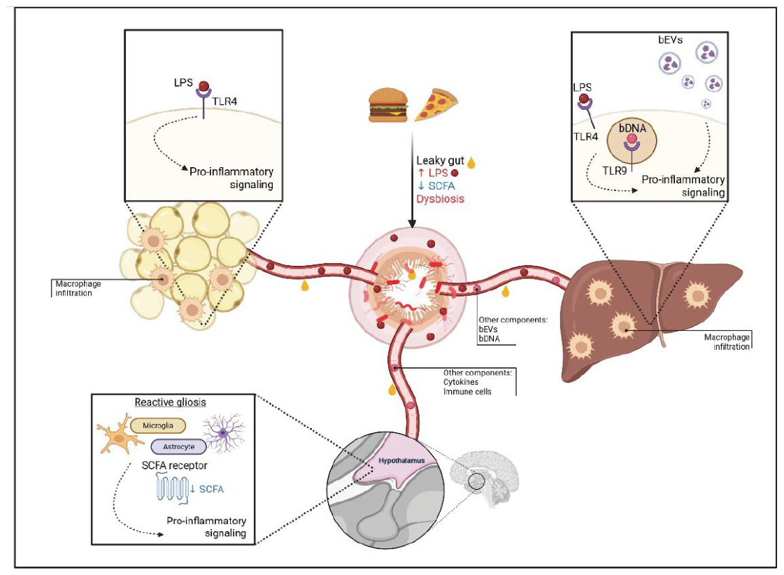
Figure 2.
Microbiota–gut–brain signaling pathways regulating appetite. Gut microbial metabolites and microbial components influence appetite-regulating circuits through multiple gut–brain communication pathways. Microbiota-derived short-chain fatty acids (SCFAs; acetate (C2), propionate (C3) and butyrate (C4)), as well as microbial components such as flagellin, interact with intestinal epithelial and enteroendocrine cells to modulate the release of gut-derived signals, including GLP-1, PYY, CCK, serotonin (5-HT) and glutamate. SCFAs can also signal through G-protein-coupled receptors GPR41/43 expressed by intestinal and neural-associated cells. These signals reach the brain through the circulation and through vagal afferent pathways, ultimately converging on hypothalamic feeding circuits. In parallel, microbial-regulated peripheral metabolic signals such as leptin act directly on leptin receptor (LepR)-expressing hypothalamic neurons. Within the hypothalamus, these convergent signals modulate the activity of anorexigenic POMC and orexigenic AgRP neurons, as well as astrocytes, thereby influencing satiety signaling and food intake. Collectively, these pathways provide multiple mechanisms through which the gut microbiota can modulate central control of energy intake. AgRP, agouti-related peptide; CCK, cholecystokinin; GABA, γ-aminobutyric acid; GPR41/43, G-protein-coupled receptor 41/43; GLP-1, glucagon-like peptide-1; LepR, leptin receptor; POMC, pro-opiomelanocortin; PYY, peptide YY; SCFAs, short-chain fatty acids; 5-HT, 5-hydroxytryptamine.
Figure 2.
Microbiota–gut–brain signaling pathways regulating appetite. Gut microbial metabolites and microbial components influence appetite-regulating circuits through multiple gut–brain communication pathways. Microbiota-derived short-chain fatty acids (SCFAs; acetate (C2), propionate (C3) and butyrate (C4)), as well as microbial components such as flagellin, interact with intestinal epithelial and enteroendocrine cells to modulate the release of gut-derived signals, including GLP-1, PYY, CCK, serotonin (5-HT) and glutamate. SCFAs can also signal through G-protein-coupled receptors GPR41/43 expressed by intestinal and neural-associated cells. These signals reach the brain through the circulation and through vagal afferent pathways, ultimately converging on hypothalamic feeding circuits. In parallel, microbial-regulated peripheral metabolic signals such as leptin act directly on leptin receptor (LepR)-expressing hypothalamic neurons. Within the hypothalamus, these convergent signals modulate the activity of anorexigenic POMC and orexigenic AgRP neurons, as well as astrocytes, thereby influencing satiety signaling and food intake. Collectively, these pathways provide multiple mechanisms through which the gut microbiota can modulate central control of energy intake. AgRP, agouti-related peptide; CCK, cholecystokinin; GABA, γ-aminobutyric acid; GPR41/43, G-protein-coupled receptor 41/43; GLP-1, glucagon-like peptide-1; LepR, leptin receptor; POMC, pro-opiomelanocortin; PYY, peptide YY; SCFAs, short-chain fatty acids; 5-HT, 5-hydroxytryptamine.
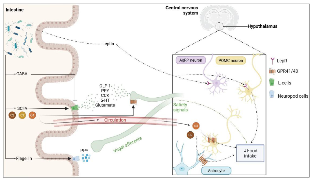
Figure 3.
A self-reinforcing gut–brain circuit linking high-fat feeding, dysbiosis and neuroinflammation. High-fat feeding promotes gut dysbiosis and impaired intestinal barrier function, characterized by increased gut permeability and elevated exposure to microbial and inflammatory signals, including lipopolysaccharide (LPS) and pro-inflammatory cytokines. These signals contribute to systemic inflammation and metabolic alterations in peripheral tissues, including the liver and adipose tissue, while also promoting neuroinflammation. Inflammatory signaling within brain circuits involved in reward and feeding behavior may alter reward processing and promote hedonic eating, favoring further consumption of high-fat foods. Increased high-fat feeding, in turn, reinforces gut dysbiosis and inflammatory signaling, establishing a self-perpetuating gut–brain–metabolic cycle that may contribute to the development and maintenance of obesity. LPS, lipopolysaccharide.
Figure 3.
A self-reinforcing gut–brain circuit linking high-fat feeding, dysbiosis and neuroinflammation. High-fat feeding promotes gut dysbiosis and impaired intestinal barrier function, characterized by increased gut permeability and elevated exposure to microbial and inflammatory signals, including lipopolysaccharide (LPS) and pro-inflammatory cytokines. These signals contribute to systemic inflammation and metabolic alterations in peripheral tissues, including the liver and adipose tissue, while also promoting neuroinflammation. Inflammatory signaling within brain circuits involved in reward and feeding behavior may alter reward processing and promote hedonic eating, favoring further consumption of high-fat foods. Increased high-fat feeding, in turn, reinforces gut dysbiosis and inflammatory signaling, establishing a self-perpetuating gut–brain–metabolic cycle that may contribute to the development and maintenance of obesity. LPS, lipopolysaccharide.
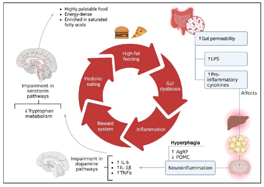

Table 1.
Summary of bacterial taxa altered across different models of obesity.
| Bacterial group altered | Change in obesity | Model | Key product/component | Functional implication | References |
| Akkermansia muciniphila (species) | ↓ | Humans, rats | SCFAs | Reduced gut barrier integrity; increased permeability and endotoxemia | [27,28] |
| Faecalibacterium prausnitzii (species) | ↓ | Humans | Butyrate | Reduced anti-inflammatory signaling; impaired epithelial health | [29] |
| Roseburia spp. (genus) | ↓ | Humans | Butyrate | Decreased SCFA production; altered gut–brain signaling | [29] |
| Clostridiales (order) | ↑ | Humans | Butyrate | Increased gut permeability; systemic low-grade inflammation | [29] |
| Proteobacteria (phylum) | ↑ | Humans, mice | LPS | Metabolic endotoxemia; activation of TLR4 signaling | [28,30,31] |
| Enterobacter cloacae (species) | ↑ | Fecal transfer (Humans → GF mice) | LPS | Causal role in adiposity and insulin resistance | [32] |
| Escherichia/Shigella (genus) | ↑ | Humans | LPS | Pro-inflammatory signaling; gut barrier disruption | [27,29] |
| Klebsiella pneumoniae (species) | ↑ | Humans | Ethanol | Hepatic inflammation; contribution to NAFLD | [33] |
Disclaimer/Publisher’s Note: The statements, opinions and data contained in all publications are solely those of the individual author(s) and contributor(s) and not of MDPI and/or the editor(s). MDPI and/or the editor(s) disclaim responsibility for any injury to people or property resulting from any ideas, methods, instructions or products referred to in the content. |
© 2026 by the authors. Licensee MDPI, Basel, Switzerland. This article is an open access article distributed under the terms and conditions of the Creative Commons Attribution (CC BY) license (http://creativecommons.org/licenses/by/4.0/).
Copyright: This open access article is published under a Creative Commons CC BY 4.0 license, which permit the free download, distribution, and reuse, provided that the author and preprint are cited in any reuse.