
Submitted:
03 September 2026
Posted:
03 September 2026
You are already at the latest version
Abstract
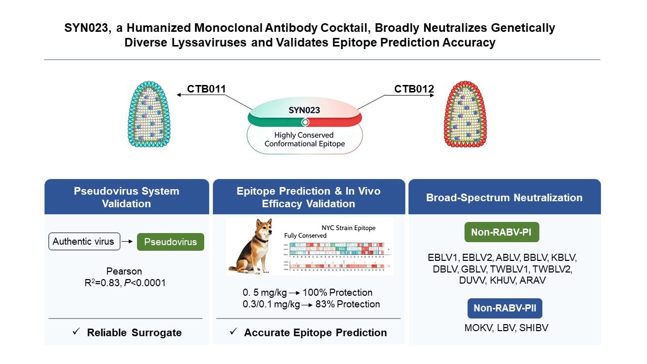
Background: Lyssaviruses remain a global public health threat, requiring broad-spectrum, high-potency neutralizing antibodies as part of recommended human rabies post-exposure prophylaxis (PEP). A humanized monoclonal antibody cocktail, SYN023, is approved for PEP in China and is under global regulatory review. Its epitope mapping data are promising. However, the neutralization breadth across diverse lyssaviruses and the reliability of pseudovirus-based neutralization assays still require comprehensive validation to support worldwide regulatory approval and clinical use. Methods: First, the correlation between authentic lyssaviruses virus and pseudotyped viruses neutralization was established experimentally to verify the reliability of the pseudovirus-based neutralization assay. Second, epitope mapping was performed using a highly virulent rabies virus (RABV) variant (New York City strain: NYC) provided by the U.S. Department of Agriculture, followed by in vivo PEP efficacy studies in a standardized canine model to functionally validate the mapped epitopes. Third, the neutralizing activity of SYN023 was evaluated against a phylogenetically diverse panel of non-RABV lyssaviruses and directly benchmarked against clinically validated comparator monoclonal antibodies, thereby providing robust, quantitative empirical evidence supporting its broad-spectrum neutralization capability. Results: A strong positive correlation was observed between neutralization titers elicited by authentic lyssaviruses and pseudotyped viruses (Pearson R2 = 0.83, P < 0.0001), validating the pseudovirus-based assay as a robust surrogate for authentic virus neutralization. Specifically, SYN023 recognized and potently neutralized the epitope mapped on the prototypic NYC RABV strain. The in vivo PEP studies conducted in a standardized canine challenge model, using the NYC strain as the challenge virus, demonstrated high protective efficacy at SYN023 doses ranging from 0.1 to 0.5 mg/kg. Furthermore, SYN023 exhibited broad, potent, and reproducible neutralizing activity against a phylogenetically diverse panel of non-RABV lyssaviruses, including European bat lyssavirus 1 (EBLV1), European bat lyssavirus 2 (EBLV2), Australian bat lyssavirus (ABLV), Bokeloh bat lyssavirus (BBLV), Taiwan bat lyssavirus 1 (TWBLV1), Taiwan bat lyssavirus 2 (TWBLV2), Kotalahti bat lyssavirus (KBLV), Divača bat lyssavirus (DBLV), Gannoruwa bat lyssavirus (GBLV), Duvenhage virus (DUVV), Khujand virus (KHUV), Aravan virus (ARAV), Mokola virus (MOKV), Lagos bat virus (LBV) and Shimoni bat virus (SHIBV). Notably, SYN023 achieved robust neutralization of MOKV (EC50 = 14.74 µg/mL), whereas clinically referenced monoclonal antibodies—Rabishield, Ormutivimab and GR1801—exhibited no detectable neutralizing activity against MOKV even at concentrations up to 1,000 µg/mL. This pan-lyssavirus neutralizing capacity was attributable mechanistically to SYN023’s targeting of a highly conserved conformational epitope within the viral glycoprotein, which is structurally preserved across all major lyssavirus phylogroups. Conclusion: This study demonstrated that the pseudovirus-based neutralization assay is a reliable surrogate platform. In addition, it confirmed the functional accuracy of the epitope mapping strategy, showed the protective efficacy against a virulent RABV challenge in dogs, and demonstrated the broad neutralization spectrum of the SYN023 antibody cocktail against both RABV and a phylogenetically diverse panel of non-RABV lyssaviruses.
Keywords:
SYN023
; lyssavirus
; pseudovirus system
; epitope prediction
; broad-spectrum neutralization
; rabies
; human monoclonal antibody
1. Introduction
The Lyssavirus genus, within the family Rhabdoviridae, comprises RABV and 18 other phylogenetically distinct non-RABV species [1]. All lyssaviruses are zoonotic pathogens capable of causing acute, progressive, and invariably fatal encephalitis that is clinically indistinguishable from classical rabies [2]. Beyond RABV, spill-over of non-RABV lyssaviruses into humans has resulted in fatal clinical cases across Africa, Europe, and Australia, representing an under-recognized public-health risk. In South Africa, two fatal cases caused by DUVV were reported in 1970 and 2006 [3,4]. In Europe, five human deaths associated with European bat lyssaviruses 1 and 2 (EBLV-1, EBLV-2) have been recorded since the 1980s [5,6,7,8]; one fatal case involved an occupationally exposed patient, where routine rabies diagnostics returned false-negative results and pathogen identification was only achieved post-mortem by next-generation sequencing [8]. In Australia, four fatal infections caused by ABLV have been reported, including a 2025 case in which a middle-aged male developed fatal neurologic disease months after receiving standard rabies PEP following exposure to a flying fox—providing compelling evidence of incomplete cross-neutralization by currently licensed rabies biologics against antigenically divergent lyssaviruses [9,10,11,12]. MOKV—the sole lyssavirus not associated with chiropteran reservoirs—was responsible for two fatal encephalitis cases in Nigeria during the 1970s following contact with small mammals [13].
Globally, lyssavirus infections account for tens of thousands of human deaths annually [14]. Although licensed rabies vaccines and PEP regimens are widely deployed, the expanding taxonomic and geographic diversity of non-RABV lyssaviruses, recurrent spillover events resulting in human mortality, and well-documented limitations of existing neutralizing biologics, including narrow spectrum and strain-dependent efficacy, highlight an urgent unmet need for next-generation, broad-spectrum prevention and control strategies. SYN023 is a novel humanized monoclonal antibody cocktail comprising two non-competing antibodies that jointly target spatially distinct, highly conserved conformational epitopes on the lyssavirus glycoprotein [15]. It has demonstrated potent in vitro neutralizing activity against RABV strains isolated from carnivorous mammals worldwide and from bats across the Americas, as well as against four phylogenetically distinct non-RABV lyssaviruses [15,16]. Approved for clinical use in China, SYN023 has been extensively administrated as part of rabies PEP in real-world settings, with observational data demonstrating 100% protection against World Health Organization (WHO) category III exposures [17,18].
However, key knowledge gaps persist regarding (i) the breadth of SYN023’s neutralizing activity against phylogenetically diverse non-RABV lyssaviruses; (ii) the regulatory acceptability and analytical rigor of the pseudovirus-based neutralization assay used for potency assessment; and (iii) experimental validation of the epitope predictions underpinning its rational design. To address these interrelated questions and generate mechanism-informed, regulatory-grade evidence supporting global implementation, we performed an integrated study comprising: (i) head-to-head comparison of the pseudovirus-based neutralization assay with the authentic virus–based neutralization assay; (ii) functional confirmation of predicted epitopes through in vivo PEP studies; and (iii) systematic cross-neutralization profiling across a representative panel of lyssaviruses, with quantitative EC50 determination and benchmarking against commercially available monoclonal antibodies. Collectively, these data fill critical knowledge gaps and provide key support for SYN023’s global regulatory submissions.
2. Materials and Methods
2.1. Correlation Analysis Between Authentic and Pseudotyped Virus
To evaluate the correlation between SYN023-mediated neutralization of authentic lyssaviruses and pseudotyped lyssaviruses, a total of 22 phylogenetically and geographically diverse lyssavirus variants was subjected to head-to-head comparative neutralization testing using both authentic virus-based and pseudovirus-based assays.
Neutralization testing using authentic virus was performed independently by the Canadian Food Inspection Agency (CFIA), the U.S. Centers for Disease Control and Prevention (CDC), the Chinese Center for Disease Control and Prevention (China CDC), and Kansas State University. Of the 22 variants, neutralization data for 14, including 2 laboratory-adapted strains (CVS-11 and ERA), 5 isolates from carnivorous mammals, and 7 bat-derived isolates, have been previously reported; all exhibited complete neutralization by SYN023 [15,16]. For the remaining 8 variants, 3 (2 carnivore-derived and 1 bat-derived isolate) were evaluated by CFIA and CDC using methodologies published previously [16]; the remaining 5, exclusively isolated from carnivorous mammals, were tested at China CDC. The China CDC neutralization assay protocol is described below: six 5-fold serial dilutions of SYN023 (1.0, 0.2, 0.04, 0.008, 0.0016 and 0.00032 μg/mL) were pre-incubated with RABV variants in a 96-well plate for 60 min at 37 °C. Subsequently, 100 μL of mouse neuroblastoma (MNA) cells (5 × 10⁵ cells/mL) were added to each well containing the antibody–virus mixture (100 μL SYN023 + 100 μL virus [50 FFU50]), with eight replicates per dilution, followed by incubation for 48 h at 37 °C under 5% CO2. Cells were then washed, fixed with cold 80% acetone, and stained with fluorescein isothiocyanate (FITC)-labeled anti-RABV immunoglobulin (Fujirebio Diagnostics, Inc.), supplemented with 0.5% Evans blue in PBS to suppress nonspecific fluorescence. Virus-infected foci were enumerated using a fluorescence microscope at ×200 magnification. The RABV neutralizing antibody (RVNA) titers were calculated from the proportion of positive wells per dilution using the Reed–Muench method and converted to EC50 for comparison analysis.
Neutralization testing using the pseudovirus system was conducted as follows: Pseudotyped lyssaviruses were generated by co-transfecting HEK293T cells with a plasmid encoding the lyssavirus glycoprotein (pCAGGS-G) to mediate pseudotyping, followed by infection with VSVΔG*-G (a glycoprotein-complemented vesicular stomatitis virus bearing a luciferase reporter and lacking its native G gene). Prior to pseudovirus infection, HEK293T cells were seeded in 96-well plates and incubated at 37 °C under 5% CO2 for 4-6 h to achieve optimal confluence. Serial dilutions of SYN023, clinically referenced monoclonal antibodies (including Rabishield, Ormutivimab and GR1801), or human rabies immune globulin (HRIG; Nanyue Biopharmaceutical Co., Ltd.) were prepared in assay medium. Each antibody dilution (50 μL) was pre-incubated with RABV pseudovirus (100 μL; 2000-4000 TCID50) for 1 h at 37 °C. The antibody–pseudovirus mixture was then added to wells containing the pre-seeded HEK293T cells. Plates were shaken gently to synchronize infection, followed by incubation at 37 °C under 5% CO2 for 24 h. At 24 h post-infection, 200 μL of culture supernatant was aspirated carefully from each well, and 50 μL of luciferase detection reagent (Vazyme) was added. After brief mixing, luminescence was quantified using a microplate luminometer. Neutralization activity was calculated as percent inhibition of luciferase signal relative to pseudovirus-only controls, and EC50 values were determined using nonlinear regression fitted to a four-parameter logistic model.
2.2. Experimental Animals
A total of 33 beagle dogs, balanced for sex and approximately 3 months of age, were enrolled in the NYC strain of rabies virus challenge dose titration and SYN023 PEP efficacy studies. Animals were sourced from Covance Research Products (Cumberland, VA, USA) and maintained under a standardized routine vaccination program against canine parvovirus (CPV), canine distemper virus (CDV), canine adenovirus(CAV), canine parainfluenza virus (CPIV), and canine coronavirus (CCV), among other endemic pathogens. Prior to study initiation, all dogs underwent serological screening on day –14 (±2 days) to confirm rabies virus seronegativity; neutralizing antibody titers were below the limit of detection (< 0.1 IU/mL) as determined by the rapid fluorescent focus inhibition test (RFFIT). Dogs were individually housed under ABSL-2 conditions with ad libitum access to certified feed and potable water. A 7-day acclimatization period was implemented prior to study procedures to ensure physiological and behavioral stabilization. Any animal exhibiting clinical signs indicative of systemic illness or distress was humanely euthanized in accordance with institutional animal welfare guidelines. Viral challenge, therapeutic intervention, and outcome assessments were conducted in a strictly defined, consistent sequence across all subjects. Cage assignments were randomized and spatially balanced within the housing room to mitigate positional confounding. To ensure blinding, animal care staff responsible for daily clinical observations had no access to treatment group assignments, including both rabies virus challenge dose levels and SYN023 administration schedules, and all related assignment documentation was physically removed from the housing area prior to study commencement. All experimental procedures, clinical monitoring, and data collection were conducted in strict accordance with the SOPs of Labcorp (formally Covance) to ensure methodological consistency and minimize experimental variability.
2.3. Epitope Analysis of the NYC Strain
Samples of highly virulent NYC street RABV (Lot No. 92-5A) were provided by the U.S. Department of Agriculture (https://www.aphis.usda.gov/sites/default/files/cvb-dat-0424.pdf). This live rabies virus was isolated from fox salivary glands and kept at -80 °C. To establish the canine challenge dose, nine dogs were inoculated intramuscularly with three serial dilutions of the NYC strain (1:10, 1:100, and 1:1000; n = 3 per group). Rabies virus RNA was extracted from brainstem tissue of four dogs that succumbed to infection. Full-length glycoprotein (G) gene sequences were generated via bidirectional Sanger sequencing: viral RNA was reverse-transcribed and amplified using overlapping RT-PCR; purified amplicons were sequenced with BigDye Terminator v3.1 chemistry on an ABI 3500xl capillary platform. Consensus sequences were assembled from forward and reverse chromatograms to obtain complete G gene coding sequences. Evolutionary conservation of the SYN023 epitope residues—relative to the reference challenge strain CVS-11—was evaluated by multiple sequence alignment using ClustalX 2.1.
2.4. PEP Efficacy of SYN023 Against the NYC Strain
Twenty-four beagle dogs were randomly allocated to four treatment groups (n = 6 per group) using a computer-generated randomization sequence (Microsoft Excel®). Group assignment was stratified by body weight and sex to ensure balanced distribution across all groups. On Day 0, all animals received an intramuscular (IM) challenge with the NYC RABV strain at a 1:100 dilution from the original virus stock [107.9 MICLD50/mL]). Groups 1–3 received a single IM dose of SYN023 at 0.5, 0.3, or 0.1 mg/kg, respectively, administered 24 ± 1 h post-challenge (Day 1) at the same anatomical site as the viral inoculation. The control group (Group 4) received an equivalent volume of PBS administered IM at the inoculation site. Clinical status was monitored daily for 90 days; survival was recorded, and protection rates were calculated as the percentage of animals surviving to the end of the study. Blood samples were collected before treatment and upon onset of clinical signs or at the humane endpoint; terminal brainstem tissue was harvested at euthanasia. Serum was analyzed for RVNA titers using the RFFIT. Brainstem sections were evaluated for RABV antigens detection by direct fluorescent antibody (DFA) test.
2.5. RFFIT for Serum RVNA Titer Quantification
RFFIT assay was performed to quantify RVNA titers in serum samples. Serial five-fold dilutions of each sample were prepared in EMEM-10 medium and loaded onto 8-well chamber slides (Lab-Tek®). A standard dilution series—1:5, 1:25, 1:125, and 1:625—was achieved by first diluting serum 1:2.5, 1:12.5, 1:62.5, and 1:312.5 in EMEM-10 (final volume per well: 0.1 mL), followed by addition of 0.1 mL of challenge virus (rabies virus CVS-11 strain; 30–50 FFU/mL). Slides were incubated for 90 min at 37 °C in a humidified CO2 incubator. Subsequently, 0.2 mL of BHK-21 cell suspension (5 × 10⁵ cells/mL) was added to each well, gently mixed, and incubated for 24 h at 37 °C under 5% CO2. Cells were then washed with PBS, fixed in cold 80% acetone, and stained with FITC-conjugated anti-rabies virus immunoglobulin (Fujirebio Diagnostics, Inc.), supplemented with 0.05% Evans blue (in PBS) to quench nonspecific fluorescence. Slides were examined under a fluorescence microscope at ×200 magnification. For each dilution, 20 microscopic fields were assessed for fluorescent foci. RVNA titers were calculated as the endpoint dilution (ED50) using the Reed–Muench method and converted to international units per milliliter (IU/mL) by comparison with the WHO International Standard for Rabies Immunoglobulin (SRIG; assigned potency: 2.0 IU/mL), according to the formula: IU/mL = (ED50 reciprocal titer of test sample ÷ ED50 reciprocal titer of SRIG) × 2.0 IU/mL.
2.6. DFAT for RABV Antigen Test in Brains Tissues
Direct fluorescent antibody testing (DFAT), widely regarded as the gold standard for rabies diagnosis, was employed for qualitative detection of rabies virus (RABV) antigen in fresh brain tissue specimens obtained from experimentally challenged animals. Monolayer impression smears were prepared by gently pressing transverse sections of brainstem tissue onto glass slides, followed by air-drying at room temperature for 15 min and fixation in cold acetone (−20 °C) for 1 hour to overnight. Concurrently, positive and negative control slides were processed under identical conditions. After fixation, residual acetone was carefully removed, and slides were allowed to air-dry at room temperature. Subsequently, slides were placed in a humidified chamber and incubated with FITC-labeled anti-rabies virus immunoglobulin (Fujirebio Diagnostics, Inc.), diluted in PBS containing 4 μL/mL Evans blue dye, at 37 °C for 30 min. Unbound conjugate was removed by gentle rinsing with PBS, followed by two sequential 5-min soaks in PBS. Slides were then vertically drained, air-dried at room temperature, mounted with 20% glycerol in Tris buffer (pH 9.0), and covered with thin glass coverslips. Fluorescence microscopy was performed using an inverted fluorescence microscope at a minimum magnification of 200×. For each brain region, 40 non-overlapping microscopic fields were systematically evaluated by two independent, blinded technicians. A result was considered positive if distinct apple-green, punctate or granular fluorescence was observed specifically within neuronal cytoplasm, in the absence of non-specific red background fluorescence; absence of such specific green fluorescence was interpreted as negative.
2.7. Statistical Analysis
Pearson correlation analysis was performed to evaluate the association between SYN023-mediated neutralizing activity against authentic RABV and rabies pseudovirus. Survival curves were compared using the log-rank (Mantel–Cox) test. The RVNA titers were compared between dogs with positive versus negative direct fluorescent antibody (DFA) test results using an unpaired two-tailed Student’s t-test. P<0.05 was considered statistically significant. All statistical analyses were conducted using GraphPad Prism version 8.0.
3. Results
3.1. Reliability of the Pseudovirus System and Accuracy of Epitope Prediction
The correlation analysis between authentic lyssavirus and pseudovirus revealed a strong positive correlation (Pearson R2=0.83, P<0.0001), indicating that the EC50 values of SYN023 measured in the pseudovirus assay closely mirrored those obtained using the authentic virus system (Figure 1 and Table S1).
As a bispecific antibody cocktail, SYN023 retains neutralizing activity as long as at least one constituent antibody (CTB011 or CTB012) effectively engages its target epitope. Notably, the CTB012 epitope residues are highly conserved across all tested RABV variants (Table S1); correspondingly, SYN023 demonstrated robust and consistent neutralization against these variants in both authentic virus and pseudovirus assays. This concordance supports the utility of epitope conservation as a predictive indicator of cross-neutralization potential. In contrast, the two non-RABV lyssaviruses, BBLV and EBLV2, harbor amino acid substitutions in the epitopes bound by both CTB011 (N355T) and CTB012 (V291I). Nevertheless, SYN023 and CTB012 alone retained measurable neutralizing activity against these divergent viruses, albeit with reduced potency (i.e., higher EC50 values; Table S1). Specifically, the V291I substitution within the CTB012 epitope partially attenuated, but did not abolish neutralization.
3.2. SYN023 Neutralization Prediction and PEP Efficacy Against the NYC Strain
All dogs succumbed to infection following challenge with the NYC rabies virus at dilutions of 1:10 (3/3) and 1:100 (3/3) of the original stock; in contrast, one of three dogs survived challenge at the 1:1000 dilution (Table S2). The 1:100 dilution was selected as the standardized challenge dose, as it represented the lowest viral concentration at which uniform, lethal infection was observed across all animals. Epitope mapping was performed using RNA extracted from brainstem tissue of four dogs that succumbed to NYC rabies virus infection.
Epitope analysis revealed that the NYC strain harbors no amino acid substitutions in either the CTB011 or CTB012 epitope (Figure 2A), indicating full conservation of both target sites and supporting the expectation that SYN023, a cocktail of CTB011 and CTB012, would potently neutralize this strain. Consistent with this prediction, SYN023 conferred robust protection in unvaccinated dogs challenged with the NYC street RABV across all tested doses (Figure 2B): 100% (6/6) survival at 0.5 mg/kg, and 83% (5/6) survival at both 0.3 mg/kg and 0.1 mg/kg. In contrast, all animals in the PBS-treated control group succumbed to infection within 10 days post-challenge. The DFA testing confirmed the absence of RABV antigens in all surviving dogs, whereas all moribund or euthanized animals tested positive (Table S3). Furthermore, the RVNA titers in survivors were significantly lower than those in non-survivors (P < 0.0001; Table S3), suggesting effective viral clearance rather than immune-mediated pathology.
3.3. Broad-Spectrum Neutralization Efficacy of SYN023 Against Non-RABV Lyssaviruses
The pseudovirus neutralization assay demonstrated that SYN023 exhibits potent and consistent neutralizing activity against all tested non-RABV lyssaviruses, with EC50 values of 0.63 ng/mL (EBLV1), 8.221 μg/mL (EBLV2), 7.65 ng/mL (ABLV), 0.96 μg/mL (BBLV), 0.29 μg/mL (TWBLV1), 15.75μg/mL (TWBLV2), 0.57 ng/mL (GBLV), 53.56 ng/mL (DUVV), 0.54 µg/mL (KHUV), 31.62 µg/mL (ARAV), 14.74 µg/mL (MOKV), 4.84 μg/mL (LBV) and 3.18 μg/mL (SHIBV) (Table 1). In marked contrast, other commercially available monoclonal antibodies Rabishield [19], Ormutivimab [20], and GR1801 [21], failed to neutralize MOKV even at the highest tested concentration of 1,000 µg/mL (Table 2). This complete lack of activity underscores SYN023’s distinctive capacity to neutralize MOKV, a capability not shared by any currently licensed rabies biologics. Collectively, these data establish that SYN023 possesses a uniquely broad lyssavirus-neutralizing spectrum, attributable to its dual-targeting mechanism engaging a highly conserved conformational epitope on the lyssavirus glycoprotein.
4. Discussion
Lyssaviruses represent a persistent and severe threat to global public health, underscoring the urgent need for broad-spectrum neutralizing agents to strengthen rabies prevention and control [2,14]. The humanized monoclonal antibody cocktail SYN023 has received marketing authorization in China and is currently under global regulatory review. The present work generated three sets of regulatory-relevant evidence: assay validation for pseudovirus neutralization, functional verification of epitope-based efficacy prediction in a canine PEP model, and systematic profiling of cross-neutralization against a panel of genetically diverse lyssaviruses.
Pseudovirus-based neutralization assays offer substantial biosafety and operational advantages over authentic high-containment lyssavirus work by eliminating the requirement for BSL-3 facilities and enabling scalable, high-throughput serological assessment [22,23,24]. Recent advances have enabled the generation of replication-incompetent pseudotypes representing all 18 known lyssavirus species, greatly expanding the capacity for in vitro antigenic profiling of divergent field isolates [25]. Nevertheless, surrogate assays require empirical validation for each therapeutic candidate rather than direct extrapolation from published platform performance [26,27]. Our data demonstrate a strong correlation between pseudovirus-derived and authentic-virus EC50 values (R2 = 0.83, P < 0.0001), formally supporting the use of this pseudovirus platform for potency assessment of SYN023. This finding aligns with meta-analytic evidence indicating that, while concordance between pseudotyped and wild-type lyssavirus neutralization readouts varies across antibody candidates, it remains quantitatively informative and mechanistically interpretable [16,28].
Beyond in vitro findings, our canine challenge study using the NYC street-virus strain provided critical in vivo proof-of-concept for epitope-conservation guided prediction. Full conservation of both CTB011 and CTB012 epitopes in this epidemiologically significant isolate accurately predicted robust protection following post-exposure administration of SYN023. Absence of viral antigen in brain tissue (as confirmed by DFA testing) alongside comparatively low RVNA titers in surviving animals supports a mechanism wherein SYN023 mediates rapid in vivo viral neutralization and clearance, rather than eliciting immune-mediated pathology. These results bridge computational epitope mapping to functional in vivo outcomes and reinforce the utility of glycoprotein epitope conservation as a predictive biomarker for real-world SYN023 performance against circulating field isolates.
A major finding of this study is that SYN023 exhibits exceptional breadth of neutralization against a broad panel of non-RABV lyssaviruses, including representatives from both phylogroup I and phylogroup II, with particularly robust activity against MOKV. In contrast, commercially available rabies immunoglobulin (RIG) and currently approved monoclonal antibody therapeutics show minimal to no neutralizing activity against phylogroup II lyssaviruses, a critical therapeutic gap in light of the documented zoonotic spillover potential of these bat-independent lyssaviruses [19,29]. Fatal human encephalitis caused by MOKV has been historically reported in Africa, and serological surveillance data indicate sustained circulation in small-mammal reservoirs, for which no validated post-exposure prophylactic interventions currently exist [13,29]. Structurally, pan-lyssavirus recognition by therapeutic antibodies is restricted to a limited number of conserved conformational epitopes on the lyssavirus glycoprotein [30]. SYN023 maintains potent activity against MOKV primarily through CTB012-mediated engagement of one such evolutionarily conserved and structurally stable conformational epitope, demonstrating how its dual-antibody cocktail design confers uniquely broad pan-lyssavirus coverage.
Although SYN023 was primarily developed for human rabies PEP, our canine efficacy data support the exploration of potential One-Health-oriented applications [31]. Current guidelines recommend pre-exposure vaccination for domestic animals at risk of lyssavirus exposure [32]. In contrast, unvaccinated companion or livestock animals that experience confirmed or suspected lyssavirus exposure are often subject to euthanasia or prolonged quarantine—measures that impose significant emotional distress on owners and substantial socioeconomic burdens on smallholder farming communities [33]. Field-based programs in regions such as Texas have demonstrated the operational feasibility of administering PEP to unvaccinated exposed animals under rigorously defined local protocols [34]. Passive immunization with neutralizing antibodies represents an underexplored yet biologically plausible alternative for naïve, exposed animals; however, licensed veterinary monoclonal antibody products specifically indicated for lyssavirus prophylaxis remain unavailable [35]. Building on our robust canine challenge data, future studies could systematically evaluate the feasibility, dosing regimen, and clinical utility of SYN023-based PEP in naïve exposed domestic animals, pending comprehensive assessment of species-specific safety, pharmacokinetics, and cost-effectiveness. Critically, any off-label use in veterinary settings would require formal regulatory review and endorsement by veterinary medical authorities, as well as integration into updated clinical practice guidelines, prior to implementation.
In summary, this study establishes that (i) the pseudovirus assay is a reliable, high-fidelity surrogate for authentic virus neutralization testing of SYN023; (ii) epitope conservation serves as a robust predictor of neutralization susceptibility across diverse lyssaviruses; and (iii) SYN023 exhibits superior, broad-spectrum neutralization against both RABV and non-RABV lyssaviruses. As an already-approved agent in China and undergoing global regulatory review, SYN023 holds substantial promise for expanding the geographic and virologic scope of rabies PEP. The potency data, mechanistic insights, and functional validation reported herein generate robust regulatory-ready evidence supporting its potential integration into national rabies-control programs and WHO guidance [36,37], positioning SYN023 as a promising component for next-generation lyssavirus prevention strategies.
Supplementary Materials
The following supporting information can be downloaded at the website of this paper posted on Preprints.org, Table S1: Neutralization activity of SYN023 against authentic and pseudotyped lyssaviruses; Table S2: Observational summary and diagnostic test results (DFA and RVNA) evaluating SYN023 efficacy in canine PEP.
Author Contributions
Conceptualization, J.Q. and E.T.; methodology, J.Q., S.H., Q.L., C.E.R., P.Y., and W.Z.; software, J.Q.; validation, J.Q. and E.T.; formal analysis, J.Q. and E.T; investigation, J.Q. and E.T; resources, E.T.; data curation, J.Q.; writing—original draft preparation, J.Q.; writing—review and editing, C.E.R., E.T.; supervision, E.T.; project administration, E.T.; funding acquisition, E.T. All authors have read and agreed to the published version of the manuscript.
Funding
This research was funded by Synermore Biologics.
Institutional Review Board Statement
The animal study described herein was conducted in strict compliance with the U.S. Department of Agriculture’s (USDA) Animal Welfare Act (9 CFR Parts 1, 2, and 3); the Guide for the Care and Use of Laboratory Animals (Institute of Laboratory Animal Resources, National Academy Press, Washington, D.C., 2011); and the National Institutes of Health, Office of Laboratory Animal Welfare. Whenever possible, procedures in this study were designed to avoid or minimize discomfort, distress, and pain to animals. This study was reviewed and approved by the Covance Institutional Animal Care and Use Committee (IACUC) under protocol number PA-0111-16.
Informed Consent Statement
Not applicable.
Data Availability Statement
The data of this study are available upon reasonable request from the authors.
Acknowledgments
We thank Wenjie Tao and Lan Han for purifying the Rabishield, Ormutivimab, and GR1801 monoclonal antibodies and for performing size-exclusion chromatography (SEC) validation to confirm their structural integrity and monomeric purity.
Conflicts of Interest
The authors declare that they have no known competing financial interests or personal relationships that could have appeared to influence the work reported in this paper.
Abbreviations
The following abbreviations are used in this manuscript:
| RABV | Rabies virus |
| NYC | New York City rabies virus strain |
| PEP | Post-exposure prophylaxis |
| EBLV1 | European bat lyssavirus 1 |
| EBLV2 | European bat lyssavirus 2 |
| ABLV | Australian bat lyssavirus |
| BBLV | Bokeloh bat lyssavirus |
| TWBLV1 | Taiwan bat lyssavirus 1 |
| TWBLV2 | Taiwan bat lyssavirus 2 |
| KBLV | Kotalahti bat lyssavirus |
| DBLV | Divača bat lyssavirus |
| GBLV | Gannoruwa bat lyssavirus |
| DUVV | Duvenhage virus |
| KHUV | Khujand virus |
| ARAV | Aravan virus |
| MOKV | Mokola virus |
| LBV | Lagos bat virus |
| SHIBV | Shimoni bat virus |
| WHO | World Health Organization |
| CFIA | Canadian Food Inspection Agency |
| CDC | Centers for Disease Control and Prevention |
| RVNA | Rabies virus neutralizing antibody |
| RFFIT | Rapid fluorescent focus inhibition test |
| DFA | Direct fluorescent antibody test |
References
- Black, E.J.; Powell, C.S.; Dempsey, D.M.; Hendrickson, R.C.; Mims, L.R.; Lefkowitz, E.J. Virus taxonomy: the database of the International Committee on Taxonomy of Viruses. Nucleic Acids Res. 2026, 54, D776–D89. [Google Scholar] [CrossRef] [PubMed]
- Fooks, A.R.; Shipley, R.; Markotter, W.; Tordo, N.; Freuling, C.M.; Muller, T.; et al. Renewed Public Health Threat from Emerging Lyssaviruses. Viruses 2021, 13. [Google Scholar] [CrossRef] [PubMed]
- Meredith, C.D.; Prossouw, A.P.; Koch, H. An unusual case of human rabies thought to be of chiropteran origin. S Afr. Med. J. 1971, 45, 767–9. [Google Scholar] [PubMed]
- Paweska, J.T.; Blumberg, L.H.; Liebenberg, C.; Hewlett, R.H.; Grobbelaar, A.A.; Leman, P.A.; et al. Fatal human infection with rabies-related Duvenhage virus, South Africa. Emerg. Infect. Dis. 2006, 12, 1965–7. [Google Scholar] [CrossRef] [PubMed]
- Lumio, J.; Hillbom, M.; Roine, R.; Ketonen, L.; Haltia, M.; Valle, M.; et al. Human rabies of bat origin in Europe. Lancet 1986, 1, 378. [Google Scholar] [CrossRef] [PubMed]
- Selimov, M.A.; Tatarov, A.G.; Botvinkin, A.D.; Klueva, E.V.; Kulikova, L.G.; Khismatullina, N.A. Rabies-related Yuli virus; identification with a panel of monoclonal antibodies. Acta Virol. 1989, 33, 542–6. [Google Scholar] [PubMed]
- Fooks, A.R.; McElhinney, L.M.; Pounder, D.J.; Finnegan, C.J.; Mansfield, K.; Johnson, N.; et al. Case report: isolation of a European bat lyssavirus type 2a from a fatal human case of rabies encephalitis. J. Med. Virol. 2003, 71, 281–9. [Google Scholar] [CrossRef] [PubMed]
- Regnault, B.; Evrard, B.; Plu, I.; Dacheux, L.; Troadec, E.; Cozette, P.; et al. First Case of Lethal Encephalitis in Western Europe Due to European Bat Lyssavirus Type 1. Clin. Infect. Dis. 2022, 74, 461–6. [Google Scholar] [CrossRef] [PubMed]
- Anthony Allworth, K.M.; John, Morgan. A human case of encephalitis due to a lyssavirus recently identified in fruit bats. Commun. Dis. Intell. 1996, 20, 24–5. [Google Scholar] [CrossRef]
- Hanna, J.N.; Carney, I.K.; Smith, G.A.; Tannenberg, A.E.; Deverill, J.E.; Botha, J.A.; et al. Australian bat lyssavirus infection: a second human case, with a long incubation period. Med. J. Aust. 2000, 172, 597–9. [Google Scholar] [CrossRef] [PubMed]
- Francis, J.R.; Nourse, C.; Vaska, V.L.; Calvert, S.; Northill, J.A.; McCall, B.; et al. Australian Bat Lyssavirus in a child: the first reported case. Pediatrics 2014, 133, e1063-7. [Google Scholar] [CrossRef] [PubMed]
- Lateef, S. A rare case of rabies-like Australian bat lyssavirus infection. Lancet Infect. Dis. 2025. [Google Scholar] [CrossRef] [PubMed]
- Familusi, J.B.; Osunkoya, B.O.; Moore, D.L.; Kemp, G.E.; Fabiyi, A. A fatal human infection with Mokola virus. Am. J. Trop. Med. Hyg. 1972, 21, 959–63. [Google Scholar] [CrossRef] [PubMed]
- Fooks, A.R.; Cliquet, F.; Finke, S.; Freuling, C.; Hemachudha, T.; Mani, R.S.; et al. Rabies. Nat. Rev. Dis. Primers 2017, 3, 17091. [Google Scholar] [PubMed]
- Chao, T.Y.; Ren, S.; Shen, E.; Moore, S.; Zhang, S.F.; Chen, L.; et al. SYN023, a novel humanized monoclonal antibody cocktail, for post-exposure prophylaxis of rabies. PLoS Negl. Trop. Dis. 2017, 11, e0006133. [Google Scholar] [CrossRef] [PubMed]
- Cao, L.; Qu, J.; Zhang, C.; Chen, R.; Wang, J.; Fehlner-Gardiner, C.; et al. Structural basis for broad neutralization of rabies virus by an antibody cocktail SYN023. Emerg. Microbes Infect. 2025, 14, 2547724. [Google Scholar] [CrossRef] [PubMed]
- McClain, J.B.; Chuang, A.; Reid, C.; Moore, S.M.; Tsao, E. Rabies virus neutralizing activity, pharmacokinetics, and safety of the monoclonal antibody mixture SYN023 in combination with rabies vaccination: Results of a phase 2, randomized, blinded, controlled trial. Vaccine 2021, 39, 5822–30. [Google Scholar] [CrossRef] [PubMed]
- Liu, X.; Li, J.; Zha, Y.; Wang, Z.; Jiang, Y.; Zhang, X.; et al. The efficacy and safety of SYN023 (Zamerovimab and Mazorelvimab injection), the recombinant humanized anti-rabies virus monoclonal antibody mixture, combined with rabies vaccine in a WHO category III rabies post-exposure population: A randomized, double-blind, positive control, phase III clinical trial. Vaccine 2025, 61, 127289. [Google Scholar] [CrossRef] [PubMed]
- Sloan, S.E.; Hanlon, C.; Weldon, W.; Niezgoda, M.; Blanton, J.; Self, J.; et al. Identification and characterization of a human monoclonal antibody that potently neutralizes a broad panel of rabies virus isolates. Vaccine 2007, 25, 2800–10. [Google Scholar] [CrossRef] [PubMed]
- Zhai, L.L.; Wang, H.; Zhao, W.; Zhang, S.F.; Miao, F.M.; Cao, Y.; et al. Efficacy of ormutivimab, a novel recombinant human anti-rabies monoclonal antibody, in post-exposure prophylaxis animal models. Travel Med. Infect. Dis. 2022, 46, 102267. [Google Scholar] [CrossRef] [PubMed]
- Long, C.; Wang, W.; Hao, X.; Yu, C.; Feng, Y.; Tu, C.; et al. Development of a novel bispecific antibody GR1801 for rabies. J. Med. Virol. 2023, 95, e29016. [Google Scholar] [CrossRef] [PubMed]
- Nie, J.; Wu, X.; Ma, J.; Cao, S.; Huang, W.; Liu, Q.; et al. Development of in vitro and in vivo rabies virus neutralization assays based on a high-titer pseudovirus system. Sci. Rep. 2017, 7, 42769. [Google Scholar] [CrossRef] [PubMed]
- Thimmiraju, S.R.; Kimata, J.T.; Pollet, J. Pseudoviruses, a safer toolbox for vaccine development against enveloped viruses. Expert Rev. Vaccines 2024, 23, 174–85. [Google Scholar] [CrossRef] [PubMed]
- Rizatdinova, S.N.; Ershova, A.E.; Astrakhantseva, I.V. Pseudotyped Viruses: A Useful Platform for Pre-Clinical Studies Conducted in a BSL-2 Laboratory Setting. Biomolecules 2025, 15. [Google Scholar] [CrossRef] [PubMed]
- Inoue, Y.; Kaku, Y.; Harada, M.; Ishijima, K.; Kuroda, Y.; Tatemoto, K.; et al. Establishment of serological neutralizing tests using pseudotyped viruses for comprehensive detection of antibodies against all 18 lyssaviruses. J. Vet. Med. Sci. 2024, 86, 128–34. [Google Scholar] [CrossRef] [PubMed]
- Nie, J.; Li, Q.; Wu, J.; Zhao, C.; Hao, H.; Liu, H.; et al. Quantification of SARS-CoV-2 neutralizing antibody by a pseudotyped virus-based assay. Nat. Protoc. 2020, 15, 3699–715. [Google Scholar] [CrossRef] [PubMed]
- Luo, X.; Wang, C.; Huang, Y.; Cong, S.; Tan, J.; Hou, W.; et al. Establishment of a neutralization assay for Nipah virus using a high-titer pseudovirus system. Biotechnol. Lett. 2023, 45, 489–98. [Google Scholar] [CrossRef] [PubMed]
- Cantoni, D.; Wilkie, C.; Bentley, E. M.; Mayora-Neto, M.; Wright, E.; Scott, S.; Ray, S.; Castillo-Olivares, J.; Heeney, J. L.; Mattiuzzo, G.; Temperton, N. J. Correlation between pseudotyped virus and authentic virus neutralisation assays, a systematic review and meta-analysis of the literature. Front. Immunol. 2023. [Google Scholar] [CrossRef] [PubMed]
- Coertse, J.; Viljoen, N.; Weyer, J.; Markotter, W. Comparative Neutralization Activity of Commercial Rabies Immunoglobulin against Diverse Lyssaviruses. Vaccines (Basel) 2023, 11. [Google Scholar]
- Callaway, H.M. Structural overview of lyssavirus glycoproteins, antibodies, and receptors. J. Virol. 2026, 100, e0178625. [Google Scholar] [CrossRef] [PubMed]
- Overgaauw, P.A.M.; Vinke, C.M.; Hagen, M.; Lipman, L.J.A. A One Health Perspective on the Human-Companion Animal Relationship with Emphasis on Zoonotic Aspects. Int. J. Environ. Res. Public Health 2020, 17. [Google Scholar] [CrossRef] [PubMed]
- National Association of State Public Health V; Compendium of Animal Rabies P; Control, C.; Brown, C.M.; Slavinski, S.; Ettestad, P.; et al. Compendium of Animal Rabies Prevention and Control, 2016. J. Am. Vet. Med. Assoc. 2016, 248, 505–17. [Google Scholar] [CrossRef] [PubMed]
- Rupprecht, C.E.; Belsare, A.V.; Cliquet, F.; Mshelbwala, P.P.; Seetahal, J.F.R.; Wicker, V.V. The Challenge of Lyssavirus Infections in Domestic and Other Animals: A Mix of Virological Confusion, Consternation, Chagrin, and Curiosity. Pathogens 2025, 14. [Google Scholar] [CrossRef] [PubMed]
- Wilson, P.J.; Hunt, P.R.; Fonken, E.P. Rabies postexposure prophylaxis protocol option for unvaccinated domestic animals, Texas: 2010-2019. J. Am. Vet. Med. Assoc. 2024, 262, 627–34. [Google Scholar] [CrossRef] [PubMed]
- Chorpunkul, A.; Boonyuen, U.; Limkittikul, K.; Saengseesom, W.; Phongphaew, W.; Putchong, I.; et al. Development of novel canine phage display-derived neutralizing monoclonal antibody fragments against rabies virus from immunized dogs. Sci. Rep. 2024, 14, 22939. [Google Scholar] [CrossRef] [PubMed]
- World Health O. Rabies vaccines: WHO position paper, April 2018 - Recommendations. Vaccine 2018, 36, 5500–3. [Google Scholar] [CrossRef] [PubMed]
- Organization WH. WHO expert consultation on rabies: third report. WHO Technical Report Series. 2018.
Figure 1.
Correlation between authentic virus and pseudovirus neutralization EC50 values across 22 phylogenetically and geographically diverse lyssavirus variants.
Figure 1.
Correlation between authentic virus and pseudovirus neutralization EC50 values across 22 phylogenetically and geographically diverse lyssavirus variants.
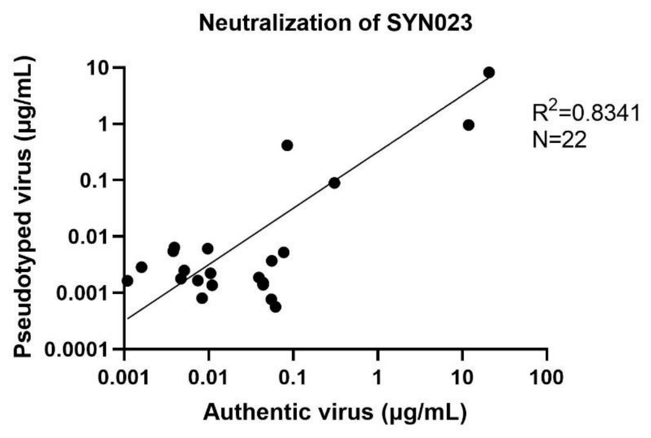
Figure 2.
Conservation of the epitope residues in the NYC strain and PEP efficacy of SYN023. (A) Glycoprotein sequences of the NYC strain derived from viral RNA isolated from the brainstem of infected dogs (designated CHR, CIJ, CIA, and CKF) were aligned with that of the CVS-11 reference strain. (B) Four groups of dogs (n = 6 per group) were challenged intramuscularly with the NYC virus and administered a single dose of SYN023 one day post-infection at 0.5, 0.3, or 0.1 mg/kg.
Figure 2.
Conservation of the epitope residues in the NYC strain and PEP efficacy of SYN023. (A) Glycoprotein sequences of the NYC strain derived from viral RNA isolated from the brainstem of infected dogs (designated CHR, CIJ, CIA, and CKF) were aligned with that of the CVS-11 reference strain. (B) Four groups of dogs (n = 6 per group) were challenged intramuscularly with the NYC virus and administered a single dose of SYN023 one day post-infection at 0.5, 0.3, or 0.1 mg/kg.
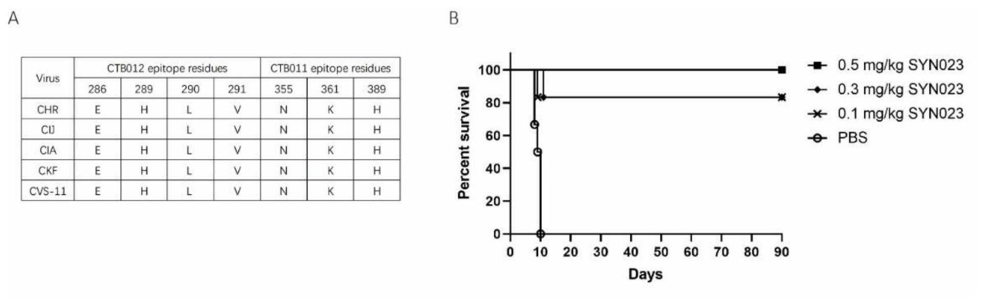

Table 1.
Neutralization activity of SYN023 against non-RABV lyssavirus variants.
| Lyssavirus variant | GenBank accession | Lineage | Neutralization of SYN023 (EC50) |
|---|---|---|---|
| EBLV1 | MF187802 | Non-RABV-PI | 0.63 ng/mL |
| EBLV2 | JX129233 | Non-RABV-PI | 8.221 μg/mL |
| ABLV | AF418014 | Non-RABV-PI | 7.65 ng/mL |
| BBLV | MF043188 | Non-RABV-PI | 0.96 μg/mL |
| TWBLV1 | ON437590 | Non-RABV-PI | 0.29 μg/mL |
| TWBLV2 | ON437589 | Non-RABV-PI | 15.75 μg/mL |
| KBLV | LR994545 | Non-RABV-PI | 0.33 μg/mL |
| DBLV | OQ428158 | Non-RABV-PI | 5.35 μg/mL |
| GBLV | APD77641 | Non-RABV-PI | 0.57 ng/mL |
| DUVV | CAG9056782 | Non-RABV-PI | 53.56 ng/mL |
| KHUV | YP_009094330 | Non-RABV-PI | 0.54 μg/mL |
| ARAV | AAP86775 | Non-RABV-PI | 31.62 μg/mL |
| MOKV | AAB26296 | Non-RABV-PII | 14.74 μg/mL |
| LBV | MH643893 | Non-RABV-PII | 4.84 μg/mL |
| SHIBV | GU170201 | Non-RABV-PII | 3.18 μg/mL# |
# The maximum infection inhibition ratio of SYN023 against the SHIBV variant was 56.27%.
Table 2.
Comparative neutralization activity of SYN023 versus other anti-rabies agents against the MOKV lyssavirus variant.
Table 2.
Comparative neutralization activity of SYN023 versus other anti-rabies agents against the MOKV lyssavirus variant.
| Neutralization against MOKV (EC50) | ||||
|---|---|---|---|---|
| SYN023 | Rabishield | Ormutivimab | GR1801 | HRIG |
| 14.74 μg/mL | >1000 μg/mL* | >1000 μg/mL* | >1000 μg/mL* | 0.229 IU/mL |
* No neutralizing activity was detected at the concentration.
Disclaimer/Publisher’s Note: The statements, opinions and data contained in all publications are solely those of the individual author(s) and contributor(s) and not of MDPI and/or the editor(s). MDPI and/or the editor(s) disclaim responsibility for any injury to people or property resulting from any ideas, methods, instructions or products referred to in the content. |
© 2026 by the authors. Licensee MDPI, Basel, Switzerland. This article is an open access article distributed under the terms and conditions of the Creative Commons Attribution (CC BY) license (http://creativecommons.org/licenses/by/4.0/).
Copyright: This open access article is published under a Creative Commons CC BY 4.0 license, which permit the free download, distribution, and reuse, provided that the author and preprint are cited in any reuse.