Preprint
Review

This version is not peer-reviewed.

Precision Hemostasis in Pediatric ECMO: An Integrated Hemostatic Network Approach to Dyshemostasis

Submitted:

02 September 2026

Posted:

03 September 2026

You are already at the latest version

Abstract

Background/Objectives: Pediatric extracorporeal membrane oxygenation (ECMO) exposes blood to surfaces, mechanical forces, and inflammatory perturbations that disrupt hemostatic homeostasis. These effects are superimposed on developmental differences in coagulation, platelet, endothelial, anticoagulant, and fibrinolytic systems. This review proposes the Integrated Hemostatic Network (IHN) as a conceptual framework for understanding ECMO-associated dyshemostasis and precision hemostatic management. Methods: Evidence addressing developmental hemostasis and determinants of ECMO-associated dyshemostasis was synthesized within a framework of blood–circuit interactions, platelet and von Willebrand factor biology, coagulation, anticoagulant pathways, fibrinolysis, endothelial dysfunction, complement, innate immunity, inflammation, hemolysis, and extracellular vesicles. Implications were examined through multimodal monitoring, phenotypic assessment, anticoagulant management, targeted therapy, transfusion stewardship, and circuit optimization. Results: The evidence supports a model in which interconnected mechanisms reshape the hemostatic phenotype during pediatric ECMO. Prothrombotic stimuli may coexist with platelet dysfunction, acquired von Willebrand abnormalities, fibrinogen depletion, altered fibrinolysis, endothelial injury, and anticoagulant effects. Three phenotypes—bleeding-predominant, thrombosis-predominant, and mixed bleeding–thrombotic—provide a framework for assessment and therapeutic reasoning. Multimodal interpretation of anticoagulation assays, viscoelastic testing, platelet and fibrinogen status, biological markers, and patient/circuit findings is preferable to reliance on laboratory targets. Conclusions: Pediatric ECMO-associated dyshemostasis reflects dynamic perturbation of an interconnected IHN, in which mechanical, developmental, cellular, coagulation, endothelial, inflammatory, fibrinolytic, and circuit-related processes interact to shape an evolving hemostatic phenotype. Precision hemostatic management should therefore be phenotype-oriented, mechanism-directed, and dynamically reassessed. The IHN framework provides a conceptual and testable platform for phenotype-adaptive management; prospective multicenter studies are needed to determine whether this approach improves patient-centered outcomes.

Keywords: 
;  ;  ;  ;  ;  ;  ;  ;  

1. Introduction

Extracorporeal membrane oxygenation (ECMO) has become an indispensable life-support modality for neonates and children with refractory respiratory and/or circulatory failure [1,2]. Despite advances in circuit technology, patient selection, and critical care, ECMO remains associated with substantial morbidity and mortality. These outcomes reflect not only the severity of the underlying disease but also the biological response generated when blood is exposed to the extracorporeal circuit [3,4].
Blood–circuit interaction activates contact and tissue factor pathways, platelets, complement, endothelial cells, leukocytes, and downstream inflammatory signaling [5,6]. These responses form an Integrated Hemostatic Network (IHN) linking hemostasis with vascular integrity, host defense, and tissue repair [7,8]. During ECMO, sustained mechanical, biochemical, and inflammatory stress can disrupt this coordinated system, generating a thromboinflammatory environment in which prothrombotic and prohemorrhagic mechanisms coexist [9,10,11]. When compensatory mechanisms are exceeded, IHN dysfunction may manifest as bleeding, thrombosis, or both, and may contribute to endothelial dysfunction, impaired tissue perfusion, and organ injury. These manifestations cannot reliably be attributed to a single abnormality or to anticoagulation intensity alone [12].
Pediatric ECMO adds a developmental dimension to this process. Throughout infancy and childhood, maturation of hemostatic and endothelial systems influences platelet reactivity, coagulation factor concentrations, endogenous anticoagulant pathways, fibrinolysis, and vascular biology [13,14]. Pediatric ECMO should therefore not be regarded as a single hemostatic entity but as a spectrum of age-dependent thromboinflammatory states [15]. Prothrombotic mechanisms—including thrombin generation, platelet activation, endothelial perturbation, and hemolysis—may coexist with platelet dysfunction, acquired von Willebrand abnormalities, fibrinolytic dysregulation, and systemic anticoagulation. The clinical phenotype consequently reflects the evolving balance among these interacting processes rather than an isolated defect within a single pathway.
Accordingly, contemporary hemostatic management during pediatric ECMO should extend beyond predefined anticoagulation targets and normalization of laboratory abnormalities. The objective is to preserve or restore coordinated IHN function through individualized anticoagulation, goal-directed correction of clinically relevant hemostatic disturbances, appropriate transfusion support, circuit optimization, and timely management of patient- and procedure-related perturbations.
This review proposes the IHN as a conceptual framework for understanding pediatric ECMO-associated dyshemostasis. It integrates platelet–von Willebrand factor–endothelial interactions, coagulation–inflammation coupling, fibrinolytic regulation, mechanical blood trauma, complement and innate immune activation, and hemolysis. Within this framework, dyshemostasis is understood as a dynamic network disorder in which bleeding and thrombosis emerge from the evolving balance of prothrombotic and prohemorrhagic forces. This provides a mechanistic basis for phenotype-oriented monitoring and adaptive precision hemostatic management.

Physiology and Ontogeny of the Integrated Hemostatic Network

Hemostasis is a dynamic process that preserves blood fluidity while enabling localized clot formation, limitation, and resolution after vascular injury. It depends on coordinated interactions among the endothelium and von Willebrand factor (vWF), platelets, coagulation and endogenous anticoagulant systems, and fibrinolysis. These components operate continuously under the influence of blood flow, cellular signaling, and inflammatory pathways; hemostatic function therefore reflects an integrated network rather than the isolated activity of individual pathways [9,16].

Vascular Injury and Initiation of the Hemostatic Response

The intact endothelium maintains hemostatic equilibrium through antiplatelet, anticoagulant, and fibrinolytic mechanisms, including regulation of platelet activation and thrombin generation, expression of thrombomodulin and endothelial protein C receptor, facilitation of antithrombin activity, and modulation of fibrinolysis. Vascular injury disrupts this balance, exposing subendothelial collagen and vWF and making tissue factor (TF) accessible to circulating blood [17].
vWF mediates platelet tethering through the glycoprotein Ib-IX-V complex, particularly under shear, while interactions with extracellular-matrix components, including collagen, promote firm adhesion and platelet activation. Activated platelets change shape, release granule contents, activate integrin αIIbβ3, recruit additional platelets, and expose phosphatidylserine, thereby providing both a structural hemostatic plug and a procoagulant surface [18]. In parallel, TF–factor VII/VIIa initiates coagulation and generates limited amounts of factor Xa and thrombin. Thrombin then activates platelets and factors V, VIII, and XI, linking initiation to the amplification and propagation phases of coagulation [19].
The cell-based model describes coagulation as three overlapping phases—initiation, amplification, and propagation—occurring on cellular surfaces within the spatial environment of vascular injury (Figure 1). During initiation, TF–FVIIa activates factor X and factor IX, generating small amounts of factor Xa and thrombin. Thrombin activates platelets and coagulation cofactors, particularly V, VIII, and XI. During amplification, activated platelets expose phosphatidylserine-rich surfaces and recruit coagulation factors. Propagation produces the thrombin burst through platelet-bound tenase and prothrombinase complexes, resulting in rapid fibrin formation [20,21].

Tissue Factor and the Contact System: Interconnected Pathways

TF-dependent and contact-dependent mechanisms are biologically distinct but converge at factor XI and subsequently engage the same platelet-dependent coagulation machinery (Figure 1) [22]. TF-dependent initiation generates thrombin, which can activate factor XI, whereas factor XII activation provides an additional route to factor XI activation through the contact system. Factor XI therefore represents a major shared coagulation node.
TF-dependent initiation is central to physiological hemostasis, whereas factor XII–dependent contact activation is not required for normal hemostatic clot formation, as demonstrated by the absence of a bleeding phenotype in congenital factor XII deficiency. Contact activation nevertheless contributes to pathological thrombosis and inflammation. The contact system also engages the kallikrein–kinin pathway and can influence vascular permeability and inflammatory signaling [23]. In ECMO, blood–surface interactions, cellular injury, hemolysis, and neutrophil activation may further engage these pathways.

Thrombin, Fibrin Formation, and Limitation of the Hemostatic Response

Thrombin occupies a central position within the hemostatic network. It converts fibrinogen to fibrin and activates platelets and factors V, VIII, XI, and XIII, thereby amplifying coagulation and stabilizing the developing clot. Endogenous regulatory mechanisms limit thrombin generation, including tissue factor pathway inhibitor, antithrombin, and the thrombomodulin–protein C system [24,25,26]. Fibrinolysis provides an additional regulatory layer through tissue plasminogen activator and plasmin generation, counterbalanced by plasminogen activator inhibitor-1, α2-antiplasmin, and thrombin-activatable fibrinolysis inhibitor [27]. Thus, clot persistence reflects the balance among fibrin formation, stabilization, and removal.

From the Cell-Based Model to the Convergent Model of Coagulation and Inflammation

Coagulation does not operate independently of inflammation, innate immunity, or vascular biology. Tissue injury, infection, and artificial surfaces can simultaneously activate endothelial cells, platelets, leukocytes, complement, coagulation, and inflammatory signaling. Inflammatory pathways can increase TF expression, alter endothelial anticoagulant properties, activate platelets, impair endogenous anticoagulant mechanisms, and modify fibrinolysis, while thrombin, activated platelets, fibrin, and fibrin degradation products can reciprocally influence inflammation. The contact system provides an additional bridge by linking coagulation with the kallikrein–kinin system [28].
Innate immune activation, including leukocyte activation, can generate extracellular DNA, histones, neutrophil extracellular traps (NETs), and polyphosphates that modify coagulation. This reciprocal interaction between coagulation and innate immunity, termed immunothrombosis, contributes to host defense when regulated but may promote pathological thromboinflammation, microvascular thrombosis, tissue ischemia, and organ dysfunction when excessive or persistent [9,29].
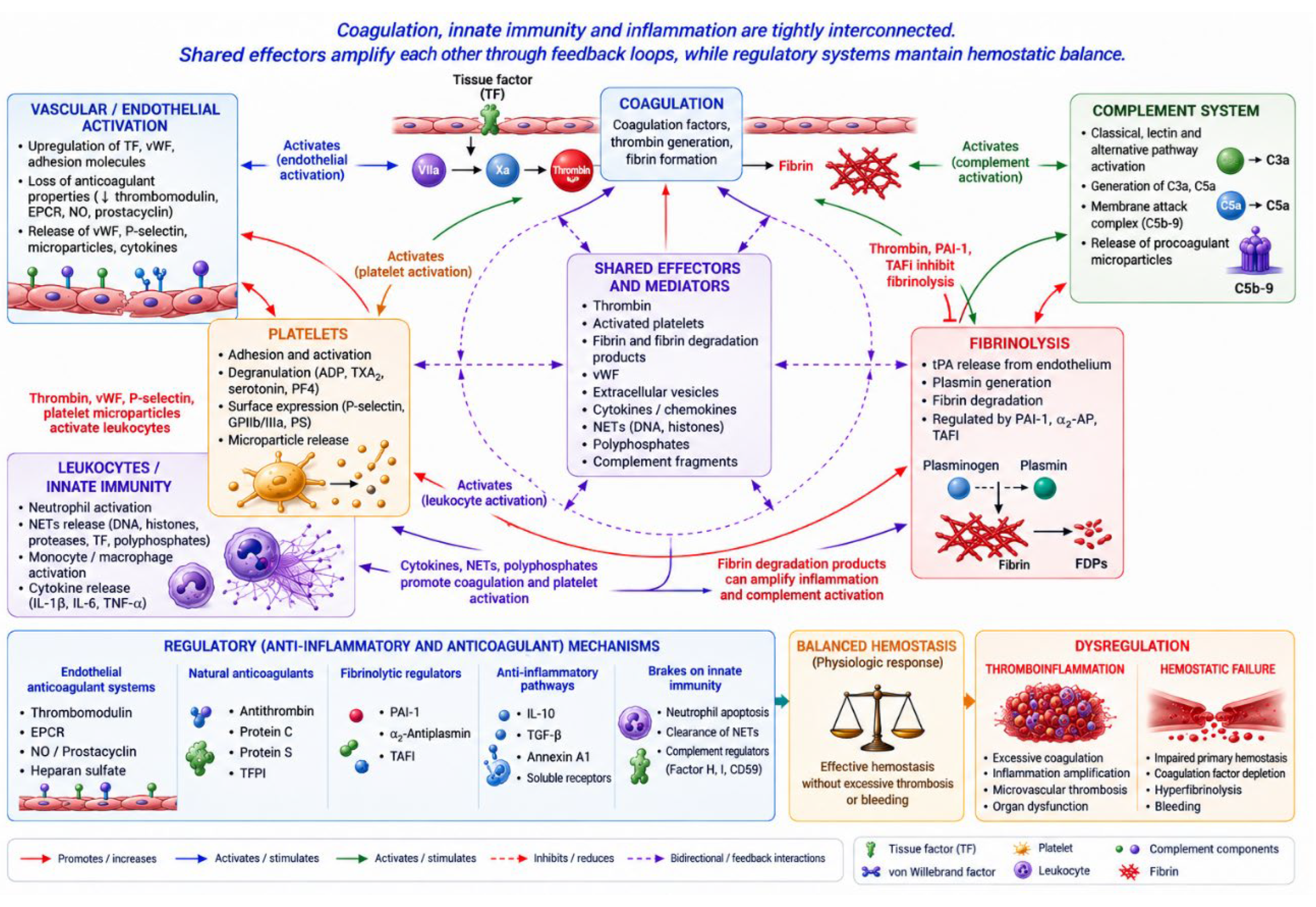
The convergent model therefore places coagulation within a broader network linking vascular injury, platelets, endothelium, TF-dependent and contact-dependent coagulation, complement, innate immunity, inflammation, fibrin formation, and fibrinolysis (Figure 2). Shared effectors—including platelets, thrombin, fibrin, endothelial cells, leukocytes, complement, and NETs—allow activation of one component to modify the others.
The integrated hemostatic and thromboinflammatory network undergoes substantial developmental changes from fetal life through childhood and adolescence. Developmental hemostasis reflects age-dependent differences in the concentrations, activities, and interactions of procoagulant, anticoagulant, platelet, endothelial, and fibrinolytic components [30].
The developing system should not be considered simply an immature or globally hypocoagulable version of adult hemostasis. Several coagulation factors, particularly vitamin K-dependent and contact pathway factors, are relatively low during fetal and neonatal life, whereas factor VIII and vWF follow different developmental trajectories [31,32]. Natural anticoagulants such as antithrombin and protein C are also relatively low early in life, while fibrinolytic mechanisms undergo parallel developmental changes [33,34]. These differences are coordinated and produce a functional, developmentally regulated hemostatic balance.
Ontogeny also affects platelet biology, platelet–endothelial interactions, innate immunity, complement, inflammatory signaling, and fibrinolysis. Consequently, functional hemostatic capacity cannot be inferred from individual coagulation-factor concentrations. In critically ill children, infection, inflammation, endothelial injury, surgery, trauma, transfusion, and organ dysfunction further perturb this developmentally regulated system.

Integrated Perspective

Hemostasis is therefore a dynamic, spatially organized, and self-regulating network. The cell-based model explains how platelets and coagulation factors generate and stabilize a clot; the convergent model places this process within a broader network linking coagulation to endothelium, innate immunity, complement, inflammation, and fibrinolysis; and ontogeny modifies the behavior of this network throughout childhood. These principles provide the physiological basis for understanding ECMO-associated dyshemostasis, in which the integrated network is exposed to additional mechanical, biochemical, inflammatory, and therapeutic perturbations.

Pathobiology of ECMO-Associated Dyshemostasis

ECMO-associated dyshemostasis is a complex and dynamic disturbance of the IHN and should not be considered synonymous with ECMO-associated coagulopathy. Its pathobiology reflects the interaction of blood with artificial surfaces, nonphysiological mechanical forces, endothelial injury, platelet and coagulation activation, contact-system and complement activation, inflammation, hemolysis, altered fibrinolysis, depletion of hemostatic components, and systemic anticoagulation. These mechanisms may operate simultaneously and in opposing directions, allowing strong prothrombotic stimuli to coexist with impaired capacity for effective hemostasis [3,35].
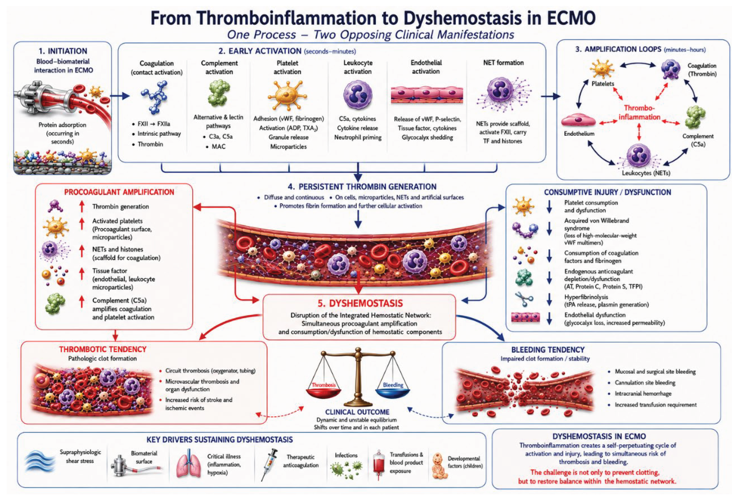
This distinction is particularly important during pediatric ECMO because the circuit is superimposed on a hemostatic system already influenced by developmental stage, underlying disease, endothelial injury, inflammation, surgery or trauma, organ dysfunction, and other acquired abnormalities. ECMO therefore does not produce a uniform “ECMO coagulopathy”; rather, it creates a continuously changing biological environment in which multiple hemostatic and thromboinflammatory pathways are activated, amplified, inhibited, or accompanied by consumption or depletion of their components. The resulting phenotype may range from predominantly bleeding to predominantly thrombotic manifestations, with both occurring simultaneously or changing over time (Figure 3).

Blood–Biomaterial Interaction: The Initiating Perturbation

The extracorporeal circuit repeatedly exposes blood to non-endothelialized artificial materials. Plasma proteins rapidly adsorb to these surfaces, creating a biologically active interface capable of promoting platelet adhesion, coagulation, complement, and inflammatory signaling [36,37].
The contact system is particularly relevant. Artificial surfaces can promote factor XII activation and subsequent factor XI activation, providing a pathway for sustained thrombin generation. Clinical observations during ECMO support contributions from contact-dependent coagulation, including increased factor XIa activity and procoagulant extracellular vesicles associated with thrombin-generation potential [38,39]. Contact activation also engages the kallikrein–kinin system, while blood–biomaterial interaction can activate complement and generate mediators that influence leukocytes, platelets, and endothelial cells [40]. The circuit therefore represents an active biological interface linking coagulation, innate immunity, and inflammation.

Mechanical Forces and Shear-Induced Hemostatic Dysfunction

Blood flowing through an ECMO circuit is exposed to mechanical forces that differ substantially from those in the native circulation. Their magnitude and distribution depend on circuit configuration, pump characteristics, cannulae, oxygenator design, flow conditions, and local disturbances [41,42].
The vWF system is particularly sensitive to shear. Excessive or sustained shear can promote proteolytic loss of high-molecular-weight vWF multimers, resulting in acquired von Willebrand syndrome and impaired primary hemostasis. This provides an important mechanism of bleeding that cannot be attributed solely to systemic anticoagulation [43]. Mechanical forces also promote platelet activation, adhesion, degranulation, receptor alteration, and aggregation, followed by consumption and functional exhaustion. ECMO can therefore produce the apparently paradoxical combination of platelet activation and platelet dysfunction [44,45].

Platelet Activation, Consumption, and Loss of Hemostatic Reserve

Platelets contribute to both thrombosis and bleeding during ECMO. Contact with artificial surfaces and abnormal shear promotes platelet activation and adhesion to circuit components and developing thrombi. Activated platelets expose phosphatidylserine and provide catalytic surfaces for tenase and prothrombinase assembly, supporting thrombin generation and fibrin formation.
At the same time, platelet activation promotes consumption within the circuit and incorporation into thrombi. Platelet-derived extracellular vesicles and other procoagulant membrane fragments may provide additional phospholipid surfaces for coagulation. With continuing extracorporeal exposure, platelet number and function may progressively decrease through receptor shedding, altered signaling, degranulation, mechanical injury, and exhaustion. Platelet count alone therefore does not fully characterize platelet contribution to hemostasis during ECMO [46,47].

Coagulation Activation and Thrombin Generation

The extracorporeal circuit provides persistent stimuli for coagulation activation. Blood contact with artificial surfaces can engage the contact system, while tissue factor (TF)-dependent mechanisms may contribute according to the patient’s inflammatory and endothelial state. Activated platelets provide the phospholipid surface for tenase and prothrombinase assembly, promoting factor Xa and thrombin generation.
Unlike physiological coagulation, which is spatially restricted to sites of vascular injury, the circuit provides a persistent surface capable of sustaining coagulation activation. Thrombin generation is therefore continuously opposed by endogenous anticoagulant mechanisms and systemic anticoagulation, creating a dynamic balance that varies with biological state and circuit conditions [35,48]. Activated coagulation factors, platelets, and extracellular vesicles may also enter the systemic circulation and interact with endothelial and inflammatory pathways, so coagulation activation during ECMO is not confined to circuit thrombosis [3,6].

Endothelial Dysfunction, Inflammation, and Thromboinflammation

ECMO-associated dyshemostasis develops in the context of endothelial activation and injury. Critical illness may already disrupt endothelial homeostasis through inflammation, hypoxemia, ischemia–reperfusion injury, infection, and tissue injury, while extracorporeal circulation can further perturb endothelial function [6,12].
Loss of endothelial homeostatic properties favors platelet adhesion, coagulation activation, leukocyte–endothelial interactions, and altered fibrinolysis. Inflammatory signaling can promote TF expression and impair endogenous anticoagulant pathways, while complement activation may amplify endothelial, platelet, and leukocyte activation [49]. These processes are reciprocally coupled: thrombin and activated platelets promote inflammatory and endothelial signaling, while inflammatory and endothelial activation further enhance coagulation and platelet activation. Thus, during ECMO, coagulation and inflammation form a self-reinforcing thromboinflammatory network [3,50].

Hemolysis as a Component of ECMO-Associated Dyshemostasis

Mechanical trauma to erythrocytes releases cell-free hemoglobin, heme, and other products that alter vascular and inflammatory biology. Free hemoglobin reduces nitric oxide bioavailability and may affect vascular tone, platelet behavior, and endothelial function, while oxidative and inflammatory effects may further promote thromboinflammatory activation [51,52,53,54]. Red-cell-derived extracellular vesicles can expose phosphatidylserine-rich procoagulant surfaces that support tenase and prothrombinase assembly and promote thrombin generation [55,56]. Hemolysis should therefore be regarded not only as a marker of mechanical injury but also as a potential participant in dyshemostasis.

Fibrinolytic Dysregulation

Fibrinolysis during ECMO is influenced by interactions among coagulation activation, endothelial and inflammatory signaling, fibrinolytic proteins, and their regulators. Sepsis and systemic inflammation may substantially modify this response, while blood–surface interactions, mechanical blood trauma, and hemolysis may further alter fibrinolytic regulation [57,58,59]. The resulting phenotype may include excessive fibrinolysis and impaired clot stability, contributing to bleeding, or impaired fibrin clearance favoring fibrin persistence. These patterns may evolve over time and differ according to disease state, inflammatory burden, anticoagulation, and circuit conditions.
Fibrinolytic dysfunction should therefore be understood as altered regulation of fibrin formation, persistence, and removal rather than as a single stereotyped ECMO phenotype.

Consumption, Depletion, Dilution, and Loss of Hemostatic Reserve

Continuous blood–circuit interaction can progressively reduce hemostatic reserve. Platelets may be activated, consumed, and retained on artificial surfaces, while sustained coagulation activation and fibrin formation contribute to consumption of coagulation factors and fibrinogen [60,61]. Blood loss from surgery, cannulation, invasive procedures, circuit sampling, and other interventions further depletes circulating hemostatic resources, while circuit priming and fluid administration can produce hemodilution [62].
The cumulative effect is not simply a reduction in component concentration. The functional adequacy of the remaining hemostatic capacity depends on the broader biological and mechanical environment. Hemostatic reserve therefore encompasses both the quantity and function of circulating components in an environment characterized by artificial surfaces, mechanical stress, inflammation, anticoagulation, and ongoing turnover. Progressive depletion may increase vulnerability to bleeding or thrombosis according to the mechanisms that predominate at a given time.

Anticoagulation as One Component of the Dyshemostatic Environment

Systemic anticoagulation is required because the extracorporeal circuit provides a persistent nonphysiological surface capable of promoting coagulation activation and thrombosis [63]. However, anticoagulation primarily modulates coagulation activation and cannot by itself normalize the broader hemostatic disturbances associated with ECMO.
Inadequate anticoagulation may permit persistent thrombin generation, fibrin deposition, and patient or circuit thrombosis, whereas excessive anticoagulant effect may compromise hemostasis when vascular integrity or other hemostatic mechanisms are impaired [64,65]. Thrombosis may therefore occur despite anticoagulation when strong procoagulant stimuli persist, while major bleeding may occur without excessive anticoagulant effect when other components of the hemostatic system are dysfunctional or depleted.
ECMO-associated dyshemostasis should consequently not be reduced to an anticoagulation problem. Anticoagulation operates within a dynamic system in which cellular, coagulation, endothelial, inflammatory, fibrinolytic, and circuit-related processes continuously interact.

ECMO-Associated Dyshemostasis as a Dynamic Network Disorder

ECMO-associated dyshemostasis emerges from the evolving interaction among circulating blood, the extracorporeal circuit, the patient’s biological state, and therapeutic interventions. Developmental stage, underlying disease, inflammatory and endothelial states, circuit characteristics, duration of support, mechanical forces, blood-component availability, anticoagulant exposure, and evolving bleeding or thrombotic processes all contribute.
Their relative contribution may change over time, so the mechanisms dominant during one phase of ECMO may differ from those operating later. Bleeding and thrombosis may therefore occur separately, sequentially, or simultaneously, and thrombotic manifestations may involve either the patient or the circuit.
Because ECMO perturbs multiple components of the hemostatic network simultaneously, no single laboratory measurement or clinical feature can fully characterize the hemostatic state. Assessment must integrate developmental and clinical context, patient- and circuit-related risk factors, laboratory and viscoelastic findings, clinical manifestations, and trajectory. The relative weight of these findings determines the prevailing hemostatic phenotype and the appropriate intensity of monitoring and general direction of management.

Clinical Phenotypes of ECMO-Associated Dyshemostasis

ECMO-associated dyshemostasis can be expressed clinically as three principal phenotypes: bleeding-predominant, thrombosis-predominant, and mixed bleeding–thrombotic. These are pragmatic, management-oriented constructs rather than discrete biological entities or validated prediction models.
Phenotype assignment integrates the patient’s developmental and clinical condition, patient- and circuit-related risk factors, laboratory and viscoelastic findings, clinical and circuit manifestations, and their trajectory. Bleeding-predominant and thrombosis-predominant phenotypes identify the principal current management concern, whereas the mixed phenotype applies when bleeding vulnerability or bleeding and thrombogenicity or thrombosis are simultaneously sufficiently consequential to constrain the overall therapeutic strategy. The coexistence of risk factors in both directions does not itself define a mixed phenotype. Patients may transition between phenotypes as their determinants evolve.

Thrombosis-Predominant Phenotype

The thrombosis-predominant phenotype represents a state in which thrombogenicity or established thrombosis is the principal current management concern. It may be supported by patient- or circuit-related prothrombotic factors, coagulation activation or inadequate anticoagulant effect, low-flow or stasis states, progressive circuit or oxygenator thrombus formation, patient thrombosis, or an evolving thrombotic trajectory.
Its biological substrate may include persistent thrombin generation, platelet activation, endothelial dysfunction, impaired endogenous anticoagulant mechanisms, altered fibrinolysis, complement activation, extracellular-vesicle-mediated procoagulant activity, inflammation, hemolysis, and circuit-related blood trauma.
Thrombosis should not automatically be interpreted as inadequate anticoagulation. Patient and circuit contributors should be assessed before escalation of anticoagulation, because correcting a reversible source of thrombogenicity may be safer and more appropriate than increasing systemic anticoagulant intensity.

Bleeding-Predominant Phenotype

The bleeding-predominant phenotype represents a state in which bleeding susceptibility or established bleeding is the principal current management concern. It may be recognized before overt bleeding when baseline characteristics, acquired risk factors, objective hemostatic abnormalities, and trajectory indicate substantial bleeding vulnerability.
Relevant contributors include neonatal developmental hemostasis, particularly prematurity; recent cardiopulmonary bypass or major surgery; thrombocytopenia or platelet dysfunction; acquired von Willebrand abnormalities; hypofibrinogenemia; coagulation-factor depletion; dysregulated fibrinolysis; endothelial injury; hemodilution; and anticoagulant effect.
When bleeding occurs, its severity, trajectory, source, and relationship to procedures or ECMO should be considered together with anticoagulant effect, platelet and fibrinogen status, coagulation-factor availability, fibrinolysis, and systemic condition. Bleeding should therefore not be attributed automatically to excessive anticoagulation.

Mixed Bleeding–Thrombotic Phenotype

The mixed bleeding–thrombotic phenotype represents a state in which bleeding vulnerability or bleeding and thrombogenicity or thrombosis are simultaneously sufficiently consequential that both constrain management. It may be evident through concurrent manifestations, such as active bleeding with progressive circuit thrombosis, or through strong, objectively supported drivers of both processes before overt complications develop.
Its biological substrate reflects the interaction of mechanisms that impair effective hemostasis and those that promote thrombosis, including platelet dysfunction or activation, consumption, endothelial injury, coagulation activation, depletion or dysfunction of coagulation factors and fibrinogen, altered fibrinolysis, inflammation, hemolysis, and circuit-related blood trauma.
The mixed phenotype should not be managed by simply selecting an intermediate anticoagulation target. The mechanisms and trajectories of both processes should be assessed in parallel, with interventions directed toward reversible contributors while minimizing additional bleeding, thrombotic, or circuit-related harm.

From Phenotype Recognition to Integrated Assessment

Risk state, dyshemostatic process, and clinical phenotype are related but distinct. A patient may have substantial bleeding or thrombotic vulnerability before an overt complication develops, and these states may change rapidly with alterations in circuit condition, inflammation, hemolysis, surgery, transfusion, organ function, anticoagulation, and recovery from the underlying disease.
Phenotype recognition provides a framework for determining monitoring intensity and the general direction of management. Laboratory, viscoelastic, clinical, and circuit findings should therefore be interpreted together to identify the prevailing phenotype, characterize its underlying mechanisms, and determine whether surveillance, preventive intervention, or targeted therapy is warranted. Because the phenotype is dynamic, reassessment is required whenever clinically important changes occur.

Precision Monitoring of Dyshemostasis During Pediatric ECMO

Phenotype-oriented monitoring integrates complementary laboratory, functional, clinical, and circuit information to characterize the prevailing hemostatic phenotype and identify mechanisms contributing to it. It differs from assay-oriented monitoring, in which an individual laboratory value becomes the principal determinant of anticoagulation or hemostatic therapy.
Anticoagulation monitoring during pediatric ECMO is challenging because the hemostatic environment evolves continuously. The circuit promotes platelet activation and dysfunction, coagulation-factor consumption, altered fibrinolysis, and thrombotic activation, while developmental hemostasis and clinical factors such as inflammation, infection, hemolysis, organ dysfunction, hemodilution, and blood-product exposure modify clotting capacity [66,67]. No single conventional assay can therefore adequately characterize the real-time balance between bleeding and thrombosis. Standard assays interrogate selected components of hemostasis, and their characteristics and reference ranges may vary substantially in neonatal and pediatric populations.
A multimodal approach integrating anticoagulant effect, clot formation, platelet and fibrinogen status, biological markers, and patient and circuit findings is therefore generally favored, although the optimal combination and monitoring frequency remain uncertain [68].

Laboratory and Functional Assessment

Activated clotting time (ACT) remains widely used because it is rapidly available and provides bedside whole-blood assessment of clot initiation. However, it is not specific for UFH and is influenced by thrombocytopenia, platelet dysfunction, hypofibrinogenemia, factor deficiencies, hypothermia, hemodilution, anemia, inflammation, and assay characteristics. Although ACT targets of approximately 180–220 seconds have traditionally been used, targets vary and have not been validated against pediatric outcomes [69,70]. ACT should therefore be interpreted with complementary laboratory and clinical information rather than used in isolation.
Activated partial thromboplastin time (aPTT) is also widely used for UFH monitoring. Targets of approximately 60–80 seconds are used in some centers, but their applicability to pediatric ECMO remains uncertain [71,72]. The relationship between aPTT and UFH depends on reagent, platform, and calibration, while developmental factor concentrations, factor consumption, inflammation, and other critical-illness abnormalities can substantially influence results. aPTT should therefore be interpreted according to locally established, assay-specific ranges and in conjunction with other measures of anticoagulant effect and hemostatic competence [73,74].
Anti-factor Xa (anti-Xa) provides a relatively direct estimate of UFH anticoagulant activity by measuring inhibition of factor Xa by the UFH–antithrombin complex [75]. It is increasingly used during ECMO and commonly employs a reference range of approximately 0.30–0.70 IU/mL [74]. However, anti-Xa is influenced by antithrombin availability, assay methodology, hemolysis/free hemoglobin, hyperbilirubinemia, hypertriglyceridemia, and other analytical or preanalytical factors [75,76]. It does not assess endogenous coagulation capacity, platelet function, fibrinogen contribution, clot strength, fibrinolysis, or overall hemostatic competence.
Heparin concentration provides an alternative estimate of circulating heparin exposure but does not necessarily reflect its biological anticoagulant effect. The concentration–effect relationship depends on antithrombin availability, patient biology, and assay characteristics. Heparin concentration may therefore provide complementary information when anticoagulation assays are discordant, but should not be interpreted as a surrogate for the overall biological anticoagulant phenotype [77,78].
Viscoelastic testing (VET) with thromboelastography or rotational thromboelastometry provides dynamic whole-blood assessment of clot initiation, kinetics, strength, stability, and lysis. It can provide information about the relative contributions of fibrin and platelets and identify functional abnormalities not apparent on conventional plasma-based assays [79,80]. VET therefore complements rather than replaces anticoagulation assays. Its interpretation remains dependent on platform, reagent, age, sampling conditions, and limited pediatric ECMO-specific validation [81,82,83,84,85].
VET-derived clot initiation reflects the combined effects of coagulation-factor activity and heparin, whereas clot propagation and strength additionally reflect the contributions of fibrinogen and platelets. VET may therefore provide complementary information when anti-Xa and aPTT are discordant by showing how coagulation-factor activity, heparin effect, fibrinogen availability, and platelet contribution translate into whole-blood clot initiation, propagation, and strength. Heparinase-modified assays may further help determine whether heparin contributes to the observed clotting abnormality, but VET is not validated as a stand-alone method for UFH dose adjustment [86].
Clot strength reflects the combined contribution of fibrin and platelets. Fibrin-specific assays, such as FIBTEM or functional fibrinogen, selectively assess the fibrin contribution to clot firmness and, when interpreted alongside platelet-related parameters, may help distinguish whether reduced clot strength is predominantly attributable to impaired fibrin formation or platelet dysfunction. These assays do not assess platelet contribution or directly measure heparin effect, and their results should not be considered interchangeable with plasma fibrinogen concentration. In clinically discordant situations, they may provide complementary functional information that helps clarify the underlying hemostatic deficit [82,86,87].
Platelet count quantifies circulating platelets but does not characterize platelet function. Quantitative thrombocytopenia may coexist with qualitative dysfunction caused by circuit-related shear, blood–surface interactions, receptor alteration, degranulation, and exhaustion [47,88]. Functional testing by aggregometry, flow cytometry, or platelet-mapping techniques may provide complementary information when available [89,90,91], although these approaches are not sufficiently standardized for routine pediatric ECMO monitoring. Acquired von Willebrand syndrome may further impair platelet adhesion through loss of high-molecular-weight vWF multimers under high shear [43,92,93,94,95].
Plasma fibrinogen concentration provides a quantitative measure of circulating fibrinogen, whereas fibrin-based VET assays, such as FIBTEM or TEG Functional Fibrinogen, assess the contribution of fibrin to whole-blood clot firmness. The two approaches therefore provide complementary information and should not be considered interchangeable [82,86,87].
PT/INR primarily reflects extrinsic and common-pathway clotting activity and has limited utility for UFH monitoring. It does not directly measure UFH effect, platelet function, or clot strength and may be affected by factor deficiency, liver dysfunction, vitamin K status, hemodilution, and assay characteristics. Abnormal results should prompt investigation of relevant factor abnormalities when clinically indicated rather than reflexive modification of UFH [96].
Because each method interrogates only selected components of hemostasis and has specific biological and analytical limitations, results should be interpreted according to what they measure, what they cannot measure, and factors that may influence reliability. The principal laboratory and functional methods are summarized in Table 1.

Adjunctive Biological Markers

Markers of coagulation activation, including thrombin–antithrombin (TAT) complexes, prothrombin fragment 1+2, soluble fibrin, and D-dimer, may provide additional information about ongoing thrombin generation, fibrin formation, and fibrinolysis. These biomarkers reflect biological processes occurring in vivo and may help identify a procoagulant phenotype or evolving thrombotic process that is not captured by routine anticoagulation assays. However, they are generally not suitable for real-time UFH dose adjustment and should be interpreted as complementary markers within the broader clinical and hemostatic context. D-dimer, in particular, reflects fibrin formation and subsequent fibrinolysis and is therefore not specific for coagulation activation [97,98].
Endothelial and immune-thrombotic biomarkers may provide additional insight into the vascular and inflammatory components of ECMO-associated dyshemostasis. von Willebrand factor, ADAMTS13, and soluble thrombomodulin may reflect endothelial activation, injury, and altered platelet–endothelial interactions, whereas cell-free DNA, histones, and neutrophil extracellular trap (NET)-related markers may provide information about immunothrombotic activation. These biomarkers remain primarily investigational in pediatric ECMO and are not validated for routine anticoagulation monitoring or UFH dose adjustment, but may help characterize evolving patterns of endothelial and immune-thrombotic activation when interpreted alongside clinical and hemostatic findings [4,12,92].

Integrated Hemostatic Patterns

No individual assay provides a comprehensive representation of the hemostatic state during ECMO. The value of multimodal monitoring lies not only in complementary measurements but also in the patterns that emerge when they are interpreted together. These patterns are not equivalent to the clinical phenotypes described previously; rather, they represent laboratory-functional or mechanistic patterns that may contribute to bleeding-predominant, thrombosis-predominant, or mixed manifestations.

Discordance Among Anticoagulation Assays

Discordance among ACT, aPTT, and anti-Xa may itself provide clinically useful information because these assays are influenced by different biological and analytical factors. aPTT provides an independent time-to-clot assessment but may be prolonged by factor deficiencies, consumption, hemodilution, liver dysfunction, developmental hemostasis, lupus anticoagulant, and other inhibitors, or shortened by elevated factor VIII, increased fibrinogen, and acute-phase changes. Its relationship with UFH is also reagent- and platform-dependent. Anti-Xa is less affected by coagulation-factor and platelet abnormalities but depends on antithrombin availability and assay methodology; hemolysis, hyperbilirubinemia, hypertriglyceridemia, sample handling, and heparin contamination may also affect results. Assay-specific calibration and the use of exogenous antithrombin should therefore be considered when interpreting anti-Xa. The principal biological, preanalytical, and analytical factors that may influence aPTT and anti-Xa results, including the expected direction of their effect, are summarized in Table 2.
Discordance between anticoagulation assays should not be interpreted automatically as inadequate or excessive UFH effect. Instead, discordant results should prompt investigation of their underlying biological and analytical determinants rather than reflexive adjustment of UFH. Anticoagulation assays are in-vitro surrogates and do not reproduce the full in-vivo interaction among blood, endothelium, inflammation, and the extracorporeal circuit; their interpretation should therefore remain embedded within the broader hemostatic context [66,99,100].

Dynamic, Phenotype-Oriented Monitoring

Phenotype-oriented monitoring integrates complementary laboratory, functional, clinical, and circuit information to characterize the prevailing hemostatic phenotype and identify the mechanisms contributing to it. Unlike assay-oriented monitoring, in which an individual laboratory value becomes the principal determinant of anticoagulation or hemostatic therapy, this approach emphasizes the concordance, trajectory, and clinical relevance of multiple measures.
The purpose of multimodal monitoring is therefore not simply to generate multiple laboratory targets, but to determine whether the prevailing phenotype is changing, which mechanisms are contributing, and whether an intervention has produced the intended biological and clinical effect.
Monitoring intensity should be adapted to the clinical trajectory. New or worsening bleeding, progressive thrombosis, circuit dysfunction, major procedures, substantial changes in anticoagulant requirements, evolving organ dysfunction, hemolysis, or other clinically relevant changes should prompt reassessment using complementary measures.
Laboratory and VET findings should be integrated with platelet and fibrinogen status, patient and circuit findings, and the trajectory of bleeding or thrombosis. The appropriate response to an abnormal or discordant result may include adjustment of anticoagulation, targeted replacement of a deficient hemostatic component, correction of a systemic or circuit-related contributor, or continued observation when the finding does not correspond to clinically meaningful dyshemostasis.
Thus, phenotype-oriented monitoring does not replace conventional anticoagulation monitoring; it places anticoagulant-effect assays within a broader assessment of hemostatic competence. Apparently contradictory findings should therefore be investigated in their biological and clinical context rather than resolved by automatically adjusting a single laboratory target.

Precision Hemostatic Management During Pediatric ECMO

Precision hemostatic management translates the prevailing hemostatic phenotype into mechanism-directed intervention. Rather than correcting laboratory abnormalities in isolation, therapeutic decisions should address the dominant disturbance while considering its expected effects on bleeding, thrombosis, and circuit integrity.
Clinical phenotype should guide therapeutic reasoning but should not be regarded as a direct surrogate for a single mechanism. Bleeding may occur despite ongoing coagulation activation, while thrombosis may develop despite an apparently adequate anticoagulant effect, and consumptive processes may accompany either or both. Management should therefore address the mechanisms contributing to the phenotype rather than treat the phenotype itself as a mechanistic diagnosis.
  • The principal therapeutic domains are:
  • Anticoagulant selection and intensity;
  • Targeted blood-component and hemostatic therapy;
  • Transfusion stewardship;
  • Circuit-directed optimization and management of clinically consequential circuit pathology;
  • Correction of systemic physiological and inflammatory drivers;
  • Bleeding source control;
  • Minimizing iatrogenic hemostatic injury;
  • Serial reassessment and therapeutic adaptation.
Their application should remain individualized and iterative as the phenotype, biological state, and circuit condition evolve.

Anticoagulant Selection and Intensity

Unfractionated Heparin Anticoagulation

Unfractionated heparin (UFH) remains the conventional first-line anticoagulant during pediatric ECMO. Its use should be guided by integrated interpretation of anti-Xa, aPTT, ACT, VET, antithrombin activity when clinically indicated, platelet and fibrinogen status, hemolysis, and patient- and circuit-level findings rather than by a single laboratory value.
The objective of anticoagulation is to limit clinically consequential patient or circuit thrombosis while minimizing bleeding. Anticoagulant intensity should therefore be positioned according to the prevailing phenotype, its trajectory, and the integrated hemostatic assessment, while considering patient- and circuit-related factors [63,101].
In patients without a predominant bleeding or thrombotic phenotype, anticoagulation can generally remain within the institutional range. When a clinically consequential phenotype emerges, intensity may be positioned toward a lower or higher portion of the institutional range according to the prevailing state. Lower-intensity positioning may be appropriate in bleeding-predominant states when anticoagulation remains necessary; higher-intensity positioning may be considered in thrombosis-predominant states when inadequate anticoagulant effect is identified after reversible patient- and circuit-related contributors have been assessed. The mixed phenotype does not correspond to a predefined intermediate target.
The commonly used ELSO reference framework includes an anti-Xa range of 0.30–0.70 IU/mL and an aPTT range of approximately 60–90 seconds [63]. Pediatric centers have reported context-dependent variation, including lower-intensity strategies in patients with increased bleeding risk [102,103]. These approaches remain pragmatic and institution-specific rather than universally validated pediatric therapeutic targets.
For bedside interpretation, phenotype-informed anticoagulation positioning can be summarized as shown in Table 3. The ranges represent potential positioning zones rather than rigid phenotype-specific prescriptions; the selected position should remain individualized according to the integrated hemostatic assessment.
For bedside management, UFH adjustment may be operationalized according to the selected institutional anti-Xa target range, as illustrated in Table 4. A measured anti-Xa value should be interpreted relative to the selected target range rather than as an isolated indication for dose adjustment. The table provides suggested dose-adjustment logic rather than a universally validated pediatric ECMO titration algorithm.
UFH titration should not be driven by a single anti-Xa, aPTT, or ACT value. A low anti-Xa accompanied by a prolonged aPTT and impaired clot strength on VET should not automatically trigger UFH escalation; this discordant pattern may instead reflect factor deficiency, hypofibrinogenemia, platelet-related impairment, broader hypocoagulability, or analytical interference. Conversely, a low anti-Xa in the presence of preserved clot strength and progressive circuit thrombogenicity should prompt reassessment of anticoagulant effect, assay validity, antithrombin activity, circuit mechanics, and other potential contributors before increasing heparin exposure [63,101,103].
The presence of thrombosis should not by itself mandate escalation of anticoagulation. Low flow or stasis, cannula or drainage problems, excessive shear, recurrent suction events, hemolysis, progressive circuit pathology, assay discordance, and antithrombin availability should be considered before increasing UFH intensity. Correction of a reversible contributor may be more appropriate than increasing systemic anticoagulation alone.
Similarly, apparent excessive anticoagulant effect should not be inferred from a single prolonged coagulation assay. Prolonged aPTT or ACT may reflect factor deficiency, hypofibrinogenemia, thrombocytopenia, consumption, or other biological or analytical factors. Anti-Xa results may also be affected by antithrombin activity and assay characteristics. Anticoagulation intensity should therefore be regarded as a modifiable component of the IHN and reassessed whenever the bleeding or thrombotic state, circuit condition, or other major determinants change.

Direct Thrombin Inhibitors

UFH remains first-line, but intravenous direct thrombin inhibitors (DTIs), particularly bivalirudin, provide an alternative when UFH is unsuitable or its effect cannot be reliably achieved or interpreted. UFH acts indirectly through antithrombin, whereas bivalirudin directly inhibits circulating and clot-bound thrombin independently of antithrombin [104]. This distinction is relevant in selected patients with suspected or confirmed heparin-induced thrombocytopenia, clinically important heparin resistance, or persistent difficulty achieving an appropriate UFH effect despite investigation of reversible causes [105].
Observational pediatric data suggest potentially favorable bleeding, transfusion, and thrombotic outcomes with bivalirudin, but findings are heterogeneous and do not establish superiority over UFH [106]. A pediatric randomized pilot trial found similar time at anticoagulation goal and bleeding, fewer RBC transfusions, but more circuit changes for thrombosis with bivalirudin [107]. Bivalirudin should therefore currently be regarded as an alternative rather than a preferred anticoagulant.
Dosing and monitoring remain incompletely standardized. Requirements vary with age, renal function, ECMO indication, and clinical course, and CRRT may further influence dosing [108,109,110,111]. No universally accepted pediatric ECMO dosing regimen or therapeutic target has been established. Switching to a DTI also does not correct broader IHN dysfunction; platelet dysfunction, acquired von Willebrand abnormalities, fibrinogen depletion, altered fibrinolysis, endothelial injury, hemolysis, inflammation, and circuit thrombogenicity may persist despite effective thrombin inhibition. DTI therapy should therefore remain within the same phenotype-oriented framework used for UFH.

Antithrombin

Antithrombin (AT) inhibits thrombin and factor Xa and substantially enhances its anticoagulant activity in the presence of heparin. UFH effect is therefore partly dependent on AT availability and function [112].
AT activity is physiologically lower in neonates and young infants and may be further reduced during ECMO through hemodilution, consumption, reduced synthesis, or redistribution [113,114]. Low AT should therefore be interpreted as a potential modifier of UFH effect rather than an independent indication for replacement.
The current evidence does not support routine AT replacement solely to normalize AT activity. Pediatric dosing studies demonstrate that conventional dosing may underestimate the amount required to achieve a desired plasma increase, while pharmacokinetic variability further complicates replacement [113,114]. Contemporary pediatric consensus supports considering AT in selected patients with impaired heparin responsiveness rather than treating a numerical AT threshold as an independent therapeutic target [115]. Recent systematic-review evidence also does not establish routine AT supplementation as improving clinically important outcomes [116].
Thus, AT replacement should be considered when low AT plausibly contributes to inadequate UFH effect after assay discordance, sampling issues, and other reversible causes have been evaluated. The decision should incorporate the clinical phenotype, thrombotic and bleeding risks, UFH requirements, and evidence of impaired heparin responsiveness.

Targeted Blood-Component and Hemostatic Therapy

Blood-component and factor therapy should correct clinically relevant functional deficits rather than normalize isolated laboratory values. The decision should integrate bleeding severity and trajectory, platelet and fibrinogen status, VET findings when available, anticoagulant effect, procedural context, and patient- and circuit-related thrombotic risk.
The principal therapeutic options and pragmatic bedside reference points are summarized in Table 5. These values are intended to support clinical reasoning rather than function as automatic transfusion or treatment triggers. A laboratory abnormality should therefore be interpreted in the context of the patient’s phenotype, trajectory, and the other interacting components of the IHN.

Platelet Replacement

Platelet transfusion should be considered when thrombocytopenia or clinically relevant platelet dysfunction contributes to impaired clot formation, particularly during clinically significant bleeding or before selected high-risk procedures. The decision should integrate platelet count and trajectory, functional information when available, fibrinogen status, hematocrit, bleeding trajectory, procedural context, age, anticoagulant effect, and thrombotic risk [117].
In nonbleeding patients, prophylactic thresholds vary substantially. Historical targets of 80–100 ×109/L are increasingly being replaced by lower, individualized strategies. A threshold around 50 ×109/L has been used in some centers and in the recent ECSTATIC feasibility trial, while lower thresholds may be selected in carefully characterized nonbleeding patients [118,119]. Neither 50 ×109/L nor lower thresholds has been established as universally appropriate; platelet therapy should therefore remain phenotype- and context-directed.

Plasma for Multiple-Factor Replacement

Plasma should be directed toward clinically relevant multiple-factor deficiency rather than isolated INR correction. In clinically significant bleeding, plasma may be considered when INR is >1.5 and relevant factor deficiency is suspected or demonstrated. Repeated administration solely to normalize INR should be avoided, and plasma should not be used as the primary strategy for fibrinogen replacement when targeted fibrinogen therapy is indicated [117].

Fibrinogen Replacement

Fibrinogen replacement addresses a distinct component of hemostatic competence. Fibrinogen concentrate or cryoprecipitate should be used when a clinically relevant disturbance is identified. Fibrinogen concentrate provides a more standardized dose, whereas cryoprecipitate provides variable fibrinogen together with additional hemostatic proteins, including factor VIII and vWF [120].
In nonbleeding patients, a fibrinogen concentration above approximately 100 mg/dL may serve as a pragmatic reference point; in clinically significant bleeding, at least approximately 150 mg/dL is reasonable [63,117]. Replacement should nevertheless incorporate functional fibrin contribution on VET when available, platelet status, bleeding severity, procedural context, and the overall phenotype rather than concentration alone.

Red Blood Cell Transfusion

RBC transfusion should be guided by the clinical scenario and adequacy of oxygen delivery rather than hemoglobin concentration alone, considering systemic oxygenation, hemodynamic status, ongoing blood loss, and the degree of cardiopulmonary support provided by the circuit. In selected hemodynamically stable pediatric ECMO patients without active bleeding or evidence of inadequate oxygen delivery, a restrictive transfusion strategy with hemoglobin thresholds around 7 g/dL may be reasonable. Higher thresholds may be appropriate when bleeding is ongoing, oxygen delivery is inadequate, or cardiopulmonary reserve is limited. No universal hemoglobin threshold applies across pediatric ECMO populations [63,117,121,122].

Recombinant Factor VIIa and PCC

rFVIIa should be reserved as an off-label rescue intervention for refractory, life-threatening bleeding after surgical or procedural causes have been addressed and conventional multimodal therapy optimized. Pediatric ECMO evidence is limited and uncontrolled [68,123].
PCC should likewise remain exceptional rescue therapy for severe bleeding refractory to multimodal management, particularly because of its potential thrombotic consequences and lack of pediatric ECMO-specific evidence [124].

Antifibrinolytic Therapy

Antifibrinolytic therapy should be phenotype- and context-directed. Tranexamic acid or ε-aminocaproic acid may be considered for clinically significant bleeding or anticipated high bleeding risk, particularly during major or high-risk procedures. VET evidence of hyperfibrinolysis may support treatment but is not mandatory when the clinical or procedural risk is substantial. Routine use for low-risk procedures or nonspecific ECMO bleeding should be avoided, particularly in the presence of clinically relevant or progressive thrombosis [125].
Overall, targeted hemostatic therapy should be understood as correction of clinically relevant disturbances within the IHN rather than normalization of laboratory variables. The same laboratory abnormality may have different therapeutic implications according to phenotype, mechanism, and competing bleeding and thrombotic risks.

Transfusion Stewardship and Transfusion as a Biological Perturbation

Transfusion may be lifesaving during active hemorrhage, clinically significant anemia, or clinically relevant hemostatic deficits, but transfusion requirements should not be interpreted independently of the underlying hemostatic phenotype. In pediatric ECMO, transfusion exposure is common and substantial, while optimal thresholds remain incompletely defined. Observational studies have associated greater RBC, plasma, and platelet transfusion exposure with adverse outcomes, including mortality, bleeding, and thrombosis, although these associations do not establish causality [126,127,128,129].
Transfused components may also modify the patient’s intravascular hemostatic and inflammatory milieu. Stored RBCs undergo biochemical and structural changes, while platelet and plasma products contain biologically active cellular and molecular constituents [130,131]. In pediatric ECMO, greater platelet transfusion exposure has been associated with subsequent bleeding and thrombosis, and platelet transfusion was associated with worsening oxygenator function among patients with the poorest pre-transfusion oxygenator function, although no overall effect on oxygenator function was observed [132]. The clinical relevance of these effects remains uncertain.
Transfusion exposure is therefore both a consequence and a potential modifier of the underlying hemostatic disturbance. The clinical processes that contribute to transfusion requirements—bleeding, anemia, thrombocytopenia, and coagulation abnormalities—are themselves associated with adverse outcomes, while transfusion may subsequently influence inflammatory, thrombotic, and circuit-related processes. This bidirectional relationship complicates the interpretation of observational associations between transfusion exposure and outcomes. Transfusion decisions should therefore be individualized to the underlying hemostatic phenotype, addressing the specific deficit while considering the potential consequences of additional transfusion exposure.

The ECMO Circuit as an Active Therapeutic Target

The ECMO circuit should be regarded as an active therapeutic target rather than merely a life-support device. It is a continuous interface between blood and artificial surfaces and contributes to platelet activation, contact and complement activation, inflammation, hemolysis, and shear-mediated injury. These effects may persist despite systemic anticoagulation.
Blood–surface interactions, abnormal flow, stasis, excessive shear, cannula-related disturbances, and mechanical blood trauma may promote thrombin generation, platelet activation, fibrin deposition, and hemolysis, while circuit thrombosis may further amplify consumption and blood trauma. The circuit can therefore function both as a source and an amplifier of dyshemostasis.
Circuit optimization should address biocompatibility, cannula configuration and position, blood flow, pump conditions, drainage, and mechanical integrity to minimize avoidable IHN activation while maintaining adequate support [133,134].
Potentially modifiable circuit and mechanical factors, their primary mechanisms, and their potential effects on the IHN are summarized in Table 6.
Circuit findings should be interpreted in the context of the patient’s hemostatic phenotype, anticoagulant effect, platelet and fibrinogen status, VET findings, hemolysis, and overall clinical condition. Progressive fibrin deposition, increasing oxygenator pressure gradients, flow instability, recurrent suction events, or increasing hemolysis may indicate evolving circuit pathology. Circuit or oxygenator exchange should therefore be considered when such abnormalities become clinically consequential, particularly when associated with worsening oxygenator function, significant hemolysis, inability to maintain adequate flow, or compromised patient support. Because exchange creates a new blood–biomaterial interface, its expected clinical benefit should outweigh the additional biological and procedural burden [133].
Circuit thrombosis should not automatically be interpreted as inadequate anticoagulation. Before increasing UFH intensity, clinicians should assess whether anticoagulant effect is adequate and whether mechanical factors are contributing, including low flow or stasis, cannula malposition or drainage obstruction, excessive shear, or recurrent suction.
Circuit optimization is therefore an upstream and potentially reversible component of precision hemostasis, complementary to rather than a substitute for anticoagulation and targeted hemostatic therapy.

Correction of Systemic Drivers of Dyshemostasis

Hemostatic management cannot be separated from the physiological conditions in which hemostasis operates. Hypothermia, acidosis, hypocalcemia, shock, severe inflammation, infection, hepatic or renal dysfunction, ongoing tissue injury, and impaired perfusion may alter hemostatic pathways and modify the effects of anticoagulant and hemostatic therapies [124,135].
These abnormalities should therefore be regarded as contributors to dyshemostasis rather than unrelated supportive-care issues. Reversible systemic drivers should be identified and corrected whenever clinically feasible.

Procedural and Surgical Source Control

Local and surgical control of bleeding is an integral component of hemostatic management during ECMO. Cannulation-site bleeding, postoperative bleeding, vascular-access bleeding, chest-tube losses, and procedure-related hemorrhage may persist despite correction of systemic abnormalities when a structural source remains uncontrolled [136].
Whenever feasible, definitive source control should be pursued concurrently with correction of clinically relevant systemic hemostatic deficits. Periprocedural decisions regarding anticoagulation interruption, transfusion, fibrinogen replacement, and antifibrinolytic therapy should be individualized according to procedural bleeding risk, the current phenotype, and the thrombotic condition of the patient and circuit.
In severe or life-threatening bleeding, temporary reduction or interruption of systemic anticoagulation may constitute a therapeutic intervention, with frequent reassessment of bleeding and thrombotic complications to guide resumption of full anticoagulation when clinically appropriate [124,137,138].

Minimizing Iatrogenic Hemostatic Injury

Precision hemostatic management should also minimize avoidable contributors to bleeding and transfusion exposure. Blood sampling should be limited to clinically necessary measurements, invasive procedures carefully planned, and medications that impair platelet or coagulation function reviewed whenever feasible. Repeated circuit manipulation and unnecessary transfusion should likewise be avoided.
These measures complement rather than replace treatment of established dyshemostasis and may reduce avoidable perturbation of the IHN during prolonged ECMO support [135].

Serial Reassessment and Therapeutic Adaptation

Precision hemostatic management is a dynamic, iterative process rather than a sequence of fixed interventions. The clinical phenotype, laboratory profile, circuit condition, and underlying drivers may change rapidly, and an intervention directed at one component of the IHN may modify others.
Therapeutic decisions should therefore be revisited after clinically meaningful changes in bleeding, thrombogenicity, anticoagulant effect, transfusion exposure, circuit performance, systemic physiology, or procedural status. Reassessment should establish whether the intended effect was achieved, whether the hemostatic phenotype has changed, and whether the mechanisms contributing to dyshemostasis remain active, have evolved, or have become reversible [66].
Persistent bleeding despite correction of an apparent coagulation abnormality should prompt reconsideration of the underlying mechanism, including surgical or procedural bleeding, platelet dysfunction, fibrinolytic abnormalities, circuit-related injury, and systemic drivers, rather than automatic escalation of hemostatic therapy. Conversely, new or progressive thrombosis should prompt reassessment of anticoagulant effect, circuit mechanics, flow and stasis, antithrombin availability when relevant, and other patient- or circuit-related contributors before intensifying anticoagulation [124].
Therapeutic adaptation should follow the evolving phenotype and observed response, with treatment escalated, de-escalated, redirected, or discontinued as the underlying mechanism and clinical context change.

Toward Precision Hemostasis

The objective is not to normalize every laboratory value or eliminate every biological response to extracorporeal support. Rather, it is to achieve sufficient coordinated hemostatic function to control clinically relevant bleeding, limit patient and circuit thrombosis, preserve circuit integrity, and avoid unnecessary treatment-related injury.
The IHN framework is therefore best regarded as a conceptual and testable platform for phenotype-adaptive management rather than a validated clinical algorithm. Its current value lies in providing a structured method for interpreting apparently contradictory findings and translating them into targeted, mechanism-directed interventions.

Current Evidence and Limitations

Despite substantial advances in understanding ECMO-associated hemostatic disturbances, evidence supporting many aspects of pediatric ECMO hemostatic management remains limited. Much of the literature consists of retrospective observational studies, single-center cohorts, case series, surveys of institutional practice, and extrapolation from adult ECMO, cardiopulmonary bypass, trauma, or other critically ill populations. Pediatric ECMO is also heterogeneous with respect to developmental stage, underlying disease, ECMO configuration, circuit characteristics, surgical context, and clinical trajectory, limiting generalizability and making universal therapeutic thresholds difficult to establish.
Important uncertainty persists regarding the optimal strategy for monitoring UFH anticoagulant effect. Anti-Xa, aPTT, ACT, and VET interrogate different dimensions of hemostasis and are subject to distinct biological and analytical limitations. Although contemporary pediatric consensus supports multimodal monitoring, evidence remains insufficient to define a single optimal assay combination or universally applicable therapeutic range. Assay discordance may itself contain clinically relevant information, but its biological and analytical basis and its implications for treatment have not been prospectively established.
Similar uncertainty applies to transfusion and factor-replacement thresholds. Current recommendations provide useful reference points, particularly for clinically significant bleeding, but specific thresholds are often supported by expert consensus, observational data, or feasibility studies rather than adequately powered comparative trials. This is particularly relevant to platelet and fibrinogen replacement, for which practice is moving toward individualized thresholds without establishing universally applicable lower limits. Uncertainty also remains regarding antithrombin replacement, antifibrinolytic therapy, PCC, rFVIIa, and other factor therapies, whose effects may depend on mechanism, phenotype, procedural context, and competing thrombotic risk.
The clinical phenotypes proposed in this review—bleeding-predominant, thrombosis-predominant, and mixed—have not been prospectively validated as reproducible biological classifications or prediction models. Similarly, phenotype-guided positioning within commonly used anticoagulation and hemostatic ranges represents a pragmatic framework integrating existing evidence and clinical reasoning rather than a validated treatment algorithm.
The IHN framework is therefore hypothesis-generating in several respects. Although thromboinflammation, endothelial activation, platelet dysfunction, complement activation, hemolysis, extracellular vesicles, fibrinolytic dysregulation, and coagulation activation are biologically interconnected, their temporal relationships and relative contributions to specific clinical phenotypes remain insufficiently characterized in pediatric ECMO. Current biomarkers and functional assays frequently identify individual components of the network without reliably establishing which mechanism is causally dominant or therapeutically actionable.
These limitations do not argue against individualized or multimodal management. Rather, they emphasize the need to distinguish established evidence from consensus-based practice and from clinically reasonable but unvalidated frameworks. At present, the principal value of the IHN approach is to provide a structured method for integrating heterogeneous information, identifying potentially correctable mechanisms, interpreting discordant findings, and avoiding treatment decisions based on isolated laboratory abnormalities.

Future Perspectives

The next generation of pediatric ECMO hemostasis research should move from static laboratory measurements toward dynamic, phenotype-oriented assessment and mechanism-directed intervention. The central question is whether integrating clinical, laboratory, functional, circuit, and biological information can identify evolving dyshemostasis early enough to change management and improve patient-centered outcomes.
Several priorities follow from this framework. First, longitudinal studies should determine whether trajectories of hemostatic and thromboinflammatory variables identify reproducible patterns preceding clinically apparent bleeding, thrombosis, or consumption. Platelet, coagulation, fibrinolytic, endothelial, inflammatory, hemolytic, and circuit-related variables should be evaluated together, with developmental hemostasis incorporated explicitly. Future studies should consistently use standardized definitions of clinically relevant bleeding and thrombosis to enable meaningful comparison across cohorts and facilitate validation of reproducible phenotypes.
Second, multimodal monitoring should be evaluated according to clinical utility rather than association with outcomes alone. Prospective studies should determine whether combinations of anti-Xa, aPTT and/or ACT, VET, platelet and fibrinogen measurements, circuit characteristics, and selected biological markers improve therapeutic decisions compared with conventional monitoring. Assay discordance should be investigated as potentially informative biological information rather than treated simply as analytical noise. Distinct combinations—for example, low anti-Xa with impaired clot strength versus low anti-Xa with preserved clot strength and progressive circuit thrombogenicity—may represent different mechanisms and require different interventions.
Third, phenotype-adaptive treatment strategies require prospective testing. The UFH positioning proposed in this review represents a testable hypothesis rather than a validated therapeutic strategy. Studies should compare conventional fixed-target approaches with adaptive strategies that modify anticoagulant intensity according to bleeding and thrombotic risk, circuit findings, and functional hemostatic assessment. Similar evaluation is needed for phenotype- and function-guided platelet and fibrinogen replacement and for plasma, antithrombin, specific factor concentrates, and antifibrinolytic therapy. Outcomes should prioritize major bleeding, patient and circuit thrombosis, transfusion exposure, circuit interventions, organ injury, and survival rather than laboratory targets alone.
Fourth, mechanistic integration and multicenter validation will be essential. Rather than studying complement, endothelial injury, platelet activation, coagulation, fibrinolysis, hemolysis, extracellular vesicles, inflammation, and circuit trauma independently, longitudinal studies should examine how these processes interact over time and whether specific combinations identify clinically meaningful trajectories or actionable mechanisms. Adequately powered pediatric studies will require multicenter collaboration, harmonized laboratory methodologies, standardized definitions, prospective data collection, and designs that account for developmental stage, ECMO configuration, underlying disease, and institutional practice. Pragmatic multicenter trials, adaptive designs, and comparative-effectiveness approaches may be particularly suitable.
Fifth, artificial intelligence and machine-learning approaches may provide new tools for integrating the high-dimensional and time-dependent data generated during pediatric ECMO. Rather than relying on isolated laboratory thresholds, predictive models could combine clinical trajectories, anticoagulant assays, VET parameters, platelet and fibrinogen measurements, circuit characteristics, hemolysis, and selected inflammatory or endothelial biomarkers to identify evolving hemostatic phenotypes and predict clinically relevant bleeding, thrombosis, or circuit dysfunction. Future studies should determine whether such models provide incremental clinical value beyond conventional monitoring and whether their predictions are sufficiently interpretable, calibrated, and robust across developmental stages, ECMO configurations, diseases, and institutions. Importantly, AI should be evaluated not only for predictive performance but also for whether its use improves therapeutic decisions and patient-centered outcomes.
Ultimately, the goal is to move from reactive correction of abnormal laboratory values toward anticipatory, mechanism-directed precision hemostasis that integrates developmental and disease context, clinical trajectory, circuit status, anticoagulant effect, functional hemostatic measurements, and selected biological markers to identify the predominant dyshemostatic state and guide intervention.

Conclusion

ECMO-associated dyshemostasis in children is a dynamic disorder of an interconnected IHN rather than a simple consequence of excessive or insufficient anticoagulation. Mechanical blood–circuit interactions, developmental hemostasis, platelet and endothelial dysfunction, coagulation activation, fibrinolytic dysregulation, complement activation, inflammation, hemolysis, and extracellular vesicles interact continuously to shape an evolving hemostatic phenotype.
Bleeding, thrombosis, and mixed manifestations should therefore be understood as clinical expressions of network dysfunction rather than isolated abnormalities of individual pathways. These clinical phenotypes should guide, but not substitute for, multimodal assessment of anticoagulant effect, clot formation, fibrinogen and platelet contribution, fibrinolysis, and circuit condition.
Precision hemostatic management should move beyond normalization of laboratory values toward phenotype-oriented, mechanism-directed, and dynamically reassessed intervention, integrating targeted therapies and circuit and physiological optimization according to the evolving clinical and biological context.
The IHN framework proposed in this review is not a validated clinical algorithm but a conceptual and testable platform for phenotype-adaptive management. Its current value lies in providing a structured approach to integrating clinical, laboratory, functional, and circuit information; interpreting apparently contradictory findings; and identifying potentially correctable mechanisms within an evolving hemostatic phenotype. Prospective, multicenter studies are needed to determine whether this approach can improve patient-centered outcomes and establish whether phenotype-adaptive precision hemostasis offers advantages over conventional threshold-based management.

Author Contributions

Conceptualization, L.F.P.d.L.; Investigation, L.F.P.d.L., I.A.M., D.V.L. and P.T.K.Y.; Writing—Original Draft Preparation, L.F.P.d.L.; Writing—Review & Editing, L.F.P.d.L., I.A.M., D.V.L. and P.T.K.Y.; Visualization, L.F.P.d.L.; Supervision, L.F.P.d.L.; Domain-specific expertise in pediatric hematology, I.A.M.; Critical review, clinical expertise, and substantive revision of the manuscript, D.V.L. and P.T.K.Y. All authors have read and agreed to the published version of the manuscript.

Funding

This research received no external funding.

Institutional Review Board Statement

Not applicable.

Data Availability Statement

No new data were created or analyzed in this study. Data sharing is not applicable to this article.

Acknowledgments

The authors gratefully acknowledge Luci Meire Pivelli Usberco, Chief Executive Officer of Hospital SEPACO, for her institutional leadership and sustained support for the implementation, development, and continuous improvement of the pediatric ECMO program at Hospital SEPACO. During the preparation of this manuscript, the authors used ChatGPT (OpenAI) for language editing, refinement of scientific wording, organization of the manuscript, editorial review, and generation of the figures. The authors critically reviewed and edited the generated output and take full responsibility for the content of this publication.

Conflicts of Interest

The authors declare no conflict of interest.

Abbreviations

ACT — activated clotting time
ADAMTS13 — a disintegrin and metalloproteinase with thrombospondin type 1 motif, member 13
AI — artificial intelligence
aPTT — activated partial thromboplastin time
AT — antithrombin
CRRT — continuous renal replacement therapy
DAMPs — damage-associated molecular patterns
D-dimer — D-dimer
DIC — disseminated intravascular coagulation
DTI — direct thrombin inhibitor
EACA — ε-aminocaproic acid
ECMO — extracorporeal membrane oxygenation
ELSO — Extracorporeal Life Support Organization
EVs — extracellular vesicles
FIBTEM — fibrin-based thromboelastometry
FFP — fresh frozen plasma
HIT — heparin-induced thrombocytopenia
IHN — integrated hemostatic network
INR — international normalized ratio
LDH — lactate dehydrogenase
NETs — neutrophil extracellular traps
PAMPs — pathogen-associated molecular patterns
PCC — prothrombin complex concentrate
PF4 — platelet factor 4
RBC — red blood cell(s)
rFVIIa — recombinant activated factor VII
ROTEM — rotational thromboelastometry
TAFI — thrombin-activatable fibrinolysis inhibitor
TAT — thrombin–antithrombin
TEG — thromboelastography
TF — tissue factor
TFPI — tissue factor pathway inhibitor
TCC — terminal complement complex
tPA — tissue plasminogen activator
TXA — tranexamic acid
UFH — unfractionated heparin
VET — viscoelastic testing
vWF — von Willebrand factor

References

  1. Maratta, C.; Potera, R.M.; van Leeuwen, G.; Castillo Moya, A.; Raman, L.; Annich, G.M. Extracorporeal Life Support Organization (ELSO): 2020 Pediatric Respiratory ELSO Guideline. ASAIO J. 2020, 66(9), 975–979. [Google Scholar] [CrossRef]
  2. Brown, G.; Moynihan, K.M.; Deatrick, K.B.; Hoskote, A.; Sandhu, H.S.; Aganga, D.; Deshpande, S.R.; Menon, A.P.; Rozen, T.; Raman, L.; Alexander, P.M.A. Extracorporeal Life Support Organization (ELSO): Guidelines for Pediatric Cardiac Failure. ASAIO J. 2021, 67(5), 463–475. [Google Scholar] [CrossRef]
  3. Millar, J.E.; Fanning, J.P.; McDonald, C.I.; McAuley, D.F.; Fraser, J.F. The inflammatory response to extracorporeal membrane oxygenation (ECMO): a review of the pathophysiology. Crit. Care 2016, 20(1), 387. [Google Scholar] [CrossRef]
  4. Li, Y.; Volleman, C.; Dubelaar, D.P.C.; Vlaar, A.P.J.; van den Brom, C.E. Exploring the Impact of Extracorporeal Membrane Oxygenation on the Endothelium: A Systematic Review. Int. J. Mol. Sci. 2024, 25(19), 10680. [Google Scholar] [CrossRef]
  5. Kuchinka, J.; Willems, C.; Telyshev, D.V.; Groth, T. Control of Blood Coagulation by Hemocompatible Material Surfaces-A Review. Bioengineering 2021, 8(12), 215. [Google Scholar] [CrossRef]
  6. Chandler, W.L. Platelet, Red Cell, and Endothelial Activation and Injury During Extracorporeal Membrane Oxygenation. ASAIO J. 2021, 67(8), 935–942. [Google Scholar] [CrossRef]
  7. Mancuso, M.E.; Santagostino, E. Platelets: much more than bricks in a breached wall. Br. J. Haematol. 2017, 178(2), 209–219. [Google Scholar] [CrossRef]
  8. Mantovani, A.; Garlanda, C. Humoral Innate Immunity and Acute-Phase Proteins. N Engl. J. Med. 2023, 388(5), 439–452. [Google Scholar] [CrossRef]
  9. Yong, J.; Toh, C.H. Rethinking coagulation: from enzymatic cascade and cell-based reactions to a convergent model involving innate immune activation. Blood 2023, 142(25), 2133–2145. [Google Scholar] [CrossRef]
  10. Iba, T.; Helms, J.; Levi, M.; Levy, J.H. Thromboinflammation in acute injury: infections, heatstroke, and trauma. J. Thromb. Haemost. 2024, 22(1), 7–22. [Google Scholar] [CrossRef]
  11. Kharnaf, M.; Zafar, F.; Hogue, S.; Rosenfeldt, L.; Cantrell, R.L.; Sharma, B.K.; Pearson, A.; Sprague, C.; Leino, D.; Abplanalp, W.A.; Zelek, W.M.; McCrae, K.R.; Shim, Y.J.; Morales, D.; Tweddell, J.; Qualls, J.E.; Palumbo, J.S. Factor XII promotes the thromboinflammatory response in a rat model of venoarterial extracorporeal membrane oxygenation. J. Thorac. Cardiovasc Surg. 2024, 168(2), e37–e53. [Google Scholar] [CrossRef]
  12. Caprarola, S.D.; Ng, D.K.; Carroll, M.K.; Tekes, A.; Felling, R.J.; Salorio, C.F.; Almuqati, R.; Schwartz, J.M.; Everett, A.D.; Bembea, M.M. Pediatric ECMO: unfavorable outcomes are associated with inflammation and endothelial activation. Pediatr. Res. 2022, 92(2), 549–556. [Google Scholar] [CrossRef]
  13. Cortesi, V.; Raffaeli, G.; Amelio, G.S.; Amodeo, I.; Gulden, S.; Manzoni, F.; Cervellini, G.; Tomaselli, A.; Colombo, M.; Araimo, G.; Artoni, A.; Ghirardello, S.; Mosca, F.; Cavallaro, G. Hemostasis in neonatal ECMO. Front Pediatr. 2022, 10, 988681. [Google Scholar] [CrossRef]
  14. Newall, F.; Ignjatovic, V.; Summerhayes, R.; Gan, A.; Butt, W.; Johnston, L.; Monagle, P. In vivo age dependency of unfractionated heparin in infants and children. Thromb. Res. 2009, 123(5), 710–4. [Google Scholar] [CrossRef]
  15. Chlebowski, M.M.; Baltagi, S.; Carlson, M.; Levy, J.H.; Spinella, P.C. Clinical controversies in anticoagulation monitoring and antithrombin supplementation for ECMO. Crit. Care 2020, 24(1), 19. [Google Scholar] [CrossRef]
  16. Lippi, G.; Favaloro, E.J. Laboratory hemostasis: from biology to the bench. Clin. Chem. Lab Med. 2018, 56(7), 1035–1045. [Google Scholar] [CrossRef]
  17. Pierce, R.W.; Giuliano, J.S., Jr.; Pober, J.S. Endothelial Cell Function and Dysfunction in Critically Ill Children. Pediatrics 2017, 140(1), e20170355. [Google Scholar] [CrossRef]
  18. Sang, Y.; Roest, M.; de Laat, B.; de Groot, P.G.; Huskens, D. Interplay between platelets and coagulation. Blood Rev. 2021, 46, 100733. [Google Scholar] [CrossRef]
  19. Al-Amer, O.M. The role of thrombin in haemostasis. Blood Coagul. Fibrinolysis 2022, 33(3), 145–148. [Google Scholar] [CrossRef]
  20. Hoffman, M.; Monroe, D.M., 3rd. A cell-based model of hemostasis. Thromb. Haemost. 2001, 85(6), 958–65. [Google Scholar] [CrossRef]
  21. Ho, K.M.; Pavey, W. Applying the cell-based coagulation model in the management of critical bleeding. Anaesth. Intensive Care 2017, 45(2), 166–176. [Google Scholar] [CrossRef]
  22. Ponczek, M.B.; Shamanaev, A.; LaPlace, A.; Dickeson, S.K.; Srivastava, P.; Sun, M.F.; Gruber, A.; Kastrup, C.; Emsley, J.; Gailani, D. The evolution of factor XI and the kallikrein-kinin system. Blood Adv. 2020, 4(24), 6135–6147. [Google Scholar] [CrossRef]
  23. Maas, C.; Renné, T. Coagulation factor XII in thrombosis and inflammation. Blood 2018, 131(17), 1903–1909. [Google Scholar] [CrossRef]
  24. Mast, A.E. Tissue Factor Pathway Inhibitor: Multiple Anticoagulant Activities for a Single Protein. Arterioscler. Thromb. Vasc. Biol. 2016, 36(1), 9–14. [Google Scholar] [CrossRef]
  25. Leon, G.; Rehill, A.M.; Preston, R.J.S. The protein C pathways. Curr. Opin. Hematol. 2022, 29(5), 251–258. [Google Scholar] [CrossRef]
  26. Nakagawa, T.; Ogiwara, K.; Tonegawa, H.; Takahashi, Y.; Nishikubo, T.; Nogami, K. Anticoagulant effects of protein C, protein S, and antithrombin levels on the protein C pathway in young children. Int. J. Hematol. 2024, 119(2), 196–204. [Google Scholar] [CrossRef]
  27. Risman, R.A.; Kirby, N.C.; Bannish, B.E.; Hudson, N.E.; Tutwiler, V. Fibrinolysis: an illustrated review. Res. Pract. Thromb. Haemost. 2023, 7(2), 100081. [Google Scholar] [CrossRef]
  28. Ekdahl, K.N.; Teramura, Y.; Hamad, O.A.; Asif, S.; Duehrkop, C.; Fromell, K.; Gustafson, E.; Hong, J.; Kozarcanin, H.; Magnusson, P.U.; Huber-Lang, M.; Garred, P.; Nilsson, B. Dangerous liaisons: complement, coagulation, and kallikrein/kinin cross-talk act as a linchpin in the events leading to thromboinflammation. Immunol. Rev. 2016, 274(1), 245–269. [Google Scholar] [CrossRef]
  29. Yong, J.; Toh, C.H. The convergent model of coagulation. J. Thromb. Haemost. 2024, 22(8), 2140–2146. [Google Scholar] [CrossRef]
  30. Khizroeva, J.; Makatsariya, A.; Vorobev, A.; Bitsadze, V.; Elalamy, I.; Lazarchuk, A.; Salnikova, P.; Einullaeva, S.; Solopova, A.; Tretykova, M.; Antonova, A.; Mashkova, T.; Grigoreva, K.; Kvaratskheliia, M.; Yakubova, F.; Degtyareva, N.; Tsibizova, V.; Gashimova, N.; Blbulyan, D. The Hemostatic System in Newborns and the Risk of Neonatal Thrombosis. Int. J. Mol. Sci. 2023, 24(18), 13864. [Google Scholar] [CrossRef]
  31. Guzzetta, N.A.; Miller, B.E. Principles of hemostasis in children: models and maturation. Paediatr. Anaesth. Epub. 2011, 21(1), 3–9. [Google Scholar] [CrossRef]
  32. Guzzardo, G.M.; Regling, K. Developmental Hemostasis: The Evolution of our Coagulation System. Neoreviews 2022, 23(2), e82–e95. [Google Scholar] [CrossRef]
  33. Al-Mugeiren, M.M.; Abdel Gader, A.G.; Al-Meshari, A.A.; Al-Rasheed, S.A.; Al-Jurayyan, N.A.; Al Hawasy, M.N. Normal levels of the natural anticoagulants (proteins C & S and antithrombin III) and the fibrinolytic factors (tPA and PAI) in Arab children. Ann. Saudi Med. 1996, 16(5), 501–4. [Google Scholar] [CrossRef]
  34. Albisetti, M. The fibrinolytic system in children. Semin Thromb. Hemost. 2003, 29(4), 339–48. [Google Scholar] [CrossRef]
  35. Doyle, A.J.; Hunt, B.J. Current Understanding of How Extracorporeal Membrane Oxygenators Activate Haemostasis and Other Blood Components. Front Med. 2018, 5, 352. [Google Scholar] [CrossRef]
  36. Cai, T.; Emery-Corbin, S.J.; McCafferty, C.; Van Den Helm, S.; Letunica, N.; Attard, C.; Barton, R.; Horton, S.; Bottrell, S.; Schultz, B.; MacLaren, G.; Chiletti, R.; Best, D.; Johansen, A.; Newall, F.; Butt, W.; d’Udekem, Y.; Dagley, L.F.; Yousef, J.M.; Monagle, P.; Ignjatovic, V. Comprehensive Characterization of Surface-Bound Proteins and Measurement of Fibrin Fiber Thickness on Extracorporeal Membrane Oxygenation Circuits Collected From Patients. Pediatr. Crit. Care Med. 2024, 25(11), 1017–1025. [Google Scholar] [CrossRef]
  37. Kuchinka, J.; Willems, C.; Telyshev, D.V.; Groth, T. Control of Blood Coagulation by Hemocompatible Material Surfaces-A Review. Bioengineering 2021, 8(12), 215. [Google Scholar] [CrossRef]
  38. Drop, J.; Letunica, N.; Van Den Helm, S.; Heleen van Ommen, C.; Wildschut, E.; de Hoog, M.; van Rosmalen, J.; Barton, R.; Yaw, H.P.; Newall, F.; Horton, S.B.; Chiletti, R.; Johansen, A.; Best, D.; McKittrick, J.; Butt, W.; d’Udekem, Y.; MacLaren, G.; Linden, M.D.; Ignjatovic, V.; Attard, C.; Monagle, P. Factors XI and XII in extracorporeal membrane oxygenation: longitudinal profile in children. Res. Pract. Thromb. Haemost. 2023, 7(8), 102252. [Google Scholar] [CrossRef]
  39. Goh, T.; Ju, L.A.; Waterhouse, A. Thrombotic response to mechanical circulatory support devices. J. Thromb. Haemost. 2025, 23(6), 1743–1757. [Google Scholar] [CrossRef]
  40. Vroman, L.; Adams, A.L.; Fischer, G.C.; Munoz, P.C. Interaction of high molecular weight kininogen, factor XII, and fibrinogen in plasma at interfaces. Blood 1980, 55(1), 156–9. [Google Scholar] [CrossRef]
  41. Maul, T.M.; Aspenleiter, M.; Palmer, D.; Sharma, M.S.; Viegas, M.L.; Wearden, P.D. Impact of Circuit Size on Coagulation and Hemolysis Complications in Pediatric Extracorporeal Membrane Oxygenation. ASAIO J. 2020, 66(9), 1048–1053. [Google Scholar] [CrossRef]
  42. Robinson, D.H.; Peter, K.; McFadyen, J.D. Shear-induced dysregulation of haemostasis during extracorporeal membrane oxygenation: From cells to circuit. Eur. J. Heart Fail 2025, 27(12), 3352–3365. [Google Scholar] [CrossRef]
  43. Liu, L.; Chen, S.; Hu, D.; Zhu, Y.; Wu, C.; Liu, A.; Hao, T.; Chen, L.; Jin, C.; Wu, J.; Fu, H.; Liu, S.; Zheng, H.; Qiu, H.; Yang, Y.; Liu, S. Von willebrand factor in ecmo: a dynamic modulator of hemorrhage and thrombosis. Shock 2025, 64(3), 291–302. [Google Scholar] [CrossRef]
  44. Fuchs, G.; Berg, N.; Broman, L.M.; Prahl Wittberg, L. Flow-induced platelet activation in components of the extracorporeal membrane oxygenation circuit. Sci. Rep. 2018, 8(1), 13985. [Google Scholar] [CrossRef]
  45. Rauch, A.; Dupont, A.; Rosa, M.; Desvages, M.; Le Tanno, C.; Abdoul, J.; Didelot, M.; Ung, A.; Ruez, R.; Jeanpierre, E.; Daniel, M.; Corseaux, D.; Spillemaeker, H.; Labreuche, J.; Pradines, B.; Rousse, N.; Lenting, P.J.; Moussa, M.D.; Vincentelli, A.; Bordet, J.C.; Staels, B.; Vincent, F.; Denis, C.V.; Van Belle, E.; Casari, C.; Susen, S. Shear Forces Induced Platelet Clearance Is a New Mechanism of Thrombocytopenia. Circ. Res. 2023, 133(10), 826–841. [Google Scholar] [CrossRef]
  46. aw, H.P.; Van Den Helm, S.; MacLaren, G.; Linden, M.; Monagle, P.; Ignjatovic, V. Platelet Phenotype and Function in the Setting of Pediatric Extracorporeal Membrane Oxygenation (ECMO): A Systematic Review. Front Cardiovasc Med. 2019, 6, 137. [Google Scholar] [CrossRef]
  47. Cashen, K.; Meert, K.; Dalton, H.J. Platelet Count and Function during Pediatric Extracorporeal Membrane Oxygenation. Semin Thromb. Hemost. 2020, 46(3), 357–365. [Google Scholar] [CrossRef]
  48. Chandler, W.L. Coagulation activation during extracorporeal membrane oxygenation (ECMO). Thromb. Res. 2022, 211, 154–160. [Google Scholar] [CrossRef]
  49. Levy, J.H.; Alexander, P.M.A.; Wolberg, A.S.; McCarty, O.J.T.; Pusateri, A.E.; Bartz, R.R.; Bergmeier, W.; Cohen, M.J.; Connors, J.M.; Morrissey, J.H.; Neal, M.D.; Tracy, E.T.; McCrae, K.; Sullenger, B.A. ECMO-induced coagulopathy: strategic initiatives for research and clinical practice (a workshop report of the NHLBI). Blood Vessel Thromb. Hemost. 2025, 2(2), 100064. [Google Scholar] [CrossRef]
  50. Chatterjee, M.; Keshava, S.; Dhara, P.; Kavadipula, P.; Mukherjee, T.; Paul, S.; Chatterjee, A.; Maitra, A.; Uddandrao, V.V.S.; Das, K.; Rao, L.V.M. Thrombin concentration shapes endothelial extracellular vesicle profiles with divergent inflammatory functions. Blood Adv. 2026, 10(15), 5199–5214. [Google Scholar] [CrossRef]
  51. Dufour, N.; Radjou, A.; Thuong, M. Hemolysis and Plasma Free Hemoglobin During Extracorporeal Membrane Oxygenation Support: From Clinical Implications to Laboratory Details. ASAIO J. 2020, 66(3), 239–246. [Google Scholar] [CrossRef]
  52. Dalton, H.J.; Cashen, K.; Reeder, R.W.; Berg, R.A.; Shanley, T.P.; Newth, C.J.L.; Pollack, M.M.; Wessel, D.; Carcillo, J.; Harrison, R.; Dean, J.M.; Meert, K.L.; Shriver, Eunice Kennedy; National Institute of Child Health and Human Development Collaborative Pediatric Critical Care Research Network (CPCCRN). Hemolysis During Pediatric Extracorporeal Membrane Oxygenation: Associations With Circuitry, Complications, and Mortality. Pediatr. Crit. Care Med. 2018, 19(11), 1067–1076. [Google Scholar] [CrossRef]
  53. Rother, R.P.; Bell, L.; Hillmen, P.; Gladwin, M.T. The clinical sequelae of intravascular hemolysis and extracellular plasma hemoglobin: a novel mechanism of human disease. JAMA 2005, 293(13), 1653–62. [Google Scholar]
  54. Frimat, M.; Boudhabhay, I.; Roumenina, L.T. Hemolysis Derived Products Toxicity and Endothelium: Model of the Second Hit. Toxins 2019, 11(11), 660. [Google Scholar] [CrossRef] [PubMed]
  55. Van Der Meijden, P.E.; Van Schilfgaarde, M.; Van Oerle, R.; Renné, T.; ten Cate, H.; Spronk, H.M. Platelet- and erythrocyte-derived microparticles trigger thrombin generation via factor XIIa. J. Thromb. Haemost. 2012, 10(7), 1355–62. [Google Scholar] [CrossRef]
  56. Koshiar, R.L.; Somajo, S.; Norström, E.; Dahlbäck, B. Erythrocyte-derived microparticles supporting activated protein C-mediated regulation of blood coagulation. PLoS ONE 2014, 9(8), e104200. [Google Scholar] [CrossRef]
  57. Alibrahim, M.N.; Sahli, K.A.; Alshehri, F.S. The fibrinolytic system in disease: From molecular pathways to clinical outcomes. Thromb. Res. 2025, 256, 109504. [Google Scholar] [CrossRef]
  58. Helms, J.; Curtiaud, A.; Severac, F.; Tschirhart, M.; Merdji, H.; Bourdin, M.; Contant, G.; Depasse, F.; Abou Rjeily, R.; Sattler, L.; Meziani, F.; Angles-Cano, E. Fibrinolysis as a Causative Mechanism for Bleeding Complications on Extracorporeal Membrane Oxygenation: A Pilot Observational Prospective Study. Anesthesiology 2024, 141(1), 75–86. [Google Scholar] [CrossRef]
  59. Helms, J.; Meziani, F.; Angles-Cano, E. Exploring elevated D-dimer in ECMO patients: a path to understanding. Intensive Care Med. 2023, 49(12), 1569–1570. [Google Scholar] [CrossRef]
  60. Retter, A.; Hunt, B.J. Consumptive coagulopathy in the ICU. Hematol. Am. Soc. Hematol. Educ. Program 2023, 2023(1), 754–760. [Google Scholar] [CrossRef]
  61. Stueber, T.; Homeier, J.M.; Gillmann, H.J.; et al. Daily biomarker trajectories predict major bleeding in patients on venovenous ECMO for ARDS: a retrospective longitudinal cohort study. Sci. Rep. 2026, 16, 12041. [Google Scholar] [CrossRef]
  62. Granja, T.; Hohenstein, K.; Schüssel, P.; Fischer, C.; Prüfer, T.; Schibilsky, D.; Wendel, H.P.; Jaschonek, K.; Serna-Higuita, L.; Schlensak, C.; Häberle, H.; Rosenberger, P.; Straub, A. Multi-Modal Characterization of the Coagulopathy Associated With Extracorporeal Membrane Oxygenation. Crit. Care Med. 2020, 48(5), e400–e408. [Google Scholar] [CrossRef]
  63. McMichael, A.B.V.; Ryerson, L.M.; Ratano, D.; Fan, E.; Faraoni, D.; Annich, G.M. 2021 ELSO Adult and Pediatric Anticoagulation Guidelines. ASAIO J. 2022, 68(3), 303–310. [Google Scholar] [CrossRef]
  64. Vandenbriele, C.; Mueller, T.; Patel, B. Consumptive coagulopathy: how low-dose unfractionated heparin can prevent bleeding complications during extracorporeal life support. Intensive Care Med. 2024, 50(8), 1358–1360. [Google Scholar] [CrossRef]
  65. Ng, W.W.; Hui, R.W.; Leung, K.C.; Ng, P.Y.; Ngai, C.W.; Sin, S.W.; Sin, C.F. Mortality and complications in low-dose vs standard-dose unfractionated heparin anticoagulation for extracorporeal membrane oxygenation: a systematic review and meta-analysis. Res. Pract. Thromb. Haemost. 2025, 10(1), 102732. [Google Scholar] [CrossRef]
  66. Ozment, C.; Alexander, P.M.A.; Chandler, W.; Emani, S.; Hyslop, R.; Monagle, P.; Muszynski, J.A.; Willems, A.; Gehred, A.; Lyman, E.; Steffen, K.; Thiagarajan, R.R. Pediatric Extracorporeal Membrane Oxygenation (ECMO) Anticoagulation CollaborativE (PEACE), in collaboration with the Pediatric Acute Lung Injury and Sepsis Investigators (PALISI) Network, the Pediatric Critical Care Blood Research Network (BloodNet), and the Pediatric ECMO subgroup of PALISI and the Extracorporeal Life Support Organization (PediECMO). Anticoagulation Monitoring and Targets: The Pediatric Extracorporeal Membrane Oxygenation Anticoagulation CollaborativE Consensus Conference. Pediatr. Crit. Care Med. 2024, 25((7) Suppl 1, e14–e24. [Google Scholar] [CrossRef]
  67. Kiskaddon, A.L.; Sochet, A.A.; Stock, A.; Ignjatovic, V.; Goldenberg, N.A. Pediatric Extracorporeal Membrane Oxygenation Anticoagulation and Monitoring: Where We Were and Where Are We Now? J. Pediatr. Pharmacol. Ther. 2023, 28(8), 687–692. [Google Scholar] [CrossRef]
  68. Alexander, P.M.A.; Bembea, M.M.; Cashen, K.; Cheifetz, I.M.; Dalton, H.J.; Himebauch, A.S.; Karam, O.; Moynihan, K.M.; Nellis, M.E.; Ozment, C.; Raman, L.; Rintoul, N.E.; Said, A.S.; Saini, A.; Steiner, M.E.; Thiagarajan, R.R.; Watt, K.; Willems, A.; Zantek, N.D.; Barbaro, R.P.; Steffen, K.; Vogel, A.M.; Almond, C.; Anders, M.M.; Annich, G.M.; Brandão, L.R.; Chandler, W.; Delaney, M.; DiGeronimo, R.; Emani, S.; Gadepalli, S.K.; Garcia, A.V.; Haileselassie, B.; Hyslop, R.; Kneyber, M.C.J.; Baumann Kreuziger, L.; Le, J.; Loftis, L.; McMichael, A.B.V.; McMullan, D.M.; Monagle, P.; Nicol, K.; Paden, M.L.; Patregnani, J.; Priest, J.; Raffini, L.; Ryerson, L.M.; Sloan, S.R.; Teruya, J.; Yates, A.R.; Gehred, A.; Lyman, E.; Muszynski, J.A. Pediatric ECMO Anticoagulation CollaborativE (PEACE), in collaboration with the Pediatric Acute Lung Injury and Sepsis Investigators (PALISI) Network, the Pediatric Critical Care Blood Research Network (BloodNet), and the Pediatric ECMO subgroup of PALISI and the Extracorporeal Life Support Organization (PediECMO). Executive Summary: The Pediatric Extracorporeal Membrane Oxygenation Anticoagulation CollaborativE (PEACE) Consensus Conference. Pediatr. Crit. Care Med. 2024, 25(7), 643–675. [Google Scholar] [CrossRef]
  69. Baird, C.W.; Zurakowski, D.; Robinson, B.; Gandhi, S.; Burdis-Koch, L.; Tamblyn, J.; Munoz, R.; Fortich, K.; Pigula, F.A. Anticoagulation and pediatric extracorporeal membrane oxygenation: Impact of activated clotting time and heparin dose on survival. Ann. Thorac. Surg. 2007, 83, 912–919. [Google Scholar] [CrossRef]
  70. Bailly, D.K.; Reeder, R.W.; Muszynski, J.A.; Meert, K.L.; Ankola, A.A.; Alexander, P.M.A.; Pollack, M.M.; Moler, F.W.; Berg, R.A.; Carcillo, J.; et al. Anticoagulation practices associated with bleeding and thrombosis in pediatric extracorporeal membrane oxygenation; a multi-center secondary analysis. Perfusion 2023, 38, 363–372. [Google Scholar] [CrossRef]
  71. Penk, J.S.; Reddy, S.; Polito, A.; Cisco, M.J.; Allan, C.K.; Bembea, M.; Giglia, T.M.; Cheng, H.H.; Thiagarajan, R.R.; Dalton, H.J. Bleeding and Thrombosis With Pediatric Extracorporeal Life Support: A Roadmap for Management, Research, and the Future From the Pediatric Cardiac Intensive Care Society: Part 2. Pediatr. Crit. Care Med. 2019, 20(11), 1034–1039. [Google Scholar] [CrossRef]
  72. Chegondi, M.; Vijayakumar, N.; Totapally, B.R. Management of Anticoagulation during Extracorporeal Membrane Oxygenation in Children. Pediatr. Rep. 2022, 14(3), 320–332. [Google Scholar] [CrossRef]
  73. Bembea, M.M.; Schwartz, J.M.; Shah, N.; Colantuoni, E.; Lehmann, C.U.; Kickler, T.; Pronovost, P.; Strouse, J.J. Anticoagulation monitoring during pediatric extracorporeal membrane oxygenation. ASAIO J. 2013, 59(1), 63–8. [Google Scholar] [CrossRef]
  74. Gouin-Thibault, I.; Frere, C.; Castellucci, L.A.; Connors, J.M.; Cuker, A.; Levy, J.H.; Lecompte, T.; Mullier, F. UFH working group. How to monitor and manage unfractionated heparin in practice (Part 1)? Guidance from the SSC of the ISTH. J. Thromb. Haemost. 2026, 24(8), 3051–3062. [Google Scholar] [CrossRef]
  75. Dean, C.L. An Overview of Heparin Monitoring with the Anti-Xa Assay. Methods Mol. Biol. 2023, 2663, 343–353. [Google Scholar] [CrossRef]
  76. Toulon, P.; Smahi, M.; De Pooter, N. APTT therapeutic range for monitoring unfractionated heparin therapy. Significant impact of the anti-Xa reagent used for correlation. J. Thromb. Haemost. 2021, 19(8), 2002–2006. [Google Scholar] [CrossRef]
  77. Kitchen, S.; Theaker, J.; Preston, F.E. Monitoring unfractionated heparin therapy: relationship between eight anti-Xa assays and a protamine titration assay. Blood Coagul. Fibrinolysis 2000, 11(2), 137–44. [Google Scholar] [CrossRef]
  78. Green, T.P.; Isham-Schopf, B.; Steinhorn, R.H.; Smith, C.; Irmiter, R.J. Whole blood activated clotting time in infants during extracorporeal membrane oxygenation. Crit. Care Med. 1990, 18(5), 494–8. [Google Scholar] [CrossRef]
  79. Moynihan, K.M.; Johnson, K.; Rane, M.; Norman, A.; Humphreys, S.; Stocker, C.; Gibbons, K.; Roy, J. Pediatric Thromboelastograph 6s and Laboratory Coagulation Reference Values. Arch. Pathol. Lab Med. 2021, 145(11), 1413–1423. [Google Scholar] [CrossRef]
  80. Goodhue Meyer, E.K.; Uffman, J.; Townsend, S.; Rice-Weimer, J.; Tobias, J.; Nicol, K. Establishing Pediatric Reference Ranges for Rotational Thromboelastometry. Am. J. Clin. Pathol. 2023, 160(1), 72–77. [Google Scholar] [CrossRef]
  81. Burton, C.L.; Furlong-Dillard, J.M.; Jawad, K.; Feygin, Y.; Berkenbosch, J.W.; Tzanetos, D.T. Analysis of Viscoelastic Testing in Pediatric Patients Using the Pediatric Extracorporeal Membrane Oxygenation Outcomes Registry. ASAIO J. 2021, 67(11), 1251–1256. [Google Scholar] [CrossRef]
  82. Regling, K.; Saini, A.; Cashen, K. Viscoelastic Testing in Pediatric Mechanical Circulatory Support. Front Med. 2022, 9, 854258. [Google Scholar] [CrossRef]
  83. Sleeper, L.A.; Mulone, M.; Diallo, F.; Nasr, V.G.; Thiagarajan, R.R.; Emani, S.; Emani, S. Stratification of Bleeding Risk Using Thromboelastography in Children on Extracorporeal Membrane Oxygenation Support. Pediatr. Crit. Care Med. 2021, 22(3), 241–250. [Google Scholar] [CrossRef]
  84. Henderson, N.; Sullivan, J.E.; Myers, J.; Wells, T.; Calhoun, A.; Berkenbosch, J.; Tzanetos, D.T. Use of Thromboelastography to Predict Thrombotic Complications in Pediatric and Neonatal Extracorporeal Membranous Oxygenation. J. Extra Corpor. Technol. 2018, 50(3), 149–154. [Google Scholar] [CrossRef]
  85. Bingham, K.R.; Riley, J.B.; Schears, G.J. Anticoagulation Management during First Five Days of Infant-Pediatric Extracorporeal Life Support. J. Extra Corpor. Technol. 2018, 50(1), 30–37. [Google Scholar] [CrossRef]
  86. Carll, T.; Wool, G.D. Basic principles of viscoelastic testing. Transfusion 2020, 60 Suppl 6, S1–S9. [Google Scholar] [CrossRef]
  87. Ranucci, M.; Di Dedda, U.; Baryshnikova, E. Trials and Tribulations of Viscoelastic-Based Determination of Fibrinogen Concentration. Anesth. Analg. 2020, 130(3), 644–653. [Google Scholar] [CrossRef]
  88. Yaw, H.P.; Van Den Helm, S.; MacLaren, G.; Linden, M.; Monagle, P.; Ignjatovic, V. Platelet Phenotype and Function in the Setting of Pediatric Extracorporeal Membrane Oxygenation (ECMO): A Systematic Review. Front Cardiovasc Med. 2019, 6, 137. [Google Scholar] [CrossRef]
  89. Van Den Helm, S.; Yaw, H.P.; Letunica, N.; Barton, R.; Weaver, A.; Newall, F.; Horton, S.B.; Chiletti, R.; Johansen, A.; Best, D.; McKittrick, J.; Butt, W.; d’Udekem, Y.; MacLaren, G.; Linden, M.D.; Ignjatovic, V.; Monagle, P. Platelet Phenotype and Function Changes With Increasing Duration of Extracorporeal Membrane Oxygenation. Crit. Care Med. 2022, 50(8), 1236–1245. [Google Scholar] [CrossRef]
  90. Spurgeon, B.E.J. Markers for platelet function testing by flow cytometry: where are we now? A clinical perspective. Res. Pract. Thromb. Haemost. 2026, 10(3), 103452. [Google Scholar] [CrossRef]
  91. Sharma, R.; Jourdi, G.; Nazy, I.; Bakchoul, T.; Lordkipanidzé, M.; Ramström, S. Aggregometry-PFT in TP study group. Consensus on Aggregometry for platelet function testing in thrombocytopenic patients: Communication from the SSC of the ISTH. J. Thromb. Haemost. 2026, 24(6), 2293–2302. [Google Scholar] [CrossRef]
  92. Van Den Helm, S.; Letunica, N.; Barton, R.; Weaver, A.; Yaw, H.P.; Karlaftis, V.; McCafferty, C.; Cai, T.; Newall, F.; Horton, S.B.; Chiletti, R.; Johansen, A.; Best, D.; McKittrick, J.; Butt, W.; d’Udekem, Y.; MacLaren, G.; Linden, M.D.; Ignjatovic, V.; Monagle, P. Changes in von Willebrand Factor Multimers, Concentration, and Function During Pediatric Extracorporeal Membrane Oxygenation. Pediatr. Crit. Care Med. 2023, 24(4), 268–276. [Google Scholar] [CrossRef]
  93. Drop, J.G.; Wildschut, E.D.; de Maat, M.P.M.; van Rosmalen, J.; de Boode, W.P.; de Hoog, M.; Heleen van Ommen, C. Acquired von Willebrand disease in children undergoing extracorporeal membrane oxygenation: a prospective observational study. J. Thromb. Haemost. 2023, 21(12), 3383–3392. [Google Scholar] [CrossRef]
  94. Ruth, A.; Meador, M.; Hui, R.; Loftis, L.; Teruya, J. Acquired von Willebrand Syndrome in Pediatric Extracorporeal Membrane Oxygenation Patients: A Single Institution’s Experience. Pediatr. Crit. Care Med. 2019, 20(10), 980–985. [Google Scholar] [CrossRef]
  95. Frere, C.; Mazzeffi, M.; Maier, C.L.; Helms, J.; Steiner, M.E.; Sullenger, B.A.; Tanaka, K.A.; Connors, J.M.; Levy, J.H. Acquired von Willebrand syndrome during extracorporeal membrane oxygenation support: a comprehensive review of current evidence: communication from the ISTH SSC on perioperative and critical care thrombosis and hemostasis. J. Thromb. Haemost. 2024, 22(9), 2608–2628. [Google Scholar] [CrossRef]
  96. Baluwala, I.; Favaloro, E.J.; Pasalic, L. Therapeutic monitoring of unfractionated heparin - trials and tribulations. Expert Rev. Hematol. 2017, 10(7), 595–605. [Google Scholar] [CrossRef]
  97. Hundalani, S.G.; Nguyen, K.T.; Soundar, E.; Kostousov, V.; Bomgaars, L.; Moise, A.; Hui, S.K.; Teruya, J. Age-based difference in activation markers of coagulation and fibrinolysis in extracorporeal membrane oxygenation. Pediatr. Crit. Care Med. 2014, 15(5), e198-205. [Google Scholar] [CrossRef]
  98. Tuan, T.A.; Xoay, T.D.; My, T.T.K.; Duyen, N.T.; Trang, N.T.; Kien, N.T.; Son, C.T.; Thang, N.V.; Quan, T.Q.; Hung, D.V. Reference Value Fibrin Monomer in Healthy Children: A Cross-Sectional Study. Clin. Appl. Thromb. Hemost. 2024, 30, 10760296241247560. [Google Scholar] [CrossRef]
  99. Carmona, C.A.; Bain, J.; Karam, O. Concordance and discordance of anticoagulation assays in children supported by ECMO: The truth is out there. Perfusion 2025, 40(7), 1592–1599. [Google Scholar] [CrossRef]
  100. Deshpande, S.J.; Vitali, S.; Thiagarajan, R.; Brediger, S.; McManus, M.; Geva, A. Coagulations Studies Do Not Correlate With Each Other or With Hematologic Complications During Pediatric Extracorporeal Membrane Oxygenation. Pediatr. Crit. Care Med. 2021, 22(6), 542–552. [Google Scholar] [CrossRef]
  101. Kanji, R.; Vandenbriele, C.; Arachchillage, D.R.J.; Price, S.; Gorog, D.A. Optimal Tests to Minimise Bleeding and Ischaemic Complications in Patients on Extracorporeal Membrane Oxygenation. Thromb. Haemost. 2022, 122(4), 480–491. [Google Scholar] [CrossRef]
  102. Lin, J.C.; Barron, L.M.; Vogel, A.M.; Colvin, R.M.; Baltagi, S.A.; Doctor, A.; Gazit, A.Z.; Mehegan, M.; O’Connor, N.; Said, A.S.; Shepard, M.; Wallendorf, M.; Spinella, P.C. Context-Responsive Anticoagulation Reduces Complications in Pediatric Extracorporeal Membrane Oxygenation. Front Cardiovasc Med. 2021, 8, 637106. [Google Scholar] [CrossRef]
  103. Clinical Outcomes of Different Heparin Anticoagulation Monitoring Targets in Extracorporeal Membrane Oxygenation: A Systematic Review and Meta-analysis. J. Cardiothorac. Vasc. Anesth. 2026, S1053-0770(26)00327-7. [CrossRef]
  104. Shammas, N.W. Bivalirudin: pharmacology and clinical applications. Cardiovasc Drug Rev. 2005, 23(4), 345–60. [Google Scholar] [CrossRef]
  105. Dunham, S.; Wieruszewski, P.M.; Gerrald, J.E. Bivalirudin in Extracorporeal Membrane Oxygenation. J. Cardiovasc Pharmacol. 2024, 84(6), 553–561. [Google Scholar] [CrossRef]
  106. Valdes, C.A.; Sharaf, O.M.; Bleiweis, M.S.; Jacobs, J.P.; Mumtaz, M.; Sharaf, R.M.; Jeng, E.I.; Peek, G.J. Heparin-based versus bivalirudin-based anticoagulation in pediatric extracorporeal membrane oxygenation: A systematic review. Front Med. 2023, 10, 1137134. [Google Scholar] [CrossRef]
  107. McMichael, A.; Weller, J.; Li, X.; Hatton, L.; Zia, A.; Raman, L. Prospective Randomized Pilot Study Comparing Bivalirudin Versus Heparin in Neonatal and Pediatric Extracorporeal Membrane Oxygenation. Pediatr. Crit. Care Med. 2025, 26(1), e86–e94. [Google Scholar] [CrossRef]
  108. Yee, D.L.; O’Brien, S.H.; Young, G. Pharmacokinetics and pharmacodynamics of anticoagulants in paediatric patients. Clin. Pharmacokinet. 2013, 52(11), 967–80. [Google Scholar] [CrossRef]
  109. Letunica, N.; Busuttil-Crellin, X.; Cowley, J.; Monagle, P.; Ignjatovic, V. Age-specific differences in the in vitro anticoagulant effect of Bivalirudin in healthy neonates and children compared to adults. Thromb. Res. 2020, 192, 167–173. [Google Scholar] [CrossRef]
  110. Smith, L.M.; Casey, F.L., Iii; Chauhan, D.; Mascio, C.E.; Mathewson, M.; Udassi, J. Evaluation of a Bivalirudin Dosing and Monitoring Protocol in Pediatric Extracorporeal Membrane Oxygenation. J. Pediatr. Pharmacol. Ther. 2025, 30(2), 218–225. [Google Scholar] [CrossRef]
  111. Lahart, M.A.; Burns, E.L.; Streb, M.M.; Gu, H.; Neumayr, T.M.; Abarbanell, A.M.; Said, A.S. Impact of Continuous Renal Replacement Therapy on Bivalirudin Dosing in Pediatric Extracorporeal Membrane Oxygenation. ASAIO J. 2022, 68(11), 1393–1398. [Google Scholar] [CrossRef]
  112. Rezaie, A.R.; Giri, H. Anticoagulant and signaling functions of antithrombin. J. Thromb. Haemost. 2020, 18(12), 3142–3153. [Google Scholar] [CrossRef]
  113. Mattke, A.C.; Johnson, K.E.; Parker, S.; Stocker, C.; Gibbons, K.; Schibler, A. Antithrombin Dosing Guidelines in Children Underestimate Dose Needed for Plasma Level Increase. Pediatr. Crit. Care Med. 2020, 21(8), 746–752. [Google Scholar] [CrossRef]
  114. Moffett, B.S.; Diaz, R.; Galati, M.; Mahoney, D.; Teruya, J.; Yee, D.L. Population pharmacokinetics of human antithrombin concentrate in paediatric patients. Br. J. Clin. Pharmacol. 2017, 83(11), 2450–2457. [Google Scholar] [CrossRef]
  115. Zantek, N.D.; Steiner, M.E.; Teruya, J.; Kreuziger, L.B.; Raffini, L.; Muszynski, J.A.; Alexander, P.M.A.; Gehred, A.; Lyman, E.; Watt, K. Pediatric ECMO Anticoagulation CollaborativE (PEACE), in collaboration with the Pediatric Acute Lung Injury and Sepsis Investigators (PALISI) Network, the Pediatric Critical Care Blood Research Network (BloodNet), and the Pediatric ECMO subgroup of PALISI and the Extracorporeal Life Support Organization (PediECMO). Recommendations on Monitoring and Replacement of Antithrombin, Fibrinogen, and Von Willebrand Factor in Pediatric Patients on Extracorporeal Membrane Oxygenation: The Pediatric Extracorporeal Membrane Oxygenation Anticoagulation CollaborativE Consensus Conference. Pediatr. Crit. Care Med. 2024, 25((7) Suppl 1, e35–e43. [Google Scholar] [CrossRef]
  116. Elliott, A.G.; Brinkman, H.M.; Seelhammer, T.G.; Klompas, A.M.; Gerberi, D.J.; Cole, K.C.; Wittwer, E.D.; Wieruszewski, P.M. Antithrombin Supplementation in Extracorporeal Membrane Oxygenation: A Systematic Review With Meta-Analysis. Artif. Organs 2026, 50(6), 797–808. [Google Scholar] [CrossRef]
  117. Nellis, M.E.; Moynihan, K.M.; Sloan, S.R.; Delaney, M.; Kneyber, M.C.J.; DiGeronimo, R.; Alexander, P.M.A.; Muszynski, J.A.; Gehred, A.; Lyman, E.; Karam, O. Pediatric Extracorporeal Membrane Oxygenation Anticoagulation CollaborativE (PEACE), in collaboration with the Pediatric Critical Care Blood Research Network (BloodNet), and Pediatric Acute Lung Injury and Sepsis Investigators (PALISI) Network, and the Pediatric ECMO subgroup of PALISI and the Extracorporeal Life Support Organization (PediECMO). Prophylactic Transfusion Strategies in Children Supported by Extracorporeal Membrane Oxygenation: The Pediatric Extracorporeal Membrane Oxygenation Anticoagulation CollaborativE Consensus Conference. Pediatr. Crit. Care Med. 2024, 25((7) Suppl 1, e25–e34. [Google Scholar] [CrossRef]
  118. Nellis, M.E.; Barney, B.J.; Coles, G.; Cholette, J.M.; Choudhury, T.A.; Furlong-Dillard, J.; Ozment, C.; Bain, J.; Niebler, R.A.; Chegondi, M.; Badheka, A.; Schiller, O.; Shostak, E.; Joashi, U.; Paden, M.; Alvey, J.S.; Muszynski, J.A.; Spinella, P.C.; Tucci, M.; Lacroix, J.; Stanworth, S.; O’Neil, E.R.; Pollak, U.; Bahr, T.; Kumar, S.R.; Karam, O. BloodNet and PediECMO; subgroups of the Pediatric Acute Lung Injury and Sepsis Investigators (PALISI). Platelet Transfusion Thresholds for Children Supported by Extracorporeal Membrane Oxygenation: The Extracorporeal Membrane Oxygenation Hemostatic Transfusions in Children (ECSTATIC) Feasibility Clinical Trial. Crit. Care Med. 2026, 54(3), 410–421. [Google Scholar] [CrossRef]
  119. Schiller, O.; Pula, G.; Shostak, E.; Manor-Shulman, O.; Frenkel, G.; Amir, G.; Yacobovich, J.; Nellis, M.E.; Dagan, O. Patient-tailored platelet transfusion practices for children supported by extracorporeal membrane oxygenation. Vox Sang. 2024, 119(4), 326–334. [Google Scholar] [CrossRef]
  120. Stanford, S.; Roy, A.; Cecil, T.; Hegener, O.; Schulz, P.; Turaj, A.; Lim, S.; Arbuthnot, E. Differences in coagulation-relevant parameters: Comparing cryoprecipitate and a human fibrinogen concentrate. PLoS ONE 2023, 18(8), e0290571. [Google Scholar] [CrossRef]
  121. Duarte, C.M.; Lopes, M.I.; Abecasis, F. Transfusion policy in pediatric extracorporeal membrane oxygenation patients: Less could be more. Perfusion 2024, 39(1), 96–105. [Google Scholar] [CrossRef]
  122. Garcia Guerra, A.; Ryerson, L.; Garros, D.; Nahirniak, S.; Granoski, D.; Calisin, O.; Sheppard, C.; Lequier, L.; Garcia Guerra, G. Standard Versus Restrictive Transfusion Strategy for Pediatric Cardiac ECLS Patients: Single Center Retrospective Cohort Study. ASAIO J. 2023, 69(7), 681–686. [Google Scholar] [CrossRef]
  123. Wittenstein, B.; Ng, C.; Ravn, H.; Goldman, A. Recombinant factor VII for severe bleeding during extracorporeal membrane oxygenation following open heart surgery. Pediatr. Crit. Care Med. 2005, 6(4), 473–6. [Google Scholar] [CrossRef]
  124. Rintoul, N.E.; McMichael, A.B.V.; Bembea, M.M.; DiGeronimo, R.; Patregnani, J.; Alexander, P.M.A.; Muszynski, J.A.; Steffen, K.; Gehred, A.; Lyman, E.; Cheifetz, I.M. Pediatric Extracorporeal Membrane Oxygenation (ECMO) Anticoagulation Collaborative (PEACE), in collaboration with the Pediatric Critical Care Blood Research Network (BloodNet), and the Pediatric ECMO subgroup of PALISI and the Extracorporeal Life Support Organization (PediECMO). Management of Bleeding and Thrombotic Complications During Pediatric Extracorporeal Membrane Oxygenation: The Pediatric Extracorporeal Membrane Oxygenation Anticoagulation CollaborativE Consensus Conference. Pediatr. Crit. Care Med. 2024, 25((7) Suppl 1, e66–e77. [Google Scholar] [CrossRef]
  125. Moynihan, K.M.; Ryerson, L.M.; Le, J.; Nicol, K.; Watt, K.; Gadepalli, S.K.; Alexander, P.M.A.; Muszynski, J.A.; Gehred, A.; Lyman, E.; Steiner, M.E. Pediatric Extracorporeal Membrane Oxygenation (ECMO) Anticoagulation CollaborativE (PEACE), in collaboration with the Pediatric Critical Care Blood Research Network (BloodNet), and Pediatric Acute Lung Injury and Sepsis Investigators (PALISI) Network, and the Pediatric ECMO subgroup of PALISI and the Extracorporeal Life Support Organization (PediECMO). Antifibrinolytic and Adjunct Hemostatic Agents: The Pediatric Extracorporeal Membrane Oxygenation Anticoagulation CollaborativE Consensus Conference. Pediatr. Crit. Care Med. 2024, 25((7) Suppl 1, e44–e52. [Google Scholar] [CrossRef]
  126. Muszynski, J.A.; Reeder, R.W.; Hall, M.W.; Berg, R.A.; Shanley, T.P.; Newth, C.J.L.; Pollack, M.M.; Wessel, D.; Carcillo, J.; Harrison, R.; Meert, K.L.; Dean, J.M.; Jenkins, T.; Tamburro, R.F.; Dalton, H.J. Eunice Kennedy Shriver National Institute of Child Health and Human Development Collaborative Pediatric Critical Care Research Network (CPCCRN). RBC Transfusion Practice in Pediatric Extracorporeal Membrane Oxygenation Support. Crit. Care Med. 2018, 46(6), e552–e559. [Google Scholar] [CrossRef]
  127. Cashen, K.; Dalton, H.; Reeder, R.W.; Saini, A.; Zuppa, A.F.; Shanley, T.P.; Newth, C.J.L.; Pollack, M.M.; Wessel, D.; Carcillo, J.; Harrison, R.; Dean, J.M.; Meert, K.L.; Shriver, Eunice Kennedy; National Institute of Child Health and Human Development Collaborative Pediatric Critical Care Research Network (CPCCRN). Platelet Transfusion Practice and Related Outcomes in Pediatric Extracorporeal Membrane Oxygenation. Pediatr. Crit. Care Med. 2020, 21(2), 178–185. [Google Scholar] [CrossRef]
  128. Luo, Z.; Qin, L.; Xu, S.; Yang, X.; Peng, Z.; Huang, C. Impact of fresh frozen plasma transfusion on mortality in extracorporeal membrane oxygenation. Perfusion 2024, 39(2), 294–303. [Google Scholar] [CrossRef]
  129. Martinez, M.J.; Romero, T.; Federman, M.D. Comparison of red blood cell transfusions and hemostatic transfusions and their relation to thromboses in pediatric patients receiving extracorporeal membrane oxygenation therapy. Perfusion 2025, 40(3), 750–756. [Google Scholar] [CrossRef]
  130. Yoshida, T.; Prudent, M.; D’alessandro, A. Red blood cell storage lesion: causes and potential clinical consequences. Blood Transfus. 2019, 17(1), 27–52. [Google Scholar] [CrossRef]
  131. Ng, M.S.Y.; Tung, J.P.; Fraser, J.F. Platelet Storage Lesions: What More Do We Know Now? Transfus. Med. Rev. 2018, S0887-7963(17)30189-X. [Google Scholar] [CrossRef]
  132. Chegondi, M.; Vijayakumar, N.; Badheka, A.; Karam, O. Effect of Platelet Transfusions on Extracorporeal Life Support Oxygenator’s Function. Front. Pediatr. 2022, 10, 826477. [Google Scholar] [CrossRef]
  133. Gajkowski, E.F.; Herrera, G.; Hatton, L.; Velia Antonini, M.; Vercaemst, L.; Cooley, E. ELSO Guidelines for Adult and Pediatric Extracorporeal Membrane Oxygenation Circuits. ASAIO J. Erratum in: ASAIO J. 2022 Jul 1;68(7):e131. doi: 10.1097/MAT.0000000000001727. 2022, 68(2), 133–152. [Google Scholar] [CrossRef]
  134. Himebauch, A.S.; Priest, J.R.; Annich, G.M.; McMullan, D.M.; Turner, D.A.; Muszynski, J.A.; Alexander, P.M.A.; Paden, M.L.; Gehred, A.; Lyman, E.; Said, A.S. Pediatric Extracorporeal Membrane Oxygenation (ECMO) Anticoagulation CollaborativE (PEACE), in collaboration with the Pediatric Acute Lung Injury and Sepsis Investigators (PALISI) Network, the Pediatric Critical Care Blood Research Network (BloodNet), and the Pediatric ECMO subgroup of PALISI and the Extracorporeal Life Support Organization (PediECMO). The Influence of the Extracorporeal Membrane Oxygenation Circuit and Components on Anticoagulation Management: The Pediatric Extracorporeal Membrane Oxygenation Anticoagulation CollaborativE Consensus Conference. Pediatr. Crit. Care Med. 2024, 25((7) Suppl 1, e1–e6. [Google Scholar] [CrossRef]
  135. Martin, A.A.; Bhat, R.; Chitlur, M. Hemostasis in Pediatric Extracorporeal Life Support: Overview and Challenges. Pediatr. Clin. North Am. 2022, 69(3), 441–464. [Google Scholar] [CrossRef]
  136. Willems, A.; Anders, M.M.; Garcia, A.V.; Vogel, A.M.; Yates, A.R.; Muszynski, J.A.; Alexander, P.M.A.; Steffen, K.; Emani, S.; Gehred, A.; Lyman, E.; Raman, L. Pediatric Extracorporeal Membrane Oxygenation [ECMO] Anticoagulation CollaborativE (PEACE), in collaboration with the Pediatric Acute Lung Injury and Sepsis Investigators (PALISI) Network, the Pediatric Critical Care Blood Research Network (BloodNet), and Pediatric Acute Lung Injury and Sepsis Investigators (PALISI) Network, and the Pediatric ECMO subgroup of PALISI and the Extracorporeal Life Support Organization (PediECMO). Management of Extracorporeal Membrane Oxygenation Anticoagulation in the Perioperative Period: The Pediatric Extracorporeal Membrane Oxygenation Anticoagulation CollaborativE Consensus Conference. Pediatr. Crit. Care Med. 2024, 25((7) Suppl 1, e53–e65. [Google Scholar] [CrossRef]
  137. Rabinowitz, E.J.; Danzo, M.T.; Anderson, M.J.; Wallendorf, M.; Eghtesady, P.; Said, A.S. Anticoagulation-Free Pediatric Extracorporeal Membrane Oxygenation: Single-Center Retrospective Study. Pediatr. Crit. Care Med. 2023, 24(6), 499–509. [Google Scholar] [CrossRef]
  138. Zhang, T.; Cheng, L.; Cheng, Z.; Shi, A.; Shao, W. Efficacy of reduced-intensity or no heparin versus standard heparin anticoagulation in patients on extracorporeal membrane oxygenation: a systematic review and meta-analysis. Front Med. 2026, 13, 1767978. [Google Scholar] [CrossRef]
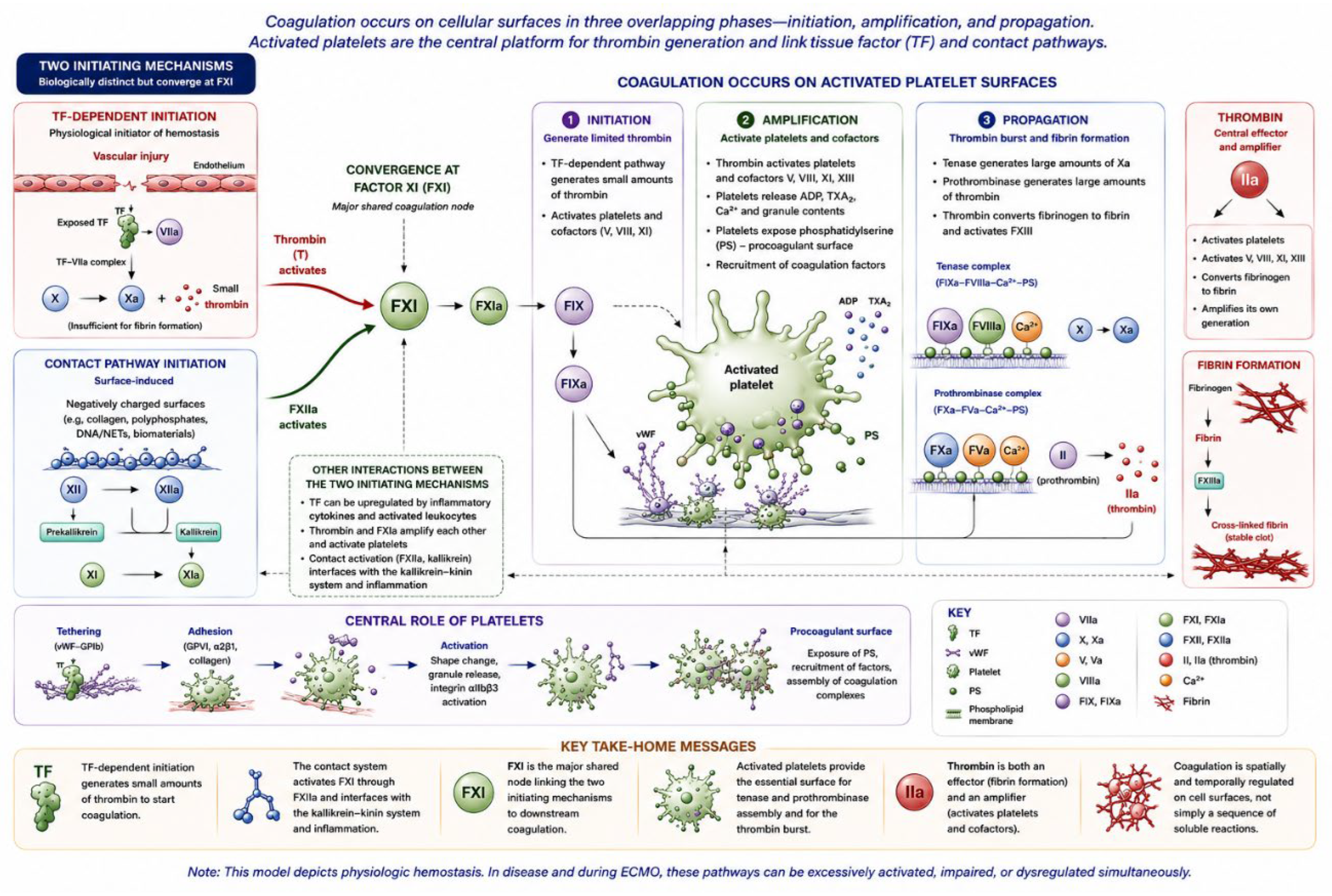
Figure 1. The cell-based model of coagulation. The figure illustrates the three overlapping phases of coagulation—initiation, amplification, and propagation—with activated platelets as the central cellular platform for coagulation complex assembly. It also emphasizes the interconnection of TF-dependent and contact-dependent mechanisms through factor XI and their convergence on platelet-dependent tenase and prothrombinase complexes.
Preprints 231442 g001
Figure 2. Convergent model of coagulation and inflammation. The figure illustrates the reciprocal interactions among vascular injury, coagulation, platelets, endothelium, complement, innate immunity, inflammation, and fibrinolysis. Endogenous anticoagulant, fibrinolytic, anti-inflammatory, and endothelial homeostatic mechanisms act as regulatory brakes; when these regulatory mechanisms are overwhelmed or dysregulated, the network may shift from regulated hemostasis toward thromboinflammation.Ontogeny of hemostasis and thromboinflammation.
Preprints 231442 g002
Figure 3. From thromboinflammation to dyshemostasis in ECMO. The figure summarizes how blood–biomaterial interaction, mechanical forces, critical illness, inflammation, anticoagulation, and other patient- and circuit-related factors initiate and sustain thromboinflammatory activation during ECMO. Persistent procoagulant activation may coexist with platelet and coagulation-factor consumption, acquired von Willebrand abnormalities, endothelial dysfunction, and altered fibrinolysis, resulting in a dynamic balance between thrombotic and bleeding manifestations.
Preprints 231442 g003
Table 1. Interpretation and therapeutic implications of hemostatic assays during pediatric ECMO*.
Method What it measures What it does not measure Main limitation in ECMO Practical interpretation Therapeutic implications
ACT Whole-blood clotting time, influenced by UFH and multiple patient- and circuit-related factors Does not provide a specific measure of heparin activity or overall hemostatic competence Influenced by thrombocytopenia, hypofibrinogenemia, hemodilution, anemia, temperature, inflammation, and other factors Useful for rapid bedside assessment; discordance with complementary assays should prompt investigation rather than automatic heparin adjustment May contribute to assessment of anticoagulant effect, but unexpected values should be interpreted with aPTT, anti-Xa, VET, and clinical/circuit findings before modifying UFH
aPTT Phospholipid-dependent clotting time, predominantly reflecting intrinsic/common pathway function and affected by UFH Does not specifically quantify heparin activity and does not assess platelet function, fibrinogen contribution, VWF, or clot strength Factor deficiencies, inflammation, elevated FVIII, lupus anticoagulant, consumptive states, developmental hemostasis, and assay/reagent variability Useful for UFH monitoring when appropriately validated; discordance with anti-Xa should be investigated rather than automatically attributed to inadequate or excessive heparin Can provide an important complementary basis for UFH adjustment; substantial discordance should prompt evaluation of biological and analytical causes before changing anticoagulation intensity
Anti-Xa Functional inhibition of factor Xa by the heparin–antithrombin complex; provides a relatively direct estimate of UFH anticoagulant activity Does not assess endogenous coagulation pathways, platelet function, fibrinogen contribution, clot strength, or the overall hemostatic phenotype AT availability, assay methodology, hemolysis/free hemoglobin, hyperbilirubinemia, hypertriglyceridemia, and other analytical or preanalytical factors may produce falsely low or otherwise misleading results; assay response to UFH is not fully standardized across platforms Useful for estimating UFH effect, but should not be interpreted in isolation. Concordance with aPTT increases confidence; marked discordance should prompt investigation. VET may provide complementary information regarding functional clot initiation and strength Heparin titration should not rely on anti-Xa alone. Before modifying UFH, consider concordance with aPTT, VET findings when available, AT availability, assay interference, sampling issues, and the patient and circuit phenotype
Heparin concentration Plasma heparin concentration Does not necessarily reflect biological anticoagulant effect Assay availability and calibration; concentration–effect relationship depends on AT and patient biology May help characterize discordance between measured heparin concentration and functional anticoagulant assays May support individualized assessment of UFH exposure when other assays are discordant, but should not replace functional assessment
VET – clot initiation Whole-blood dynamics of clot initiation and propagation, integrating coagulation factors, fibrinogen, platelets, and cellular components Does not provide a specific measure of UFH concentration or a validated stand-alone measure of anticoagulant activity Influenced by multiple plasma and cellular components and assay-specific conditions; pediatric ECMO-specific thresholds for UFH effect are not standardized Provides complementary functional information and may be particularly useful when anti-Xa and aPTT are discordant May help determine whether an apparent anticoagulation discrepancy is accompanied by functional hypo- or hypercoagulability; should inform rather than independently dictate UFH adjustment
VET – clot strength Integrated contribution of fibrin and platelets to clot firmness Does not independently distinguish platelet dysfunction from fibrinogen deficiency without appropriate adjunctive assays Cannot identify the specific molecular cause of reduced or increased clot strength Particularly useful for identifying functional abnormalities in clot strength and assessing the relative contribution of fibrin and platelets when paired with fibrin-specific testing May support targeted consideration of platelet or fibrinogen therapy rather than empiric transfusion, when consistent with the clinical phenotype
FIBTEM / functional fibrinogen Fibrin-based contribution to clot firmness estimated after pharmacologic inhibition of platelet contribution Does not assess platelet contribution or provide a direct measure of heparin effect Interpretation depends on assay platform and pediatric context; does not directly equal plasma fibrinogen concentration Helps determine whether impaired fibrin contribution substantially contributes to reduced clot strength May support targeted fibrinogen replacement when clinically indicated and interpreted together with plasma fibrinogen, VET, and clinical phenotype
Platelet count Number of circulating platelets Does not measure platelet function or the broader platelet phenotype Platelet count may not reflect functional platelet competence during ECMO Quantifies thrombocytopenia but does not characterize platelet contribution to hemostasis May contribute to platelet transfusion decisions, but intervention should consider bleeding/thrombotic phenotype and functional information, including VET when available
Fibrinogen Plasma fibrinogen concentration Does not measure functional fibrin formation or clot architecture Concentration may not reflect the functional contribution of fibrinogen to clot strength Useful for identifying quantitative fibrinogen deficiency; interpretation is strengthened by functional fibrin assessment on VET May support consideration of fibrinogen replacement when clinically indicated, particularly when reduced fibrin contribution is demonstrated functionally
PT/INR Extrinsic/common pathway clotting time Does not measure UFH effect, platelet function, or clot strength Limited sensitivity to UFH; affected by factor deficiency, liver dysfunction, vitamin K status, dilution, and assay characteristics May identify abnormalities in extrinsic/common pathway factor activity or broader acquired coagulopathy, but has limited utility for UFH monitoring Should prompt investigation and correction of relevant factor abnormalities when clinically indicated rather than reflexive modification of UFH
*This table is intended to support integrated interpretation of hemostatic assays and should not be used as a stand-alone treatment algorithm. Therapeutic decisions should incorporate assay results together with the clinical and circuit phenotype, serial trends, and relevant biological and analytical confounders.
Table 2. Factors influencing aPTT and anti-Xa results and their expected direction of effect.
Factor aPTT Anti-Xa
UFH exposure ↑ ↑
Heparin contamination from line ↑ ↑
Intrinsic/common factor deficiency ↑ ↔
Factor consumption / DIC ↑ ↔
Liver dysfunction ↑ ↔
Developmental low factor levels ↑ ↔
Elevated factor VIII ↓ ↔
Increased fibrinogen / acute-phase response ↓ ↔
Lupus anticoagulant / inhibitors ↑ ↔
Antithrombin deficiency ↔* ↓*
Thrombocytopenia / platelet dysfunction usually ↔ ↔
Hemolysis / ↑ plasma-free Hb variable ↓**
Hyperbilirubinemia variable ↓**
Hypertriglyceridemia / lipemia variable ↓**
Assay/reagent/platform characteristics ↑ / ↓ ↑ / ↓
Delayed/inappropriate sample handling ↑ / ↓ ↑ / ↓
* Depends on assay methodology. Anti-Xa assays relying on endogenous AT may show a lower result with AT deficiency; assays containing excess exogenous AT are less influenced by the patient’s AT level. ** Potential analytical interference with chromogenic anti-Xa assays; magnitude and even direction may depend on the platform.
Table 3. Phenotype-informed positioning of UFH anticoagulation intensity during pediatric ECMO*.
Prevailing clinical context Potential anticoagulation positioning Anti-Xa positioning
Bleeding-predominant phenotype Lower-intensity positioning when anticoagulation remains necessary 0.10–0.30 IU/mL
Stable hemostatic state Conventional positioning 0.30–0.50 IU/mL
Thrombosis-predominant phenotype Higher-intensity positioning when inadequate anticoagulant effect is identified after other patient- and circuit-related contributors have been assessed 0.50–0.70 IU/mL
Mixed bleeding–thrombotic phenotype No predefined target; individualize according to the competing processes —
*The ranges in Table 3 represent positioning zones rather than validated phenotype-specific therapeutic targets. The lower 0.10–0.30 IU/mL and higher 0.50–0.70 IU/mL zones should be considered selectively according to the integrated hemostatic assessment. Anti-Xa and aPTT are alternative, non-interchangeable measures of UFH effect; when aPTT is used as the primary assay, its target should be defined independently by the institutional protocol.
Table 4. Operational approach to UFH infusion adjustment according to selected anti-Xa target range during pediatric ECMO.
Selected anti-Xa target range Measured anti-Xa Suggested UFH infusion adjustment
0.10–0.30 IU/mL <0.10 Consider increasing by 10–20% after integrated assessment
0.10–0.30 No change
>0.30 Consider decreasing by 10–20% after integrated assessment
0.30–0.50 IU/mL <0.30 Consider increasing by 10–20% after integrated assessment
0.30–0.50 No change
>0.50 Consider decreasing by 10–20% after integrated assessment
0.50–0.70 IU/mL <0.50 Consider increasing by 10–20% after integrated assessment
0.50–0.70 No change
>0.70 Consider decreasing by 10–20% after integrated assessment
Table 5. Pragmatic reference points and phenotype-directed considerations for targeted blood-component and hemostatic therapy during pediatric ECMO.
Hemostatic intervention Conservative bedside reference Bleeding-predominant phenotype Thrombosis-predominant phenotype Mixed bleeding–thrombotic phenotype
Platelet transfusion ≥100 ×109/L in bleeding patients;
≥50–100 ×109/L in nonbleeding patients. Lower individualized thresholds have been reported.
Consider when thrombocytopenia or suspected platelet dysfunction is present in the setting of clinically significant bleeding or before a high-risk procedure. Platelet count, trajectory, function, and VET-derived clot strength should be interpreted together with fibrinogen and clinical bleeding. Avoid unnecessary platelet exposure. Transfuse only for a defined bleeding, procedural, or severe thrombocytopenic indication; consider the potential contribution of platelet replacement to patient or circuit thrombosis. Consider when platelet deficiency or dysfunction contributes to clinically consequential bleeding. Balance replacement against active or progressive patient or circuit thrombosis.
Fresh Frozen Plasma (FFP) For clinically significant bleeding, an initial transfusion may be considered when
INR >1.5 and relevant multiple-factor deficiency is suspected or demonstrated. Avoid prophylactic FFP solely to correct INR.
Consider when clinically significant bleeding is associated with relevant multiple-factor deficiency or impaired clot initiation. Avoid repeated administration solely to normalize INR. Generally avoid plasma solely to correct an elevated INR when there is no clinically relevant bleeding or demonstrated factor deficiency. Consider when multiple-factor deficiency materially contributes to bleeding; avoid INR-driven correction when thrombosis is clinically consequential.
Fibrinogen replacement A fibrinogen level ≥100 mg/dL may be acceptable in nonbleeding patients; a target of ≥150 mg/dL is reasonable in clinically significant bleeding or before high-risk intervention. Optimal thresholds remain uncertain. Consider when low fibrinogen or impaired fibrin contribution contributes to clinically significant bleeding. Integrate fibrinogen concentration with platelet contribution and functional fibrin assessment when available. Avoid unnecessary fibrinogen loading when fibrin contribution is adequate, particularly in the presence of progressive patient or circuit thrombosis. Consider when low fibrinogen or impaired fibrin contribution materially contributes to bleeding; balance replacement against progressive thrombosis and circuit thrombus burden.
Red blood cells (RBC) No universal pediatric ECMO threshold. Restrictive strategies, including approximately 7 g/dL, have been used in selected populations. Individualize according to blood loss, hemoglobin, systemic oxygen delivery and consumption, hemodynamics, and clinical trajectory; transfuse when anemia or blood loss compromises oxygen delivery. Individualize according to oxygen-delivery requirements and patient/circuit condition; avoid transfusion solely to normalize hemoglobin when oxygen delivery is adequate. Individualize according to blood loss and oxygen-delivery requirements while considering the concurrent thrombotic and circuit context.
Prothrombin complex concentrate (PCC) No validated pediatric ECMO threshold or dose; exceptional intervention. Consider only as rescue therapy for severe, refractory, life-threatening bleeding after surgical/procedural causes have been addressed and conventional multimodal therapy optimized. Generally avoid, particularly with active or progressive patient or circuit thrombosis, because of substantial thrombotic potential. Generally avoid; consider only in exceptional life-threatening bleeding after multidisciplinary assessment of the competing thrombotic risk.
Recombinant activated factor VII (rFVIIa) No validated pediatric ECMO threshold or dose; exceptional rescue intervention. Reserve for refractory, life-threatening bleeding after conventional measures and correction of reversible causes; pediatric ECMO evidence is limited and uncontrolled. Generally avoid because of substantial thrombotic potential and uncertain benefit. Generally avoid; consider only in exceptional life-threatening bleeding after multidisciplinary assessment of thrombotic risk.
Antifibrinolytic therapy No universal laboratory threshold; consider when clinically significant bleeding or high bleeding risk is associated with suspected or likely hyperfibrinolysis. Consider when hyperfibrinolysis materially contributes to clinically significant bleeding, particularly in a surgical or high-risk procedural context. VET evidence may support the diagnosis but is not mandatory when clinical/procedural risk is compelling. Generally avoid when clinically relevant or progressive thrombosis is present, particularly when hyperfibrinolysis is not contributing to the phenotype. Consider only when hyperfibrinolysis materially contributes to bleeding and the anticipated benefit outweighs the potential effect on patient or circuit thrombosis.
Table 6. Circuit-Based Strategies for Hemostatic Optimization During Pediatric ECMO.
Circuit strategy Primary mechanism addressed Potential hemostatic implication
Biocompatible circuit surfaces/coatings Reduce protein adsorption and contact activation May reduce platelet, coagulation, complement, and inflammatory activation
Minimize unnecessary blood–biomaterial interface Reduce artificial-surface exposure May reduce coagulation, platelet, and inflammatory activation
Reduce priming volume when feasible Limit hemodilution and unnecessary artificial-surface exposure May preserve hemostatic substrate concentrations and reduce transfusion requirements
Optimize cannula size and position Improve drainage and reduce abnormal flow and shear May reduce flow instability, mechanical blood trauma, and hemolysis
Optimize pump speed and blood flow for the circuit configuration and required support Balance adequate flow and drainage against excessive shear and stasis May limit shear-mediated blood injury, hemolysis, and circuit thrombogenicity while maintaining effective circuit function
Prevent recurrent suction/chattering events Reduce excessive negative pressures and flow instability May reduce hemolysis and mechanical blood trauma
Avoid unnecessary circuit manipulation Reduce additional blood–biomaterial and air exposure May limit additional coagulation, complement, and inflammatory activation
Avoid unnecessary circuit/oxygenator exchanges Avoid renewed blood–biomaterial exposure and procedural burden May reduce additional thromboinflammatory activation when exchange is not clinically necessary
Promptly investigate progressive fibrin deposition or circuit thrombosis Identify evolving circuit pathology and its mechanical and hemostatic consequences May facilitate early recognition of increasing thrombotic burden, hemolysis, or oxygenator dysfunction and inform subsequent circuit management
Circuit/oxygenator intervention when clinically consequential Remove or replace a component compromising circuit function or patient support May reduce ongoing circuit-related thrombogenicity and mechanical blood injury when the circuit has become a clinically significant source of dyshemostasis
Disclaimer/Publisher’s Note: The statements, opinions and data contained in all publications are solely those of the individual author(s) and contributor(s) and not of MDPI and/or the editor(s). MDPI and/or the editor(s) disclaim responsibility for any injury to people or property resulting from any ideas, methods, instructions or products referred to in the content.
Copyright: This open access article is published under a Creative Commons CC BY 4.0 license, which permit the free download, distribution, and reuse, provided that the author and preprint are cited in any reuse.