
Submitted:
02 September 2026
Posted:
03 September 2026
You are already at the latest version
Abstract
Background: Metabolic dysfunction-associated fatty liver disease (MASLD) is the most common chronic liver disease in children and adolescents. While magnetic resonance imaging–proton density fat fraction (MRI-PDFF) provides quantification of hepatic fat, its cost and accessibility restrict its use for screening and monitoring. Emerging quantitative ultrasound techniques offer non-invasive alternatives for pediatric hepatic steatosis assessment. This scoping review synthesized evidence on emerging quantitative ultrasound techniques in children. Methods: A scoping review was conducted in accordance with PRISMA-ScR guidelines. MEDLINE (PubMed), Embase, and Scopus were searched for studies published between January 2019 and October 2025. Studies evaluating quantitative ultrasound techniques for children were included. Controlled attenuation parameter (CAP) studies were identified but excluded to focus on emerging non-CAP modalities. Data extraction included study characteristics, reference standards, and findings. Results: Fifty-five studies met the eligibility criteria for quantitative ultrasound. After excluding 40 CAP only studies, fifteen evaluating non-CAP quantitative ultrasound modalities were included in the final synthesis. The most studied techniques included ultrasound-derived fat fraction (UDFF), attenuation-based techniques, and backscatter-based analysis. Several studies reported correlations with MRI-PDFF and good to excellent diagnostic performance, with proposed attenuation cutoffs ranging from 0.54 to 0.73 dB/cm/MHz. However, heterogeneity was observed in study design, populations, acquisition protocols, and reference standards. Conclusion: Emerging quantitative ultrasound techniques show promising diagnostic accuracy for pediatric hepatic steatosis and may represent accessible alternatives for noninvasive liver fat quantification. Their implementation could facilitate earlier identification of pediatric MASLD. However, methodological heterogeneity and lack of standardization, require prospective multicenter validation and pediatric-specific thresholds.
Keywords:
MASLD
; pediatric hepatic steatosis
; quantitative ultrasound
; attenuation imaging
; ultrasound-derived fat fraction
1. Introduction
Metabolic dysfunction-associated fatty liver disease (MASLD), previously referred to as non-alcoholic fatty liver disease (NAFLD), has emerged as the most common chronic liver disease in children and adolescents worldwide [1,2]. Its prevalence is estimated at approximately 7–8% in the general pediatric population and rises substantially among children with obesity or metabolic comorbidities [3]. Early identification of hepatic steatosis is therefore clinically important, as pediatric MASLD is associated with increased long-term risk of cardiometabolic complications including type 2 diabetes, cardiovascular disease, and progressive liver injury [1,4].
Liver biopsy historically represented the reference standard for diagnosing and staging hepatic steatosis, but its invasiveness, cost, and sampling variability limit its use in routine pediatric care [5]. Magnetic resonance imaging-based techniques, particularly proton density fat fraction (MRI-PDFF), now provide accurate non-invasive quantification of hepatic fat with excellent correlation to histologic steatosis [5,6]. However, MRI remains limited by cost, availability, and the frequent need for sedation in younger children, restricting its use as a screening tool in many clinical settings.
Ultrasound is widely used as a first-line imaging modality for evaluating suspected pediatric hepatic steatosis because it is safe, inexpensive, and readily available. Conventional B-mode ultrasound relies on qualitative features such as increased hepatic echogenicity relative to the renal cortex, attenuation of the ultrasound beam, and reduced visualization of intrahepatic vessels [7]. However, these qualitative criteria are operator-dependent and demonstrate limited sensitivity for mild steatosis, highlighting the need for objective and reproducible imaging biomarkers [7].
To overcome these limitations, several quantitative ultrasound (QUS) techniques have been developed to provide objective and reproducible measurements of hepatic fat. These include controlled attenuation parameter (CAP), ultrasound-derived fat fraction (UDFF) and attenuation imaging (ATI/TAI) [8,9,10,11]. Although these technologies have shown promising diagnostic performance, their clinical adoption in pediatric populations remains limited and the existing literature shows variability in study design, imaging protocols, patient populations and reference standards [7,12].
In this context, a comprehensive mapping of quantitative ultrasound techniques used to evaluate pediatric hepatic steatosis is needed. The aim of this scoping review is therefore to map and synthesize the current evidence on quantitative ultrasound-based modalities applied to pediatric hepatic steatosis. To our knowledge, no previous review has specifically synthesized the evidence regarding emerging non-CAP quantitative ultrasound techniques in children.
2. Materials and Methods
This scoping review was conducted according to the Preferred Reporting Items for Systematic Reviews and Meta-Analyses extension for Scoping Reviews (PRISMA-ScR) guidelines [13] and followed the framework proposed by Arksey and O’Malley [14].
This scoping review aimed to map current evidence on quantitative ultrasound techniques for assessing hepatic steatosis in pediatric populations and identify knowledge gaps for future research. The review focused on imaging modalities, technical parameters, diagnostic performance, and clinical feasibility.
The research question was structured according to the Population, Intervention, Comparator, Outcome (PICO) framework (Table 1). The population included children and adolescents (0–18 years) with suspected or confirmed hepatic steatosis. Intervention consisted of non-invasive quantitative ultrasound techniques excluding CAP-only studies, with MRI-PDFF or liver biopsy as reference standards. Outcomes included diagnostic performance, quantitative parameters, and feasibility in clinical practice.
Studies were eligible if they included pediatric participants (<18 years) with suspected or confirmed hepatic steatosis. Studies including participants up to 21 years were retained if the cohort was predominantly pediatric.
The review focused on non-invasive quantitative ultrasound techniques for hepatic steatosis assessment, including UDFF, ATI/TAI, and other quantitative ultrasound approaches. CAP was included in the search strategy to ensure comprehensive study identification but studies exclusively evaluating CAP were excluded because the review focused on emerging non-CAP quantitative modalities. Studies reporting both CAP and other quantitative ultrasound techniques were retained if non-CAP measurements were provided.
Studies focusing exclusively on liver fibrosis, qualitative ultrasound, or lacking quantitative measurements were excluded.
Only original research articles published between January 2019 and October 2025 were included to capture recent developments in quantitative ultrasound techniques, which have been increasingly developed in recent years [7]. No language restrictions were applied during the search, although only English-language studies were included after screening.
Studies involving predominantly adult populations, mixed cohorts without separate pediatric analysis, CAP-only evaluations, or non-original research (e.g., reviews, editorials, guidelines, or protocols) were excluded (Table 2).
A systematic search was conducted in MEDLINE (via PubMed), Embase, and Scopus for studies published between January 1, 2019 and October 1, 2025. The final search was performed on October 1, 2025. Reference lists of included studies were also screened to identify additional relevant articles.
The search strategy combined controlled vocabulary (MeSH and Emtree terms) and free-text keywords related to hepatic steatosis (NAFLD/MAFLD/MASLD), pediatric populations, and quantitative ultrasound techniques. Terminology was harmonized using current MASLD nomenclature. Data originally reported as NAFLD or MAFLD were synthesized under the MASLD framework to ensure alignment with contemporary guidelines. Full search strategies are provided in Appendix A.
All records were imported into Zotero and duplicates were manually removed (n=68). One reviewer (PA) screened titles, abstracts, and full-text of potentially relevant studies. Screening decisions were verified by a second reviewer (ST).
Data were manually extracted using a standardized form. Extraction was performed by one reviewer (PA) and verified by a second (ST). Extracted data included study characteristics (author, year, country, study design, sample size, and age), ultrasound modality and manufacturer, comparator (reference standards), and key findings, including diagnostic performance, cutoff values, and other relevant outcomes related to hepatic steatosis.
Results were synthesized descriptively, focusing on the diversity of quantitative ultrasound modalities, diagnostic performance, feasibility, and methodological heterogeneity. For non-CAP studies, the methodological quality and risk of bias were evaluated using the QUADAS-2 tool.
3. Results
3.1. Study Selection
The initial database search yielded 403 records (322 from the primary search and 81 from the updated search). After removing 68 duplicates, 335 articles remained for screening. During title and abstract screening, 230 records were excluded because of non-pediatric populations (n = 78), absence of US techniques (n=41), inappropriate publication types (n = 30), or lack of relevance based on title (n=81).
A total of 105 full-text articles were assessed for eligibility. Of these, 50 studies were excluded, primarily due to the absence of quantitative ultrasound modalities (n = 40), a focus on fibrosis without steatosis assessment (n = 9), or inability to retrieve the full text (n = 1). While 55 studies met the initial inclusion criteria for quantitative ultrasound, an additional 40 studies were excluded as they focused exclusively on CAP, which was outside the scope of this review. Ultimately, 15 studies were included in the final qualitative synthesis. The study selection process is summarized in the PRISMA flow diagram (Figure 1).
This section may be divided by subheadings. It should provide a concise and precise description of the experimental results, their interpretation, and the experimental conclusions that can be drawn.
3.2. Study Characteristics
Of the 15 included studies evaluating non-CAP modalities, their characteristics are summarized in Table 3 [8,10,15,16,17,18,19,20,21,22,23,24,25,26,27]. The majority of the research was published between 2021 and 2025, reflecting the rapid expansion of QUS technologies in pediatric hepatology. Prospective observational designs predominated, followed by retrospective cohorts and cross-sectional studies. Geographically, there was a strong preponderance of studies from East Asia (South Korea, China, Japan, Taiwan), with emerging contributions from Europe and North America [8,10,15,16,17,18,19,20,21,22,23,24,25,26,27]. Sample sizes ranged from small single-center pilot cohorts (n = 32) [25] to larger clinical evaluations, such as the cohort of 268 participants evaluated by Liu et al. [26]. MRI-proton density fat fraction (MRI-PDFF) served as the primary reference standard in high-quality studies, followed by histology and transient elastography [8,10,15,16,17,18,19,20,21,22,23,24,25,26,27].
Technological implementation was vendor-specific across the included studies. Attenuation Imaging (ATI) was evaluated on Canon Aplio systems [15,18,19,20,21], while Ultrasound-Derived Fat Fraction (UDFF) was assessed exclusively on Siemens Acuson Sequoia platforms [10,25,26]. Ultrasound-Guided Attenuation Parameter (UGAP) was implemented on GE LOGIQ systems [19,27]. Attenuation coefficient (ATT) and shear-wave measurements (SWM) were performed using Arietta 850 ultrasound systems [16]. Specialized backscatter analysis and envelope statistics were primarily performed using Terason platforms [17,23]. Other quantitative tools included Tissue Attenuation Imaging (TAI) and Tissue Scatter Imaging (TSI) on Samsung systems [8,22], and acoustic attenuation coefficient measurements on Philips platforms [24].
3.3. Attenuation Imaging (ATI), Tissue Attenuation Imaging (TAI), and Related Attenuation-Based Techniques
Attenuation-based ultrasound modalities including ATI, TAI, ATT, and UGAP were evaluated in several pediatric cohorts. Early prospective pediatric data from 2021, by D’Hondt et al., including 48 children aged 7–17 years, reported moderate correlation with MRI-PDFF (r = 0.76), with AUROC values of 0.86 for detecting steatosis (MRI-PDFF ≥5%) and 0.91 for moderate steatosis (MRI-PDFF ≥10%), with cutoffs of 0.54 and 0.60 dB/cm/MHz respectively, and excellent interobserver agreement (ICC = 0.92) (24).
In 2022, Yoon et al. investigated UGAP in a retrospective cohort of 118 children aged 0–18 years on a GE LOGIQ E10 system, with a measurement success rate of 98.3%. Using MRI-PDFF as the reference standard in a subset of 45 patients, UGAP correlated positively with PDFF (r = 0.498). A cutoff of 0.699 dB/cm/MHz yielded 90.2% sensitivity and 100% specificity for fatty liver detection. The same year, Song et al. assessed ATI feasibility in 111 children aged 6–18 years. Using B-mode ultrasonography as the primary comparator and MRI-PDFF in a subset of patients. They reported that ATI values increased across steatosis grades (0.54 ± 0.09, 0.63 ± 0.08, and 0.73 ± 0.11 dB/cm/MHz for normal, mild, and moderate–severe steatosis, respectively), with AUROC values of 0.853 and 0.91 for differentiating fatty liver from normal liver and moderate-to-severe steatosis, respectively, with corresponding optimal cutoffs of 0.59 and 0.69 dB/cm/MHz. However, technical failure occurred in approximately 20% of examinations, particularly in younger children and those with lower BMI, highlighting feasibility limitations in this population [21]. In 2024, Shin et al. further investigated ATI reliability over 83 children aged 6–17 years, focusing on the impact of respiratory motion. Using B-mode ultrasonography as primary comparator and MRI-PDFF as secondary comparator, they found no significant difference between free-breathing and breath-hold acquisitions, with excellent agreement for ATI measurements (ICC = 0.95) [19]. Notably, measurement variability was slightly higher in younger children, but did not affect overall agreement, supporting reliable ATI acquisition without strict breath-holding.
Next, Dardanelli et al. (2023) evaluated ATI in 174 children aged 6-18 years with metabolic risk factors using B-mode ultrasonography and reported significantly higher median ATI values in children with risk factors compared to healthy controls (0.64 vs. 0.54 dB/cm/MHz), with strong correlation with B-mode and excellent inter-operator reproducibility (ICC ≈ 0.94) [15].
In 2023, a prospective study from Turkey demonstrated high diagnostic performance of ATI for MRI-PDFF ≥5%. In a cohort of 140 children aged 9-18 years, Bulakci et al. reported AUROC values of 0.94, 0.98, and 0.97 for detecting increasing steatosis grades, with an optimal cutoff of 0.65 dB/cm/MHz [18]. In contrast, in 2025, Bozbeyoglu et al., in a single-center Turkish cohort of 51 children aged 5-17 years also using MRI-PDFF as the reference standard, reported more moderate accuracy (AUROC 0.817) with a similar cutoff (0.67 dB/cm/MHz) [22].
In 2025, Cetinic et al. evaluated ATI feasibility in children aged 9-18 years with severe obesity (mean BMI 41.0 ± 5.8 kg/m²) [20]. No external reference standard was used; ATI measurements were compared with clinical, serological and technical parameters. The median ATI value was 0.58 dB/cm/MHz, with a wide range (0.32–0.97), and scan–rescan reproducibility was moderate (ICC = 0.61). ATI values were significantly influenced by measurement depth (β = −4.2; p < 0.0001), with greater depth associated with lower ATI values. Despite severe obesity, ATI values remained relatively low, compared to diagnostic thresholds reported in other studies.
Wang et al. evaluated the combination of ATT with SWM, in a prospective cohort of 62 children with Wilson’s disease, reporting AUROC values between 0.714 and 0.867 for steatosis detection and an optimal cutoff of approximately 0.73 dB/cm/MHz (16).
Overall, most studies reported ATI cutoff values ranging from 0.59 to 0.67 dB/cm/MHz. Lower values were observed in selected cohorts, such as D’Hondt et al. (0.54 dB/cm/MHz) and Cetinic et al. (0.58 dB/cm/MHz) [20,24]. Higher thresholds were reported in studies with specific methodological or population characteristics, including Yoon et al. (0.699 dB/cm/MHz) and Wang et al. (~0.73 dB/cm/MHz) [16,27].
3.4. Specialized Backscatter Analysis
Several studies evaluated specialized backscatter-based approaches, frequently in combination with other quantitative ultrasound techniques, echogenicity ratios such as the automated Hepato-Renal Index (ezHRI), a semi-quantitative evaluation of steatosis based on the comparison of liver and kidney signal intensity on B-mode or clinical scores such as the Hepatic Steatosis Index (HSI), a biochemical score based on BMI, ALT, AST, sex, and diabetes status used to estimate the risk of hepatic steatosis.
In a prospective Italian study including 36 children, Polti et al. assessed TSI, ezHRI, TAI, using MRI-PDFF as the reference standard. Pediatric abnormal thresholds were defined as TSI ≥91.95, ezHRI ≥1.215, and TAI ≥0.625 dB/cm/MHz, demonstrating good discrimination of steatosis [8].
In 2021, in a Taiwanese population, Chuang et al. explored another backscatter-based approach, using envelope statistics [23]. Using HSI as comparator, they reported progressive increases in the Nakagami parameter across steatosis grades in 68 children aged 3-18 years, supporting early steatosis detection [23]. In 2023, Hsieeh et al., in a larger prospective cohort of 236 children of the same age range, extended this approach by evaluating Nakagami imaging, Homodyned-K modeling, and entropy analysis, using HSI as the comparator [17]. AUROC values ranged from 0.83 to 0.97 for steatosis grading and 0.82–0.84 for fibrosis detection in a transient elastography validation subgroup.
3.5. Ultrasound-Derived Fat Fraction (UDFF)
Three studies evaluated UDFF in pediatric populations, all using the Siemens Acuson Sequoia platform. Zalcman et al. [10] conducted a prospective study in 46 children aged 7–18 years referred for abdominal MRI, using MRI-PDFF as the reference standard. Among the cohort, 36 children had normal liver fat fraction and 10 had confirmed steatosis. UDFF demonstrated excellent agreement with MRI-PDFF (ICC = 0.92) and an AUROC of 0.95 for detecting steatosis ≥6%, with 90% sensitivity and 94% specificity. BMI was the only independent predictor of UDFF on multivariable analysis.
Huang et al. prospectively evaluated UDFF in 32 children aged 10–14 years with confirmed MASLD, using MRI-PDFF as the reference standard [25]. UDFF correlated positively with MRI-PDFF (r = 0.72), with AUROCs of 0.84, 0.88, and 0.88 for detecting steatosis grades ≥S1, ≥S2, and S3 respectively, and optimal cutoffs of 9.5%, 10.5%, and 12.8%. Overall measurement reproducibility was high (ICC = 0.90).
In the largest clinical evaluation to date, Liu et al. [26] enrolled 230 children aged 6–12 years with MASLD alongside 38 healthy controls. Steatosis grading was based on B-mode ultrasound rather than MRI-PDFF. UDFF values increased significantly across steatosis grades (11.52% for Grade 1 vs. 19.10% for Grade 2; p < 0.001), and showed the strongest correlation with steatosis severity.
Overall, QUS-based methods demonstrated high diagnostic accuracy for the detection of pediatric steatosis, with AUROC values frequently exceeding 0.85 across studies. However, diagnostic performance should be interpreted cautiously because of heterogeneous reference standards, which limits direct comparison between techniques. Attenuation-based methods, particularly ATI, showed consistent performance across multiple cohorts, with relatively clustered cutoff values, while UDFF demonstrated high agreement with MRI-PDFF and strong diagnostic performance. In contrast, other quantitative approaches, including backscatter-based techniques such as TSI, were evaluated in fewer studies.
3.6. Risk of Bias Assessment
Risk of bias was assessed using the QUADAS-2 tool for the 15 included studies. Risk of bias distribution across domains is illustrated in Figure 2. Overall, most studies demonstrated high risk of bias in the patient selection and index test domains. High-risk in-patient selection was mainly related to single-center recruitment, selected clinical populations, and unclear consecutive inclusion. In the index test domain, cutoffs were frequently derived without external validation, and blinding was rarely reported. Risk was lower when MRI-PDFF was used as reference standard than with biochemical markers or indirect indices. Reporting in the flow and timing domain was often incomplete, particularly regarding the interval between index test and reference standard. A summary of the risk-of-bias assessment is presented in Table 4.
4. Discussion
This scoping review identified 55 studies meeting the initial eligibility criteria for quantitative ultrasound, of which 15 evaluated non-CAP modalities and were included in the final synthesis. While Controlled Attenuation Parameter (CAP) remained the most frequently studied modality, a growing body of literature now explores emerging approaches, including Ultrasound-Derived Fat Fraction (UDFF), Attenuation Imaging (ATI), attenuation coefficient measurements, and backscatter-based techniques. These findings highlight both the historical dominance of CAP and a progressive shift toward fat-centered imaging strategies in pediatric hepatology.
CAP remains the most widely studied quantitative ultrasound modality in pediatric populations, largely due to its early integration into FibroScan® platforms and its widespread use in hepatology since the early 2010s [7,28]. Initially developed within a fibrosis-oriented framework, CAP reflects an adult MASLD paradigm, in which fibrosis determines the main long-term outcomes (cirrhosis, hepatocellular carcinoma, and mortality), and therefore plays a central role in clinical decision-making [1,2].
However, applying this paradigm to pediatrics introduces a conceptual mismatch. In children, MASLD typically represents an early metabolic phenotype in which steatosis is the predominant hepatic abnormality, whereas advanced fibrosis remains uncommon [1,17,20,23,24]. Steatosis represents the main target for early detection and metabolic risk stratification [29]. Despite its widespread use, CAP has several limitations in pediatric populations, including reduced sensitivity for mild steatosis and the lack of standardized pediatric thresholds [7]. In addition, the current evidence base is extensive, with multiple meta-analyses and pediatric-focused reviews, leading to a relative saturation of CAP-related research. For these reasons, this review deliberately focused on emerging quantitative ultrasound modalities that remain comparatively under-explored in pediatric imaging. This shift reflects increasing interest in fat-centered imaging approaches for pediatric MASLD.
Emerging quantitative ultrasound techniques expand the assessment of hepatic steatosis in children. Beyond CAP, approaches such as UDFF, ATI/TAI, and backscatter-derived methods aim to directly quantify hepatic lipid content [8,10,15,18,23,25,26].
This diversification reflects a shift toward a fat-centered imaging paradigm, more closely aligned with the pediatric MASLD phenotype. Pediatric-specific factors, including thinner acoustic windows and a lower prevalence of advanced disease, may further support the feasibility and performance of these techniques compared with adult populations [8].
Among these modalities, UDFF appears particularly promising, demonstrating a strong correlation with MRI-PDFF, without major technical limitations in children [10,25,26]. Evidence from adult populations further strengthens this robustness, showing high diagnostic accuracy and strong agreement with MRI-PDFF across a broad spectrum of steatosis severity [30]. Concordant pediatric and adult findings support the biological and technical validity of UDFF. Although pediatric evidence remains limited, the suitability of UDFF for pediatric imaging and its strong validation against MRI-PDFF support future clinical integration. However, direct head-to-head comparisons between emerging quantitative ultrasound techniques remain scarce, precluding definitive conclusions regarding the relative performance of individual modalities.
This review has several limitations. First, the search was restricted to studies published from 2019 onward, potentially excluding earlier relevant evidence. Screening and data extraction were performed by a single reviewer, which may introduce selection bias despite adherence to a predefined protocol. Included studies were highly heterogeneous regarding design, population characteristics, acquisition protocols, and reference standards, precluding quantitative synthesis and limiting direct comparison across modalities. In addition, most non-CAP studies remained single-center and exploratory.
Clinical integration of emerging QUS modalities remains limited by predominantly single-center, cross-sectional, or retrospective studies with small sample sizes and heterogeneous reference standards, preventing the establishment of reproducible diagnostic thresholds. This variability is reflected in the attenuation thresholds reported across studies. For example, Yoon et al. reported a relatively high UGAP cutoff (0.699 dB/cm/MHz), which may partly be explained by the markedly imbalanced distribution of steatosis grades within their cohort, with only five children classified as S1 compared with thirty classified as S3 [27]. Cross-vendor harmonization is also lacking, with distinct acquisition protocols and limited MRI–US calibration complicating multiparametric integration [16,17,20,23]. The vendor-dependent nature of these technologies remains a significant barrier to clinical implementation. Proprietary algorithms, such as UDFF, are restricted to specific manufacturers. Furthermore, even similar physical parameters, such as the attenuation coefficient, are processed differently across various platforms. Consequently, the diagnostic thresholds identified in this review cannot be considered universal. This technical fragmentation underscores the need for standardized phantoms and cross-vendor calibration.
Beyond methodological differences, patient characteristics may also contribute to variability in quantitative measurements. In the cohort studied by Cetinic et al., ATI values remained relatively low despite severe obesity. One possible explanation is the greater subcutaneous tissue thickness observed in these patients, which may have increased the distance between the transducer and the region of interest and consequently affected attenuation measurements [20].
Future studies should prioritize prospective multicenter validation using MRI-PDFF as reference standard, with assessment of inter-operator and inter-equipment reproducibility and establishment of pediatric-specific thresholds adjusted for age, BMI, and metabolic phenotype [8,9,10,25].
Beyond technical performance, the feasibility and acceptability of these protocols in pediatric populations remain insufficiently documented. Although quantitative ultrasound appears well suited to children, data remain lacking regarding examination time, sedation requirements, and tolerance; the development of simplified acquisition protocols may improve adherence without compromising accuracy [15,18].
Early detection of steatosis may help identify children at cardiometabolic risk and support targeted interventions [17,20,29]. If validated, quantitative ultrasound techniques could provide a practical and widely accessible tool for longitudinal monitoring of pediatric MASLD in routine clinical care. However, the clinical and economic impact of these tools remains uncertain.
Artificial intelligence may enhance standardization by automating region-of-interest placement, artifact suppression, and parameter extraction, potentially reducing operator dependency and improving reproducibility [17,22].
Overall, this review highlights gaps in pediatric MASLD imaging literature and supports the need for prospective multicenter studies integrating MRI-PDFF validation, standardized protocols, and pediatric-adjusted thresholds.
5. Conclusions
This scoping review highlights the predominance of CAP in pediatric quantitative ultrasound research while demonstrating the emergence of alternative fat-focused modalities such as UDFF, ATI, attenuation coefficient measurements, and backscatter techniques. These modalities reflect a shift toward direct or indirect quantification of hepatic fat, more closely aligned with the early metabolic phenotype of pediatric MASLD.
Despite encouraging diagnostic performance, the current evidence remains limited and heterogeneous. Most studies are small, single-center, and exploratory, with variability in acquisition protocols and reference standards, precluding the establishment of robust pediatric thresholds and standardized clinical workflows.
Future research should prioritize prospective multicenter validation studies using MRI-PDFF as the reference standard, along with cross-vendor harmonization and the definition of pediatric-specific cutoffs. Evaluation of reproducibility, feasibility, and real-world clinical integration will be essential. Quantitative ultrasound techniques may ultimately support earlier detection and phenotyping of pediatric MASLD, facilitating preventive strategies to reduce long-term cardiometabolic risk and integration into multiparametric liver imaging workflows.
Author Contributions
Conceptualization, P.A. and S.T.; methodology, P.A. and S.T.; validation, P.A. and S.T.; formal analysis, P.A.; investigation, P.A.; data curation, P.A.; writing original draft preparation, P.A.; writing review and editing, P.A. and S.T.; visualization, P.A.; supervision, S.T.; project administration, P.A. All authors have read and agreed to the published version of the manuscript.
Funding
This research received no external funding.
Institutional Review Board Statement
Not applicable.
Informed Consent Statement
Not applicable.
Data Availability Statement
Data sharing is not applicable to this article as no datasets were generated or analyzed during the current study.
Acknowledgments
During the preparation of this manuscript, the authors used ChatGPT (OpenAI) for the purpose of improving the English language, clarity and readability of the manuscript. The authors have reviewed and edited the output and take full responsibility for the content of this publication.
Conflicts of Interest
The authors declare no conflicts of interest.
Abbreviations
The following abbreviations are used in this manuscript:
| UDFF | Ultrasound-Derived Fat Fraction |
| MRI-PDFF | Magnetic Resonance Imaging Proton Density Fat Fraction |
| MASLD | Metabolic Dysfunction Associated Steatotic Liver Disease |
| CAP | Controlled Attenuation Parameter |
| US | Ultrasound |
| QUS | Quantitative Ultrasound |
| ATI | Attenuation imaging |
| TSI | Tissue Scatter Imaging |
| SWM | Shear Wave Measurement |
| SWE | Shear Wave Elastography |
| SWD | Shear Wave Dispersion |
| UGAP | Ultrasound Guided Attenuation Parameter |
| EzHRI | Enhanced Hepato-Renal Index |
| HSI | Hepatic Steatosis Index |
| HRI | Hepato-Renal Index |
| UGAP | Ultrasound Guided Attenuation Parameter |
Appendix A.
Appendix A.1. Detailed Search Strategies
The following detailed search strategies were developed and applied across all database.
PubMed (MEDLINE)
(“Ultrasonography”[Mesh] OR ultrasonograph*[tiab] OR ultrasound[tiab] OR sonograph*[tiab]
OR “Ultrasound Imaging”[tiab]
OR “point of care ultrasound”[tiab]
OR “point-of-care ultrasound”[tiab]
OR POCUS[tiab])
AND
(“Fatty Liver”[Mesh] OR steatosis[tiab] OR “hepatic steatosis”[tiab] OR “liver steatosis”[tiab]
OR “fatty liver”[tiab] OR NAFLD[tiab] OR NASH[tiab] OR MAFLD[tiab] OR MASLD[tiab]
OR “nonalcoholic fatty liver disease”[tiab] OR “steatotic liver disease”[tiab])
AND
(“Child”[Mesh] OR “Infant”[Mesh] OR “Adolescent”[Mesh]
OR child*[tiab] OR infant*[tiab] OR adolescen*[tiab] OR pediatric*[tiab]
OR paediatric*[tiab] OR teen*[tiab] OR youth*[tiab])
AND
(“controlled attenuation parameter”[tiab] OR CAP[tiab]
OR “attenuation coefficient”[tiab] OR “ultrasound attenuation”[tiab]
OR UGAP[tiab] OR “ultrasound-guided attenuation parameter”[tiab]
OR “backscatter coefficient”[tiab] OR BSC[tiab]
OR “quantitative ultrasound”[tiab] OR QUS[tiab]
OR “radiofrequency ultrasound”[tiab] OR “RF ultrasound”[tiab]
OR “raw ultrasound data”[tiab] OR “ultrasound RF”[tiab]
OR “ultrasound grading”[tiab] OR “ultrasound score”[tiab]
OR “hepatic fat quantification”[tiab] OR “fat quantification”[tiab]
OR “steatosis quantification”[tiab] OR “steatosis grade”[tiab]
OR “ultrasound fat fraction”[tiab])
Embase
(‘ultrasonography’/exp OR ultrasonograph*:ti,ab OR ultrasound:ti,ab
OR sonograph*:ti,ab OR ‘ultrasound imaging’:ti,ab OR ‘point of care ultrasound’:ti,ab OR ‘point-of-care ultrasound’:ti,ab OR POCUS:ti,ab)
AND
(‘fatty liver’/exp OR steatosis:ti,ab OR ‘hepatic steatosis’:ti,ab
OR ‘liver steatosis’:ti,ab OR ‘fatty liver’:ti,ab OR NAFLD:ti,ab
OR NASH:ti,ab OR MAFLD:ti,ab OR MASLD:ti,ab
OR ‘nonalcoholic fatty liver disease’:ti,ab OR ‘steatotic liver disease’:ti,ab)
AND
(‘child’/exp OR ‘infant’/exp OR ‘adolescent’/exp
OR child*:ti,ab OR infant*:ti,ab OR adolescen*:ti,ab
OR pediatric*:ti,ab OR paediatric*:ti,ab OR teen*:ti,ab OR youth*:ti,ab)
AND
(‘controlled attenuation parameter’:ti,ab OR CAP:ti,ab
OR ‘attenuation coefficient’:ti,ab OR ‘ultrasound attenuation’:ti,ab
OR UGAP:ti,ab OR ‘ultrasound-guided attenuation parameter’:ti,ab
OR ‘backscatter coefficient’:ti,ab OR BSC:ti,ab
OR ‘quantitative ultrasound’:ti,ab OR QUS:ti,ab
OR ‘radiofrequency ultrasound’:ti,ab OR ‘RF ultrasound’:ti,ab
OR ‘raw ultrasound data’:ti,ab OR ‘ultrasound RF’:ti,ab
OR ‘ultrasound grading’:ti,ab OR ‘ultrasound score’:ti,ab
OR ‘hepatic fat quantification’:ti,ab OR ‘fat quantification’:ti,ab
OR ‘steatosis quantification’:ti,ab OR ‘steatosis grade’:ti,ab
OR ‘ultrasound fat fraction’:ti,ab)
Scopus
TITLE-ABS-KEY(ultrasonograph* OR ultrasound OR sonograph* OR “ultrasound imaging” OR POCUS)
AND
TITLE-ABS-KEY(steatosis OR “hepatic steatosis” OR “liver steatosis”
OR “fatty liver” OR NAFLD OR NASH OR “nonalcoholic fatty liver disease”)
D
TITLE-ABS-KEY(child* OR infant* OR adolescen* OR pediatric* OR paediatric*
OR teen* OR youth*)
AND
TITLE-ABS-KEY(“controlled attenuation parameter” OR CAP
OR “attenuation coefficient” OR “ultrasound attenuation”
OR UGAP OR “ultrasound-guided attenuation parameter”
OR “backscatter coefficient” OR BSC
OR “quantitative ultrasound” OR QUS
OR “radiofrequency ultrasound” OR “RF ultrasound”
OR “ultrasound grading” OR “ultrasound score”
OR “hepatic fat quantification” OR “fat quantification”
OR “steatosis quantification” OR “steatosis grade”
OR “ultrasound fat fraction”)
References
- Vajro, P.; Paolella, G.; Fasano, A. Metabolic Fatty Liver Disease in Children: A Growing Public Health Problem. Nutrients 2018, 10(11), 1576. [Google Scholar]
- Eslam, M.; Sanyal, A.J.; George, J.; et al. MAFLD: A consensus-driven proposed nomenclature for metabolic associated fatty liver disease. Gastroenterology 2020, 158(7), 1999–2014.e1. [Google Scholar] [CrossRef]
- Anderson, E.L.; Howe, L.D.; Jones, H.E.; et al. The Prevalence of Non-Alcoholic Fatty Liver Disease in Children and Adolescents: A Systematic Review and Meta-Analysis. PLoS ONE 2015, 10(10), e0140902. [Google Scholar] [CrossRef]
- Newton, K.P.; Hou, J.; Bradley, J.S.; et al. Incidence of Type 2 Diabetes in Children With Nonalcoholic Fatty Liver Disease. Clin. Gastroenterol. Hepatol. 2020, 18(9), 2091–2099. [Google Scholar]
- Schwimmer, J.B.; Middleton, M.S.; Behling, C.; et al. Magnetic Resonance Imaging Predicts Histology in Pediatric Nonalcoholic Fatty Liver Disease. Hepatology 2015, 61(2), 565–573. [Google Scholar] [CrossRef]
- Noureddin, M.; Lam, J.; et al. Utility of MRI-PDFF for quantification of hepatic steatosis in children. J. Pediatr. Gastroenterol. Nutr. 2020, 71(5), 606–611. [Google Scholar]
- Ferraioli, G.; Soares Monteiro, L.B. Ultrasound-based techniques for the diagnosis of liver steatosis. World J. Gastroenterol. 2019, 25(40), 6053–6062. [Google Scholar] [CrossRef]
- Polti, G.; Frigerio, F.; Del Gaudio, G.; Pacini, P.; Dolcetti, V.; Renda, M.; et al. Quantitative ultrasound fatty liver evaluation in a pediatric population: comparison with magnetic resonance imaging proton density fat fraction. Pediatr. Radiol. 2023, 53(12), 2458–2465. [Google Scholar] [CrossRef]
- Runge, J.H.; van Giessen, J.; Draijer, L.G.; Deurloo, E.E.; Smets, A.; Benninga, M.A.; et al. Accuracy of controlled attenuation parameter compared with ultrasound for detecting hepatic steatosis in children with severe obesity. Eur. Radiol. 2021, 31(3), 1588–1596. [Google Scholar] [CrossRef]
- Zalcman, M.; Barth, R.A.; Rubesova, E. Real-time ultrasound-derived fat fraction in pediatric population: feasibility validation with MR-PDFF. Pediatr. Radiol. 2023, 53(12), 2466–2475. [Google Scholar] [CrossRef]
- Kim, P.H.; Cho, Y.A.; Yoon, H.M.; Bak, B.; Lee, J.S.; Jung, A.Y.; et al. Accuracy of attenuation imaging in the assessment of pediatric hepatic steatosis: correlation with the controlled attenuation parameter. Ultrasonography 2022, 41(4), 761–769. [Google Scholar] [CrossRef]
- Alves, V.; Trout, A.T.; Dewit, M.; Mouzaki, M.; Arce-Clachar, A.C.; Bramlage, K.; et al. Clinical performance of transient elastography with comparison to quantitative magnetic resonance imaging, ultrasound, and biopsy in children and adolescents with known or suspected fatty liver disease. Child Obes. 2023, 19(7), 461–469. [Google Scholar] [CrossRef]
- Tricco, A.C.; Lillie, E.; Zarin, W.; et al. PRISMA extension for scoping reviews (PRISMA-ScR): checklist and explanation. Ann. Intern Med. 2018, 169(7), 467–473. [Google Scholar] [CrossRef]
- Arksey, H.; O’Malley, L. Scoping studies: towards a methodological framework. Int. J. Soc. Res. Methodol. 2005, 8(1), 19–32. [Google Scholar] [CrossRef]
- Dardanelli, E.P.; Orozco, M.E.; Oliva, V.; Lutereau, J.F.; Ferrari, F.A.; Bravo, M.G.; et al. Ultrasound attenuation imaging: a reproducible alternative for the noninvasive quantitative assessment of hepatic steatosis in children. Pediatr. Radiol. 2023, 53(8), 1618–28. [Google Scholar] [CrossRef]
- Wang, J.; Wang, J.; Wang, H.; Li, B.; Wang, Y.; Sun, L.; et al. Application of attenuation coefficient in the assessment of hepatic involvement in children and adolescents with Wilson’s disease. BMC Med. Imaging 2023, 23(1), 24. [Google Scholar] [CrossRef]
- Hsieh, C.S.; Lai, M.W.; Chen, C.C.; Chao, H.C.; Wang, C.Y.; Wan, Y.L.; et al. Quantitative ultrasound envelope statistics imaging as a screening approach for pediatric hepatic steatosis and liver fibrosis: using biomarker and transient elastography as reference standards. Heliyon 2023, 9(12), e22743. [Google Scholar] [CrossRef]
- Bulakci, M.; Ercan, C.C.; Karapinar, E.; Aksakal, M.Z.T.; Aliyev, S.; Bicen, F.; et al. Quantitative evaluation of hepatic steatosis using attenuation imaging in a pediatric population: a prospective study. Pediatr. Radiol. 2023, 53(8), 1629–39. [Google Scholar] [CrossRef]
- Shin, H.J.; Song, K.; Hwang, S.; Han, K.; Ryu, L. Impact of Respiratory Motion on the Quantification of Pediatric Hepatic Steatosis Using Two Different Ultrasonography Machines. Yonsei Med. J. 2024, 65(10), 602–10. [Google Scholar] [CrossRef]
- Cetinic, I.; de Lange, C.; Lagerstrand, K.; Kindblom, J.M.; Sjögren, L.; Hebelka, H. Applicability of multiple quantitative ultrasound liver biomarkers in children and adolescents with severe obesity. BMC Pediatr. 2025, 25(1), 390. [Google Scholar] [CrossRef]
- Song, K.; Son, N.H.; Chang, D.R.; Chae, H.W.; Shin, H.J. Feasibility of Ultrasound Attenuation Imaging for Assessing Pediatric Hepatic Steatosis. Biology 2022, 11(7), 1087. [Google Scholar] [CrossRef]
- Bozbeyoglu, S.G.; Asik, M. Comparison of Ultrasound-Based Techniques and Magnetic Resonance Imaging Proton Density Fat Fraction in Measuring the Amount of Hepatic Fat in Children with Hepatosteatosis. Medeni. Med. J. 2025, 40(2), 46–52. [Google Scholar] [CrossRef]
- Chuang, Y.H.; Hsieh, C.S.; Lai, M.W.; Chen, C.C.; Chao, H.C.; Yeh, H.Y.; et al. Detection of pediatric hepatic steatosis through ultrasound backscattering analysis. Eur. Radiol. 2021, 31(5), 3216–25. [Google Scholar] [CrossRef]
- D’Hondt, A.; Rubesova, E.; Xie, H.; Shamdasani, V.; Barth, R.A. Liver Fat Quantification by Ultrasound in Children: A Prospective Study. AJR Am. J. Roentgenol. 2021, 217(4), 996–1006. [Google Scholar] [CrossRef]
- Huang, Y.; Sun, C.; Cheng, J.; Wang, Y.; Qiu, Y.; Wang, S.; et al. Quantitative ultrasound-derived fat fraction for identifying pediatric patients with metabolic dysfunction-associated steatotic liver disease: a prospective study. Pediatr. Radiol. 2025, 55(12), 2590–600. [Google Scholar] [CrossRef]
- Liu, Q.; Lyu, X.; Ren, H.; Wang, M.; Liu, Y.; Tang, R.; et al. Assessment of hepatic steatosis in children with metabolic dysfunction-associated steatotic liver disease using ultrasound derived fat fraction: first large-scale clinical evaluation. Quant. Imaging Med. Surg. 2025, 15(6), 5582–5592. [Google Scholar] [CrossRef]
- Yoon, H.; Kim, J.; Lim, H.J.; Kamiyama, N.; Oguri, T.; Koh, H.; et al. Attenuation Coefficient Measurement Using a High-Frequency (2-9 MHz) Convex Transducer for Children Including Fatty Liver. Ultrasound Med. Biol. 2022, 48(6), 1070–1077. [Google Scholar] [CrossRef]
- Sasso, M.; Beaugrand, M.; de Ledinghen, V.; et al. Controlled attenuation parameter (CAP): a novel VCTE™ guided ultrasonic attenuation measurement for the evaluation of hepatic steatosis. Ultrasound Med. Biol. 2010, 36(11), 1825–1835. [Google Scholar] [CrossRef]
- Xanthakos, S.A.; Ibrahim, S.H.; Adams, K.; Kohli, R.; Sathya, P.; Sundaram, S.; et al. AASLD Practice Statement on the evaluation and management of metabolic dysfunction–associated steatotic liver disease in children. Hepatology 2025, 82(5), 1352–1394. [Google Scholar] [CrossRef]
- Kavvadas, D.; Rafailidis, V.; Liakos, A.; Sinakos, E.; Partovi, S.; Papamitsou, T.; et al. Quantitative Ultrasound for Hepatic Steatosis: A Systematic Review Highlighting the Diagnostic Performance of Ultrasound-Derived Fat Fraction. Diagnostics 2025, 15(20), 2640. [Google Scholar] [CrossRef]
Figure 1.
PRISMA flow diagram of study selection.
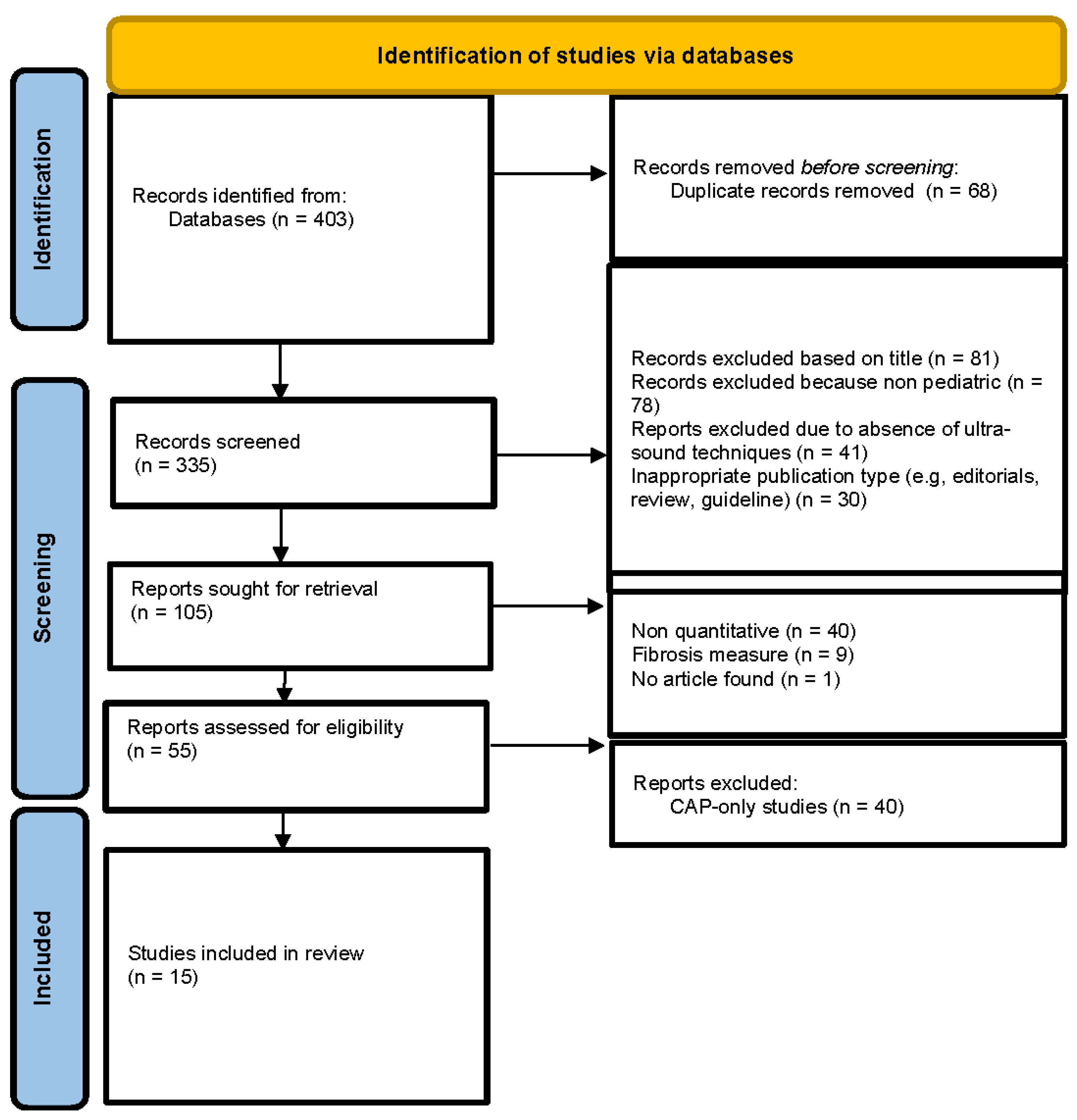
Figure 2.
Risk of bias assessment using the QUADAS-2 tool.
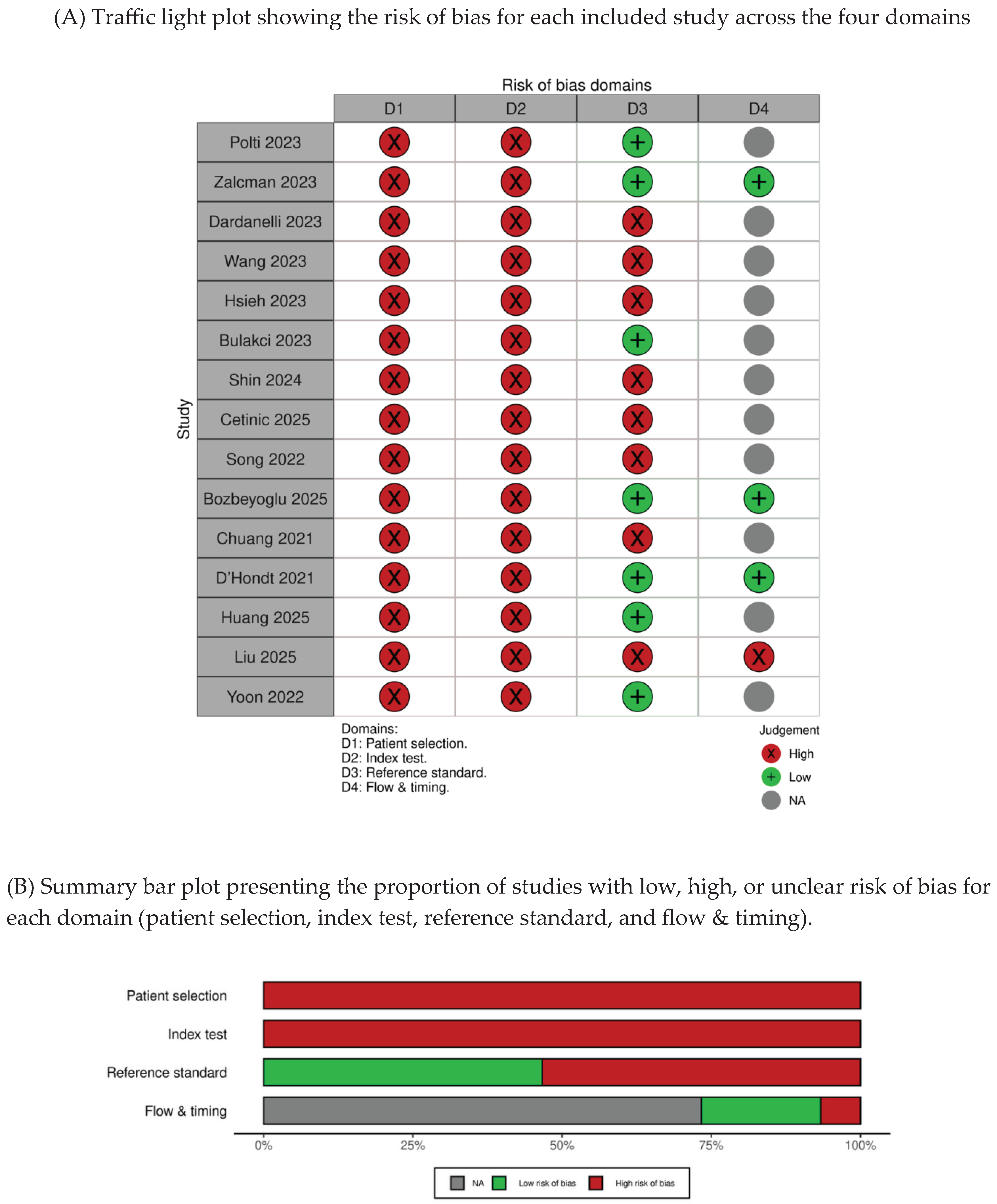

Table 1.
Population, intervention, comparator, outcome (PICO) framework.
| PICO | Detail |
| Population | Children and adolescents (0–18 years) with suspected or confirmed MASLD. |
| Intervention | Quantitative ultrasound-based techniques for hepatic steatosis assessment |
| Comparator | Reference standards such as MRI-PDFF, liver biopsy, elastography, or biochemical biomarkers. |
| Outcome | Primary outcome: Outcome 1 : Detection of hepatic steatosis by ultrasound measurement of sensitivity, specificity and correlation with reference standards. Outcome 2 : Quantification of hepatic steatosis using quantitative parameters (e.g., hepatic/renal ratio, attenuation in dB/cm) and correlation with percentage of hepatic fat. Outcome 3 : Assessment of MASLD severity performance of US to differentiate disease stages and concordance with clinical, biological scores, or histological findings. |
Table 2.
Inclusion & Exclusion criteria Applied for study selection in this scoping review, focusing on pediatric population and quantitative ultrasound-based methods for hepatic steatosis assessment.
Table 2.
Inclusion & Exclusion criteria Applied for study selection in this scoping review, focusing on pediatric population and quantitative ultrasound-based methods for hepatic steatosis assessment.
| Inclusion criteria | • Studies including pediatric populations (<18 years). Studies with mixed-age populations were included if the cohort was predominantly pediatric and participants did not exceed 21 years of age. • Non-invasive ultrasound-based imaging modalities specifically developed for quantitative assessment of hepatic steatosis were included. These include, but are not limited to : UDFF, ATI and QUS. • Studies specifically addressing MASLD, regardless of whether the aim is to quantify the disease, screen for it, or both. • Studies must report a quantitative measurement of hepatic steatosis, performed using ultrasound-based methods (e.g., dB/cm/MHz, percentage fat fraction, attenuation coefficient). • No language restrictions were applied during the search ; however, only studies published in English were included after screening. • Studies published between January 2019 and October 2025. |
| Exclusion criteria | • Studies predominantly including adult populations or mixed cohorts without separate pediatric analysis. • Studies that do not employ an ultrasound-based imaging modality. • Studies that do not focus on MASLD. • Studies not reporting any quantitative ultrasound-based measurement of hepatic steatosis (e.g., those focusing exclusively on fibrosis or using qualitative ultrasound only). • Studies exclusively evaluating controlled attenuation parameter (CAP) without reporting additional non-CAP quantitative ultrasound measurements. • Descriptive articles, literature reviews, guideline documents, or protocols. • Editorials, letters to the editor, and comments without original clinical or technical data. |
Table 3.
Characteristics of the included studies evaluating non-Cap quantitative ultrasound techniques.
Table 3.
Characteristics of the included studies evaluating non-Cap quantitative ultrasound techniques.
| REF | AUTHOR (YEAR, COUNTRY) | STUDY DESIGN | SAMPLE (N, AGE) | MODALITY | MANUFACTURER | COMPARATOR | KEY FINDINGS |
| 26 | Liu et al. (2025, China) | Cross-sectional study | n=268 (6-12y) |
UDFF | Siemens Acuson Sequoia | Convensional US B-mode | Cutoff: NR; Performance: UDFF values increased with steatosis grade, demonstrating strong quantitative discrimination across disease severity. |
| 10 | Zalcman et al. (2023, USA) | Prospective study | n=46 (7-18y) |
UDFF | Siemens Acuson Sequoia | MRI-PDFF | Cutoff: UDFF ≥6%; Performance: strong correlation with MRI-PDFF for quantitative assessment of hepatic steatosis. |
| 25 | Huang et al. (2025, China) |
Prospective study | n=32 (10-14y) |
UDFF |
Siemens Acuson Sequoia |
MRI-PDFF | Cutoff: ≥S1: 9.5%; ≥S2: 10.5%; ≥S3: 12.8%; Performance: UDFF demonstrated strong agreement with MRI-PDFF and good discrimination across steatosis grades. |
| 24 | D’Hondt et al. (2021, USA) |
Prospective study |
n=48 (7-17y) |
QUS (ATT, HRI, Nakagami) | Philips EPIQ 7G | MRI-PDFF | Cutoff: ≥0.54 dB/cm/MHz; Performance: attenuation coefficient correlated with MRI-PDFF and demonstrated good diagnostic performance for detecting hepatic steatosis |
| 27 | Yoon et al. (2022, South Korea) |
Retrospective study |
n=118 (0-18y) |
UGAP |
GE LOGIQ E10 |
MRI-PDFF (primary); CAP and transient elastography | Cutoff: ≥0.699 dB/cm/MHz; Performance: UGAP demonstrated high diagnostic accuracy for detecting hepatic steatosis with strong agreement with MRI-PDFF. |
| 21 | Song K et al. (2022, South Korea) | Retrospective study | n = 111 (6–18 y) |
ATI | Canon Aplio i800 | Ultrasound steatosis grading (primary); MRI-PDFF (secondary) | Cutoff: ≥0.59 dB/cm/MHz (AUC 0.853) for detecting steatosis; ≥0.69 dB/cm/MHz (AUC up to 0.91) for grading severity Performance: ATI values increased with steatosis severity and demonstrated good diagnostic performance for both detection and grading of hepatic steatosis |
| 19 | Shin et al. (2024, South Korea) | Technical feasibility study | n=83 (6-17y) |
ATI and UGAP | ATI: Canon Aplio i800 UGAP: GE LOGIQ E10 |
Ultrasound steatosis grading (primary); MRI-PDFF (secondary) | Cutoff: NR; Performance: ATI and UGAP were feasible in pediatric populations, with measurement variability influenced by respiratory motion. |
| 15 | Dardanelli et al. (2023, Argentina) | Prospective study | n=174 (6-18y) |
ATI | Canon Aplio a | Conventional US B-mode | Cutoff: NR; Performance: ATI demonstrated good reproducibility and feasibility for non-invasive assessment of hepatic steatosis in children. |
| 18 | Bulakci et al. (2023, Turkey) | Prospective study | n=140 (9-18y) |
ATI | Canon Aplio i800 | MRI-PDFF | Cutoff: ATI ≥0.65 dB/cm/MHz; Performance: high diagnostic accuracy for hepatic steatosis with good correlation with MRI-PDFF. |
| 22 | Bozbeyoglu et al. (2025, Turkey) | Prospective study | n=51 (5-17y) |
QUS (TAI, TSI, ezHRI) | Samsung RS85 Prestige | MRI-PDFF | Cutoff: TAI ≥0.67 dB/cm/MHz; TSI ≥88.03; Performance: quantitative ultrasound parameters showed good diagnostic performance for detecting hepatic steatosis compared with MRI-PDFF. |
| 20 | Cetinic et al. (2025, Sweden) | Prospective study |
n=56 (9-18y) |
ATI | Canon Aplio i800 |
NR | Cutoff: ATI ≥0.58 dB/cm/MHz; SWE ≥7.2 kPa; SWD ≥11.9 (m/s)/kHz; Performance: multiparametric ultrasound biomarkers demonstrated feasibility for assessing hepatic steatosis and liver stiffness in pediatric obesity. |
| 16 | Wang et al. (2023, China) | Prospective study | n=62 (5–18y) |
ATT | Hitachi Arietta 850 | Liver enzymes and clinical assessment | Cutoff: NR; Performance: attenuation and shear wave measurements showed clinical utility for assessing hepatic involvement with correlation to biochemical markers. |
| 8 | Polti et al. (2023, Italy) | Prospective study | n=36 (< 18 y) |
QUS (TAI, TSI, ezHRI) | Samsung RS85 Prestige | MRI-PDFF | Cutoff: ezHRI ≥1.215, TAI ≥0.625 dB/cm/MHz, TSI ≥91.95. Performance: All three parameters showed good correlation with MRI-PDFF and high diagnostic accuracy for detecting pediatric steatosis. |
| 23 | Chuang et al. (2021, Taiwan) | Prospective study | n=68 (3-18y) |
QUS (Nakagami/ envelope statistics) | Terason Model 3000 | Hepatitic steatosis index (HSI) | Cutoff: ≥G1: 0.69; ≥G2: 0.71; ≥G3: 0.73; Performance: Nakagami imaging values increased with steatosis severity, showing good discrimination across steatosis grades. |
| 17 | Hsieeh et al. (2023, Taiwan) | Prospective study | n=236 (3-18y) |
QUS (Nakagami/ envelope statistics) | Terason Model 3000 | Hepatitic steatosis index (HSI) | Cutoff: Nakagami ≥0.78; Performance: Nakagami, entropy and HK parameters showed good diagnostic performance for detecting hepatic steatosis with correlation to HSI. |
Table 4.
Risk of bias.
| Study (Author, Year, Ref) | Modality | Patient Selection | Index Test | Reference Standard (MRI-PDFF) | Flow & Timing | Overall Risk of Bias |
| Polti et al. 2023, (8) | QUS | High | High | Low | Unclear | High |
| Zalcman et al. 2023 (10) | UDFF | High | High | Low | Low (timing between index test and MRI not explicitly detailed) | Moderate |
| Dardanelli et al. 2023(15) | ATI | High | High | High (B-mode ultrasound used as an imperfect and non-validated reference standard for steatosis assessment) | Unclear | High |
| Wang et al. 2023 (16) | ATT + SWM | High (highly selected population with Wilson’s disease, not representative of MASLD population) | High | High (use of biochemical markers instead of validated imaging or histological reference standard) | Unclear | High |
| Hsieh et al. 2023 (17) | QUS envelope statistics | High | High | Moderate (combination of transient elastography and biochemical markers rather than a validated imaging reference standard) | Unclear | High |
| Bulakci et al. 2023 (18) | ATI | High | High | Low | Unclear | High |
| Shin et al. 2024 (19) | ATI / UGAP | High | High (measurement variability influenced by respiratory motion and lack of standardized acquisition conditions) | Moderate (The reference standard relied mainly on conventional ultrasound assessment, with MRI-PDFF available only in a limited subset, leading to a high risk of bias). | Unclear | Moderate |
| Cetinic et al. 2025 (20) | Multiparametric QUS | High | High | High (absence of a validated reference standard such as MRI-PDFF or liver biopsy) | Unclear | High |
| Song et al. 2022 (21) | ATI | High | High | Moderate (MRI-PDFF used as secondary reference standard rather than primary comparator) | Unclear (MRI was performed within one month of ultrasound in a subset of patients; however, the interval was not consistently reported for all participants) | High |
| Bozbeyoglu et al. 2025 (22) | QUS (TAI / TSI / ezHRI) | High | High | Low | Low (Ultrasound and MRI-PDFF were performed on the same day for all participants) | High |
| Chuang et al. 2021 (23) | Backscatter analysis | High | High | High (Hepatic steatosis was assessed using the hepatic steatosis index (HSI), a non-imaging and non-histological surrogate marker, which may not accurately reflect true liver fat content) | Unclear | High |
| D’Hondt et al. 2021 (24) | Attenuation coefficient | High | High | Low | Low (Ultrasound and MRI-PDFF were performed on the same day for all participants) | Moderate |
| Huang et al. 2025 (25) | UDFF | High | High | Low | Unclear (MRI was performed within one week of ultrasound; however, patient flow was not fully detailed.) | High |
| Liu et al. 2025 (26) | UDFF | High | High | High (Steatosis was assessed using conventional ultrasound without a validated reference standard such as MRI-PDFF or histology.) | High (Index test and reference standard were derived from the same ultrasound examination) | High |
| Yoon et al. 2022 (27) | UGAP | High | High | Low | Unclear | High |
Patient Selection: Most studies included selected pediatric cohorts with suspected steatosis, limiting generalizability → high risk. Index Test: Quantitative ultrasound techniques were often non-standardized with limited external validation → high risk. Reference Standard: MRI-PDFF was used as reference in most studies → low risk. Flow and Timing: Timing between ultrasound and reference standard was frequently not clearly reported → unclear risk.
Disclaimer/Publisher’s Note: The statements, opinions and data contained in all publications are solely those of the individual author(s) and contributor(s) and not of MDPI and/or the editor(s). MDPI and/or the editor(s) disclaim responsibility for any injury to people or property resulting from any ideas, methods, instructions or products referred to in the content. |
© 2026 by the authors. Licensee MDPI, Basel, Switzerland. This article is an open access article distributed under the terms and conditions of the Creative Commons Attribution (CC BY) license.
Copyright: This open access article is published under a Creative Commons CC BY 4.0 license, which permit the free download, distribution, and reuse, provided that the author and preprint are cited in any reuse.