

Submitted:
02 September 2026
Posted:
03 September 2026
You are already at the latest version
Abstract
Aim: To evaluate the effectiveness of functional device treatment using both 3D-printed and conventional Twin Block appliances on soft tissue changes during growth modification in Class II malocclusion. Background: Enhancing facial aesthetics is the main target of orthodontic treatment, as appearance greatly influences a person’s social confidence and self-esteem. Skeletal Class II malocclusion is quite common, and the pubertal growth spurt is the perfect time for functional treatment to promote mandibular advancement. The Twin-Block, one of the most widely used functional appliances, is a popular choice for achieving this goal. Patients and methods: Twenty patients divided into two groups with mixed or early permanent dentition were enrolled. Lateral cephalometric radiographs were taken at the start (T0; before treatment) and end (T1; at the end of the active phase) using the same machine. Those of each patient treated with a conventional Twin Block appliance for class II malocclusion were compared to those of patients treated with a 3D-printed Twin Block appliance for a 6-month follow-up period. Both groups were evaluated for soft tissue changes using various angular and linear geometric measurements, and differences between pre- and post-treatment were analyzed with a paired t-test. Results: The cephalometric analysis showed that the Twin Block appliance encouraged mandibular growth. Patients using it experienced a significant (p < 0.001) improvement in mandibular length and lower facial height in both groups, with no significant differences between the two groups. All angular and linear measurements also changed notably from before to after treatment in each group. Similarly, there was improvement in the facial profile; thus, mean soft tissue measurements improved significantly over the treatment period, but there were no significant differences between the two groups. Conclusion: The results showed that functional treatment for class II malocclusion, whether using a traditional or 3D-printed device, shows promising effects. The most noticeable soft tissue changes appeared in the lower facial region, particularly the lower lip, soft tissue pogonion, and soft tissue gnathion, due to forward movement. Further research with larger samples, 3D imaging, and long-term follow-ups could offer a clearer understanding of the impact of functional appliances.
Keywords:
class II malocclusion
; functional treatment
; growth modification
; twin block
; lower lip
; soft tissue changes
Introduction
One of the major concerns for orthodontic patients with skeletal malocclusion is improving their facial profile. Skeletal Class II malocclusions are a common orthodontic problem, affecting around a third of the population and often affecting facial aesthetics. They can result from maxillary prognathism, mandibular retrognathism, or a combination of both [1].
While there are various treatment options depending on the case, the Twin Block (TB) is the most popular functional appliance for treating patients with skeletal Class II malocclusion caused by mandibular retrognathia during growth and development. Developed by William J. Clark [2], it stimulates and enhances mandibular growth by moving the mandible forward using two separate removable acrylic appliances for the maxilla and mandible, each with inclined planes. As the desire for facial attractiveness grows, soft tissue changes that improve the facial profile become increasingly important. After TB therapy for mandibular retrognathia, noticeable soft tissue adaptation occurs in the lower facial area, and previous studies have shown that the TB appliance enhances the facial profile aesthetically [3].
Most studies agree on the skeletal and dentoalveolar effects of functional appliances, though opinions vary on their influence on facial profile [4]. Previous research on the Twin Block appliance, based on lateral cephalograms, has shown reduced maxillomandibular discrepancy and overjet. However, some studies note that not all patients experience an improved facial profile after Class II treatment. Soft tissue changes have mainly been examined with two-dimensional (2D) lateral cephalograms. This retrospective study aims to evaluate a 2D method for assessing the soft-tissue change following conventional Twin Block versus 3D-printed Twin Block functional appliance therapy.
Patients and Methods
Study setting & design:
This comparative, prospective clinical and radiographic study was conducted on patients from the Orthodontics Department’s outpatient clinic, Faculty of Dental Medicine, Boys, Assiut, Al-Azhar University
It used cephalometric and dental cast analyses to evaluate post-treatment soft-tissue changes after the active phase of functional therapy. Twenty patients were divided into two groups of ten: Group I (7 males and 3 females) received treatment with a conventional twin block appliance, while Group II (4 males and 6 females) were treated with a 3D-printed twin block appliance.
Ethical considerations: The research protocol was approved by the ethics committee of the Faculty of Dental Medicine at Al-Azhar University (Assiut Branch), and all enrolled patients received a written consent form. (Number AUARC20240003-7).
ClinicalTrials.gov Identifier: NCT07795138
Randomization: A restricted randomization strategy was utilized to ensure the equitable allocation of patients to the therapy group. Patients were randomly assigned using computer randomization (http://www.randomlists.com) into two groups of ten patients each: group one (seven males and three females) and group two (four males and six females).
Blinding: Participants in each group were blinded, although the primary investigator who carried out the intervention could not be. However, the supervisors who reviewed the pre- and post-cephalometric images for both groups, as well as the statistician, were unaware of which group each image was from.
Inclusion criteria:
- Young, growing age range from 9 to 16 years.
- Class II malocclusion.
- Increased overjet.
Exclusion criteria:
- Severe proclination of anterior teeth.
- Severe crowding of anterior teeth.
- Systemic disease that affects bone and growth.
Sample size calculation:
Using the G*Power statistical analysis program (version 3.1.9.4) for sample size determination, a total sample size of n8 per group was sufficient to detect a large effect size (f) = 1.5503 between two groups, with an actual power (1-β error) of 0.8 (80%) and a significance level (α error) of 0.05 (5%) for a two-tailed hypothesis test. To account for patient dropout, the sample size was increased by 20%, resulting in n=20 (10 per group).
Diagnostic records:
Include orthodontic study casts. The set includes frontal, right-side, and left-side views in centric occlusion, along with upper and lower occlusal views. It also features extraoral and intraoral photographs, with the extraoral shots showing a frontal view with lips at rest, a frontal view while smiling, and a profile view. Radiographic images include a digital panoramic radiograph, a digital lateral cephalometric radiograph, and intraoral scanning.
Clinical procedures:
Group 1 (conventional twin block):
1. Patient preparation: All patients underwent dental prophylaxis procedures, including scaling, polishing, and periodontal treatment with a chlorhexidine-containing mouthwash.
2. Conventional twin block appliance fabrication: The upper plate is placed first, ensuring complete coverage of the teeth and palate, secure clasp retention, and no pressure on surrounding tissues. Next, the lower plate is fitted, checking lingual coverage, clasp engagement, and proper occlusal alignment with the upper plate. The patient is instructed to bite into the prescribed therapeutic position, usually involving mandibular advancement, while evaluating the interlocking of the inclined bite blocks. Any premature contacts or pressure points are identified using articulating paper, and minor adjustments are made to relieve soft tissue pressure or high occlusal spots.
A written home care instruction form was provided, and all patients in the study participated in a special oral hygiene program for one month to standardize pretreatment oral care as much as possible before expansion. The same orthodontist carried out all treatments.
3. Patient instructions included proper insertion and removal of the appliance, cleaning routines, and a typical wear schedule of 12 hours per day. Advice on managing initial discomfort and easing soreness was also given to encourage compliance.
4. Post-treatment lateral cephalometric records were collected after six months.
Group 2 (3D-printed twin block):
1) Patient preparation: All patients underwent dental prophylaxis, including scaling, polishing, and periodontal treatment, with a chlorhexidine mouthwash used for each case.
2) The 3-D Twin Block appliance fabrication: First, the upper plate is placed to fully cover the teeth and palate, ensuring a secure fit with digital retention features and avoiding any pressure on soft tissue. Next, the lower plate is inserted, checking for proper lingual coverage and alignment with the upper plate. The patient then bites in the therapeutic mandibular position, usually with the jaw moved slightly forward, while the fit is evaluated for interlocking bite blocks, pressure points, or premature contacts, making adjustments as needed to ease soft tissue pressure and fix bite alignment issues.
3) Patient instructions:
Patients are shown how to properly insert and remove the appliance, follow cleaning routines, and stick to a wear schedule of about 12 hours a day. Tips for handling initial discomfort and easing soreness are given to help ensure they follow the plan.
4) Post-treatment lateral cephalometric records: Records were collected after 6 months.
Cephalometric analysis:
Lateral cephalometric radiographs were taken for each patient before removable functional therapy (T1) and right after mandibular protraction (T2). All images were captured using the same X-ray equipment under identical conditions, with a consistent 10% magnification factor. Each radiograph was traced, digitized, and analyzed by a single examiner using AudaxCeph orthodontic software suite version 6 (dHal Software, Athens, Greece) after proper adjustments. All examinations were performed by one calibrated examiner, with calibration based on repeated measurements of five cephalograms from participants not involved in the study.
- -
- Soft tissue measurements were recorded.
Statical analysis
The data were entered into the computer and analyzed using IBM SPSS Statistics for Windows, Version 22.0 (IBM Corp., 2013, Armonk, NY). Qualitative data were presented as numbers and percentages, while quantitative data were summarized using the mean and standard deviation for parametric data after testing for normality with the Shapiro–Wilk test. The significance of the results was determined at the 0.05 level. Independent-samples t tests were used to compare different groups, and paired-samples t tests were used for comparisons within groups.
Group (A) (Control Group)
Results: A total of 20 patients were assessed for eligibility to participate in the study and were divided into two groups of ten: Group I (7 males and 3 females) received treatment with a conventional twin block appliance, while Group II (4 males and 6 females) were treated with a 3D-printed twin block appliance, with a mean age of 13.71 with SD(1.66) in group I, and the means 13.72 with SD(1.88) in group II. Mean values and standard deviations of soft tissue measurements and comparisons for T0 and T1 are given in Table [1]. Independent-samples T-tests were conducted to assess significant differences between the study groups before treatment. The p-values for all variables were well above 0.05, showing there were no statistically significant differences between the groups at the 95% confidence level. This means both groups were essentially equivalent before treatment in terms of angular and linear values.
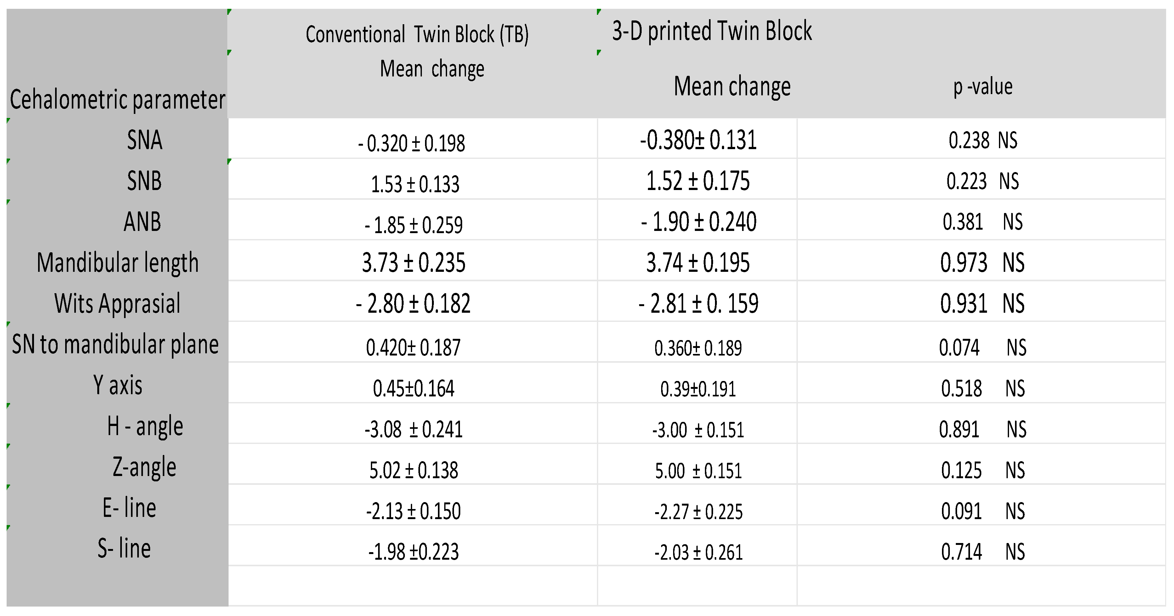
In evaluating the skeletal effect on the maxilla, this study found that the SNA mean value for group (1) was 82.54 before treatment and decreased to 82.22 after, showing a statistically significant change. For group (2), the mean dropped from 82.31 to 81.93, also statistically significant. The mean change for the conventional twin block (-0.320) was slightly less than for the 3D-printed twin block (-0.380), but the difference was not significant. For the mandible, the SNB mean value for group (1) increased from 76.69 before treatment to 78.22 after, without statistical significance. Group (2) went from 76.45 to 77.97, also not significant. The mean change for the conventional twin block (1.53) was slightly higher than that of the 3D-printed version (1.52), but the difference was not significant. Comparing skeletal differences between the maxilla and mandible, the ANB mean value for group (1) fell from 5.85 to 4.00, a significant change, while group (2) went from 5.86 to 3.96, also significant. The mean change for the conventional twin block (-1.85) was slightly less than that of the 3D-printed twin block (-1.90), but the difference was not significant.
In evaluating the skeletal effect, this study recorded the mean SN to mandibular plane angle for group (1) as 33.68 before treatment, which changed to 34.04 after treatment, showing a statistically significant difference. For group (2), the mean was 33.24 before treatment and 33.66 after, also a statistically significant change. The mean change for the conventional twin block (0.420) was higher than that for the 3D-printed twin block (0.360), though this difference was not statistically significant. While, In evaluating the Y-axis mean values for group (1) as 65.65 before treatment and slightly increased to 66.10 after treatment, showing a statistically significant difference. For group (2), the mean values were 65.50 before treatment and 65.89 after, also statistically significant. The mean change for the conventional twin block (0.450) was higher than that of the 3D-printed twin block (0.390), though this difference was not statistically significant.
In evaluating the sagittal relationship between the maxilla and mandible, this study recorded Wits Appraisal mean values of 5.24 for group (1) before treatment, which decreased to 2.44 afterward, a statistically significant change. For group (2), the mean values were 4.82 before treatment and 2.01 after, also a statistically significant difference. The mean change for the conventional twin block (-2.80) was slightly less than that of the 3D-printed twin block (-2.81), but this difference was not statistically significant. Moreover, the recorded mean value of mandibular body length for Group (1) was 109.43 mm before treatment, increasing to 113.16 mm after treatment, a difference that was statistically significant. For Group (2), the mean was 109.78 mm before treatment and increased to 113.52 mm after treatment, also a statistically significant change. The mean change for the conventional twin block (2.07 mm) was slightly higher than that for the 3D-printed twin block (2.06 mm), but this difference was not statistically significant.
Soft tissue effects showed a reduction in H angle for group (1) from 18.50 to 15.41, a significant change. Group (2) decreased from 18.11 to 15.11, also significant. The mean for the conventional twin block (-3.08) was greater than that for the 3D-printed version. On the other hand, assessing the soft tissue effects, the study found that the Z angle increased for Group 1 from 68.10 before treatment to 73.12 afterward, a statistically significant change. Group 2 showed a similar increase from 68.10 to 73.10, which was also significant. The average change for the conventional twin block (5.02) was just slightly higher than for the 3D-printed twin block (5.00), though the difference wasn’t significant.
The study also reported a drop in linear measurements (S line mm) for Group 1, from 3.11 mm before treatment to 1.12 mm after treatment, a significant change. For Group 2, values declined from 3.55 mm to 1.51 mm, again showing significance. The mean change for the conventional twin block (-1.98) was a bit less than that for the 3D-printed twin block (-2.00), but the difference wasn’t significant.
Figure 1.
Lateral cephalometry representing soft tissue measurements used in this study.
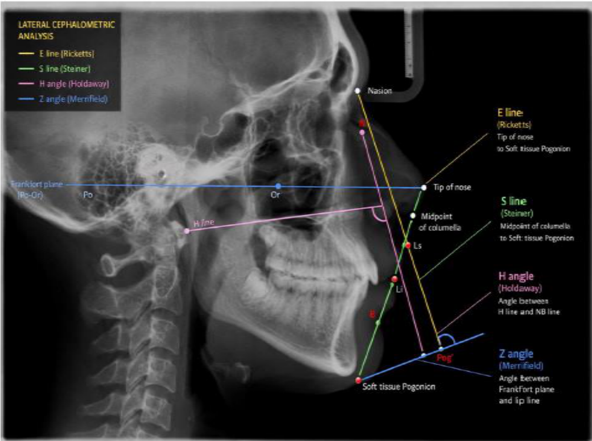
Figure 2.
Extraoral photographs of a case representing group (1) before treatment.
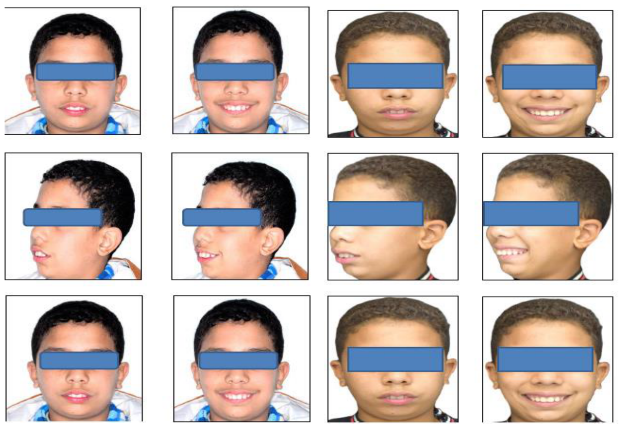
Figure 3.
Intraoral photographs after insertion of the appliance in the patient’s mouth in group (1) before treatment.
Figure 3.
Intraoral photographs after insertion of the appliance in the patient’s mouth in group (1) before treatment.
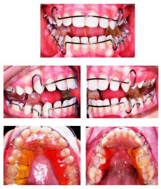
Figure 4.
Cephalometry of a case representing group (1) before treatment.
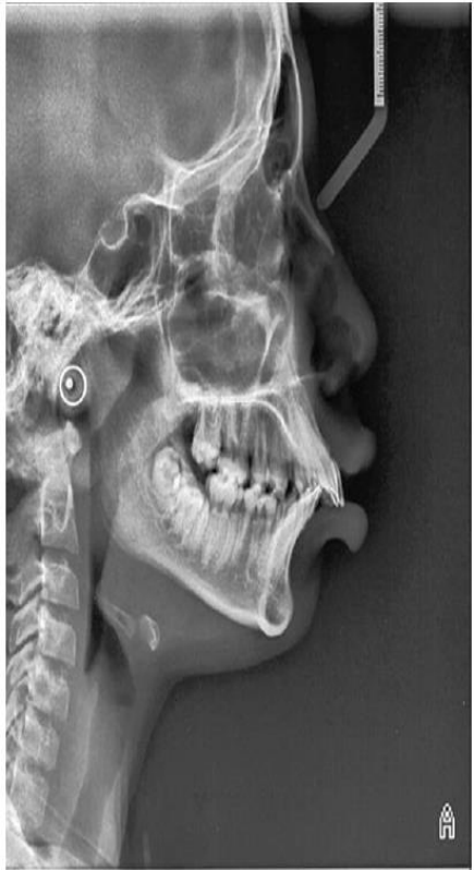
Figure 5.
Cephalometry of a case representing group (1) after 6 months.
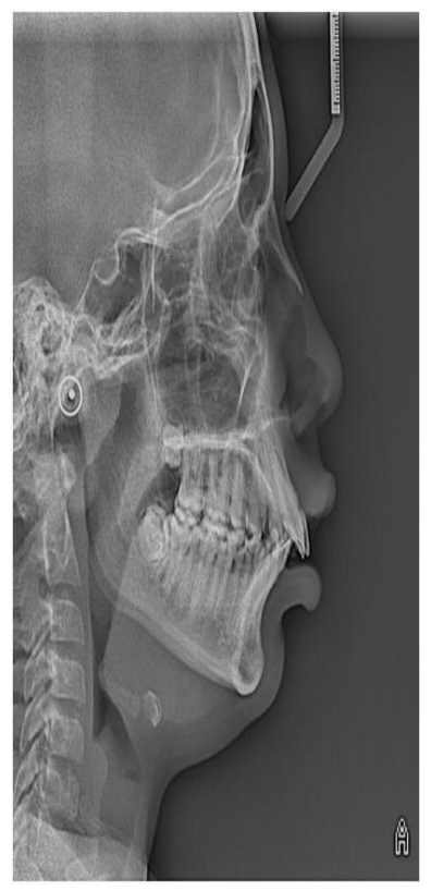
Figure 6.
Extraoral photographs of a case representing group (2) before treatment.
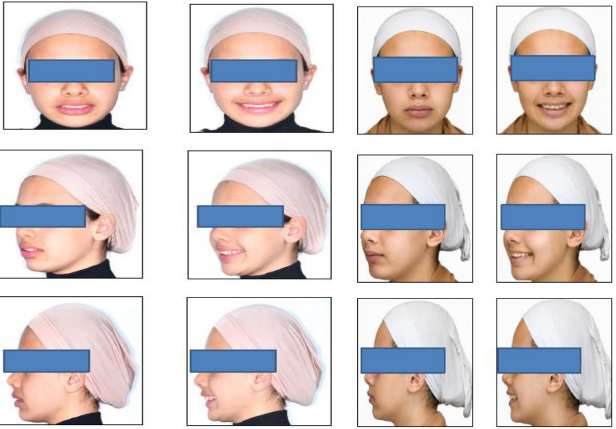
Figure 7.
Intraoral photographs after insertion of the appliance in the patient’s mouth in group (2) before treatment.
Figure 7.
Intraoral photographs after insertion of the appliance in the patient’s mouth in group (2) before treatment.
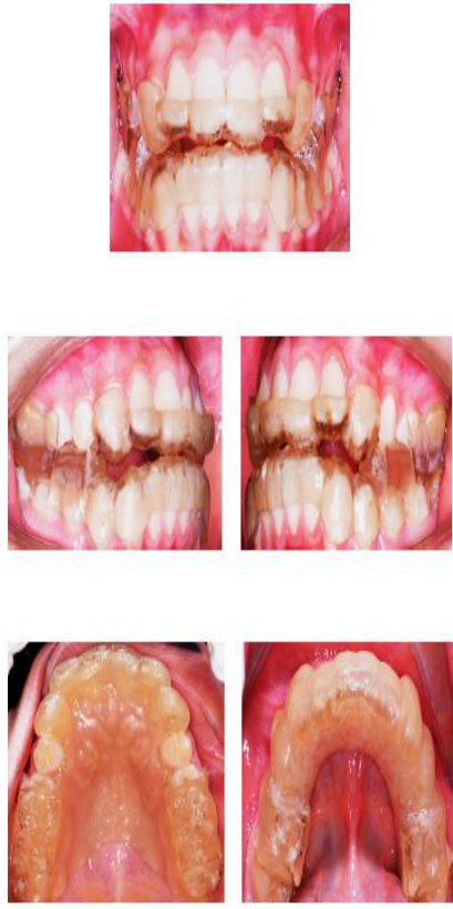
Figure 8.
Cephalometry of a case representing group (2) before treatment.
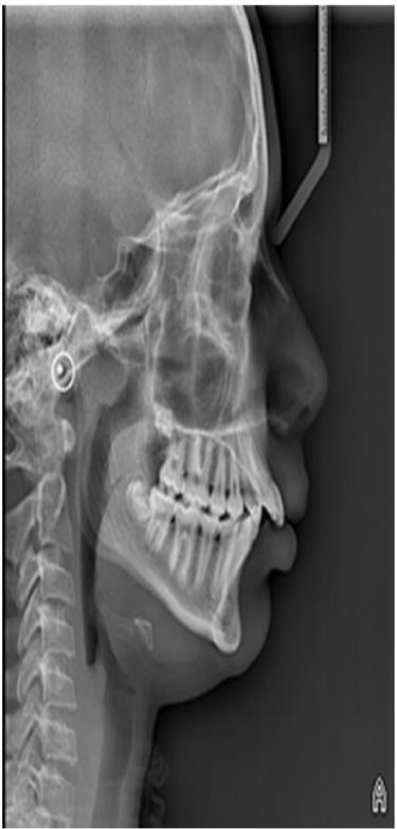
Figure 9.
Cephalometry of a case representing group (2) after 6 months.
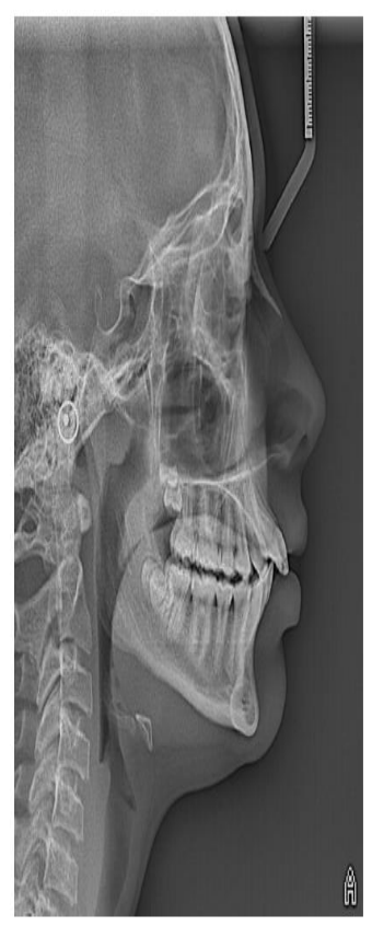
Figure 10.
Flow diagram following consort’s guidelines (2010) [5].
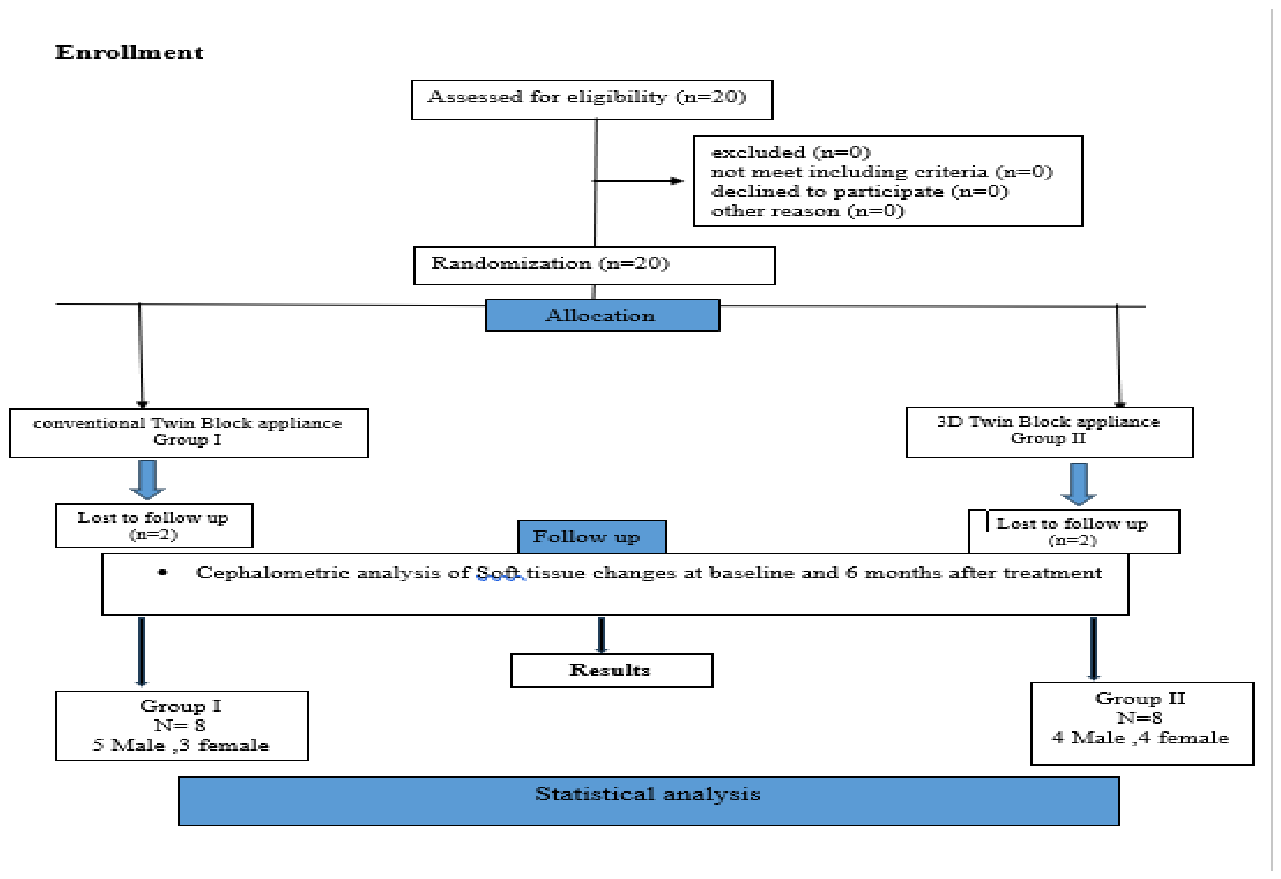

Table 1.
Comparison of Soft tissue measurement mean values between groups.
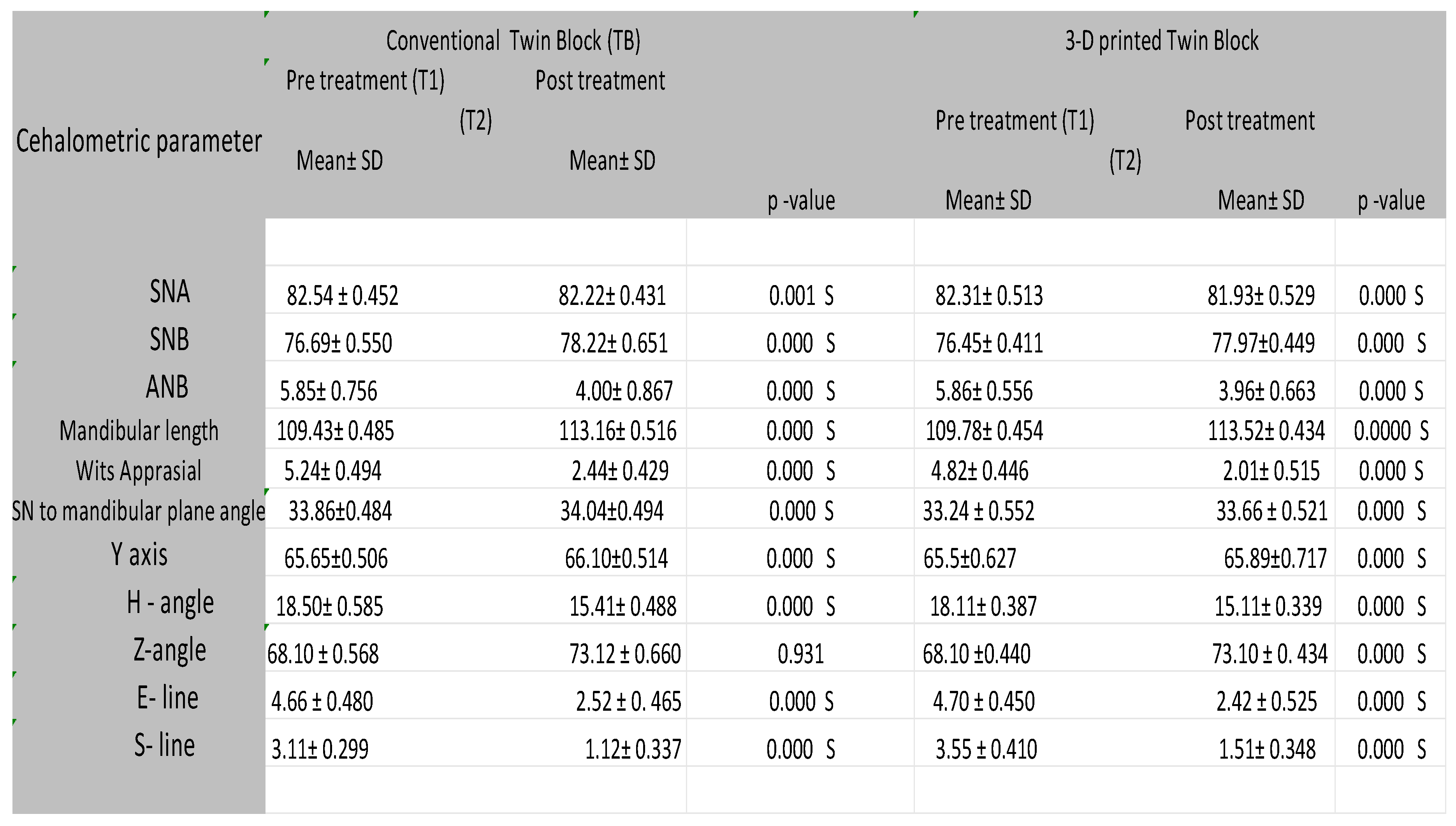
Table 2.
Comparison of Soft tissue measurement mean change values & P-value between groups.
Abbreviations: TB: Twin Block. ANB: The angle between 3 point landmarks, A point, N, and B point, determining the anteroposterior relation between the maxilla and the mandible relative to the cranium. Mandibular Body Length (mm): Distance between the Gonion and Gnathion points. Y-axis: The angle between the Sella–Gnathion (S-Gn) line and the Frankfort Horizontal (Po- Or) plane. SN to mandibular plane angle: The angle between the Sella–Nasion (SN) plane and the mandibular plane (Go–Me). Wits appraisal: The linear distance between the perpendicular projections of Point A (AO) and Point B (BO) on the functional occlusal plane. LFH: Distance between the anterior nasal spine and menton. (H-angle: The angle between the Holdaway line (tangent to the upper lip and soft tissue pogonion) and the soft tissue facial plane (N–Pog). Z-angle: The angle between the Frankfort Horizontal plane and the profile line connecting the most prominent lip to the soft tissue pogonion.) Angular measurement. [Linear measurments]: E- Line: The linear distance from the lower lip to the esthetic line drawn from the tip of the nose (Pronasale) to the soft tissue pogonion (Pog). S- line: The linear distance from the lower lip to the S-line, drawn from the midpoint of the columella of the nose to the soft tissue pogonion.
As for the E-line, the conventional twin block’s mean ± SD values fell from 4.66 ± 0.480 before treatment to 2.52 ± 0.465 after treatment, a highly significant change (P = .000). Likewise, the 3D-printed twin block decreased from 4.70 ± 0.450 to 2.42 ± 0.525, also highly significant (P = .000).
Discussion
The Twin Block appliance is a popular choice for treating skeletal Class II issues caused by a small lower jaw in growing children. Since a recessed jaw often leads to these orthodontic problems, using functional appliances to move the mandible forward can be an effective solution during the growth stage [6,7].
The study found a “twin block effect” on the maxilla in Group I, with only a slight drop in the SNA angle from before to after treatment, similar to Group II. It also showed a “twin block effect” on the mandible in Group I, with a clear increase in the SNB angle, much like in Group II. For maxillomandibular changes, both Twin Block groups showed better sagittal relationships, reflected in the ANB angle and Wits appraisal. This suggests sagittal skeletal discrepancy is mainly corrected by lengthening the mandible and limiting maxillary growth, as seen in the reduced ANB angle and lower Wits value. These results align with findings from Elfeky et al. [8], Cozza et al. [9], Koretsi et al. [10], Perinetti et al. [11], Hirji et al. [12], D’Antò et al. [13], and Giuntini al. [14], who reported similar soft tissue changes with Twin Block therapy.
The present study showed that changes in the lower lip and chin soft tissue landmarks, along with facial soft tissue volume after treatment, were more pronounced than those observed before TB treatment in both groups. In both cases, the Twin Block appliance applied a forward and downward force on the mandible, increasing both lower anterior and posterior facial height, which was consistent with previous studies [7,8,9,10,11,12,13].
Similarly, Quintão et al. [3] examined Class II patients treated with a Twin Block, reporting a decrease in the ANB angle and a notable increase in mandibular length. The upper lip moved backward, the soft tissue pogonion shifted forward, and the lower lip stayed in the same position relative to the E-line. Changes in the facial soft tissue profile showed a significant decrease in the facial convexity angle in both treatment groups (P = 0.000), leading to improved facial profiles. Most studies used the E-line to assess upper and lower lip positions. In the current study, both groups showed significant backward movement of the upper lip relative to the E-line (P = 0.009). However, comparisons between the groups showed no statistically significant difference (p > 0.05), possibly due to greater maxillary incisor retraction in both. These findings align with earlier reports of significant upper lip retraction with the Twin Block. The E-line might not be the most reliable way to assess lip position changes, since the pronasale and soft tissue pogonion points can shift due to growth or treatment. This could explain why many studies report no noticeable forward movement of the lower lip after Twin Block therapy [15].
Moreover, in both groups using the Twin Block appliance, there was a small, nonsignificant increase in the nasolabial angle (p > 0.05), likely due to maxillary incisor retraction causing the upper lip to shift, consistent with Khoja et al.’s findings [4]. The TB group actually showed a slight, nonsignificant decrease in this angle (p > 0.05), with no notable difference between groups (p > 0.05), possibly because TB tends to cause less upper incisor retraction. Other studies have also found no significant difference in nasolabial angle between the two Twin Block groups, which might be explained by thicker upper lips from reduced lip strain, lessening the effect of incisor retraction. On the other hand, the mentolabial angle increased significantly in both groups (P = 0.000), with no difference between them. The Twin Block group did show less maxillary lip retraction, but again, there was no significant difference (P > 0.05). These results were probably due to the lower jaw moving forward and the lower lip being freed from its previous position behind the upper front teeth. Quintão et al. [3] observed only minor facial changes in both groups over the 12 months, and given the short timeline of the present study, it seems the soft tissue changes occurred independently of natural growth. While Singh and Clark [16], using color-coded finite element analysis, noted less prominence in the labio-mental groove after TB treatment. On the other hand, Gulec and Goymen [17] disagree with the present study; they noted Class II patients with TB appliances and found they encouraged mandibular growth and lowered the ANB angle, but didn’t change soft tissue parameters like lip position to the E-line. While Baysal and Uysal [18] show agreement with the current study, they reported a rise in the SNB angle and more forward movement of the soft tissue pogonion and lower lip in TB patients.
Conclusions:
- There’s no major difference between the two study groups using Functional Twin Block appliances for treating Class II malocclusion in terms of soft tissue changes. The selection of either appliance should be based on clinical requirements, patient preference, treatment workflow, and cost considerations rather than treatment effectiveness alone
- Both groups showed a clear positive impact on facial soft tissue.
- These appliances help reduce lower face convexity, particularly around the lower lip, soft tissue pogonion, and soft tissue gnathion, by encouraging forward movement. This results in a more balanced lower facial profile and increased concavity in the lower lip area.
Recommendation: Future research with long-term follow-up should explore soft tissue changes using 3D CBCT imaging alongside Class II Twin Block treatment, to assess both the quantitative and qualitative aspects of these changes after functional therapy.
Limitation: Most of the data comes from systematic review papers and lacks randomized clinical trials, as there are only a limited number of trials in this area.
Informed Consent: Written consent was obtained from all participating patients.
Data Availability: Publicly available.
Ethics Statement
Ethical approval for the study was obtained from the ethical committee of the Faculty of Dental Medicine, Al-Azhar University, Assiut Branch. (Approval number: AUARC20240003-7).
ClinicalTrials.gov Identifier: NCT07795138.
Funding Statement: This research received no external funding.
Conflicts of Interest
The authors declare no conflict of interest.
References
- Yıldırım, Ersin; Karacay, Seniz; Tekin, Dilek. Three-Dimensional Evaluation of Soft Tissue Changes after Functional Therapy. Scanning 2021, 9928101. [Google Scholar] [CrossRef] [PubMed]
- Clark, W. J. The twin block traction technique. Eur. J. Orthod. 1982, 4(2), 129–138. [Google Scholar] [CrossRef] [PubMed]
- Quintão, C.; Brunharo, I. H. V. P.; Menezes, R. C.; Almeida, M. A. O. Soft tissue facial profile changes following functional appliance therapy. Eur. J. Orthod. 2006, 28(1), 35–41. [Google Scholar] [CrossRef] [PubMed]
- Khoja, A.; Fida, M.; Shaikh, A. Cephalometric evaluation of the effects of the twin block appliance in subjects with class II, division 1 malocclusion amongst different cervical vertebral maturation stages. Dent. Press J. Orthod. 2016, 21(3), 73–84. [Google Scholar] [CrossRef] [PubMed]
- Merkow, R.P.; Kaji, A.H.; Itani, K.M.F. The CONSORT Framework. JAMA Surg. 2021, 1;156(9), 877–8. [Google Scholar] [CrossRef] [PubMed]
- Alsilq, M.N.; Youssef, M. Soft tissue effects of aesthetic and conventional twin block appliances in the treatment of skeletal class II malocclusion: a randomized controlled trial. Sci. Rep. 2025, 15(1), 37627. [Google Scholar] [CrossRef] [PubMed]
- Salloum, E.; Millett, D.T.; Kelly, N.; McIntyre, G.T.; Cronin, M.S. Soft tissue changes: a comparison between changes caused by the construction bite and by successful treatment with a modified Twin-block appliance. Eur. J. Orthod. 2018, 40(5), 512–8. [Google Scholar] [CrossRef] [PubMed]
- Elfeky, H.Y.; Fayed, M.S.; Alhammadi, M.S.; Soliman, S.A.Z.; El Boghdadi, D.M. Three-dimensional skeletal, dentoalveolar and temporomandibular joint changes produced by Twin Block functional appliance References. J. Orofac. Orthop. Der Kieferorthopädie 2018, 79(4). [Google Scholar] [CrossRef] [PubMed]
- Cozza, P.; Baccetti, T.; Franchi, L.; De Toffol, L.; McNamara, J.A., Jr. Mandibular changes produced by functional appliances in Class II malocclusion: a systematic review. In American Journal of Orthodontics and Dentofacial Orthopedics; Thiruvenkatachari, B., Harrison, J.E., Worthington, H.V.D., Eds.; 2006; Volume 129, 5, pp. 599. e1–e12. 10. [Google Scholar]
- Koretsi, V.; Zymperdikas, V.F.; Papageorgiou, S.N.; Papadopoulos, M.A. Treatment effects of removable functional appliances in patients with Class II malocclusion: a systematic review and meta-analysis. Eur. J. Orthod. 2015, 37(4), 418–34. [Google Scholar] [CrossRef] [PubMed]
- Perinetti, G.; Primožič, J.; Franchi, L.; Contardo, L. Treatment effects of removable functional appliances in pre-pubertal and pubertal Class II patients: a systematic review and meta-analysis of controlled studies. PLoS ONE 2015, 10(10), e0141198. [Google Scholar] [PubMed]
- Hirji, S.N.; Qamruddin, I.; Mudassar, M.A.; Khurshid, Z.; Alam, M.K. Treatment of Class II Malocclusion With Removable Functional Appliances: A Narrative Review. Eur. J. General. Dent. 2021, 10(03), 170–5. [Google Scholar] [CrossRef]
- D'Antò, V.; Bucci, R.; Franchi, L.; Rongo, R.; Michelotti, A.; Martina, R. Class II functional orthopaedic treatment: a systematic review of systematic reviews. J. Oral Rehabil. 2015, 42(8), 624–42. [Google Scholar] [CrossRef] [PubMed]
- Giuntini, V.; Vangelisti, A.; Masucci, C.; Defraia, E.; McNamara, J.A., Jr.; Franchi, L. Treatment effects produced by the Twin-block appliance vs the Forsus Fatigue Resistant Device in growing Class II patients. Angle Orthod. 2015, 85(5), 784–9. [Google Scholar] [CrossRef] [PubMed]
- Yıldırım, E.; Karaçay, Ş.; Tekin, D. Three-Dimensional Evaluation of Soft Tissue Changes after Functional Therapy. Scanning 2021, 2021, 9928101. [Google Scholar] [CrossRef] [PubMed]
- Singh, G. D.; Clark, W. J. Soft tissue changes in patients with class II division 1 malocclusions treated using twin block appliances: finite-element scaling analysis. Eur. J. Orthod. 2003, vol. 25(no. 3), 225–230. [Google Scholar] [CrossRef] [PubMed]
- Gulec; Goymen, M. Treatment of class II malocclusion: a comparative study of the effects of twin-block and fatigue-resistant device. Niger. J. Clin. Pract. 2018, vol. 21(no. 12), 1557–1563. [Google Scholar] [CrossRef] [PubMed]
- Baysal, 18; Uysal, T. Soft tissue effects of twin block and Herbst appliances in patients with class II division 1 mandibular retrognathia. Eur. J. Orthod. 2013, vol. 35(no. 1), 71–81. [Google Scholar] [CrossRef] [PubMed]
Disclaimer/Publisher’s Note: The statements, opinions and data contained in all publications are solely those of the individual author(s) and contributor(s) and not of MDPI and/or the editor(s). MDPI and/or the editor(s) disclaim responsibility for any injury to people or property resulting from any ideas, methods, instructions or products referred to in the content. |
© 2026 by the authors. Licensee MDPI, Basel, Switzerland. This article is an open access article distributed under the terms and conditions of the Creative Commons Attribution (CC BY) license.
Copyright: This open access article is published under a Creative Commons CC BY 4.0 license, which permit the free download, distribution, and reuse, provided that the author and preprint are cited in any reuse.