
Submitted:
03 September 2026
Posted:
03 September 2026
You are already at the latest version
Abstract
Auditory evoked potentials (AEPs) comprise a family of electrophysiological responses that follow acoustic stimulation from the auditory nerve and brainstem to thalamocortical and association networks. Their interpretive value lies in the combination of millisecond temporal resolution and hierarchical sensitivity: early responses mainly index the fidelity and timing of ascending sensory conduction, whereas later responses increasingly depend on cortical context, attention, prediction, and behavioral relevance. This review develops a state-aware framework for interpreting AEPs during wakefulness, natural sleep, pharmacologic sedation, general anesthesia, and impaired consciousness. We first describe the ascending auditory pathway and relate approximate latency ranges to distributed neural generators. We then examine how attention and task engagement modify early cortical processing; how non-rapid eye movement and rapid eye movement sleep preserve some forms of acoustic analysis while reorganizing or suppressing others; and how anesthetic effects vary with response latency, drug concentration, and cortical hierarchy. The review also distinguishes transient event-related potentials from steady-state auditory evoked potentials (SSAEPs), and explains why phase-locked activity can be recovered by trial averaging whereas non-phase-locked induced activity requires single-trial time-frequency analysis. Separate sections address cross-species translation, click, chirp, oddball, steady-state, and naturalistic speech paradigms, as well as clinical applications in hearing assessment, intraoperative monitoring, anesthesia, and disorders of consciousness. The central conclusion is that an AEP is interpretable only when its generator, stimulus, analysis domain, recording conditions, and cognitive state are considered together.
Keywords:
auditory evoked potential
; auditory brainstem response
; mismatch negativity
; P300
; steady-state auditory evoked potential
; sleep
; sedation
; anesthesia
; consciousness
1. Introduction and Scope
Auditory evoked potentials are voltage fluctuations recorded at the scalp or directly from neural tissue after an acoustic event. The first reproducible far-field human brainstem responses demonstrated that very small, time-locked signals generated deep in the auditory pathway could be recovered noninvasively by repeated stimulation and averaging [1,2]. Soon afterward, systematic studies showed that the auditory response is not a single waveform but a succession of components extending from the first milliseconds after a click to late positive potentials several hundred milliseconds later [3]. This latency hierarchy remains useful, but latency alone is not a complete taxonomy: components also differ in neural generator, dependence on stimulus regularity, susceptibility to attention, and vulnerability to changes in arousal.
The distinction between predominantly stimulus-driven (exogenous) and context-dependent (endogenous) responses provides a second organizing axis [3,4]. Early waves are strongly constrained by stimulus timing and peripheral conduction, although longer-term experience and descending control can also influence subcortical encoding. Later cortical components depend increasingly on how the current event relates to preceding events. Selective attention increases cortical responses to attended sounds [5,6], rare or uncertain targets evoke the P300 family [7], and violations of an auditory regularity can produce mismatch negativity (MMN) even when the auditory stream is not the focus of voluntary attention [8]. These findings suggest that AEPs provide a bridge between clinical neurophysiology and cognitive neuroscience.
Cognitive state is therefore not a nuisance variable to be averaged away. Wakefulness, drowsiness, non-rapid eye movement (NREM) sleep, rapid eye movement (REM) sleep, sedation, and general anesthesia alter neuromodulatory tone, background oscillations, behavioral responsiveness, and effective communication between cortical regions. The same physical sound can consequently produce an intact auditory brainstem response (ABR), an attenuated middle-latency response (MLR), a residual deviance response, or no task-dependent P300. A preserved early component shows that sound activated the auditory nerve or brainstem level being tested; it does not establish intact conscious perception. Conversely, a late context-sensitive response can reveal processing not evident from behavior, especially in a patient who cannot communicate.
This review uses that hierarchical dissociation as its central framework. It asks four practical questions: (1) which generator populations contribute to each response; (2) which components are relatively preserved or strongly modulated across cognitive states; (3) which findings from animal AEP studies can be reasonably translated to humans; and (4) which stimulus and analysis choices are required to test a particular level of processing. The scope includes ABR, MLR, N1/P2, MMN, P300, frequency-following responses (FFRs), and SSAEPs. Otoacoustic emissions are faint sounds generated mainly by cochlear outer hair cells and recorded in the ear canal. They are discussed only as an independent measure of cochlear function, not as neural AEPs or as a source of AEP noise. Purely spontaneous EEG is included only when it provides a complementary measure of state.
The framework is probabilistic rather than absolute. Latency ranges overlap; scalp peaks combine activity from multiple sources; and pharmacologic effects depend on the agent, concentration, stimulus, montage, and physiologic conditions. Accordingly, terms such as "preserved," "attenuated," and "absent" describe tendencies observed in groups under defined acquisition conditions. They should not be treated as universal properties or used alone to classify an individual patient's state. This distinction is especially important when AEPs are used to infer awareness or prognosis.
2. Neurophysiological Foundations of AEPs
2.1. Ascending Auditory Pathways and Distributed Generators
Inner hair cells transduce basilar-membrane motion into neural activity, and auditory-nerve fibers encode the timing and frequency content of that activity. The ascending stream then passes through the cochlear nuclei, superior olivary complex, nuclei and fibers of the lateral lemniscus, inferior colliculus, medial geniculate body, and auditory cortex. Beginning in the brainstem, projections are substantially bilateral (Figure 1), so input from either ear reaches both sides of the central pathway. This organization supports binaural computations but prevents a simple one-wave-to-one-nucleus mapping. Human lesion studies nevertheless established a clinically useful relationship between ABR timing and lesion level: abnormalities in early peaks and interpeak intervals can help distinguish peripheral or lower-pontine dysfunction from more rostral conduction abnormalities [9].
Controlled experiments in cats clarified why that clinical map is informative but not one-to-one. In a three-part series, investigators used selective kainic-acid lesions of the cochlear nucleus or superior olivary complex, related the lesion sites to far-field waveform changes, and integrated the results with anatomical and physiological evidence [10,11,12]. In this cat nomenclature, P2, P3, P4, and P5 are early brainstem auditory peaks, not the human cortical P2 or P300. The earliest extrema arose from cells peripheral to the cochlear nucleus; cat P2 was associated chiefly with cochlear-nuclear populations; cat P3 reflected mixed cochlear-nuclear and superior-olivary activity; and cat P4-P5 reflected distinct parallel pathways involving cochlear-nuclear and bilateral superior-olivary populations. A scalp peak can include stationary currents in aligned cell populations, activity propagating along fiber tracts, and downstream effects of interrupting an earlier relay. Generator labels should therefore be treated as dominant associations rather than exclusive sources.
Temporal synchrony is as important as anatomy. A neural population contributes strongly to a scalp-recorded far-field response when many neurons activate within a narrow time window and their current dipoles have orientations that sum rather than cancel at the scalp. In click-ABR protocols, alternating click polarity can reduce the cochlear microphonic and electrical stimulus artifact because those artifacts reverse with stimulus polarity, whereas the principal neural peaks are more stable. Click polarity can still alter neural waveform details and must be reported. Conduction delay, desynchronization, hearing threshold, stimulus presentation level, temperature, and electrode montage can change peak latency or amplitude without implying destruction of a generator.
At the cortical level, organization is more complex than a single "auditory cortex." Human postmortem mapping distinguishes core, belt, and parabelt regions in and around Heschl's gyrus and the superior temporal plane [13]. Direct intracranial recordings show that the earliest cortical responses usually occur in posteromedial Heschl's gyrus, but later activity does not always proceed through a strictly serial core-to-belt-to-parabelt chain [14]. Instead, primary and non-primary fields exchange information through parallel and recurrent routes. These fields differ in response latency, spectral selectivity, and sensitivity to complex sounds, so a single scalp component can combine activity from several cortical stages.
Auditory cortical geometry also shapes the scalp signal. Much of Heschl's gyrus lies within the lateral sulcus; synchronous postsynaptic currents in tangentially oriented pyramidal populations can therefore produce frontocentral voltage maxima rather than maxima directly over the temporal bone. The measured N1, for example, is a composite of overlapping processes rather than a single generator. Direct recordings from human auditory cortex further place several MLR components along Heschl's gyrus, supporting a transition from subcortical conduction to early cortical processing within approximately the first 80 ms [15].
Descending and recurrent influences complete the circuit. In humans, selective attention changes early auditory-cortical responses within tens of milliseconds [16]. In behaving ferrets, task engagement rapidly reshapes spectrotemporal receptive fields in primary auditory cortex [17]. Corticothalamic and corticofugal pathways provide routes by which learning and expectation can also influence subcortical encoding. These examples do not imply that every early ABR peak changes with moment-to-moment attention; the magnitude and time scale of modulation depend on the response, task, and analysis. A useful working model is a predominantly feedforward early volley embedded in a recurrent system whose later components express progressively more context.
2.2. Components, Generators, and Time Scales
AEP components are commonly grouped by latency, but the boundaries are approximate and shift with age, stimulus presentation level, repetition rate, filtering, and nomenclature. The ABR occupies roughly the first 10 ms after stimulus onset in adults and is dominated by auditory-nerve and brainstem activity. MLR components such as Na, Pa, Nb, and Pb extend from approximately 10 to 80 ms and include thalamic, thalamocortical, and early cortical contributions. Long-latency auditory evoked potentials include P1, N1, and P2, typically between about 50 and 250 ms. Context-dependent components such as MMN, P3a, and P3b overlap these windows and are defined by experimental contrasts and cognitive function, not latency alone. Figure 1 (right panel) shows the approximate response windows, and Table 1 summarizes the terminology, generators, analyses, state sensitivity, and uses.
The click-evoked ABR reflects precise neural conduction timing. Peak amplitudes are small, so hundreds or thousands of repetitions are often required. Absolute latency is measured from acoustic stimulus arrival to a peak. An interpeak interval subtracts the latency of an earlier peak from that of a later peak in the same ear; this cancels delay shared by both peaks and emphasizes conduction between the corresponding pathway levels. Wave I is most closely associated with the distal auditory nerve, and wave V is the most robust later peak in routine human recordings. Waves II-IV may be less distinct, especially near threshold or in noisy conditions. The clinically useful inference comes from the pattern across peaks within each ear and from interaural comparisons within the same patient, not from a single textbook morphology.
MLRs bridge the brainstem and late cortical response. Intracranial recordings show that Pa and later middle-latency components have generators along the supratemporal plane [15]. Because these components depend more on thalamocortical and early cortical synchrony than the ABR, they are more sensitive to arousal and anesthetic concentration. Their measured waveform can also reflect overlap between successive neural responses at rapid presentation rates and contamination from myogenic activity, particularly postauricular muscle responses.
N1 and P2 are prominent cortical responses to sound onset, but the term "obligatory" does not mean state-invariant. The N1 family contains several overlapping processes with different scalp distributions and sensitivities to stimulus onset, repetition, attention, and arousal [4]. P2 is also shaped by stimulus probability, learning, and recording context. Changes in N1/P2 across states can therefore reflect changes in the relative contribution and overlap of several cortical processes, not the on-off state of one neuronal population.
MMN is usually estimated as the difference between responses to frequent standards and physically or relationally deviant sounds [8,19]. The subtraction tests sensitivity to violation of an auditory regularity, but unequal adaptation to standards and deviants can also contribute. Equiprobable or many-standards control sequences help separate regularity effects from adaptation. P3a is commonly associated with involuntary orienting toward novelty, whereas P3b is most robust when a participant detects or evaluates a task-relevant event. These distinctions matter in sleep, anesthesia, and disorders of consciousness because a local deviance response can persist when a later response to a global or task-defined rule is absent.
SSAEPs provide a complementary frequency-domain view. In this review, "steady-state auditory evoked potential" (SSAEP) is used consistently; the commonly used term "auditory steady-state response" (ASSR) denotes the same response class. Periodic acoustic stimulation produces neural activity at the modulation or repetition frequency. Responses may also occur at integer multiples of that frequency; these response harmonics reflect the nonsinusoidal shape of the neural waveform and should not be confused with acoustic harmonics in the carrier. SSAEP amplitude and phase are typically estimated in the frequency domain, although coherent time-domain averaging is also possible. The classic 40-Hz response was originally interpreted as constructive overlap of successive MLRs [20]. Rate-manipulation and deconvolution studies show that simple linear superposition is incomplete and that adaptation and rate-dependent circuit dynamics also contribute [21,22]. The response is sensitive to drowsiness, sleep, and anesthesia, and careful montage selection is needed to avoid postauricular muscle contamination [23]. Faster modulation rates are often used for objective audiometry because they weight subcortical generators more strongly and can be easier to detect during sleep; generator composition nevertheless varies continuously rather than switching at one frequency.
FFRs also track periodic stimulus structure, but they should not be collapsed conceptually with all SSAEPs. The human FFR can represent the stimulus fundamental frequency and individual spectral harmonics of complex sounds, including synthetic vowels [24]. Early work often interpreted the scalp FFR as brainstem-dominated, but human magnetoencephalography and intracortical recordings demonstrate cortical contributions, particularly at lower represented frequencies; the measured source balance depends on stimulus frequency and montage [25,26]. Reports should therefore specify the acoustic carrier, modulation or fundamental frequency, electrode configuration, and whether the measure is a sustained time-domain waveform or a frequency-domain estimate.
2.3. from Acoustic Event to Measured Waveform
An AEP is the product of the stimulus, auditory system, behavioral state, acquisition hardware, and analysis pipeline. Reports should specify whether stimulation is monotic (one ear only), diotic (the same signal to both ears), or dichotic (different signals to the two ears), as well as masking of the non-test ear when applicable. Click polarity, rise time, presentation level, interstimulus interval, and transducer delay also influence the input. Transducer delay is the interval between the electrical trigger recorded by the acquisition system and the arrival of sound at the ear canal. Electrode impedance, reference placement, amplifier bandwidth, digitization rate, and line-noise control influence the recording. Artifact rejection, filtering, baseline correction, averaging, peak measurement, and statistical thresholding influence the reported component. An apparent cross-state change can arise from the neural response, the background EEG signal, or a state-dependent interaction with the processing pipeline.
The distinction between near-field and far-field recording is also important. Intracranial contacts sample local or regional activity with high spatial specificity, whereas scalp electrodes record volume-conducted mixtures from multiple sources. Magnetoencephalography measures the magnetic counterpart of synchronized postsynaptic currents and is less distorted by skull conductivity, but it is preferentially sensitive to certain source orientations. Findings from EEG, magnetoencephalography, and intracranial recordings are therefore complementary rather than interchangeable.
Interpretive decisions associated with component detection should be separated from the cognitive claims made after detection. A statistically significant response at a prespecified latency or frequency establishes that the recording contains reproducible stimulus-related activity. Inferring hearing, attention, memory, or consciousness requires additional assumptions supported by the paradigm and comparison condition. Stronger cognitive claims require evidence that lower-level sensory responses were present, artifacts were excluded, and the relevant effect was reliably detectable in that individual.
3. Cognitive-State Modulation of AEPs
3.1. Wakefulness: Attention, Expectation, and Task Engagement
Wakefulness supplies the broadest range of top-down influences on auditory processing. In the classic selective-attention paradigm, identical tones elicited larger cortical responses when they were assigned to an attended rather than ignored stream [5]. Subsequent magnetoencephalography localized attentional modulation to auditory-cortical activity within approximately 20-50 ms in demanding dichotic-listening tasks [16]. These findings show that attentional selection can influence early cortical processing, although the latency and magnitude of the effect depend on stimulus rate, perceptual load, and the overlapping components being measured.
Attention does not simply amplify every sound. It changes the relative gain assigned to task-relevant features and streams. Human functional mapping has shown that sensory responses to acoustic features and attention-related modulations have partly distinct distributions across auditory cortex, with stronger attention effects in lateral higher-order regions than in medial core regions [27]. At the scalp, this distributed reweighting can alter N1/P2 amplitude, sustained processing negativity, induced oscillations, and late target responses. A participant who stops following instructions may therefore produce a physiologically valid but cognitively different dataset.
A second mechanism is expectation based on acoustic regularity. Repetition establishes a short-term model of the acoustic environment, and a deviant can elicit a difference response even when the stream is unattended [8]. When the deviant is also task relevant, additional orienting and decision processes contribute to P3a and P3b. MMN and P300 should not be treated as two points on a single amplitude scale: they rely on different contrasts, have different task requirements, and can dissociate across states.
Awake recordings are sometimes treated as an uncomplicated baseline, yet they are often the most behaviorally variable condition. Vigilance, fatigue, motivation, hearing effort, medication, and mind wandering can change late AEPs over minutes. Strong studies verify audibility, document task performance, balance trial numbers, and model within-session drift. When the scientific question concerns state change, repeated measures within the same participant reduce interindividual variance but create additional concerns about learning, habituation, and order effects.
3.2. Natural Sleep: Preserved Monitoring Within a Reorganized Cortex
Sleep is not a uniform state of sensory shutdown. Early auditory conduction remains comparatively stable, whereas cortical responses are reorganized by sleep stage, ongoing slow waves, spindles, and the probability that a sound evokes a K-complex. The resulting waveform may not resemble a smaller waking ERP; it may contain sleep-specific components that overlap or replace waking components. Accurate staging from concurrent polysomnography is therefore essential. Pooling N1, N2, N3, and REM epochs into a single "sleep" condition can obscure the very physiology under study.
During NREM sleep, N1 amplitude can be markedly reduced or indistinguishable from baseline; latency estimates become unreliable when N1 is suppressed or overlapped by sleep-specific activity. P2, by contrast, can increase during both NREM and REM sleep [28]. During N2, sounds can evoke a large K-complex with a negative component near 500-600 ms followed by a positive deflection. Intracranial recordings identify the K-complex as a widespread cortical down-state rather than an enlarged N1 or P300 [29]. Its presence changes the interpretation of late averages, which should distinguish trials that do and do not evoke K-complexes whenever the question concerns residual stimulus discrimination.
Oddball studies demonstrate that differential processing can persist during behavioral unresponsiveness, but the form of the effect changes with stage. In eight volunteers, sleep stages II-IV produced K-complex-like responses to both frequent and deviant tones, with rare-tone responses four-to-fivefold larger, probably because of differential K-complex habituation. During REM sleep, a deviant-selective P3-like response with a waking-like topography reappeared [30]. In ten adults, the subject's own name selectively enhanced the early K-complex during stage II and elicited a posterior P3-like response during REM sleep [31]. These findings support continued environmental monitoring, but they do not establish waking-like conscious access to every stimulus.
REM sleep has a desynchronized scalp EEG and reduced muscle tone, and auditory responses remain altered. Some mismatch-like or salience-related effects can be observed, whereas task-dependent P3b is unreliable because a sleeping participant is not maintaining the waking task set. Dream mentation and internal sensory activity may also compete with external input. Comparisons between REM and wakefulness should therefore control stimulus probability and arousal and should not assume that similar gross EEG appearance implies similar cognitive processing.
MLR effects are also component- and stage-dependent. In one adult natural-sleep study, Na and Pa latencies increased and amplitudes decreased as sleep deepened from stage II to stage IV, whereas REM values were similar to wakefulness [32].
SSAEPs reveal another level of state sensitivity. The seminal Galambos study characterized the 40-Hz response and noted prior reports of its disappearance under surgical anesthesia [20]. Direct studies subsequently showed reduced amplitude during drowsiness and sleep [23] and marked attenuation during selected general-anesthetic protocols [33]. Faster-rate responses used in hearing assessment may be more robust during natural sleep, but a protocol must not generalize the state behavior of one modulation frequency to all SSAEPs.
The overarching pattern is hierarchical, not absolute. Auditory nerve and brainstem conduction usually persist; thalamocortical and early cortical responses are attenuated or reshaped; automatic deviance and salience detection may survive under some conditions; and task-dependent target evaluation is the least reliable. Because spontaneous arousals can transiently restore waking-like responses, analyses should exclude or separately model epochs with microarousals and should report the time between stimulus and any sleep-stage transition.
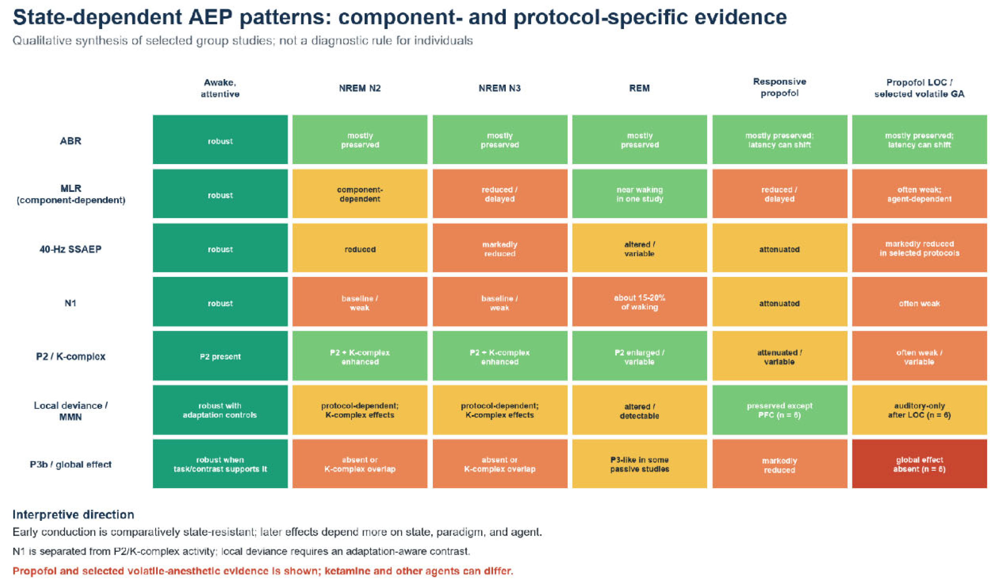
Figure 2.
State-dependent AEP patterns. The cells summarize component- and protocol-specific group findings, including selected propofol and volatile-anesthetic studies; they are not individual diagnostic thresholds or universal drug effects. N1 is separated from P2/K-complex activity, and local deviance is separated from P3b/global-rule processing [23,28,29,30,31,32,33,34,35,36,37,38].
Figure 2.
State-dependent AEP patterns. The cells summarize component- and protocol-specific group findings, including selected propofol and volatile-anesthetic studies; they are not individual diagnostic thresholds or universal drug effects. N1 is separated from P2/K-complex activity, and local deviance is separated from P3b/global-rule processing [23,28,29,30,31,32,33,34,35,36,37,38].
3.3. Sedation and General Anesthesia: Concentration- and Network-Dependent Effects
Sedation and anesthesia differ from natural sleep because drugs alter synaptic transmission and network dynamics in agent-specific ways. A useful design therefore reports the drug, target or measured concentration, co-administered analgesics, behavioral responsiveness, airway and physiologic variables, and the time course of induction and recovery. Terms such as "light," "moderate," and "deep" sedation are insufficient without an operational behavioral or pharmacologic definition.
Propofol studies illustrate the latency hierarchy. In nine surgical patients, propofol markedly affected middle-latency activity while producing smaller but measurable latency changes in ABR waves I, III, and V [34]. In 41 women, each studied at one of seven propofol concentration regimens, Nb latency and other MLR features varied with drug exposure and responsiveness; Nb latency discriminated eyelash response better than propofol concentration or dose rate [35]. Thus, the ABR is comparatively resistant and remains useful for pathway monitoring, but it is not invariant: temperature, anesthetic concentration, cochlear perfusion, and physiologic disturbance can still change its latency.
Late cognitive components are more vulnerable. During conscious propofol sedation, P3 amplitude fell substantially and reaction time increased, even though participants remained oriented and responsive [39]. Local-global auditory paradigms further show that propofol does not suppress the hierarchy uniformly. In six neurosurgical patients, local-deviance effects persisted outside prefrontal cortex during responsive sedation, whereas global-deviance responses were markedly reduced and became absent after loss of responsiveness; at that point, local-deviance effects were restricted to auditory cortex [36]. In eight patients with drug-resistant epilepsy studied during just-hypnotic propofol anesthesia, stimulus-evoked primary-auditory spiking and local field potentials persisted after loss of responsiveness, whereas higher-order spiking was largely attenuated. Induced gamma-band power did not always follow the local spiking response [37]. Together, these small intracranial studies support progressive restriction of auditory processing rather than complete cessation of sensory input.
The 40-Hz SSAEP and MLR have both been investigated as pharmacodynamic measures because they depend on synchronized thalamocortical and cortical activity. Under many propofol and volatile-anesthetic protocols, 40-Hz amplitude or power decreases as arousal is lost, and MLR latencies lengthen; however, neither response is a universal binary marker of consciousness. A response can be small because of montage, myogenic artifact control, hearing loss, poor stimulus delivery, or background EEG noise. A response can also persist despite unresponsiveness because some auditory circuits remain active. A controlled study showed that mastoid referencing can capture postauricular muscle activity and create misleading increases during drowsiness [23].
Surgical stimulation adds a further complication. In 30 cardiac-surgery patients receiving high-dose fentanyl plus propofol, isoflurane, or flunitrazepam, baseline MLRs differed by hypnotic regimen, but skin incision and sternotomy produced no significant additional MLR changes in any group [40]. The authors interpreted this lack of activation as consistent with blockade of surgical-stimulus effects by high-dose fentanyl. Thus, an AEP-based depth measure reflects hypnotic regimen within its analgesic and physiologic context; lack of activation does not independently prove adequate hypnosis. A monitor must also distinguish true cortical activation from electromyography, electrical interference, and changes in stimulus delivery.
Anesthetic-agent comparisons should not assume a common mechanism or response direction. Propofol and volatile anesthetics commonly suppress or delay cortical AEP measures, whereas ketamine can increase the 40-Hz response [38]. Nitrous oxide provides another dissociation: it hardly affected the MLAEP even at concentrations that produced unresponsiveness, although xenon, isoflurane, and sevoflurane produced graded MLAEP changes during emergence [41]. Direct cross-agent comparisons using the same stimulus, behavioral endpoint, and analysis pipeline are therefore more informative than comparisons among laboratories using different methodologies.
4. Human and Animal Studies
Animal models are indispensable because they allow recordings and causal manipulations at spatial scales that are rarely available in humans. The cat ABR generator experiments established how localized inactivation or lesions alter a scalp far-field response [10,11,12]. In behaving ferrets, task engagement rapidly changed spectrotemporal receptive fields in primary auditory cortex [17]. In awake rats, epidural potentials to frequency and duration deviants included effects that could not be explained solely by stimulus-specific adaptation, while anesthesia changed the deviance response [42]. These studies provide mechanistic models for human state effects.
Translation is strongest when the measurement and computation are conserved. Auditory-nerve timing, brainstem conduction, tonotopy, onset responses, and some forms of deviance sensitivity have recognizable cross-species analogues and can be reasonably translated when the paradigm is matched. Absolute latencies do not translate directly because brain size, pathway length, cochlear mechanics, hearing range, and stimulus calibration differ. A "wave V" in one species cannot be equated with a human wave solely by ordinal position, and a rodent MMN-like difference should not be assumed to have the same relationship to conscious report as human P3b.
Anesthetic state is a major cross-species confound. Many animal auditory experiments are performed under agents that alter cortical gain, adaptation, oscillations, and long-range communication. A finding obtained under ketamine, or isoflurane may describe the drug-shaped circuit as much as the awake circuit. Studies intended to explain human sleep or consciousness should include awake recordings when feasible, quantify state continuously, and test whether the relevant effect survives changes in anesthetic regimen.
Human intracranial studies bridge noninvasive human recordings and invasive animal experiments. They preserve human cortical organization and behavior while providing local field potentials, high-gamma activity, and sometimes single-unit recordings. Limitations include small clinically selected samples, nonuniform electrode coverage, anti-seizure medications, and the need to avoid epileptogenic tissue. Convergence among intracranial human recordings, noninvasive scalp AEPs, and causal animal experiments is therefore more persuasive than any one modality alone.
5. Pathway-Specific AEPs
5.1. Brainstem Responses and Frequency-Following Activity
The click-evoked ABR is the most clinically mature AEP because it is reproducible, rapid, and relatively resistant to changes in attention. Its principal measures are the presence and morphology of waves, absolute peak latency, interpeak latency, interaural differences, and amplitude ratios. Near threshold, wave V is usually the most robust component. At higher levels, waves I and V and the I-V interval help distinguish peripheral delay from central conduction abnormalities. Interpretation requires age- and protocol-appropriate norms because neonatal maturation, hearing loss, temperature, and stimulus rate influence timing.
"Brainstem response" should not be used as a synonym for "state independent." The foundational human study found stable responses across repeated recordings and sleep [2], but anesthetic studies have documented smaller latency changes even when gross morphology remains usable [34]. The clinically relevant statement is that early waves are less state-sensitive than cortical AEPs under many common conditions, not that they are immune to state or physiology.
Complex sounds produce sustained responses that preserve periodic acoustic structure. In synthetic vowels, the human FFR represents the stimulus fundamental periodicity and distinct spectral peaks associated with vowel identity [24]. These measures can be collected without an active task and can characterize temporal fidelity under difficult recording conditions. However, the term "brainstem FFR" is not universally valid: cortical contributions become more prominent at lower fundamental frequencies and depend on montage [25,26]. Source claims should therefore be supported by stimulus frequency, latency, polarity behavior, and, ideally, source-sensitive recording.
In state research, brainstem responses provide an essential control. If an ABR or high-rate subcortical response is absent, a missing cortical response cannot be interpreted cleanly as loss of higher-order processing because the sound may not have reached the cortex effectively. Conversely, an intact ABR with absent P300 supports a dissociation between preserved ascending conduction and impaired context-dependent evaluation. This hierarchical control logic is one of the strongest reasons to combine early and late components in the same protocol.
5.2. Cortical Responses: N1/P2, MMN, and P300
N1/P2 reflects early cortical registration of acoustic onset and change. The N1 is a superposition of processes arising from supratemporal and association regions and is shaped by onset sharpness, presentation level, interstimulus interval, refractoriness, attention, and arousal [4]. A smaller N1 during sleep or sedation can reflect lower cortical gain, changed synchronization, overlap with slow activity, or altered adaptation. Peak amplitude alone cannot distinguish among these mechanisms.
MMN is a relational response. It is computed from the difference between responses to standards and deviants and therefore depends on how the standard regularity is established. Deviants can differ in frequency, duration, intensity, location, phoneme, or an abstract acoustic pattern. MMN is often described as preattentive because it can be observed when participants ignore the sounds [8], but attention, stimulus salience, and state can modulate it. A deviant-minus-standard difference also contains physical and adaptation differences unless the design includes appropriate control sequences.
The P300 family adds task and context. The original uncertainty experiments showed a late positive potential associated with resolution of stimulus uncertainty [7]. In contemporary oddball designs, P3a is often elicited by unexpected distractors and has a frontocentral distribution, while P3b is typically largest over parietal regions when a participant detects or categorizes a task-relevant target. The presence of a P3b-like global effect is stronger evidence of long-range, context-dependent processing than the presence of an early local deviance effect, but absence remains difficult to interpret in an individual because sensitivity is imperfect.
These cortical components should be analyzed as a cascade rather than competing biomarkers. N1/P2 asks whether the cortex registered the event; MMN asks whether the event violated a regularity; P3a asks whether novelty recruited orienting; and P3b asks whether the event entered a task-defined context strongly enough to support updating or report. The stages overlap in time and anatomy, but the cascade makes explicit which inference is justified by each contrast.
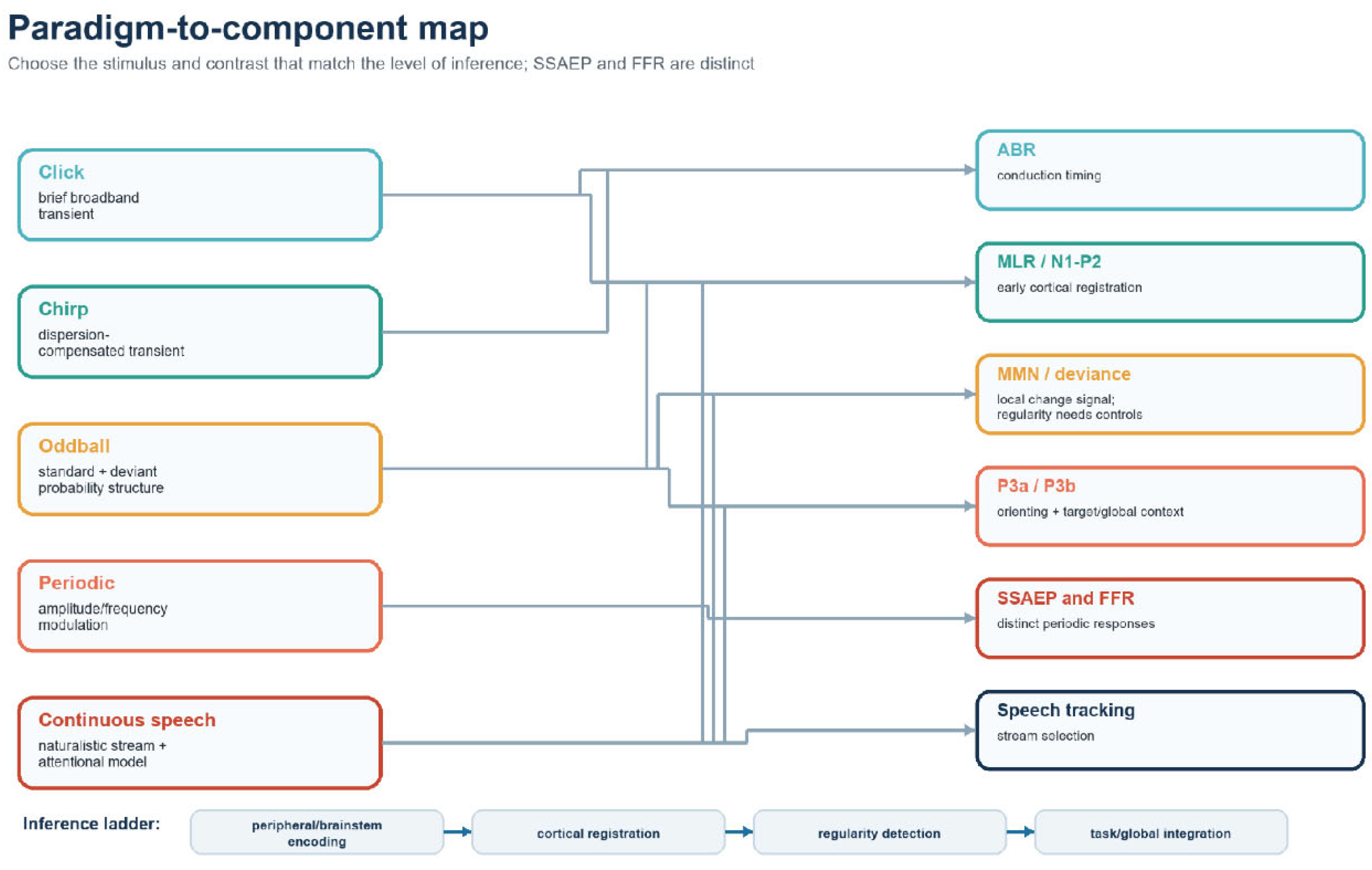
Figure 3.
Paradigm-to-component map. Arrows show principal elicitation routes rather than exclusive relationships; inference becomes stronger only when earlier pathway levels and the relevant experimental contrast are verified.
Figure 3.
Paradigm-to-component map. Arrows show principal elicitation routes rather than exclusive relationships; inference becomes stronger only when earlier pathway levels and the relevant experimental contrast are verified.
6. Stimulus Paradigms
6.1. Click-Evoked Responses
A click is a brief broadband transient that produces synchronized activation across a substantial portion of the auditory nerve. It is the canonical stimulus for ABR because its abrupt onset yields precisely timed early peaks [1,2]. Rarefaction, condensation, and alternating click polarity emphasize different cochlear and neural contributions; click polarity must therefore be reported. Insert earphones require correction for the acoustic delay between the transducer and ear canal, and contralateral masking may be needed when a high presentation level could stimulate the non-test ear.
The click's strength is also its limitation. Cochlear traveling-wave delay means that high-frequency basal regions respond before lower-frequency apical regions, so the click does not activate the entire cochlea simultaneously. Its response is weighted toward the basal cochlea and does not provide frequency-specific threshold information without additional techniques such as tone bursts, masking, or derived-band analysis. Because the ABR is dominated by early conduction, a click is also poorly suited to testing auditory prediction or conscious evaluation unless it is embedded within a higher-order paradigm.
6.2. Chirp Stimuli and Cochlear-Dispersion Compensation
A transient sound does not reach all characteristic-frequency places on the basilar membrane at the same time. High-frequency energy reaches maximum excitation near the cochlear base earlier than low-frequency energy reaches more apical regions. The resulting dispersion spreads auditory-nerve firing over time and reduces the amount of activity that sums synchronously in the far-field waveform.
A rising-frequency chirp compensates for this delay by presenting low-frequency energy earlier and high-frequency energy later. Dau and colleagues designed an optimized chirp using a cochlear delay model so that excitation maxima across frequency regions would occur more nearly together [43]. The intended result is not that sound physically travels instantaneously through the cochlea, but that the timing of the stimulus counteracts the normal traveling-wave delay. More synchronized neural activation can produce a larger wave V than a conventional click, especially at low to moderate levels.
The optimal chirp delay pattern depends on stimulus presentation level because cochlear response latency changes with level. Elberling and Don therefore derived chirps from human derived-band ABR latencies and described a direct method for designing level-specific stimuli [44]. This development is clinically important: a chirp optimized at one presentation level can be suboptimal at another, and a larger amplitude does not automatically indicate better neural function. Reports should specify chirp family, presentation level, polarity, rate, calibration, and whether latency norms were established for that exact stimulus.
Chirps improve synchrony at the input to the ascending pathway; they do not make ABR immune to sleep, anesthesia, hearing loss, or neural dyssynchrony. Their benefit should be evaluated separately for response amplitude, detection time, threshold accuracy, and diagnostic specificity. When state is the independent variable, the same calibrated chirp and acquisition protocol must be used in every condition.
6.3. Oddball Paradigms: Probability, Regularity, and Task Relevance
The oddball paradigm is not merely a way to "produce MMN and P300." It is a family of designs that manipulates probability, regularity, and task relevance. A frequent standard establishes a local model; an infrequent deviant violates one or more properties of that model. The participant may ignore the sequence, monitor it for targets, or learn a higher-order rule across groups of sounds. Each instruction changes the inference that can be drawn from the resulting waveform.
The classic P300 experiments manipulated stimulus uncertainty and associated its resolution with a late positive potential [7]. Later work distinguished an early negative deviance response from prolonged attention-related negativity that can overlap the N1 range [8]. In a passive oddball task, the deviant-minus-standard difference around 100-250 ms is commonly called MMN. In an active target task, a later P3b is expected when the participant detects, counts, or responds to the task-relevant target. Novel distractors may evoke P3a even when they are not the designated target.
Design choices strongly influence the measured components. Deviant probability determines how much evidence supports a standard regularity and how surprising the deviant is. The interstimulus interval influences refractoriness and sensory memory, and the magnitude and dimension of deviance affect discriminability. A duration deviant changes the physical time at which standard and deviant waveforms cease to be identical, whereas a frequency deviant differs from the standard beginning at acoustic onset. These properties determine the valid subtraction window and should be considered before a negative difference is interpreted as MMN.
Adaptation is the principal alternative explanation for a simple oddball difference. Neurons tuned to the frequent standard respond less after repetition, whereas neurons tuned to the rare deviant are less adapted. A many-standards or equiprobable control presents the deviant with the same probability but without a single repetitive standard. If the deviant still differs from its control, the evidence for regularity violation is stronger. Animal studies using such controls have demonstrated deviance-sensitive potentials beyond simple rarity under some awake conditions [42].
State changes alter both the component and the task. During wakefulness, an active oddball can separate early sensory registration, deviance detection, orienting, and target evaluation. During sleep or deep sedation, the participant cannot maintain the same overt task, so comparison with an awake active condition confounds state with instruction and behavior. Passive paradigms are better matched across states, but they usually provide weaker evidence for conscious access. A local-global design addresses this limitation by nesting short-range violations within longer-range sequence rules. In healthy awake participants, violations of a global rule evoke a late response associated with conscious processing [45]. In noncommunicating patients and during propofol anesthesia, local and global effects can dissociate [36,46].
An oddball result should therefore specify at least five elements: the physical standard and deviant; their probabilities; the sequence rule; the participant's instruction and performance; and the contrast used to define each component. Reporting only "MMN was present" is insufficient. The same negative deflection could reflect adaptation, local deviance detection, attention-related processing, or overlap with a sleep-specific component.
6.4. Periodic Stimulation and Naturalistic Listening
SSAEP paradigms use tones, clicks, noise, or amplitude- and frequency-modulated carriers presented periodically. The neural response is evaluated at the stimulation frequency and, when present, its response harmonics. Multiple carriers can be modulated at different frequencies and presented simultaneously to estimate frequency-specific hearing sensitivity, but interactions, masking, and unequal signal-to-noise ratios require attention. Modulation rate shifts relative generator weighting: responses near 40 Hz contain strong auditory-cortical contributions, whereas responses near 80-100 Hz are relatively more brainstem weighted; neither range is source-pure [18].
Naturalistic paradigms trade tight event isolation for ecological validity. In competing-speech experiments, the neural response can be related to the temporal envelope of each talker. Single-trial EEG has been used to decode which of two speakers a listener attends [47]. Such responses are not conventional peak-labeled AEPs, but they extend the same system-identification logic to continuous input. They are particularly promising for hearing devices and bedside communication assessment, provided that acoustic preprocessing, stimulus reconstruction, and temporal response functions are reported transparently.
7. Analysis Domains and Response Types
7.1. Acquisition, Preprocessing, and Reproducibility
AEP analysis begins before recording. The scientific question determines whether the protocol needs submillisecond ABR timing, wide-band cortical EEG, high trial counts, active behavioral responses, or uninterrupted sleep staging. The amplifier sampling rate and analog filters must preserve the component of interest. High-pass filtering can distort slow late potentials and generate artifactual pre-stimulus activity; low-pass filtering can remove sharp ABR features or induced high-frequency activity. Filter type, order, cutoff definition, direction, and application to continuous or epoched EEG data should be reported.
The reference electrode changes waveform amplitude and topography. Mastoid or earlobe references may be appropriate for some cortical ERPs but can capture postauricular muscle responses in 40-Hz work [23]. Common-average reference requires adequate scalp coverage. Linked references can introduce correlations and hide lateral asymmetry. Because no reference is electrically neutral, cross-state differences should be confirmed under a physiologically defensible montage rather than treated as invariant to rereferencing.
Artifact structure changes with state. Awake participants produce eye movements, blinks, speech, and muscle activity; sleeping participants produce slow eye movements, spindles, and stage transitions; surgical recordings include electrocautery, pumps, warming devices, and stimulus-delivery disturbances. Automated rejection thresholds applied identically across states may remove unequal proportions of data or preferentially reject physiologically large sleep responses. Reports should provide retained trial counts by condition and explain how ocular, muscle, line, stimulation, and movement artifacts were handled.
Consensus ERP guidance emphasizes prespecified components, explicit measurement windows, comparable trial counts, and complete reporting of acquisition and processing decisions [19]. These principles are particularly important in small clinical samples, where flexible peak selection can turn background fluctuations into apparent components. When possible, analysis code, de-identified summary data, and machine-readable event definitions should accompany the paper.
7.2. Transient, Phase-Locked Evoked Responses
Traditional ABR and ERP averaging assumes that the response recurs with consistent latency and phase relative to the onset of the acoustic stimulus while unrelated background EEG activity varies across trials. Averaging reinforces the repeated response and reduces uncorrelated noise. The resulting waveform can be measured by peak amplitude, mean amplitude within a time window, area, onset latency, fractional-area latency, or model-based component estimates. Each measure answers a different question and has different noise sensitivity.
Peak latency is intuitive for distinct ABR waves but unstable for broad or multi-peaked cortical components. Mean amplitude over a prespecified window is often more robust for MMN and P300, provided that the window is not chosen after inspecting condition differences. Difference waves can isolate condition-sensitive activity, but they combine noise and any physical differences between the contributing conditions. Topographic or source analyses require the same latency rule across electrodes; selecting a separate local peak at every channel can create a physiologically impossible map.
Single-subject detection demands a statistical criterion, not visual resemblance alone. Replication across independent trial subsets, residual-noise estimates, bootstrap confidence intervals, or frequency-domain tests can quantify reliability. A component visible in a group average may not be detectable in most individual participants, and a classifier trained on a group contrast may not be calibrated for a clinical decision about one patient. Sensitivity, specificity, test-retest reliability, and indeterminate results should be reported.
7.3. SSAEP Analysis
SSAEPs are phase-locked periodic responses and can be analyzed in the time or frequency domain. In the frequency domain, the primary question is whether energy and phase consistency at the stimulation frequency exceed the surrounding noise distribution. Measures include Fourier amplitude, signal-to-noise ratio, phase coherence, circular statistics, and multivariate combinations across channels. The analysis window should contain an integer number of modulation cycles or otherwise account for spectral leakage.
Time-domain averaging remains useful because it preserves phase and waveform shape. A controlled arousal study [23] found that 40-Hz response detection depended on analysis and montage; the spectral-averaging implementation used in that study was less efficient than time-domain averaging. Modern frequency-domain methods can preserve phase by averaging complex-valued Fourier coefficients. The general lesson is that "frequency-domain" does not guarantee optimal detection and that averaging power spectra is not equivalent to coherently averaging complex coefficients.
Throughout this review, SSAEP denotes the response class. If a cited paper uses ASSR in its title, that title is retained verbatim in the reference list, but the narrative term remains SSAEP. Reports should state carrier frequency, modulation frequency and depth, stimulus level, number and duration of epochs, phase treatment, rejection criteria, and statistical detection threshold. These details strongly influence both threshold inference and state sensitivity.
7.4. Phase-Locked Versus Non-Phase-Locked Activity
The phrase "non-phase-locked response" can be confusing because the response may still be stimulus-linked. The key distinction is between reproducibility of event timing and reproducibility of oscillatory phase. A phase-locked evoked response has a similar sequence of positive and negative voltage deflections on each trial. If its peaks align to stimulus onset, direct voltage averaging preserves it. ABR, MLR, N1/P2, MMN, P300, and SSAEPs are usually analyzed primarily in this way.
An induced oscillatory response can occur after the same event on every trial even when its instantaneous phase differs across trials. For example, a sound may increase power at 40 Hz between 200 and 400 ms. One trial may begin that interval with a positive gamma cycle and another with a negative cycle, so direct voltage averaging cancels the oscillation despite a power increase in both trials. Investigators recover such effects by calculating a time-frequency representation for each trial, using a wavelet transform, short-time Fourier transform, or related method, and then averaging power or another magnitude measure across trials.
Intertrial phase coherence and induced power answer different questions. Intertrial phase coherence measures whether oscillatory phase is consistent across repetitions. Power at a specified frequency, such as 40 Hz, measures the strength of the oscillation regardless of phase. A stimulus can increase both measures, increase phase coherence without much power change, or increase power without phase alignment. To isolate non-phase-locked activity, investigators sometimes subtract the phase-locked ERP from each trial before calculating power; this procedure has assumptions and can introduce bias.
Baseline normalization also matters. Decibel change, percent change, and subtraction can yield different results when baseline power differs across wakefulness, sleep, and anesthesia. A baseline contaminated by anticipatory activity or a preceding stimulus is not neutral. Time-frequency analyses also involve many time-frequency-electrode comparisons, so cluster-based or other multiplicity-aware inference should be prespecified. Tallon-Baudry and Bertrand's distinction between evoked and induced gamma remains conceptually useful when implemented with these analytic cautions [48].
In plain language, phase-locked analysis asks, "Does the same waveform recur at the same point in every trial?" Induced-power analysis asks, "Does the event change the strength of an oscillation even if its peaks and troughs do not line up?" Both can be physiologically meaningful. They should not be placed in a table as mutually exclusive types of AEP without explaining that SSAEPs are phase-locked periodic responses, whereas induced activity is a broader stimulus-related oscillatory phenomenon.
7.5. Cross-State Comparisons
Cross-state inference is strongest when stimulus delivery, electrode geometry, preprocessing, and statistical detection remain constant while the state changes. Even then, background noise and artifact distributions may differ. Equalizing accepted trial counts, estimating residual noise separately, and reporting within-participant effect sizes reduce the risk that a smaller response in sleep or anesthesia merely reflects a noisier recording.
State should be measured rather than inferred from clock time or drug infusion alone. Awake tasks require performance and vigilance measures; sleep requires stage scoring and arousal detection; anesthesia requires concentration or dose history, behavioral responsiveness, and physiologic monitoring. For noncommunicating patients, diagnostic category, sedation exposure, etiology, time since injury, and repeated assessment are essential covariates.
8. Clinical and Translational Implications
8.1. Hearing Screening and Diagnostic Audiology
ABR supports objective assessment when a patient cannot provide a reliable behavioral threshold. In newborn programs, automated ABR and otoacoustic emissions identify infants who need diagnostic follow-up. Otoacoustic emissions are ear-canal sounds generated mainly by healthy cochlear outer hair cells and therefore assess cochlear, rather than neural, function. The public-health value comes from the full chain of screening, diagnostic confirmation, amplification or other intervention, and language support-not from the screening waveform alone. Children born in hospitals with universal screening showed better early speech and language outcomes within an early-intervention cohort than matched children born without such screening [49]. Loss to follow-up and delayed diagnostic ABR remain practical barriers.
Frequency-specific tone-burst or chirp responses and SSAEPs can supplement click ABR when estimating an audiogram. Auditory neuropathy spectrum disorder illustrates why cochlear and neural measures must be combined: otoacoustic emissions or cochlear microphonics may show preserved outer-hair-cell function, while disrupted auditory-nerve synchrony produces an absent or distorted ABR. Sedation may facilitate recording but should be documented because it can influence cortical timing and, to a smaller degree, brainstem timing.
8.2. Intraoperative Monitoring
Intraoperative BAEP monitoring is used when surgery risks the cochlea, eighth nerve, or brainstem. The objective is continuous acquisition and rapid detection of a change that permits correction before permanent injury. Wave V latency, amplitude, and persistence, together with wave I and interpeak intervals, help distinguish a peripheral stimulus or cochlear problem from more central compromise. A prospective series of microvascular decompression procedures related progressive wave V delay and loss to postoperative hearing outcome and proposed graded warning values [50]. Warning thresholds from one procedure or laboratory should not be imported uncritically into another.
Mechanistic interpretation matters in real time. Loss of wave I suggests a problem before or at the distal nerve, poor stimulus delivery, or cochlear dysfunction; preserved wave I with delayed or lost wave V suggests more central conduction compromise. Hypothermia, hypotension, irrigation, drilling noise, anesthetic change, earphone displacement, and electrical interference can mimic neural deterioration. A monitoring response should therefore trigger a structured technical and physiologic check before a surgical conclusion.
8.3. Sedation and Anesthesia Monitoring
MLR, 40-Hz SSAEP, P300, and other auditory paradigms have been evaluated as measures of hypnotic state because they probe evoked cortical processing rather than spontaneous EEG alone. Commercial AEP-based depth monitors were developed and independently evaluated. In 22 volunteers exposed to desflurane and propofol, approximately 40-Hz power predicted responsiveness with a prediction probability of 0.96 under both agents, compared with 0.86 and 0.88 for Nb latency [51]. Propofol concentration-response studies also show systematic changes in midlatency features [35], and artificial neural networks have classified anesthetic states from MLR latency patterns more accurately than hemodynamic variables in a small surgical cohort [52]. These findings support continued development of AEP-informed monitoring, but not use of an auditory index as a standalone measure of consciousness.
Commercialization also exposed important limitations. In 13 patients, a composite A-Line index that combined MLAEP, spontaneous EEG, and burst-suppression information tracked calculated propofol effect-site concentration better than an earlier MLAEP-only index, although clinical performance remained to be established [53]. In ten awake, unmedicated volunteers, an earlier A-Line monitor produced index values from 15 to 99; 11.4% fell below the manufacturer's range for surgical anesthesia because contaminated MLAEP signals were not reliably excluded [54]. No auditory measure should therefore be treated as a standalone consciousness meter. Hearing loss, stimulus disconnection, myogenic contamination, analgesia, and agent-specific pharmacology can all change the signal. A credible monitor should combine verified auditory input, early pathway integrity, a cortical evoked measure, spontaneous EEG, drug history, signal-validity checks, and clinical endpoints.
8.4. Disorders of Consciousness and the Intensive-Care Setting
Patients with coma, unresponsive wakefulness syndrome, or a minimally conscious state may have preserved sensory pathways but inconsistent motor output. A hierarchical auditory battery can test for evidence that sound activates the brainstem, reaches cortex, violates a local regularity, and engages a global context. The local-global paradigm demonstrates the logic: healthy participants show early responses to local violations and a late effect to violations of a longer-range rule [45]. In patients with impaired consciousness, local effects may occur across diagnostic categories, whereas a late global effect is more specific but not sufficiently sensitive to rule out consciousness when absent [46].
Repeated measurements are crucial because arousal fluctuates; a single negative recording can result from noise, medication, hearing impairment, or a transient state. Prognostic studies should blind waveform interpretation to outcome and report indeterminate cases. The ethical implication is asymmetric: a reproducible late context-dependent response may provide positive evidence of preserved higher-order processing, but its absence should be interpreted cautiously.
ICU implementation also requires attention to competing sounds, ventilators, cranial injury, edema, hypothermia, and sedative exposure. Ear-specific delivery and an early response control help confirm effective auditory-pathway activation. Bedside paradigms should prioritize short acquisition, rapid analysis, and repeated single-patient reliability rather than maximizing the number of group-level components.
8.5. Developmental and Neuropsychiatric Applications
AEP latency and morphology change with maturation, so pediatric norms must be age specific. ABR maturation reflects myelination and conduction changes, while cortical P1, N1, P2, and mismatch responses follow distinct developmental trajectories. A developmental difference is not automatically a deficit; it may reflect age, sleep state, hearing experience, or stimulus familiarity.
Representative case-control studies have reported reduced 40-Hz SSAEP synchronization in schizophrenia, stimulus-dependent MMN and P3a differences in autistic children, and altered speech-evoked brainstem/FFR encoding in autism [55,56,57]. Effect direction and magnitude vary by stimulus and cohort. Clinical translation requires prospective individual-level validation, appropriate hearing controls, and demonstration that the measure adds value beyond behavioral and standard clinical assessment. Broad diagnostic labels should not be inferred from a single component.

Table 2.
Clinical translation matrix.
| Application | Primary response | Decision supported | Essential controls | Main limitation |
| Newborn screening | Automated ABR with/without otoacoustic emissions | Referral for diagnostic hearing evaluation | Calibration, ear-specific testing, follow-up pathway | Screening does not establish etiology or guarantee intervention |
| Diagnostic audiology | Click, tone-burst, chirp ABR; SSAEP | Threshold and pathway assessment | Age norms, masking, cochlear measures | Sedation, maturation, and neural dyssynchrony alter responses |
| Intraoperative monitoring | BAEP waves I and V; interpeak timing | Detect reversible cochlear/nerve/brainstem compromise | Stimulus integrity, temperature, blood pressure, artifact | Warning criteria vary by procedure and laboratory |
| Anesthesia monitoring | MLR, 40-Hz SSAEP, late auditory ERP | Track drug-related changes in cortical and thalamocortical processing | Hearing, montage, drug/concentration, stimulation | No single component is a binary consciousness marker |
| Disorders of consciousness | ABR + N1/MMN + P3b/global effect | Identify preserved hierarchical processing | Repeated testing, medication, stimulus delivery and early pathway response, etiology | Positive late effects are informative; absence has limited sensitivity |
| Wearable monitoring | Ear-/around-ear N1/P300; ABR and envelope-following/steady-state responses | Test feasibility of unobtrusive extended auditory monitoring | Synchronization, motion, contact stability | Clinical state decisions remain unvalidated; real-world signal quality varies |
9. Future Directions
Naturalistic listening is likely to connect laboratory components with real-world hearing. Continuous-speech models can estimate how neural activity tracks attended and ignored speakers. A two-talker study demonstrated that single-trial EEG could decode which speaker a listener attended [47]. Future work should combine this cortical tracking with verified peripheral or brainstem measures so that weak decoding can be attributed to the appropriate level of the auditory pathway.
Wearable systems can extend state research beyond short laboratory sessions. Around-the-ear and in-ear electrodes have captured N1- and P300-range effects during active hearing-device use [58], and cEEGrid configurations have also been evaluated for subcortical auditory potentials in normal-hearing and hearing-impaired listeners [59]. The remaining challenges include stable long-duration contact, motion and muscle artifact, accurate stimulus synchronization, user comfort, and validation against conventional montages.
Modern computational models should move from retrospective discrimination to transparent, calibrated decision support. Early work showed that nonlinear models could classify propofol anesthetic states from MLR features [52]. Models can now learn from time series or time-frequency representations, but high within-dataset accuracy does not establish clinical validity. Promising approaches include hierarchical calibration to an individual's awake baseline, mixed-effects models that separate person- and state-level variation, and domain-adaptation methods that normalize differences among hospitals, amplifiers, montages, drugs, hearing profiles, and demographic groups. External validation remains essential. A clinical model should expose the signal features driving its decision and return uncertainty or "insufficient signal quality" rather than force every recording into a single state category.
Multimodal monitoring can test whether evoked and spontaneous measures provide complementary information. Propofol produces reproducible changes in spontaneous frontal EEG around loss and recovery of responsiveness [60]. Intracranial auditory studies show relative preservation of early primary-auditory responses and attenuation or spatial restriction of higher-order evoked and spiking responses; stimulus-induced power can also diverge from local spiking [36,37]. Combining these measurements may distinguish failure of sensory input, local cortical processing, and long-range integration. Such a system should be evaluated prospectively against behavioral responsiveness, explicit memory, and clinically relevant outcomes.
Cross-species studies should use shared stimuli and analysis definitions. The same click, chirp, oddball, or amplitude-modulated waveform can be calibrated for species-specific hearing ranges while preserving the computational contrast. Awake animal recordings, causal perturbation, laminar physiology, and human intracranial data can then be connected to scalp biomarkers. Pre-registration of the predicted generator and direction of state effect would reduce post hoc mapping between unlike components.
Finally, the field needs a minimum reporting set for state-aware AEP research: calibrated stimulus waveform and transducer; ear and masking; electrode montage and impedance; state definition and monitoring; medications and concentrations; artifact and trial-retention statistics; filter and baseline parameters; component definition; individual-level detectability; and persistent identifiers for every cited source. Reference lists should include stable PubMed or DOI links so that readers can verify the underlying evidence efficiently.
10. Conclusions
AEPs should be interpreted as a hierarchy of related measurements rather than as a single test. Early ABR components provide a comparatively robust index of auditory-nerve and brainstem conduction. MLR and 40-Hz SSAEP measures probe thalamocortical and early cortical synchrony. N1/P2 reflects cortical registration, MMN tests sensitivity to auditory regularity, and P300 or global-rule effects provide stronger evidence of task- or context-dependent integration. Each level requires a stimulus and analysis matched to its generator.
Cognitive state determines which stages of the auditory hierarchy remain measurable and how response amplitude, latency, phase consistency, induced power, and spatial distribution change. Sleep reorganizes cortical responses and introduces stage-specific events. Sedation and anesthesia alter auditory processing according to agent, concentration, network level, and response measure; later and higher-order responses are generally more vulnerable than primary responses. Disorders of consciousness can preserve sensory activity despite absent behavioral output. None of these states is adequately summarized by "response present" or "response absent."
The practical rule is simple: interpret a late component only after verifying the earlier pathway, and interpret any state difference only after verifying stimulus delivery, recording quality, analysis sensitivity, and the state itself. A multimodal, repeated, and component-specific approach offers the clearest route from auditory physiology to reliable biomarkers of hearing, arousal, and residual cognition.
Author Contributions
Conceptualization by Zhibin Zhou and Kevin Goehl. All authors contribute to original draft preparation and revision. All authors have read and agreed to the published version of the manuscript
Funding
This research received no external funding.
Acknowledgments
Thanks to Professor Virginia Richards, from University of California, Irvine, for her valuable inputs on the manuscript.
Conflicts of Interest
Kevin Goehl is the CEO of KBO Systems. And Nathaen Weitzel is the co-founder of the company. The other authors declare no conflicts of interest.
Abbreviations
The following abbreviations are used in this manuscript:
| AEP | Auditory evoked potential |
| ABR | Auditory brainstem response |
| ASSR | Auditory steady-state response |
| BAEP | Brainstem auditory evoked potential |
| BIS | Bispectral index |
| EEG | Electroencephalography |
| ERP | Event-related potential |
| FFR | Frequency-following response |
| ICU | Intensive care unit |
| MEG | Magnetoencephalography |
| MLAEP | Middle-latency auditory evoked potential |
| MLR | Middle-latency response |
| MMN | Mismatch negativity |
| NREM | Non-rapid eye movement |
| REM | Rapid eye movement |
| SSAEP | Auditory steady-state evoked potential |
References
- Jewett, D.L.; Romano, M.N.; Williston, J.S. Human auditory evoked potentials: possible brain stem components detected on the scalp. Science 1970, 167(3924), 1517–8. [Google Scholar] [CrossRef] [PubMed]
- Jewett, D.L.; Williston, J.S. Auditory-evoked far fields averaged from the scalp of humans. Brain 1971, 94(4), 681–96. [Google Scholar] [CrossRef] [PubMed]
- Picton, T.W.; Hillyard, S.A.; Krausz, H.I.; Galambos, R. Human auditory evoked potentials. I. Evaluation of components. Electroencephalogr. Clin. Neurophysiol. 1974, 36(2), 179–90. [Google Scholar] [CrossRef] [PubMed]
- Näätänen, R.; Picton, T. The N1 wave of the human electric and magnetic response to sound: a review and an analysis of the component structure. Psychophysiology 1987, 24(4), 375–425. [Google Scholar] [CrossRef] [PubMed]
- Hillyard, S.A.; Hink, R.F.; Schwent, V.L.; Picton, T.W. Electrical signs of selective attention in the human brain. Science 1973, 182(4108), 177–80. [Google Scholar] [CrossRef] [PubMed]
- Picton, T.W.; Hillyard, S.A. Human auditory evoked potentials. II. Effects of attention. Electroencephalogr. Clin. Neurophysiol. 1974, 36(2), 191–9. [Google Scholar] [CrossRef] [PubMed]
- Sutton, S.; Braren, M.; Zubin, J.; John, E.R. Evoked-potential correlates of stimulus uncertainty. Science 1965, 150(3700), 1187–8. [Google Scholar] [CrossRef] [PubMed]
- Näätänen, R.; Gaillard, A.W.; Mäntysalo, S. Early selective-attention effect on evoked potential reinterpreted. Acta Psychol. (Amst) 1978, 42(4), 313–29. [Google Scholar] [CrossRef] [PubMed]
- Starr, A.; Achor, J. Auditory brain stem responses in neurological disease. Arch. Neurol. 1975, 32(11), 761–8. [Google Scholar] [CrossRef] [PubMed]
- Melcher, J.R.; Knudson, I.M.; Fullerton, B.C.; Guinan, J.J.; Norris, B.E.; Kiang, N.Y. Generators of the brainstem auditory evoked potential in cat. I. An experimental approach to their identification. Hear Res. 1996, 93(1–2), 1–27. [Google Scholar] [CrossRef] [PubMed]
- Melcher, J.R.; Guinan, J.J.; Knudson, I.M.; Kiang, N.Y. Generators of the brainstem auditory evoked potential in cat. II. Correlating lesion sites with waveform changes. Hear Res. 1996, 93(1–2), 28–51. [Google Scholar] [CrossRef] [PubMed]
- Melcher, J.R.; Kiang, N.Y. Generators of the brainstem auditory evoked potential in cat. III: Identified cell populations. Hear Res. 1996, 93(1–2), 52–71. [Google Scholar] [CrossRef] [PubMed]
- Sweet, R.A.; Dorph-Petersen, K.A.; Lewis, D.A. Mapping auditory core, lateral belt, and parabelt cortices in the human superior temporal gyrus. J. Comp. Neurol. 2005, 491(3), 270–89. [Google Scholar] [CrossRef] [PubMed]
- Nourski, K.V.; Steinschneider, M.; McMurray, B.; Kovach, C.K.; Oya, H.; Kawasaki, H.; et al. Functional organization of human auditory cortex: investigation of response latencies through direct recordings. Neuroimage 2014, 101, 598–609. [Google Scholar] [CrossRef] [PubMed]
- Liégeois-Chauvel, C.; Musolino, A.; Badier, J.M.; Marquis, P.; Chauvel, P. Evoked potentials recorded from the auditory cortex in man: evaluation and topography of the middle latency components. Electroencephalogr. Clin. Neurophysiol. 1994, 92(3), 204–14. [Google Scholar] [CrossRef] [PubMed]
- Woldorff, M.G.; Gallen, C.C.; Hampson, S.A.; Hillyard, S.A.; Pantev, C.; Sobel, D.; et al. Modulation of early sensory processing in human auditory cortex during auditory selective attention. Proc. Natl. Acad. Sci. U S A 1993, 90(18), 8722–6. [Google Scholar] [CrossRef] [PubMed]
- Fritz, J.; Shamma, S.; Elhilali, M.; Klein, D. Rapid task-related plasticity of spectrotemporal receptive fields in primary auditory cortex. Nat. Neurosci. 2003, 6(11), 1216–23. [Google Scholar] [CrossRef] [PubMed]
- Herdman, A.T.; Lins, O.; Van Roon, P.; Stapells, D.R.; Scherg, M.; Picton, T.W. Intracerebral sources of human auditory steady-state responses. Brain Topogr. 2002, 15(2), 69–86. [Google Scholar] [CrossRef] [PubMed]
- Picton, T.W.; Bentin, S.; Berg, P.; Donchin, E.; Hillyard, S.A.; Johnson, R.; et al. Guidelines for using human event-related potentials to study cognition: recording standards and publication criteria. Psychophysiology 2000, 37(2), 127–52. [Google Scholar] [CrossRef] [PubMed]
- Galambos, R.; Makeig, S.; Talmachoff, P.J. A 40-Hz auditory potential recorded from the human scalp. Proc. Natl. Acad. Sci. U S A 1981, 78(4), 2643–7. [Google Scholar] [CrossRef] [PubMed]
- Santarelli, R.; Conti, G. Generation of auditory steady-state responses: linearity assessment. Scand. Audiol. Suppl. 1999, 51, 23–32. [Google Scholar] [PubMed]
- Presacco, A.; Bohórquez, J.; Yavuz, E.; Özdamar, Ö. Auditory steady-state responses to 40-Hz click trains: relationship to middle latency, gamma band and beta band responses studied with deconvolution. Clin. Neurophysiol. 2010, 121(9), 1540–1550. [Google Scholar] [CrossRef] [PubMed]
- Picton, T.W.; John, M.S.; Purcell, D.W.; Plourde, G. Human auditory steady-state responses: the effects of recording technique and state of arousal. Anesth. Analg. 2003, 97(5), 1396–1402. [Google Scholar] [CrossRef] [PubMed]
- Krishnan, A. Human frequency-following responses: representation of steady-state synthetic vowels. Hear Res. 2002, 166(1–2), 192–201. [Google Scholar] [CrossRef] [PubMed]
- Coffey, E.B.; Herholz, S.C.; Chepesiuk, A.M.; Baillet, S.; Zatorre, R.J. Cortical contributions to the auditory frequency-following response revealed by MEG. Nat. Commun. 2016, 7, 11070. [Google Scholar] [CrossRef] [PubMed]
- Gnanateja, G.N.; Rupp, K.; Llanos, F.; Remick, M.; Pernia, M.; Sadagopan, S.; et al. Frequency-Following Responses to Speech Sounds Are Highly Conserved across Species and Contain Cortical Contributions. eNeuro 2021, 8(6), ENEURO.0451–21. [Google Scholar] [CrossRef] [PubMed]
- Woods, D.L.; Stecker, G.C.; Rinne, T.; Herron, T.J.; Cate, A.D.; Yund, E.W.; et al. Functional maps of human auditory cortex: effects of acoustic features and attention. PLoS ONE 2009, 4(4), e5183. [Google Scholar] [CrossRef] [PubMed]
- Campbell, K.; Muller-Gass, A. The extent of processing of near-hearing threshold stimuli during natural sleep. Sleep 2011, 34(9), 1243–9. [Google Scholar] [CrossRef] [PubMed]
- Cash, S.S.; Halgren, E.; Dehghani, N.; Rossetti, A.O.; Thesen, T.; Wang, C.; et al. The human K-complex represents an isolated cortical down-state. Science 2009, 324(5930), 1084–7. [Google Scholar] [CrossRef] [PubMed]
- Bastuji, H.; García-Larrea, L.; Franc, C.; Mauguière, F. Brain processing of stimulus deviance during slow-wave and paradoxical sleep: a study of human auditory evoked responses using the oddball paradigm. J. Clin. Neurophysiol. 1995, 12(2), 155–67. [Google Scholar] [CrossRef] [PubMed]
- Perrin, F.; García-Larrea, L.; Mauguière, F.; Bastuji, H. A differential brain response to the subject's own name persists during sleep. Clin. Neurophysiol. 1999, 110(12), 2153–64. [Google Scholar] [CrossRef] [PubMed]
- Deiber, M.P.; Ibañez, V.; Bastuji, H.; Fischer, C.; Mauguière, F. Changes of middle latency auditory evoked potentials during natural sleep in humans. Neurology 1989, 39(6), 806–13. [Google Scholar] [CrossRef] [PubMed]
- Plourde, G.; Picton, T.W. Human auditory steady-state response during general anesthesia. Anesth. Analg. 1990, 71(5), 460–8. [Google Scholar] [CrossRef] [PubMed]
- Chassard, D.; Joubaud, A.; Colson, A.; Guiraud, M.; Dubreuil, C.; Banssillon, V. Auditory evoked potentials during propofol anaesthesia in man. Br. J. Anaesth. 1989, 62(5), 522–6. [Google Scholar] [CrossRef] [PubMed]
- Tooley, M.A.; Greenslade, G.L.; Prys-Roberts, C. Concentration-related effects of propofol on the auditory evoked response. Br. J. Anaesth. 1996, 77(6), 720–6. [Google Scholar] [CrossRef] [PubMed]
- Nourski, K.V.; Steinschneider, M.; Rhone, A.E.; Kawasaki, H.; Howard, M.A.; Banks, M.I. Auditory Predictive Coding across Awareness States under Anesthesia: An Intracranial Electrophysiology Study. J. Neurosci. 2018, 38(39), 8441–8452. [Google Scholar] [CrossRef] [PubMed]
- Krom, A.J.; Marmelshtein, A.; Gelbard-Sagiv, H.; Tankus, A.; Hayat, H.; Hayat, D.; et al. Anesthesia-induced loss of consciousness disrupts auditory responses beyond primary cortex. Proc. Natl. Acad. Sci. U S A 2020, 117(21), 11770–11780. [Google Scholar] [CrossRef] [PubMed]
- Plourde, G.; Baribeau, J.; Bonhomme, V. Ketamine increases the amplitude of the 40-Hz auditory steady-state response in humans. Br. J. Anaesth. 1997, 78(5), 524–9. [Google Scholar] [CrossRef] [PubMed]
- Reinsel, R.A.; Veselis, R.A.; Wronski, M.; Marino, P. The P300 event-related potential during propofol sedation: a possible marker for amnesia? Br. J. Anaesth. 1995, 74(6), 674–80. [Google Scholar] [CrossRef] [PubMed]
- Schwender, D.; Golling, W.; Klasing, S.; Faber-Züllig, E.; Pöppel, E.; Peter, K. Effects of surgical stimulation on midlatency auditory evoked potentials during general anaesthesia with propofol/fentanyl, isoflurane/fentanyl and flunitrazepam/fentanyl. Anaesthesia 1994, 49(7), 572–8. [Google Scholar] [CrossRef] [PubMed]
- Goto, T.; Nakata, Y.; Saito, H.; Ishiguro, Y.; Niimi, Y.; Morita, S. The midlatency auditory evoked potentials predict responsiveness to verbal commands in patients emerging from anesthesia with xenon, isoflurane, and sevoflurane but not with nitrous oxide. Anesthesiology 2001, 94(5), 782–9. [Google Scholar] [CrossRef] [PubMed]
- Nakamura, T.; Michie, P.T.; Fulham, W.R.; Todd, J.; Budd, T.W.; Schall, U.; et al. Epidural Auditory Event-Related Potentials in the Rat to Frequency and duration Deviants: Evidence of Mismatch Negativity? Front Psychol. 2011, 2, 367. [Google Scholar] [CrossRef] [PubMed]
- Dau, T.; Wegner, O.; Mellert, V.; Kollmeier, B. Auditory brainstem responses with optimized chirp signals compensating basilar-membrane dispersion. J. Acoust. Soc. Am. 2000, 107(3), 1530–40. [Google Scholar] [CrossRef] [PubMed]
- Elberling, C.; Don, M. A direct approach for the design of chirp stimuli used for the recording of auditory brainstem responses. J. Acoust. Soc. Am. 2010, 128(5), 2955–64. [Google Scholar] [CrossRef] [PubMed]
- Bekinschtein, T.A.; Dehaene, S.; Rohaut, B.; Tadel, F.; Cohen, L.; Naccache, L. Neural signature of the conscious processing of auditory regularities. Proc. Natl. Acad. Sci. U S A 2009, 106(5), 1672–7. [Google Scholar] [CrossRef] [PubMed]
- Faugeras, F.; Rohaut, B.; Weiss, N.; Bekinschtein, T.; Galanaud, D.; Puybasset, L.; et al. Event related potentials elicited by violations of auditory regularities in patients with impaired consciousness. Neuropsychologia 2012, 50(3), 403–18. [Google Scholar] [CrossRef] [PubMed]
- O'Sullivan, J.A.; Power, A.J.; Mesgarani, N.; Rajaram, S.; Foxe, J.J.; Shinn-Cunningham, B.G.; et al. Attentional Selection in a Cocktail Party Environment Can Be Decoded from Single-Trial EEG. Cereb. Cortex 2015, 25(7), 1697–706. [Google Scholar] [CrossRef] [PubMed]
- Tallon-Baudry, C.; Bertrand, O. Oscillatory gamma activity in humans and its role in object representation. Trends Cogn. Sci. 1999, 3(4), 151–162. [Google Scholar] [CrossRef] [PubMed]
- Yoshinaga-Itano, C.; Coulter, D.; Thomson, V. The Colorado Newborn Hearing Screening Project: effects on speech and language development for children with hearing loss. J. Perinatol. 2000, 20 8 Pt 2, S132–7. [Google Scholar] [CrossRef] [PubMed]
- Polo, G.; Fischer, C.; Sindou, M.P.; Marneffe, V. Brainstem auditory evoked potential monitoring during microvascular decompression for hemifacial spasm: intraoperative brainstem auditory evoked potential changes and warning values to prevent hearing loss--prospective study in a consecutive series of 84 patients. Neurosurgery 2004, 54(1), 97–104; discussion 104–6. [Google Scholar] [CrossRef] [PubMed]
- Dutton, R.C.; Smith, W.D.; Rampil, I.J.; Chortkoff, B.S.; Eger, E.I., 2nd. Forty-hertz midlatency auditory evoked potential activity predicts wakeful response during desflurane and propofol anesthesia in volunteers. Anesthesiology 1999, 91(5), 1209–20. [Google Scholar] [CrossRef] [PubMed]
- Zhang, X.S.; Roy, R.J.; Schwender, D.; Daunderer, M. Discrimination of anesthetic states using mid-latency auditory evoked potential and artificial neural networks. Ann. BioMed Eng. 2001, 29(5), 446–53. [Google Scholar] [CrossRef] [PubMed]
- Vereecke, H.E.M.; Martinez Vasquez, P.; Jensen, E.W.; Thas, O.; Vandenbroecke, R.; Mortier, E.P.; et al. New composite index based on midlatency auditory evoked potential and electroencephalographic parameters to optimize correlation with propofol effect site concentration: comparison with bispectral index and solitary used fast extracting auditory evoked potential index. Anesthesiology 2005, 103(3), 500–7. [Google Scholar] [CrossRef] [PubMed]
- Wenningmann, I.; Paprotny, S.; Strassmann, S.; Ellerkmann, R.K.; Rehberg, B.; Soehle, M.; et al. Correlation of the A-Line ARX index with acoustically evoked potential amplitude. Br. J. Anaesth. 2006, 97(5), 666–75. [Google Scholar] [CrossRef] [PubMed]
- Kwon, J.S.; O'Donnell, B.F.; Wallenstein, G.V.; Greene, R.W.; Hirayasu, Y.; Nestor, P.G.; et al. Gamma frequency-range abnormalities to auditory stimulation in schizophrenia. Arch. Gen. Psychiatry 1999, 56(11), 1001–5. [Google Scholar] [CrossRef] [PubMed]
- Lepistö, T.; Kujala, T.; Vanhala, R.; Alku, P.; Huotilainen, M.; Näätänen, R. The discrimination of and orienting to speech and non-speech sounds in children with autism. Brain Res. 2005, 1066(1–2), 147–57. [Google Scholar] [CrossRef] [PubMed]
- Russo, N.; Nicol, T.; Trommer, B.; Zecker, S.; Kraus, N. Brainstem transcription of speech is disrupted in children with autism spectrum disorders. Dev. Sci. 2009, 12(4), 557–67. [Google Scholar] [CrossRef] [PubMed]
- Denk, F.; Grzybowski, M.; Ernst, S.M.A.; Kollmeier, B.; Debener, S.; Bleichner, M.G. Event-Related Potentials Measured From In and Around the Ear Electrodes Integrated in a Live Hearing Device for Monitoring Sound Perception. Trends Hear 2018, 22, 2331216518788219. [Google Scholar] [CrossRef] [PubMed]
- Garrett, M.; Debener, S.; Verhulst, S. Acquisition of Subcortical Auditory Potentials With Around-the-Ear cEEGrid Technology in Normal and Hearing Impaired Listeners. Front Neurosci. 2019, 13, 730. [Google Scholar] [CrossRef] [PubMed]
- Purdon, P.L.; Pierce, E.T.; Mukamel, E.A.; Prerau, M.J.; Walsh, J.L.; Wong, K.F.; et al. Electroencephalogram signatures of loss and recovery of consciousness from propofol. Proc. Natl. Acad. Sci. U S A 2013, 110(12), E1142–51. [Google Scholar] [CrossRef] [PubMed]
Figure 1.
Major auditory stations and approximate AEP response windows. The anatomy is a conceptual synthesis, and the transient-response windows are not drawn to scale. Scalp components reflect distributed generators and overlapping windows; SSAEPs are sustained periodic responses rather than single-latency peaks [10,11,12,13,14,15,18].
Figure 1.
Major auditory stations and approximate AEP response windows. The anatomy is a conceptual synthesis, and the transient-response windows are not drawn to scale. Scalp components reflect distributed generators and overlapping windows; SSAEPs are sustained periodic responses rather than single-latency peaks [10,11,12,13,14,15,18].
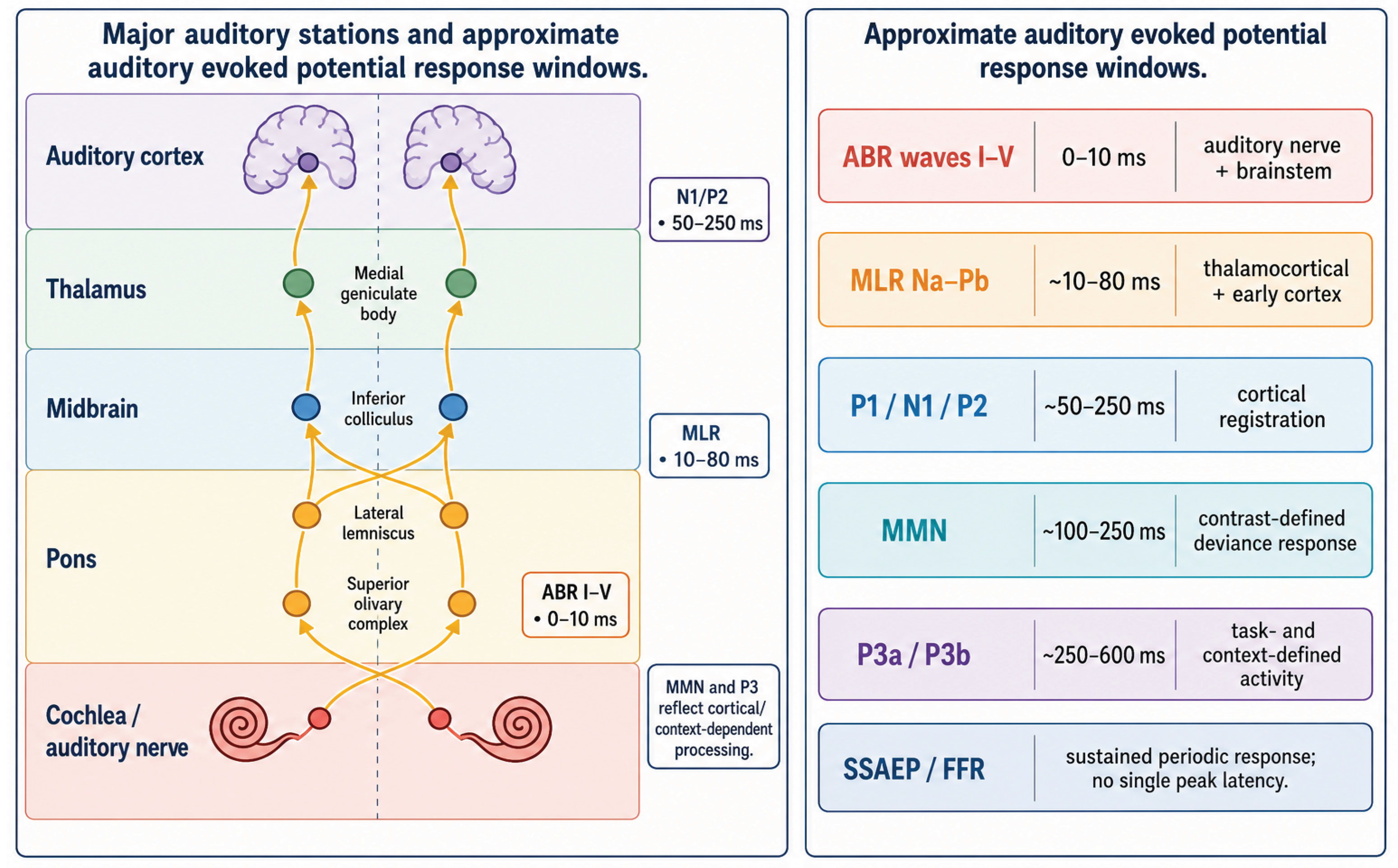
Table 1.
AEP components by latency, generator, analysis, state sensitivity, and use.
| Component | Latency and dominant generator | Typical paradigm / analysis | State sensitivity | Principal use |
| ABR | 0-10 ms; auditory nerve and brainstem | Click or chirp; coherent time-domain averaging | Low relative sensitivity; latency can still change | Hearing threshold, neural conduction, intraoperative monitoring |
| MLR | 10-80 ms; thalamocortical and early auditory cortex | Transient clicks/tones; peak amplitude and latency | Moderate to high | Early cortical integrity and anesthetic pharmacodynamics |
| N1/P2 | 50-250 ms; auditory cortical networks | Sound onset or change; ERP amplitude and latency | High; affected by attention, sleep, and drugs | Cortical registration, attention, development |
| MMN | About 100-250 ms; auditory-cortical deviance response, with protocol-dependent frontal contribution | Passive oddball; deviant-minus-control difference | Variable; may persist when P3b is absent | Auditory regularity and sensory-memory testing |
| P3a/P3b | About 250-600 ms; distributed frontoparietal networks | Novelty or active target/global rule | Very high | P3a: orienting; P3b: evaluation of task-relevant targets or global rules |
| 40-Hz SSAEP | Periodic; cortical/thalamocortical weighting | 40-Hz modulation; complex-valued Fourier coefficients or coherent averaging | High; sensitive to drowsiness and anesthesia | State tracking and research audiology |
| FFR | Sustained; frequency- and montage-dependent cortical/subcortical balance | Periodic or quasi-periodic complex sounds; waveform, spectral, and phase analysis | Frequency-, montage-, and state-dependent | Representation of stimulus periodicity and spectral harmonics |
| High-rate SSAEP (about 80-100 Hz) | Periodic; predominantly brainstem-weighted in adults | Faster-rate amplitude/frequency modulation; Fourier detection | Often more sleep-robust than 40 Hz; protocol-dependent | Objective threshold estimation |
Disclaimer/Publisher’s Note: The statements, opinions and data contained in all publications are solely those of the individual author(s) and contributor(s) and not of MDPI and/or the editor(s). MDPI and/or the editor(s) disclaim responsibility for any injury to people or property resulting from any ideas, methods, instructions or products referred to in the content. |
© 2026 by the authors. Licensee MDPI, Basel, Switzerland. This article is an open access article distributed under the terms and conditions of the Creative Commons Attribution (CC BY) license (http://creativecommons.org/licenses/by/4.0/).
Copyright: This open access article is published under a Creative Commons CC BY 4.0 license, which permit the free download, distribution, and reuse, provided that the author and preprint are cited in any reuse.