
Submitted:
28 August 2026
Posted:
03 September 2026
You are already at the latest version
Abstract
Autoimmune hemolytic anemia is a rare but potentially life-threatening disorder characterized by immune-mediated red blood cell destruction. Warm AIHA accounts for most pediatric cases and in childhood AIHA differs substantially from adult disease because of its stronger association with infections, immune dysregulation syndromes and inborn errors of immunity. The disease is clinically heterogeneous, ranging from self-limited episodes to severe chronic relapsing forms requiring prolonged immunosuppression. Diagnostic evaluation should not be limited to the direct antiglobulin test, but must include confirmation of hemolysis, exclusion of alternative causes of anemia, and a systematic search for underlying disorders. Corticosteroids (CS) remain the cornerstone of first-line therapy for warm AIHA, whereas rituximab has emerged as the preferred second-line treatment in CS-dependent or CS-refractory patients. Management of cold-antibody AIHA, paroxysmal cold hemoglobinuria, Evans syndrome, and AIHA associated with giant cell hepatitis requires specific diagnostic and therapeutic considerations. Novel targeted therapies directed against B cells, plasma cells, complement activation, Fc receptors, and intracellular signaling pathways are expanding the therapeutic landscape and may improve outcomes in refractory disease.
Keywords:
autoimmune hemolytic anemia
; children
; pediatric
; corticosteroids
; rituximab
Introduction
Autoimmune hemolytic anemia (AIHA) is a rare acquired disorder characterized by premature destruction of red blood cells (RBC) caused by autoantibodies directed against erythrocyte surface antigens. Although its estimated incidence is 0.81/100,000 children under 18 years old per year [1], AIHA represents the most important cause of acquired hemolytic anemia in childhood. The disease results from a heterogeneous group of immune mechanisms leading to either extravascular hemolysis, mainly occurring in the spleen and liver, or intravascular hemolysis mediated by complement activation; the intravascular hemolysis is usually more severe, being able to lyse around 200 mL erythrocytes/hour vs. 17 mL/hour of the extravascular type.
At disease onset, AIHA in children is frequently severe. Treatment recommendations remain largely based on retrospective studies, small pediatric series, expert opinion, and extrapolation from adult data. CS continue to represent the cornerstone of first-line therapy for wAIHA, whereas rituximab has emerged as the preferred second-line treatment. However, several unresolved questions remain regarding the management of refractory disease, the role of immunosuppressive agents, and the treatment of AIHA associated with immune dysregulation disorders. Furthermore, targeted therapies directed against B cells, plasma cells, Fc receptors, complement activation pathways, and intracellular signalling molecules are rapidly expanding the therapeutic landscape.
Here, I try to provide a practical pediatric-oriented approach to the diagnosis, etiological work-up, treatment, and long-term management of AIHA, integrating current evidence with real-world clinical experience and focusing on the challenges most commonly encountered in everyday pediatric practice.
Direct Antiglobulin Test
Direct antiglobulin test (DAT), also known as the Direct Coombs Test, whose principles are illustrated in Figure 1, is the cornerstone for the diagnosis of AIHA. The test detects immunoglobulins and/or complement components bound to the erythrocyte surface. In routine clinical practice, DAT is generally performed using polyspecific antisera directed against IgG and complement component C3d. Additional monospecific testing can identify IgA, IgM, or complement deposition individually [2], but these reagents are not available in many blood banks.
Despite its central role, AIHA cannot be diagnosed solely on the basis of a positive DAT. Evidence of hemolysis must be present, and alternative causes of anemia should be excluded. In my practice, the diagnosis is based on anemia, laboratory evidence of hemolysis and DAT positivity. However, a small proportion of patients have DAT-negative AIHA (see later). False-positive DAT results may be observed after administration of intravenous immunoglobulins, following blood transfusions, during infections, in autoimmune diseases, in hypergammaglobulinemic states, and occasionally in otherwise healthy individuals.
The Indirect Antiglobulin Test (IAT) detects free anti-RBC antibodies in serum and is useful almost exclusively in case of Mixed AIHA (see later).
Classification of Pediatric AIHA
Two complementary classification systems are currently used in AIHA. The first is based on the thermal characteristics of the autoantibodies responsible for hemolysis and applies to all forms of AIHA, whereas the second one is based on pathogenesis and primarily applies to warm AIHA.
Temperature-Reaction-Based Classification of AIHA
- •
- Warm AIHA. Warm AIHA (wAIHA) is the most common subtype, accounting for approximately 65-85% of pediatric cases [3,4,5]. It is usually mediated by IgG autoantibodies that react optimally at 37 °C and is characterized predominantly by extravascular hemolysis occurring in the spleen and, to a lesser extent, in the liver (Figure 2a). Warm AIHA is frequently (65-70% of patients) associated with IgG positivity alone: in a previous report combined IgG and C3d positivity and C3d isolated positivity have been observed in 20% and 13% respectively [6]; rarely wAIHA can be due to IgM or IgA, but the positivity is usually associated to IgG (IgA “only” AIHA are 1% of warm AIHA [5]). In some cases, more commonly in adulthood, wAIHA can be drug-associated [2,7] and the hemolysis abruptly ceases when the specific drug is stopped; in children antibiotics (i.e., ceftriaxone) are the most associated drugs [8,9].
- •
- Cold-Antibody AIHA. Cold-antibody-mediated AIHA (cAIHA) accounts for approximately 15% of pediatric cases [10] and comprises a heterogeneous group of disorders characterized by autoantibodies that bind erythrocytes at temperatures below normal body temperature, usually between 28 °C and 31 °C, and activate the classical complement pathway. Thermal amplitude is defined as the highest temperature at which agglutination occurs: the broader the thermal amplitude (TA) of these antibodies, the greater their pathogenic potential. TA reaches temperatures close to physiologic temperature in about 85% of pediatric cAIHA [5] (TA exceeding approximately 28 °C is active in the acral circulation and therefore pathogenic). Notably up to 35% of wAIHA [7,11] can present low titers of not clinically significant cold agglutinin which are non-reactive at physiologic body temperature (narrow TA). cAIHA is usually mediated by IgM autoantibodies and is characterized by both intravascular and extravascular hemolysis occurring predominantly in the liver (Figure 2b). The DAT is typically positive for C3d (and negative or weakly positive with IgG) and the cold agglutinin (CA) titre level is usually 5 > 64 at 4 °C. Three principal entities are recognized:
- ❖
- Cold Agglutinin Disease (CAD). The disease is typically chronic and primitive, caused by monoclonal cold-reactive autoantibodies, usually of the IgM class, and is therefore primarily encountered in adults (true pediatric CAD is virtually non-existent). Hemolysis is mainly complement-mediated and predominantly extravascular, occurring in the liver through clearance of C3b-coated erythrocytes. Patients may also develop (more rarely in childhood) cold-induced circulatory symptoms such as acrocyanosis, causing skin discoloration with or without numbness and tingling, and Raynaud-like phenomena.
- ❖
- Cold Agglutinin Syndrome (CAS). CAS accounts for approximately 10% of pediatric cases1, is typically acute and secondary principally to infections, in childhood particularly Mycoplasma pneumoniae and Epstein–Barr [12], although numerous other infectious agents have been implicated. Unlike CAD, CAS is generally transient and self-limited: most cases resolve with treatment of the underlying condition and supportive care. It may be occasionally secondary to autoimmune disorders or malignancy [2].
- ❖
- Paroxysmal Cold Hemoglobinuria (PCH) is a distinctive form of cAIHA mediated by the biphasic Donath–Landsteiner antibody: the IgG binds erythrocytes at cold temperatures in acral circulation and, although the antibody dissociates at higher temperatures, complement remains bound to erythrocytes and causes intravascular hemolysis after rewarming at 37 °C in the central circulation. In the Donath-Landsteiner test the disease is recapitulated in vitro, by first incubating patient plasma and RBCs at 4 °C, followed by warming at 37 °C: the test is technically demanding and not routinely available outside of specialized laboratories. PCH is really rare, with an incidence of 0.04 cases per 100 000 persons, and almost exclusive of pediatric AIHA with a median age at onset of 5 years [13]: it represents about 5% of all pediatric AIHA cases [5].
- •
- Mixed AIHA is characterized by the coexistence of warm-reactive IgG autoantibodies and clinically significant CA. Serologically, patients usually demonstrate DAT positivity for both IgG and C3d together with a significant CA titre showing a TA ≥30° C and evidence of a warm IgG antibody evidenced in the eluate with the IAT. It is very uncommon in childhood, representing less than 5% of all pediatric AIHA3,5; it is often associated with severe hemolysis and may be more difficult to manage than isolated warm disease because both warm-antibody-mediated and complement-mediated mechanisms contribute to red blood cell destruction.
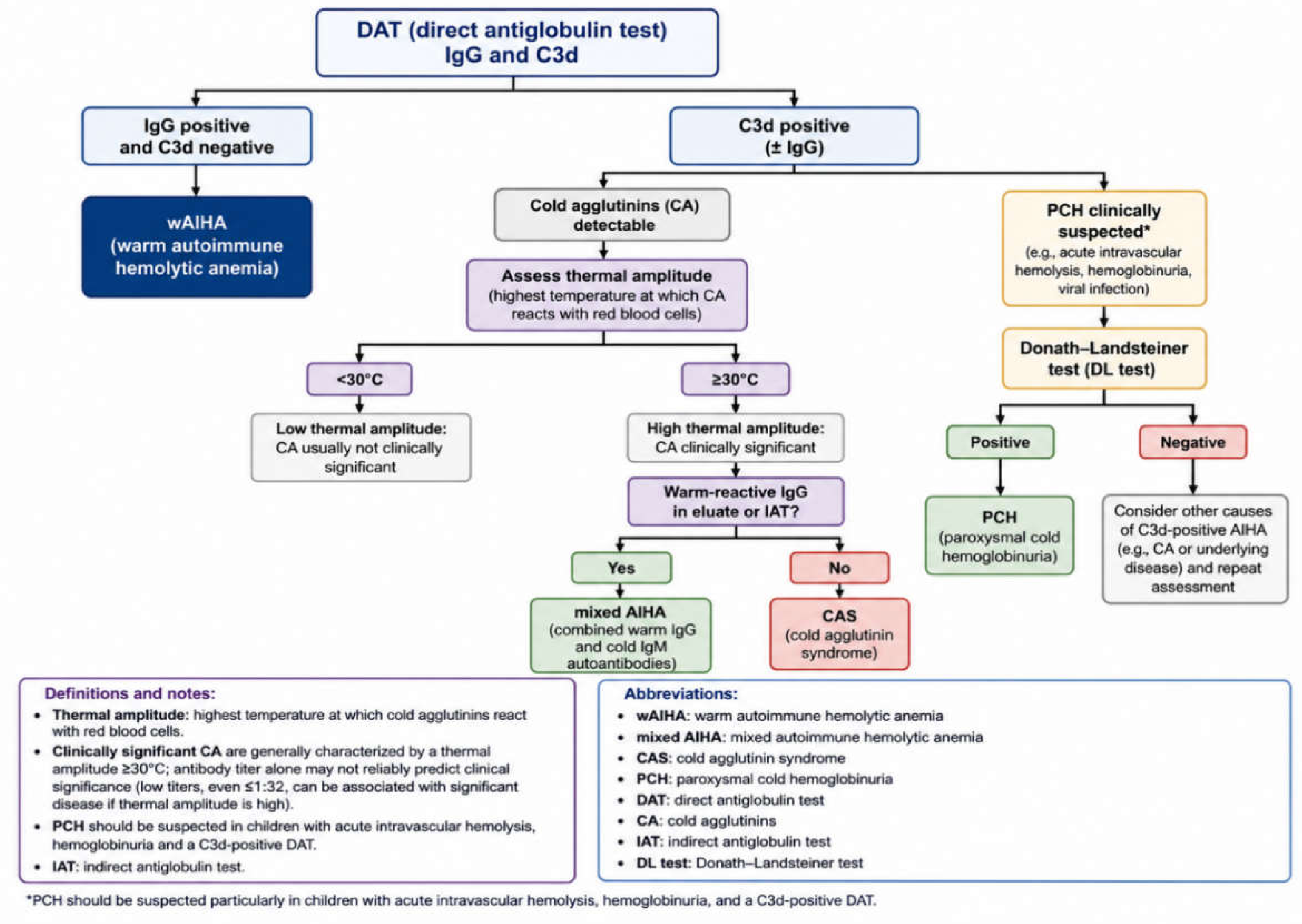
Figure 3.
proposes a simplified step-by-step diagnostic flowchart for the diagnosis of pediatric AIHA.
Figure 3.
proposes a simplified step-by-step diagnostic flowchart for the diagnosis of pediatric AIHA.
Pathogenesis-Based Classification
Primary AIHA. Primary (idiopathic) wAIHA is diagnosed when no associated condition can be identified despite appropriate clinical and laboratory investigation. Historically, primary disease was considered the predominant pediatric form. However, increasing recognition of immune dysregulation disorders and inborn errors of immunity has progressively reduced the proportion of cases classified as idiopathic. Current studies suggest that truly primary AIHA accounts for approximately 40% of pediatric cases [2,5,14].
Search for Underlying Disorders
The question is no longer whether a child with AIHA should be investigated for an underlying disorder, but rather how extensively and how early such investigations should be performed: identifying the cause of AIHA is often as important as treating the hemolysis itself because management must address both hemolysis and the underlying pathological process. In my opinion at least the following three categories of underlying disorders should be investigated:
- Infectious Triggers. Numerous infections have been associated with pediatric AIHA in about 10% of cases: [4] AIHA is often self-limited, however, severe hemolysis may still require treatment and transfusion support. The infectious agents most frequently implicated are Epstein–Barr virus (EBV) and Mycoplasma pneumoniae, but also cytomegalovirus (CMV), parvovirus B19, hepatitis viruses, and human immunodeficiency virus (HIV) have been described: [4] I usually add, during winter, research for influenza, para-influenza and SARS-CoV-2 viruses.
- Autoimmune diseases represent one of the most common causes of secondary AIHA. Systemic lupus erythematosus (SLE) is the best recognized association and may present with AIHA either at diagnosis or during the course of the disease with a risk that is increased in presence of ANA [16]. Importantly, a positive DAT is considerably more frequent than overt hemolysis in patients with SLE. Additional autoimmune disorders associated with AIHA include Autoimmune Lymphoproliferative Syndrome (ALPS), autoimmune hepatitis, autoimmune thyroid disease, inflammatory bowel disease, and juvenile idiopathic arthritis [4,17].
- Inborn Errors of Immunity and Immune Dysregulation. One of the most important advances in pediatric AIHA over the last decade has been the recognition that a substantial proportion of affected children harbour an underlying inborn error of immunity (IEI), especially Common variable immunodeficiency (CVID) [3,18,19]. In some series, defects of the immune regulation pathways have been identified in up to 40-50% of pediatric AIHA patients [4,18]. The possibility of an underlying IEI should be strongly considered particularly in children presenting with chronic lymphadenopathy, persistent splenomegaly, recurrent infections, multiple autoimmune manifestations, ES, and family history suggestive of immune dysfunction [18]. Importantly, recognition of these disorders may profoundly influence treatment decisions: in fact in these patients, successful long-term control of AIHA depends more on management of the underlying immune dysregulation than on treatment of hemolysis itself.
Other associated conditions include AIHA with hepatitis, AIHA associated to solid organ or hematopoietic stem cell transplantation (HSCT) (see later), and AIHA associated to malignancies or lymphoproliferative disorders (almost prerogative of adulthood).
How I Diagnose AIHA
Clinical Presentation
The clinical presentation of childhood AIHA is less variable than in adulthood, being uncommon, in my experience, cases developing gradually or fully compensated: the majority of pediatric patients, in fact, develop a rapidly progressive anemia. Children may present with jaundice, dark urine, irritability, poor feeding, tachycardia, fatigue, headache, and, more rarely, cardiovascular instability and exertional dyspnea. The presence of acrocyanosis and Raynaud phenomenon must raise suspicion of a cAIHA and, in my experience, are very uncommon in childhood. Physical examination may reveal splenomegaly, due to the trapping of spherocytes and autoantibody-opsonized RBCs in the spleen, in about 1/3 of patients; [20,21] hepatomegaly is much less common. The risk of venous thromboembolism, present in about 10% of adults, [21,22] is exceedingly rare in childhood.
Notably the seriousness of anemia is not determined exclusively by the rate of erythrocyte destruction but also by the ability of the bone marrow to mount an adequate compensatory erythropoietic response; this response is age related and explains why toddler and infants, whose bone marrow is less efficient, may exhibit particularly severe clinical manifestations.
Initial Laboratory Evaluation
The diagnosis of AIHA requires demonstration of both hemolysis and an immune mechanism of RBC destruction. The evaluation should include:
- Complete blood count with red cell indices: increase of Mean Corpuscular Volume (MCV) and Red Cell Distribution Width (RDW), due to the higher volume of reticulocyte compared to mature red cells, may be appreciated not at AIHA onset but after some days/weeks.
- Reticulocyte count. Reticulocytosis is one of the most important indicators of hemolysis and, in presence of anemia, is the starting point of the diagnostic work-up. It should be remembered that absolute count and not percentage of reticulocytes should be evaluated: the median value is 208 x 109/L. [4] However reticulocytopenia does not exclude AIHA (see later) and reticulopenic patients often represent a particularly severe subgroup.
- Direct antiglobulin test or DAT.
- Lactate dehydrogenase (LDH): LDH tends to be more markedly elevated in intravascular hemolysis and occasionally can be normal [23]. A frequent difficulty is the evaluation of LDH levels, profoundly different in children compared to adults: the thresholds for normal LDH levels are < 900 U/L for children < 1 year aged, < 700 U/L for children 1–3 years aged, < 640 U/L for children 3–6 years aged, <600 U/L for children 6–12 years aged, and <500 U/L at 12–18 years of age [24,25]. The reported LDH median value at AIHA onset is 2048 IU/L4 and the more severe the anemia, the higher the LDH level [21].
- Total and unconjugated bilirubin, whose level are higher in extravascular red cell destruction.
- Haptoglobin: typically decreased with a median value of 0.3 mg/dL [4] but its levels may be influenced by liver disease, inflammation, recent transfusions, intense physical activity, and rare congenital haptoglobin deficiency. It must be emphasized that haptoglobin value is physiologically low during the first months of life.
- Peripheral blood smear examination remains an important diagnostic tool. Microspherocytes are frequently observed in warm AIHA (wAIHA) and reflect partial membrane loss following macrophage-mediated erythrocyte injury. Although not specific, their presence supports the diagnosis of immune-mediated hemolysis. Evidence of nucleated RBCs is the counterpart of reticulocytosis. Furthermore, red cell agglutination and neutrophil erythrocyte rosettes suggest more cold-than warm-antibody disease.
- Urine examination: hemoglobinuria is typically observed only in patients with severe intravascular hemolysis, whereas hemosiderinuria is indicative of ongoing subacute or chronic hemolysis.
- Identification of blood groups and sub blood groups with extensive red cell antigen typing.
- Extensive pre-transfusion crossmatching.
- Immunological evaluation, including:
- IgG, IgA, IgM and, possibly, IgG subclasses and lymphocyte subsets.
- Auto antibody screening (antinuclear antibodies (ANA), extractable nuclear antigen antibodies (ENA), anti-double-stranded DNA antibodies, lupus anticoagulant, anti-cardiolipin and anti-beta-2 antibodies and anti-thyroid antibodies.
Why Immunological Assessment Should Be Performed Early
A practical message that deserves particular emphasis is that immunological evaluation should ideally be performed before administration of CS, intravenous immunoglobulin (IVIg), rituximab or immunosuppressive therapy. Failure to obtain these investigations before treatment may significantly complicate subsequent interpretation because the therapy can profoundly alter immunological parameters for months. Another relevant aspect is that the infectious risk, obviously increased in an undiscovered immunodeficiency, may be exaggerated by the therapies of the AIHA [21,26].
How I Initially Treat wAIHA
The management of AIHA continues to rely predominantly on expert opinion and individual clinical experience, as high-quality evidence remains scarce. Only a few prospective therapeutic trials have been performed, whereas most large clinical studies are outdated and predominantly involve adult or mixed-age populations, limiting their applicability to children.
Patient 1
A previously healthy 6-year-old boy was admitted because of progressive pallor, severe fatigue, jaundice, and dark urine developing over 5 days. Physical examination revealed tachycardia (135 beats/min), mild scleral icterus, and splenomegaly (3 cm below the left costal margin).
Laboratory evaluation showed: hemoglobin (HB) 6.7 g/dL (Hb fell to 4.8 g/dL within 6 hours), reticulocyte count 156 x 109/L, LDH 1950 U/L, total bilirubin 4.1 mg/dL (predominantly unconjugated), undetectable haptoglobin, and a DAT strongly positive for IgG. The peripheral blood smear demonstrated numerous microspherocytes. No evidence of infection or underlying autoimmune disease was identified during the initial evaluation. Because of symptomatic severe anemia, the patient received an urgent red blood cell transfusion together with intravenous methylprednisolone (10 mg/kg/d for 3 days) followed by oral prednisone (2 mg/kg/d). Three weeks later, Hb had increased to 11.2 g/dL with complete resolution of hemolysis signs.
Commentary on Patient 1
This patient represents the typical presentation of acute pediatric wAIHA. The most important initial questions are not whether the child should receive CS, but rather:
- Is transfusion necessary? Answer: yes, this patient has rapidly decreasing Hb level and is symptomatic.
- Is hemolysis sufficiently severe to justify high-dose CS therapy? Answer: yes, the severity of hemolysis justified high-dose of CS.
At diagnosis, I rapidly evaluate four key elements:
- Clinical status. Some patients tolerate remarkably low Hb levels because of gradual disease onset and adequate compensatory erythropoiesis, whereas others become hemodynamically unstable despite higher values. Particular attention is paid to signs of cardiovascular compromise, including tachycardia, orthostatic symptoms, dyspnea, reduced exercise tolerance, altered mental status, and evidence of heart failure.
- Severity of anemia and rate (speed) of Hb decline.
- Adequacy of reticulocyte response.
- Baseline risk of relapse. Although treatment decisions are driven by the severity of the acute presentation, I also assess clinical features that are associated with a higher risk of therapy refractoriness or subsequent relapse. These include younger age, more severe anemia at presentation, absence of a documented infectious trigger, Evans Syndrome (ES), AIHA subtypes other than wAIHA, and combined IgG/C3 positivity on the DAT rather than isolated IgG or C3 positivity;3,4,21,27 acute renal failure, described as adverse prognostic factor in adults, [27] has, in my opinion, no role in childhood where it is really infrequent [28].
Red Blood Cell Transfusion
One of the most common mistakes in the management of AIHA is delaying transfusion because of concerns regarding serological incompatibility. Transfusion should never be withheld when clinically indicated: actually, after few days from diagnosis 75-90% of children have been transfused.[3,4,12]. Because autoantibodies often react with all reagent red cells, complete compatibility is frequently impossible to achieve, and, if needed, patients should be transfused judiciously with least incompatible, ABO and Rh/Kell phenotype-matched, RBC: close collaboration with transfusion medicine specialists is therefore essential. Whenever possible, underlying alloantibodies should be excluded, but life-saving transfusions should not be delayed while attempting to obtain perfectly compatible blood [7,12,29]. Furthermore transfusions seems to be safe in the vast majority of patients [28,30]. I usually transfuse RBC at 10 ml/kg in the presence of clinical signs and/or when Hb is below 4–5 g/dL, and/or in the case of a rapid decline in Hb levels (also taking into account the reticulocyte count).
Corticosteroids: First-Line Therapy
I usually initiate intravenous high-dose methylprednisolone at 8-20 mg/kg/day (maximum 1 g/day) over no more than 60 minutes for 1–3 consecutive days: [3,14,17,21,27,31] it is historically reported that this therapy should be reserved to patients with severe hemolysis, profound anemia, cardiovascular compromise, or rapidly progressive disease but, actually, in my experience, this is the typical onset of pediatric wAIHA. High-dose methylprednisolone often produces a rapid suppression of immune-mediated erythrocyte destruction and may reduce transfusion requirements: after 2-3 days of iv CS administration I continue with oral prednisone or prednisolone 1.5-2 mg/kg/day (maximum 80 mg/day). In the (rare) event of a mild and gradual anemia it is possible to start directly with prednisone or prednisolone or methylprednisolone 1.5-2 mg/kg/day [17,32]. The response rate is around 80%, [33] the median time to response (stabilization of Hb levels, reduction of hemolysis markers, and progressive correction of anemia) is 4-9 days [32,33,34] and complete remission (normal Hb value plus negativity of hemolysis markers) is reached in 1 month in 58% of pediatric patients. [4] Once Hb has normalized and laboratory evidence of hemolysis has resolved, CS should be tapered gradually over 6-12 months: [17,21,31,35] I usually prefer extending the taper to approximately 12 months with extremely low doses and supplement with folic acid (on average 1 mg/day) during the period of active hemolysis. Excessively rapid tapering is one of the most frequent causes of early relapse [17,35].
Intravenous Immunoglobulin
The role of IVIg in isolated wAIHA remains less clearly established than in immune thrombocytopenia (ITP) [17,21,36]. In my opinion the effectiveness of IVIg is very mild but, since the AIHA onset is frequently challenging, even a modest therapeutic benefit may be clinically valuable, and so, with the exception of mild cases not at transfusion risk, according to other experiences, [28] I usually administer IVIg at 1-2 g/kg (single dose, repeatable if necessary) in 6-8 hours: some other schemes (i.e., 0.4 gr-1.0/kg/day for 5 days) have probably the same efficacy [37,38].
Comprehensive Initial Care Steps
That is what I do at every AIHA onset:
- Establish large-bore vascular access.
- Order packed red blood cells (PRBC) from the blood bank: in cases of life-threatening anemia, when waiting for typed and crossmatched blood is not feasible, request O Rh-negative PRBC.
- Laboratory evaluation (see above): in cases of severe anemia, initially collect only the blood volume essential for red blood cell typing and crossmatching, and complete the remaining blood sampling after the arrival of PRBC from the blood bank.
- Administer IVIg: the infusion should be temporarily interrupted to administer the transfusion, if required, as soon as PRBC become available.
- Transfuse PRBC if needed.
Monitoring Response
During follow-up I monitor Hb, reticulocyte count and laboratory markers of hemolysis (reticulocytes, LDH, bilirubin and haptoglobin), together with transfusion requirements. A rising Hb level accompanied by progressive normalization of hemolysis markers generally indicates adequate disease control. Persistence of severe hemolysis beyond 2-3 weeks of appropriate CS therapy should prompt early consideration of second-line treatment. Routine monitoring of the DAT is, in my experience, of limited clinical value: I observed many patients with remission lasting more than 10 years maintaining a persistent DAT positivity (persisting low hemolysis well compensated by the bone marrow response).
How I Treat Steroid-Refractory or Steroid-Dependent Warm AIHA
Patient 2
An 8-year-old girl was diagnosed with wAIHA after presenting with severe hemolytic anemia (Hb 4.1 g/dL, positive DAT for IgG, elevated LDH, and undetectable haptoglobin). She responded rapidly to high-dose methylprednisolone + IVIg with following prednisone 2 mg/kg/day, achieving complete hematological remission within 3 weeks. However, each attempt to reduce prednisone below 0.5 mg/kg/day resulted in recurrent hemolysis with declining Hb levels, increasing reticulocytosis, and rising bilirubin concentrations. Furthermore, she experienced one severe relapse requiring a temporary increase of prednisone at 2 mg/kg/day for 10 days. Four months after diagnosis, she remained CS-dependent and had developed weight gain, plus initial cushingoid facies, and mood changes.
Immunological evaluation performed at diagnosis had shown normal immunoglobulin levels and normal lymphocyte subsets, with no evidence of an underlying immune dysregulation disorder.
Commentary on Patient 2
This patient illustrates one of the most common therapeutic challenges in pediatric AIHA. Although CS remain highly effective as first-line therapy, prolonged exposure is associated with substantial toxicity, particularly in growing children.
- When should a patient be considered CS-dependent? In my opinion, CS dependency can be defined when unsuccessful attempts at tapering require maintenance of prednisone at doses exceeding 0.2 mg/kg/day [17].
- When should a patient be considered CS-refractory? In my opinion, CS refractoriness can be defined when no satisfactory response is achieved after 2 weeks of therapy, including high-dose intravenous CS. In patients with a doubtful or partial response, it may be reasonable to wait up to 3 weeks before considering second-line treatment [17].
CS refractoriness, persistent CS dependence, inability to taper CS within a reasonable timeframe, or frequent relapses during tapering should prompt early consideration of second-line treatment.
Rituximab
Rituximab has become the preferred second-line treatment for most children with wAIHA both in cases of CS refractoriness or dependency [2,17,40,41]. The rationale for its use is straightforward. Since wAIHA is predominantly mediated by autoreactive B cells producing pathogenic antibodies, depletion of CD20-positive B lymphocytes frequently results in durable disease control while allowing CS withdrawal. Published pediatric and adult series [21,41,42] consistently demonstrate 70-80% response rates, with many patients achieving CS-free remission. Responses generally can become evident within 3-6 weeks (almost never after 1st dose), although delayed responses may occur [20,27]. Potential adverse effects include infusion reactions, increased susceptibility to infection, prolonged hypogammaglobulinemia, delayed B-cell reconstitution, progressive multifocal encephalopathy [17,41]. Despite these concerns, rituximab remains my preferred second-line treatment in most patients with CS-dependent or CS-refractory wAIHA. In my opinion its use as first-line therapy in association to CS, proposed in severe wAIHA of adulthood, [2] is not supported by sufficient evidence in childhood.
Mycophenolate Mofetil
Mycophenolate mofetil (MMF) at the usual dose of 600 mg/m2 twice daily is one of preferred CS-sparing agents in pediatric AIHA. By inhibiting inosine monophosphate dehydrogenase, MMF selectively suppresses lymphocyte proliferation and reduces autoantibody production. The drug has accumulated substantial pediatric experience through its use in transplantation, rheumatology, and immune dysregulation disorders. One of the major advantages of MMF is its favourable safety profile compared with many traditional immunosuppressive agents. The main limitation is its relatively slow onset of action, which may require several weeks or months. In patients with mild CS dependency (i.e., prednisone 0.2-0.3 mg/kg/day) it can be considered, with the agreement of parents, in place of rituximab as second step [44,45].
Sirolimus
Sirolimus has emerged as an increasingly attractive option for patients with AIHA associated with immune dysregulation disorders. By inhibiting the mammalian target of rapamycin (mTOR) pathway, sirolimus modulates lymphocyte activation, promotes immune tolerance, and may restore regulatory immune mechanisms [45]. The drug appears particularly useful, in my opinion, in patients with AIHA in an ALPS context or in CS-dependent disease if MMF failed [45]. Failure to respond to MMF does not preclude a subsequent response to sirolimus. In the largest pediatric series of refractory autoimmune cytopenias, 8 patients who failed MMF subsequently achieved a response with sirolimus, suggesting that these agents have distinct mechanisms of action and should not be considered interchangeable [45,46]. Conversely, published evidence supporting the reverse sequence (MMF efficacy after sirolimus failure) is currently lacking.
Bortezomib
Bortezomib, a selective proteasome inhibitor extensively used against multiple myeloma, targets antibody-producing plasma cells and therefore addresses a pathogenic compartment not eliminated by rituximab. The drug has shown very promising activity in highly refractory AIHA, [2,20,47,48,49,50] particularly after multiple treatment failures and is the drug I use after rituximab or MMF/sirolimus failure. Although current evidence is largely limited to case reports and small series, responses have often been rapid and clinically impressive: in my opinion, it should no longer be considered a future therapy; at the same time it must be underlined that, although pediatric experience is increasing, [50] it has no official pediatric indication in major regulatory jurisdictions and its use in childhood remains off-label. The usual dose is 1.3 mg/m2 administered intravenously on days 1, 4, 8, and 11 of a 21-day cycle [50,51]. Most responses are observed within the first cycle [51]. In patients who respond, subsequent cycles can be administered using a weekly schedule or at longer intervals to reduce toxicity while maintaining efficacy (this is my preferred option). The drug is generally manageable.
Splenectomy
Historically, splenectomy has been associated with response rates of approximately 75% in AIHA [21]. However, durable remission is not universal, as nearly 1/3 of patients eventually relapse.[21,31]. Combined with surgical complications in 12% of patients,[52] the irreversibility of the procedure, the lifelong risk of overwhelming post-splenectomy infection, a possible increase in mortality [21] and the availability of several effective medical alternatives, these limitations have led to a progressive decline in the use of splenectomy, particularly in pediatric patients [17].
Cyclosporine
Cyclosporine inhibits T-cell activation through calcineurin blockade and has been successfully used in refractory AIHA. Its use is generally limited by nephrotoxicity, hypertension, and the need for therapeutic drug monitoring. I do not currently use it [17].
Azathioprine
Cyclophosphamide
Cyclophosphamide is a potent alkylating agent capable of suppressing both B-cell and T-cell immune responses. Historically, it was used more frequently in severe autoimmune cytopenias, whereas its use today is generally restricted to highly refractory disease because of concerns regarding cumulative toxicity. I do not currently use it [17].
HSCT
How I Choose Among Second- and Third-Line Therapies in Warm AIHA
The optimal sequencing of second-line therapies remains uncertain: it must be emphasized that at 3 years follow-up 28% are on therapy4 and that a second-line therapy is necessary in about 30% of patients [41]. For most children with CS-refractory wAIHA, rituximab remains my preferred second-line treatment, and for those requiring prolonged CS-sparing treatment, I usually choose MMF, followed by sirolimus in selected cases. As third step, before recourse to splenectomy or another “new” drug, I usually choose a trial with bortezomib.
Relapses are more common in younger age,[21] and, if they occur, another attempt with CS, in my opinion, may be done: I frequently associate immediately MMF.
My personal approach to the treatment of wAIHA is outlined in Figure 4.
Special Situations
Patient 3
A 7-year-old boy was referred because of recurrent wAIHA associated with ITP. During the previous 3 years, he had experienced 2 episodes of severe hemolysis requiring CS and transfusion support. Physical examination revealed persistent splenomegaly and generalized lymphadenopathy. Laboratory investigations demonstrated DAT-positive hemolytic anemia, thrombocytopenia, elevated double-negative T cells, and hypergammaglobulinemia. Genetic evaluation subsequently confirmed ALPS.
Commentary on Patient 3
This patient illustrates an important principle in pediatric AIHA: not all autoimmune hemolytic anemias are biologically equivalent. In some children, hemolysis represents only one manifestation of a broader immune dysregulation syndrome. In these situations, treatment directed exclusively at hemolysis is often insufficient and long-term disease control requires targeting the underlying immunological abnormality.
Evans Syndrome
Evans Syndrome (ES), the concomitant or sequential occurrence of 2 autoimmune cytopenias, most frequently AIHA and ITP, deserves special consideration, since these patients, which seem more frequent among pediatric than adult patients, representing more than 1/3 of AIHA, [4,21] have a higher probability of chronic or relapsing disease and treatment dependence: [4,21,41,55,56]. at 20 years follow-up of pediatric ES AIHA is in remission is about ¾ of cases [56]. Survival at 15 years after diagnosis is 84%, infection being the main cause of death [56]. Risk factors of fatal outcome are age < 24 months, family history of inborn errors of immunity, splenomegaly, and hepatomegaly [57]. ES is now increasingly recognized as a warning sign of underlying immune dysregulation which in some studies reach 65-80% of all ES patients, [55,56] and immunological plus genetic investigations (NGS panel for immunodeficiency/immune dysregulation) should be broader than in isolated AIHA.
Acute management generally follows the same principles used for isolated wAIHA. First-line treatment includes CS + IVIg (the vast majority of ES are with ITP where IVIg are very efficient) and transfusion support when required. However, long-term management often differs substantially. Rituximab remains an effective option in many patients, but relapses are frequent and it is contraindicated in ALPS patients, given the risk of infectious complications [44]. Because repeated CS exposure is common and frequently poorly tolerated, I have a low threshold for introducing CS-sparing therapies such as MMF or, particularly in patients with ALPS, sirolimus.
DAT-Negative AIHA
A negative DAT does not exclude AIHA. Approximately 5% of patients [14,21] with clinically convincing AIHA may have negative results using standard DAT methodologies. Several mechanisms may account for this finding, including low-density erythrocyte-bound antibodies, low-affinity antibodies removed during washing procedures, or technical limitations of conventional assays (i.e., in case of IgA-mediated disease or warm IgM autoantibodies without availability of monospecific sera). When clinical suspicion remains high despite a negative DAT, additional investigations should be pursued. More sensitive methods include microcolumn assays, solid-phase testing, washing RBCs with low ionic strength solutions (LISS), immunoradiometric assays, ELISA, flow cytometric DAT, Dual Direct Antiglobulin Test (DDAT) and ultra-specialized reference laboratory investigations such as the mitogen stimulated DAT; [20,58] furthermore Donath-Landsteiner testing (see later) should also be considered, being DAT occasionally negative in Paroxysmal Cold Hemoglobinuria (PCH) [13]. In children with a suspected DAT-negative AIHA I suggest, in order to exclude a lympho or myeloproliferative disease, to perform a bone marrow aspiration before starting CS therapy. The treatment of DAT-negative AIHA is similar to those used for the treatment of typical wAIHA (see later).
AIHA with Reticulocytopenia or Inadequate Reticulocytosis
Reticulocytosis is generally considered a hallmark of AIHA. However, up to 20% of patients can present with severe reticulocytopenia despite ongoing hemolysis: [21] this phase can be limited to the early phase of the disease [4] or may be longer; remarkably, adding patients presenting inadequate reticulocytosis, the proportion of patients, in adult studies, increases up to 50-70% [21,59]. In my experience, according to other reports, [28] the proportion is much lower in children, and, many potential mechanisms, including immune-mediated destruction of erythroid precursors or transient infectious marrow suppression, have been proposed to explain inadequate reticulocyte response [59,60,61,62]. In my opinion the first hypothesis appears more reasonable and frequent. When presenting, I suggest to perform in these patients a bone marrow examination to exclude alternative diagnoses [17]. Since reticulocytopenic patients frequently experience more severe anemia than expected from the degree of hemolysis,[21] transfusion requirement is often higher than in classical wAIHA, and so, erythropoietin (EPO) treatment must be considered:[20,21,59,60,63,64] since the reported doses regard only adult patients,[27] I suggest to use the same dose proposed in infants with transfusion dependent spherocytosis: 750 IU/kg/week in 3 doses[65].
AIHA Associated with Giant-Cell Hepatitis
One of the most distinctive pediatric forms of secondary AIHA is AIHA associated with giant-cell hepatitis. The disease predominantly affects infants and toddlers < 1 year of age and is characterized by the association of wAIHA with severe hepatitis showing multinucleated giant hepatocytes on liver histology. AIHA and hepatitis frequently have a contemporary onset but AIHA in about 1/3 of cases precedes the onset of hepatitis by several weeks or months, making early recognition challenging. The clinical presentation is often severe, with warm-antibody hemolysis, markedly elevated transaminases, and progressive liver dysfunction [66].
Recognition of this entity is crucial because management differs substantially from that of conventional wAIHA and requires close collaboration among pediatric haematologists and hepatologists. Initial treatment usually includes CS + cyclosporine or azathioprine. However, prolonged CS dependence is common and relapses frequently occur during tapering: for this reason, rituximab has become a central component of modern therapy [66]. Several reports have documented dramatic improvements following B-cell depletion, and many centres now introduce rituximab relatively early in the disease course [67,68].
Post-Transplant AIHA
Autoimmune hemolytic anemia is a recognized complication following both hematopoietic stem cell transplantation (HSCT)[69,70] and, less frequently, solid organ transplantation (above all liver and heart-lung transplantation) [71,72].
AIHA may develop several months after HSCT: it is very unusual after less than 1 month [73]. Although relatively uncommon (it develops in about 5% of allogenic HSCT)[73], AIHA is often severe, associated with substantial morbidity, and frequently refractory to conventional therapies. Its pathogenesis is multifactorial, involving delayed immune reconstitution, donor–recipient immune dysregulation, loss of immune tolerance, viral infections, and, in some cases, mixed chimerism or graft-versus-host disease. Compared with primary pediatric AIHA, post-HSCT AIHA is characterized by a more aggressive clinical course and poorer therapeutic responses. Management should be individualized and tailored to the transplant setting, taking into account immune reconstitution, graft status, concomitant infections, and graft-versus-host disease. CS remain the standard first-line treatment, but complete responses are uncommon. Rituximab is therefore often introduced early and represents a cornerstone of therapy. Additional treatment options include optimization of baseline immunosuppression, MMF, sirolimus, bortezomib, and, in selected refractory cases, daratumumab or complement inhibitors (see later), depending on the underlying pathogenic mechanism [20,69,70,73].
Immune-mediated hemolysis after solid organ transplantation, affecting approximately 10–15% of recipients, is known as passenger lymphocyte syndrome (PLS) and results from the transient production of IgG by donor-derived B lymphocytes passively transferred with the graft and specificity within ABO (anti-A or anti-B) or Rh (anti-D) blood groups. Hemolysis typically develops within 1–3 weeks after transplantation, may be severe, but is usually self-limited [72,74].
Cold AIHA
Patient 4
A previously healthy 9-year-old girl presented with pallor, jaundice, and dark urine one week after a respiratory infection. Laboratory investigations revealed severe anemia, elevated LDH, low haptoglobin, and a DAT positive for C3d. Peripheral blood smear examination showed erythrocyte agglutination. Serological testing demonstrated recent Mycoplasma pneumoniae infection.
Commentary on Patient 4
This patient illustrates the most common form of cold-antibody-mediated AIHA encountered in childhood: secondary cold agglutinin syndrome following infection. Unlike adults, where primary cAIHA is often associated with primary clonal lymphoproliferative disorders, pediatric cAIHA is usually secondary, transient and self-limited.
How I Treat Secondary Cold Agglutinin Syndrome
The first step should be distinguishing secondary Cold Agglutinin Syndrome (CAS) from primary Cold Agglutinin Disease (CAD), but the latter is virtually non-existent in childhood [4,75], and I do not discuss it in this paper. The vast majority of pediatric cases require only supportive care, treatment of the underlying infection when appropriate, avoidance of cold exposure by blanketing or maintaining room temperatures at 37 °C–40 °C, monitoring urine output in the presence of significant intravascular haemolysis, and transfusion support if clinically indicated; the extremity chosen for transfusion should be kept warm, using, if possible, an in-line blood warmer.
First-Line Treatments
CS are generally less effective than in wAIHA [17,20,21,40] and should not be expected to produce the same response rates. At the same time, in my opinion, it should be remarked that in some studies they maintained a certain efficacy,[3,76] which is in the order of 50-60% of cases if both partial and complete response are considered,[21] and that they continued to be frequently used in cAIHA patients:[12,77]: on these basis, also considering that pediatric cAIHA is usually self-limiting and that rituximab carries a ultra-rare but lethal side effect such as the progressive multifocal encephalopathy,[78,79] whose risk is less acceptable in treating “benign” hematological diseases, I suggest, in accordance to what recently reported in pediatric AIHA with C3d only DAT positivity,[12] if treatment needed, a brief cycle of CS (7-10 days) as first-line therapy.
Second-Line Treatments
In the rare cases of persisting significant hemolysis after CS administration, rituximab represents the most reasonable therapeutic option [17,77]. Splenectomy is generally ineffective in these patients Lechner [21].
The increasing availability of complement inhibitors may eventually modify the management of severe cold-antibody disease because complement activation represents the central pathogenic mechanism. So, therapeutic inhibition of the complement cascade has rapidly evolved from a theoretical concept to a clinical reality and is particularly attractive because targeting a specific pathogenic mechanism while potentially avoiding the broader immunosuppressive effects associated with conventional therapies.
Sutimlimab, which selectively blocks C1s and inhibits activation of the classical complement pathway, has demonstrated remarkable efficacy in CAD.[2,80,81,82,83,84], Its rapid inhibition of classical complement-mediated hemolysis provides a strong pathophysiological rationale for its use as rescue or bridging therapy in severe, life-threatening CAS;[85] however, the drug is really expensive, and it is not currently approved for pediatric use, safety and efficacy not being established.
Pegcetacoplan is a subcutaneously administered C3 inhibitor that prevents downstream complement activation and demonstrated robust efficacy in paroxysmal nocturnal hemoglobinuria (PNH) with preliminary encouraging results and rapid onset of efficacy in treatment of both cAIA and wAIHA [86]. It is approved for pediatric use from 12 years of age in selected complement-mediated renal diseases, but it is not currently approved for childhood AIHA.
Plasma exchange may provide temporary benefit in selected patients with severe IgM-mediated hemolysis by removing circulating pathogenic antibodies; human albumin or fresh frozen plasma may be used as replacement fluids, with 1–1.5 times the estimated plasma volume exchanged. However, its effect is usually transient [21,38,87].
How I Treat Paroxysmal Cold Hemoglobinuria
PCH occurred predominantly in childhood [13,88] as post-infectious complication after an upper respiratory tract infection; other rarer causes include vaccination, autoimmune diseases, and hematopoietic neoplasms [10,13,89]. Clinical manifestations may include marked intravascular hemolysis, chills, fever, cramps, lower back and abdominal pain, and vasomotor phenomena. In the peripheral blood smear RBC-neutrophil rosettes and erythrophagocytosis by neutrophils can be observed [10]. Despite the often-dramatic clinical presentation, the disease is usually self-limited and most patients recover completely with supportive treatment, avoidance of cold exposure,[88,90] and red blood cell transfusions,[20,21] which are necessary in 72% of cases [13].
The role of CS remains controversial, and evidence supporting their routine use is very limited [13,15] even though in some cases they proved to be efficient and many clinicians continue to administer CS (+/- IVIg) in severe presentations [91,92,93] (52% of patients in the review of Jacobs et al. [13]). In severe cases rituximab [94,95], eculizumab [96,97] and plasmapheresis [13,97,98] can also be considered. Recurrence is fairly uncommon [93,95].
The main characteristics of wAIHA, CAS and PCH are presented in Table 1.
How I Treat Mixed AIHA
Mixed AIHA represents probably much less than 5% of all pediatric AIHA [3,4,5], and is the only AIHA type with a sex preference (female-to-male ratio of 2.25:1) [99]. Many case reports [100,101,102,103,104,105,106] illustrate the therapeutic challenges: [38,72,100,101,102,103,104,105,106] the cold antibody titer does not affect the prognosis [99]. I suggest to start with CS (+/- IVIg), which in some reports have shown complete efficacy; [100,101,107] in severe cases or in absence of initial response to CS after 4-7 days, in my opinion, rituximab should be administered. [38] Notably, due to its efficacy in case of IgM mediated AIHA, plasmapheresis may be highly effective in these patients [38,103] whereas splenectomy, similarly to cAIHA, is often ineffective [99].
Future Perspectives
Advances in the understanding of AIHA pathogenesis are now driving the development of targeted therapies directed against specific components of the immune system, creating opportunities for more effective and less toxic treatment strategies; perhaps the most important conceptual shift is the progressive transition from empiric immunosuppression to mechanism-based therapy. Rather than broadly suppressing immune function, emerging treatments aim to target the precise cellular and molecular pathways responsible for autoantibody production, complement activation, and immune dysregulation. A comprehensive review of emerging therapeutic options for pediatric AIHA is beyond the scope of this paper and I will limit the discussion to the agents that appear most promising in this setting, although a serious limitation of what I am going to discuss is that, almost no reported evidence regards the pediatric population. Unless otherwise specified, all the following agents are currently off-label for pediatric AIHA.
Daratumumab
Even though rituximab effectively depletes CD20-positive B lymphocytes, it does not eliminate mature plasma cells, which can continue producing pathogenic autoantibodies despite complete B-cell depletion. This observation probably explains a substantial proportion of rituximab failures and relapses. Accordingly, while bortezomib represents the first plasma cell-directed approach explored in refractory AIHA, daratumumab, an anti-CD38 monoclonal antibody targeting plasma cells has emerged as a more selective therapeutic strategy and some reports have documented remarkable responses in patients with multiply relapsed or treatment-refractory disease [108,109]. It is used in various schedules, the most frequent being 16 mg/kg/week intravenously.
FcRn Inhibitors
The neonatal Fc receptor (FcRn) plays a crucial role in protecting IgG from intracellular degradation and thereby prolongs antibody half-life. Blocking FcRn accelerates IgG catabolism and reduce circulating pathogenic autoantibodies, providing a biologically attractive strategy for antibody-mediated diseases such as wAIHA. Monoclonal antibodies including rozanolixizumab, batoclimab, and, above all, nipocalimab have demonstrated promising activity in several autoimmune disorders and are currently being investigated in immune-mediated cytopenias. These molecules may represent a valuable future option for patients with refractory wAIHA, but at present, should be considered investigational therapies [20,90,100,101,102,103,104,105,106,107,108,109,110,111,112].
BTK Inhibitors
Targeting B-cell receptor (BCR) signaling is a promising therapeutic approach, as this pathway regulates B-cell survival, proliferation, and activation through key kinases, including Bruton tyrosine kinase (BTK), phosphoinositide 3-kinase (PI3K), and spleen tyrosine kinase (SYK). Inhibition of BTK suppresses pathogenic B-cell responses and may reduce autoantibody-mediated hemolysis. Given their targeted mechanism of action and favourable theoretical rationale, BTK inhibitors may become important components of future treatment algorithms, particularly in patients with relapsing disease who have failed conventional immunosuppressive therapies and B-cell-directed approaches [90]. Several BTK inhibitors, including ibrutinib, rilzabrutinib, and other emerging compounds [84,113,114] are currently under investigation in autoimmune hematological disorders [20]. Preliminary studies suggest potential activity in refractory AIHA, although clinical experience remains limited. Ibrutinib is approved for pediatric use from 1 year of age in the United States for chronic graft-versus-host disease, but not for AIHA, where the use remains off-label.
Fostamatinib, a spleen tyrosine kinase (Syk) inhibitor, reduces autoantibody-mediated RBC and platelet destruction by disrupting Fc receptor (FcR)-dependent signaling in macrophages. It is approved for the treatment of chronic ITP in adults, and emerging evidence suggests a potential therapeutic role in wAIHA [115,116,117]. However, fostamatinib is not approved for pediatric use, mainly because of adverse effects on actively growing bones observed in nonclinical studies. Therefore, it cannot currently be considered a treatment option for childhood AIHA, and its future role in this setting appears limited.
Conclusion
The management of autoimmune hemolytic anemia (AIHA) is undergoing a profound transformation. For decades, treatment strategies relied largely on CS, splenectomy, and non-specific immunosuppressive agents. Although these approaches remain effective in many patients, they are frequently associated with significant toxicity and often fail to provide durable disease control in refractory cases. Perhaps the most important future development in pediatric AIHA will likely be characterized by earlier identification of underlying immune dysregulation, broader use of molecular diagnostics, and increasing availability of targeted therapies directed against specific pathogenic pathways. Under this paradigm, two children with apparently identical wAIHA could ultimately receive completely different treatments based on their underlying immunological abnormalities. As our understanding of immune regulation continues to evolve, AIHA may become one of the clearest examples of precision medicine applied to pediatric hematology. Until that time, careful clinical judgment, early recognition of underlying immune dysregulation, and individualized treatment remain the cornerstones of pediatric AIHA management.
Acknowledgments
The author thanks Angela Guarina (Palermo, Italy) for valuable discussions and insightful comments.
References
- Aladjidi, N.; Jutand, M.A.; Beaubois, C.; Fernandes, H.; Jeanpetit, J.; Coureau, G.; Gilleron, V.; Kostrzewa, A.; Lauroua, P.; Jeanne, M.; et al. Reliable assessment of the incidence of childhood autoimmune hemolytic anemia. Pediatr. Blood Cancer 2017, 64. [CrossRef]
- Jäger, U.; Barcellini, W.; Broome, C.M.; Gertz, M.A.; Hill, A.; Hill, Q.A.; Jilma, B.; Kuter, D.J.; Michel, M.; Montillo, M.; et al. Diagnosis and treatment of autoimmune hemolytic anemia in adults: Recommendations from the First International Consensus Meeting. Blood Rev. 2020, 41, 100648. [CrossRef]
- Sankaran, J.; Rodriguez, V.; Jacob, E.K.; Kreuter, J.D.; Go, R.S. Autoimmune Hemolytic Anemia in Children: Mayo Clinic Experience. J. Pediatr. Hematol. Oncol. 2016, 38, e120-4. [CrossRef]
- Aladjidi, N.; Leverger, G.; Leblanc, T.; Picat, M.Q.; Michel, G.; Bertrand, Y.; Bader-Meunier, B.; Robert, A.; Nelken, B.; Gandemer, V.; et al. New insights into childhood autoimmune hemolytic anemia: A French national observational study of 265 children. Haematologica 2011, 96, 655–663. [CrossRef]
- Vaglio, S.; Arista, M.C.; Perrone, M.P.; Tomei, G.; Testi, A.M.; Coluzzi, S.; Girelli, G. Autoimmune hemolytic anemia in childhood: Serologic features in 100 cases. Transfusion 2007, 47, 50–54. [CrossRef]
- Loriamini, M.; Cserti-Gazdewich, C.; Branch, D.R. Autoimmune hemolytic anemias: Classifications, pathophysiology, diagnoses, and management. Int. J. Mol. Sci. 2024, 25, 4296. [CrossRef]
- Garratty, G.; Petz, L.D. Approaches to selecting blood for transfusion to patients with autoimmune hemolytic anemia. Transfusion 2002, 42, 1390–1392. [CrossRef]
- Arndt, P.A. Drug-induced immune hemolytic anemia: The last 30 years of changes. Immunohematology 2014, 30, 44–54. [CrossRef]
- Sun, X.M.; Liu, L.H.; Wu, Q.; Wang, H.G. Cefoperazone/sulbactam-induced hemolytic anemia. J. Postgrad. Med. 2023, 69, 46–49. [CrossRef]
- Sokol, R.J.; Booker, D.J.; Stamp, R. Erythropoiesis: Paroxysmalcold haemoglobinuria: A clinic-pathological study ofpatients with a positive Donath-Landsteiner test. Hematology 1999, 4, 137–164. [CrossRef]
- Petz, L.D.; Garratty, G. Immune Hemolytic Anemias, 2nd ed.; Churchill Livingstone, 2004.
- Blachez, M.; Aladjidi, N.; Modot, T.; Fernandes, H.; Garnier, N.; Chastagner, P.; Bayart, S.; Armari-Alla, C.; Piguet, C.; Pasquet, M.; et al. Childhood isolated C3d-positive AIHA: Favorable prognosis but rare relapse or associated immunodeficiency. Blood Adv. 2026, 10, 521–530. [CrossRef]
- Jacobs, J.W.; Figueroa Villalba, C.A.; Booth, G.S.; Woo, J.S.; Stephens, L.D.; Adkins, B.D. Clinical and epidemiological features of paroxysmal cold hemoglobinuria: A systematic review. Blood Adv. 2023, 7, 2520–2527. [CrossRef]
- Abdel-Salam, A.; Bassiouni, S.T.; Goher, A.M.; Shafie, E.S. Autoimmune Hemolytic Anemia in the Pediatric Age Group: The Egyptian Experience. Ann. Hematol. 2023, 102, 1687–1694. [CrossRef]
- Berentsen, S.; D’Sa, S.; Randen, U.; Malecka, A.; Vos, J.M.I. Cold agglutinin disease: Improved understanding of pathogenesis helps define targets for therapy. Hemato 2022, 3, 574–594. [CrossRef]
- Granel, J.; Fernandes, H.; Bader-Meunier, B.; Guth, A.; Richer, O.; Pillet, P.; Leverger, G.; Ducassou, S.; Fahd, M.; Pasquet, M.; et al. Antinuclear antibody-associated autoimmune cytopenia in childhood is a risk factor for systemic lupus erythematosus. Blood 2024, 143, 1576–1585. [CrossRef]
- Ladogana, S.; Maruzzi, M.; Samperi, P.; Perrotta, S.; Del Vecchio, G.C.; Notarangelo, L.D.; Farruggia, P.; Verzegnassi, F.; Masera, N.; Saracco, P.; et al. Diagnosis and management of newly diagnosed childhood autoimmune haemolytic anaemia. Recommendations from the Red Cell Study Group of the Paediatric Haemato-Oncology Italian Association. Blood Transfus. 2017, 15, 259–267. [CrossRef]
- Zama, D.; Conti, F.; Moratti, M.; Cantarini, M.E.; Facchini, E.; Rivalta, B.; Rondelli, R.; Prete, A.; Ferrari, S.; Seri, M.; et al. Immune cytopenias as a continuum in inborn errors of immunity: An in-depth clinical and immunological exploration. Immun. Inflamm. Dis. 2021, 9, 583–594. [CrossRef]
- Hill, Q.A.; Stamps, R.; Massey, E.; Grainger, J.D.; Provan, D.; Hill, A.; British Society for Haematology Guidelines. Guidelines on the management of drug-induced immune and secondary autoimmune, haemolytic anaemia. Br. J. Haematol. 2017, 177, 208–220. [CrossRef]
- Barcellini, W.; Zaninoni, A.; Giannotta, J.A.; Fattizzo, B. New Insights in Autoimmune Hemolytic Anemia: From Pathogenesis to Therapy Stage 1. J. Clin. Med. 2020, 9, 3859. [CrossRef]
- Barcellini, W.; Fattizzo, B.; Zaninoni, A.; Radice, T.; Nichele, I.; Di Bona, E.; Lunghi, M.; Tassinari, C.; Alfinito, F.; Ferrari, A.; et al. Clinical heterogeneity and predictors of outcome in primary autoimmune hemolytic anemia: A GIMEMA study of 308 patients. Blood 2014, 124, 2930–2936. [CrossRef]
- Ungprasert, P.; Tanratana, P.; Srivali, N. Autoimmune hemolytic anemia and venous thromboembolism: A systematic review and meta-analysis. Thromb. Res. 2015, 136, 1013–1017. [CrossRef]
- Mizuno, H.; Hangaishi, A.; Saika, M.; Morioka, T.; Ando, Y.; Kida, M.; Usuki, K. Autoimmune hemolytic anemia with normal serum lactate dehydrogenase level. Rinsho Ketsueki 2015, 56, 2341–2345. [CrossRef]
- Delvin, E.E.; Marcil, V.; Lambert, M.; O’Loughlin, J.; Paradis, G.; Levy, E. Pediatric reference intervals for biochemical markers in a Caucasian population. Clin. Chem. Lab Med. 2011, 49, 1093–1102.
- Soldin, S.J.; Wong, E.C. Pediatric Reference Intervals; AACC Press: Washington, DC, 2017.
- Barcellini, W.; Zaninoni, A.; Fattizzo, B.; Giannotta, J.A.; Lunghi, M.; Ferrari, A.; Leporace, A.P.; Maschio, N.; Scaramucci, L.; Cantoni, S.; et al. Predictors of refractoriness to therapy and healthcare resource utilization in 378 patients with primary autoimmune hemolytic anemia from eight Italian reference centers. Am. J. Hematol. 2018, 93, E243–E246. [CrossRef]
- Barcellini, W.; Fattizzo, B. Management of autoimmune hemolytic anemia. Hematol. Am. Soc. Hematol. Educ. Program. 2025, 2025, 305–311. [CrossRef]
- Tang, X.; Li, X.; Wang, Y.; Yang, M.; Luo, L.L. Retrospective analysis of 26 children with severe autoimmune hemolytic anemia treated with transfusions of red blood cells. Pediatr. Hematol. Oncol. 2025, 42, 257–264. [CrossRef]
- Yürek, S.; Mayer, B.; Almahallawi, M.; Pruss, A.; Salama, A. Precautions surrounding blood transfusion in autoimmune haemolytic anaemias are overestimated. Blood Transfus. 2015, 13, 616–621. [CrossRef]
- Chen, C.; Wang, L.; Han, B.; et al. Autoimmune hemolytic anemia in hospitalized patients: 450 patients and their red blood cell transfusions. Medicine 2020, 99, e18739. [CrossRef]
- Zanella, A.; Barcellini, W. Treatment of autoimmune hemolytic anemias. Haematologica 2014, 99, 1547–1554. [CrossRef]
- Kwon, Y.D.; Jung, E.S.; Lim, Y.J. Treatment and clinical outcomes of pediatric autoimmune hemolytic anemia: Real-world single-center data from Korea. Clin. Exp. Pediatr. 2025, 68, 522–529. [CrossRef]
- Naithani, R.; Agrawal, N.; Mahapatra, M.; Kumar, R.; Pati, H.P.; Choudhry, V.P. Autoimmune hemolytic anemia in children. Pediatr. Hematol. Oncol. 2007, 24, 309–315. [CrossRef]
- Pirofsky, B. Immune haemolytic disease: The autoimmune haemolytic anaemias. Clin. Haematol. 1975, 4, 167–180. [CrossRef]
- Dussadee, K.; Taka, O.; Thedsawad, A.; Wanachiwanawin, W. Incidence and risk factors of relapses in idiopathic autoimmune hemolytic anemia. J. Med. Assoc. Thai 2010, 93, S165–170.
- Flores, G.; Cunningham-Rundles, C.; Newland, A.C.; Bussel, J.B. Efficacy of intravenous immunoglobulin in the treatment of autoimmune hemolytic anemia: Results in 73 patients. Am. J. Hematol. 1993, 44, 237–242. [CrossRef]
- Barcellini, W.; Fattizzo, B. How I treat warm autoimmune hemolytic anemia. Blood 2021, 137, 1283–1294. [CrossRef]
- Turudic, D.; Dejanovic Bekic, S.; Mucavac, L.; Pavlovic, M.; Milosevic, D.; Bilic, E. Case report: Autoimmune hemolytic anemia caused by warm and cold autoantibodies with complement activation-etiological and therapeutic issues. Front Pediatr. 2023, 11, 1217536. [CrossRef]
- Gehrs, B.C.; Friedberg, R.C. Autoimmune hemolytic anemia. Am. J. Hematol. 2002, 69, 258–271. [CrossRef]
- Lechner, K.; Jäger, U. How I treat autoimmune hemolytic anemias in adults. Blood 2010, 116, 1831–1838. [CrossRef]
- Ducassou, S.; Leverger, G.; Fernandes, H.; Chambost, H.; Bertrand, Y.; Armari-Alla, C.; Nelken, B.; Monpoux, F.; Guitton, C.; Leblanc, T.; et al. Benefits of rituximab as a second-line treatment for autoimmune haemolytic anaemia in children: A prospective French cohort study. Br. J. Haematol. 2017, 177, 751–758. [CrossRef]
- Parodi, E.; Rivetti, E.; Amendola, G.; Bisogno, G.; Calabrese, R.; Farruggia, P.; Giordano, P.; Matarese, S.M.; Nardi, M.; Nobili, B.; et al. Long-term follow-up analysis after rituximab therapy in children with refractory symptomatic ITP: Identification of factors predictive of a sustained response. Br. J. Haematol. 2009, 144, 552–558. [CrossRef]
- Fattizzo, B.; Zaninoni, A.; Pettine, L.; Cavallaro, F.; Di Bona, E.; Barcellini, W. Low-dose rituximab in autoimmune hemolytic anemia: 10 years after. Blood 2019, 133, 996–998. [CrossRef]
- Rao, V.K.; Dugan, F.; Dale, J.K.; Davis, J.; Tretler, J.; Hurley, J.K.; Fleisher, T.; Puck, J.; Straus, S.E. Use of mycophenolate mofetil for chronic, refractory immune cytopenias in children with autoimmune lymphoproliferative syndrome. Br. J. Haematol. 2005, 129, 534–538. [CrossRef]
- Miano, M.; Scalzone, M.; Perri, K.; Palmisani, E.; Caviglia, I.; Micalizzi, C.; Svahn, J.; Calvillo, M.; Banov, L.; Terranova, P.; et al. Mycophenolate mofetil and Sirolimus as second or further line treatment in children with chronic refractory Primitive or Secondary Autoimmune Cytopenias: A single centre experience. Br. J. Haematol. 2015, 171, 247–253. [CrossRef]
- Miano, M.; Calvillo, M.; Palmisani, E.; Fioredda, F.; Micalizzi, C.; Svahn, J.; et al. Sirolimus for the treatment of multi-resistant autoimmune haemolytic anaemia in children. Br. J. Haematol. 2014, 167, 571–574. [CrossRef]
- Chen, M.; Zhuang, J.; Yang, C.; Zhang, L.; Wang, W.; Cai, H.; Yu, Y.; Li, J.; Zhou, D.; Han, B. Rapid response to a single-dose rituximab combined with bortezomib in refractory and relapsed warm autoimmune hemolytic anemia. Ann. Hematol. 2020, 99, 1141–1143. [CrossRef]
- Fadlallah, J.; Michel, M.; Crickx, E.; Limal, N.; Costedoat, N.; Malphettes, M.; Fieschi, C.; Galicier; Oksenhendler, L.; Godeau, E.; et al. Bortezomib and dexamethasone, an original approach for treating multi-refractory warm autoimmune haemolytic anaemia. Br. J. Haematol. 2019, 187, 124–128. [CrossRef]
- Ratnasingam, S.; Walker, P.A.; Tran, H.; Kaplan, Z.S.; McFadyen, J.D.; Tran, H.; et al. Bortezomib-based antibody depletion for refractory autoimmune hematological diseases. Blood Adv. 2016, 1, 31–35. [CrossRef]
- Pasquale, R.; Giannotta, J.A.; Barcellini, W.; Fattizzo, B. Bortezomib in autoimmune hemolytic anemia and beyond. Ther. Adv. Hematol. 2021, 12, 20406207211046428. [CrossRef]
- Khandelwal, P.; Davies, S.M.; Grimley, M.S.; Jordan, M.B.; Curtis, B.R.; Jodele, S.; Marsh, R.; Filipovich, A.H. Bortezomib for refractory autoimmunity in pediatrics. Biol. Blood Marrow Transplant. 2014, 20, 1654–1659. [CrossRef]
- Ogbue, O.D.; Bahaj, W.; Kewan, T.; Ahmed, R.; Dima, D.; Willimas, N.; Durmaz, A.; Visconte, V.; Maskal, S.M.; Gurnari, C.; et al. Splenectomy outcomes in immune cytopenias: Treatment outcomes and determinants of response. J. Intern. Med. 2024, 295, 229–241. [CrossRef]
- Passweg, J.R.; Rabusin, M. Hematopoetic stem cell transplantation for immune thrombocytopenia and other refractory autoimmune cytopenias. Autoimmunity 2008, 41, 660–665. [CrossRef]
- Snowden, J.A.; Saccardi, R.; Allez, M.; et al. EBMT Autoimmune Disease Working Party (ADWP); Paediatric Diseases Working Party (PDWP). Haematopoietic SCT in severe autoimmune diseases: Updated guidelines of the European Group for Blood and Marrow Transplantation. Bone Marrow Transplant. 2012, 47, 770–790. [CrossRef]
- Hadjadj, J.; Aladjidi, N.; Fernandes, H.; Leverger, G.; Magérus-Chatinet, A.; Mazerolles, F.; Stolzenberg, M.C.; Jacques, S.; Picard, C.; Rosain, J.; et al. Members of the French Reference Center for Pediatric Autoimmune Cytopenia (CEREVANCE). Pediatric Evans syndrome is associated with a high frequency of potentially damaging variants in immune genes. Blood 2019, 134, 9–21. [CrossRef]
- Pincez, T.; Fernandes, H.; Leblanc, T.; Michel, G.; Barlogis, V.; Bertrand, Y.; Neven, B.; Chahla, W.A.; Pasquet, M.; Guitton, C. Long term follow-up of pediatric-onset Evans syndrome: Broad immunopathological manifestations and high treatment burden. Haematologica 2022, 107, 457–466. [CrossRef]
- Ben Khaled, M.; Ben Ayed, M.; Zaroui, Z.; Ben Fraj, I.; Rekaya, S.; Mekki, N.; Chebbi, Y.; Ben Taieb, A.; Merdassi, A.; Zaiter, I.; et al. Predictive factors for severity and poor treatment response in children with Evans syndrome: A retrospective cohort study. Front Immunol. 2026, 17, 1701492. [CrossRef]
- Barcellini, W.; Revelli, N.; Imperiali, F.G.; Villa, M.A.; Manera, M.C.; Paccapelo, C.; Zaninoni, A.; Zanella, A. Comparison of traditional methods and mitogen-stimulated direct antiglobulin test for detection of anti-red blood cell autoimmunity. Int. J. Hematol. 2010, 91, 762–769. [CrossRef]
- Fattizzo, B.; Michel, M.; Zaninoni, A.; Giannotta, J.; Guillet, S.; Frederiksen, H.; Vos, J.M.I.; Mauro, F.R.; Jilma, B.; Patriarca, A.; et al. Efficacy of recombinant erythropoietin in autoimmune hemolytic anemia: A multicenter international study. Haematologica 2021, 106, 622–625. [CrossRef]
- Fattizzo, B.; Pedone, G.L.; Brambilla, C.; Pettine, L.; Zaninoni, A.; Passamonti, F.; et al. Recombinant erythropoietin in autoimmune hemolytic anemia with inadequate bone marrow response: A prospective analysis. Blood Adv. 2024, 8, 1322–1327. [CrossRef]
- Tohda, S.; Nara, N.; Tanikawa, S.; Imai, Y.; Murakami, N.; Aoki, N. Pure red cell aplasia following autoimmune haemolytic anaemia. Cell-mediated suppression of erythropoiesis as a possible pathogenesis of pure red cell aplasia. Acta Haematol. 1992, 87, 98–102. [CrossRef]
- Mangan, K.F.; Besa, E.C.; Shadduck, R.K.; Tedrow, H.; Ray, P.K. Demonstration of two distinct antibodies in autoimmune hemolytic anemia with reticulocytopenia and red cell aplasia. Exp. Hematol. 1984, 12, 788–793.
- Arbach, O.; Funck, R.; Seibt, F.; Salama, A. Erythropoietin May Improve Anemia in Patients with Autoimmune Hemolytic Anemia Associated with Reticulocytopenia. Transfus. Med. Hemother 2012, 39, 221–223. [CrossRef]
- Salama, A.; Hartnack, D.; Lindemann, H.W.; Lange, H.J.; Rummel, M.; Loew, A. The effect of erythropoiesis-stimulating agents in patients with therapy-refractory autoimmune hemolytic anemia. Transfus. Med. Hemother 2014, 41, 462–468. [CrossRef]
- Farruggia, P.; Puccio, G.; Ramenghi, U.; Colombatti, R.; Corti, P.; Trizzino, A.; Barone, A.; Boscarol, G.; Ferraro, F.; Grotto, P.; et al. Recombinant erythropoietin vs. blood transfusion care in infants with hereditary spherocytosis: A retrospective cohort study of A.I.E.O.P. patients (Associazione Italiana Emato-Oncologia Pediatrica). Am. J. Hematol. 2017, 92, E103–E105. [CrossRef]
- Maggiore, G.; Sciveres, M.; Fabre, M.; Gori, L.; Pacifico, L.; Resti, M.; Choulot, J.J.; Jacquemin, E.; Bernard, O. Giant cell hepatitis with autoimmune hemolytic anemia in early childhood: Long-term outcome in 16 children. J. Pediatr. 2011, 159, 127–132.e1. [CrossRef]
- Bakula, A.; Socha, P.; Klaudel-Dreszler, M.; Karolczyk, G.; Wozniak, M.; Rutynowska-Pronicka, O.; Matysiak, M. Giant cell hepatitis with autoimmune hemolytic anemia in children: Proposal for therapeutic approach. J. Pediatr. Gastroenterol. Nutr. 2014, 58, 669–673. [CrossRef]
- Zhang, X.Y.; Gong, J.Y.; Wang, J.S.; Feng, J.Y.; Chen, L.; Xie, X.B.; Lu, Y. Efficacy of rituximab-containing regimens used as first-line and rescue therapy for giant cell hepatitis with autoimmune hemolytic anemia a retrospective study. Clin. Res. Hepatol. Gastroenterol. 2024, 48, 102392. [CrossRef]
- Miller, P.D.E.; Snowden, J.A.; De Latour, R.P.; Iacobelli, S.; Eikema, D.J.; Knol, C.; Marsh, J.C.W.; Rice, C.; Koh, M.; Fagioli, F.; et al. Autoimmune cytopenias (AIC) following allogeneic haematopoietic stem cell transplant for acquired aplastic anaemia: A joint study of the Autoimmune Diseases and Severe Aplastic Anaemia Working Parties (ADWP/SAAWP) of the European Society for Blood and Marrow Transplantation (EBMT). Bone Marrow Transplant. 2020, 55, 441–451. [CrossRef]
- Neunert, C.E.; Despotovic, J.M. Autoimmune hemolytic anemia and immune thrombocytopenia following hematopoietic stem cell transplant: A critical review of the literature. Pediatr. Blood Cancer 2019, 66, e27569. [CrossRef]
- Marcus, N.; Amir, A.Z.; Grunebaum, E.; Dipchand, A.; Hebert, D.; Ng, V.L.; Walters, T.; Avitzur, Y. De Novo Allergy and Immune-Mediated Disorders Following Solid-Organ Transplantation-Prevalence, Natural History, and Risk Factors. J. Pediatr. 2018, 196, 154–160.e2. [CrossRef]
- Haller, W.; Hind, J.; Height, S.; Mitry, R.; Dhawan, A. Successful treatment of mixed-type autoimmune hemolytic anemia with rituximab in a child following liver transplantation. Pediatr. Transplant. 2010, 14, E20-5. [CrossRef]
- Chang, T.Y.; Jaing, T.H.; Wen, Y.C.; Huang, I.A.; Chen, S.H.; Tsay, P.K. Risk factor analysis of autoimmune hemolytic anemia after allogeneic hematopoietic stem cell transplantation in children. Medicine 2016, 95, e5396. [CrossRef]
- Kanellopoulou, T. Autoimmune hemolytic anemia in solidorgan transplantation—The role of immunosuppression. Clin. Transplant. 2017, 31, e13031. [CrossRef]
- Bozzi, S.; Umarje, S.; Hawaldar, K.; Tyma, J.; Ward, B.; Schinkel, J.; Agatep, B.; Pulungan, Z.; Petruski-Ivleva, N. Prevalence and incidence of primary autoimmune hemolytic anemia and cold agglutinin disease in the United States, 2016-2023. PLoS ONE 2025, 20, e0323843. [CrossRef]
- Swiecicki, P.L.; Hegerova, L.T.; Gertz, M.A. Cold agglutinin disease. Blood 2013, 122, 1114–1121. [CrossRef]
- Berentsen, S.; Ulvestad, E.; Gjertsen, B.T.; et al. Rituximab for primary chronic cold agglutinin disease: A prospective study of 37 courses of therapy in 27 patients. Blood 2004, 103, 2925–2928. [CrossRef]
- Tan, C.S.; Koralnik, I.J. Progressive multifocal leukoencephalopathy and other disorders caused by JC virus: Clinical features and pathogenesis. Lancet Neurol. 2010, 9, 425–437. [CrossRef]
- Carson, K.R.; Evens, A.M.; Richey, E.A.; et al. Progressive multifocal leukoencephalopathy after rituximab therapy in HIV-negative patients: A report of 57 cases from the research on adverse drug events and reports project. Blood 2009, 113, 4834–4840. [CrossRef]
- Röth, A.; Berentsen, S.; Barcellini, W.; D’Sa, S.; Jilma, B.; Michel, M.; et al. Sutimlimab in patients with cold agglutinin disease: Results of the randomized placebo-controlled phase 3 cadenza trial. Blood 2022, 140, 980–991. [CrossRef]
- Röth, A.; Berentsen, S.; Barcellini, W.; D’Sa, S.; Jilma, B.; Michel, M.; et al. Long-term efficacy and safety of continued complement C1s inhibition with sutimlimab in cold agglutinin disease: CADENZA study Part B. E Clin. Med. 2024, 74, 102733. [CrossRef]
- Röth, A.; Barcellini, W.; D’Sa, S.; Miyakawa, Y.; Broome, C.M.; Michel, M.; Kuter, D.J.; Jilma, B.; Tvedt, T.H.A.; Weitz, I.C.; et al. Sustained inhibition of complement C1s with sutimlimab over 2 years in patients with cold agglutinin disease. Am. J. Hematol. 2023, 98, 1246–1253. [CrossRef]
- Frey, S.M.; Halberstadt, P.; Alashkar, F.; Lenz, V.; Reinhardt, H.C.; Röth, A. Sutimlimab in Patients With Cold Agglutinin Disease (CAD): Results From a Managed Access Program. Eur. J. Haematol. 2026, 116, 409–415. [CrossRef]
- Costa, A.; Mulas, O.; Mereu, A.M.; Schintu, M.; Greco, M.; Caocci, G. Beneath the surface in autoimmune hemolytic anemia: Pathogenetic networks, therapeutic advancements and open questions. Front Immunol. 2025, 16, 1624667. [CrossRef]
- Rybinski, B.; James, M.; Hardy, N.; Koka, R.; Rapoport, A.P. Successful treatment of refractory cold agglutinin syndrome using fixed-duration daratumumab, bortezomib and dexamethasone with a sutimlimab bridge. Haematologica 2025, 110, 2230–2234. [CrossRef]
- Roman, E.; Fattizzo, B.; Shum, M.; Hanna, W.; Lentz, S.R.; Araujo, S.S.S.; Al-Adhami, M.; Grossi, F.V.; Gertz, M.A. Safety and efficacy of pegcetacoplan treatment for cold agglutinin disease and warm antibody autoimmune hemolytic anemia. Blood 2025, 145, 397–408. [CrossRef]
- Szczepiorkowski, Z.M.; Winters, J.L.; Bandarenko, N.; Kim, H.C.; Linenberger, M.L.; Marques, M.B.; Sarode, R.; Schwartz, J.; Weinstein, R.; Shaz, B.H.; et al. Guidelines on the use of therapeutic apheresis in clinical practice--evidence-based approach from the Apheresis Applications Committee of the American Society for Apheresis. J. Clin. Apher. 2010, 25, 83–177. [CrossRef]
- Göttsche, B.; Salama, A.; Mueller-Eckhardt, C. Donath-Landsteiner autoimmune hemolytic anemia in children. A study of 22 cases. Vox Sang. 1990, 58, 281–286. [CrossRef]
- Shanbhag, S.; Spivak, J. Paroxysmal cold hemoglobinuria. Hematol. Oncol. Clin. N Am. 2015, 29, 473–478. [CrossRef]
- Berentsen, S.; Fattizzo, B.; Barcellini, W. The choice of new treatments in autoimmune hemolytic anemia: How to pick from the basket? Front Immunol. 2023, 14, 1180509. [CrossRef]
- Shao, E.X.; Raju, N. Paroxysmal cold hemoglobinuria with acute renal failure. Blood 2018, 131, 2506. [CrossRef]
- Karafin, M.S.; Shirey, R.S.; Ness, P.M.; King, K.E.; Keefer, J. A case study of a child with chronic hemolytic anemia due to a Donath-Landsteiner positive, IgM anti-I autoantibody. Pediatr. Blood Cancer 2012, 59, 953–955. [CrossRef]
- Prince, S.D.; Winestone, L.E.; Nance, S.J.; Friedman, D.F. Recurrent Donath-Landsteiner hemolytic anemia: A pediatric case report. Transfusion 2017, 57, 1401–1406. [CrossRef]
- Hiranuma, N.; Koba, Y.; Kawata, T.; Tamekane, A.; Watanabe, M. Successful Treatment of Warm Autoimmune Hemolytic Anemia with a Positive Donath-Landsteiner Test Using Rituximab. Intern Med. 2024, 63, 2173–2176. [CrossRef]
- Koppel, A.; Lim, S.; Osby, M.; Garratty, G.; Goldfinger, D. Rituximab as successful therapy in a patient with refractory paroxysmal cold hemoglobinuria. Transfusion 2007, 47, 1902–1904. [CrossRef]
- Lau-Braunhut, S.A.; Stone, H.; Collins, G.; Berentsen, S.; Braun, B.S.; Zinter, M.S. Paroxysmal cold hemoglobinuria successfully treated with complement inhibition. Blood Adv. 2019, 3, 3575–3578. [CrossRef]
- Pelletier, J.; Ward, C.; Borloz, M.; Ickes, A.; Guelich, S.; Edwards, E., Jr. A Case of Childhood Severe Paroxysmal Cold Hemoglobinuria with Acute Renal Failure Successfully Treated with Plasma Exchange and Eculizumab. Case Rep. Pediatr. 2022, 2022, 3267189. [CrossRef]
- Roy-Burman, A.; Glader, B.E. Resolution of severe Donath-Landsteiner autoimmune hemolytic anemia temporally associated with institution of plasmapheresis. Crit. Care Med. 2002, 30, 931–934. [CrossRef]
- Jacobs, J.W.; Raza, S.; Clark, L.M.; Stephens, L.D.; Allen, E.S.; Woo, J.S.; Walden, R.L.; Figueroa Villalba, C.A.; Tormey, C.A.; et al. Mixed Autoimmune Hemolytic Anemia: A Systematic Review of Epidemiology, Clinical Characteristics, Therapies, and Outcomes. Am. J. Hematol. 2025, 100, 1397–1407. [CrossRef]
- Sudha Reddy, V.R.; Samayam, P.; Ravichander, B.; Bai, U. Autoimmune hemolytic anemia: Mixed type-a case report. Indian J. Hematol. Blood Transfus. 2011, 27, 107–110. [CrossRef]
- Rai, P.; Sharma, G.; Singh, D.; Garg, J. Rare presentation of mixed autoimmune hemolytic anemia in children: Report of 2 cases. J. Lab Physicians 2017, 9, 332–336. [CrossRef]
- Hanna, M.; Carcao, M. Rare case of refractory mixed autoimmune hemolytic anemia in a 6-year-old child: A case report. J. Med. Case Rep. 2023, 17, 418. [CrossRef]
- Kononowicz, J.E.; Farhan Ali, M.; Palko, W.; Pyper, S.; Agasthya, N. Rapidly Progressing Autoimmune Hemolytic Anemia in a Pediatric Patient With COVID-19. Cureus 2023, 15, e45633. [CrossRef]
- Wasnik, M.; Lahare, S.; Jagzape, T.; Chandrakar, R.K. Blood group discrepancy in mixed-type autoimmune hemolytic anemia in a pediatric patient. Asian J. Transfus. Sci. 2021, 15, 247–249. [CrossRef]
- Sheth, S.S.; Karande, S.C.; Nadkarni, V.B.; Lahiri, K.; Jain, M.K.; Shah, M.D. Autoimmune hemolytic anemia–mixed type. Indian Pediatr. 1991, 28, 303–304.
- Hirano, Y.; Itonaga, T.; Yasudo, H.; Isojima, T.; Miura, K.; Harita, Y.; et al. Systemic lupus erythematosus presenting with mixed-type fulminant autoimmune hemolytic anemia. Pediatr. Int. 2016, 58, 527–530. [CrossRef]
- Kumar, S.; Priyadarshi, A.; Kumari, S. A Case Report of a 14-year-old Boy with Mixed-type Autoimmune Hemolytic Anemia. Ann. Afr. Med. 2026, 25, 951–954. [CrossRef]
- Schuetz, C.; Hoenig, M.; Moshous, D.; Weinstock, C.; Castelle, M.; Bendavid, M.; et al. Daratumumab in life-threatening autoimmune hemolytic anemia following hematopoietic stem cell transplantation. Blood Adv. 2018, 2, 2550–2553. [CrossRef]
- Jalink, M.; Jacobs, C.F.; Khwaja, J.; et al. Daratumumab monotherapy in refractory warm autoimmune hemolytic anemia and cold agglutinin disease. Blood Adv. 2024, 8, 2622–2634. [CrossRef]
- Sandhu, M.; Murakhovskaya, I. FcRn Blockade as a Targeted Therapeutic Strategy in Antibody-Mediated Autoimmune Diseases: A Focus on Warm Autoimmune Hemolytic Anemia. Antibodies 2025, 14, 65. [CrossRef]
- Moise, K.J., Jr.; Ling, L.E.; Oepkes, D.; Tiblad, E.; Verweij, E.J.T.J.; Lopriore, E.; Smoleniec, J.; Sachs, U.J.; Bein, G.; Kilby, M.D.; et al. Nipocalimab in Early-Onset Severe Hemolytic Disease of the Fetus and Newborn. N Engl. J. Med. 2024, 391, 526–537. [CrossRef]
- Patel, D.D.; Bussel, J.B. Neonatal Fc receptor in human immunity: Function and role in therapeutic intervention. J. Allergy Clin. Immunol. 2020, 146, 467–478. [CrossRef]
- Jäger, U.; D’Sa, S.; Schörgenhofer, C.; Bartko, J.; Derhaschnig, U.; Sillaber, C.; Jilma-Stohlawetz, P.; Fillitz, M.; Schenk, T.; Patou, G.; et al. Inhibition of complement C1s improves severe hemolytic anemia in cold agglutinin disease: A first-in-human trial. Blood 2019, 133, 893–901. [CrossRef]
- Barcellini, W.; Fattizzo, B. The Changing Landscape of Autoimmune Hemolytic Anemia. Front. Immunol. 2020, 11, 946. [CrossRef]
- Kuter, D.J.; Piatek, C.; Röth, A.; Siddiqui, A.; Numerof, R.P.; Dummer, W. Fostamatinib for warm antibody autoimmune hemolytic anemia: Phase 3, randomized, double-blind, placebo-controlled, global study (FORWARD). Am. J. Hematol. 2024, 99, 79–87. [CrossRef]
- Kuter, D.J.; Rogers, K.A.; Boxer, M.A.; Choi, M.; Agajanian, R.; Arnold, D.M.; Broome, C.M.; Field, J.J.; Murakhovskaya, I.; Numerof, R.; et al. Fostamatinib for the treatment of warm antibody autoimmune hemolytic anemia: Phase 2, multicenter, open-label study. Am. J. Hematol. 2022, 97, 691–699. [CrossRef]
- Ruiz Lopez, J.N.; Shao, L.; Jiang, D.; Bailey, L.S.; Bikkani, T.; Boothby, A.; Moore, D.C.; Murakhovskaya, I.; Piatek, C.I.; Pineda-Roman, M. A multicentre analysis of efficacy, safety and molecular response correlates of fostamatinib in warm autoimmune haemolytic anaemia and Evans syndrome. Br. J. Haematol. 2026, 208, 1878–1884. [CrossRef]
Figure 1.
Principles of the DAT.
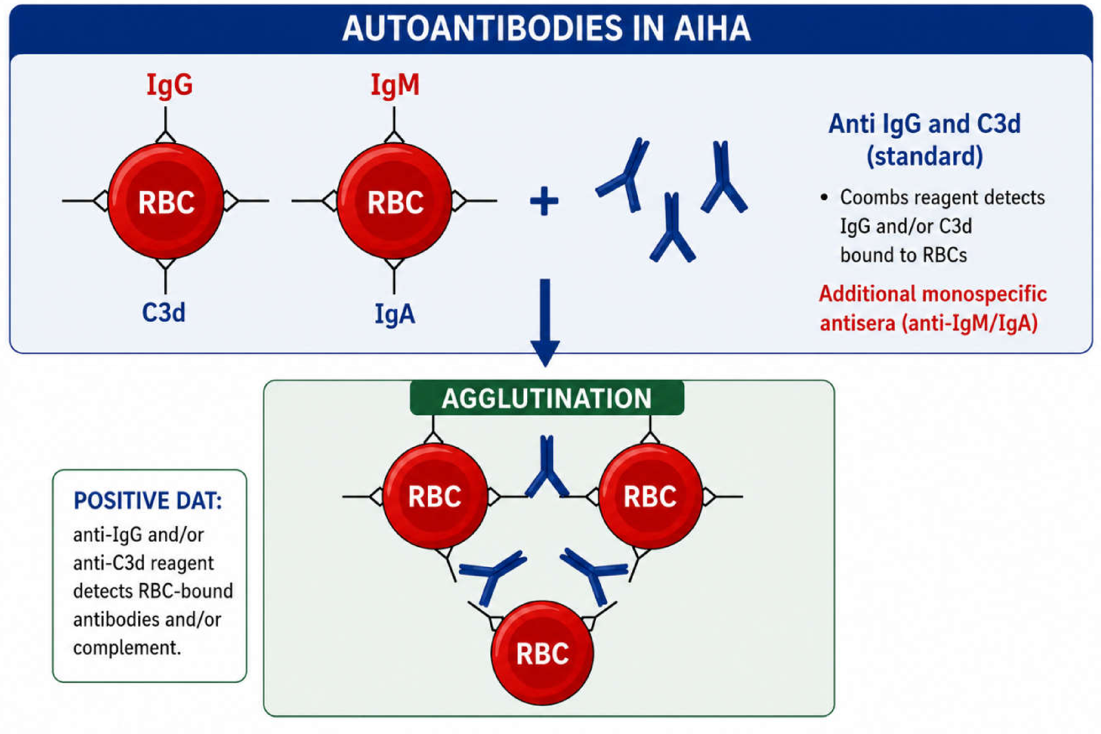
Figure 2.
Mechanisms of hemolysis in wAIHA and cAIHA.
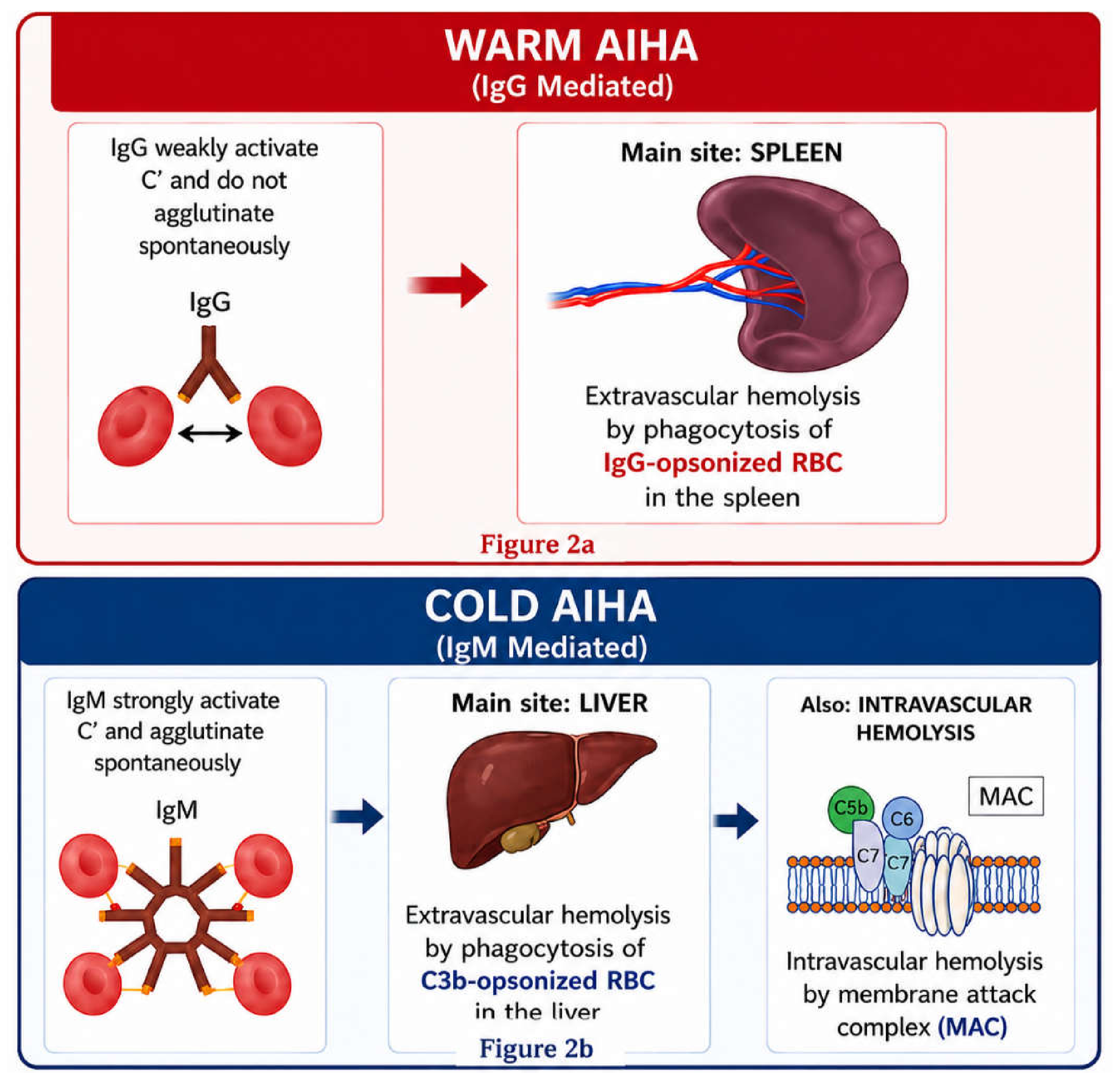
Figure 4.
Author’s practical therapeutic approach to wAIHA.
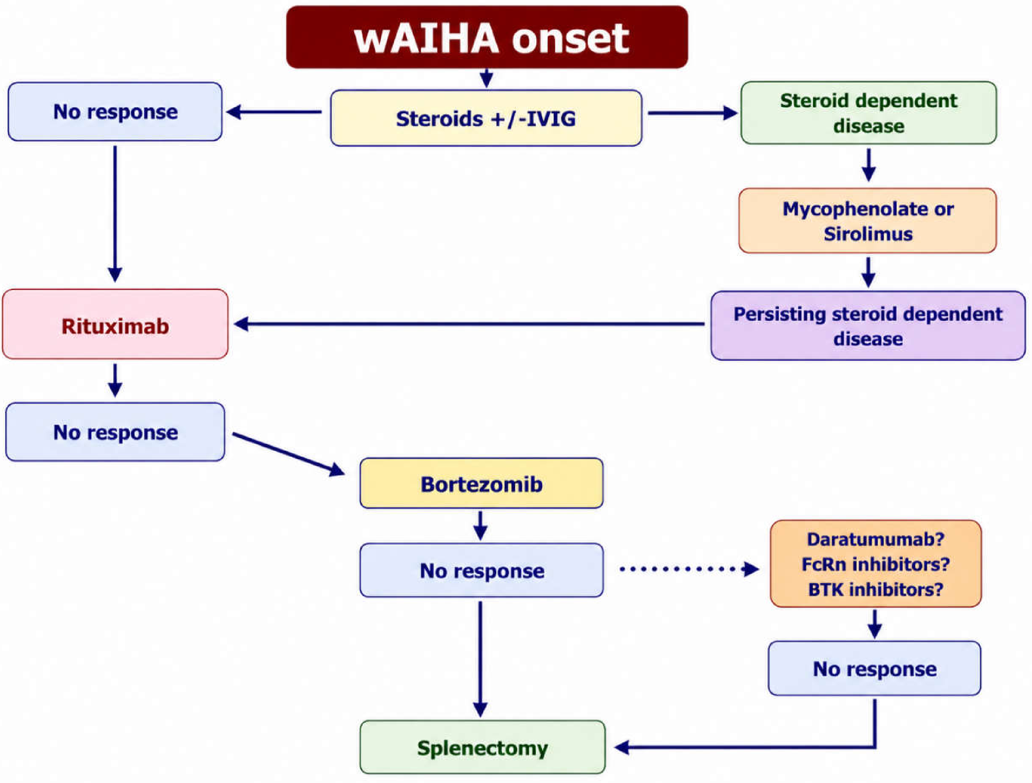

Table 1.
Main laboratory, clinical and prognostic characteristics of wAIHA, CAS and PCH.
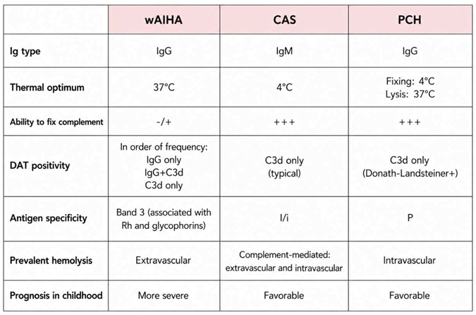 |
Disclaimer/Publisher’s Note: The statements, opinions and data contained in all publications are solely those of the individual author(s) and contributor(s) and not of MDPI and/or the editor(s). MDPI and/or the editor(s) disclaim responsibility for any injury to people or property resulting from any ideas, methods, instructions or products referred to in the content. |
© 2026 by the authors. Licensee MDPI, Basel, Switzerland. This article is an open access article distributed under the terms and conditions of the Creative Commons Attribution (CC BY) license (http://creativecommons.org/licenses/by/4.0/).
Copyright: This open access article is published under a Creative Commons CC BY 4.0 license, which permit the free download, distribution, and reuse, provided that the author and preprint are cited in any reuse.