

Submitted:
01 September 2026
Posted:
03 September 2026
You are already at the latest version
Preprints on COVID-19 and SARS-CoV-2
Abstract
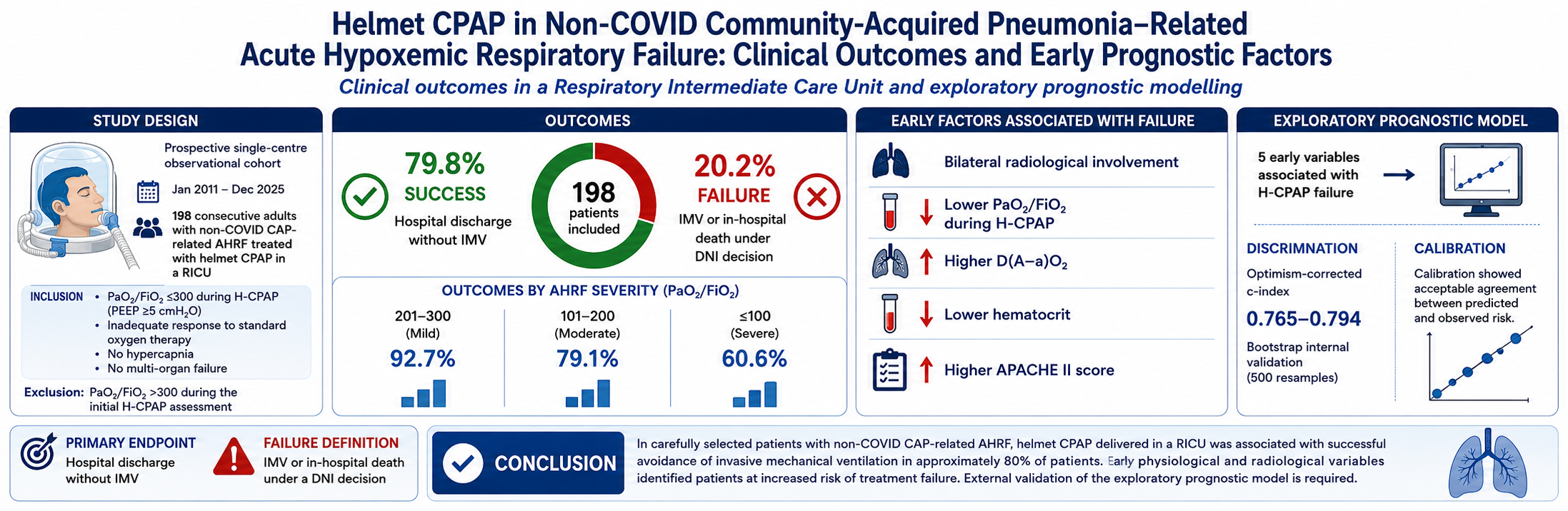
Non-invasive respiratory support for acute hypoxemic respiratory failure (AHRF) remains controversial. Continuous positive airway pressure (CPAP) provides sustained positive end-expiratory pressure, while helmet interface (H-CPAP) may improve tolerance and re-duce air leaks. We aimed to evaluate clinical outcomes of H-CPAP delivered in a respira-tory intermediate care unit (RICU) by pulmonologists and identify early predictors of treatment failure. We conducted a prospective single-centre observational cohort study in-cluding consecutive adults with community-acquired pneumonia (CAP)-AHRF treated with H-CPAP in a RICU. H-CPAP success was defined as hospital discharge without in-vasive mechanical ventilation (IMV). Clinical, radiological, gas exchange, laboratory var-iables were collected at baseline and after 1 hour of H-CPAP. Among 1100 screened pa-tients, 198 met inclusion criteria. H-CPAP success occurred in 158/198 patients (79.8%), while overall in-hospital mortality was 9.1%. Success rates decreased with increasing AHRF severity. Lower haematocrit, bilateral pulmonary infiltrates, lower PaO₂/FiO₂ dur-ing H-CPAP, higher alveolar–arterial oxygen gradient, and higher APACHE II score were the main predictors of treatment failure. In selected patients with CAP-AHRF, H-CPAP delivered in a RICU was associated with high rates of avoidance of IMV and low in-hospital mortality. Early physiological and radiological variables identified patients at increased risk of H-CPAP failure with acceptable predictive performance.
Keywords:
helmet CPAP
; acute hypoxemic respiratory failure
; community-acquired pneumonia
; respiratory intermediate care unit
; pulmonologist
1. Introduction
Acute hypoxemic respiratory failure (AHRF) is a frequent cause of intensive care unit (ICU) admission in adult patients [1], often requiring endotracheal intubation and invasive mechanical ventilation (IMV), and is commonly driven by pneumonia [2]. A substantial proportion of patients with AHRF fulfills criteria for acute respiratory distress syndrome (ARDS), a syndrome characterized by acute inflammatory lung injury, impaired oxygenation, and a high risk of IMV and death. Mortality increases with the severity of hypoxemia, making early recognition of patients at risk of treatment failure clinically important [2,3,4]. The role of non-invasive respiratory support (NIRS) in AHRF remains debated, particularly regarding the identification of patients at high risk of treatment failure [5,6,7,8]. Potential advantages of NIRS include avoidance of sedation, neuromuscular blockade, endotracheal intubation, and complications associated with IMV. However, prolonged NIRS in the absence of clinical improvement may delay tracheal intubation and IMV [9], potentially exposing patients to further lung injury due to excessive spontaneous breathing efforts and large tidal volumes, a mechanism known as patient self-inflicted lung injury (P-SILI). A relevant limitation is that tidal volume cannot be routinely measured during helmet-CPAP (H-CPAP) outside experimental settings [10], limiting bedside assessment of potentially injurious spontaneous breathing patterns.Current guidelines do not provide definitive recommendations regarding the optimal choice of device or interface for AHRF [5,6,7]. Among NIRS modalities, continuous positive airway pressure (CPAP) may be particularly relevant in AHRF because it provides positive end-expiratory pressure (PEEP) without inspiratory assistance, potentially improving lung recruitment while avoiding excessive increases in tidal volume. The helmet interface has been shown to improve tolerance, reduce air leaks, and allow prolonged application compared with face-mask interfaces [11,12,13,14,15,16]. Nevertheless, evidence regarding H-CPAP in non-COVID community-acquired pneumonia-related AHRF (CAP-AHRF) remains limited [15,16], particularly in the setting of a respiratory intermediate care unit (RICU) with close collaboration with ICU specialists.The primary objective of this study was to describe H-CPAP success and in-hospital mortality among consecutive patients with non-COVID CAP-AHRF managed in a RICU. The secondary objective was to identify baseline and early physiological variables associated with H-CPAP failure. H-CPAP success was defined as hospital discharge without IMV. H-CPAP failure was defined as the need for IMV or in-hospital death without IMV under a do-not-intubate (DNI) decision.
2. Materials and Methods
2.1. Study Design and Setting
We conducted a prospective single-centre observational cohort study including consecutive adult patients with CAP-AHRF treated with H-CPAP in the RICU of Vimercate Hospital, Italy, between January 2011 and December 2025. Participants were followed prospectively from hospital admission until hospital discharge or in-hospital death. The study is reported according to the Strengthening the Reporting of Observational Studies in Epidemiology (STROBE) recommendations for cohort studies. The RICU is equipped with continuous multiparametric monitoring, high-flow oxygen and compressed air sources, devices enabling precise FiO₂ delivery, and immediate access to advanced life-support equipment and endotracheal intubation. Throughout the study period, patients were managed by pulmonologists and dedicated nurses (nurse-to-patient ratio 1:6) experienced in H-CPAP, in close collaboration with the ICU team for decisions regarding treatment escalation.
2.2. Study Population
AHRF was defined as PaO₂ <60 mmHg while breathing room air. H-CPAP was initiated in patients with inadequate response to standard oxygen therapy (SOT), delivered through a Venturi mask or non-rebreathing mask, defined by persistent oxygen saturation ≤92%, respiratory rate >24 breaths/min, or signs of increased work of breathing, including thoraco-abdominal dyssynchrony. Consecutive adult patients treated with H-CPAP were eligible if they met all of the following predefined criteria: age ≥18 years; radiological evidence of pulmonary infiltrates; de novo AHRF not explained by cardiogenic pulmonary oedema or acute exacerbation of chronic respiratory disease; PaO₂/FiO₂ (P/F) ≤300 during H-CPAP with PEEP ≥5 cmH₂O (patients who achieved a P/F ratio >300 during the initial H-CPAP assessment were excluded); haemodynamic stability; Glasgow Coma Scale (GCS) ≥14; absence of multi-organ failure, acidosis or hypercapnia. Patients with COVID-19 pneumonia were excluded.
2.3. H-CPAP Protocol
Clinical and physiological variables were recorded at RICU admission and one hour after H-CPAP initiation. H-CPAP was delivered using commercially available helmet (DIMAR, Medolla, Italy), using a Venturi flow generator providing continuous fresh gas flow through the inlet port, while airway pressure was regulated by a PEEP valve positioned on the outlet port [17,18,19]. A minimum fresh gas flow of 60 L/min was maintained throughout treatment to minimize CO₂ rebreathing. PEEP was initially set between 5 and 12.5 cmH₂O according to the patient's clinical condition and gas exchange, whereas FiO₂ was titrated between 50% and 100% to maintain peripheral oxygen saturation between 92% and 98%.
2.4. Treatment Escalation
Predefined criteria for escalation to IMV included reduced level of consciousness, persistent hypoxemia with ongoing respiratory distress despite H-CPAP, intolerance to the interface, haemodynamic instability, or development of multi-organ failure. Decisions regarding ICU admission and endotracheal intubation were made jointly by pulmonologists and intensivists according to the patient's clinical condition and goals of care [20]. For patients with a DNI order, H-CPAP represented the ceiling of respiratory support. The decision to establish a DNI order was based on the patient's clinical characteristics, comorbidities, premorbid functional status, and shared goals of care. DNI criteria were considered only in cases in which IMV was necessary, not at the admission stage.
2.5. Weaning Protocol and Data Collection
In patients showing clinical improvement during H-CPAP, ventilatory support was progressively reduced according to a standardized weaning protocol. FiO₂ was first decreased to 0.50 while maintaining the same PEEP level, followed by daily PEEP reductions of 2.5 cmH₂O to a minimum of 5 cmH₂O. Daily exposure to H-CPAP was subsequently reduced according to clinical tolerance.Weaning from H-CPAP was considered when patients achieved clinical stability, defined as the absence of respiratory distress while receiving oxygen through a Venturi mask or high-flow nasal cannula (HFNC) with FiO₂ ≤0.50, stable oxygenation, body temperature <37°C for at least 24 hours, and improvement in inflammatory biomarkers. Microbiological investigations included urinary antigen tests for Streptococcus pneumoniae and Legionella pneumophila, serological testing for Mycoplasma pneumoniae, nasopharyngeal swabs for viral and bacterial pathogens, and blood, sputum cultures. All patients received antimicrobial therapy according to the American Thoracic Society/Infectious Diseases Society of America guidelines [21,22]. Demographic characteristics, comorbidities, disease severity scores, radiological findings, microbiological results, baseline gas exchange variables, laboratory parameters, and initial H-CPAP settings were collected at RICU admission, before H-CPAP initiation, as appropriate. The P/F ratio during H-CPAP was assessed one hour after treatment initiation and was therefore considered an indicator of early physiological response rather than a baseline variable. Subsequent clinical outcomes, including ICU admission, IMV, DNI status, and in-hospital mortality, were recorded.
2.6. Bias
Potential sources of bias were considered a priori. Selection bias was minimized by including consecutive eligible patients, although the cohort remained restricted to patients considered suitable for NIRS. Potential confounding related to the long enrolment period was addressed by describing temporal changes before and after January 1, 2020, and by comparing baseline characteristics, early physiological response, and model-predicted risk across the two periods. Because treatment allocation, ICU admission, and decisions regarding IMV were not randomized, all observed associations should be interpreted as prognostic rather than causal.
2.7. Study Size
The study size was determined by the number of consecutive eligible patients treated during the study period. No formal sample size calculation was performed because of the observational design of the study.
2.8. Statistical Analysis
Continuous variables are presented as mean ± standard deviation (SD), whereas categorical variables are reported as counts and percentages. Between-group comparisons were performed using Student's t-test or Fisher's exact test, as appropriate. The change in the P/F ratio from SOT to H-CPAP was compared between patients with H-CPAP success and failure using the Wilcoxon rank-sum test. All statistical tests were two-sided, and statistical significance was set at P <0.05. The primary outcome for multivariable modelling was H-CPAP failure. An exploratory multivariable logistic regression model was developed to identify baseline and early physiological variables associated with the probability of H-CPAP failure. Candidate predictors (Box 1) were selected a priori on clinical and pathophysiological grounds. To improve model stability and reduce redundancy in this modest-sized dataset, variables with negligible prognostic information (Information Value, IV <0.1) or excessive collinearity (variance inflation factor, VIF ≥5) were excluded before model fitting [23]. Continuous predictors were standardized before modelling, and the assumption of linearity on the logit scale was assessed graphically. To account for the limited number of outcome events and reduce overfitting, variable selection was performed using group lasso penalization [24]. The penalization parameter was selected by minimizing the Akaike Information Criterion (AIC). Only main effects were considered, with no interaction terms included. Because of the limited sample size, the entire dataset was used for model development. Internal validation was performed using 500 bootstrap resamples to estimate optimism-corrected model performance and evaluate the stability of predictor selection. Variable stability was quantified as the proportion of bootstrap samples in which each predictor, and the final model specification, was retained. Given the exploratory nature of the analysis, the final model was also compared with the three most frequently selected alternative models across bootstrap samples. Because predictor selection was partly data-driven, regression coefficient P values were not interpreted. Model performance was assessed in terms of discrimination and calibration, including both calibration-in-the-large and calibration slope [25] (Box A1). All performance measures were optimism-corrected using the 0.632 bootstrap method [26]. Calibration stability was assessed by graphical inspection, as recommended by Riley et al. [27]. Ninety-five percent confidence intervals (95% CIs) were calculated using the location-shifted bootstrap approach [28]. Variables included in the final analysis had complete data; therefore, no imputation was performed. The number of available observations was reported for each variable in the tables when missing values were present. Given the long inclusion period and the potential changes in respiratory care pathways following the COVID-19 pandemic, patients were additionally stratified according to calendar period: before 1 January 2020 and on or after 1 January 2020. No patient was admitted on the cut-off date. Differences in baseline characteristics, early physiological response, and model-based estimated risk of H-CPAP failure were explored between the two periods to assess potential changes in case mix and clinical management over time. All analyses and graphical representations were performed using SAS software version 9.4M9.
Box 1. A priori candidate predictors considered for the exploratory prognostic model of H-CPAP failure.
Sex, Age, GCS, Bilateral involvement,
Pulmonary comorbidities, Cardiovascular disease, Diabetes, Neoplasia (current
or previous), Microbiological isolation, Calendar period, Mean arterial
pressure, Haematocrit, P/F during H-CPAP, P/F during SOT, D(A–a)O₂, APACHE II
score, HACOR score during H-CPAP, Symptoms onset (hours).
Note: Early response variables were those measured 1 hour after H-CPAP initiation, including P/F during H-CPAP and HACOR score during H-CPAP.
3. Results
3.1. Patient Characteristics and Clinical Outcomes
During the study period, 1100 patients with CAP-AHRF were screened. Among these, 198 consecutive patients fulfilled the predefined eligibility criteria and were treated with H-CPAP in the RICU (Figure 1). Patients with less severe AHRF who achieved adequate oxygenation with conventional oxygen therapy and therefore did not require escalation to H-CPAP, as well as patients who achieved a P/F ratio >300 mmHg during the initial H-CPAP assessment, were excluded from the analysis. H-CPAP success, defined as hospital discharge without IMV, occurred in 158/198 patients (79.8%). H-CPAP failure occurred in 40/198 patients (20.2%). Overall in-hospital mortality was 18/198 (9.1%): 15 patients died after ICU admission and IMV, whereas 3 patients died in the RICU under a DNI decision.
3.2. Baseline Characteristics
Baseline and early physiological characteristics according to H-CPAP outcome are reported in Table 1. The mean age of the cohort was 63.5 years, and 51 patients (25.8%) were female. The most frequent comorbidities were cardiovascular disease, chronic respiratory disease, diabetes, and current or previous malignancy. Age and sex distribution did not differ significantly between outcome groups. Compared with patients successfully treated with H-CPAP, patients experiencing failure had markers of greater disease severity, including more frequent bilateral pulmonary involvement, lower haematocrit values, lower P/F ratio during H-CPAP, higher alveolar–arterial oxygen gradient [D(A–a)O₂], and higher APACHE II scores.
3.3. Severity Stratification
According to P/F ratio during H-CPAP, 55 patients (27.8%) had mild AHRF, 110 (55.6%) moderate AHRF, and 33 (16.7%) severe AHRF. H-CPAP success progressively decreased with increasing severity of hypoxemia: 51/55 patients (92.7%) with mild AHRF, 87/110 (79.1%) with moderate AHRF, and 20/33 (60.6%) with severe AHRF (p=0.001).
3.4. Microbiology
A microbiological diagnosis was obtained in 121/198 patients (61.1%). The most frequently identified pathogen was Legionella pneumophila (n=45), followed by Streptococcus pneumoniae (n=29). Additional microbiological findings are reported in Table 1.
3.5. Modelling Section
In the exploratory prognostic analysis, the outcome of interest was H-CPAP failure. Among the a priori candidate prognostic factors, variables with Information Value <0.10 or relevant collinearity were excluded before penalized modelling. Infectious comorbidities were excluded because of quasi-separation. Variables retained after screening entered the group lasso selection procedure. Variables excluded before modelling were sex, GCS, diabetes, current or previous malignancy, and AHRF severity. Odds ratio of the final model identified in the original dataset (M1) and of the three most frequently selected alternative models across bootstrap samples (M2–M4) are reported on the logit scale in Table 2. Positive coefficients indicate a higher estimated probability of H-CPAP failure, whereas negative coefficients indicate a lower estimated probability. Because predictor selection involved data-driven procedures, odds ratio estimates should be interpreted descriptively and formal hypothesis testing was not performed.
The direction of associations was consistent across the selected models: higher D(A–a)O₂ and APACHE II scores were associated with increased estimated risk of H-CPAP failure, whereas higher haematocrit values and higher P/F ratio during CPAP were associated with lower estimated risk. Predictor stability was moderate. Variables included in the final model and in the three most frequently selected alternative models were among those most frequently retained across the 500 bootstrap samples (Supplementary Figure S2). However, the exact predictor combination identified in the original dataset was not among the most frequently occurring model specifications, indicating uncertainty regarding the optimal set of predictors despite a relatively consistent prognostic signal (Supplementary Figure S2). The four candidate models showed comparable performance, suggesting that reducing the number of predictors did not substantially compromise discrimination. Optimism-corrected c-index values ranged from 0.765 to 0.794 (Figure 2), indicating moderate discrimination.Brier scores were low and similar across models (0.121–0.128), reflecting comparable overall accuracy. However, scaled Brier scores were modest and associated with wide confidence intervals, suggesting limited improvement over a non-informative model and considerable uncertainty due to the limited number of outcome events.
Calibration intercepts and slopes indicated satisfactory average calibration. However, calibration plots revealed some deviation from ideal calibration, particularly at the extremes of predicted risk (Figure 3). At lower predicted probabilities, models tended to overestimate observed risk, whereas underestimation was observed in part of the intermediate-to-higher risk range. This pattern was more evident in reduced models, particularly Models 2 and 3. Bootstrap calibration curves showed similar overall behaviour, with increasing uncertainty at higher predicted risks because of fewer observations. (Supplementary Figure S2).
When the cohort was stratified according to calendar period, differences were observed in baseline characteristics, early physiological response, and estimated risk of H-CPAP failure. Patients treated before 1 January 2020 showed a less favourable clinical profile compared with those treated after this date, with differences in the distribution of several prognostic variables (Supplementary Materials, Table S2). Consistently, the estimated risk of H-CPAP failure derived from the exploratory prognostic model was higher in the earlier period (Supplementary Figure S1).
These findings should be interpreted cautiously. The inclusion period spanned major changes in respiratory care pathways, including increased availability of HFNC, evolving ICU admission criteria, changes in antimicrobial strategies, and implementation of prone positioning in awake spontaneously breathing patients. Therefore, the observed temporal differences may reflect changes in case mix and clinical management rather than a direct effect of calendar period itself.
Complications occurring during hospitalization are summarized in the Supplementary Materials, Table S1.
4. Discussion
The main findings of this prospective single-centre observational cohort study are that approximately 80% of carefully selected patients with non-COVID CAP-AHRF treated with H-CPAP in a RICU were discharged without IMV, while overall in-hospital mortality was 9.1%. H-CPAP failure was associated with markers of greater disease severity and impaired gas exchange.
The potential harms of spontaneous breathing in AHRF derive from the vicious circle generated by hypoxemia, dysregulated inspiratory effort, altered respiratory mechanics and inhomogeneous lung inflation. Effects of intense inspiratory efforts in spontaneous breathing carries the risk of lung damage induced by excessively large tidal volumes, overinflating the normally aerated lung tissue which is markedly reduced because of inflammatory oedema (baby lung) [29].
HFNC has progressively become a widely used first-line strategy for AHRF because of its ability to improve oxygenation, reduce anatomical dead space, decrease work of breathing, and improve patient comfort [30]. However, the optimal initial respiratory support strategy for patients with CAP-AHRF remains uncertain, particularly among those with moderate-to-severe hypoxemia.
Randomized trials comparing HFNC with other non-invasive strategies enrolled heterogeneous populations of patients with AHRF, including a substantial proportion of patients with less severe hypoxemia than those included in the present cohort [31]. Furthermore, NIV was predominantly delivered through facemask interfaces rather than helmet systems, limiting direct comparisons with H-CPAP.
Compared with HFNC, H-CPAP provides continuous PEEP, promoting alveolar recruitment, improving ventilation homogeneity, and increasing functional residual capacity. Experimental studies have demonstrated that H-CPAP reduces the intratidal redistribution of gas from non-dependent to dependent lung regions (the Pendelluft phenomenon) [32] and exerts a favorable mechanical effect on diaphragmatic function by modifying the force-length relationship of diaphragmatic fibers [33]. These physiological effects may reduce excessive inspiratory effort and potentially limit p-SILI, providing a strong physiological rationale for the use of H-CPAP in selected patients with pneumonia-related AHRF [34,35].
Additional physiological evidence supports this concept. In a recent bench and healthy volunteer study, Haudebourg et al. compared different NIRS devices, including HFNC, facemask and helmet CPAP, and facemask and helmet NIV [36]. CPAP was associated with the lowest tidal volume, whereas NIV generated the highest tidal volume. Since excessive tidal volume and high minute ventilation have both been associated with NIV failure and worse outcomes [30,37], limiting spontaneous inspiratory effort may represent an important mechanism through which CPAP exerts its beneficial effects.
Recent clinical evidence is also consistent with this physiological rationale. A systematic review and network meta-analysis of randomized trials [38] reported that, compared with SOT, CPAP probably reduces the need for endotracheal intubation and may also decrease mortality in patients with AHRF. Among the available NIRS modalities, CPAP showed the highest probability of reducing short-term mortality [12,13,39]. Our findings are consistent with these observations. However, because all patients in our cohort were treated with H-CPAP, our study cannot establish the comparative effectiveness of H-CPAP versus HFNC or NIV.
For these reasons, strategies based on the application of moderate-to-high levels of PEEP through CPAP have gained increasing attention for the non-invasive management of AHRF [40]. The helmet interface represents an attractive alternative to conventional facemask because it is generally better tolerated, minimizes air leaks, and reduces pressure-related skin injury [14,15,16].
Despite increasing interest in H-CPAP, most available evidence derives from COVID-19-related respiratory failure [20,39,41,42,43]. Data specifically addressing non-COVID CAP-AHRF remain limited [15,16]. Furthermore, previous studies frequently included heterogeneous etiologies of hypoxemic respiratory failure, making it difficult to extrapolate their findings to patients with pneumonia alone. In contrast, our study evaluated a clinically homogeneous cohort of patients with CAP-related AHRF, excluding respiratory failure due to concomitant causes. Moreover, our patients were substantially more severe than those enrolled in the largest randomized trials evaluating HFNC [30,31].
H-CPAP has been routinely implemented in our RICU as part of a structured treatment pathway for patients with AHRF who fail to achieve adequate oxygenation with SOT but do not require immediate IMV [20,43,44]. This strategy is based on the concept that, in patients with persistent hypoxemia despite conventional oxygen therapy, optimization of both FiO₂ and PEEP represents the cornerstone of respiratory support. While previous randomized trials and recent meta-analyses have primarily compared H-CPAP with SOT [15,16,38,45], our study provides real-world evidence regarding the performance of this strategy in a well-defined population of patients with severe CAP-related AHRF managed according to a standardized clinical protocol.
To our knowledge, this represents the largest observational cohorts specifically evaluating H-CPAP in patients with CAP-related AHRF, a population that has been underrepresented in previous randomized trials.
In addition, patients considered at higher risk of deterioration could be transferred to the ICU while continuing H-CPAP, allowing closer monitoring and timely escalation if needed. Patients transferred to the ICU but discharged without IMV were classified as H-CPAP successes according to the predefined outcome definition. Among patients requiring IMV, ICU mortality was 42%, a finding consistent with previous reports in severe pneumonia and AHRF populations [46].
Our findings suggest that early physiological response to H-CPAP may provide clinically relevant prognostic information. In particular, P/F ratio measured after H-CPAP initiation was associated with treatment outcome, whereas baseline oxygenation during conventional oxygen therapy was not.
Literature indicates a cut-off of P/F ≤ 150 as a limit for NIRS [40,47,48]. We split our moderate AHRF 110 patients in two subgroups A (200 < P/F ≤ 150) and B (150 < P/F ≤ 100). H-CPAP success in subgroup A was 44/55 (80%) and in subgroup B 43/55 (78.2%), without any statistical differences.
Our findings raise the hypothesis that patients with P/F ≤100 may represent a subgroup at particularly high risk of H-CPAP failure. More studies are needed in order to deepen this new possible limit.
ARDS Criteria and Unilateral Involvement
Although bilateral pulmonary opacities are a key component of the definition of ARDS [4,49], recent discussions have questioned whether the presence of unilateral opacities should necessarily exclude patients with otherwise compatible clinical features of ARDS [50]. In our cohort, all patients fulfilled the oxygenation criterion for ARDS, with a mean P/F ratio of 165.4 ± 58.7 measured during H-CPAP with PEEP ≥5 cmH₂O; however, 71 of 198 patients (35.9%) presented with unilateral radiological involvement and therefore did not meet the full ARDS definition. Patients with unilateral involvement appeared to experience lower rates of H-CPAP failure and in-hospital mortality than those with bilateral disease, despite similar P/F ratios both before and during H-CPAP. Previous studies have suggested that the apparent prognostic advantage associated with unilateral opacities may disappear after adjustment for illness severity and the extent of lung involvement. Since the extent of radiological involvement was not quantitatively assessed in our study, these findings should be considered exploratory and interpreted with caution [51].
Microbiological Findings
The number of patients infected with individual pathogens was insufficient to allow pathogen-specific prognostic analyses. Therefore, the association between microbiological isolation and H-CPAP success may be explained by the use of more targeted antimicrobial therapy [52].
APACHE II Score
Unlike COVID-19-related AHRF, where disease phenotype and respiratory mechanics may differ substantially over time [20,42,43], general markers of clinical severity may retain prognostic value in non-COVID CAP-AHRF. In our cohort, APACHE II score was higher among patients experiencing H-CPAP failure, suggesting that global severity assessment remains useful for early risk stratification.
P/F
P/F in CPAP is higher in the SUCCESS group (p<0,0001), in contrast to P/F in oxygen (p 0.59). The improvement in P/F observed after H-CPAP initiation may reflect effective alveolar recruitment (Figure 4).
D(A-a)O₂ and Haematocrit
The association between D(A–a)O₂ and H-CPAP failure is physiologically plausible. The alveolar–arterial oxygen gradient integrates multiple determinants of oxygen transfer, including ventilation-perfusion mismatch, shunt fraction, and impaired diffusion. In our cohort, higher D(A–a)O₂ values were strongly associated with treatment failure (p<0.0001), supporting its potential role as a marker of more severe pulmonary involvement.
Haematocrit was significantly higher in the H-CPAP success group (p = 0.003). This observation is consistent with established physiological principles, as haemoglobin is a major determinant of oxygen delivery (DO₂), together with cardiac output and arterial oxygenation. Higher haematocrit values may therefore reflect a greater capacity to maintain tissue oxygen delivery despite severe respiratory failure [53].
Mortality Comparison with Literature
Previous studies have reported substantial mortality among patients with severe AHRF, often exceeding 20–30% depending on disease severity and inclusion criteria [2]. In our cohort, in-hospital mortality was 9.1%, and H-CPAP success occurred in 79.8% of patients. However, these findings should not be interpreted as evidence of a mortality benefit associated with H-CPAP, because our cohort included selected patients considered appropriate candidates for NIRS and lacked a control group.
Patients underwent early respiratory assessment in the emergency department, allowing appropriate triage to either the RICU or ICU. Moreover, selection criteria for NIRS (haemodynamic stability, GCS ≥14, absence of multi-organ failure, acidosis or hypercapnia) [8] defined a subgroup of AHRF patients with a better prognosis, manageable in RICU. These findings may support the feasibility of H-CPAP in selected patients managed in an appropriately monitored intermediate-care setting.
Exploratory Prognostic Model Interpretation
The exploratory prognostic analysis should be interpreted as hypothesis-generating rather than as a tool for clinical decision-making. Internal validation suggested that variables reflecting gas-exchange impairment and overall disease severity captured a relevant prognostic signal. However, the instability of the exact predictor combination across bootstrap samples indicates uncertainty regarding the optimal model specification. Despite acceptable internal performance, the selected predictor combination showed instability across bootstrap samples and should therefore not be considered a definitive bedside decision tool.
Models including fewer predictors showed similar discrimination, suggesting that risk ranking was preserved despite model simplification. Conversely, calibration appeared more sensitive to predictor selection, with some deviation from ideal calibration at different ranges of predicted risk.
Calendar period was explored because the study covered a prolonged timeframe during which respiratory care pathways changed substantially. Although period was not retained as a consistent predictor in bootstrap analyses, patients treated in the earlier period showed higher estimated risk of failure, likely reflecting differences in patient characteristics, referral pathways, and available therapeutic strategies. These findings reinforce the importance of considering temporal changes when interpreting long observational cohorts.
Study Strengths
This study has several strengths. First, it included a clinically homogeneous population of patients with non-COVID CAP-AHRF, excluding respiratory failure due to other concomitant causes. Second, patients were managed within a dedicated RICU with extensive experience in H-CPAP and close collaboration with intensivists, allowing structured monitoring and timely escalation when required. Third, consecutive eligible patients were included over a prolonged observation period, and both clinical outcomes and early physiological responses were systematically collected, providing a comprehensive evaluation of H-CPAP implementation in routine clinical practice.
Study Limitations
This study has some limitations. First, it was conducted in a single centre with specific expertise in H-CPAP and close collaboration between the RICU and ICU, which may limit external generalizability. Second, the observational design and absence of a control group prevent causal conclusions regarding the effectiveness of H-CPAP compared with HFNC, NIV, or other respiratory support strategies. Third, the cohort included only patients considered suitable for NIRS and therefore excluded individuals with severe haemodynamic instability, multi-organ failure, respiratory acidosis, hypercapnia, or impaired consciousness. Fourth, the long inclusion period may have introduced temporal changes in case mix, clinical pathways, antimicrobial strategies, ICU admission thresholds, availability of HFNC, and use of awake prone positioning (Supplementary Figure S1). Fifth, the number of failure events was limited, increasing the risk of model instability despite penalization and bootstrap validation. Sixth, P/F ratio during H-CPAP was measured after treatment initiation and should therefore be interpreted as an early response variable rather than a baseline predictor. External validation and assessment of clinical utility are required before considering implementation of the prognostic model.
5. Conclusions
In this prospective single-centre observational cohort study, H-CPAP delivered in a RICU was associated with successful avoidance of IMV in approximately 80% of carefully selected patients with CAP-related AHRF. Early physiological response to H-CPAP and markers of disease severity were associated with treatment outcome, although the exploratory prognostic model requires external validation before clinical application. These findings support the feasibility of H-CPAP within a structured intermediate-care pathway and highlight the importance of early identification of patients at increased risk of treatment failure.
Supplementary Materials
Table S1: in-hospital complications according to H-CPAP outcome; Table S2: baseline and early clinical characteristics according to calendar period; Figure S1: individual predicted probability of H-CPAP failure according to calendar period; Figure S2: stability of calibration across 500 bootstrap samples.
Author Contributions
Conceptualization, C.B., E.R., A.G. and P.S.; Data Curation C.B., A.G., R.R., S.P., P.U., S.R., M.P., L. P., C.M. (Chiara Melacini), C.M. (Claudio Macaluso), L.L., F.D., R.C., M.B., S.B, F.S., C.P. and P.S.; Methodology, C.B., E.R., S.G. and P.S.; Writing-Original Draft, C.B., E.R., S.G., A.G., R.R. and P.S.; All authors have read and approved the final version of the manuscript.
Funding
This research received no specific grant from any funding agency in the public, commercial, or not-for-profit sectors.
Institutional Review Board Statement
Human participants were involved in this research; the study was conducted in accordance with the Declaration of Helsinki. Our study was approved by the local institution, Vimercate Hospital, ASST-Brianza, according to the legal requirements concerning observational studies (Resolution No. 0000133, 22 February 2023). Patient consent was waived.
Informed Consent Statement
Due to the nature of the present observational study and data anonymization, the patients’ consent to participate was not required, as declared by the ASST Brianza Ethic Committee.
Data Availability Statement
The datasets analyzed during the current study are available from the corresponding author on reasonable request.
Acknowledgments
The authors thank all nurses and healthcare support workers of the Respiratory Intermediate Care Unit (RICU) for their valuable assistance in patient care.
Conflicts of Interest
The authors declare that they have no competing interests.
Abbreviations
The following abbreviations are used in this manuscript:
| AHRF | acute hypoxemic respiratory failure; |
| AIC | akaike information criterion; |
| ARDS | acute respiratory distress syndrome; |
| CAP-AHRF | community-acquired pneumonia-related AHRF; |
| CPAP | continuous positive airway pressure; |
| D(A–a)O₂ | alveolar–arterial oxygen gradient; |
| DNI | do-not-intubate; |
| DO₂ | oxygen delivery; |
| GCS | Glasgow Coma Scale; |
| H-CPAP | helmet-CPAP; |
| HFNC | high-flow nasal cannula; |
| ICU | intensive care unit; |
| IMV | invasive mechanical ventilation; |
| IV | information value; |
| NIRS | non-invasive respiratory support; |
| P/F | PaO₂/FiO₂; |
| PEEP | positive end-expiratory pressure; |
| P-SILI | patient self-inflicted lung injury; |
| RICU | respiratory intermediate care unit; |
| SOT | standard oxygen therapy; |
| STROBE | strengthening the reporting of observational studies in epidemiology; |
| VIF | variance inflation factor |
References
- Bellani, G.; Laffey, J. G.; Pham, T.; et al. Epidemiology, Patterns of Care, and Mortality for Patients with Acute Respiratory Distress Syndrome in Intensive Care Units in 50 Countries. JAMA 2016, 315(8), 788–800. [Google Scholar] [CrossRef]
- Cavallazzi, R.; Furmanek, S.; Arnold, F. W.; et al. The Burden of Community-Acquired Pneumonia Requiring Admission to ICU in the United States. Chest 2020, 158(3), 1008–1016. [Google Scholar] [CrossRef]
- Matthay, M. A.; Arabi, Y.; Arroliga, A. C.; et al. A New Global Definition of Acute Respiratory Distress Syndrome. Am. J. Respir. Crit. Care Med. 2024, 209, 37–47. [Google Scholar] [CrossRef]
- Ranieri, V. M.; Rubenfeld, G. D.; Thompson, B. T.; et al. Acute Respiratory Distress Syndrome: The Berlin Definition. JAMA 2012, 307(23), 2526–2533. [Google Scholar] [CrossRef]
- Carteaux, G.; Haudebourg, A. F. Noninvasive Ventilation in Acute Hypoxemic Respiratory Failure: What Is the Future? J. Intensive Med. 2025, 5(3), 237–245. [Google Scholar] [CrossRef]
- Pittao, M. L. V.; Nava, S.; Hill, N. S.; Pisani, L. Tailoring Non-Invasive Respiratory Supports in Acute Hypoxemic Respiratory Failure: A Practical Approach for Clinicians. Eur. J. Intern. Med. 2025, 137, 21–32. [Google Scholar] [CrossRef]
- Thille, A. W.; Balen, F.; Carteaux, G.; et al. Oxygen Therapy and Noninvasive Respiratory Supports in Acute Hypoxemic Respiratory Failure: A Narrative Review. Ann. Intensive Care 2024, 14(1). [Google Scholar] [CrossRef]
- Ferrer, M.; Esquinas, A.; Leon, M.; Gonzalez, G.; Alarcon, A.; Torres, A. Noninvasive Ventilation in Severe Hypoxemic Respiratory Failure: A Randomized Clinical Trial. Am. J. Respir. Crit. Care Med. 2003, 168(12), 1438–1444. [Google Scholar] [CrossRef]
- Bellani, G.; Laffey, J. G.; Pham, T.; et al. Noninvasive Ventilation of Patients with Acute Respiratory Distress Syndrome: Insights from the LUNG SAFE Study. Am. J. Respir. Crit. Care Med. 2017, 195(1), 67–77. [Google Scholar] [CrossRef]
- LoMauro, A.; De Luca, A.; Scarpazza, P.; Aliverti, A. In Vivo Measurement of Tidal Volume during Non-Invasive Respiratory Support by Continuous-Flow Helmet CPAP. Ann. Biomed. Eng. 2024, 52(9), 2546–2555. [Google Scholar] [CrossRef]
- Chiumello, D.; Brochard, L.; Marini, J. J.; et al. Respiratory Support in Patients with Acute Respiratory Distress Syndrome: An Expert Opinion. Crit. Care 2017, 21(1). [Google Scholar] [CrossRef]
- Sakuraya, M.; Okano, H.; Masuyama, T.; Kimata, S.; Hokari, S. Efficacy of Non-Invasive and Invasive Respiratory Management Strategies in Adult Patients with Acute Hypoxaemic Respiratory Failure: A Systematic Review and Network Meta-Analysis. Crit. Care 2021, 25(1). [Google Scholar] [CrossRef]
- Ferreyro, B. L.; Angriman, F.; Munshi, L.; et al. Association of Noninvasive Oxygenation Strategies with All-Cause Mortality in Adults with Acute Hypoxemic Respiratory Failure: A Systematic Review and Meta-Analysis. JAMA 2020, 324(1), 57–67. [Google Scholar] [CrossRef]
- Patel, B. K.; Wolfe, K. S.; Pohlman, A. S.; Hall, J. B.; Kress, J. P. Effect of Noninvasive Ventilation Delivered by Helmet vs Face Mask on the Rate of Endotracheal Intubation in Patients with Acute Respiratory Distress Syndrome: A Randomized Clinical Trial. JAMA 2016, 315(22), 2435–2441. [Google Scholar] [CrossRef]
- Cosentini, R.; Brambilla, A. M.; Aliberti, S.; et al. Helmet Continuous Positive Airway Pressure vs Oxygen Therapy to Improve Oxygenation in Community-Acquired Pneumonia: A Randomized, Controlled Trial. Chest 2010, 138(1), 114–120. [Google Scholar] [CrossRef]
- Brambilla, A. M.; Aliberti, S.; Prina, E.; et al. Helmet CPAP vs Oxygen Therapy in Severe Hypoxemic Respiratory Failure Due to Pneumonia. Intensive Care Med. 2014, 40(7), 942–949. [Google Scholar] [CrossRef]
- Cutuli, S. L.; Grieco, D. L.; Michi, T.; et al. Personalized Respiratory Support in ARDS: A Physiology-to-Bedside Review. J. Clin. Med. 2023, 12(13). [Google Scholar] [CrossRef]
- Chiumello, D.; Esquinas, A. M.; Moerer, O.; Terzi, N. A Systematic Technical Review of the Systems for the Continuous Positive Airway Pressure. Minerva Medica 2012, 78(12), 1385–1393. [Google Scholar]
- Rezoagli, E.; Nova, A.; Carteaux, G.; et al. A Clinical Guide to Non-Invasive Respiratory Support in Acute Respiratory Failure: Ventilation Settings, Technical Optimization and Clinical Indications. Crit. Care 2025, 29(1). [Google Scholar] [CrossRef]
- Piluso, M.; Ferrari, C.; Pagani, S.; et al. COVID-19 Acute Respiratory Distress Syndrome: Treatment with Helmet CPAP in Respiratory Intermediate Care Unit by Pulmonologists in the Three Italian Pandemic Waves. Adv. Respir. Med. 2023, 91(5), 383–396. [Google Scholar] [CrossRef]
- Mandell, L. A.; Wunderink, R. G.; Anzueto, A.; et al. Infectious Diseases Society of America/American Thoracic Society Consensus Guidelines on the Management of Community-Acquired Pneumonia in Adults. Clin. Infect. Dis. 2007, 44 (Suppl. 2). [Google Scholar] [CrossRef]
- Metlay, J. P.; Waterer, G. W.; Long, A. C.; et al. Diagnosis and Treatment of Adults with Community-Acquired Pneumonia. Am. J. Respir. Crit. Care Med. 2019, 200(7), E45–E67. [Google Scholar] [CrossRef]
- Siddiqi, N. Credit Risk Scorecards: Developing and Implementing Intelligent Credit Scoring; Wiley: Hoboken, NJ, 2006. [Google Scholar] [CrossRef]
- Yuan, M.; Lin, Y. Model Selection and Estimation in Regression with Grouped Variables. J. R. Stat. Soc. Ser. B Stat. Methodol. 2006, 68(1), 49–67. [Google Scholar] [CrossRef]
- Van Calster, B.; Nieboer, D.; Vergouwe, Y.; De Cock, B.; Pencina, M. J.; Steyerberg, E. W. A Calibration Hierarchy for Risk Models Was Defined: From Utopia to Empirical Data. J. Clin. Epidemiol. 2016, 74, 167–176. [Google Scholar] [CrossRef]
- Efron, B. Estimating the Error Rate of a Prediction Rule: Improvement on Cross-Validation. J. Am. Stat. Assoc. 1983, 78(382), 316–331. [Google Scholar] [CrossRef]
- Riley, R. D.; Collins, G. S. Stability of Clinical Prediction Models Developed Using Statistical or Machine Learning Methods. Biom. J. 2023, 65(8). [Google Scholar] [CrossRef]
- Noma, H.; Shinozaki, T.; Iba, K.; Teramukai, S.; Furukawa, T. A. Confidence Intervals of Prediction Accuracy Measures for Multivariable Prediction Models Based on the Bootstrap-Based Optimism Correction Methods. Stat. Med. 2021, 40(26), 5691–5701. [Google Scholar] [CrossRef]
- Gattinoni, L.; Pesenti, A. The Concept of “Baby Lung”. Intensive Care Med. 2005, 31(6), 776–784. [Google Scholar] [CrossRef]
- Frat, J. P.; Thille, A. W.; Mercat, A.; et al. High-Flow Oxygen through Nasal Cannula in Acute Hypoxemic Respiratory Failure. N. Engl. J. Med. 2015, 372(23), 2185–2196. [Google Scholar] [CrossRef]
- Maia, I. S.; Kawano-Dourado, L.; Tramujas, L.; et al. High-Flow Nasal Oxygen vs Noninvasive Ventilation in Patients with Acute Respiratory Failure: The RENOVATE Randomized Clinical Trial. JAMA 2025, 333(10), 875–890. [Google Scholar] [CrossRef]
- Cesarano, M.; Grieco, D. L.; Michi, T.; et al. Helmet Noninvasive Support for Acute Hypoxemic Respiratory Failure: Rationale, Mechanism of Action and Bedside Application. Ann. Intensive Care 2022, 12(1). [Google Scholar] [CrossRef]
- Evans, C. L.; Hill, A. V. The Relation of Length to Tension Development and Heat Production on Contraction in Muscle. J. Physiol. 1914, 49(1–2), 10–16. [Google Scholar] [CrossRef]
- Grieco, D. L.; Patel, B. K.; Antonelli, M. Helmet Noninvasive Support in Hypoxemic Respiratory Failure. Intensive Care Med. 2022, 48(8), 1072–1075. [Google Scholar] [CrossRef]
- Buell, K. G.; Patel, B. K. Helmet Noninvasive Ventilation in Acute Hypoxic Respiratory Failure. Curr. Opin. Crit. Care 2023, 29(1), 8–13. [Google Scholar] [CrossRef]
- Haudebourg, A. F.; Maraffi, T.; Tuffet, S.; Le Corvoisier, P.; Mekontso Dessap, A.; Carteaux, G. Influence of Different Noninvasive Oxygenation Support Devices on Tidal Volume. Ann. Intensive Care 2023, 13(1). [Google Scholar] [CrossRef]
- He, H.; Sun, B.; Liang, L.; et al. A Multicenter RCT of Noninvasive Ventilation in Pneumonia-Induced Early Mild Acute Respiratory Distress Syndrome. Crit. Care 2019, 23(1). [Google Scholar] [CrossRef]
- Lee, K. G.; Hopkins, M.; Couban, R.; et al. Non-Invasive Respiratory Supports and Criteria for Intubation in Randomised Trials of Acute Hypoxaemic Respiratory Failure: A Systematic Review and Network Meta-Analysis. Lancet Respir. Med. 2026, 14(6), 533–544. [Google Scholar] [CrossRef]
- Perkins, G. D.; Ji, C.; Connolly, B. A.; et al. Effect of Noninvasive Respiratory Strategies on Intubation or Mortality among Patients with Acute Hypoxemic Respiratory Failure and COVID-19: The RECOVERY-RS Randomized Clinical Trial. JAMA 2022, 327(6), 546–558. [Google Scholar] [CrossRef]
- Grieco, D. L.; Maggiore, S. M.; Roca, O.; et al. Non-Invasive Ventilatory Support and High-Flow Nasal Oxygen as First-Line Treatment of Acute Hypoxemic Respiratory Failure and ARDS. Intensive Care Med. 2021, 47(8), 851–866. [Google Scholar] [CrossRef]
- Barbeta, E.; Motos, A.; Torres, A.; et al. SARS-CoV-2-Induced Acute Respiratory Distress Syndrome: Pulmonary Mechanics and Gas-Exchange Abnormalities. Ann. Am. Thorac. Soc. 2020. [Google Scholar] [CrossRef]
- Chiumello, D.; Busana, M.; Coppola, S.; et al. Physiological and Quantitative CT-Scan Characterization of COVID-19 and Typical ARDS: A Matched Cohort Study. Intensive Care Med. 2020, 46(12), 2187–2196. [Google Scholar] [CrossRef]
- Piluso, M.; Scarpazza, P.; Oggionni, E.; et al. Helmet Continuous Positive Airway Pressure in COVID-19 Related Acute Respiratory Distress Syndrome in Respiratory Intermediate Care Unit. Austin J. Infect. Dis.-Vol. 2021, Volume 8, 4–2021. [Google Scholar]
- Brambilla, A. M.; Prina, E.; Ferrari, G.; et al. Non-Invasive Positive Pressure Ventilation in Pneumonia Outside Intensive Care Unit: An Italian Multicenter Observational Study. Eur. J. Intern. Med. 2019, 59, 21–26. [Google Scholar] [CrossRef]
- Luo, Y.; Luo, Y.; Li, Y.; et al. Helmet CPAP versus Oxygen Therapy in Hypoxemic Acute Respiratory Failure: A Meta-Analysis of Randomized Controlled Trials. Yonsei Med. J. 2016, 57(4), 936–941. [Google Scholar] [CrossRef]
- Nolley, E. P.; Sahetya, S. K.; Hochberg, C. H.; et al. Outcomes among Mechanically Ventilated Patients with Severe Pneumonia and Acute Hypoxemic Respiratory Failure from SARS-CoV-2 and Other Etiologies. JAMA Netw. Open 2023, 6(1). [Google Scholar] [CrossRef]
- Bellani, G.; Grasselli, G.; Cecconi, M.; et al. Noninvasive Ventilatory Support of Patients with COVID-19 Outside the Intensive Care Units (WARD-COVID). Ann. Am. Thorac. Soc. 2021, 18(6), 1020–1026. [Google Scholar] [CrossRef]
- Coppadoro, A.; Benini, A.; Fruscio, R.; et al. Helmet CPAP to Treat Hypoxic Pneumonia Outside the ICU: An Observational Study during the COVID-19 Outbreak. Crit. Care 2021, 25(1). [Google Scholar] [CrossRef]
- Grasselli, G.; Calfee, C. S.; Camporota, L.; et al. ESICM Guidelines on Acute Respiratory Distress Syndrome: Definition, Phenotyping and Respiratory Support Strategies. Intensive Care Med. 2023, 49(7), 727–759. [Google Scholar] [CrossRef]
- Khemani, R. G.; Smith, L. S.; Zimmerman, J. J.; Erickson, S. Pediatric Acute Respiratory Distress Syndrome: Definition, Incidence, and Epidemiology: Proceedings from the Pediatric Acute Lung Injury Consensus Conference. Pediatr. Crit. Care Med. 2015, 16 (Suppl. 1), S23–S40. [Google Scholar] [CrossRef]
- Pham, T.; Pesenti, A.; Bellani, G.; et al. Outcome of Acute Hypoxaemic Respiratory Failure: Insights from the LUNG SAFE Study. Eur. Respir. J. 2021, 57(6). [Google Scholar] [CrossRef]
- Kojicic, M.; Li, G.; Hanson, A. C.; et al. Risk Factors for the Development of Acute Lung Injury in Patients with Infectious Pneumonia. Crit. Care 2012, 16(2). [Google Scholar] [CrossRef]
- Ferrer, M.; Cosentini, R.; Nava, S. The Use of Non-Invasive Ventilation during Acute Respiratory Failure Due to Pneumonia. Eur. J. Intern. Med. 2012, 23(5), 420–428. [Google Scholar] [CrossRef]
Figure 1.
flow diagram of patient screening, eligibility, inclusion, helmet CPAP outcome, invasive mechanical ventilation, and in-hospital mortality.
Figure 1.
flow diagram of patient screening, eligibility, inclusion, helmet CPAP outcome, invasive mechanical ventilation, and in-hospital mortality.
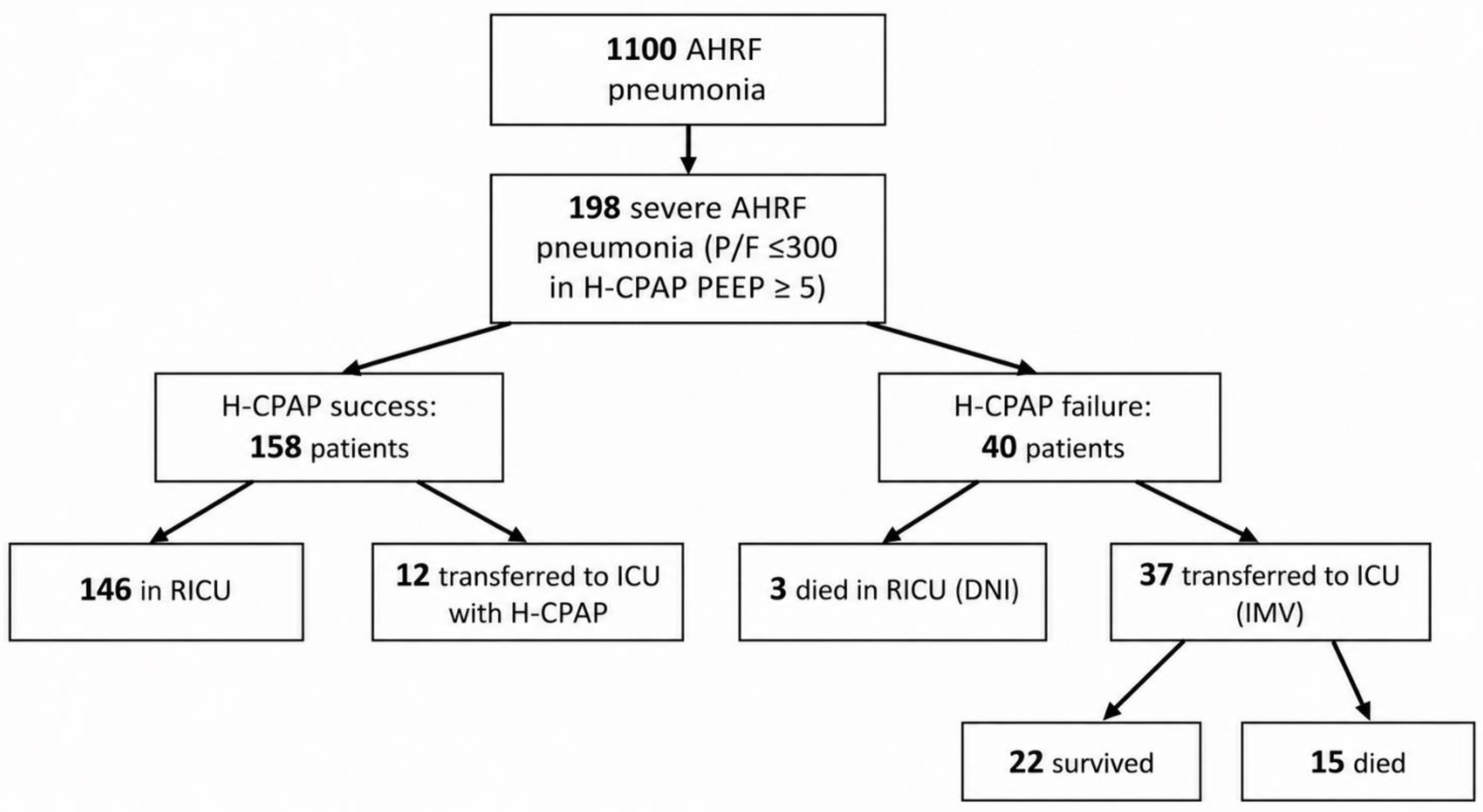
Figure 2.
Model performance across metrics. Optimism-corrected predictive performance of the four candidate models, expressed as c-index, Brier score, and scaled Brier score with 95% confidence intervals. For the c-index, values closer to 1 indicate better discrimination, whereas a value of 0.5 indicates no discriminative ability. For the Brier score, lower values indicate better overall prediction accuracy, with 0 representing perfect prediction. For the scaled Brier score, higher values indicate greater improvement over a non-informative model based on the outcome prevalence, with 1 representing perfect prediction and values close to 0 indicating limited improvement. Across models, discrimination was moderate, Brier scores were low and similar, and scaled Brier scores were modest with wide confidence intervals.
Figure 2.
Model performance across metrics. Optimism-corrected predictive performance of the four candidate models, expressed as c-index, Brier score, and scaled Brier score with 95% confidence intervals. For the c-index, values closer to 1 indicate better discrimination, whereas a value of 0.5 indicates no discriminative ability. For the Brier score, lower values indicate better overall prediction accuracy, with 0 representing perfect prediction. For the scaled Brier score, higher values indicate greater improvement over a non-informative model based on the outcome prevalence, with 1 representing perfect prediction and values close to 0 indicating limited improvement. Across models, discrimination was moderate, Brier scores were low and similar, and scaled Brier scores were modest with wide confidence intervals.
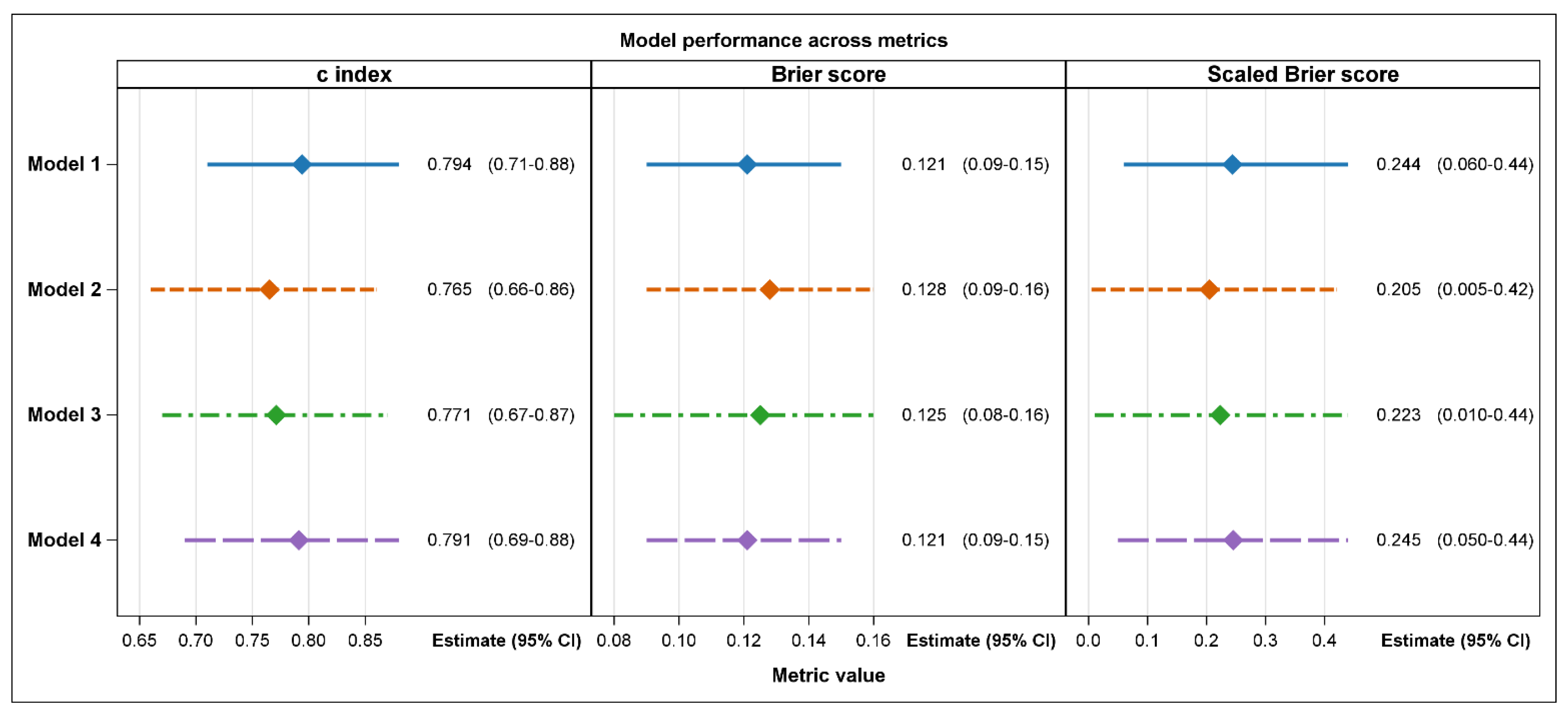
Figure 3.
Calibration plots by model. Calibration plots comparing observed and predicted risks for the four candidate models. Points represent grouped observed risks with 95% confidence intervals, the dashed diagonal line represents perfect calibration, and the solid line shows the loess calibration curve. Ideally, points and the loess curve should lie close to the diagonal reference line, indicating agreement between predicted and observed risks across the full range of predicted probabilities. Calibration intercepts and slopes close to 0 and 1, respectively, indicate good average calibration. Although average calibration was satisfactory, the plots suggested some overestimation at low predicted probabilities and underestimation in parts of the intermediate-to-higher risk range, particularly in the more reduced models. Wider confidence intervals at higher predicted risks indicate greater uncertainty due to fewer observations in these ranges.
Figure 3.
Calibration plots by model. Calibration plots comparing observed and predicted risks for the four candidate models. Points represent grouped observed risks with 95% confidence intervals, the dashed diagonal line represents perfect calibration, and the solid line shows the loess calibration curve. Ideally, points and the loess curve should lie close to the diagonal reference line, indicating agreement between predicted and observed risks across the full range of predicted probabilities. Calibration intercepts and slopes close to 0 and 1, respectively, indicate good average calibration. Although average calibration was satisfactory, the plots suggested some overestimation at low predicted probabilities and underestimation in parts of the intermediate-to-higher risk range, particularly in the more reduced models. Wider confidence intervals at higher predicted risks indicate greater uncertainty due to fewer observations in these ranges.
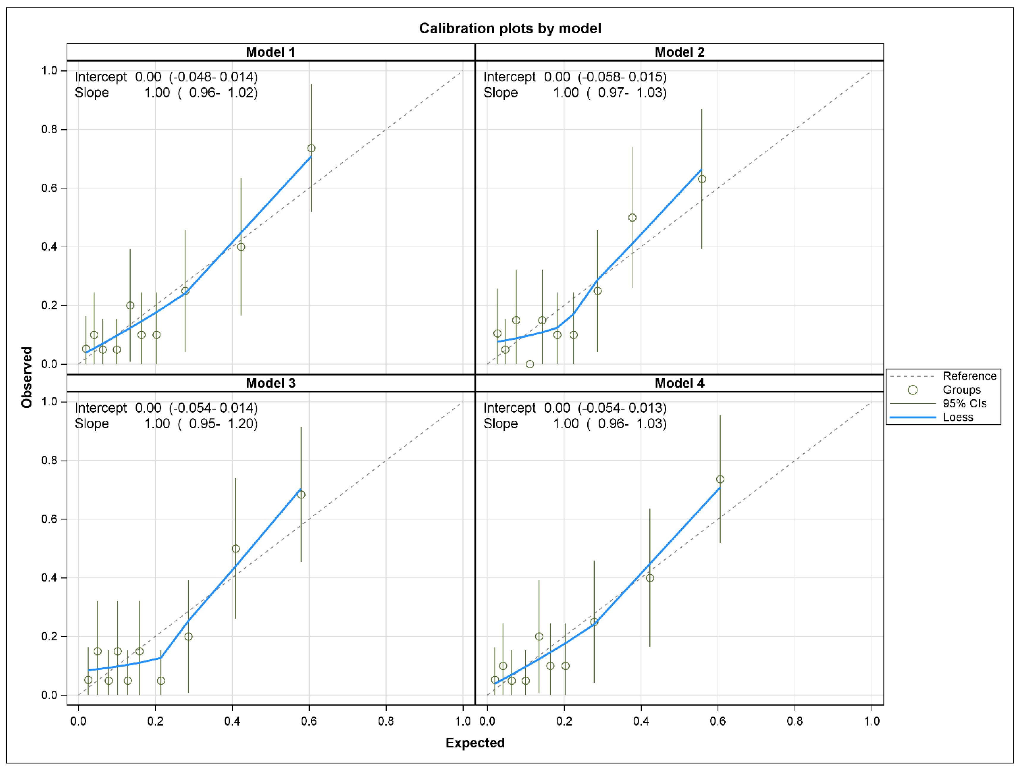
Figure 4.
Change in P/F ratio from standard oxygen therapy to helmet CPAP according to helmet CPAP outcome.
Figure 4.
Change in P/F ratio from standard oxygen therapy to helmet CPAP according to helmet CPAP outcome.
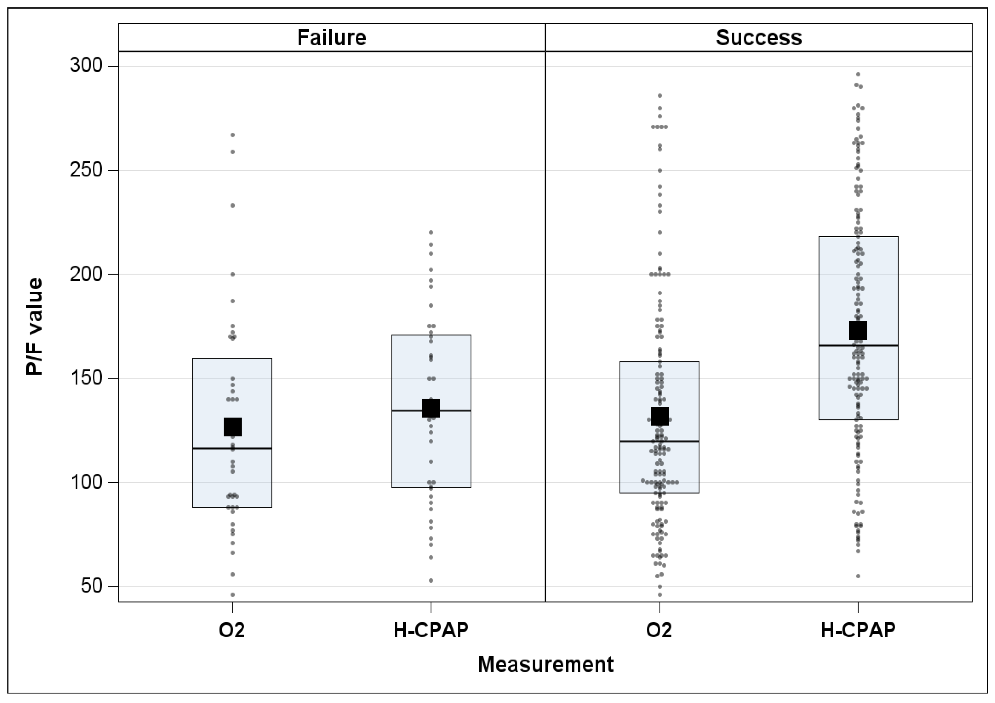

Table 1.
Baseline and early clinical characteristics according to H-CPAP outcome.
| OUTCOME | ||||
|---|---|---|---|---|
| SUCCESS (N=158) |
FAILURE (N=40) |
Total (N=198) |
P-value | |
| Sex (M/F), n (%) | 0.54481 | |||
| F | 39 (76.5%) | 12 (23.5%) | 51 (25.8%) | |
| M | 119 (81.0%) | 28 (19.0%) | 147 (74.2%) | |
| Age | 0.12632 | |||
| Mean (SD) | 62.7 (14.37) | 66.6 (13.99) | 63.5 (14.34) | |
| Period, n (%) | 0.03311 | |||
| Before 01/01/2020 | 80 (74.1%) | 28 (25.9%) | 108 (54.5%) | |
| After 01/01/2020 | 78 (86.7%) | 12 (13.3%) | 90 (45.5%) | |
| AHRF severity, n (%) | 0.00141 | |||
| Mild | 51 (92.7%) | 4 (7.3%) | 55 (27.8%) | |
| Moderate | 87 (79.1%) | 23 (20.9%) | 110 (55.6%) | |
| Severe | 20 (60.6%) | 13 (39.4%) | 33 (16.7%) | |
| Bilateral involvement, n (%) | 0.02591 | |||
| NO | 63 (88.7%) | 8 (11.3%) | 71 (35.9%) | |
| YES | 95 (74.8%) | 32 (25.2%) | 127 (64.1%) | |
| GCS, n (%) | 0.04291 | |||
| Confusion | 13 (61.9%) | 8 (38.1%) | 21 (10.6%) | |
| Normal | 145 (81.9%) | 32 (18.1%) | 177 (89.4%) | |
| Microbiological isolation, n (%) | 0.02851 | |||
| NO | 55 (71.4%) | 22 (28.6%) | 77 (38.9%) | |
| YES | 103 (85.1%) | 18 (14.9%) | 121 (61.1%) | |
| Legionella, n (%) | 0.09431 | |||
| NO | 118 (77.1%) | 35 (22.9%) | 153 (77.3%) | |
| YES | 40 (88.9%) | 5 (11.1%) | 45 (22.7%) | |
| Streptococcus pneumoniae, n (%) | 0.80521 | |||
| NO | 134 (79.3%) | 35 (20.7%) | 169 (85.4%) | |
| YES | 24 (82.8%) | 5 (17.2%) | 29 (14.6%) | |
| Mycoplasma pneumoniae, n (%) | 0.12501 | |||
| NO | 147 (78.6%) | 40 (21.4%) | 187 (94.4%) | |
| YES | 11 (100.0%) | 0 (0.0%) | 11 (5.6%) | |
| Influenza, n (%) | 0.71121 | |||
| NO | 149 (80.1%) | 37 (19.9%) | 186 (93.9%) | |
| YES | 9 (75.0%) | 3 (25.0%) | 12 (6.1%) | |
| Others, n (%) | 1.0000 | |||
| NO | 133 (79.6%) | 34 (20.4%) | 167 (84.3%) | |
| YES | 25 (80.6%) | 6 (19.4%) | 31 (15.7%) | |
| Pulmonary comorbidities, n (%) | 0.06661 | |||
| NO | 133 (82.6%) | 28 (17.4%) | 161 (81.3%) | |
| YES | 25 (67.6%) | 12 (32.4%) | 37 (18.7%) | |
| Cardiovascular disease, n (%) | 0.07741 | |||
| NO | 81 (85.3%) | 14 (14.7%) | 95 (48.0%) | |
| YES | 77 (74.8%) | 26 (25.2%) | 103 (52.0%) | |
| Diabetes, n (%) | 0.49961 | |||
| NO | 130 (80.7%) | 31 (19.3%) | 161 (81.3%) | |
| YES | 28 (75.7%) | 9 (24.3%) | 37 (18.7%) | |
| Neoplasia (current or previous), n (%) | 1.00001 | |||
| NO | 126 (79.7%) | 32 (20.3%) | 158 (79.8%) | |
| YES | 32 (80.0%) | 8 (20.0%) | 40 (20.2%) | |
| Mean arterial pressure | 0.43332 | |||
| Mean (SD) | 91.2 (14.28) | 89.2 (14.41) | 90.8 (14.29) | |
| Haematocrit | 0.00302 | |||
| Mean (SD) | 39.2 (5.16) | 35.8 (6.42) | 38.5 (5.59) | |
| p/F CPAP | <.00012 | |||
| Mean (SD) | 172.9 (59.37) | 135.9 (45.59) | 165.4 (58.67) | |
| p/F O₂ | 0.59082 | |||
| Mean (SD) | 131.9 (55.32) | 126.8 (52.61) | 130.9 (54.69) | |
| D(A–a)O₂ | <.00012 | |||
| Mean (SD) | 295.9 (91.57) | 435.4 (145.87) | 324.1 (118.52) | |
| APACHE2 | 0.00542 | |||
| Mean (SD) | 14.5 (4.26) | 17.2 (5.32) | 15.1 (4.60) | |
| HACORE SCORE CPAP | 0.72762 | |||
| Mean (SD) | 4.4 (2.00) | 4.5 (1.80) | 4.4 (1.96) | |
| Symptoms onset (hours) | 0.77342 | |||
| Mean (SD) | 107.9 (73.41) | 111.4 (66.71) | 108.6 (71.97) | |
|
Values are n (%) unless otherwise indicated. Continuous variables are reported as mean (standard deviation). P-values were calculated using 1Fisher Exact p-value and 2Unequal variance two sample t-test; | ||||
Table 2.
Odds ratio for the final model (M1) and for the three most frequently selected alternative models in 500 bootstrap samples (M2-M3-M4). Coefficients are reported on the logit scale. Positive values indicate a higher predicted probability of H-CPAP failure. A ‘/’ means that the variable was not selected for that model. The reference (baseline) category for bilateral involvement is ”Yes”.
Table 2.
Odds ratio for the final model (M1) and for the three most frequently selected alternative models in 500 bootstrap samples (M2-M3-M4). Coefficients are reported on the logit scale. Positive values indicate a higher predicted probability of H-CPAP failure. A ‘/’ means that the variable was not selected for that model. The reference (baseline) category for bilateral involvement is ”Yes”.
| Variables | Odds Ratio | |||
|---|---|---|---|---|
| M1 | M2 | M3 | M4 | |
| Intercept | 0.23 | 0.18 | 0.18 | 0.15 |
| Bilateral involvement | 0.39 | / | / | 0.63 |
| Htc | 0.57 | 0.52 | 0.55 | 0.57 |
| P/F CPAP | 0.80 | / | / | / |
| D(A-a)O₂ | 1.87 | 2.45 | 2.24 | 2.22 |
| APACHE2 | 1.44 | / | 1.41 | 1.47 |
Disclaimer/Publisher’s Note: The statements, opinions and data contained in all publications are solely those of the individual author(s) and contributor(s) and not of MDPI and/or the editor(s). MDPI and/or the editor(s) disclaim responsibility for any injury to people or property resulting from any ideas, methods, instructions or products referred to in the content. |
© 2026 by the authors. Licensee MDPI, Basel, Switzerland. This article is an open access article distributed under the terms and conditions of the Creative Commons Attribution (CC BY) license (http://creativecommons.org/licenses/by/4.0/).
Copyright: This open access article is published under a Creative Commons CC BY 4.0 license, which permit the free download, distribution, and reuse, provided that the author and preprint are cited in any reuse.