

Submitted:
01 September 2026
Posted:
02 September 2026
You are already at the latest version
Abstract
Metachromatic leukodystrophy (MLD) is a rare inherited lysosomal storage disorder that results from a deficiency of the arylsulfatase A (ARSA) enzyme and leads to progressive neurodegeneration and demyelination. Due to its narrow pre-symptomatic window of opportunity for disease-modifying therapies, the implementation of newborn screening (NBS) for MLD seems crucial and the incorporation of MLD screening into existing NBS programs is required. In many NBS programs, DNA-based assays have already been established, for instance to screen for severe combined immunodeficiency, sickle cell disease, or spinal muscular atrophy. Utilizing these DNA eluates, we established high-multiplex mutation testing (HMMT), a novel DNA-based method, as a genetic first-tier to screen for MLD. By multiplex allele-specific amplification, the assay detects thirteen frequent ARSA variants within the European population identifying newborns with MLD with a modelled, gnomAD-based sensitivity of approximately 92%. Anonymized DNA eluates from 3,078 newborns were tested retrospectively. Variants were identified in nine samples (0.3%) confirmed by an allele-specific differentiation assay and next-generation sequencing. HMMT proved capable to identify frequent ARSA variants as a genetic first-tier test, while panel composition and achievable sensitivity remain to be tested and optimised in a real-world cohort. When biochemical testing cannot be implemented as first tier screening, our approach might be useful to set up as simple screening algorithm for MLD.
Keywords:
metachromatic leukodystrophy
; newborn screening
; high multiplex mutation testing
; PCR
; MLD
; ARSA
; genetic newborn screening
1. Introduction
Metachromatic leukodystrophy (MLD) is a devastating lysosomal storage disorder caused by a deficiency of the enzyme arylsulfatase A (ARSA) (OMIM #250100), which results in an accumulation of sulfatides throughout the body, particularly in the central and peripheral nervous system [1]. The disease is characterized by progressive neurodegeneration and subsequent demyelination, clinically manifesting as rapid motor and cognitive decline leading to premature death, particularly in early onset phenotypes. According to the age of onset of clinical symptoms, the disease is classified into four subtypes: late-infantile (<30 months), early-juvenile (30 months – 7 years), late-juvenile (7 years – 16 years), and adult (>16 years). Late-infantile and early-juvenile MLD subtypes are referred to as early onset phenotypes and comprise the majority of cases [2,3]. MLD is a rare disease with an estimated birth prevalence of approximately one per 100,000 live births (1:40,000 to 1:160,000) [4]. To date, more than 250 disease-causing variants have been described in the underlying ARSA gene (ClinVar as of August 2026).
For patients with early onset MLD phenotypes, treatment with a lentiviral autologous hematopoietic stem and progenitor cell gene therapy (atidarsagene autotemcel/Libmeldy, Orchard therapeutics, London, United Kingdom) has been available since 2020 (approval by the European Medicines Agency) [2]. For late-onset phenotypes, HSCT has been shown to be effective [5]. However, this disease-modifying therapy needs to be administered in a pre-symptomatic stage to achieve the most favourable outcomes [6].
Thus, implementing MLD in newborn screening (NBS) programs seems vital. Several pilot studies have been published [7,8,9,10,11] and a three-tier screening algorithm has been proposed based on their results [12]. As first European country, Norway implemented NBS for MLD in January 2025. The three-tier algorithm consists of the determination of sulfatide species (first-tier), followed by ARSA activity in samples with elevated sulfatide levels (second-tier), and sequencing of the ARSA gene in samples with elevated sulfatides and decreased ARSA activity (third-tier). The algorithm has been described to be reliable and sensitive.
However, implementing biochemical testing in screening laboratories comes with infrastructural challenges as the determination of sulfatide species cannot be integrated into the existing flow injection analysis tandem mass spectrometry (FIA-MS/MS) platforms used for the determination of amino acids and acylcarnitines. A separate platform utilizing the more sensitive liquid chromatography (LC) MS/MS would need to be available. Compared to FIA-MS/MS, analysis time of LC-MS/MS is considerably longer and thus less suitable for high-throughput analyses required in NBS [7,10,13]. To date, LC-MS/MS is most commonly used as a second-tier technique in a small fraction of samples.
In addition, defining adequate cut-offs for sulfatide determination is challenging despite existing protocols [8,14]. In healthy infants, sulfatide concentrations are known to increase with age within the first two years of life [7,10,13]. However, knowledge is scarce about sulfatide levels in premature babies [15]. Furthermore, sulfatide levels have been shown to be unspecifically elevated in a considerable number of samples (up to 0.7%) [8,10,11,12] and are also elevated in individuals suffering from Saposin B deficiency caused by variants in the prosaposin (PSAP) gene encoding for the sphingolipid activator protein B of ARSA (OMIM #249900) [16] or multiple sulfatase deficiency (MSD) caused by variants in sulfatase modifying factor 1 (SUMF1) (OMIM #607939) [17,18,19]. Lacking therapeutic options, both disorders do not meet the Wilson and Jungner criteria for NBS and must not be identified [20].
ARSA testing from dried blood spots (DBS) (second-tier) has been described to be analytically challenging, not only due to pre-analytical effects [11,21]. ARSA activity could not be determined in 151 out of 381 (40%) samples of the prospective pilot study [22]. In addition, determination of ARSA activity does not dissect patients with MLD from patients with MSD.
We aimed to implement a genetic pre-screening as first-tier similar to that already established for sickle cell disease (SCD) [23] or as described for cystinosis [24]. For cystinosis, pre-screening for three frequent variants within the cystinosin (lysosomal cystine transporter; CTNS) gene followed by preselected next-generation sequencing (NGS) targeting 101 CTNS variants provided a calculated detection rate of 98.5% [25].
Since many current NBS programs include DNA-based screening, e.g. for severe combined immunodeficiency (SCID) and spinal muscular atrophy (SMA) as a high-throughput process, a PCR-based test seems feasible to reduce the number of samples for sulfatide screening appropriate for a LC-MS/MS second-tier method.
The primary objective of the present concept study was to evaluate whether genetic pre-screening could be used to detect frequent disease causing variants in the ARSA gene in a single PCR reaction. This novel method was called high multiplex mutation testing (HMMT).
In addition, we established a variant-specific real-time stripe assay including melting analysis for further differentiation of the variants. Complemented with the determination of sulfatides and/or ARSA sequencing, these assays provide a novel tool to establish simple screening algorithms that integrate with the existing resources.
2. Materials and Methods
2.1. Samples
For this study, irreversibly anonymized residual DNA eluates left over from routine newborn screening were used. Samples were collected from January 15th 2026 to February 27th 2026. Due to this study design, the determination of sulfatides could not be performed.
In addition, two DBS from previously identified affected MLD children beyond the newborn period were available to evaluate our assay (provided by University Children’s Hospital Tübingen, Neuropaediatrics), in the following referred to as “affected MLD patients”.
2.2. Rapid Nucleic Acid Extraction from DBS
2.3. Multiplex Screening qPCR (HMMT)
Vacuum dried LightMix® KIT Newborn MLD (HMMT) (40-0465-RUO, TIB Molbiol, Berlin, Germany) was dissolved as described in the manufacturer’s instructions. First, 7 µl of reaction mix consisting of 0.5 µl LightMix® KIT Newborn, 2.0 µl Multiplex DNA Master (Roche, Mannheim, Germany) and 4.5 µl H2O were distributed per reaction to a 384-well plate (Roche). Second, 3 µl of eluted DNA were added to the reaction mix using the ViaFlo96 system (Integra, Zizers, Switzerland) to transfer the DNA elution from the 96-well plates to the 384-well qPCR plates (Roche). qPCR was performed on a LightCycler480 II instrument (Roche) with the following PCR profile: initial denaturation at 95 °C for 5 min, followed by 45 cycles in three steps at 95 °C for 5 s, 61 °C for 15 s, and 72 °C for 15 s. A single acquisition of fluorescence signals was included in the 61 °C step. The fit point analysis of the LightCycler® software (Roche) was used to analyse qPCR fluorescence signals. Four standardised positive controls were included in each qPCR run. An additional detection assay for the housekeeping gene of Ribonuclease P (RNase P) was utilised as an internal control in the same reaction.
The assay covered the following thirteen ARSA variants at twelve positions: c.465+1G>A (rs80338815), c.1283C>T (rs28940893), c.542T>G (rs74315457), c.293C>T (rs74315456), c.257G>A (rs74315458), c.542dup (rs776748338), c.736C>T (rs74315470), c.862A>C (rs28940894), c.869G>A (rs199476355), c.925G>C/A (rs199476360), c.1010A>T (rs74315475), and c.1136C>T (rs74315478). All variants are classified as disease causing in ClinVar.
An earlier increase in fluorescence signal compared to the other samples was considered a positive result indicating heterozygosity for at least one of the included variants. The test does not differentiate between the presence of different variants and whether they are heterozygous, compound heterozygous, or homozygous. This set of variants comprises the most frequent ARSA variants in the European population (see results section).
2.4. Variant-Specific qPCR Assay
A variant-specific assay to differentiate between the different ARSA variants was provided as qPCR stripes with each variant being detected in separate wells (Newborn MLD, 40-2004-RUO, TIB Molbiol).
For each well 4.5 µl Multiplex DNA Master (Roche), 16.75 µl H2O and 1.25 µl sample DNA were mixed. A total of 20 µl of this mixture were then pipetted into the variant-specific well.
qPCR was performed on a LightCycler480 II instrument (Roche) with the following PCR profile: initial denaturation at 95 °C for 5 min, followed by 45 cycles in three steps at 95 °C for 5 s, 60 °C for 15 s, and 72 °C for 15 s. A single acquisition of fluorescence signals was included in the 60 °C step. Melting analyses were done for 30 s at 95 °C followed by 2 min at 40 °C and slowly increasing the temperature to 75 °C with continuous measuring of the fluorescence (3 acquisitions per °C).
Of note, this test gives information on whether the detected variations are present in a heterozygous or a homozygous state.
2.5. Next-Generation Sequencing
The same nucleic acid extracts as for the qPCR assays were used for the generation of targeted enriched NGS-Libraries according to the protocols “Library Preparation EF 2.0 with Enzymatic Fragmentation and Twist Universal Adapter System” and “Twist Target Enrichment Standard Hybridization v1 Protocol” (Twist Bioscience, San Francisco, CA, USA). The Unique Dual Index (UDI) System and a customized target panel were used, both from Twist Bioscience, USA. Hybridization-based targeted enrichment was done using a Twist custom panel targeting all CDS of the ARSA gene (NM_000487= ENST00000216124) including +/- 10 bp of intron exon borders (Twist Bioscience). Library preparation and enrichment was done according to manufacturer’s instructions.
Short-read sequencing was performed on the Illumina MiniSeq using the 2x 150 bp paired-ends (PE) Mid-Output Kit (Illumina, San Diego, CA, USA). Processing of the resulting fastq-files and variant calling was done using a proprietary workflow of QIAGEN CLC Genomics Workbench, Version 25.0.2 (Qiagen, Hilden, Germany).
3. Results
3.1. Sensitivity Estimation
To validate the population-genetic approach, we estimated the genetic birth prevalence of ARSA-related MLD from allele frequencies in gnomAD v4.1.1, following the approach described previously [28]. The cumulative allele frequency of pathogenic ARSA variants in the European (non-Finnish) population was 0.00314 (list of pathogenic variants can be found in Supplementary Table S1). Assuming Hardy-Weinberg equilibrium and complete penetrance, this corresponds to an expected birth prevalence of q2=9.89×10⁻⁶ or approximately 0.99 per 100,000 births, which is consistent with published estimates [4,29]. Assay sensitivity was estimated from the cumulative frequency of the thirteen screened variants relative to the cumulative frequency of all pathogenic ARSA alleles. As an affected individual is detected whenever at least one of the two pathogenic alleles is included in the assay, only individuals carrying two non-screened pathogenic alleles are missed. Sensitivity was therefore calculated as. The modelled genetic sensitivity was 92.03% (95% CI, 91.19–92.83%), with the uncertainty interval estimated by parametric bootstrapping of variant-specific allele counts from gnomAD v4.1.1 (100,000 iterations); this interval reflects sampling uncertainty of the gnomAD allele frequencies only and does not account for uncertainty related to variant classification, penetrance, or population structure. The modelled sensitivity will be an overestimation as this approach only considers pathogenic variants that are listed in gnomAD..
3.2. qPCR Results
Anonymized DNA eluates from 3,078 newborns screening samples were screened for ARSA variants using the novel HMMT approach. Nine samples (0.3%) showed an increase in fluorescence indicative of the presence of one of the thirteen screened variants (Fig. 1). These samples, which were considered to be at least heterozygous, were analysed by the variant-specific assay and NGS. Two additional samples showed a later and weaker increase of fluorescence at the final measurement points (Figure 1). These signals were considered to be the result of non-specific PCR amplification. However, to rule out the possibility of missing ARSA variants with late amplification, we analyzed those late amplifiers for variants via NGS. In addition, both samples of the affected MLD patients showed a characteristic increase of the fluorescence signal.
3.3. Variant-Specific Assay
All of the nine newborn screening samples exhibiting an early increase of fluorescence were confirmed to harbour one of the screened ARSA variants in a heterozygous state using the variant-specific assay. Three samples were heterozygous for c.465+1G>A, two for c.542T>G, two for c.869G>A, one for c.293C>T and one for c.736C>T. None of the newborn screening samples showed a homozygous signal or heterozygous signals for two different variants indicating compound heterozygosity.
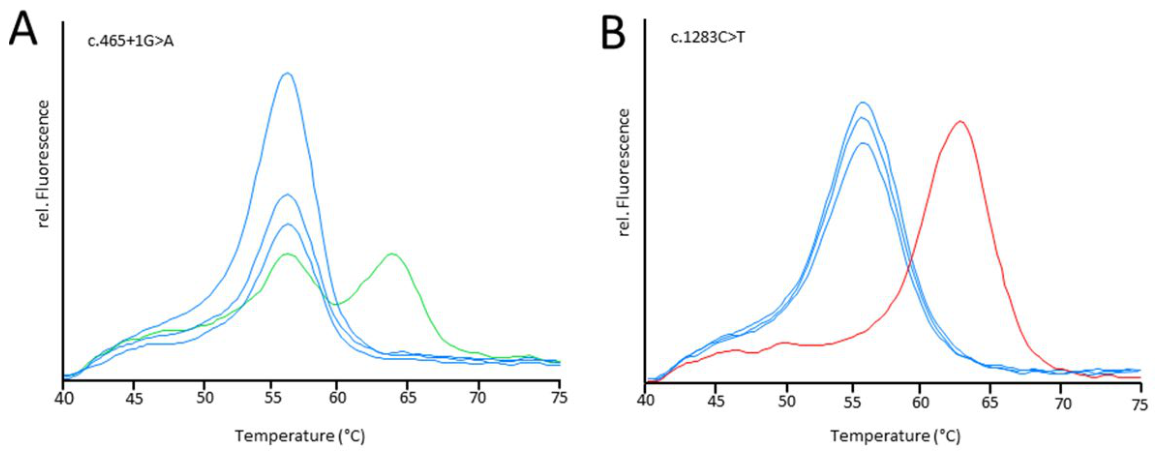
Of the two affected MLD patients, one showed heterozygosity for c.465+1G>A, while the other patient revealed homozygosity for c.1283C>T (Figure 2).
3.4. Next Generation Sequencing
No second (likely) pathogenic variant within the ARSA gene was identified in the 9 heterozygous newborn screening samples, thus excluding compound heterozygosity with a second rare coding pathogenic variant. Additionally, all variants determined with the variant-specific qPCR assay were confirmed by targeted NGS analysis of the ARSA gene. None of the two samples showing late and unspecific amplification (Fig. 1, late amplifiers) in the HMMT qPCR revealed one of the thirteen ARSA variants included in the test or another pathogenic variant.
In the MLD patient samples, homozygosity for c.1283C>T was confirmed in one patient. In the other patient a second pathogenic ARSA variant, c.899delT (p.Leu300fs) was identified.
4. Discussion
With the availability of a disease-modifying therapy, which is most effective when applied in pre-symptomatic and early symptomatic stages of early onset phenotypes of the disease, an urgent need to include screening for MLD in newborn screening programs has evolved [22]. However, the proposed three-tier screening algorithm comprising the determination of sulfatide species, ARSA enzyme activity, and ARSA gene sequencing is technically challenging to implement depending on local resources and established pipelines. This is even more significant given the fact that only a small number of individuals are expected to be affected by this rare condition each year, for instance 4-16 individuals are expected to be affected in Germany [4].
In particular, the first-tier measurement of sulfatides cannot be incorporated in the existing FIA-MS/MS measurements. It needs to be executed by LC-MS/MS, a method only partially applicable as high-throughput assay and mainly employed as a classical second-tier method following positive first-tier results in a small subset of samples to reduce recall rates and increase positive predictive values [30,31,32,33].
We evaluated a screening algorithm that would integrate better with the existing structures and resources of most newborn screening laboratories, using sulfatide LC-MS/MS as a complementary (second-tier) test, and thus reducing costs and resources.
Adding HMMT pPCR as first-tier analysis to the screening algorithm makes use of a technique established in most newborn screening laboratories. It seems feasible in view of the fact, that only a few pathogenic variants within the ARSA gene have a high allele frequency in the European population. With the set of variants described, we calculated to identify up to 92% of MLD patients of the European population (estimation based on gnomAD Dataset v4.1.1, GRCh38). However, these calculations need to be interpreted with caution as ultra-rare pathogenic variants not listed in gnomAD would not be identified and reduce the sensitivity of the test. It would also have a higher sensitivity in outbred populations with a higher likelihood of compound heterozygous variants in comparison to inbred populations with a higher probability of homozygous presence of disease causing variants. However, it cannot achieve the sensitivity of 100% described in retrospective biochemical first tier studies [8]. A combination of screening heterozygotes (first-tier) with targeted ARSA NGS will identify all MLD patients, if at least one pathogenic variant from the set of thirteen is present. In comparison, NBS for SMA screens for the most frequent deletion within the survival of motor-neuron 1 (SMN1) gene identifying approximately 95% of SMA patients [26,34]. Notably, all three MLD screen positives of the German pilot study would have been detected with our approach [10]. Since then, nine screen-positive newborns have been identified and confirmed in the German and Austrian pilot programs (www.researchsquare.com/article/rs-9496450/v1). Applying the present panel to their published genotypes, seven of these nine infants carry at least one of the thirteen variants and would have entered a genetic first-tier algorithm. The two infants that would not have been captured were both predicted to develop late-infantile MLD. Although these numbers are small, they illustrate the overestimation of the modelled sensitivity of 92% as this approach only considers 162 variants that are listed in gnomAD out of more than 500 pathogenic variants in ClinVar.
The observed frequency of variants at 0.003 (nine out of 3,078) is close to the calculated frequency of pathogenic variants of 0.0022 (see Supplementary Table S1). Of note, we designed this project as a proof-of-concept study. The number as well as the selection of target variants should be updated in a clinical trial to yield the highest feasible sensitivity.
In the present series, 0.3% of samples (nine of 3,078) were first-tier positive and all of them were heterozygous carriers, corresponding to approximately one in 340 newborns requiring second-tier biochemical analysis, preferably the measurement of sulfatides. Unfortunately, this was not possible in our concept study due to the use of anonymized DNA eluates. Since heterozygous carriers of clinically relevant ARSA variants may show isolated elevations of single sulfatide species, not all of these samples will be resolved unambiguously by biochemical analysis. Only samples with normal concentrations of sulfatides can be considered unaffected. Hence, comprehensive genetic testing as third tier is required. The variant-specific assay might be used to reduce the number of costly NGS tests. The homozygous affected MLD patient in our study would have been identified by this practice. However, when the variant-specific assay fails to identify the second pathogenic variant, targeted NGS ARSA sequencing needs to be performed in individuals with elevated sulfatide concentrations. This strategy ensures that rare variants are not missed in these individuals. A similar approach is applied in the Dutch newborn screening for cystic fibrosis, where extended gene analysis was implemented as safety net analysis to complement the DNA analysis for a smaller, distinct set of CFTR variants in individuals with elevated concentrations of immunoreactive trypsin and pancreatitis-associated protein [35].
However, NBS laboratories do not generally have the technical resources for targeted NGS or Sanger sequencing of entire genes, which also requires the expertise of a clinical laboratory geneticist experienced in MLD genetic testing. As an alternative to targeted NGS or Sanger sequencing of ARSA, a set of pathogenic variants could be defined. This procedure would be comparable to the current German NBS for cystic fibrosis, which tests for 31 CFTR variants in covering 98% of the variants in the Caucasian population. For MLD, the set of ARSA variants could be designed to include or exclude specific variants associated with certain clinical presentations.
Our concept study comprised thirteen ARSA variants, but could be expanded to include further eligible variants with a manageable amount of effort. Specific limitations regarding the assay design have not yet been identified, but are determined mainly by the structure of the reference sequence.
Implementing a genetic test as first-tier test provides further important benefits. It utilizes the same DNA eluates, which are also required for other genetic tests within the newborn screening panel, for instance SCID and SMA, and by that saves on valuable and limited sample material. No extra punch of dried blood is required in the vast majority of (>99%) samples. This aspect will become increasingly important with a growing number of conditions meeting the criteria to be screened for and an increasing number of screening algorithms requiring second-tier strategies to improve specificity, i.e. congenital adrenal hyperplasia, methylmalonic acidurias, propionic acidemia, and isovaleric academia [36,37,38,39]. Furthermore, individuals with Saposin B deficiency or MSD are not detected. Both conditions do not meet the criteria to be implemented in NBS programs but do lead to elevated concentrations of sulfatides and reduced ARSA activity (only MSD). In the three-tier algorithm proposed by Laugwitz et al. [12], both conditions may cause positive results in the first (Saposin B deficiency) or first two tests (MSD) and require genetic testing.
Since PSAP and SUMF1 variants are not included in our first-tier HMMT, it would even be conceivable to reduce the NBS for MLD to the HMMT qPCR and the determination of sulfatides. Individuals with clearly elevated concentrations of sulfatides after genetic pre-screening for ARSA variants are highly suspicious to be affected by MLD and would be referred to treatment centres for confirmatory diagnostics i.e. ARSA activity and ARSA gene sequencing. However, as this approach would still identify heterozygous carriers, optimizing the sulfatide cut-offs would be necessary in order to reduce the number of false-positives.
The limitations of the present study need to be stated explicitly: the sensitivity reported here is modelled from population allele frequencies rather than measured in an affected cohort, and it is derived from a database dominated by individuals of European ancestry. Sulfatide concentrations could not be determined in the anonymised DNA eluates used here, so the algorithm we propose has not been validated end to end. A prospective study addressing these points as well as revisiting the number and selection of target variants against clinically characterised cohorts is required.
In general, a genetic first-tier HMMT could be a promising approach for newborn screening. It might well be applicable to further conditions which are caused by a distinct number of frequent variants or conditions lacking feasible biochemical parameters. For instance, detecting only six variants in the lysosomal tripeptidyl peptidase 1 gene (TPP1) would provide a sensitivity >95% in a screening for ceroidlipofuscinosis type 2 (CLN2), another lysosomal storage disorder. In combination with a variant-specific stripe assay, NGS or another suitable second-tier assay, HMMT is a simple method of expanding NBS to include more treatable diseases.
Our findings with a modelled sensitivity estimate should be regarded as a proof-of-concept requiring prospective validation. We propose HMMT as a useful tool that can be combined with other screening assays in various ways depending on country and laboratory specific regulations and resources.
Supplementary Materials
The following supporting information can be downloaded at the website of this paper posted on Preprints.org. Table S1: Pathogenic and likely pathogenic ARSA variants included in the population-based prevalence estimation.
Author Contributions
Conceptualization, J.B., L.C., S.B., and E.M.M.; methodology, J.B., L.C., S.B., and M.K.; validation, J.B., L.C., S.B., M.B., and J.D.; formal analysis, J.B., L.C., S.B., E.S., A.T., C.C., N.S., E.M.M., K.V., M.W., S.G., and J.D.; investigation, J.B., L.C., S.B., E.M.M., M.B., and J.D.; resources, J.B., L.C., S.B., E.M.M., M.B., and J.D.; data curation, J.B., L.C., S.B., E.M.M., and J.D.; writing—original draft preparation, J.B., E.M.M., M.W., L.C., and S.B.; writing—review and editing, J.B., L.C., S.B., L.L., S.G., H.R., E.M.M., M.W., M.B., and J.D.; visualization, J.B. and L.C.; supervision, S.B., E.M.M., M.B., and J.D.; project administration, J.B., L.C., S.B., M.B., E.M.M., and J.D. All authors have read and agreed to the published version of the manuscript.
Funding
This research received no external funding.
Institutional Review Board Statement
The study was conducted in accordance with the Declaration of Helsinki, and approved by the Ethics Committee of the Medical Faculty of Ludwig-Maximilians-Universität München, Munich, Germany (25-0699 KB, issued 11th August 2025) and the local institutional Review Board of the Medical Faculty of Tübingen, Germany (948/2018BO2).
Informed Consent Statement
Patient consent was waived due to the retrospective analyses of irreversibly anonymized data.
Data Availability Statement
The raw data supporting the conclusions of this article will be made available by the authors on request.
Acknowledgments
M.W. was supported by a Else Kröner excellence fellowship (2025_EKES.12) and a clinician scientist fellowship of the Hertie foundation (Hertie Network of Excellence in Clinical Neuroscience, P1230037).
Conflicts of Interest
L.L. and S.G. received institutional research support by Orchard Therapeutics, unrelated to this study. Author M.K. is employed by the company TIB Molbiol Syntheselabor GmbH. The research was conducted in the absence of any commercial or financial relationships that could be construed as a potential conflict of interest. The remaining authors declare no conflicts of interest.
Abbreviations
The following abbreviations are used in this manuscript:
| HMMT | High multiplex mutation testing |
| MLD | Metachromatic leukodystrophy |
| ARSA | Arylsulfatase A |
| NBS | Newborn screening |
| FIA-MS/MS | Flow injection analysis tandem mass spectrometry |
| LC-MS/MS | Liquid chromatography tandem mass spectrometry |
| PSAP | Prosaposin |
| SUMF1 | Sulfatase modifying factor 1 |
| MSD | Multiple sulfatase deficiency |
| SCD | Sickle cell disease |
| SCID | Severe combined immunodeficiency |
| CTNS | Lysosomal cystine transporter |
| SMA | Spinal muscular atrophy |
| DBS | Dried blood spot |
| SMN1 | Survival of motor neuron 1 |
| CFTR | Cystic fibrosis transmembrane conductance regulator |
| VUS | Variants of unknown significance |
| TTP1 | Tripeptidyl peptidase 1 |
| CLN2 | Ceroidlipofuscinosis type 2 |
References
- Asbreuk, M.; Schoenmakers, D.H.; Adang, L.A.; Beerepoot, S.; Bergner, C.; Bley, A.; Boelens, J.J.; Bugiani, M.; Calbi, V.; Garcia-Cazorla, A.; et al. Metachromatic Leukodystrophy: New Therapy Advancements and Emerging Research Directions. Neurology 2025, 105(2), e213817. [Google Scholar] [CrossRef] [PubMed]
- Fumagalli, F.; Calbi, V.; Natali Sora, M.G.; Sessa, M.; Baldoli, C.; Rancoita, P.M.V.; Ciotti, F.; Sarzana, M.; Fraschini, M.; Zambon, A.A.; et al. Lentiviral haematopoietic stem-cell gene therapy for early-onset metachromatic leukodystrophy: long-term results from a non-randomised, open-label, phase 1/2 trial and expanded access. Lancet 2022, 399(10322), 372–383. [Google Scholar] [CrossRef] [PubMed]
- van Rappard, D.F.; Boelens, J.J.; Wolf, N.I. Metachromatic leukodystrophy: Disease spectrum and approaches for treatment. Best Pract. Res. Clin. Endocrinol. Metab. 2015, 29(2), 261–273. [Google Scholar] [CrossRef] [PubMed]
- Chang, S.C.; Bergamasco, A.; Bonnin, M.; Bisono, T.A.; Moride, Y. A systematic review on the birth prevalence of metachromatic leukodystrophy. Orphanet J. Rare Dis. 2024, 19(1), 80. [Google Scholar] [CrossRef] [PubMed]
- Videbaek, C.; Stokholm, J.; Sengelov, H.; Fjeldborg, L.U.; Larsen, V.A.; Krarup, C.; Nielsen, J.E.; Gronborg, S. Allogenic hematopoietic stem cell transplantation in two siblings with adult metachromatic leukodystrophy and a systematic literature review. JIMD Rep. 2021, 60(1), 96–104. [Google Scholar] [CrossRef] [PubMed]
- Fumagalli, F.; Calbi, V.; Gallo, V.; Zambon, A.A.; Recupero, S.; Ciotti, F.; Sarzana, M.; Fraschini, M.; Scarparo, S.; De Mattia, F.; et al. Long-Term Effects of Atidarsagene Autotemcel for Metachromatic Leukodystrophy. N Engl. J. Med. 2025, 392(16), 1609–1620. [Google Scholar] [CrossRef] [PubMed]
- Bekri, S.; Bley, A.; Brown, H.A.; Chanson, C.; Church, H.J.; Gelb, M.H.; Hong, X.; Janzen, N.; Kasper, D.C.; Mechtler, T.; et al. Higher precision, first tier newborn screening for metachromatic leukodystrophy using 16:1-OH-sulfatide. Mol. Genet Metab. 2024, 142(1), 108436. [Google Scholar] [CrossRef] [PubMed]
- Hong, X.; Daiker, J.; Sadilek, M.; Ruiz-Schultz, N.; Kumar, A.B.; Norcross, S.; Dansithong, W.; Suhr, T.; Escolar, M.L.; Ronald Scott, C.; et al. Toward newborn screening of metachromatic leukodystrophy: results from analysis of over 27,000 newborn dried blood spots. Genet Med. 2021, 23(3), 555–561. [Google Scholar] [CrossRef] [PubMed]
- Wu, T.H.Y.; Brown, H.A.; Church, H.J.; Kershaw, C.J.; Hutton, R.; Egerton, C.; Cooper, J.; Tylee, K.; Cohen, R.N.; Gokhale, D.; et al. Improving newborn screening test performance for metachromatic leukodystrophy: Recommendation from a pre-pilot study that identified a late-infantile case for treatment. Mol. Genet Metab. 2024, 142(1), 108349. [Google Scholar] [CrossRef] [PubMed]
- Laugwitz, L.; Mechtler, T.P.; Janzen, N.; Oliva, P.; Kasper, A.R.; Teunissen, C.E.; Burger, F.; Janda, J.; Doring, M.; Weitz, M.; et al. Newborn Screening and Presymptomatic Treatment of Metachromatic Leukodystrophy. N Engl. J. Med. 2024, 391(13), 1256–1258. [Google Scholar] [CrossRef] [PubMed]
- Malvagia, S.; Bettiol, A.; Porcaro, M.; Mura, M.; Funghini, S.; Ombrone, D.; Forni, G.; Scolamiero, E.; Coppi, F.; Damiano, R.; et al. Newborn Screening for Metachromatic Leukodystrophy in Tuscany: The Paradigm of a Successful Preventive Medicine Program. Int. J. Neonatal Screen. 2025, 11(2). [Google Scholar] [CrossRef] [PubMed]
- Laugwitz, L.; Shenker, A.; Sluys, E.F.; Pintat, S.; Whiteman, D.; Chanson, C. Newborn Screening for Metachromatic Leukodystrophy: A Systematic Literature Review. Int. J. Neonatal Screen. 2025, 11(4). [Google Scholar] [CrossRef] [PubMed]
- Gelb, M.H.; Basheeruddin, K.; Burlina, A.; Chen, H.J.; Chien, Y.H.; Dizikes, G.; Dorley, C.; Giugliani, R.; Hietala, A.; Hong, X.; et al. Liquid Chromatography-Tandem Mass Spectrometry in Newborn Screening Laboratories. Int. J. Neonatal Screen. 2022, 8(4). [Google Scholar] [CrossRef] [PubMed]
- Shaff, A.; Basheeruddin, K.; Bekri, S.; Brown, H.A.; Church, H.J.; Gianares, J.; Hong, X.; Jones, S.A.; Kappell, T.; Kubaski, F.; et al. Newborn screening for metachromatic leukodystrophy: Preparation of reagents and methodology for measurement of sulfatides and arylsulfatase A enzymatic activity in dried blood spots. Mol. Genet Metab. 2025, 145(3), 109138. [Google Scholar] [CrossRef] [PubMed]
- van der Ham, M.; Hoytema van Konijnenburg, E.; van Rossum, W.; Gerrits, J.; van Hasselt, P.; Prinsen, H.; Jans, J.; Schlotawa, L.; Laugwitz, L.; de Sain-van der Velden, M. Profiling and semi-quantitation of urine sulfatides by UHPLC-Orbitrap-HRMS. Anal. Chim. Acta 2025, 1350, 343824. [Google Scholar] [CrossRef] [PubMed]
- Cesani, M.; Lorioli, L.; Grossi, S.; Amico, G.; Fumagalli, F.; Spiga, I.; Filocamo, M.; Biffi, A. Mutation Update of ARSA and PSAP Genes Causing Metachromatic Leukodystrophy. Hum. Mutat. 2016, 37(1), 16–27. [Google Scholar] [CrossRef] [PubMed]
- Fraldi, A.; Biffi, A.; Lombardi, A.; Visigalli, I.; Pepe, S.; Settembre, C.; Nusco, E.; Auricchio, A.; Naldini, L.; Ballabio, A.; et al. SUMF1 enhances sulfatase activities in vivo in five sulfatase deficiencies. Biochem J. 2007, 403(2), 305–312. [Google Scholar] [CrossRef] [PubMed]
- Cosma, M.P.; Pepe, S.; Parenti, G.; Settembre, C.; Annunziata, I.; Wade-Martins, R.; Di Domenico, C.; Di Natale, P.; Mankad, A.; Cox, B.; et al. Molecular and functional analysis of SUMF1 mutations in multiple sulfatase deficiency. Hum. Mutat. 2004, 23(6), 576–581. [Google Scholar] [CrossRef] [PubMed]
- Schlotawa, L.; Adang, L.A.; Radhakrishnan, K.; Ahrens-Nicklas, R.C. Multiple Sulfatase Deficiency: A Disease Comprising Mucopolysaccharidosis, Sphingolipidosis, and More Caused by a Defect in Posttranslational Modification. Int. J. Mol. Sci. 2020, 21(10). [Google Scholar] [CrossRef]
- Andermann, A.; Blancquaert, I.; Beauchamp, S.; Dery, V. Revisiting Wilson and Jungner in the genomic age: a review of screening criteria over the past 40 years. Bull. World Health Organ. 2008, 86(4), 317–319. [Google Scholar] [CrossRef] [PubMed]
- Hong, X.; Kumar, A.B.; Daiker, J.; Yi, F.; Sadilek, M.; De Mattia, F.; Fumagalli, F.; Calbi, V.; Damiano, R.; Della Bona, M.; et al. Leukocyte and Dried Blood Spot Arylsulfatase A Assay by Tandem Mass Spectrometry. Anal. Chem. 2020, 92(9), 6341–6348. [Google Scholar] [CrossRef] [PubMed]
- Laugwitz, L.; Schoenmakers, D.H.; Adang, L.A.; Beck-Woedl, S.; Bergner, C.; Bernard, G.; Bley, A.; Boyer, A.; Calbi, V.; Dekker, H.; et al. Newborn screening in metachromatic leukodystrophy - European consensus-based recommendations on clinical management. Eur. J. Paediatr. Neurol. 2024, 49, 141–154. [Google Scholar] [CrossRef] [PubMed]
- Janda, J.; Hegert, S.; Bzdok, J.; Tesorero, R.; Holtkamp, U.; Burggraf, S.; Schuhmann, E.; Horster, F.; Hoffmann, G.F.; Janzen, N.; et al. High Throughput Newborn Screening for Sickle Cell Disease - Application of Two-Tiered Testing with a qPCR-Based Primary screen. Klin. Padiatr. 2023, 235(6), 366–372. [Google Scholar] [CrossRef] [PubMed]
- Fleige, T.; Burggraf, S.; Czibere, L.; Haring, J.; Gluck, B.; Keitel, L.M.; Landt, O.; Harms, E.; Hohenfellner, K.; Durner, J.; et al. Next generation sequencing as second-tier test in high-throughput newborn screening for nephropathic cystinosis. Eur. J. Hum. Genet. 2020, 28(2), 193–201. [Google Scholar] [CrossRef] [PubMed]
- Hohenfellner, K.; Bergmann, C.; Fleige, T.; Janzen, N.; Burggraf, S.; Olgemoller, B.; Gahl, W.A.; Czibere, L.; Froschauer, S.; Roschinger, W.; et al. Molecular based newborn screening in Germany: Follow-up for cystinosis. Mol. Genet Metab. Rep. 2019, 21, 100514. [Google Scholar] [CrossRef] [PubMed]
- Czibere, L.; Burggraf, S.; Fleige, T.; Gluck, B.; Keitel, L.M.; Landt, O.; Durner, J.; Roschinger, W.; Hohenfellner, K.; Wirth, B.; et al. High-throughput genetic newborn screening for spinal muscular atrophy by rapid nucleic acid extraction from dried blood spots and 384-well qPCR. Eur. J. Hum. Genet. 2020, 28(1), 23–30. [Google Scholar] [CrossRef]
- Bzdok, J.; Czibere, L.; Burggraf, S.; Landt, O.; Maier, E.M.; Roschinger, W.; Albert, M.H.; Hegert, S.; Janzen, N.; Becker, M.; et al. Quality considerations and major pitfalls for high throughput DNA-based newborn screening for severe combined immunodeficiency and spinal muscular atrophy. PLoS ONE 2024, 19(6), e0306329. [Google Scholar] [CrossRef] [PubMed]
- Tan, J.; Wagner, M.; Stenton, S.L.; Strom, T.M.; Wortmann, S.B.; Prokisch, H.; Meitinger, T.; Oexle, K.; Klopstock, T. Lifetime risk of autosomal recessive mitochondrial disorders calculated from genetic databases. EBioMedicine 2020, 54, 102730. [Google Scholar] [CrossRef] [PubMed]
- Brimley, C.J.; Lopez, J.; van Haren, K.; Wilkes, J.; Sheng, X.; Nelson, C.; Korgenski, E.K.; Srivastava, R.; Bonkowsky, J.L. National variation in costs and mortality for leukodystrophy patients in US children's hospitals. Pediatr. Neurol. 2013, 49(3), 156–162 e151. [Google Scholar] [CrossRef] [PubMed]
- la Marca, G.; Malvagia, S.; Pasquini, E.; Innocenti, M.; Donati, M.A.; Zammarchi, E. Rapid 2nd-tier test for measurement of 3-OH-propionic and methylmalonic acids on dried blood spots: reducing the false-positive rate for propionylcarnitine during expanded newborn screening by liquid chromatography-tandem mass spectrometry. Clin. Chem. 2007, 53(7), 1364–1369. [Google Scholar] [CrossRef] [PubMed]
- Turgeon, C.T.; Magera, M.J.; Cuthbert, C.D.; Loken, P.R.; Gavrilov, D.K.; Tortorelli, S.; Raymond, K.M.; Oglesbee, D.; Rinaldo, P.; Matern, D. Determination of total homocysteine, methylmalonic acid, and 2-methylcitric acid in dried blood spots by tandem mass spectrometry. Clin. Chem. 2010, 56(11), 1686–1695. [Google Scholar] [CrossRef] [PubMed]
- Janzen, N.; Peter, M.; Sander, S.; Steuerwald, U.; Terhardt, M.; Holtkamp, U.; Sander, J. Newborn screening for congenital adrenal hyperplasia: additional steroid profile using liquid chromatography-tandem mass spectrometry. J. Clin. Endocrinol. Metab. 2007, 92(7), 2581–2589. [Google Scholar] [CrossRef] [PubMed]
- Wudy, S.A.; Hartmann, M.; Svoboda, M. Determination of 17-hydroxyprogesterone in plasma by stable isotope dilution/benchtop liquid chromatography-tandem mass spectrometry. Horm. Res. 2000, 53(2), 68–71. [Google Scholar] [CrossRef] [PubMed]
- Wirth, B. An update of the mutation spectrum of the survival motor neuron gene (SMN1) in autosomal recessive spinal muscular atrophy (SMA). Hum. Mutat. 2000, 15(3), 228–237. [Google Scholar] [CrossRef]
- Dankert-Roelse, J.E.; Bouva, M.J.; Jakobs, B.S.; Janssens, H.M.; de Winter-de Groot, K.M.; Schonbeck, Y.; Gille, J.J.P.; Gulmans, V.A.M.; Verschoof-Puite, R.K.; Schielen, P.; et al. Newborn blood spot screening for cystic fibrosis with a four-step screening strategy in the Netherlands. J. Cyst. Fibros. 2019, 18(1), 54–63. [Google Scholar] [CrossRef] [PubMed]
- Maier, E.M.; Mutze, U.; Janzen, N.; Steuerwald, U.; Nennstiel, U.; Odenwald, B.; Schuhmann, E.; Lotz-Havla, A.S.; Weiss, K.J.; Hammersen, J.; et al. Collaborative evaluation study on 18 candidate diseases for newborn screening in 1.77 million samples. J. Inherit. Metab. Dis. 2023, 46(6), 1043–1062. [Google Scholar] [CrossRef] [PubMed]
- Pajares, S.; Arranz, J.A.; Ormazabal, A.; Del Toro, M.; Garcia-Cazorla, A.; Navarro-Sastre, A.; Lopez, R.M.; Meavilla, S.M.; de Los Santos, M.M.; Garcia-Volpe, C.; et al. Implementation of second-tier tests in newborn screening for the detection of vitamin B(12) related acquired and genetic disorders: results on 258,637 newborns. Orphanet J. Rare Dis. 2021, 16(1), 195. [Google Scholar] [CrossRef] [PubMed]
- Murko, S.; Aseman, A.D.; Reinhardt, F.; Gramer, G.; Okun, J.G.; Mutze, U.; Santer, R. Neonatal screening for isovaleric aciduria: Reducing the increasingly high false-positive rate in Germany. JIMD Rep. 2023, 64(1), 114–120. [Google Scholar] [CrossRef] [PubMed]
- Mak, J.; Peng, G.; Le, A.; Gandotra, N.; Enns, G.M.; Scharfe, C.; Cowan, T.M. Validation of a targeted metabolomics panel for improved second-tier newborn screening. J. Inherit. Metab. Dis. 2023, 46(2), 194–205. [Google Scholar] [CrossRef] [PubMed]
Figure 1.
High Multiplex Mutation Testing (HMMT) of thirteen variants of the ARSA gene by qPCR. Red lines with a clear amplification signal indicate the presence of a variant allele that requires further analysis. Green lines with late amplification (circled) or no amplification do not harbour the tested variants.
Figure 1.
High Multiplex Mutation Testing (HMMT) of thirteen variants of the ARSA gene by qPCR. Red lines with a clear amplification signal indicate the presence of a variant allele that requires further analysis. Green lines with late amplification (circled) or no amplification do not harbour the tested variants.
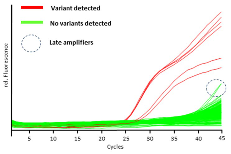
Figure 2.
Results of the variant-specific qPCR to clarify the High Multiplex Mutation Testing (HMMT) of the ARSA gene. Melting curve genotyping of (A) wildtype samples in blue and a heterozygous sample in green for variant c.465+1G>A and (B) wildtype samples in blue and a homozygous sample in red for variant c.1283C>T.
Figure 2.
Results of the variant-specific qPCR to clarify the High Multiplex Mutation Testing (HMMT) of the ARSA gene. Melting curve genotyping of (A) wildtype samples in blue and a heterozygous sample in green for variant c.465+1G>A and (B) wildtype samples in blue and a homozygous sample in red for variant c.1283C>T.
Disclaimer/Publisher’s Note: The statements, opinions and data contained in all publications are solely those of the individual author(s) and contributor(s) and not of MDPI and/or the editor(s). MDPI and/or the editor(s) disclaim responsibility for any injury to people or property resulting from any ideas, methods, instructions or products referred to in the content. |
© 2026 by the authors. Licensee MDPI, Basel, Switzerland. This article is an open access article distributed under the terms and conditions of the Creative Commons Attribution (CC BY) license (http://creativecommons.org/licenses/by/4.0/).
Copyright: This open access article is published under a Creative Commons CC BY 4.0 license, which permit the free download, distribution, and reuse, provided that the author and preprint are cited in any reuse.