

Submitted:
01 September 2026
Posted:
02 September 2026
You are already at the latest version
Abstract
Professor Sir Simon Baron-Cohen’s public acknowledgment that the phrase “extreme male brain” is clinically unhelpful marks a critical juncture for neurodevelopmental psychiatry. However, current mainstream perspectives continue to attribute the primary etiology of autism spectrum disorder (ASD) to hardwired, intrinsic prenatal sex steroid pathways interacting with genetic determinism. This opinion presents an alternative systems-biology framework: the prenatal steroid profiles observed across historical cohorts are indicators of a reactive, compensatory maternal-fetal endocrine shield attempting to protect the fetal brain from an environmental phenocopy. We model how ambient anthropogenic nitrous oxide (N₂O) exposure—which our live emissions data tracks at more than twice standard regulatory thresholds—passivates cobalamin and blocks NMDA receptors, forcing a chronological neuroendocrine "coiled-spring" compression in the maternal host that manifests as obesity, polycystic ovary syndrome (PCOS), and severe premenstrual disarray. Upon gas withdrawal, the resulting explosive hypothalamic-pituitary-gonadal (HPG) rebound drives a massive influx of excess sex steroids into the amniotic space. Phenotypic resolution is determined by sex-divergent fetal utilization: while the female fetus leverages a robust cell-autonomous chromosomal buffer and an ERβ-driven MAT2A-BHMT metabolic pump to preserve its methylation reservoir, the male fetus faces structural vulnerability via metabotropic GluN2B networks. Within this toxicological paradigm, the autism phenotype represents a sub-lethal evolutionary triage compromise prioritizing pregnancy viability over absolute reproductive loss. This unified N₂O axis effectively maps the entire neurochemical, metabolic, behavioral, and structural landscape of autism.
Keywords:
nitrous oxide
; Cobalamin passivation
; HPG axis rebound
; prenatal sex steroids
; GluN2B networks
; Cell‐autonomous methylation
; sub‐lethal evolutionary triage
; polycystic ovary syndrome (PCOS)
; premenstrual syndrome (PMS)
; premenstrual dysphoric disorder (PDD)
; autism spectrum disorder
; homosexuality
; estrogen
1. The Environmental Alternative: An Over-Correction of the Endocrine Shield
Professor Sir Simon Baron-Cohen’s public acknowledgment that the phrase “extreme male brain” is clinically unhelpful marks a critical juncture for neurodevelopmental psychiatry [1]. However, his team’s 25-year perspective continues to attribute the primary etiology of Autism Spectrum Disorder (ASD) to hardwired, intrinsic prenatal sex steroid pathways interacting with genetic determinism [2,3]. By failing to account for the unique neuro-endocrinological signature of environmental gaseous neurotoxins—specifically ambient nitrous oxide (N₂O)—the current framework misinterprets a reactive maternal-fetal biomarker as a primary causal agent.
Synthesis of modern environmental toxicology data suggests that the prenatal steroid profiles observed by the Cambridge team are actually indicators of an active, failing, or asymmetrical endocrine shield attempting to protect the fetal brain from an environmental phenocopy. The empirical foundation for this environmental alternative has advanced from macro-ecological tracking to direct, source-level empirical testing [4,5]. While macroeconomic models often assume ambient N₂O exposure remains within safe federal baselines, localized empirical sampling in highly populated metropolitan transit zones in late 2025 suggests these thresholds are frequently breached [5]. Localized field sampling in urban parking structures and street traffic corridors routinely detected peaks reaching 50 ppm, utilizing dual-wavelength infrared technology combined with an inline soda-lime filter to completely isolate N₂O and eliminate CO₂ cross-reactivity [5]. Individual maternal exposure vectors can actively reach the exact 50 ppm threshold considered neurotoxic, validated by both human [6] and animal studies [7] investigating the cognitive and neurochemical effects of N₂O-induced neurotoxicity. Because this 50 ppm environmental footprint causes central neurochemical disruption while tracking at more than double the safety thresholds mandated by regulatory bodies like the National Institute for Occupational Safety and Health (NIOSH), which sets the maximum airborne occupational limit at 25 ppm [8], the genetic determinism of the prenatal sex steroid theory is directly undermined.
Crucially, the validity of this toxicological alternative is reinforced by the fact that ambient N2O exposure functions as a precise molecular phenocopy, replicating virtually every recognized neurochemical, metabolic, behavioral, and structural hallmark of the autism phenotype. Rather than an unanchored environmental stressor, N₂O kinetics target the exact biological nodes that genetic determinism attributes to inherited mutations:
- Centrally/Structurally: Its non-competitive NMDA receptor antagonism perfectly mirrors the GRIN-family signaling deficits that alter the baseline excitation-inhibition balance of the developing central nervous system. The resulting energy-dependent apoptotic cascades and subsequent late-luteal or withdrawal-induced glutamatergic rebounds provide a direct, unified framework for the disrupted hemispheric lateralization, synaptic pruning alterations, and severe neurological comorbidities—such as epilepsy and autonomic dysregulation—that define the clinical presentation of the disorder [4,5].
- Behaviorally and Autonomically: Its intense hyper-stimulation of the endogenous opioid system provides a direct molecular explanation for the most distinct clinical and autonomic features of the disorder. This toxicant-driven opioid hyper-activation replicates the specific signaling profiles that induce repetitive motor stereotypies—phenocopying clinical stimming behavior—while concurrently disrupting hypothalamic thermoregulatory set-points and nociceptive gates to force the elevated pain thresholds and cyclical fevers of unknown origin (FUO) that characterize central dysautonomia [4,5].
By demonstrating that an atmospheric toxicant can actively format the entire neurobiological and behavioral signature of ASD, the necessity of an exclusively hardwired genetic etiology is entirely bypassed.
2. Chronic Opioid Signaling and the Induction of Maternal Obese Phenotypes
Classical neurobehavioral toxicology demonstrates that N2O exposure directly induces immediate, concentration-dependent feeding in non-deprived animal models [9]. In landmark rodent experiments, completely sated, non-deprived rats exposed to low concentrations of N₂O exhibited profound hyperphagia [9]. Crucially, this hyperphagic response was blocked not by traditional benzodiazepine antagonists, but by the opioid receptor blocker naltrexone [9]. This reveals that N₂O-stimulated eating is fundamentally mediated by an upstream activation of the endogenous opioid system.
Under chronic, low-dose environmental exposure, this opioid-driven appetite stimulation forms a dangerous feedback loop with maternal physiology. By demonstrating that N₂O forces caloric consumption independent of metabolic need, the proposed model suggests a critical reframing of maternal obesity: obesity is not an isolated, lifestyle-driven “confounding variable” in autism epidemiology, but rather a clinical manifestation of chronic, low-grade environmental opioid hyperstimulation. This environment-induced obesity then acts as a heavy physiological anchor, worsening baseline gonadotropin suppression and setting the stage for more severe endocrine crashes and subsequent rebounds.
3. Reframing the Maternal Phenotype: The Unified Origin of PCOS, PMS, and PMDD
This neuroendocrine cascade completely reframes the correlation between elevated prenatal amniotic estrogens ASD risk reported in landmark cohort studies [2]. Historically, the short-term toxicological footprint of acute N₂O exposure has been defined strictly by a profound paralysis of hypothalamic luteinizing hormone-releasing hormone (LHRH/GnRH) pathways, leading to arrested ovulatory surges, collapsed estrogen secretion, and systemic subfertility [10]. However, under chronic, intermittent environmental exposure conditions, it is hypothesized that the maternal endocrine axis triggers a powerful compensatory feedback loop. This loop unifies the seemingly distinct clinical pathologies of polycystic ovary syndrome (PCOS), premenstrual syndrome (PMS), and premenstrual dysphoric disorder (PMDD) into a singular spectrum of environmental resistance.
Polycystic Ovary Syndrome as an Ovarian Over-Correction
A profound drop in primary ovarian estrogen secretion during the gas-exposure phase strips the brain of its negative feedback mechanism. When coupled with the toxicant-driven maternal obesity described above—which independently depresses baseline gonadotropins while multiplying twinning rates—this compounded suppression acts like a tightly coiled mechanical spring. Once the exposure window clears, the dual-layered reproductive blockade forces the neuroendocrine axis to execute an explosive, compensatory upsurge. Crucially, this over-correction shifts gonadotropin-releasing hormone (GnRH) pulse dynamics into a rapid, hyper-pulsatile state. This selectively accelerates pituitary Luteinizing Hormone (LH) secretion over Follicle-Stimulating Hormone (FSH), establishing the classic elevated LH:FSH ratio that defines clinical PCOS profiles [11,12].
While a secondary, concurrent spike in FSH drives the erratic multi-follicular capture and characteristic fluid-filled cystic morphology, it is this dominant, unmitigated LH hyper-secretion that directly hyper-stimulates the ovarian stroma, multiplying testosterone production and generating systemic hyperandrogenemia [11]. This mechanism marks a multiple birth pregnancy, and most especially twinning with extra placental tissue, not as an intrinsic genetic anomaly, but as a direct indicator of an active female compensatory response to environmental gas toxicity. This culminates in a secondary, massive compensatory estrogenic burst intended to function as a neuroprotectant to stabilize embryonic NMDA pathways against gas-induced cellular apoptosis.
The Broken Methylation Engine of the Surge
However, this compensatory estrogenic shield is fundamentally crippled at its metabolic root. Because N₂O concurrently inactivates vitamin B₁₂ [13,14], this explosive neuroendocrine rebound occurs within a maternal-fetal environment starved of S-adenosylmethionine (SAM) [15]. The resulting lack of local methylation capacity impairs the precise epigenetic silencing of androgenic pathways, forcing an overactivation of androgen-receptor-dependent genes [3,16], while simultaneously preventing the proper synthesis and translation of monoamine neurotransmitters via the downstream collapse of methylation-driven cofactor cycles [17].
While conventional frameworks view the resulting high-estrogen/high-androgen amniotic environment as a primary marker of an intrinsically “masculinized” or hyperextended fetal steroid pathway, it represents a hyper-reactive HPG axis attempting an emergency upsurge with a broken biochemical engine—producing a massive volume of protective hormones that are rendered ineffective due to the systemic failure of the underlying methylation-dependent cellular machinery.
PMS and PMDD as Late-Luteal GABAergic Failures
This neuroendocrine over-correction is dynamically compounded by the cyclical architecture of PMS and PMDD. In the late luteal phase, the compromised follicular development initiated during the gas-exposure phase culminates in an unstable, structurally weak corpus luteum. This structural deficit triggers a premature, jagged collapse of progesterone and its calming neurosteroid metabolites, specifically allopregnanolone [18]. This sudden hormonal drop strips the central nervous system of its primary inhibitory GABAergic braking mechanism, throwing the brain’s baseline glutamate/NMDA balance into acute disarray.
When an environmental gas-withdrawal window overlaps with this vulnerable premenstrual threshold, the resulting central glutamatergic rebound is profoundly amplified and can manifest, at its extreme, as drug-withdrawal-induced epileptogenic activity. This neurological crisis is heavily exacerbated by the B₁₂-methylation deficit: without functional cobalamin, the methionine synthase arrest drives up intracellular homocysteine, which directly acts as an NMDA receptor agonist, further accelerating the central excitatory panic [19]. Starved of both metabolic protection and luteal negative feedback, the HPG axis undergoes a synchronized, highly volatile over-correction. This establishes severe PMS and PMDD symptoms not merely as localized behavioral complaints, but as reliable clinical indicators of a chronically compressed, hyper-reactive neuroendocrine axis [18]. The recurring loop of luteal suppression and withdrawal continuously widens the follicular selection window, forcing the multi-follicular rescue that drives twinning while generating the volatile embryonic environment linked to altered neurodevelopment.
4. Asymmetrical Intrauterine Defense and FPE
Evaluating this dynamic hormonal defense system reveals a broader behavioral spectrum regarding brain sexual differentiation, while providing a biochemically cohesive explanation for the skewed male-to-female ratio in autism. Proponents of the prenatal sex steroid theory have historically struggled with a core paradox: if the traditional paradigm links elevated amniotic estrogens to increased autism risk, attributing the female protective effect (FPE) to higher absolute fluid baselines introduces a profound structural contradiction [2]. The environmental model resolves this flaw by defining female resilience through receptor-level protection and signaling efficiency rather than absolute fluid volumes.
Classic neuroprotective models demonstrate that estradiol shields the brain from acute neuroexcitatory insults [20,21]. However, this protection is not merely a function of absolute circulating hormone levels; it also reflects a dynamic synergy with the host’s underlying cell-autonomous chromosomal architecture [22]. Somatic cells with an XX karyotype possess an inherently high-capacity framework driven by active expression from the inactive X chromosome (Xi) [23]. These dosage-sensitive X-chromosome escapees act as global molecular modifiers that buffer downstream cellular and pathway disruptions [23]. This genetic buffer operates in direct synergy with sex-divergent endocrine architectures; specifically, the developing female brain exhibits robust, natively higher expression of Estrogen Receptor Beta (ERβ).
When ambient gaseous neurotoxicity passivates cobalamin and paralyzes primary methionine synthase cycling, this elevated ERβ-driven pathway acts as a coordinated, two-gear metabolic pump to preserve a superior maternal-fetal methylation reservoir. First, high-flux estrogen signaling actively enhances and drives alternative betaine-homocysteine S-methyltransferase (BHMT) pathways to synthesize L-methionine from localized betaine and choline pools [24], completely bypassing the blocked cobalamin-dependent engine.
Second, ERβ activation directly upregulates the transcription of methionine adenosyltransferase 2A (MAT2A) while simultaneously stabilizing mitochondrial ATP kinetics [25]. This immediate energetic surge drives the MAT2A engine at maximum velocity, rapidly converting the raw intermediate L-methionine into active S-adenosylmethionine (SAM). By ensuring continuous, high-volume SAM donor synthesis from alternative metabolic inputs under toxicant stress, this receptor-mediated priming engine maintains the epigenetic stability of developing synaptic networks. By pairing systemic estrogenic cascades with this robust genetic foundation, the female brain possesses a highly responsive, high-capacity system capable of actively stabilizing and shielding developing embryonic NMDA and opioid loops from ambient gaseous toxicity.
Conversely, a male fetus lacks this highly responsive receptor-level framework. When a male fetus receives a lower baseline or suboptimal distribution of this prenatal estrogen shield, it is stripped of its primary defense against ambient gaseous neurotoxicity [26,27]. This allows the N₂O insult to drive the unmitigated NMDA hypofunction and kappa opioid receptor (KOR) dysregulation that dictates the classic hyper-masculinized cognitive profile historically termed the ‘extreme male brain’ [2].
However, because estrogen is a foundational molecule responsible for the sexual differentiation and organizational features of specific hypothalamic nuclei in the developing brain, an over-saturation of this defensive endocrine surge can shift the neuroanatomical trajectory away from typical male patterns. Long-term cohort studies on prenatal exposure to potent synthetic estrogens like diethylstilbestrol (DES) consistently support this highly sensitive neuro-organizational architecture, demonstrating an elevated prevalence of non-heterosexual phenotypes among prenatally exposed male offspring [28].
This framework exposes a profound flaw in the “Equal Environments Assumption” underpinning traditional twin studies. When a pregnant mother experiences severe environmental N₂O stress, her system mounts this significant, FSH-driven estrogenic shield, frequently inducing a twin pregnancy when supported by a highly receptive metabolic baseline like maternal obesity [11]. However, the physical architecture of the uterus prevents a completely symmetrical distribution of these protective hormones. If one twin receives a lower localized share of this protective estrogenic shield due to intrauterine crowding or asymmetrical placental vascularity, its developing NMDA and KOR systems are left fully exposed to ambient N₂O neurotoxicity, driving an autistic presentation. Meanwhile, the co-twin receiving an excessive share of the estrogenic burst avoids neurotoxicity but bears the burden of localized neurodevelopmental alterations. Standard epidemiological models misinterpret this discordance as a genetic failure, entirely missing a shared environmental insult that was simply resisted unequally due to structural hormonal asymmetry. Consequently, where these receptor-mediated clearing mechanisms are suboptimal or absent, the maternal-fetal unit is forced to transition from active metabolic defense to severe evolutionary triage
5. Autism as a Sub-Lethal Triage Response
When the physical constraints of intrauterine crowding or male sex-chromosomal architecture breach the limits of this protective clearing network, this model introduces a novel theoretical framework to interpret neurodevelopmental phenotypes as adaptive outcomes of gestational toxicological stress. At critical embryonic formatting windows, persistent or severe N₂O exposure acts as a potent non-competitive NMDA receptor antagonist, functioning as a primary driver of embryonic lethality. Extreme, unmitigated NMDA receptor hypofunction during these highly sensitive windows triggers widespread cascades of central cellular apoptosis, leading fundamentally to spontaneous abortion [29].
Concurrently, the proposed framework reinterprets the elevated prenatal sex steroids documented in historical cohort data [2]. It is conceptualized here that the massive maternal-placental endocrine surge is a sub-lethal evolutionary triage adaptation designed to salvage pregnancy viability. This endocrine rescue interacts distinctively with sex-divergent micro-circuitry. Recent mammalian neurobiology establishes that while both sexes rely on non-classical, ion-flux-independent (metabotropic) signaling for structural synaptic stabilization, they execute it through entirely separate molecular pathways [30]. Specifically, oxygen-starved or threatened males rely on a metabotropic NMDA receptor pathway inherently dependent on the GluN2B subunit, whereas females utilize a distinct, non-canonical pathway, reliant on synaptic estrogen receptor alpha (ERα) to trigger structural consolidation [30].
Because the male fetus is structurally reliant on the GluN2B subunit for metabotropic stabilization, it presents a highly vulnerable target to an environmental NMDA antagonist like N₂O. The incoming maternal estrogen burst cannot wholly neutralize gas-induced neurotoxicity or prevent downstream structural miswiring via this blocked male pathway. This vulnerability is compounded by the systemic metabolic depletion taking place concurrently. While the massive, compensatory estrogenic burst is sufficient to execute a partial, blunt stabilization of embryonic NMDA loops—attenuating the wholesale cellular apoptosis cascade and preserving fetal life—it cannot reverse the parallel, irreversible oxidation of Vitamin B₁₂. The resulting arrest of methionine synthase permanently starves the salvaged male brain of the localized SAM required for typical DNA methylation [31].
Within this toxicological paradigm, the autism phenotype represents a biological compromise favoring preservation over termination. The elevated amniotic sex steroids identified by the Cambridge team are viewed not as the pathognomonic cause of a genetic disease [2,3], but as the downstream endocrine marker of a protective selection mechanism favoring a viable neurodivergent trajectory over absolute reproductive loss.
Figure 1.
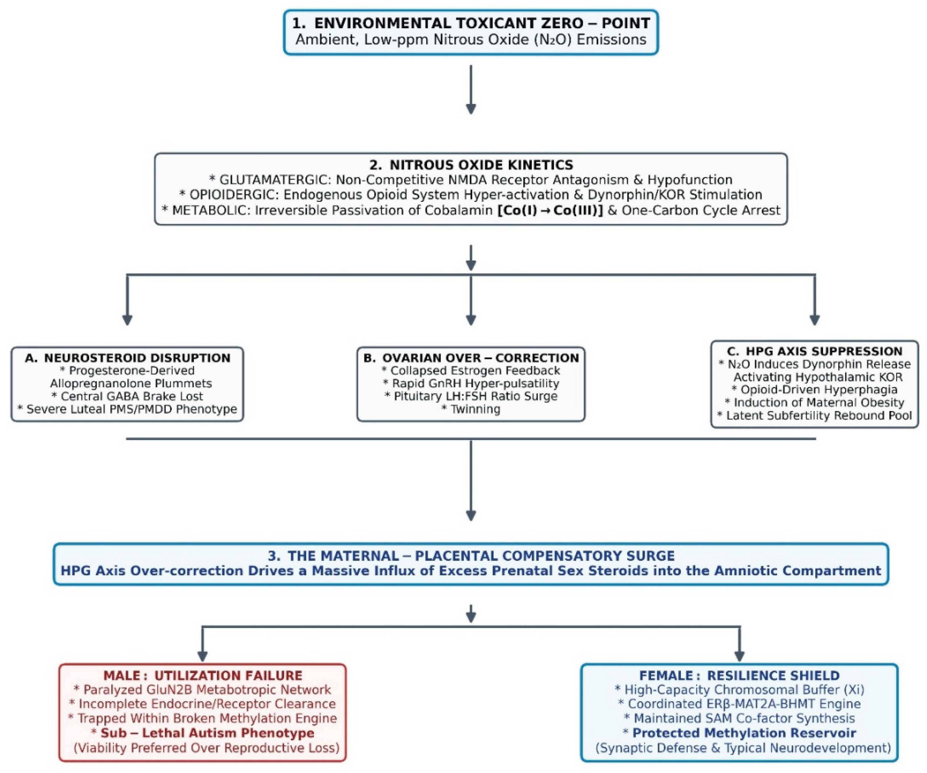
The Grand Unified Matrix of Environmental Resistance and Sex-Divergent Triage. Schematic workflow illustrating the multi-systemic transgenerational cascade under background anthropogenic nitrous oxide (N₂O) load. (1) Environmental Zero-Point: Low-ppm exposure vectors transcend safe federal baselines. (2) Nitrous Oxide Kinetics: Placental toxicant intrusion drives concurrent disruptions across three distinct molecular parameters: glutamatergic non-competitive NMDA receptor antagonism, opioidergic endogenous system/KOR hyper-stimulation, and metabolic passivation of active Cob(I)alamin to the trivalent Cob(III) state. This primary block cascade drives sequential failures in the maternal host: (A) Neurosteroid disruption, where progesterone-derived allopregnanolone depletion terminates the central GABA brake to trigger premenstrual (PMS/PMDD) glutamate disarray; (B) Ovarian over-correction, where collapsed estrogen feedback triggers rapid, hyper-pulsatile GnRH pulses and pituitary LH:FSH surges, leading to polycystic ovary syndrome (PCOS) profiles and twinning; and (C) HPG axis suppression, where toxicant-induced dynorphin release directly binds to and activates hypothalamic kappa opioid receptors (KOR) to compress neuroendocrine pathways, forcing opioid-mediated hyperphagia and maternal obesity. (3) The Compensatory Endocrine Hyper-Surge: This underlying host compression triggers an explosive HPG axis rebound, driving an influx of excess prenatal sex steroids into the amniotic space. Resolution is determined by fetal utilization: the male fetus faces metabotropic GluN2B vulnerability and incomplete clearance within a broken methylation engine, selecting for a sub-lethal neurodivergent autism phenotype to prevent total reproductive loss. Conversely, the female fetus leverages a high-capacity cell-autonomous chromosomal buffer and a coordinated ERβ-MAT2A-BHMT engine to actively maintain its methylation reservoir, securing typical neurodevelopment.
Figure 1.
The Grand Unified Matrix of Environmental Resistance and Sex-Divergent Triage. Schematic workflow illustrating the multi-systemic transgenerational cascade under background anthropogenic nitrous oxide (N₂O) load. (1) Environmental Zero-Point: Low-ppm exposure vectors transcend safe federal baselines. (2) Nitrous Oxide Kinetics: Placental toxicant intrusion drives concurrent disruptions across three distinct molecular parameters: glutamatergic non-competitive NMDA receptor antagonism, opioidergic endogenous system/KOR hyper-stimulation, and metabolic passivation of active Cob(I)alamin to the trivalent Cob(III) state. This primary block cascade drives sequential failures in the maternal host: (A) Neurosteroid disruption, where progesterone-derived allopregnanolone depletion terminates the central GABA brake to trigger premenstrual (PMS/PMDD) glutamate disarray; (B) Ovarian over-correction, where collapsed estrogen feedback triggers rapid, hyper-pulsatile GnRH pulses and pituitary LH:FSH surges, leading to polycystic ovary syndrome (PCOS) profiles and twinning; and (C) HPG axis suppression, where toxicant-induced dynorphin release directly binds to and activates hypothalamic kappa opioid receptors (KOR) to compress neuroendocrine pathways, forcing opioid-mediated hyperphagia and maternal obesity. (3) The Compensatory Endocrine Hyper-Surge: This underlying host compression triggers an explosive HPG axis rebound, driving an influx of excess prenatal sex steroids into the amniotic space. Resolution is determined by fetal utilization: the male fetus faces metabotropic GluN2B vulnerability and incomplete clearance within a broken methylation engine, selecting for a sub-lethal neurodivergent autism phenotype to prevent total reproductive loss. Conversely, the female fetus leverages a high-capacity cell-autonomous chromosomal buffer and a coordinated ERβ-MAT2A-BHMT engine to actively maintain its methylation reservoir, securing typical neurodevelopment.
6. Methodological Limitations
While this neuroendocrine framework offers a comprehensive, alternative interpretation of prenatal sex steroid profiles, several critical limitations must be explicitly acknowledged to guide future empirical validation:
- Biomarker Scarcity and Exposure Extrapolation: While theoretical and source-level environmental modeling has tracked localized ambient N₂O concentrations exceeding 50 ppm [5], large-scale, individual-level biomarker data directly pairing maternal ambient N₂O exposure during gestation with subsequent infant neurodevelopmental outcomes remains scarce. Moving this framework beyond a speculative baseline requires recognizing that the gestational body burden may be driven by a cumulative, multi-source environmental footprint—spanning concentrated agricultural nitrogen runoff into local bodies of water, occupational exposure in unscavenged medical facilities, and industrial combustion—rather than uniform urban traffic distribution alone. Establishing an empirical exposure-response gradient thus demands direct maternal-fetal bio-monitoring cohorts that transcend isolated geographic or industrial boundaries to quantify real-time neurochemical thresholds.
- One-Carbon Metabolic Kinetics and Epigenetic Validation: Although the biochemical pathway of N₂O-induced cobalamin B₁₂ oxidation is thoroughly mapped, real-time quantification of maternal-fetal methylation flux remains a significant hurdle. Directly linking an ambient gas exposure to the precise epigenetic failure of the CYP19A1 (aromatase) gene in human gestations requires advanced, non-invasive isotopic tracking of SAM consumption and concurrent cell-free fetal DNA methylation mapping [32]. Without this granular data, calculating the exact toxicological threshold where a normal compensatory endocrine surge degrades into systemic methylation failure remains an approximation.
- Receptor Homeostasis and Tissue Extrapolation: The primary evidence mapping sex-divergent metabotropic NMDA stabilization via GluN2B and ERα originates from juvenile and adult mammalian hippocampal slice paradigms [30]. Extrapolating these adult synaptic mechanics backward to early embryonic formatting windows represents a substantial conceptual step. Furthermore, driving hyperovulation in an obesity-primed ovarian microenvironment requires rigorous, longitudinal validation in controlled mammalian models to prove receptor thresholds can successfully bypass localized cellular replication bottlenecks.
- DES Cohort Translation: Extrapolating from high-dose synthetic diethylstilbestrol (DES) cohorts to assume endogenous, placenta-derived surges can exert an equivalent neuro-organizational shift in typical twin gestations demands closer investigation [28]. The differences in receptor affinity, bioavailability, and clearing kinetics between synthetic compounds and endogenous placental steroids require advanced placental vascular mapping and fetal neuroimaging to establish mechanistic equivalence.
- Teleological Nomenclature Constraints: Describing these complex endocrine adjustments using phrases like “rescue mission” or “triage response” functions strictly as a teleological metaphor for a probabilistic, selectionist evolutionary adaptation. These terms do not imply conscious physiological intent or directional awareness within the maternal-fetal unit, but rather illustrate how pathways preserving sub-lethal viability under intense environmental stress may be naturally selected to prevent total reproductive loss.
7. Conclusion
The male bias in autism spectrum disorder is not an intrinsic inevitability of male neurology, but rather the structural consequence of a male fetus lacking the specific, high-capacity ERα-mediated metabotropic protection and the upstream estrogen-primed methylation reservoir required to shield early NMDA networks and one-carbon metabolic pathways from a pervasively disrupted global nitrogen cycle. Within this toxicological framework, the elevated prenatal sex steroids traditionally viewed as pathognomonic risk markers are repositioned as the downstream endocrine signatures of an active, sub-lethal evolutionary triage prioritizing pregnancy viability over absolute reproductive loss.
Neurodevelopmental psychiatry must transition away from an exclusive focus on static genetic traits and broaden its diagnostic and theoretical scope to investigate the real-time, transgenerational biochemical feedback loops induced by the transformative industrialization of modern society and, particularly, the profound anthropogenic manipulation of nitrogen cycling.
Statement of AI Assistance
The conceptual framework, transdisciplinary synthesis, and longitudinal hypothesis linking ambient nitrous oxide to neurodevelopmental phenotypes presented in this manuscript represent over a decade of the author’s original, independent research. Generative AI text tools were utilized strictly during the final writing phase as an advanced editing assistant to refine sentence structure, optimize grammatical flow, and ensure stylistic alignment with the journal’s Opinion guidelines. The author has systematically cross-verified every biochemical mechanism, nomenclature update, and literature citation against primary sources, and assumes sole intellectual property ownership and full accountability for the scientific integrity of the final text.
Declaration of Generative AI in Scientific Writing
During the preparation of this work, the author utilized generative artificial intelligence (AI) technologies exclusively for language editing, typographical normalization, and the rendering of structural layout coordinates for the accompanying display item. The core conceptual synthesis, transdisciplinary biochemical mapping, and theoretical conclusions presented in this manuscript were conceived, analyzed, and generated entirely by the author. The author maintains absolute responsibility and accountability for the accuracy, scientific integrity, and bibliographic validity of the compiled work.
Declaration of Interests
The author declares no competing financial, professional, or personal interests that could influence the synthesis, execution, or presentation of the work described in this manuscript.
Funding Acknowledgments
This independent theoretical contribution received no specific grant, funding allocation, or financial underwriting from any regulatory body, commercial enterprise, or institutional research sector.
References
- Devlin, H. (2026) Pioneer of ‘extreme male brain’ theory of autism now says phrase unhelpful. The Guardian.
- Baron-Cohen, S.; et al. Foetal oestrogens and autism. Mol. Psychiatry 2020, 25, 2970–2978. [Google Scholar] [CrossRef] [PubMed]
- Baron-Cohen, S.; et al. The prenatal sex steroid theory of autism after 25 years. Nat. Hum. Behav. 2026, 10, 841–848. [Google Scholar] [CrossRef] [PubMed]
- Fluegge, K. Does environmental exposure to the greenhouse gas, N₂O, contribute to etiological factors in neurodevelopmental disorders? A mini-review of the evidence. Environ. Toxicol. Pharmacol. 2016, 47, 6–18. [Google Scholar] [CrossRef] [PubMed]
- Fluegge, K.; Fluegge, K. Comment on Chu et al. Acetaminophen’s Role in Autism and ADHD: A Mitochondrial Perspective. Int. J. Mol. Sci. 2025, 26, 8585, Int. J. Mol. Sci. 26, 11966. [Google Scholar] [CrossRef] [PubMed]
- Bruce, D.L.; Bach, M.J. Effects of trace anaesthetic gases on behavioural performance of volunteers. Br. J. Anaesth. 1976, 48, 871–876. [Google Scholar] [CrossRef] [PubMed]
- Abdul-Kareem, H.S.; et al. Effects of repeated intermittent exposures to nitrous oxide on central neurotransmitters and hepatic methionine synthetase activity in CD-1 mice. Toxicol. Ind. Health 1991, 7, 97–108. [Google Scholar] [CrossRef]
- National Institute for Occupational Safety and Health (NIOSH) (1977) Criteria for a Recommended Standard: Occupational Exposure to Waste Anesthetic Gases and Vapors (DHEW/NIOSH Publication No. 77-140). 1977.
- Czech, D.A. Nitrous oxide induces feeding in the nondeprived rat that is antagonized by naltrexone. Physiol. Behav. 1995, 58, 251–255. [Google Scholar] [CrossRef] [PubMed]
- Kugel, G.; et al. Nitrous oxide and infertility. Anesth. Prog. 1990, 37, 176–180. [Google Scholar] [CrossRef] [PubMed]
- Kosidou, K.; et al. Maternal polycystic ovary syndrome and the risk of autism spectrum disorder in the offspring: a population-based nation-wide study in Sweden. Mol. Psychiatry 2016, 21, 1441–1448. [Google Scholar] [CrossRef] [PubMed]
- Cherskov, A.; et al. Polycystic ovary syndrome and autism: A test of the prenatal sex steroid theory. Transl. Psychiatry 2018, 8, 136. [Google Scholar] [CrossRef] [PubMed]
- Deacon, R.; et al. Inactivation of methionine synthase by nitrous oxide. Eur. J. Biochem. 1980, 104, 419–423. [Google Scholar] [CrossRef] [PubMed]
- Chanarin, I. Cobalamins and nitrous oxide: a review. J. Clin. Pathol. 1980, 33, 909–916. [Google Scholar] [CrossRef] [PubMed]
- Chen, L.; et al. Oxidative stress marker aberrations in children with autism spectrum disorder: a systematic review and meta-analysis of 87 studies (N = 9109). Transl. Psychiatry 2021, 11, 15. [Google Scholar] [CrossRef] [PubMed]
- Mirza, S.; et al. Expression of DNA methyltransferases in breast cancer patients and to analyze the effect of natural compounds on DNA methyltransferases and associated proteins. J. Breast Cancer 2013, 16, 23–31. [Google Scholar] [CrossRef] [PubMed]
- Deth, R.; et al. How environmental and genetic factors combine to cause autism: A redox/methylation hypothesis. Neurotoxicology 2008, 29, 190–201. [Google Scholar] [CrossRef] [PubMed]
- Bäckström, T.; et al. Allopregnanolone and mood disorders. Prog. Neurobiol. 2014, 113, 88–94. [Google Scholar] [CrossRef] [PubMed]
- Ljungblad, U.W.; et al. Nitrous oxide in labour predicted newborn screening total homocysteine and is a potential risk factor for infant vitamin B12 deficiency. Acta Paediatr. 2022, 111, 2315–2321. [Google Scholar] [CrossRef] [PubMed]
- Liu, S.B.; et al. G-protein-coupled receptor 30 mediates rapid neuroprotective effects of estrogen via depression of NR2B-containing NMDA receptors. J. Neurosci. 2012, 32, 4887–4900. [Google Scholar] [CrossRef] [PubMed]
- Weaver, C.E.; et al. 17β-Estradiol protects against NMDA-induced excitotoxicity by direct inhibition of NMDA receptors. Brain Res. 1997, 761, 338–341. [Google Scholar] [CrossRef] [PubMed]
- Blencowe, M.; et al. Relative contributions of sex hormones, sex chromosomes, and gonads to sex differences in tissue gene regulation. Genome Res. 2022, 32, 807–824. [Google Scholar] [CrossRef] [PubMed]
- Talukdar, M.; Page, D.C. The inactive X chromosome as a female protector in autism and beyond. Nat. Genet. 2026, 58, 687–694. [Google Scholar] [CrossRef] [PubMed]
- Resseguie, M.; et al. Phosphatidylethanolamine N-methyltransferase (PEMT) gene expression is induced by estrogen in human and mouse primary hepatocytes. FASEB J. 2007, 21, 2622–2632. [Google Scholar] [CrossRef] [PubMed]
- Chu, P.Y.; et al. Translocation of methionine adenosyl transferase MAT2A and its prognostic relevance for liver hepatocellular carcinoma. Int. J. Mol. Sci. 2023, 24, 9103. [Google Scholar] [CrossRef] [PubMed]
- Adamson, A.; et al. Sex differences in dopaminergic vulnerability to environmental toxicants - implications for Parkinson’s disease. Curr. Environ. Health Rep. 2022, 9, 563–573. [Google Scholar] [CrossRef] [PubMed]
- Torres-Rojas, C.; Jones, B.C. Sex differences in neurotoxicogenetics. Front. Genet. 2018, 9, 196. [Google Scholar] [CrossRef] [PubMed]
- Titus-Ernstoff, L.; et al. Gender identity and sexual orientation identity in women and men prenatally exposed to diethylstilbestrol. Arch. Sex. Behav. 2020, 49, 447–454. [Google Scholar] [CrossRef] [PubMed]
- Savage, S.; Ma, D. The neurotoxicity of nitrous oxide: the facts and “putative” mechanisms. Brain Sci. 2014, 4, 73–90. [Google Scholar] [CrossRef] [PubMed]
- Le, A.A.; et al. Metabotropic NMDAR signaling contributes to sex differences in synaptic plasticity and episodic memory. J. Neurosci. 2024, 44, e0438242024. [Google Scholar] [CrossRef] [PubMed]
- Vogel Ciernia, A.; LaSalle, J. The landscape of DNA methylation amid a perfect storm of autism aetiologies. Nat. Rev. Neurosci. 2016, 17, 411–423. [Google Scholar] [CrossRef] [PubMed]
- Belardo, A.; et al. The concomitant lower concentrations of vitamins B6, B9 and B12 may cause methylation deficiency in autistic children. J. Nutr. Biochem. 2019, 70, 38–46. [Google Scholar] [CrossRef] [PubMed]
Disclaimer/Publisher’s Note: The statements, opinions and data contained in all publications are solely those of the individual author(s) and contributor(s) and not of MDPI and/or the editor(s). MDPI and/or the editor(s) disclaim responsibility for any injury to people or property resulting from any ideas, methods, instructions or products referred to in the content. |
© 2026 by the authors. Licensee MDPI, Basel, Switzerland. This article is an open access article distributed under the terms and conditions of the Creative Commons Attribution (CC BY) license (http://creativecommons.org/licenses/by/4.0/).
Copyright: This open access article is published under a Creative Commons CC BY 4.0 license, which permit the free download, distribution, and reuse, provided that the author and preprint are cited in any reuse.