

Submitted:
01 September 2026
Posted:
02 September 2026
You are already at the latest version
Abstract
Background: Tumor Treating Fields (TTFields) are being investigated for pelvic malignancies, while total hip arthroplasty (THA) is increasingly common. This study evaluated whether THA prosthetic materials influence TTFields distribution and target dosimetry in ovarian tumors. Methods: Patient-specific finite element models were generated from computed tomography datasets of four patients with ovarian carcinoma. Three-dimensional simulations assigned cortical bone or Ti6Al4V titanium alloy, Co28Cr6Mo cobalt-chromium alloy, or tantalum to both femurs. Electric field (E), specific absorption rate (SAR), and current density (CD) were quantified in the clinical target volume (CTV), femur, and skin using 95%, 50%, and 5% volume-based metrics. Results: Metallic implant properties produced minor CTV dosimetric changes, with mean and median percent differences generally within ±3.4% across metrics. Tantalum most closely approximated cortical bone, typically within ±1%. Implants substantially reduced femoral distributions of E and SAR, while increasing CD. Skin exposure decreased modestly across 95% and 50% volumes, with slight increases in localized hotspots. Conclusions: THA materials alter femoral distributions of TTFields while largely preserving clinically relevant CTV dosimetry, of which tantalum closely approximated cortical bone. Tantalum also reduced exposure across skin volumes. Implant composition alone will unlikely alter TTFields treatment planning for patients with ovarian malignancies undergoing THA.
Keywords:
TTFields
; total hip arthroplasty
; orthopedics
; pelvic malignancies
; prosthesis
1. Introduction
Ovarian cancer is a heterogeneous malignancy and remains one of the leading causes of gynecologic cancer-related mortality worldwide [1]. Because symptoms are often nonspecific, many patients present with advanced-stage disease requiring multimodal treatment [2]. Current standard-of-care management includes cytoreductive surgery followed by platinum- and taxane-based chemotherapy, with or without targeted therapies such as bevacizumab [3,4]. Despite these treatments, recurrence is common, highlighting the need for additional therapeutic approaches [5].
Total hip arthroplasty (THA) is among the most successful and widely performed orthopedic procedures, with more than 600,000 operations performed annually in the United States alone [6,7,8]. It is primarily indicated for osteoarthritis, osteonecrosis, congenital hip disorders, and inflammatory arthritis [9,10], and most patients report a high degree of satisfaction, particularly with restoration of normal ambulatory function [6]. Although THA is less common in the oncology population, approximately 3% of patients with gynecological cancers undergo the procedure [11]. As demand for THA is projected to increase by 284% by 2040 [12] and survival among patients with ovarian cancer improves [13], clinicians are increasingly likely to encounter patients with both ovarian cancer and hip arthroplasties [14,15]. Furthermore, the ovary is a recognized site of metastasis from several nongynecological malignancies, including colorectal, gastric, breast, and, less commonly, lung cancers, further expanding the population of patients with ovarian involvement [16,17]. This evolving clinical intersection highlights the importance of understanding how orthopedic implants may influence emerging cancer therapies.
Tumor Treating Fields (TTFields) are low-intensity, intermediate frequency (150 to 300 kHz) alternating electric fields that exert anticancer effects through multiple mechanisms, including disruption of mitosis, interference with DNA repair, induction of immunogenic cell death and autophagy, increased membrane permeability, and inhibition of tumor cell migration [18,19]. TTFields are delivered through two pairs of orthogonally positioned transducer arrays placed on the body or scalp [20]. The therapy is generally well tolerated, with skin irritation representing the most common treatment-related adverse effect [21,22]. TTFields are currently approved by the U.S. Food and Drug Administration for newly diagnosed and recurrent glioblastoma, malignant pleural mesothelioma, metastatic non-small cell lung cancer after platinum failure, and locally advanced pancreatic adenocarcinoma, while ongoing clinical trials continue to evaluate their efficacy in additional malignancies, including brain metastases from non-small cell lung cancer (NCT02973789), gastric cancer (NCT04281576), and ovarian cancer (NCT03940196) [23,24,25,26,27,28,29]. Although the phase III INNOVATE-3 trial did not demonstrate a significant survival benefit for the overall ovarian cancer population, post hoc analyses suggested improved outcomes among patients naïve to pegylated liposomal doxorubicin [30], supporting continued investigation of TTFields in ovarian malignancies.
The therapeutic efficacy of TTFields depends on the distribution of electric fields within the target volume. Previous computational studies have demonstrated that patient-specific anatomy, tissue electrical properties, highly conductive fluids (e.g., urine, cerebrospinal fluid, pleural fluid, and edema), and electrically attenuating structures (e.g., cortical bone, ribs, and sternum) can substantially influence TTFields distributions [31,32,33,34]. However, to our knowledge, no previous study has evaluated the influence of THA implant materials on TTFields delivery to pelvic malignancies. Because commonly used THA materials—including Ti6Al4V titanium alloy, Co28Cr6Mo cobalt-chromium alloy, and tantalum (Ta)—possess electrical conductivities that differ substantially from cortical bone [35,36,37,38], they may alter local electric field distributions and consequently influence TTFields dosimetry.
2. Materials and Methods
2.1. Patient Models and Anatomical Segmentation
Four patient-specific models of ovarian carcinoma were generated for finite element analysis using a previously established workflow [32]. Two patients’ datasets (BM002 and NS001) were obtained from a prior publication [32], whereas two additional datasets (AP003 and OY004) were obtained from The Cancer Imaging Archive [39]. Computed tomography (CT) images were imported into ScanIP 2022 (Simpleware Ltd., Exeter, UK) for anatomical segmentation.
The clinical target volume (CTV), femurs, and relevant abdominal and pelvic organs were segmented using a combination of manual contouring, grayscale thresholding, and TotalSegmentator, an automated deep-learning segmentation framework that has demonstrated high accuracy for CT-based anatomical segmentation [40]. All segmentations were reviewed and verified by an experienced physician. Patient-specific electrode arrays and conductive hydrogel layers consisting of orthogonal anteroposterior and lateral array pairs were then virtually positioned as described by [32]. Hydrogel layers were placed between each transducer array and the skin surface to provide a continuous conductive interface for uniform TTFields delivery. Three-dimensional finite element models were subsequently generated from the segmented anatomies. Meshes were generated using linear tetrahedral elements with a target minimum edge length of 1 mm, a maximum edge length of 3 mm, and a target maximum error of 0.1 mm. Additional mesh-quality improvement was performed using the Jacobian quality metric.
2.2. THA Material Modeling
To evaluate the effects of THA on TTFields delivery, the electrical properties assigned to both femurs were simultaneously replaced with those of three commonly used implant materials: Ti6Al4V alloy, Co28Cr6Mo alloy, and Ta (Table 1) [41,42,43,44]. Simulations were compared with a baseline model in which the femurs retained the electrical properties of cortical bone.
Electric conductivity and mass density values were assigned to each anatomic structure according to published literature (Table 1) [32,45]. To provide a preliminary assessment of the effects of THA implant materials on TTFields distribution, each femur was modeled as a homogeneous material rather than explicitly segmenting individual prosthetic components (e.g., acetabular cup, liner, femoral stem, and femoral head). This approach enabled isolation of the influence of implant material properties while establishing general trends in TTFields redistribution. All other anatomical structures retained identical material properties across simulations.
2.3. Finite Element Simulations
Electric field distributions were solved using the AC/DC module in the frequency domain of COMSOL 6.1 (COMSOL, Burlington, MA). TTFields were modeled at 200 kHz, the reported optimal therapeutic frequency for ovarian cancer cells [46]. We followed similar electro-quasistatic approximations of Maxwell’s equations and boundary conditions previously described by Korshoej A, et al. [47]. Electric field strength (E), specific absorption rate (SAR), and current density (CD) were computed throughout each patient model.
Model implementation was verified by reproducing the cortical-bone baseline simulations for BM002 and NS001 using the previously published ovarian TTFields modeling workflow. The resulting E-, SAR-, and CD-volume metrics were consistent with the corresponding values reported by Lok et al. [32].
2.4. Dosimetric Analysis
For each anatomical structure, field distributions were evaluated at the 95%, 50%, and 5% volume levels. Specifically, E95%, SAR95%, and CD95% represent the minimum values received by 95% of the structure volume; E50%, SAR50%, and CD50% represent the median values; and E5%, SAR5%, and CD5% represent the highest-exposed 5% of the volume (“hotspot”).
Percent changes relative to the cortical bone model were calculated for each implant material according to
where X denotes the corresponding dosimetric metric.
3. Results
3.1. THA Materials Minimally Affect CTV Dosimetry
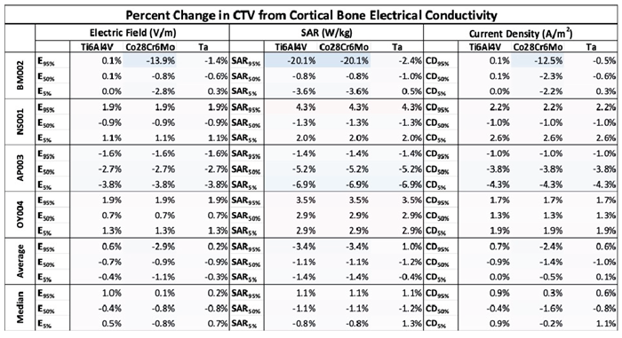
Across all four patient-specific CTVs, altering the electrical conductivity of the femur to represent Ti6Al4V, Co28Cr6Mo, and Ta implants produced only modest changes in TTFields dosimetry within the target volume (Figure 1, Table 2). Although some patient-specific variability was observed, particularly for BM002, CTV dosimetry remained largely preserved across all implant materials.
Across all E, SAR, and CD metrics within the CTV, mean and median percent changes generally remained within approximately ± 3.4% relative to the cortical bone model (Table 2). Among the three implant materials, tantalum most closely approximated the cortical bone model, with average and median changes remaining within approximately ± 1.3% across all evaluated metrics, whereas titanium alloy and cobalt-chromium exhibited slightly larger, but still modest, differences.
No qualitative relationship was observed between CTV dosimetry and tumor volume, target morphology, or average femur-to-CTV distance (Figure 2). Despite variations in patient anatomy, only minimal average and median percent changes in TTFields delivery to the CTV were produced. In contrast, substantially larger changes were observed within the femur itself following modification of its electrical conductivity.
3.2. THA Materials Substantially Alter TTFields Distribution Within the Femurs
Modifying femoral electrical conductivity substantially altered TTFields distribution within the femur itself (Figure 3, Table A1). Across cobalt-chromium and tantalum models, E and SAR values were reduced by approximately 100%, throughout the femoral volume, whereas CD increased by more than 850% relative to cortical bone.
Titanium alloy demonstrated a similar overall trend, although BM002 differed from the remaining patient models. In BM002, E95%, E50%, and E5% increased, whereas CD decreased relative to the cortical bone model. Despite this exception, all implant materials produced substantially greater changes within the femur than within the CTV.
3.3. THA Materials Reduce Skin TTFields Exposure with Mild Hotspot Increases
To evaluate potential changes in TTFields exposure within the skin, TTFields distributions were quantified within the skin (Figure 4, Table A2). Across the majority (95%) and median (50%) skin volumes, all metrics generally decreased by approximately 1.1 – 9.0% following replacement of cortical bone with metallic implant properties.
In contrast, the highest-exposure regions of the skin (5% volume) demonstrated small increases, ranging from approximately 1.7 – 3.7% across the evaluated metrics. These findings suggest that although overall skin exposure was modestly reduced, localized hotspots increased slightly.
Among the implant materials, tantalum yielded the largest reductions within the 95% skin volume, although these differences between materials remained relatively small.
3.4. THA Materials Produce Spatially Heterogeneous Effects in Adjacent Pelvic Structures
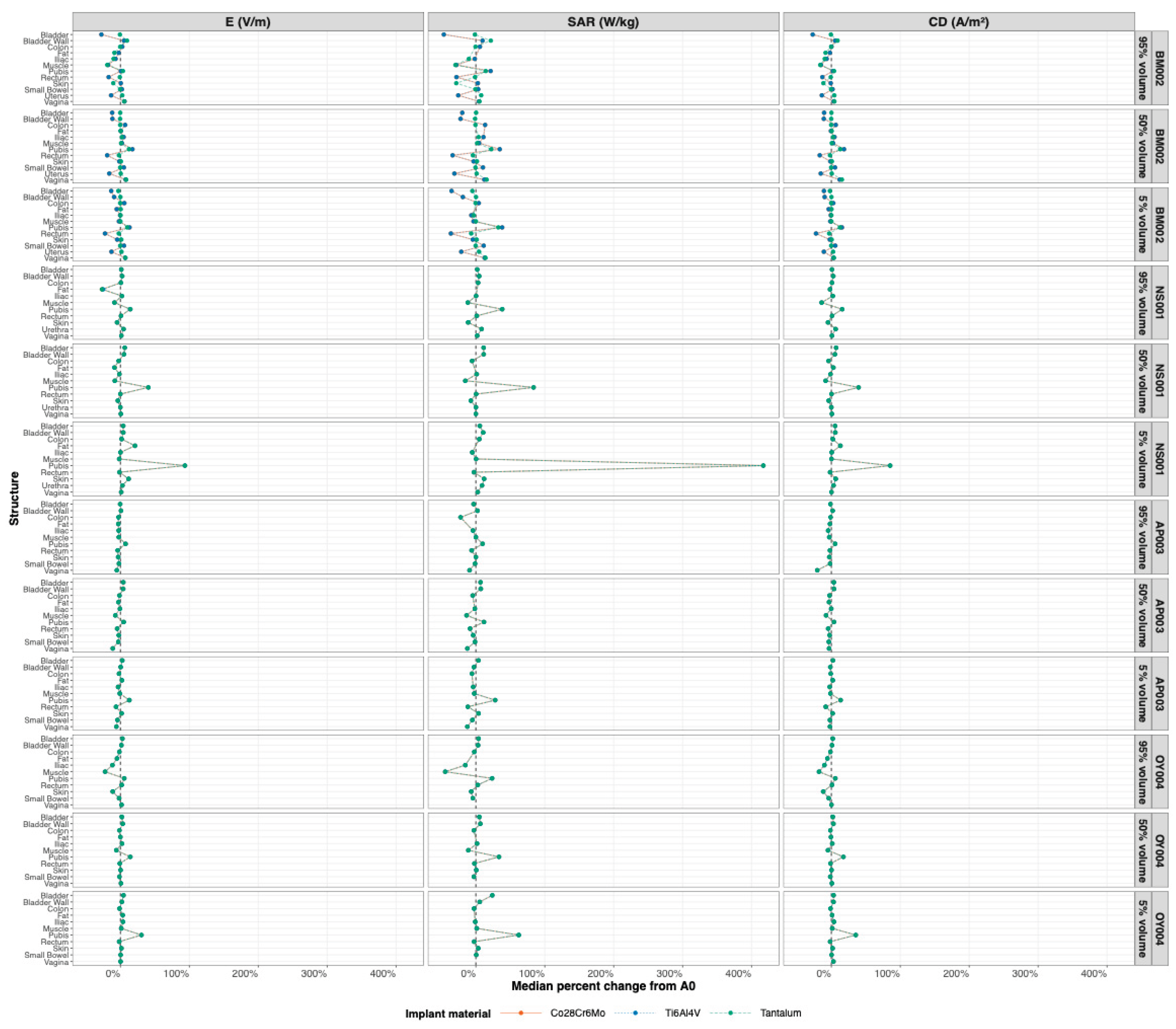
Additional structures proximal to the CTV and femur—including the pubis, fat, muscle, bladder, and rectum—were evaluated to characterize the effects of THA implants on the surrounding pelvic anatomy. Relative to the cortical-bone reference, changes in field metrics varied by structure, implant material, and evaluated volume percentile (Table A3, Figure A1).
The largest increases were observed in the pubis and were most pronounced within the 5% volume. Across implant materials, pubic SAR5% increased by 47.4% to 50.2%, while E5% and CD5% increased by 21.7% to 21.9% and 24.5% to 25.4%, respectively. Pubic SAR50% also increased by 28.0% to 34.1%. In contrast, muscle and rectum predominantly demonstrated reductions in field metrics, particularly in SAR. Changes in the remaining proximal structures were generally smaller and varied in direction across metrics and volume percentiles. Overall, these findings demonstrate that implant-associated field changes were spatially heterogeneous and were concentrated in specific proximal structures and volume regions.
3.5. Comparison Among THA Materials
Comparison of the three implant materials demonstrated similar overall effects on TTFields distribution. All materials produced minimal changes within the CTV and skin, while substantially altering field distributions within the femur. Compared with titanium alloy, cobalt chromium alloy and tantalum yielded greater amplification of current density within the femur (Table A1). Furthermore, tantalum closely approximated the cortical bone model within the CTV and produced slightly greater reductions within the 95% skin volume (Table 2, Table A2).
Collectively, these findings demonstrate that replacing cortical bone with clinically relevant THA implant materials substantially alters TTFields distributions within the femur but exhibits limited changes in the CTV and surrounding skin.
This section may be divided by subheadings. It should provide a concise and precise description of the experimental results, their interpretation, as well as the experimental conclusions that can be drawn.
4. Discussion
4.1. Principal Findings
This study investigated the effects of three commonly THA implant materials, Ti6Al4V, Co28Cr6Mo, and Ta, on TTFields distributions using patient-specific finite element models of pelvic malignancies. The principal finding is that replacing cortical bone with metallic implant properties generally produced minor changes in TTFields dosimetry within the CTV, despite substantially altering fields within the femur itself. Additionally, skin exposure was modestly reduced throughout most of the skin volume with only slight increases confined to localized hotspot regions. Collectively, these findings suggest that common THA implant materials may have limited influence on TTFields delivery to ovarian tumors.
4.2. Local Redistribution of TTFields Within the Femur Does Not Significantly Affect CTV Do-Simetry
Although replacing cortical bone with metallic implant materials substantially altered TTFields distributions within the femur, these changes were not propagated to the target volume as shown in Figure 2. This observation is likely explained by the localized influence of implant conductivity on current flow. TTFields are generated by external transducer arrays that establish electric field distributions throughout the pelvis rather than through a single localized current pathway. Furthermore, the CTV is anatomically distant from the femoral implants and positioned closer to the field-generating arrays. These spatial relationships, which may limit the extent to which implant-induced perturbations affect the field reaching the target, were previously shown by Wong and Lok [33]. Accordingly, the large changes observed within the femur may represent localized field redistribution around the conductive implant rather than a substantial alteration of the broader pelvic field.
These findings extend previous computational studies of TTFields distributions in ovarian malignancies by demonstrating that commonly used THA implant materials did not meaningfully compromise target dosimetry despite altering local electric-field behavior around the implant. This result is analogous to the bladder findings reported by Lok et al. [32], in which increased bladder conductivity affected surrounding structures without significantly altering the CTV. Together, these observations suggest that conductivity-induced perturbations may remain localized to the altered tissue or material and its immediate surroundings rather than propagating substantially to the target volume.
No consistent qualitative relationships were observed between CTV dosimetry and tumor volume, target morphology, or average femur-to-CTV distance as shown in Figure 2. Although BM002 demonstrated somewhat larger deviations than the other patient models, evaluation of the individual patient–structure profiles did not identify a single structure or response pattern that clearly accounted for this variability as displayed in Figure A1. BM002 also had the greatest average femur-to-CTV distance; however, the limited number of models precluded determining whether this characteristic was associated with the observed dosimetric differences. Overall, the dosimetric differences were not consistently associated with any single geometric characteristic. These findings suggest that simple anatomical descriptors alone may not adequately predict TTFields distributions and that additional patient-specific anatomical, geometric, or electrical factors may contribute to localized variability. Future studies involving larger patient cohorts and more detailed anatomical analyses will be necessary for identifying these relationships.
4.3. Effects of Metallic Hip Implants on Femoral Dosimetry
The largest changes observed in this study occurred within the femur itself. Across cobalt-chromium and tantalum models, E and SAR decreased substantially throughout the femoral volume, whereas CD increased relative to cortical bone. Titanium alloy demonstrated similar overall behavior, although differences were observed in one patient model.
These findings reflect the underlying electrical properties of metallic implants. Because CD is proportional to electrical conductivity, highly conductive implant materials preferentially conduct current, thereby increasing CD within the implant. Simultaneously, redistribution of current pathways reduces E intensity within surrounding femoral tissues. Since SAR depends on both electrical conductivity and E2 while also being inversely related to tissue density, the marked reduction in E together with the high density of metallic implants results in substantially attenuated SAR values despite increased conductivity.
From a safety perspective, the observed reduction in SAR suggests that metallic hip implants are unlikely to increase thermal deposition within the femur. Although thermal effects were not directly modeled in this study, these findings suggest that the presence of common THA materials is unlikely to increase the risk of implant-associated heating during TTFields therapy [48,49].
4.4. Skin Exposure and Potential Clinical Implications
Skin irritation remains one of the most frequently reported adverse effects associated with TTFields therapy because electric fields are introduced through transducer arrays placed directly on the skin [49]. In the present study, replacement of cortical bone with metallic implant properties produced modest reductions in field density across the majority (95%) of the skin volume while slightly increasing exposure within the highest-exposure (5%) regions.
Although localized hotspot increases were observed, these changes remained relatively small compared to the reductions seen across 95% and 50% skin volumes. The reductions observed across larger skin volumes may provide a broader representation of overall skin exposure than isolated hotspot regions. Consequently, the presence of metallic hip implants is unlikely to substantially increase the risk of TTFields-associated skin toxicity. Nevertheless, future experimental and clinical investigations are warranted to determine whether these modest hotspot increases have measurable effects on patient-reported skin reactions.
4.5. Comparison Among THA Materials
Among the three evaluated implant materials, tantalum is most closely approximated to the cortical bone model, producing the smallest changes within the CTV across all evaluated dosimetric metrics. Titanium alloy and cobalt-chromium demonstrated slightly larger deviations but produced comparable overall effects on TTFields distributions. Within the femur, cobalt-chromium and tantalum produced greater increases in CD than titanium alloy, while tantalum also demonstrated slightly larger reductions across the 95% skin volume.
Despite these material-specific differences, all three implant materials produced only minor changes in clinically relevant CTV dosimetry. These findings suggest that differences among currently used THA materials are unlikely to require modifications to TTFields treatment planning solely based on implant composition.
4.6. Clinical Significance
The prevalence of THA continues to increase as the population ages, and a growing number of patients with pelvic malignancies may undergo TTFields therapy following hip replacement surgery. The present findings suggest that common THA implant materials do not substantially compromise TTFields delivery to the target volume, indicating that patients with metallic hip implants should remain appropriate candidates for TTFields therapy without requiring major modifications to treatment planning. Furthermore, implants were shown to help reduce skin exposure at the 95% and 50% dose volumes.
Although patient-specific finite element modeling remains valuable for treatment optimization, these results provide reassurance that commonly implanted metallic prostheses are unlikely to interfere meaningfully with therapeutic electric field delivery. This finding may simplify future treatment planning workflows while supporting broader clinical implementation of TTFields in patients with prior THA.
4.7. Limitations
This study has several limitations. First, only four patient-specific models were evaluated, limiting the ability to identify precise relationships between anatomical characteristics and TTFields distributions. Larger patient cohorts are needed to validate the generalizability of these findings and to investigate whether additional anatomical factors influence patient-specific variability. Although model implementation was verified for consistency with previously published results, independent experimental validation and additional sensitivity analyses were not performed in the present study. Still, our study is first to integrate the relationship between THA and ovarian malignancies. Second, each hip prosthesis was modeled as a homogeneous material rather than incorporating the multiple components present in clinical THA systems, including femoral stems, acetabular shells, liners, and femoral heads. This simplification was intentional for this initial modeling study to isolate the influence of implant material properties on TTFields distributions and establish general trends. Although all three homogeneous implant materials produced only minor changes in CTV dosimetry, the effects of anatomically detailed, multi-material prostheses remain unknown. Future studies should incorporate realistic implant geometries and heterogeneous material compositions to better represent clinical THA devices and determine whether additional local perturbations influence TTFields distributions. Finally, only three metallic implant materials were investigated. Additional studies should evaluate other prosthetic materials, including polyethylene acetabular liners and ceramic components, as well as patient-specific implant geometries, to further characterize implant-related effects on TTFields distributions.
5. Conclusions
Our study is first to evaluate the effects of three commonly used THA implant materials on TTFields distributions using patient-specific finite element models of ovarian malignancies. Although metallic implant properties altered field distributions within the femur, these effects remained largely localized and produced only minor changes in TTFields dosimetry within the CTV and modest changes within the skin. Among the evaluated materials, tantalum most closely approximated the cortical bone model, while all three implant materials demonstrated similarly limited effects on target dosimetry. Tantalum was also associated with slightly greater reductions in the majority of the skin volume compared to titanium alloy and cobalt-chromium.
Overall, these findings demonstrate that commonly used THA implant materials are unlikely to compromise TTFields delivery to pelvic tumors, supporting the continued use of TTFields therapy in patients with total hip arthroplasty and suggesting that implant composition alone is unlikely to require substantial modifications to treatment planning.
Supplementary Materials
The following supporting information can be downloaded at website of this paper posted on Preprints.org, Table A1: Percent change in PQM at the femur; Table A2: Percent change in PQM at the skin; Table A3: Median changes in TTFields distributions within the CTV and adjacent pelvic structures for THA implant materials relative to cortical bone; Figure A1: Patient-specific changes in TTFields distributions within adjacent pelvic structures for THA implant materials relative to cortical bone.
Author Contributions
Olivia Liang: conceptualization, data curation, formal analysis, writing- original draft preparation, reviewing and editing. Edwin Lok: conceptualization, validation, writing- original draft preparation, reviewing and editing, supervision. Bassel G Diebo: writing- reviewing and editing. Monika Haack: Writing- reviewing and editing. Eric T Wong: writing- original draft preparation, reviewing and editing, supervision. All authors have read and agreed to the published version of the manuscript.
Funding
Eric T. Wong reports financial support was provided by A Reason to Walk & Run Research Fund and The Musella Foundation.
Institutional Review Board Statement
This is not applicable because our datasets were obtained from a previous publication [32] and from the Cancer Imaging Archive (https://www.cancerimagingarchive.net/).
Informed Consent Statement
Informed consent is not applicable because images were obtained from a previous publication [32] and from the Cancer Imaging Archive (https://www.cancerimagingarchive.net/).
Data Availability Statement
Research data are stored in an institutional repository and will be shared upon request to the corresponding author.
Conflicts of Interest
Eric T. Wong reports a relationship with Bristol-Myers Squibb Company that includes: consulting or advisory. Bassel Diebo reports a relationship with Spinal Alignment Solutions, Medtronic, SpineArt, Clariance, and Spine Vision that includes: consulting or advisory. Edwin Lok reports a relationship with US Oncology that includes: employment. Eric T. Wong and Edwin Lok have patent #No.16/335,920 “System and Methods For Cancer Treatment Using Alternating Electric Fields” issued to Beth Israel Deaconess Medical Center. Eric T. Wong and Edwin Lok have patent “Hydrogel Conductivity Impacts Skin Dose from Tumor Treating Fields” pending to Brown University Health. If there are other authors, they declare that they have no known competing financial interests or personal relationships that could have appeared to influence the work reported in this paper.:
Abbreviations
The following abbreviations are used in this manuscript:
| CD | Current Density |
| Co28Cr6Mo | Cobalt Chromium Alloy |
| CT | Computed Tomography |
| CTV | Clinical Target Volume |
| E | Electric Field Strength |
| SAR | Specific Absorption Rate |
| Ta | Tantalum |
| THA | Total Hip Arthroplasty |
| Ti6Al4V | Titanium Alloy |
| TTFields | Tumor Treating Fields |
Appendix A

Table A1.
Percent change in PQM at the femur. E, SAR, and CD at 95%, 50% and 5% coverage of the femur were compared to cortical among the various types of THA material (titanium, cobalt-chromium, and tantalum) with different electrical properties. A/m2, ampere per meter squared; CTV, clinical treatment volume, Co28Cr6Mo, cobalt-chromium; PQM, plan quality metrics; Ta, tantalum; Ti6Al4V, titanium; V/m, volt per meter; W/kg, watt per kilogram.
Table A1.
Percent change in PQM at the femur. E, SAR, and CD at 95%, 50% and 5% coverage of the femur were compared to cortical among the various types of THA material (titanium, cobalt-chromium, and tantalum) with different electrical properties. A/m2, ampere per meter squared; CTV, clinical treatment volume, Co28Cr6Mo, cobalt-chromium; PQM, plan quality metrics; Ta, tantalum; Ti6Al4V, titanium; V/m, volt per meter; W/kg, watt per kilogram.
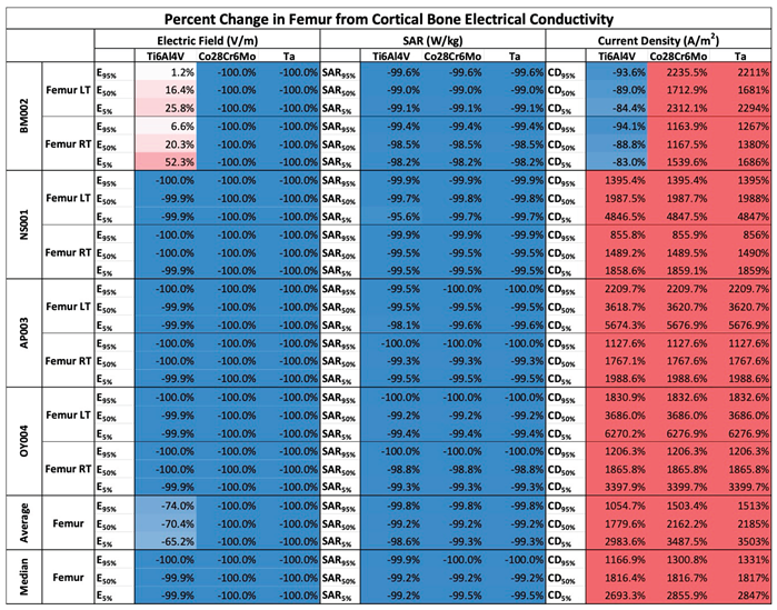 |
Table A2.
Percent change in PQM at the skin. E, SAR, and CD at 95%, 50% and 5% coverage of the skin were compared to cortical among the various types of THA material (titanium, cobalt-chromium, and tantalum) with different electrical properties. A/m2, ampere per meter squared; CTV, clinical treatment volume, Co28Cr6Mo, cobalt-chromium; PQM, plan quality metrics; Ta, tantalum; Ti6Al4V, titanium; V/m, volt per meter; W/kg, watt per kilogram.
Table A2.
Percent change in PQM at the skin. E, SAR, and CD at 95%, 50% and 5% coverage of the skin were compared to cortical among the various types of THA material (titanium, cobalt-chromium, and tantalum) with different electrical properties. A/m2, ampere per meter squared; CTV, clinical treatment volume, Co28Cr6Mo, cobalt-chromium; PQM, plan quality metrics; Ta, tantalum; Ti6Al4V, titanium; V/m, volt per meter; W/kg, watt per kilogram.
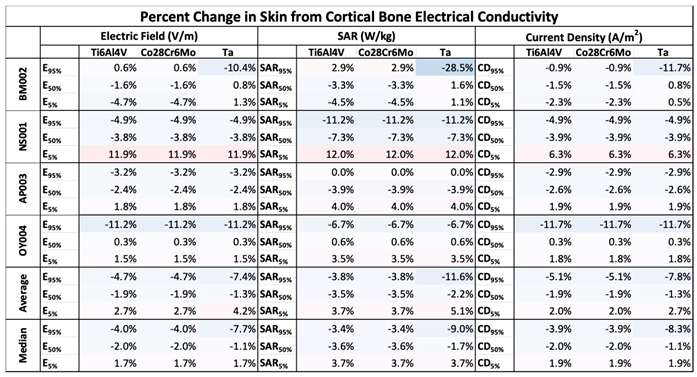 |
Table A3.
Median changes in TTFields distributions within the CTV and adjacent pelvic structures for THA implant materials relative to cortical bone. Heatmaps show the median percentage change across patient models in electric field intensity (E; V/m), specific absorption rate (SAR; W/kg), and current density (CD; A/m²) at the 95%, 50%, and 5% volume levels. Changes are presented for Ti6Al4V, Co28Cr6Mo, and Ta implants.
Table A3.
Median changes in TTFields distributions within the CTV and adjacent pelvic structures for THA implant materials relative to cortical bone. Heatmaps show the median percentage change across patient models in electric field intensity (E; V/m), specific absorption rate (SAR; W/kg), and current density (CD; A/m²) at the 95%, 50%, and 5% volume levels. Changes are presented for Ti6Al4V, Co28Cr6Mo, and Ta implants.
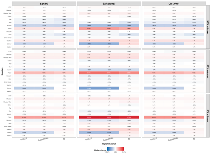 |
Figure A1.
Patient-specific changes in TTFields distributions within adjacent pelvic structures for THA implant materials relative to cortical bone. Profiles show the percentage changes in electric field intensity (E; V/m), specific absorption rate (SAR; W/kg), and current density (CD; A/m²) at the 95%, 50%, and 5% volume levels for each patient model. Changes for Co28Cr6Mo (orange), Ti6Al4V (blue), and tantalum (green) are normalized to the corresponding cortical-bone reference case (A0), and the corresponding colors may converge across structures.
Figure A1.
Patient-specific changes in TTFields distributions within adjacent pelvic structures for THA implant materials relative to cortical bone. Profiles show the percentage changes in electric field intensity (E; V/m), specific absorption rate (SAR; W/kg), and current density (CD; A/m²) at the 95%, 50%, and 5% volume levels for each patient model. Changes for Co28Cr6Mo (orange), Ti6Al4V (blue), and tantalum (green) are normalized to the corresponding cortical-bone reference case (A0), and the corresponding colors may converge across structures.
References
- Matulonis, U.A.; Sood, A.K.; Fallowfield, L.; et al. Ovarian cancer. Nat. Rev. Dis. Primer 2016, 2, 16061. [Google Scholar] [CrossRef] [PubMed]
- Dilley, J.; Burnell, M.; Gentry-Maharaj, A.; et al. Ovarian cancer symptoms, routes to diagnosis and survival – Population cohort study in the ‘no screen’ arm of the UK Collaborative Trial of Ovarian Cancer Screening (UKCTOCS). Gynecol. Oncol. 2020, 158, 316–322. [Google Scholar] [CrossRef] [PubMed]
- Burger, R.A.; Brady, M.F.; Bookman, M.A.; et al. Incorporation of Bevacizumab in the Primary Treatment of Ovarian Cancer. N Engl. J. Med. 2011, 365, 2473–2483. [Google Scholar] [CrossRef] [PubMed]
- Ozols, R.F.; Bundy, B.N.; Greer, B.E.; et al. Phase III trial of carboplatin and paclitaxel compared with cisplatin and paclitaxel in patients with optimally resected stage III ovarian cancer: a Gynecologic Oncology Group study. J. Clin. Oncol. Off. J. Am. Soc. Clin. Oncol. 2003, 21, 3194–3200. [Google Scholar] [CrossRef] [PubMed]
- Committee on the State of the Science in Ovarian Cancer Research, Board on Health Care Services, Institute of Medicine, National Academies of Sciences, Engineering, and Medicine (2016) Diagnosis and Treatment. In Ovarian Cancers: Evolving Paradigms in Research and Care; National Academies Press (US).
- Varacallo, M.; Chakravarty, R.; Denehy, K.; Star, A. Joint perception and patient perceived satisfaction after total hip and knee arthroplasty in the American population. J. Orthop. 2018, 15, 495–499. [Google Scholar] [CrossRef] [PubMed]
- Varacallo, M.A.; Luo, T.D.; Johanson, N.A. Total Hip Arthroplasty Techniques. In StatPearls; StatPearls Publishing: Treasure Island (FL), 2025. [Google Scholar]
- Patel, I.; Nham, F.; Zalikha, A.K.; El-Othmani, M.M. Epidemiology of total hip arthroplasty: demographics, comorbidities and outcomes. Arthroplasty 2023, 5, 2. [Google Scholar] [CrossRef] [PubMed]
- Rosas, S.; Sabeh, K.; Kurowicki, J.; et al. National use of total hip arthroplasty among patients with a history of breast, lung, prostate, colon or bladder cancer—an analysis of the Medicare population. Ann. Transl. Med. 2017, 5, S34–S34. [Google Scholar] [CrossRef] [PubMed]
- Singh, J.A.; Chen, J.; Inacio, M.C.S.; et al. An underlying diagnosis of osteonecrosis of bone is associated with worse outcomes than osteoarthritis after total hip arthroplasty. BMC Musculoskelet. Disord. 2017, 18, 8. [Google Scholar] [CrossRef] [PubMed]
- Dybvik, E.; Furnes, O.; Fosså, S.D.; et al. Pelvic irradiation does not increase the risk of hip replacement in patients with gynecological cancer. A cohort study based on 8,507 patients. Acta Orthop. 2014, 85, 652–656. [Google Scholar] [CrossRef] [PubMed]
- Singh, J.A.; Yu, S.; Chen, L.; Cleveland, J.D. Rates of Total Joint Replacement in the United States: Future Projections to 2020–2040 Using the National Inpatient Sample. J. Rheumatol. 2019. [Google Scholar] [CrossRef] [PubMed]
- Wu, J.; Sun, H.; Yang, L.; et al. Improved survival in ovarian cancer, with widening survival gaps of races and socioeconomic status: a period analysis, 1983-2012. J. Cancer 2018, 9, 3548–3556. [Google Scholar] [CrossRef] [PubMed]
- Brewster, D.H.; Stockton, D.L.; Reekie, A.; et al. Risk of cancer following primary total hip replacement or primary resurfacing arthroplasty of the hip: a retrospective cohort study in Scotland. Br. J. Cancer 2013, 108, 1883–1890. [Google Scholar] [CrossRef] [PubMed]
- Hailer, N.P.; Garland, A.; Gordon, M.; et al. No generally increased risk of cancer after total hip arthroplasty performed due to osteoarthritis. Int. J. Cancer 2020, 147, 76–83. [Google Scholar] [CrossRef] [PubMed]
- Lee, S.-J.; Bae, J.-H.; Lee, A.-W.; et al. Clinical Characteristics of Metastatic Tumors to the Ovaries. J. Korean Med. Sci. 2009, 24, 114–119. [Google Scholar] [CrossRef] [PubMed]
- Moore, R.G.; Chung, M.; Granai, C.O.; et al. Incidence of metastasis to the ovaries from nongenital tract primary tumors. Gynecol. Oncol. 2004, 93, 87–91. [Google Scholar] [CrossRef] [PubMed]
- Kirson, E.D.; Dbalý, V.; Tovaryš, F.; et al. Alternating electric fields arrest cell proliferation in animal tumor models and human brain tumors. Proc. Natl. Acad. Sci. U S A 2007, 104, 10152–10157. [Google Scholar] [CrossRef] [PubMed]
- Kirson, E.D.; Giladi, M.; Gurvich, Z.; et al. Alternating electric fields (TTFields) inhibit metastatic spread of solid tumors to the lungs. Clin. Exp. Metastasis 2009, 26, 633–640. [Google Scholar] [CrossRef] [PubMed]
- Novocure GmbH. NovoTTFTM-100L System for Unresectable Malignant Pleural Mesothelioma - Patient Information and Operation Manual. 2019.
- Lacouture, M.E.; Anadkat, M.J.; Ballo, M.T.; et al. Prevention and Management of Dermatologic Adverse Events Associated With Tumor Treating Fields in Patients With Glioblastoma. Front Oncol. 2020, 10. [Google Scholar] [CrossRef] [PubMed]
- Lacouture, M.E.; Elizabeth Davis, M.; Elzinga, G.; et al. Characterization and Management of Dermatologic Adverse Events With the NovoTTF-100A System, a Novel Anti-mitotic Electric Field Device for the Treatment of Recurrent Glioblastoma. Semin Oncol. 2014, 41, S1–S14. [Google Scholar] [CrossRef] [PubMed]
- Stupp, R.; Wong, E.T.; Kanner, A.A.; et al. NovoTTF-100A versus physician’s choice chemotherapy in recurrent glioblastoma: A randomised phase III trial of a novel treatment modality. Eur. J. Cancer 2012, 48, 2192–2202. [Google Scholar] [CrossRef] [PubMed]
- Stupp, R.; Taillibert, S.; Kanner, A.; et al. Effect of Tumor-Treating Fields Plus Maintenance Temozolomide vs Maintenance Temozolomide Alone on Survival in Patients With Glioblastoma: A Randomized Clinical Trial. JAMA 2017, 318, 2306–2316. [Google Scholar] [CrossRef] [PubMed]
- Ceresoli, G.L.; Aerts, J.G.; Dziadziuszko, R.; et al. Tumour Treating Fields in combination with pemetrexed and cisplatin or carboplatin as first-line treatment for unresectable malignant pleural mesothelioma (STELLAR): a multicentre, single-arm phase 2 trial. Lancet Oncol. 2019, 20, 1702–1709. [Google Scholar] [CrossRef] [PubMed]
- Leal, T.; Kotecha, R.; Ramlau, R.; et al. Tumor Treating Fields therapy with standard systemic therapy versus standard systemic therapy alone in metastatic non-small-cell lung cancer following progression on or after platinum-based therapy (LUNAR): a randomised, open-label, pivotal phase 3 study. Lancet Oncol. 2023, 24, 1002–1017. [Google Scholar] [CrossRef] [PubMed]
- Li, X.; Liu, K.; Xing, L.; Rubinsky, B. A review of tumor treating fields (TTFields): advancements in clinical applications and mechanistic insights. Radiol. Oncol. 2023, 57, 279–291. [Google Scholar] [CrossRef] [PubMed]
- Gondi, V.; Ahluwalia, M.S.; Roberge, D.; et al. Results from METIS (EF-25), an international, multicenter phase III randomized study evaluating the efficacy and safety of tumor treating fields (TTFields) therapy in NSCLC patients with brain metastases. J. Clin. Oncol. 2024. [Google Scholar] [CrossRef]
- Babiker, H.M.; Picozzi, V.; Chandana, S.R.; et al. Tumor Treating Fields With Gemcitabine and Nab-Paclitaxel for Locally Advanced Pancreatic Adenocarcinoma: Randomized, Open-Label, Pivotal Phase III PANOVA-3 Study. J. Clin. Oncol. Off. J. Am. Soc. Clin. Oncol. 2025, JCO2500746. [Google Scholar] [CrossRef] [PubMed]
- Vergote, I.; Copeland, L.J.; Gorp, T.V.; et al. Tumor Treating Fields therapy in platinum-resistant ovarian cancer: Results of the ENGOT-ov50/GOG-3029/INNOVATE-3 pivotal phase 3 randomized study. Eur. J. Cancer 2025, 219. [Google Scholar] [CrossRef] [PubMed]
- Lok, E.; San, P.; Liang, O.; et al. Finite element analysis of Tumor Treating Fields in a patient with posterior fossa glioblastoma. J. Neurooncol 2020, 147, 125–133. [Google Scholar] [CrossRef] [PubMed]
- Lok, E.; San, P.; White, V.; et al. Tumor Treating Fields for Ovarian Carcinoma: A Modeling Study. Adv. Radiat. Oncol. 2021, 6, 100716. [Google Scholar] [CrossRef] [PubMed]
- Lok, E.; Liang, O.; Malik, T.; Wong, E.T. Computational Analysis of Tumor Treating Fields for Non-Small Cell Lung Cancer in Full Thoracic Models. Adv. Radiat. Oncol. 2023, 8, 101203. [Google Scholar] [CrossRef] [PubMed]
- Lok, E.; Liang, O.; Haack, M.; Wong, E.T. Hydrogel and scalp/skin conductivities impact dose from tumor treating fields. Front Bioeng. Biotechnol. 2025, 13. [Google Scholar] [CrossRef] [PubMed]
- Huang, G.; Pan, S.-T.; Qiu, J.-X. The Clinical Application of Porous Tantalum and Its New Development for Bone Tissue Engineering. Materials 2021, 14, 2647. [Google Scholar] [CrossRef] [PubMed]
- Hailer, N. 20 years of porous tantalum in primary and revision hip arthroplasty—time for a critical appraisal. Acta Orthop. 2018, 89, 254–255. [Google Scholar] [CrossRef] [PubMed]
- Katti, K.S. Biomaterials in total joint replacement. Colloids Surf. B Biointerfaces 2004, 39, 133–142. [Google Scholar] [CrossRef] [PubMed]
- Merola, M.; Affatato, S. Materials for Hip Prostheses: A Review of Wear and Loading Considerations. Materials 2019, 12, 495. [Google Scholar] [CrossRef] [PubMed]
- Welcome to The Cancer Imaging Archive. In Cancer Imaging Arch; TCIA, 2026; Available online: https://www.cancerimagingarchive.net/ (accessed on 14 July 2026).
- Wasserthal, J.; Breit, H.-C.; Meyer, M.T.; et al. TotalSegmentator: Robust Segmentation of 104 Anatomic Structures in CT Images. Radiol. Artif. Intell. 2023, 5, e230024. [Google Scholar] [CrossRef] [PubMed]
- Aherwar, A.; Bahraminasab, M. Biocompatibility evaluation and corrosion resistance of tungsten added Co-30Cr-4Mo-1Ni alloy. BioMed Mater. Eng. 2017, 28, 687–701. [Google Scholar] [CrossRef] [PubMed]
- Arajs, S.; Dunmyre, G.R.; Dechter, S.J. Electrical Resistivity Studies of Chromium-Rich Chromium-Cobalt Alloys. Phys. Rev. 1967, 154, 448–452. [Google Scholar] [CrossRef]
- Javed, A.; Durrani, H.G.; Zhu, C. The effect of vacuum annealing on the microstructure, mechanical and electrical properties of tantalum films. Int. J. Refract Met. Hard Mater. 2016, 54, 154–158. [Google Scholar] [CrossRef]
- Bolzoni, L.; Ruiz-Navas, E.M.; Gordo, E. Flexural properties, thermal conductivity and electrical resistivity of prealloyed and master alloy addition powder metallurgy Ti–6Al–4V. Mater. Des. 2013, 52, 888–895. [Google Scholar] [CrossRef]
- Gabriel, C.; Gabriel, S.; Corthout, E. The dielectric properties of biological tissues: I. Literature survey. Phys. Med. Biol. 1996, 41, 2231. [Google Scholar] [CrossRef] [PubMed]
- Giladi, M.; Schneiderman, R.S.; Voloshin, T.; et al. Mitotic Spindle Disruption by Alternating Electric Fields Leads to Improper Chromosome Segregation and Mitotic Catastrophe in Cancer Cells. Sci. Rep. 2015, 5, 18046. [Google Scholar] [CrossRef] [PubMed]
- Korshoej, A.R. Estimation of TTFields Intensity and Anisotropy with Singular Value Decomposition: A New and Comprehensive Method for Dosimetry of TTFields. In Brain and Human Body Modeling: Computational Human Modeling at EMBC 2018; Makarov, S., Horner, M., Noetscher, G., Eds.; Springer: Cham (CH), 2019. [Google Scholar]
- Matthes, R.; Bernhardt, J.H. Guidelines on limiting exposure to non-ionizing radiation: a reference book based on the guidelines on limiting exposure to non-ionizing radiation and statements on special applications; International Commission on Non-Ionizing Radiation Protection: Oberschleißheim, 1999. [Google Scholar]
- Osaigbovo, I.A.; Isabona, J. Current Density and SAR Analysis of Biological Tissues due to Radiant Electromagnetic Waves from Base Station Antennas. 2018.
Figure 1.
Plan quality metrics of the CTV. E, SAR, and CD at 95%, 50%, and 5% coverage of the CTV are displayed according to femur/cortical bone and various types of THA material (titanium, cobalt-chromium, and tantalum) with different electrical properties. CTV, clinical treatment volume; E, electric field; SAR, specific absorption rate; CD, current density; Ti6Al4V, titanium; Co28Cr6Mo, cobalt-chromium; Ta, tantalum.
Figure 1.
Plan quality metrics of the CTV. E, SAR, and CD at 95%, 50%, and 5% coverage of the CTV are displayed according to femur/cortical bone and various types of THA material (titanium, cobalt-chromium, and tantalum) with different electrical properties. CTV, clinical treatment volume; E, electric field; SAR, specific absorption rate; CD, current density; Ti6Al4V, titanium; Co28Cr6Mo, cobalt-chromium; Ta, tantalum.
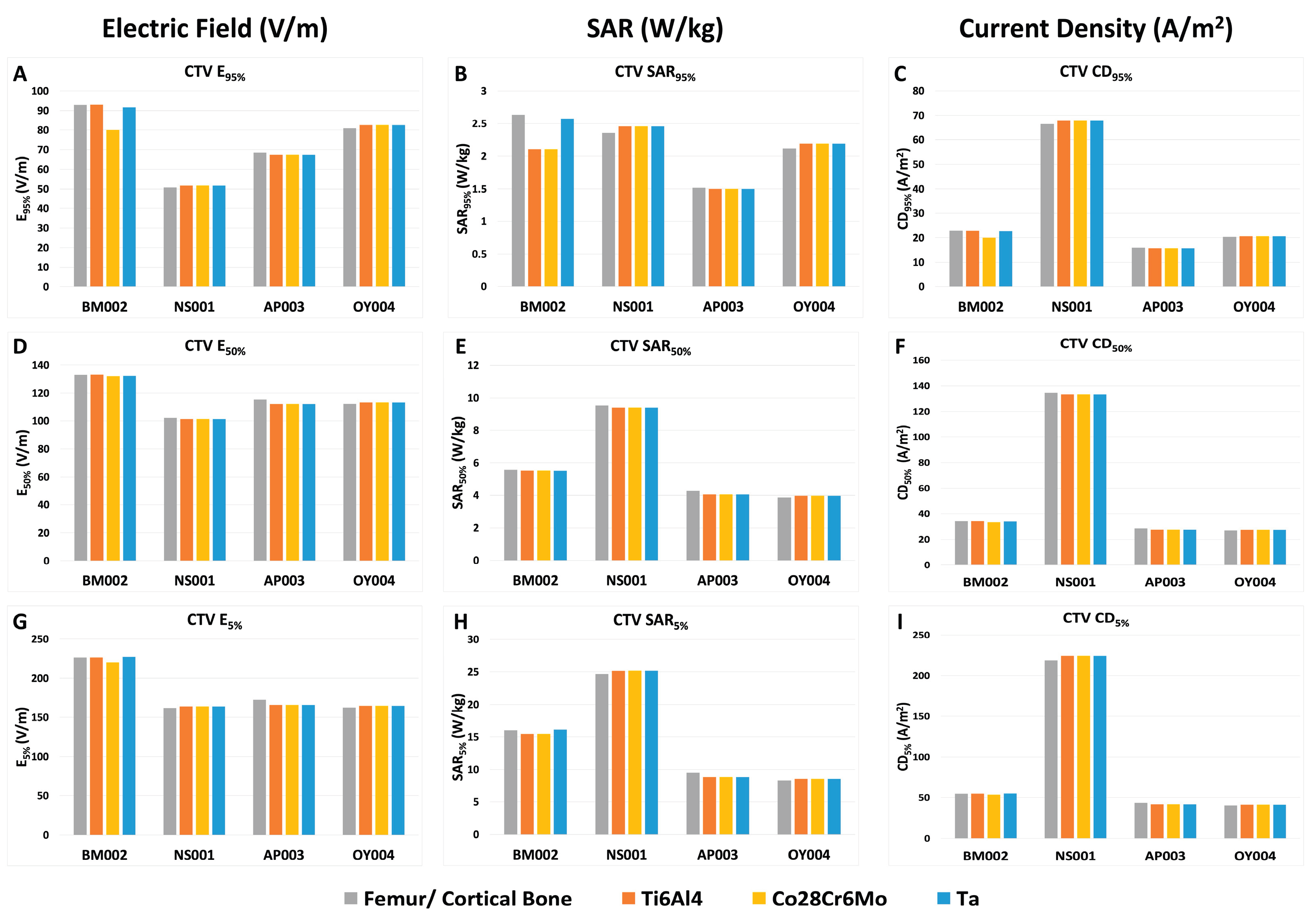
Figure 2.
CTV morphology and proximity to the femora across patient models. Three-dimensional renderings of the CTV (red), right femur (green), and left femur (blue) are shown for (A) BM002, (B) NS001, (C) AP003, and (D) OY004. Labels indicate the minimum distance from the CTV to each femur. The accompanying table summarizes CTV volume, CTV surface area, left and right femur-to-CTV distances, and the mean bilateral femur-to-CTV distance for each model.
Figure 2.
CTV morphology and proximity to the femora across patient models. Three-dimensional renderings of the CTV (red), right femur (green), and left femur (blue) are shown for (A) BM002, (B) NS001, (C) AP003, and (D) OY004. Labels indicate the minimum distance from the CTV to each femur. The accompanying table summarizes CTV volume, CTV surface area, left and right femur-to-CTV distances, and the mean bilateral femur-to-CTV distance for each model.
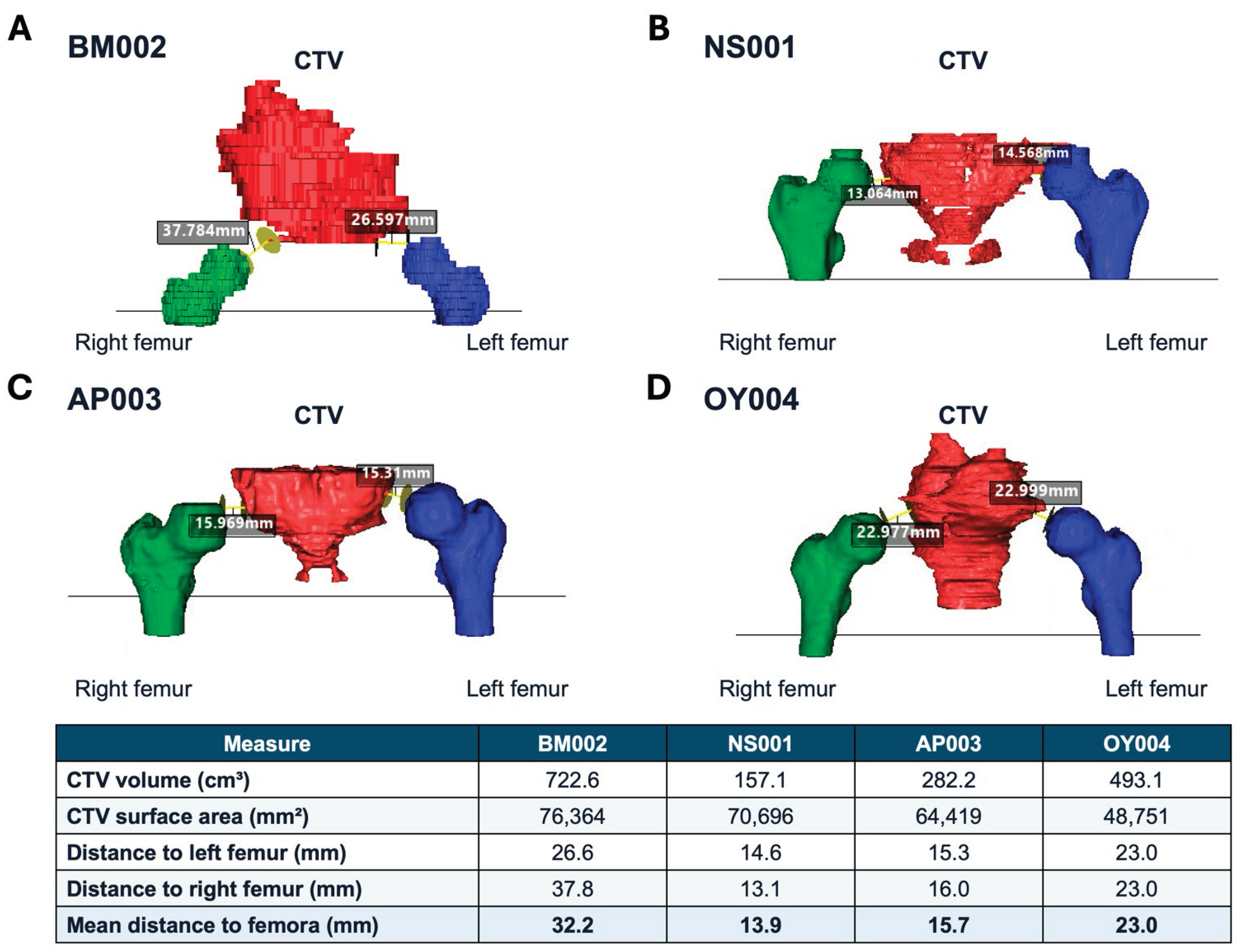
Figure 3.
Plan quality metrics of the femur. E, SAR, and CD at 95%, 50%, and 5% coverage of the femur are displayed according to femur/cortical bone and various types of THA material (titanium, cobalt-chromium, and tantalum) with different electrical properties. CTV, clinical treatment volume; E, electric field; SAR, specific absorption rate; CD, current density; Ti6Al4V, titanium; Co28Cr6Mo, cobalt-chromium; Ta, tantalum.
Figure 3.
Plan quality metrics of the femur. E, SAR, and CD at 95%, 50%, and 5% coverage of the femur are displayed according to femur/cortical bone and various types of THA material (titanium, cobalt-chromium, and tantalum) with different electrical properties. CTV, clinical treatment volume; E, electric field; SAR, specific absorption rate; CD, current density; Ti6Al4V, titanium; Co28Cr6Mo, cobalt-chromium; Ta, tantalum.
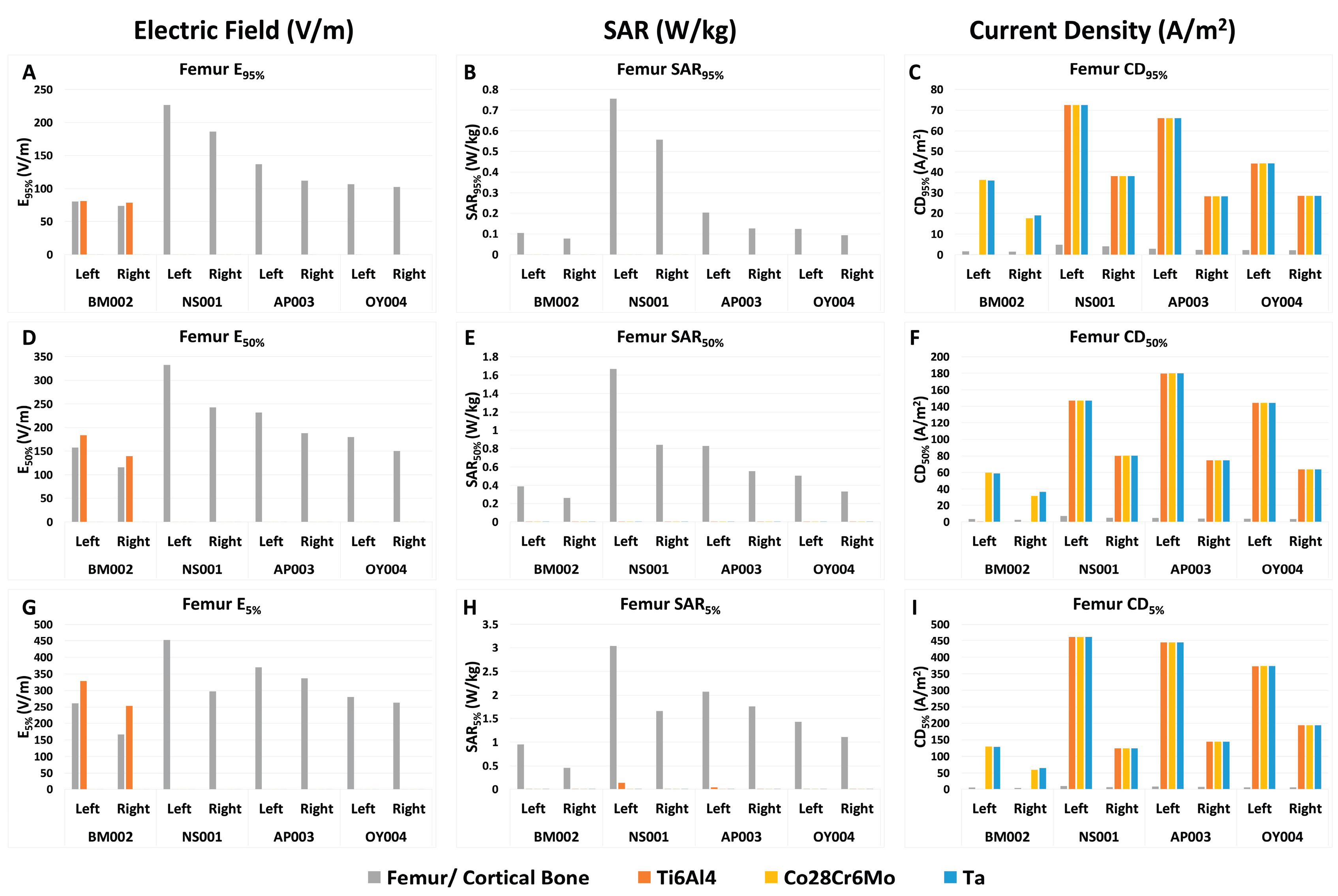
Figure 4.
Plan quality metrics of the skin. E, SAR, and CD at 95%, 50%, and 5% coverage of the skin are displayed according to femur/cortical bone and various types of THA material (titanium, cobalt-chromium, and tantalum) with different electrical properties. CTV, clinical treatment volume; E, electric field; SAR, specific absorption rate; CD, current density; Ti6Al4V, titanium; Co28Cr6Mo, cobalt-chromium; Ta, tantalum.
Figure 4.
Plan quality metrics of the skin. E, SAR, and CD at 95%, 50%, and 5% coverage of the skin are displayed according to femur/cortical bone and various types of THA material (titanium, cobalt-chromium, and tantalum) with different electrical properties. CTV, clinical treatment volume; E, electric field; SAR, specific absorption rate; CD, current density; Ti6Al4V, titanium; Co28Cr6Mo, cobalt-chromium; Ta, tantalum.
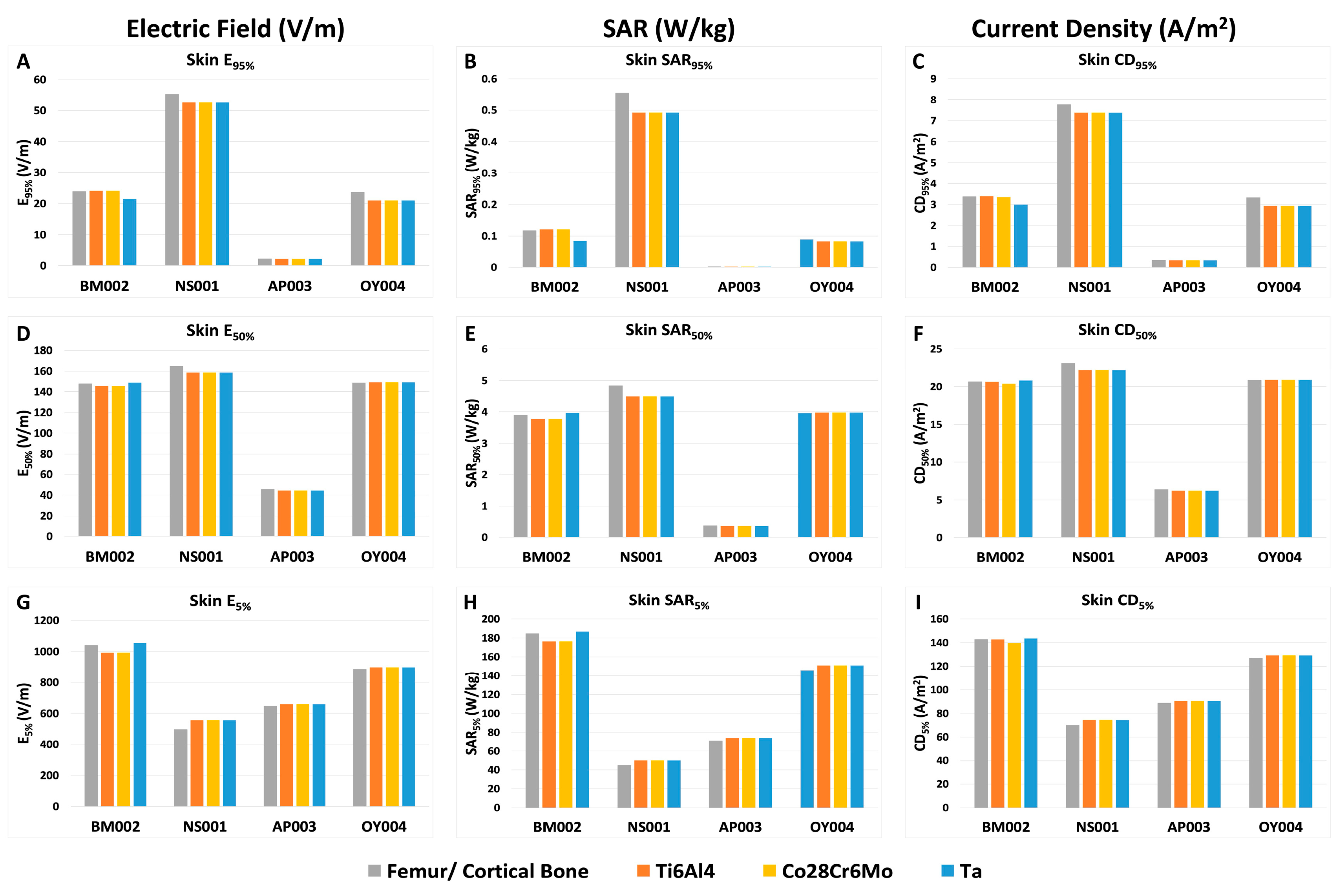
Table 1.
Physical and electrical properties of the femur, THA materials (titanium, cobalt-chromium, and tantalum), CTV, and skin. CTV, clinical tumor volume.
Table 1.
Physical and electrical properties of the femur, THA materials (titanium, cobalt-chromium, and tantalum), CTV, and skin. CTV, clinical tumor volume.
| Tissue Structure |
Electrical Conductivity σ (S/m) |
Mass Density ρ (kg/m3) |
| Femur (left/right) | 2.09E-02 | 1908 |
| Titanium Alloy (Ti6Al4V) | 5.85E+05 | 4430 |
| Cobalt Chromium (Co28Cr6Mo) | 1.25E+06 | 8400 |
| Tantalum (Ta) | 7.70E+06 | 16650 |
| CTV (NS001) | 1.30E+00 | 2000 |
| CTV (BM002, AP003, OY004) | 2.50E-01 | 1100 |
| Skin | 1.05E-03 | 1109 |
Table 2.
Percent change in PQM at the CTV. E, SAR, and CD at 95%, 50% and 5% coverage of the CTV was compared to cortical bone among the various types of THA material (titanium, cobalt-chromium, and tantalum) with different electrical properties. A/m2, ampere per meter squared; CTV, clinical treatment volume, Co28Cr6Mo, cobalt-chromium; PQM, plan quality metrics; Ta, tantalum; Ti6Al4V, titanium; V/m, volt per meter; W/kg, watt per kilogram.
Table 2.
Percent change in PQM at the CTV. E, SAR, and CD at 95%, 50% and 5% coverage of the CTV was compared to cortical bone among the various types of THA material (titanium, cobalt-chromium, and tantalum) with different electrical properties. A/m2, ampere per meter squared; CTV, clinical treatment volume, Co28Cr6Mo, cobalt-chromium; PQM, plan quality metrics; Ta, tantalum; Ti6Al4V, titanium; V/m, volt per meter; W/kg, watt per kilogram.
 |
Disclaimer/Publisher’s Note: The statements, opinions and data contained in all publications are solely those of the individual author(s) and contributor(s) and not of MDPI and/or the editor(s). MDPI and/or the editor(s) disclaim responsibility for any injury to people or property resulting from any ideas, methods, instructions or products referred to in the content. |
© 2026 by the authors. Licensee MDPI, Basel, Switzerland. This article is an open access article distributed under the terms and conditions of the Creative Commons Attribution (CC BY) license (http://creativecommons.org/licenses/by/4.0/).
Copyright: This open access article is published under a Creative Commons CC BY 4.0 license, which permit the free download, distribution, and reuse, provided that the author and preprint are cited in any reuse.