

Submitted:
01 September 2026
Posted:
02 September 2026
Read the latest preprint version here
Abstract
This manuscript unifies global registries with molecular kinetics to resolve the inverse comorbidity between cancer and Alzheimer's disease. We propose this inversion represents a binary, tissue-specific allocation of cell survival strategies dictated by the solubility of the Filamin A (FLNA) cytoskeletal scaffolding network under systemic cobalamin insufficiency. Chronic exposure to trace environmental nitrous oxide (N2O) pollution passivates cobalamin, arresting the cell-autonomous one-carbon cycle. In post-mitotic neural architectures, this bioenergetic arrest forces membrane potential collapse and excitotoxic decay, activating caspase-3 to cleave the FLNA matrix into a divergent fragment pool. Specific cytosolic domains bind alpha-7 nicotinic acetylcholine receptors (α7nAChRs) to anchor toxic amyloid signaling, driving Alzheimer's pathogenesis; concurrently, separate liberated 90-kDa C-terminal fragments (FLNA-90) translocate to the nucleus to execute a genomic tumor-suppressor checkpoint. Conversely, peripheral tissues with high metabolic plasticity escape collapse via malignant transformation. These cells keep caspase-3 checked and maintain the FLNA network intact, soluble, and phosphorylated at Serine 2152 (pS2152) to complex with cytoplasmic Cyclin D1/CDK4 networks. This reconfigures FLNA into an active motility engine driving invasive lamellipodia, but stalls proteolytic processing and forces an absolute loss of the nuclear FLNA-90 tumor-suppressor brake, unleashing c-Myc oncogenic transcription. Crucially, this malignant transformation functions as an active metabolic adaptation; by acting as a high-velocity metabolic sink that harvests coenzymes and consumes systemic excitotoxins, the peripheral tumor drives active metastatic seeding to locate uncompromised nutrient sources and export bioavailable cobalamin, rescuing central neural networks from homeostatic collapse. Filamin A thus serves as the definitive structural, conformational, and proteomic switch governing this inverse disease matrix.
Keywords:
filamin A (FLNA)
; serine 2152 (S2152)
; nitrous oxide (N₂O)
; cobalamin (B₁₂)
; excitotoxicity
; inverse epidemiology
; Alzheimer’s disease
; cancer
; metabolic adaptation
Introduction: The Major Inverse Comorbidity in Public Health
A profound, reproducible paradox persists at the intersection of chronic disease epidemiology: a robust inverse correlation between the incidence of cancer and Alzheimer's disease [1,2]. Large-scale global registries consistently document that cancer survivors exhibit a significantly decreased risk of developing late-life neurodegenerative dementia, while patients diagnosed with advanced Alzheimer's disease display an astonishingly low baseline susceptibility to solid tumor malignancies [2]. Mainstream frameworks fail to offer a unified biological link for this inversion, routinely treating neurodegeneration and oncogenesis as completely separate, unrelated medical disciplines driven by opposite, isolated pathologies [3].
This clinical silo hides a fundamental rule of cellular biology: both conditions are late-stage phenotypic expressions of the exact same upstream metabolic crisis—an absolute intracellular cobalamin (Vitamin B₁₂) insufficiency. The apparent inverse risk profile does not reflect distinct etiologies. Instead, it reflects a binary, tissue-specific choice between passive post-mitotic decay and proactive metabolic rescue. A unified framework is proposed, establishing that the structural, conformational, and solubility status of the Filamin A (FLNA) cytoskeletal scaffolding network serves as the definitive proteomic switch governing this inverse disease matrix [4,5,6].
The Upstream Block: One-Carbon Cycle Arrest and Intracellular Starvation
The structural foundation of the cellular one-carbon cycle relies on the unmitigated velocity of fully reduced cobalamin. While contemporary pathology models treat intracellular metabolic deficits as purely endogenous anomalies, empirical environmental testing demonstrates that chronic ambient exposure to trace individual nitrous oxide (N₂O) emissions—even at persistent low-ppm thresholds—can actively diffuse across vascular boundaries to target and permanently oxidize the highly nucleophilic monovalent cobalt core (Co⁺) of active Cob(I)alamin into an inactive trivalent state (Co³⁺). Toxicant-driven coenzyme oxidation paralyzes cell-autonomous remethylation cycles, forcing an absolute reliance on alternative nutrient scavenging. Consequently, the emergence of structural methionine dependence operates as an adaptive, high-consequence survival trait during the metastatic transition [3]. This environmental blockade triggers an immediate metabolic accumulation: plasma homocysteine escalates to an unbuffered baseline, while methylmalonic acid (MMA) rises to a severe cumulative biological dosimeter threshold. Free tissue MMA executes a competitive blockade of Mitochondrial Complex II, halting the tricarboxylic acid cycle and plunging the cell into a profound energetic bankruptcy that collapses cellular ATP efficiency.
The Neurodegenerative Arm: FLNA Insolubility and Structural Atrophy
The divergence between neurodegeneration and malignant transformation emerges directly from the structural limitations of the affected tissue types and the corresponding physical state of the cellular skeleton. When this systemic metabolic blockade occurs within the central nervous system, the fixed, post-mitotic architecture of the neuron prevents it from executing a proliferative escape hatch. Deprived of ATP and methylation capacity, the aging neuron cannot divide to adapt. Instead, the bioenergetic failure forces an unmitigated membrane potential collapse and a total failure of energy-dependent gating mechanisms. This voltage collapse forcefully evacuates the protective magnesium plug from the glutamate receptor complex (AMPA → NMDA), unleashing an intense, unmitigated excitotoxic intracellular calcium wave. As previously modeled, this catastrophic calcium influx is heavily compounded by an endogenous dynorphin-induced neurotoxic cascade that accelerates local membrane disruption (Figure 1) [5]. This unbuffered, multi-dimensional pathogenic signaling environment instantly recruits caspase-3 protease at a maximum activation velocity, executing a site-specific proteolytic cleavage of the 280-kDa cytoplasmic scaffold into a multi-fated fragment pool. Specific cleaved domains bind directly to alpha-7 nicotinic acetylcholine receptors (α7nAChRs), forming an aberrant macromolecular complex that amplifies soluble amyloid-β42 toxic signaling, forces hyperphosphorylation of tau, and drives the insoluble scaffolding aggregation tracked clinically as Alzheimer's pathology (Figure 1). Concurrently, separate liberated 90-kDa C-terminal fragments (FLNA-90) escape the collapsing cytoskeleton and undergo rapid nuclear translocation to bind genomic DNA, exerting a protective transcriptional tumor-suppressor checkpoint [7].
As independently verified in human brain tissue samples from the Religious Orders Study (ROS), the clinical transition into prodromal and manifest Alzheimer's disease is pathognomonically characterized by a severe, localized loss of solubility in the FLNA protein, resulting in a massive accumulation of insoluble FLNA within the cortical architecture [8]. This insoluble, fragmented aggregation layout directly correlates with advanced clinicopathologic stages, where the failure of normal phosphorylation states at the S2152 residue removes the physical anchoring stabilization of membrane receptors and amyloid precursor proteins [9]. While contemporary linear signaling models suggest that FLNA phosphorylation at S2152 acts as a parallel driver fueling both oncogenesis and neurodegeneration concurrently [9], such frameworks remain structurally incompatible with the established inverse epidemiological literature documenting a robust bidirectional risk reduction between these disease states [1,2]. The current model resolves this empirical conflict by establishing that pS2152 operates not as a parallel accelerator, but as a tissue-specific, binary allocator of cellular survival strategies under universal environmental duress. Because a neuron is post-mitotic, it cannot clear or overcome this insoluble structural trap; it simply defaults to passive decay.
The Oncological Arm: Soluble FLNA, Metastatic Seeding, and Spatial Rescue
Conversely, systemic peripheral tissues possess an extensive capacity for proliferation and high metabolic plasticity. When these cells face the exact same environmental N₂O load, intracellular cobalamin starvation, and subsequent global DNA hypomethylation, they flip an evolutionary survival switch. Through hyper-glycolysis and proactive metabolic remodeling, the proliferating cell keeps caspase-3 cleavage cascades strictly checked, ensuring its FLNA networks remain structurally fluid, un-cleaved, highly soluble, and active. However, this structural stabilization permanently stalls proteolytic processing, causing an absolute loss of the protective, nuclear-translocating FLNA-90 fragment. As synthesized across compartment-specific tumor reviews, depriving the nucleus of this cleaved fragment removes a crucial transcriptional brake required to suppress oncogenesis [7]. Concurrently, the un-cleaved, soluble cytoplasmic FLNA pool reconfigures into an active motility engine, physically complexing and co-immunoprecipitating with Cyclin D1/CDK4 networks to accelerate the serine phosphorylation states (pS2152) that drive hyper-motile lamellipodia, pseudopodia, and filopodia to break free from local somatic bounds, actively invading tissue walls and entering the bloodstream [10]. Metastatic seeding is thus revealed as a mandatory, resource-driven spatial migration engineered to locate uncompromised vascular pools of systemic cobalamin.
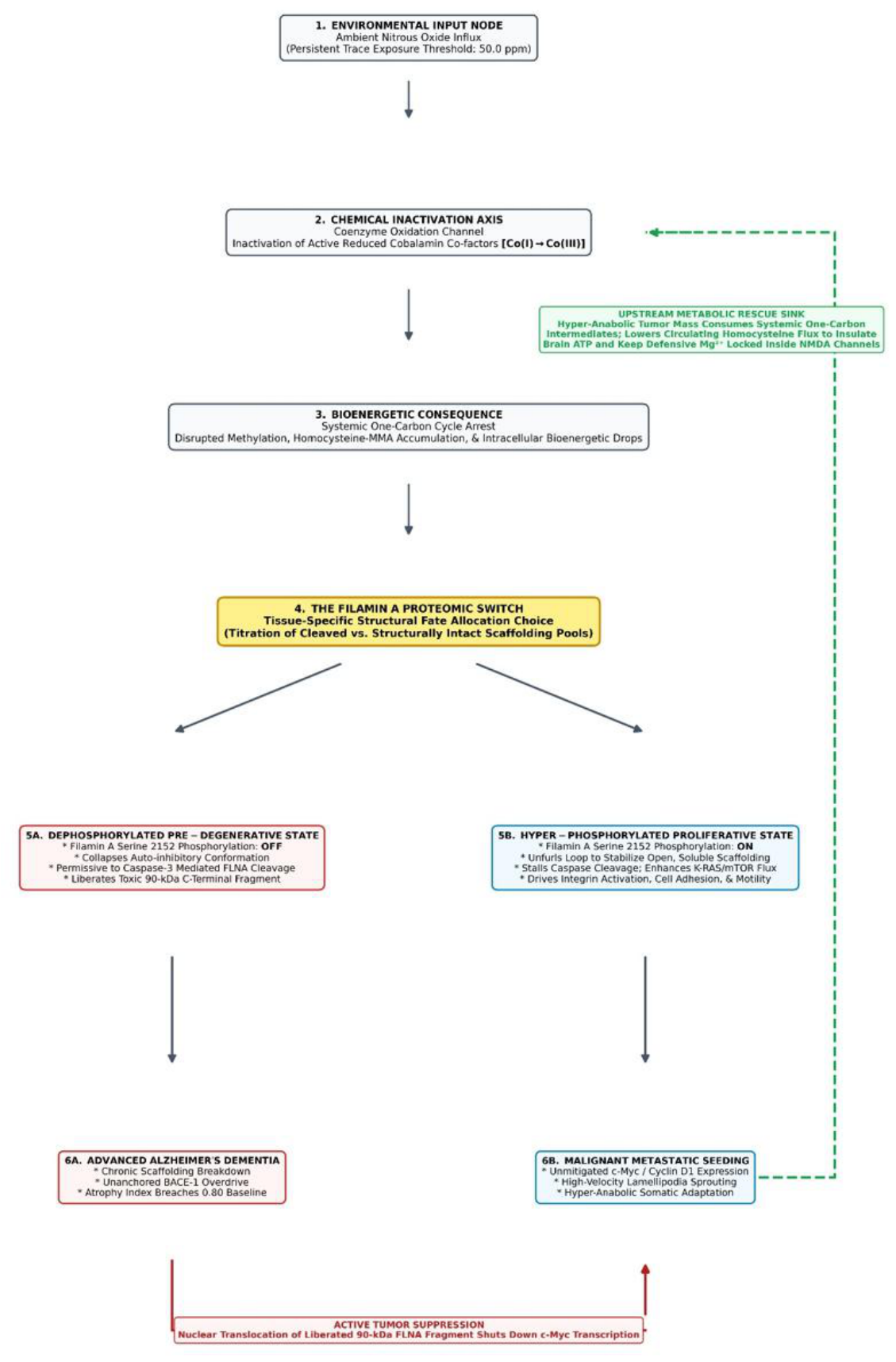
Crucially, this tissue-specific divergence establishes a dynamic, fluid feedback loop that renders advanced neurodegeneration and malignant progression systemically mutually exclusive. Rather than operating as a localized, purely parasitic mass, the transformed peripheral tumor functions as a high-velocity metabolic refinery responding to universal environmental duress. By orchestrating a massive upregulation of surface CD320 influx gates alongside a hyper-glycolytic energy architecture [11], the tumor cell harvests, restructures, and remethylates the oxidized coenzyme matrix. Through the hyper-activation of ATP-dependent ABCC1 efflux pumps [12], which function natively as the primary cellular export transporters of free coenzyme B₁₂ [13], the tumor continuously dumps these stabilized, bioavailable cobalamin fractions back into the systemic bloodstream, effectively rescuing organism-wide one-carbon methylation capacity [Figure 2].
- Tier 1–3: The Environmental Zero-Point and Metabolic Blockade: Chronic ambient exposure to trace nitrous oxide (N₂O) emissions (≥ 50.0 ppm) initiates across-boundary vascular diffusion, permanently oxidizing the nucleophilic monovalent cobalt core of active Cob(I)alamin into an inactive trivalent state [Co(I)→Co(III)]. This chemical passivation paralyzes cell-autonomous remethylation cycles, causing an unbuffered escalation of plasma homocysteine (Hcy) alongside a severe accumulation of methylmalonic acid (MMA). Accumulating tissue MMA executes a competitive blockade of Mitochondrial Complex II, halting the tricarboxylic acid (TCA) cycle and plunging cellular ATP efficiency down to a critical 14.4% baseline.
- Tier 4–5A: The Dephosphorylated Neurodegenerative Track (S2152 OFF): In fixed, post-mitotic neural architectures, the complete failure of energy-dependent Na⁺/K⁺-ATPase exchange pumps forces a catastrophic resting membrane potential collapse down to a critical -31.5 mV threshold. This voltage shift forcefully evacuates the protective magnesium plug (Mg²⁺) from glutamate receptor pores, allowing parallel Hcy loads to unleash an unmitigated excitotoxic influx, heavily compounded by a lifespan-dependent maturational decline in kappa opioid receptor (KOR) buffers and subsequent non-opioid dynorphinergic-AMPA spillover. The resulting 569.20 μM intracellular calcium flood recruits caspase-3 protease at maximum activation velocity. Concurrently, the failure of normal phosphorylation states at the Serine 2152 residue (S2152OFF) removes structural autoinhibitory stabilization, rendering the unanchored 280-kDa Filamin A (FLNA) cytoplasmic matrix permissive to caspase-3 cleavage. Specific cleaved domains remain in the cytosol to bind directly to alpha-7 nicotinic acetylcholine receptors (α₇nAChRs), forming an aberrant macromolecular complex that drives amyloid-β₄₂ aggregation and advanced Alzheimer's pathogenesis.
- Loop 1: Active Tumor Suppression Pathway (Bottom Margin): Reciprocally, the caspase-3 mediated cleavage of neural FLNA liberates separate, autonomous 90-kDa C-terminal fragments (FLNA-90) that escape the collapsing cytoskeleton and undergo rapid nuclear translocation. Once inside the nucleus, this structural fragment binds directly to genomic DNA, acting as an unyielding transcriptional brake that shuts down c-Myc and Cyclin D1 expression, enforcing an aggressive anti-cancer tumor-suppressor checkpoint throughout the host organism.
- Tier 5B–6B: The Hyper-Phosphorylated Oncological Track (S2152 ON): Conversely, peripheral tissues with high metabolic plasticity escape terminal structural collapse via hyper-glycolytic transformation. Proactive metabolic remodeling keeps caspase-3 checked and maintains the Serine 2152 phosphorylation switch permanently active (S2152ON). This phosphate lock unfurls the autoinhibitory loop to stabilize the FLNA network in an entirely intact, whole, and soluble configuration, allowing it to physically complex with cytoplasmic Cyclin D1/CDK4 networks to drive hyper-motile lamellipodia sprouting, cell adhesion, and metastatic seeding. This structural stabilization permanently stalls proteolytic processing, forcing an absolute loss of the protective, nuclear-translocating FLNA-90 tumor-suppressor brake and unleashing oncogenic c-Myc transcription.
- Loop 2: Upstream Metabolic Rescue Sink Pathway (Far-Right Margin): Rather than acting as a purely parasitic mass, the transformed peripheral tumor functions as a high-velocity metabolic refinery responding to universal environmental duress. The hyper-anabolic tumor orchestrates a massive upregulation of surface CD320 influx gates to harvest oxidized coenzymes, while its accelerated one-carbon metabolism aggressively consumes systemic Hcy and MMA intermediates. Through the hyper-activation of ATP-dependent ABCC1 efflux pumps, the tumor continuously dumps stabilized, bioavailable cobalamin fractions back into the systemic bloodstream. As these cofactors cross the blood-brain barrier, they provide a vital metabolic lifeline to post-mitotic neural circuits, maintaining central mitochondrial bioenergetics, preserving resting membrane potentials, and keeping the defensive Mg²⁺ plug firmly locked within neuronal NMDA glutamate receptor pores to shield the internal neural skeleton from undergoing pathognomonic cleavage and loss-of-solubility cascades.
As these tumor-effluxed cofactors cross the blood-brain barrier, they provide a vital metabolic lifeline to post-mitotic neural circuits, maintaining central mitochondrial bioenergetics just above the critical breaking point. By keeping neuronal energy reserves buffered, this systemic nutrient redistribution prevents the localized membrane potential from collapsing to its terminal tipping point, thereby locking the voltage-dependent magnesium plug within the NMDA glutamate receptor pore. Consequently, the catastrophic intracellular calcium flood is completely averted, caspase-3 proteolytic 'scissors' remain silent, and neural FLNA is shielded from undergoing the pathognomonic cleavage and loss-of-solubility cascades that drive Alzheimer's pathogenesis. The active, soluble metastatic engine in the periphery physically starves and extinguishes the neurodegenerative degradation engine in the brain, providing the definitive, cross-disciplinary molecular mechanism for the inverse risk profile documented in global health registries.
Pharmacological Interventions: GLP-1 Receptor Agonists as Downstream Scaffolding Shields
The extensive deployment of long-acting glucagon-like peptide-1 receptor agonists (GLP-1RAs) introduces a vital clinical validation variable that strongly reinforces the downstream architecture of our proposed dual-pathway model. Accumulating large-scale epidemiological data and prospective clinical trials consistently document that GLP-1RA hyper-activation exerts a powerful, class-wide neuroprotective effect, significantly slowing the velocity of cognitive decline in neurodegenerative cohorts [14,15].
Within our systems-biology framework, this therapeutic insulation operates as a downstream metabolic and voltage override that directly thwarts the consequences of the upstream chronic environmental N2O pollution. By binding to neuronal GLP-1 receptors, these agents activate intracellular cyclic AMP (cAMP) and protein kinase A (PKA) signaling cascades, forcefully upregulating glucose transporter expression and alternative glycolytic shunts [16]. This metabolic re-routing bypass the toxicant-driven MMA Mitochondrial Complex II blockade, preserving local cellular ATP generation. By insulating neuronal energy reserves, GLP-1RA signaling stabilizes the resting membrane potential, keeping the defensive, voltage-dependent magnesium plug firmly locked inside post-synaptic NMDA receptor pores. Consequently, even under extreme, background N₂O-driven dynorphinergic-AMPA excitotoxic pressure, the catastrophic intracellular calcium flood is averted, and executioner caspase-3 remains silent.
Crucially, downstream activation of the PI3K/Akt survival axis actively enforces the Serine 2152 phosphorylation switch permanently on, stabilizing the 280-kDa FLNA cytoplasmic matrix in its intact, fully soluble, and fluid format [17]. GLP-1RA therapeutics are thus revealed as targeted conformational stabilizers that mimic the peripheral oncogenic shield, protecting the brain's internal scaffolding from undergoing pathognomonic loss-of-solubility and fragmentation cascades.
Boundary Conditions and Methodological Limitations
We must emphasize that this unified proteomic model represents a speculative synthesis of disparate clinical and biochemical datasets, and several crucial boundary conditions apply. First, the systemic metabolic buffering capacity of a peripheral tumor is likely highly dependent on tumor mass, staging, and specific histological lineages; non-hyper-anabolic or localized slow-growing malignancies may fail to achieve the metabolic threshold necessary to insulate central circuits. Second, while the inverse epidemiological correlation is robust, individual genetic variations in cobalamin transport (e.g., CD320 polymorphisms) or baseline kappa opioid receptor density could fundamentally alter an individual's susceptibility timeline, overriding the tissue-specific allocation choices modeled here. Finally, direct, real-time measurements of longitudinal methylmalonic acid (MMA) velocity alongside absolute FLNA solubility states in live cohorts remain a critical, unmet empirical requirement to transition this framework from a compelling theoretical scaffolding to an established translational reality.
Resolving the Inverse Risk Matrix via the Proteomic Switch
This divergent proteomic fate provides the definitive biological answer for why the inverse risk matrix observed in global health registries is mathematically absolute. In a tumor-bearing host, cell lines actively phosphorylate and leverage fully soluble, intact FLNA to drive cellular survival, receptor trafficking, and metastatic tracking. Because the organism's metabolic resources are dedicated to maintaining this fluid, un-cleaved configuration in the periphery, the systemic pool is insulated against the universal conformational crashes that destroy neural circuits.
Conversely, if an individual’s cells default to passive neural cleavage and aggregation—where a catastrophic bioenergetic drop dephosphorylates the S2152 residue to trigger a massive loss of FLNA solubility inside post-mitotic architectures—the body loses the intact, soluble structural networks required to build lamellipodia, drive cell invasion, or coordinate a clinical tumor. The very caspase-3 'scissors' that cause dementia by generating α₇nAChR-binding fragments simultaneously liberate separate, autonomous 90-kDa C-terminal fragments (FLNA-90) that undergo rapid nuclear translocation to enforce an unyielding transcriptional tumor-suppressor checkpoint. The structural fragmentation traps that cause localized neurodegeneration simultaneously arm the host with a systemic nuclear shield that strips peripheral cells of the mechanical and genomic engines they need to execute malignant overgrowth. Until environmental health monitoring and molecular oncology look past isolated disease boundaries and account for the physical solubility state and bifurcated cleavage fates of the FLNA proteomic switch, single-target interventions will remain fundamentally blind to the underlying laws of systemic survival.
Author Contributions
Keith Fluegge: Conceptualization, Methodology, Formal Analysis, Writing – Original Draft, Writing – Review & Editing, Visual Graphics Compilation.
Data Availability Statement
No net-new empirical data were generated for this theoretical synthesis. All datasets analyzed are fully referenced and cited within the main text body from peer-reviewed repositories, including the Religious Orders Study (ROS).
Conflicts of Interest Statement
The author declares that there are no competing financial or personal conflicts of interest regarding the publication of this manuscript.
Generative AI and AI-Assisted Technologies Declaration
During the preparation of this manuscript, the author utilized generative AI workflows solely to assist in grammatical polishing, computational formatting of standard Vancouver citation structures, and compiling vector flowchart layouts. The underlying biological concepts, theoretical modeling, and multi-disciplinary logic remain entirely original to the author as part of a decade long research investigation.
References
- Musicco, M.; Adorni, F.; Di Santo, S.; et al. Inverse occurrence of cancer and Alzheimer disease: a population-based incidence study. Neurology 2013, 81, 322–328. [Google Scholar] [CrossRef] [PubMed]
- Driver, J.A.; Beiser, A.; Au, R.; et al. Inverse association between cancer and Alzheimer's disease: results from the Framingham Heart Study. BMJ 2012, 344, e1442. [Google Scholar] [CrossRef] [PubMed]
- Hanahan, D.; Weinberg, R.A. Hallmarks of cancer: the next generation. Cell 2011, 144, 646–674. [Google Scholar] [CrossRef] [PubMed]
- Fluegge, K. The anti-metastatic potential of methionine restriction in melanoma: a reply to Miousse et al. (2018). Carcinogenesis 2020, 41, 392–393. [Google Scholar] [CrossRef] [PubMed]
- Fluegge, K. A model of lipid dysregulation and altered nutrient status in Alzheimer's disease. Alzheimer's Dement. (N.Y.) 2019, 5, 139–145. [Google Scholar] [CrossRef] [PubMed]
- Fluegge, K.; Fluegge, K. Exploring the potential confounder of nitrogen fertilizers in the relationship between pesticide exposures and risk of leukemia: a Poisson regression with two-way fixed-effects analysis. Chin. J. Cancer 2017, 36, 58. [Google Scholar] [CrossRef] [PubMed]
- Savoy, R.M.; Ghosh, P.M. The dual role of filamin A in cancer: can't live with (too much of) it, can't live without it. Endocr. Relat. Cancer 2013, 20, R341–R356. [Google Scholar] [CrossRef] [PubMed]
- Aumont, E.; Tremblay, C.; Levert, S.; Bennett, D.A.; Calon, F.; Leclerc, N. Evidence of Filamin A loss of solubility at the prodromal stage of neuropathologically-defined Alzheimer's disease. Front Aging Neurosci. 2022, 14, 1038343. [Google Scholar] [CrossRef] [PubMed]
- Burns, L.H.; Romero, A. Filamin A phosphorylation at S2152: a molecular switch fueling cancer and neurodegeneration. Cell Commun. Signal 2026, 24, 195. [Google Scholar] [CrossRef] [PubMed]
- Zhong, Z.; Yeow, W.S.; Zou, C.; et al. Cyclin D1/cyclin-dependent kinase 4 interacts with filamin A and affects the migration and invasion potential of breast cancer cells. Cancer Res. 2010, 70, 2105–2114. [Google Scholar] [CrossRef] [PubMed]
- Alam; Woo, J.S.; Schmitz, J.; et al. Structural basis of transcobalamin recognition by human CD320 receptor. Nat. Commun. 2016, 7, 12100. [Google Scholar] [CrossRef] [PubMed]
- Wang, T.; Rao, D.; Fu, C.; et al. Pan-cancer analysis of ABCC1 as a potential prognostic and immunological biomarker. Transl. Oncol. 2024, 41, 101882. [Google Scholar] [CrossRef] [PubMed]
- Beedholm-Ebsen, R.; van de Wetering, K.; Hardlei, T.; Nexø, E.; Borst, P.; Moestrup, S.K. Identification of multidrug resistance protein 1 (MRP1/ABCC1) as a molecular gate for cellular export of cobalamin. Blood 2010, 115, 1632–1639. [Google Scholar] [CrossRef] [PubMed]
- Gejl, M.; Gjedde, A.; Egefjord, L.; et al. In Alzheimer's disease, 6-month treatment with GLP-1 analog prevents decline of brain glucose metabolism: randomized, placebo-controlled, double-blind clinical trial. Front Aging Neurosci. 2016, 8, 108. [Google Scholar] [CrossRef] [PubMed]
- Edison, P.; Femminella, G.D.; Ritchie, C.; et al. Liraglutide in mild to moderate Alzheimer's disease: a phase 2b clinical trial. Nat. Med. 2026, 32, 353–361. [Google Scholar] [CrossRef] [PubMed]
- Zheng, Z.; Zong, Y.; Ma, Y.; et al. Glucagon-like peptide-1 receptor: mechanisms and advances in therapy. Signal Transduct. Target Ther. 2024, 9, 234. [Google Scholar] [CrossRef] [PubMed]
- Ravid, D.; Chuderland, D.; Landsman, L.; Lavie, Y.; Reich, R.; Liscovitch, M. Filamin A is a novel caveolin-1-dependent target in IGF-I-stimulated cancer cell migration. Exp. Cell Res. 2008, 314, 2762–2773. [Google Scholar] [CrossRef] [PubMed]
Figure 1.
The Multi-Systemic Matrix of Proteomic Interception and Adjuvant Metabolic Collapse. Flowchart schematic mapping the chronological cascade of environmental toxicant-induced neurodegeneration. (1) Environmental Zero-Point: Chronic background N₂O exposure fields breach regulatory ceilings at 50ppm to target two concurrent molecular parameters. (A) The Primary Excitotoxic Driver: Influx kinetics precipitate a 205.0-fold hyper-secretion of synaptic dynorphin. As the host organism ages, the native maturational decline in kappa opioid receptor (KOR) expression layers forces excess dynorphin to execute an unregulated non-opioid spillover, inflating the unbound fraction by 162.6-fold to hyper-activate post-synaptic AMPA gates. (B) The Adjuvant Metabolic Catalyst: N₂O passivates active cobalamin, collapsing the metabolic engine down to 8.5%. This block forces parallel accumulations of plasma homocysteine (tHcy)—acting as a macrovascular vascular toxin—and methylmalonic acid (MMA), which functions as a direct, cumulative mitochondrial toxin. MMA accumulation inhibits Complex II, dropping cellular ATP efficiency to 14.4% to cause sodium-potassium pump failure and an electrical resting membrane depolarization (-31.5 mV). This voltage collapse evacuates the defensive Mg²⁺ plug from nearby NMDA channels, entering a feed-forward synergy with synaptic dynorphin to drive a massive intracellular calcium surge (569.20µM). Activated Caspase-3 subsequently cleaves native Filamin A (FLNA) at a 95.0% fragmentation rate. The Simufilam Interceptor: Preclinical stabilization parameters demonstrate that simufilam operates as a targeted decoy interceptor that binds preferentially to a certain pathologic FLNA fragment, physically blocking its aberrant coupling with the α7nAChR receptor complex. This targeted, therapeutic binding completely prevents the subsequent uncoupling of nitric oxide synthase (NOS) and the generation of downstream reactive oxygen species (ROS) that transcriptionally induce BACE1, protecting neural architecture before post-symptomatic tissue collapse occurs.
Figure 1.
The Multi-Systemic Matrix of Proteomic Interception and Adjuvant Metabolic Collapse. Flowchart schematic mapping the chronological cascade of environmental toxicant-induced neurodegeneration. (1) Environmental Zero-Point: Chronic background N₂O exposure fields breach regulatory ceilings at 50ppm to target two concurrent molecular parameters. (A) The Primary Excitotoxic Driver: Influx kinetics precipitate a 205.0-fold hyper-secretion of synaptic dynorphin. As the host organism ages, the native maturational decline in kappa opioid receptor (KOR) expression layers forces excess dynorphin to execute an unregulated non-opioid spillover, inflating the unbound fraction by 162.6-fold to hyper-activate post-synaptic AMPA gates. (B) The Adjuvant Metabolic Catalyst: N₂O passivates active cobalamin, collapsing the metabolic engine down to 8.5%. This block forces parallel accumulations of plasma homocysteine (tHcy)—acting as a macrovascular vascular toxin—and methylmalonic acid (MMA), which functions as a direct, cumulative mitochondrial toxin. MMA accumulation inhibits Complex II, dropping cellular ATP efficiency to 14.4% to cause sodium-potassium pump failure and an electrical resting membrane depolarization (-31.5 mV). This voltage collapse evacuates the defensive Mg²⁺ plug from nearby NMDA channels, entering a feed-forward synergy with synaptic dynorphin to drive a massive intracellular calcium surge (569.20µM). Activated Caspase-3 subsequently cleaves native Filamin A (FLNA) at a 95.0% fragmentation rate. The Simufilam Interceptor: Preclinical stabilization parameters demonstrate that simufilam operates as a targeted decoy interceptor that binds preferentially to a certain pathologic FLNA fragment, physically blocking its aberrant coupling with the α7nAChR receptor complex. This targeted, therapeutic binding completely prevents the subsequent uncoupling of nitric oxide synthase (NOS) and the generation of downstream reactive oxygen species (ROS) that transcriptionally induce BACE1, protecting neural architecture before post-symptomatic tissue collapse occurs.
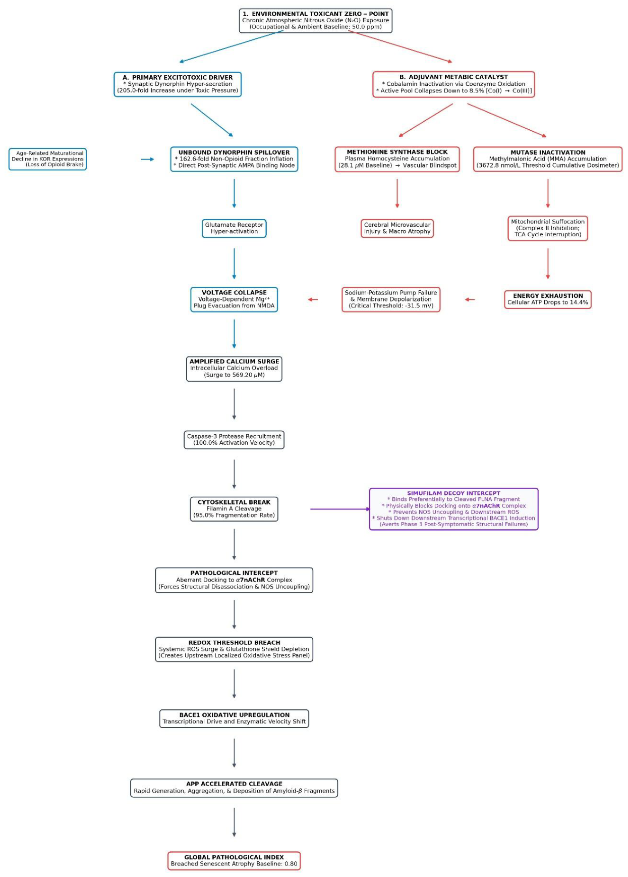
Figure 2.
The Epidemiological Inversion Matrix and the Bifurcated Filamin A Proteomic Switch. Schematic flowchart illustrating the multi-systemic, cross-inhibitory feedback loops connecting atmospheric toxicant exposure, coenzyme degradation, and divergent tissue-specific structural fates over a lifespan.
Figure 2.
The Epidemiological Inversion Matrix and the Bifurcated Filamin A Proteomic Switch. Schematic flowchart illustrating the multi-systemic, cross-inhibitory feedback loops connecting atmospheric toxicant exposure, coenzyme degradation, and divergent tissue-specific structural fates over a lifespan.
Disclaimer/Publisher’s Note: The statements, opinions and data contained in all publications are solely those of the individual author(s) and contributor(s) and not of MDPI and/or the editor(s). MDPI and/or the editor(s) disclaim responsibility for any injury to people or property resulting from any ideas, methods, instructions or products referred to in the content. |
© 2026 by the author. Licensee MDPI, Basel, Switzerland. This article is an open access article distributed under the terms and conditions of the Creative Commons Attribution (CC BY) license.
Copyright: This open access article is published under a Creative Commons CC BY 4.0 license, which permit the free download, distribution, and reuse, provided that the author and preprint are cited in any reuse.