

Submitted:
01 September 2026
Posted:
02 September 2026
You are already at the latest version
Abstract
Background: Cancer therapy-related cardiac dysfunction (CTRCD) remains an important complication of human epidermal growth factor receptor 2 (HER2)-targeted therapy in patients with breast cancer. Although conventional cardiovascular risk factors and serial cardiac imaging are routinely incorporated into cardio-oncology surveillance, the potential contribution of global comorbidity burden and cumulative anti-HER2 treatment exposure remains incompletely characterized. We investigated the occur-rence, clinical course, and factors associated with CTRCD in a large real-world population of patients with breast cancer receiving HER2-targeted therapy. Methods: Adult patients with breast cancer exposed to HER2-targeted therapy and undergoing longitudinal cardio-oncology surveillance at a single tertiary center between January 2014 and December 2024 were included. CTRCD was defined according to contemporary European cardio-oncology criteria integrating left ventricular ejection fraction (LVEF), left ventricular global longitudinal strain (LV-GLS), and cardiac biomarkers when available. Baseline comorbidity burden was quantified using the Charlson Comorbidity Index (CCI), while cumulative anti-HER2 exposure was quantified according to the total number of treatment cycles. Factors associated with CTRCD were investigated using univariable and parsimonious multivariable logistic regression. Results: A total of 850 patients with breast cancer receiving anti-HER2 therapy were included. CTRCD occurred in 179 patients (21.1%) and was predominantly mild, whereas clinically overt heart failure occurred in 2.9%. LVEF and LV-GLS deteriorated at nadir and subsequently showed substantial recovery during follow-up. Conventional cardiovascular risk factors, previous anthracycline exposure, tumor grade, baseline LVEF, and baseline LV-GLS were not significantly associated with CTRCD. In multivariable analysis, increasing CCI (adjusted OR 1.35 per one-point increase, 95% CI 1.12–1.63; p=0.002) and cumulative anti-HER2 exposure (adjusted OR 1.41 per five additional cycles, 95% CI 1.10–1.80; p=0.006) were independently associated with CTRCD. CTRCD probability progressively increased across higher CCI categories and with increasing cumulative anti-HER2 exposure. Conclusions: In this 10-year cardio-oncology cohort of patients with breast cancer, CTRCD was relatively frequent during HER2-targeted therapy but was predominantly mild, infrequently associated with overt heart failure, and frequently reversible. Global comorbidity burden and cumulative anti-HER2 exposure were independently associated with CTRCD, suggesting that cardiotoxic risk may reflect the interplay between baseline patient vulnerability and treatment-related bur-den. If prospectively validated, these readily available variables may provide additional information for longitudinal cardiovascular risk assessment and help individualize surveillance during prolonged HER2-targeted therapy.
Keywords:
cardio-oncology
; cardiotoxicity
; cancer therapy-related cardiac dysfunction
; HER2
; trastuzumab
; pertuzumab
; Charlson Comorbidity Index
; global longitudinal strain
; left ventricular ejection fraction
1. Introduction
Advances in cancer diagnosis and treatment have substantially improved survival across a broad spectrum of malignancies, resulting in a growing population of patients exposed to potentially cardiotoxic therapies [1,2,3]. Consequently, cardiovascular complications have emerged as an important determinant of morbidity, treatment discontinuation, and long-term prognosis among cancer patients and survivors [4,5,6]. Cancer therapy-related cardiac dysfunction (CTRCD) represents one of the most clinically relevant manifestations of cardiovascular toxicity, encompassing a continuum from asymptomatic myocardial dysfunction to overt heart failure [7,8,9]. Contemporary cardio-oncology has therefore progressively shifted from the treatment of established cardiac injury toward its early identification and prevention through baseline cardiovascular risk assessment, longitudinal imaging surveillance, cardiac biomarkers, and timely implementation of cardioprotective strategies [10,11,12].
Human epidermal growth factor receptor 2 (HER2)-targeted therapies represent a paradigmatic yet challenging setting in which this preventive approach is particularly relevant. Trastuzumab and other HER2-directed agents have profoundly improved oncological outcomes in patients with HER2-positive breast cancer in the neoadjuvant, adjuvant, and metastatic settings [13,14,15]. However, interference with HER2-dependent myocardial signaling may impair cardiomyocyte adaptive and repair mechanisms, exposing susceptible patients to left ventricular dysfunction during treatment [16,17,18]. Although anti-HER2-related cardiotoxicity is frequently reversible when promptly recognized and appropriately managed, its occurrence may require treatment interruption or modification and may compromise the optimal delivery of anticancer therapy [19,20,21]. Accordingly, serial assessment of left ventricular ejection fraction (LVEF), left ventricular global longitudinal strain (LV-GLS), and, when appropriate, cardiac biomarkers has become an integral component of cardiovascular surveillance during HER2-targeted treatment [22,23,24,25].
Nevertheless, prediction of CTRCD remains challenging. Cardiotoxicity is unlikely to result from a single determinant and may instead reflect the interaction between baseline patient characteristics, pre-existing cardiovascular disease, conventional cardiovascular risk factors, concomitant comorbidities, previous cardiotoxic treatments, and the intensity and duration of ongoing anticancer therapy [26,27,28]. In routine cardio-oncology practice, these factors frequently coexist, particularly in older and medically complex patients. Consequently, assessment of individual cardiovascular risk factors alone may provide an incomplete representation of the overall vulnerability of a patient undergoing potentially cardiotoxic treatment. A more comprehensive evaluation of comorbidity burden may therefore complement conventional cardiovascular risk stratification.
The Charlson Comorbidity Index (CCI) is a simple and widely applicable instrument that summarizes the burden of multiple clinically relevant comorbidities within a single score [29]. Although originally developed to estimate mortality according to comorbidity burden [30], its potential utility may extend to clinical settings in which reduced physiological reserve could influence susceptibility to treatment-related complications [31,32]. In cardio-oncology, such an integrated measure may be particularly attractive because patients often present with heterogeneous combinations of cardiovascular and non-cardiovascular conditions [33,34]. However, the potential relationship between increasing global comorbidity burden and the development of CTRCD during contemporary anticancer treatment remains insufficiently characterized [35].
Treatment-related exposure represents another potentially important component of cardiotoxic risk. Previous anthracycline exposure is an established determinant of cardiovascular toxicity [36,37,38], whereas risk stratification during HER2-targeted therapy has traditionally relied largely on baseline cardiovascular characteristics and serial changes in cardiac function during treatment [39,40]. However, among patients receiving HER2-targeted agents, cumulative exposure may vary considerably according to disease stage, treatment setting, therapeutic response, recurrence, and metastatic progression [41,42,43]. Consequently, the cardiovascular impact of HER2-targeted therapy may not be adequately captured by considering treatment exposure as a simple dichotomous variable. Whether the cumulative duration of anti-HER2 therapy, expressed as the number of administered treatment cycles, provides additional information for identifying patients at increased risk of CTRCD remains insufficiently characterized [44]. Clarifying this relationship may contribute to a more dynamic assessment of cardiotoxic risk and support individualized cardiovascular surveillance throughout the oncological treatment pathway.
Therefore, the present study specifically focused on a large cohort of patients with breast cancer exposed to HER2-targeted therapy and was designed to comprehensively characterize the incidence, severity, echocardiographic features, clinical management, and recovery of CTRCD over a 10-year observation period. We also sought to investigate clinical, oncological, treatment-related, and baseline echocardiographic factors associated with CTRCD within this anti-HER2-treated breast cancer population. Particular attention was devoted to the potential contribution of overall comorbidity burden, assessed by the CCI, and cumulative anti-HER2 treatment exposure. By integrating baseline patient characteristics with the longitudinal burden of HER2-targeted treatment, the study aimed to identify readily accessible factors associated with CTRCD that may provide additional information for longitudinal cardiovascular risk assessment and help inform surveillance strategies in patients with breast cancer receiving anti-HER2 therapy.
2. Materials and Methods
2.1. Study Design and Population
This study represents a focused analysis of the retrospective component of the CARTOX-PREDICT project, an observational, retro-prospective, single-center cardio-oncology study conducted at IRCCS MultiMedica, Ospedale San Giuseppe, Milan, Italy. The original CARTOX-PREDICT study protocol, provided in Supplementary Material File S1, was designed to investigate cardiovascular toxicity associated with different contemporary anticancer treatments, including anthracyclines, HER2-targeted therapies, fluoropyrimidines, vascular endothelial growth factor inhibitors, and immune checkpoint inhibitors.
For the present investigation, we specifically selected the subpopulation of adult patients with breast cancer exposed to HER2-targeted therapy and undergoing longitudinal cardio-oncology surveillance. Thus, unlike the broader CARTOX-PREDICT population, the current study was restricted to patients with breast cancer who had received anti-HER2 treatment and had serial cardiac assessments available. Patients were identified from the institutional oncology and cardiology databases over the 10-year study period, from January 1, 2014 to December 31, 2024.
Eligible patients were aged >18 years, had a diagnosis of breast cancer, and had received at least one HER2-targeted agent, predominantly trastuzumab alone or in combination with pertuzumab, in the neoadjuvant, adjuvant, maintenance, and/or metastatic setting. HER2-targeted treatment could be administered alone or in combination or sequence with other systemic anticancer therapies. To permit longitudinal assessment of cardiac function, patients were required to have a baseline cardiovascular evaluation performed before or at the initiation of HER2-targeted therapy and at least one subsequent echocardiographic examination during follow-up. Patients for whom only a baseline echocardiographic examination was available, without any subsequent cardiac imaging assessment, as well as those with insufficient clinical documentation to reliably establish treatment exposure or cardiovascular outcomes, were excluded.
2.2. Data Collection and Clinical Assessment
Demographic, anthropometric, cardiovascular, oncological, laboratory, therapeutic, and echocardiographic information was retrospectively retrieved from institutional electronic medical records and cardio-oncology archives. The original protocol established collection of baseline information before anticancer treatment together with serial follow-up data obtained during treatment.
Recorded clinical variables included age at initiation of cancer therapy, sex, body mass index (BMI), conventional cardiovascular risk factors—including hypertension, diabetes mellitus, dyslipidemia, and current or previous smoking—and pre-existing cardiovascular disease. Relevant non-cardiovascular comorbidities were also systematically reviewed.
Rather than considering comorbid conditions exclusively as isolated variables, overall comorbidity burden was summarized using the CCI [30]. The CCI was calculated from comorbid conditions documented at baseline and was subsequently analyzed both as a continuous variable and according to clinically interpretable categories (CCI = 2, CCI 3–4, and CCI ≥5).
Laboratory variables included complete blood cell count, renal and hepatic function, electrolytes, C-reactive protein, troponin, and N-terminal pro-B-type natriuretic peptide (NT-proBNP), when available.
2.3. Oncological Characteristics and Treatment Exposure
Tumor-related information included primary cancer site, histological subtype, tumor grade, hormone-receptor expression, HER2 status, Ki-67 proliferation index, disease stage, and oncological status during follow-up, whenever available. Data regarding systemic anticancer treatment, radiotherapy, and endocrine therapy were systematically retrieved from the institutional medical records.
Detailed treatment history was reconstructed for each patient, including previous or concomitant exposure to anthracyclines and taxanes and the use of HER2-targeted agents. For HER2-directed therapy, exposure to trastuzumab and pertuzumab was recorded together with the treatment setting and duration. HER2-targeted therapy could be administered in the neoadjuvant or adjuvant setting or, in patients with advanced or metastatic disease, continued according to oncological indication until disease progression, unacceptable toxicity, or treatment discontinuation.
To provide a quantitative measure of treatment burden, cumulative anti-HER2 exposure was determined from the total number of treatment cycles received during the observation period. Neoadjuvant and adjuvant exposure were initially recorded separately when this information was available, while the overall cumulative number of anti-HER2 cycles was subsequently derived for each patient. Temporary or permanent interruption of HER2-targeted treatment because of suspected or established cardiotoxicity was also documented. Information regarding subsequent resumption of anticancer therapy, treatment completion, disease progression, and oncological outcome was collected throughout follow-up.
2.4. Transthoracic Echocardiography
Transthoracic echocardiography (TTE) was performed as part of routine cardio-oncology surveillance. All echocardiographic examinations were performed using the same ultrasound system (Sparq, Philips Healthcare, Andover, MA, USA) throughout the study period. Examinations were acquired with patients in the left lateral decubitus position, and measurements were obtained in accordance with contemporary recommendations for cardiac chamber quantification and assessment of LV diastolic function [45,46]. The majority of echocardiographic examinations were performed and interpreted by the same experienced cardiologist (A.S.), whereas a smaller proportion was performed by a second experienced operator (M.B.), thereby ensuring substantial consistency in image acquisition and measurement methodology throughout the study period.
The present analysis focused on echocardiographic variables that could be consistently retrieved across the retrospective study period. These included interventricular septal thickness, LV end-diastolic diameter, left atrial volume, transmitral E/A ratio, E/e’ ratio, LV ejection fraction (LVEF), and pulmonary artery systolic pressure (PASP). LVEF was assessed using the biplane modified Simpson method whenever technically feasible.
For longitudinal assessment, LVEF was recorded at three clinically relevant time points: baseline, defined as the examination performed before or at initiation of anticancer treatment; nadir, defined as the lowest documented LVEF during oncological treatment and cardio-oncology follow-up; and final follow-up, corresponding to the last available echocardiographic examination. The absolute change in LVEF from baseline to nadir was calculated for each patient. In accordance with the 2022 ESC Guidelines on cardio-oncology [25], a decline in LVEF of ≥10 percentage points from baseline and the occurrence of an LVEF <50% at nadir were additionally recorded as categorical variables for the subsequent assessment of LV systolic dysfunction.
2.5. Assessment of Myocardial Deformation
Speckle-tracking echocardiography (STE) was used for the assessment of LV myocardial deformation. Two-dimensional grayscale images acquired from the standard apical four-chamber, two-chamber, and three-chamber views were used for longitudinal strain analysis, in accordance with the methodology prespecified in the CARTOX-PREDICT protocol. All strain analyses included in the present study were performed offline by the same experienced operator (A.S.) using Philips QLAB software, version 10.3.1 (Philips Healthcare, Andover, MA, USA), thereby avoiding inter-observer variability related to post-processing.
For each apical view, the LV endocardial border was identified and the region of interest was adjusted when necessary to ensure adequate tracking of the myocardial wall throughout the cardiac cycle. Segmental longitudinal strain values obtained from the three apical views were combined to generate the LV-GLS, together with the corresponding bull’s-eye representation. This approach was consistent with standardized recommendations for two-dimensional STE [47,48].
For longitudinal assessment, LV-GLS was evaluated at baseline, at the examination corresponding to the lowest documented myocardial deformation during treatment (GLS nadir), and at the last available follow-up examination. The relative change in GLS from baseline to nadir was calculated for each patient. A new relative decline in LV-GLS >15% from baseline was considered clinically relevant and was incorporated into the assessment of CTRCD in accordance with contemporary European cardio-oncology criteria [25].
Because of the retrospective nature and extended duration of the study, adequate images for longitudinal strain analysis were not uniformly available at all time points in every patient. Therefore, GLS was analyzed whenever technically feasible and was interpreted in conjunction with serial LVEF measurements and the overall clinical assessment.
2.6. Definition and Grading of Cancer Therapy-Related Cardiac Dysfunction
CTRCD was defined and graded according to contemporary European cardio-oncology criteria [25], integrating changes in LVEF, GLS, and cardiac biomarkers when available.
Mild asymptomatic CTRCD was defined by preserved LVEF (≥50%) accompanied by a new relative reduction in GLS >15% from baseline and/or a new increase in cardiac biomarkers. Moderate asymptomatic CTRCD was defined by a new LVEF reduction ≥10 percentage points resulting in an LVEF of 40–49%, or by an LVEF reduction <10 percentage points resulting in an LVEF of 40–49% when accompanied by a >15% relative GLS decline and/or a new increase in cardiac biomarkers. Severe asymptomatic CTRCD was defined as a new reduction in LVEF to <40%. Symptomatic CTRCD was identified when cardiac dysfunction was accompanied by clinical manifestations of heart failure.
For each patient developing CTRCD, the severity, echocardiographic pattern, clinical heart-failure status, modification or interruption of anticancer treatment, initiation of cardioprotective therapy, and subsequent recovery of LV systolic function were recorded.
2.7. Management of CTRCD and LVEF Recovery
Clinical management following CTRCD was reconstructed from cardiology and oncology records. Recorded interventions included temporary or permanent interruption of anticancer treatment and initiation or intensification of cardiovascular therapy, including angiotensin-converting enzyme inhibitors, angiotensin receptor blockers or angiotensin receptor–neprilysin inhibitors, beta-blockers, mineralocorticoid receptor antagonists, sodium-glucose cotransporter-2 (SGLT2) inhibitors, and diuretics when clinically indicated.
LV systolic recovery was classified as complete, partial, or absent according to the subsequent echocardiographic course, based on the previously established thresholds for clinically significant changes in cardiac function (≥10 percentage points for LVEF and >15% for LV-GLS). When temporal information was sufficiently reliable, the interval between CTRCD/LVEF nadir and the first examination documenting recovery was also recorded.
2.8. Study Outcomes
The primary outcome of the present study was the occurrence of CTRCD during longitudinal follow-up in patients with breast cancer receiving HER2-targeted therapy, according to the predefined criteria described above.
Secondary outcomes included CTRCD severity; occurrence of clinically overt heart failure; magnitude of LVEF and LV-GLS deterioration during treatment; subsequent recovery of LV systolic function; temporary or permanent interruption of HER2-targeted therapy because of cardiotoxicity; initiation or intensification of cardioprotective treatment; and time to recovery of cardiac function when available.
An additional exploratory objective was to identify clinical, oncological, treatment-related, and baseline echocardiographic factors associated with CTRCD. Particular attention was devoted to the potential contribution of baseline comorbidity burden, quantified by the CCI, and cumulative anti-HER2 exposure, quantified according to the total number of treatment cycles received. These analyses were intended to explore whether patient-related vulnerability and treatment-related exposure were associated with CTRCD and could provide additional information for longitudinal cardiovascular risk assessment within the anti-HER2-treated breast cancer population.
2.9. Statistical Analysis
Initial data screening was performed to identify inconsistencies, implausible values, and missing observations before statistical analysis. The distribution of continuous variables was assessed using the Kolmogorov–Smirnov test together with visual inspection of Q–Q plots. Normally distributed continuous variables are presented as mean ± standard deviation (SD), whereas non-normally distributed variables are reported as median and interquartile range [IQR]. Categorical variables are expressed as absolute numbers and percentages. Analyses involving variables with missing observations were performed using available cases, without imputation of missing values unless otherwise specified.
The occurrence of CTRCD during follow-up was considered the dependent variable for regression analyses. Candidate variables were selected among baseline demographic and cardiovascular characteristics, global comorbidity burden, oncological variables, treatment-related exposures, and baseline echocardiographic parameters. Associations between individual candidate variables and CTRCD were initially examined using univariable binary logistic regression. For each predictor, odds ratios (ORs), corresponding 95% confidence intervals (CIs), and p values were calculated. Continuous variables were modeled using clinically interpretable increments whenever appropriate. Age was evaluated per 10-year increment, body mass index per 5 kg/m² increment, and the CCI per one-point increment. Cumulative anti-HER2 exposure was quantified as the total number of treatment cycles administered during the oncological treatment course and modeled per five additional cycles. Baseline LVEF and LV-GLS were modeled per one-percentage-point decrease. For ease of interpretation, LV-GLS values were expressed as positive absolute values throughout the statistical analyses, with lower values indicating worse myocardial deformation. The use of univariable and multivariable logistic regression to investigate factors associated with CTRCD was prespecified in the original CARTOX-PREDICT study protocol (Supplementary Material File S1), and the present analytical approach was therefore consistent with the predefined statistical plan. A time-to-event analysis using Cox proportional hazards regression was not performed because the retrospective dataset did not permit sufficiently reliable ascertainment of the exact date of first CTRCD occurrence for all affected patients or reconstruction of anti-HER2 exposure with adequate temporal granularity. In routine clinical practice, cardiac assessments were performed intermittently, commonly at intervals of approximately 1–3 months, such that CTRCD could generally be localized to an interval between consecutive assessments rather than to a precise date of onset. Consequently, the temporal relationship between cumulative anti-HER2 exposure and CTRCD could not be modeled with sufficient accuracy to support a Cox analysis incorporating treatment exposure as a time-dependent covariate.
To identify factors independently associated with CTRCD, variables showing the strongest associations at univariable analysis and considered clinically relevant were subsequently evaluated in a parsimonious multivariable binary logistic regression model. The number of covariates included in the final model was deliberately restricted in relation to the number of CTRCD events to minimize overfitting and reduce the likelihood of unstable effect estimates. Adjusted odds ratios (aORs) with corresponding 95% CIs were calculated for variables retained in the final model.
Two complementary secondary analyses were performed to further characterize the relationship between CTRCD and the variables emerging as independently associated with its occurrence. First, patients were stratified according to baseline comorbidity burden into three predefined CCI categories: CCI = 2, CCI 3–4, and CCI ≥5. CTRCD occurrence was evaluated across progressively higher CCI categories to assess the presence of a graded relationship between comorbidity burden and cardiotoxicity. Second, cumulative anti-HER2 exposure was categorized as <10 cycles, 10–16 cycles, and ≥17 cycles, allowing the relationship between increasing treatment exposure and CTRCD to be examined across clinically interpretable treatment-duration strata. These categorical analyses complemented the continuous-variable logistic regression models and were primarily intended to facilitate clinical interpretation of the observed risk gradients.
All statistical analyses were performed using IBM SPSS Statistics for Windows, version 28.0 (IBM Corp., Armonk, NY, USA). All statistical tests were two-sided, and a p value <0.05 was considered statistically significant.
2.10. Ethical Considerations
The CARTOX-PREDICT study was conducted in accordance with the principles of the Declaration of Helsinki and applicable standards for clinical research. The study protocol was approved by the Comitato Etico Territoriale Lombardia 5 (CET Lombardia 5; Protocol No. 678/25), with final ethical clearance granted on 9 May 2026. The official Ethics Committee approval document is provided in Supplementary Material File S2. Patient information was handled in anonymized form and in accordance with institutional requirements for confidentiality, privacy, and data protection.
3. Results
3.1. Study Population and Baseline Clinical Characteristics
A total of 850 adult patients with breast cancer exposed to HER2-targeted therapy and undergoing longitudinal cardio-oncology surveillance at IRCCS MultiMedica, Ospedale San Giuseppe, Milan, Italy, between January 1, 2014 and December 31, 2024 were included in the present analysis. The mean duration of follow-up was approximately 2.0 years (range, 0.5–9.0 years).
The study population was almost entirely composed of women and had a mean age in the mid-fifties at initiation of cancer therapy. The overall anthropometric profile was characterized by a normal mean BMI, although overweight and obese individuals were also represented. Hypertension was the most frequent conventional cardiovascular risk factor, followed by dyslipidemia and current or previous smoking, whereas diabetes mellitus was less common (Table 1).
As shown in Table 2, approximately one-third of patients had evidence of pre-existing cardiac disease.
Mitral valve prolapse (MVP)/billowing with or without mitral regurgitation represented the most frequently documented cardiac phenotype, followed by LV hypertrophy/hypertensive heart disease, aortic valve or aortic disease, arrhythmias or conduction disorders, and ischemic cardiovascular disease. The non-cardiovascular comorbidity profile was heterogeneous, with osteopenia/osteoporosis, thyroid disease, hepatic steatosis and metastatic cancer among the most frequently observed conditions. Overall comorbidity burden was relatively low in most patients, with a median CCI of 2; nevertheless, a clinically relevant subgroup showed intermediate or high CCI values.
Baseline laboratory assessment showed an overall preserved hematological, renal, hepatic, and electrolyte profile, without major abnormalities (Table 3). Because of the retrospective nature and extended study period, inflammatory and cardiac biomarkers were not systematically assessed, with CRP, troponin, and NT-proBNP available in approximately 70%, 60%, and 55% of patients, respectively.
3.2. Oncological Characteristics and Anticancer Treatments
The study population was entirely composed of patients with breast cancer, with a relatively balanced distribution between left- and right-sided tumors. Invasive ductal/no special type (NST) carcinoma represented the predominant histological subtype, and high-grade tumors were frequently observed. Approximately two-thirds of tumors expressed estrogen receptors, while the HER2 profile reflected the heterogeneous biological characteristics documented over the oncological disease course, including changes in HER2 expression or amplification at recurrence or metastatic progression (Table 4).
By study design, all patients included in the present cohort had been exposed to HER2-targeted therapy, predominantly trastuzumab-based treatment, while pertuzumab was administered in approximately one-fifth of patients. Exposure to antibody–drug conjugates, including trastuzumab emtansine (T-DM1) and trastuzumab deruxtecan (T-DXd), was less frequent. Anti-HER2 therapy was delivered across different phases of the oncological pathway, including neoadjuvant and adjuvant treatment, with a longer cumulative exposure generally observed in the adjuvant setting. The therapeutic background was highly multimodal: anthracyclines and taxanes were commonly administered, and a substantial proportion of patients also received radiotherapy and endocrine therapy. Overall, these treatment patterns reflect the complex sequential and combined therapeutic strategies encountered in contemporary management of HER2-targeted breast cancer (Table 5).
3.3. Echocardiographic Changes and Occurrence of CTRCD
Baseline echocardiography showed, on average, preserved LV chamber dimensions and systolic function. During longitudinal surveillance, a measurable reduction in LVEF was observed, with subsequent improvement toward baseline values at final follow-up. A similar temporal pattern was evident for myocardial deformation, with LV-GLS worsening at nadir and subsequently improving. Reliable longitudinal LV-GLS assessment was not feasible in approximately 100 patients (11.8% of the overall cohort), primarily because of inadequate acoustic windows and/or insufficient image quality for retrospective speckle-tracking analysis. Accordingly, GLS-derived longitudinal measurements and categorical changes should be interpreted as referring to patients with technically adequate images available for analysis. In contrast, indices of LV filling pressure and PASP remained comparatively stable during follow-up (Table 6).
Overall, CTRCD occurred in 179 patients (21.1%). Most cases were classified as mild, whereas moderate and severe dysfunction represented progressively smaller proportions of the total cardiotoxicity burden. Clinically overt heart failure was considerably less frequent than imaging-defined CTRCD, indicating that most events were detected during the asymptomatic or subclinical phase of cardiac dysfunction. During longitudinal surveillance, deterioration in LVEF and LV-GLS generally occurred in parallel, without evidence of a consistent temporal precedence of LV-GLS changes over LVEF deterioration. Notably, when the myocardial distribution of CTRCD could be reliably characterized, cardiac dysfunction was more frequently circumscribed than diffuse, with predominant involvement of the inferior and inferoseptal myocardial territories, particularly at the basal or mid-basal level. Conversely, global or diffuse LV dysfunction and mixed patterns combining global impairment with inferior/inferoseptal predominance were less frequently observed. These findings indicate that anti-HER2-related myocardial dysfunction in our cohort frequently manifested as a localized regional abnormality rather than as homogeneous impairment of the entire LV myocardium, as illustrated by the representative LV speckle-tracking bull’s-eye plots shown in Figure 1.
3.4. Management and Recovery of Cardiac Dysfunction
Development of CTRCD resulted in temporary or permanent interruption of oncological therapy in a minority of patients. These decisions were primarily driven by the magnitude of LVEF deterioration, particularly when LVEF declined by ≥10 percentage points and/or fell below 50%, the concomitant deterioration in LV-GLS (>15% relative decline from baseline), and, in the more clinically relevant cases, the occurrence of heart failure symptoms and/or biomarker elevation. Treatment decisions were individualized according to the overall severity of cardiac dysfunction and the clinical and oncological context.
Conversely, initiation or intensification of cardioprotective treatment was frequent, most commonly involving beta-blockers and renin–angiotensin system inhibitors, with mineralocorticoid receptor antagonists, diuretics, and SGLT2 inhibitors less frequently used according to the clinical setting (Table 7).
LV systolic dysfunction showed a predominantly favorable course after recognition and management of cardiotoxicity. Complete recovery of LVEF was documented in most patients with available follow-up, while partial recovery occurred in a smaller subset and persistent dysfunction was comparatively uncommon. These findings support the largely reversible phenotype of cardiac dysfunction observed in this population when detected during structured cardio-oncology surveillance.
3.5. Oncological Outcomes
At the latest available oncological assessment, approximately half of the cohort was free from evidence of active disease. Among patients with metastatic cancer, a substantial proportion achieved complete or partial response or maintained stable disease, whereas a smaller group experienced progressive disease or entered a palliative phase. A further subgroup was still receiving neoadjuvant or adjuvant treatment with curative intent at the end of the observation period (Table 8).
3.6. Clinical and Treatment-Related Factors Associated with CTRCD
Univariable logistic regression analysis was performed to investigate the association of clinical, oncological, treatment-related, and baseline echocardiographic variables with the occurrence of CTRCD (Table 9).
Among conventional cardiovascular risk factors, age, hypertension, diabetes mellitus, dyslipidemia, and current or previous smoking were not significantly associated with CTRCD. BMI showed a borderline association, although statistical significance was not reached. Similarly, neither tumor grade nor previous anthracycline exposure or the number of anthracycline cycles was associated with a significantly increased risk of cardiac dysfunction. Baseline echocardiographic measures, including LVEF and LV-GLS, were also not significantly associated with subsequent CTRCD.
Conversely, overall comorbidity burden and cumulative anti-HER2 exposure emerged as the two main factors associated with CTRCD. Increasing CCI was associated with progressively higher odds of cardiac dysfunction, with an OR of 1.32 (95% CI 1.10–1.58; p=0.003) for each one-point increase. A similar association was observed for cumulative anti-HER2 treatment, with an OR of 1.37 (95% CI 1.09–1.71; p=0.006) for every five additional treatment cycles. Notably, this association was observed for cumulative anti-HER2 exposure rather than for previous anthracycline treatment, suggesting that the duration of HER2-targeted therapy may provide additional information when evaluating treatment-related cardiac risk.
A parsimonious multivariable logistic regression model was subsequently constructed including CCI and cumulative anti-HER2 exposure (Table 10).
Both variables retained independent associations with CTRCD after mutual adjustment. Each one-point increase in CCI was associated with a 35% increase in the odds of CTRCD (adjusted OR 1.35, 95% CI 1.12–1.63; p=0.002), while every five additional anti-HER2 cycles were associated with a 41% increase in the odds of CTRCD (adjusted OR 1.41, 95% CI 1.10–1.80; p=0.006). These findings identify two complementary dimensions potentially contributing to cardiotoxic risk: the patient's underlying comorbidity burden and the cumulative intensity of treatment exposure.
3.7. CTRCD Across Increasing Comorbidity Burden and Anti-HER2 Exposure
To further characterize these associations and facilitate their clinical interpretation, CTRCD probability was examined across increasing levels of CCI and cumulative anti-HER2 exposure.
A clear graded relationship emerged across CCI categories (Figure 2).
CTRCD probability was lowest among patients with CCI = 2 (18.2%), increased to 25.2% among those with CCI 3–4, and reached 37.0% in patients with CCI ≥5. Thus, patients in the highest comorbidity category showed approximately twice the probability of CTRCD observed in those with the lowest CCI burden. The progressive rather than abrupt increase across categories was consistent with the continuous association observed in logistic regression and suggests that the relationship between comorbidity burden and cardiac vulnerability extends across the spectrum of CCI rather than being confined to patients with very high scores.
A similarly progressive pattern was observed according to cumulative anti-HER2 treatment exposure (Figure 3).
CTRCD probability increased from 16.6% among patients exposed to <10 cycles, to 24.7% with 10–16 cycles, and to 33.7% among those receiving ≥17 cycles. The near-doubling of CTRCD probability between the lowest and highest exposure categories was consistent with the association observed when treatment duration was modeled as a continuous variable, for which each five-cycle increment was associated with higher odds of CTRCD.
Taken together, the two analyses showed parallel risk gradients: increasing CTRCD probability was observed both with greater baseline comorbidity burden and with longer cumulative anti-HER2 exposure. These findings complement the multivariable analysis and suggest that assessment of cardiotoxic risk may benefit from considering both the clinical vulnerability of the patient before treatment and the cumulative therapeutic burden accrued during the oncological pathway.
4. Discussion
4.1. Main Findings
The present study provides a comprehensive characterization of CTRCD in a large cohort of patients with breast cancer exposed to HER2-targeted therapy and undergoing longitudinal cardiovascular surveillance within a dedicated cardio-oncology program over a 10-year period, with a mean follow-up duration of approximately 2.0 years (range, 0.5–9.0 years). The findings provide a real-world perspective on the frequency, clinical expression, echocardiographic evolution, management, and reversibility of cardiac dysfunction associated with anti-HER2 treatment. CTRCD occurred in approximately one-fifth of the study population and was predominantly represented by mild forms of cardiac dysfunction, whereas moderate and severe events were less frequent and clinically overt heart failure occurred only in a minority of patients. Importantly, when the myocardial distribution of dysfunction could be characterized, anti-HER2-related myocardial impairment more frequently exhibited a regional rather than diffuse pattern, with preferential involvement of the inferior and inferoseptal segments, particularly at the basal and mid-basal levels. This distribution emphasizes the importance of systematic cardiac surveillance during HER2-targeted therapy, since a substantial proportion of cardiotoxic events may be identified at an asymptomatic or subclinical stage, before progression to overt heart failure.
The longitudinal echocardiographic findings further support this concept. LVEF and LV-GLS showed a parallel deterioration during treatment, reaching their lowest values at nadir, followed by substantial improvement toward baseline levels at subsequent follow-up. Together with the high proportion of patients showing recovery of LV systolic function after CTRCD, with generally concordant improvement in LVEF and LV-GLS, this pattern is consistent with the potentially reversible phenotype traditionally associated with HER2-related myocardial dysfunction, particularly when cardiac abnormalities are recognized early and appropriate cardiovascular management is implemented. Cardioprotective therapy was frequently initiated or intensified after detection of CTRCD, whereas interruption of oncological treatment was required in a considerably smaller proportion of the overall cohort. These observations are clinically relevant because contemporary cardio-oncology management should aim not only to prevent progression of myocardial dysfunction, but also to preserve the possibility of continuing effective anticancer treatment whenever safely feasible.
A particularly relevant finding of the present analysis concerns the factors associated with CTRCD. Conventional cardiovascular risk factors considered individually, including age, hypertension, diabetes mellitus, dyslipidemia, and smoking, were not significantly associated with cardiac dysfunction. Likewise, previous anthracycline exposure, anthracycline treatment burden, tumor grade, and baseline measures of LV systolic function were not significantly associated with CTRCD. Conversely, global comorbidity burden, quantified by the CCI, and cumulative anti-HER2 treatment exposure were independently associated with CTRCD. Each one-point increase in CCI was associated with higher odds of CTRCD, while increasing cumulative anti-HER2 exposure showed a parallel association when evaluated per five additional treatment cycles. Both variables retained their associations after mutual adjustment in the parsimonious multivariable model.
These findings suggest that susceptibility to anti-HER2-related cardiac dysfunction may reflect the interaction between patient-related vulnerability and treatment-related burden rather than isolated cardiovascular risk factors. The CCI may capture reduced physiological reserve by integrating cardiovascular and non-cardiovascular comorbidities, while cumulative anti-HER2 exposure may provide a dynamic measure of treatment-related burden, particularly during prolonged or multiple lines of therapy.
The progressive increase in CTRCD probability across higher CCI categories and longer anti-HER2 exposure further supports this concept. These parallel gradients suggest that cardiotoxic risk is not a fixed baseline characteristic but may evolve throughout treatment. Accordingly, integrating baseline comorbidity burden and cumulative anti-HER2 exposure with conventional clinical and echocardiographic assessment may help identify patients requiring closer cardiovascular surveillance during prolonged HER2-targeted therapy.
4.2. Comparison with Previous Evidence on Anti-HER2-Related Cardiotoxicity
The incidence of CTRCD observed in the present study is broadly consistent with, although somewhat higher than, that reported in several contemporary cohorts of patients receiving trastuzumab-based therapy. Differences across studies are expected, since the reported frequency of anti-HER2-related cardiotoxicity is highly dependent on patient selection, previous exposure to cardiotoxic treatments, duration of HER2-targeted therapy, frequency of cardiac surveillance, and, importantly, the definition used to identify cardiac dysfunction [49,50,51]. In a large real-world cohort of 931 patients receiving trastuzumab for HER2-positive early breast cancer, cardiotoxicity was reported in 16.6% of patients, whereas an LVEF decline ≥10 percentage points occurred in 15.1% and LVEF <50% in 5.9%. Symptomatic heart failure was substantially less frequent [52]. These findings closely resemble the pattern observed in our population, in which CTRCD was identified in 21.1%, an LVEF decline ≥10 percentage points occurred in 17.1%, LVEF <50% at nadir in 5.9%, and clinically overt heart failure in only 2.9%.
The slightly higher overall CTRCD rate in our cohort may be explained, at least in part, by the broader contemporary definition of cardiac dysfunction adopted in the present study. Current cardio-oncology criteria incorporate not only clinically meaningful reductions in LVEF but also changes in myocardial deformation and cardiac biomarkers, thereby permitting recognition of mild asymptomatic CTRCD before conventional systolic impairment becomes evident. The ESC cardio-oncology framework specifically distinguishes mild, moderate, and severe asymptomatic CTRCD and emphasizes the integration of symptoms, LVEF, GLS, and biomarkers in patients receiving HER2-targeted therapy [25]. Accordingly, direct comparison of overall CTRCD incidence with historical studies based predominantly on LVEF decline or symptomatic heart failure should be interpreted cautiously. In our population, the predominance of mild CTRCD and the relatively low occurrence of clinical heart failure support the concept that structured surveillance shifts detection toward earlier stages of myocardial dysfunction. An additional finding was the predominantly regional rather than diffuse distribution of myocardial dysfunction, with preferential involvement of the inferior and inferoseptal territories, particularly at the mid-basal level. Previous trastuzumab studies have largely focused on changes in global LV-GLS, demonstrating that strain deterioration may precede LVEF decline and provide an early marker of CTRCD, whereas the segmental distribution of myocardial impairment has been less extensively characterized [53,54,55]. Interestingly, MVP/billowing was the most frequently documented pre-existing cardiac phenotype in our cohort. Previous studies have shown that MVP may be associated with preferential impairment of basal myocardial deformation, potentially related to mechanical traction exerted by the mitral apparatus and, in some individuals, to extrinsic compressive effects associated with a reduced antero-posterior thoracic diameter or anterior chest wall deformity [56,57,58]. These observations raise the hypothesis that basal inferior and inferoseptal myocardial territories may represent mechanically more vulnerable regions in some patients with MVP, potentially increasing their susceptibility to additional functional impairment during HER2-targeted therapy. Our findings therefore suggest that early anti-HER2-related dysfunction may not necessarily involve the LV myocardium homogeneously and highlight the potential value of considering regional strain patterns in addition to global LV-GLS.
Previous evidence has also consistently demonstrated that the cardiovascular safety profile of HER2-targeted therapy is strongly influenced by the characteristics of the population under investigation. Clinical trials generally report relatively low rates of severe cardiac events, whereas observational cohorts—including older patients and individuals with cardiovascular comorbidities or reduced cardiac reserve—have reported substantially greater cardiotoxicity [59,60,61,62,63]. This distinction is relevant to our study because the population was derived from routine cardio-oncology practice and included patients with heterogeneous cardiovascular profiles, previous anthracycline exposure, metastatic disease, and variable durations of HER2-targeted treatment. Approximately one-third had pre-existing cardiac disease, while the CCI identified a subgroup with substantial global comorbidity burden. Such heterogeneity arguably reflects the clinical population encountered in contemporary cardio-oncology more closely than highly selected registration trials.
Our findings concerning the clinical course of CTRCD are also consistent with previous observations suggesting that trastuzumab-related myocardial dysfunction is frequently, although not invariably, reversible [64,65]. In a real-world cohort of patients with metastatic HER2-positive breast cancer and impaired baseline LVEF, most patients developing severe cardiotoxicity subsequently experienced either complete or partial recovery [66]. Similarly, studies evaluating a permissive cardiotoxicity strategy have shown that many patients recover LV function despite continuation or subsequent completion of trastuzumab, although persistent dysfunction may remain in a minority at longer-term follow-up [67,68]. In our cohort, complete LVEF recovery was considerably more frequent than partial or absent recovery, while oncological treatment interruption for cardiotoxicity was required in only a minority of patients. These observations are aligned with contemporary ESC recommendations supporting continuation of HER2-targeted therapy, accompanied by cardioprotective treatment and intensified surveillance, in appropriately selected patients with asymptomatic moderate CTRCD, rather than systematic treatment withdrawal [25].
The comparison with previous literature becomes particularly relevant when treatment duration is considered. Evidence from randomized trials and meta-analyses has suggested that longer trastuzumab exposure may carry a greater cardiac burden. A meta-analysis including more than 11,000 patients showed that 12 months of trastuzumab was associated with higher odds of clinically relevant cardiac dysfunction than shorter treatment schedules, including a significant excess risk when 12 months was compared specifically with 6 months [69]. Earlier evidence similarly suggested that extending trastuzumab treatment beyond the conventional duration did not improve oncological efficacy while increasing cardiotoxicity [70,71]. These observations provide an important context for our finding that cumulative anti-HER2 exposure, when considered according to the number of administered cycles, was associated with CTRCD. Rather than considering HER2-targeted treatment as a simple binary exposure, our results therefore extend previous evidence by supporting the potential relevance of cumulative treatment burden within an entirely anti-HER2-treated population.
At the same time, our findings differ in part from previous reports regarding conventional determinants of trastuzumab cardiotoxicity. Advanced age, cardiovascular risk factors, reduced baseline LVEF, and particularly previous anthracycline exposure have frequently been associated with increased risk of cardiac dysfunction [72,73,74,75]. In the present analysis, however, these individual variables were not significantly associated with CTRCD, whereas overall comorbidity burden quantified by the CCI did. This apparent discrepancy may be clinically informative rather than contradictory. It raises the possibility that, in a heterogeneous real-world population universally exposed to anti-HER2 treatment, the aggregate burden of coexisting disease may provide information not fully captured by individual cardiovascular risk factors considered separately. This hypothesis is supported by previous real-world evidence showing progressively higher trastuzumab cardiotoxicity across increasing baseline cardiovascular risk categories. The specific potential role of CCI as an integrated marker of susceptibility to anti-HER2-related CTRCD therefore represents one of the most distinctive findings of the present study and deserves separate consideration.
4.3. Pathophysiological Considerations
The association between increasing comorbidity burden, cumulative anti-HER2 exposure, and CTRCD may be interpreted within a pathophysiological framework in which myocardial susceptibility reflects the balance between pre-existing cardiovascular reserve and treatment-related stress. HER2 signaling has an important role in cardiomyocyte survival, adaptive responses, and maintenance of myocardial homeostasis, particularly under conditions of cellular stress [76,77]. Pharmacological inhibition of this pathway may therefore reduce the ability of cardiomyocytes to tolerate additional hemodynamic, metabolic, inflammatory, or oxidative insults [78,79,80]. Unlike the cumulative structural myocardial injury classically associated with anthracyclines, HER2-related dysfunction has traditionally been considered predominantly functional and potentially reversible [81]. However, the clinical expression of this toxicity is heterogeneous, suggesting that the myocardial response to HER2 inhibition is substantially influenced by the biological substrate on which treatment acts.
The association observed with CCI supports this concept. A higher CCI should not necessarily be interpreted as the effect of any single comorbid condition, but rather as a surrogate of reduced global physiological reserve. Cardiovascular disease, diabetes, renal dysfunction, pulmonary disease, metabolic abnormalities, and other chronic conditions may converge through endothelial dysfunction, neurohormonal activation, systemic inflammation, oxidative stress, impaired mitochondrial function, and reduced capacity for cardiovascular adaptation [82,83,84]. In this setting, inhibition of HER2-dependent cardioprotective signaling may represent an additional stressor capable of revealing previously compensated myocardial vulnerability. This interpretation may also explain why individual conventional cardiovascular risk factors were not independently associated with CTRCD in our analysis, whereas their broader clinical context, captured by an integrated comorbidity index, provided greater discriminatory information. The CCI may therefore reflect a multidimensional susceptibility phenotype that is not adequately represented by evaluating hypertension, diabetes, dyslipidemia, or smoking separately.
A complementary mechanism may underlie the association with cumulative anti-HER2 exposure. Repeated treatment cycles result in prolonged inhibition of cardiomyocyte HER2 signaling and may progressively reduce the capacity of the myocardium to compensate for concurrent biological and hemodynamic stress [85,86]. The observed relationship should not necessarily be interpreted as evidence of irreversible dose-dependent myocardial injury analogous to anthracycline toxicity. Rather, longer exposure may increase the temporal opportunity for cardiac dysfunction to emerge, particularly in patients with limited cardiovascular reserve or concomitant exposure to other cardiotoxic treatments. The progressive increase in CTRCD probability across longer treatment-duration categories is consistent with this concept and suggests that cardiotoxic risk may evolve during therapy rather than remaining determined exclusively by baseline characteristics [87].
The longitudinal echocardiographic course further supports a predominantly functional and potentially reversible process, with LVEF and LV-GLS improving toward baseline after deterioration at nadir and complete recovery being more frequent than persistent LV dysfunction. Increasing cumulative exposure may therefore identify a period of greater myocardial vulnerability without necessarily implying permanent damage. Patients with greater comorbidity burden may reach the threshold for functional myocardial impairment earlier during treatment, supporting a dynamic interaction between host vulnerability, myocardial reserve, and cumulative anti-HER2 exposure.
4.4. Clinical Implications
The present findings may have relevant implications for cardiovascular risk assessment and surveillance in patients receiving HER2-targeted therapy. Contemporary cardio-oncology strategies rely heavily on baseline cardiovascular evaluation followed by serial assessment of LVEF, LV-GLS, and cardiac biomarkers. Although this approach remains essential, our results suggest that longitudinal cardiovascular risk assessment may potentially be informed by considering two readily obtainable and complementary dimensions: baseline global comorbidity burden and cumulative anti-HER2 exposure. Rather than considering cardiotoxic risk as a static characteristic established before treatment, these findings support a more dynamic model in which baseline susceptibility interacts with the progressive therapeutic burden accumulated during the oncological pathway.
The CCI may be particularly useful in this context because it provides a simple means of summarizing multiple coexisting diseases that, when considered individually, may have limited predictive value. In the present population, conventional cardiovascular risk factors did not independently identify patients at increased risk of CTRCD, whereas increasing CCI remained associated with cardiac dysfunction after adjustment for cumulative anti-HER2 exposure. The progressive increase in CTRCD across CCI categories further suggests that comorbidity burden should be regarded as a continuum rather than a binary high- versus low-risk condition. From a practical perspective, calculation of CCI at the initial cardio-oncology assessment is inexpensive, rapid, reproducible, and based entirely on routinely available clinical information. It could therefore complement, rather than replace, established cardiovascular risk-assessment tools and imaging parameters.
The association with cumulative anti-HER2 exposure has a different but equally relevant clinical implication. Unlike baseline comorbidity, treatment exposure is time-dependent and progressively accumulates during therapy. A patient initially considered at relatively low cardiovascular risk may therefore require reassessment as the number of anti-HER2 cycles increases, particularly when treatment extends beyond conventional schedules because of maintenance therapy, recurrence, or metastatic disease. The progressive increase in CTRCD across the <10, 10–16, and ≥17-cycle categories supports the potential usefulness of incorporating cumulative exposure into surveillance planning. Importantly, these thresholds should not presently be interpreted as rigid cut-offs for treatment interruption. Rather, they may identify clinically intuitive stages at which cardiovascular risk should be reconsidered and surveillance intensity potentially adapted to the evolving risk profile.
The combination of these two dimensions may be particularly informative. Patients with higher CCI and prolonged anti-HER2 exposure could represent a subgroup in whom closer echocardiographic surveillance, greater attention to changes in LV-GLS and biomarkers, aggressive control of modifiable cardiovascular risk factors, and a lower threshold for initiation of cardioprotective therapy may be appropriate. Conversely, patients with a low comorbidity burden and limited cumulative exposure might potentially avoid unnecessarily intensive surveillance, although this possibility requires prospective validation before influencing clinical practice. The objective should therefore not be to use CCI or treatment duration as isolated reasons to withhold effective oncological therapy, but to improve identification of patients in whom the balance between oncological benefit and cardiovascular risk warrants closer multidisciplinary attention.
A hypothesis-generating framework integrating preliminary CCI assessment with longitudinal consideration of cumulative anti-HER2 exposure as potential complementary information for dynamic cardiovascular risk reassessment is summarized in Figure 4.
Our findings regarding reversibility are equally relevant to treatment decisions.
Most CTRCD events were mild, clinical heart failure was uncommon, and LV systolic recovery was frequently observed, while interruption of oncological treatment for cardiotoxicity occurred in only a minority of patients. These observations reinforce the contemporary principle that detection of CTRCD should not automatically lead to permanent discontinuation of HER2-targeted therapy [88]. When clinically appropriate, early recognition of myocardial dysfunction, prompt cardioprotective treatment, intensified surveillance, and close collaboration between cardiologists and oncologists may allow continuation or resumption of an effective anticancer treatment while limiting progression toward symptomatic or irreversible cardiac dysfunction.
Taken together, these findings suggest a pragmatic evolution from baseline risk stratification toward longitudinal risk reassessment during HER2-targeted treatment. A patient's cardiovascular risk profile at the beginning of therapy represents only the starting point; comorbidity burden defines the underlying substrate, while cumulative treatment exposure progressively modifies risk throughout follow-up. If prospectively validated, integrating these readily accessible variables with conventional echocardiographic and biomarker surveillance could contribute to a more individualized cardio-oncology pathway, directing resources and monitoring intensity toward patients most likely to develop CTRCD while preserving effective HER2-targeted therapy whenever safely possible.
Finally, emerging evidence suggests a potential cardioprotective role for SGLT2 inhibitors in patients receiving cardiotoxic cancer therapies, although their efficacy in this setting requires confirmation in randomized clinical trials [89].
4.5. Strengths and Limitations
The present study has several strengths, including the relatively large sample size, the extended 10-year observation period, with a mean follow-up duration of approximately 2.0 years (range, 0.5–9.0 years), and the specific focus on patients with breast cancer exposed to HER2-targeted therapy in a real-world cardio-oncology setting. The availability of longitudinal echocardiographic examinations allowed cardiac function to be evaluated at baseline, at nadir, and during subsequent follow-up rather than relying on a single assessment. Moreover, most echocardiographic examinations were performed by the same experienced cardiologist, while all STE analyses were performed offline by a single operator using the same software platform, thereby limiting variability related to image interpretation and post-processing. The simultaneous evaluation of conventional cardiovascular risk factors, global comorbidity burden, previous anthracycline exposure, cumulative anti-HER2 treatment, and echocardiographic parameters also permitted a multidimensional assessment of factors potentially associated with CTRCD.
Several limitations should nevertheless be acknowledged. The retrospective, single-center design inherently exposes the study to selection bias, incomplete documentation, and residual or unmeasured confounding, and limits the generalizability of the findings to other cardio-oncology populations and healthcare settings. The cohort was almost exclusively composed of women with breast cancer, reflecting the population most commonly receiving HER2-targeted therapy but limiting the generalizability of our findings to male patients with breast cancer and to patients receiving anti-HER2 agents for other malignancies. Furthermore, treatment strategies were heterogeneous and included different combinations and sequences of HER2-targeted agents, anthracyclines, taxanes, radiotherapy, and endocrine therapy. Consequently, the independent contribution of individual therapeutic components cannot be completely disentangled from the overall treatment pathway.
The long observation period represents both a strength and a potential source of heterogeneity. During the decade covered by the study, oncological strategies, indications for HER2-targeted agents, cardiovascular surveillance protocols, and management of CTRCD evolved considerably. In addition, the timing and number of echocardiographic examinations were determined by routine clinical practice rather than by a uniform research schedule. Although most examinations were performed by the same operator and all strain analyses were centralized, adequate images for retrospective STE analysis were not available at every time point in all patients. This may have introduced variability in the identification of GLS nadir and in the classification of mild CTRCD. Similarly, cardiac biomarkers were not uniformly available throughout follow-up, and therefore their contribution to CTRCD detection could not be assessed systematically.
The intrinsic methodological limitations of both LVEF and GLS assessment should also be acknowledged, particularly because relatively small longitudinal changes in these parameters may influence the diagnosis and grading of CTRCD in cardio-oncology practice. LVEF assessment is dependent on adequate image quality and endocardial border definition, geometric assumptions, loading conditions, and operator experience, and is characterized by non-negligible intra- and inter-observer variability and limited sensitivity for detecting subtle changes in LV systolic function [90,91,92,93]. Although GLS may provide greater sensitivity for identifying subclinical myocardial dysfunction, STE-derived measurements are themselves influenced by image quality, frame rate, tracking quality, loading conditions, operator-related factors, and inter-vendor/software variability [94,95,96,97]. Furthermore, extrinsic anthropometric factors, particularly chest wall conformation and a reduced antero-posterior thoracic diameter, may affect cardiac motion and the reproducibility of both LVEF and GLS measurements [98]. These limitations are especially relevant in cardio-oncology, where serial examinations are used to identify relatively small treatment-related changes from baseline and may consequently affect CTRCD classification and subsequent clinical management. Accordingly, changes in LVEF and GLS observed during longitudinal surveillance should be interpreted within the overall clinical context and, whenever possible, considering acquisition quality, technical consistency, and loading conditions rather than as isolated numerical changes.
The CCI also warrants specific consideration. Although simple, reproducible, and readily applicable, it was originally developed as a prognostic measure of overall comorbidity and was not specifically designed to predict cardiotoxicity. Some of its components may therefore carry different cardiovascular implications, while the same total score can arise from substantially different combinations of underlying diseases. Moreover, because malignancy and metastatic disease contribute to the conventional CCI, part of the score reflects oncological rather than purely cardiovascular or systemic vulnerability. The association observed between CCI and CTRCD should consequently be interpreted as evidence supporting the potential relevance of global comorbidity burden, rather than as validation of CCI as a dedicated cardio-oncology risk score. Prospective comparison with established cardio-oncology risk-stratification tools will be required to determine its incremental predictive value.
Cumulative anti-HER2 exposure also requires cautious interpretation. Treatment duration was quantified according to the number of administered cycles, which provides a pragmatic measure of exposure but does not fully account for differences in drug type, dosing schedule, treatment interruptions, combination regimens, or changes in therapy during recurrent or metastatic disease. Furthermore, longer treatment exposure may partly reflect longer survival, treatment response, or different disease trajectories, creating the possibility of time-dependent and indication-related confounding. The observed association between cumulative anti-HER2 cycles and CTRCD should therefore not be interpreted as establishing a direct causal dose–toxicity relationship. Similarly, the categorical thresholds of <10, 10–16, and ≥17 cycles were intended to facilitate clinical interpretation of the exposure gradient rather than to define validated cardiotoxicity thresholds.
A time-to-event analysis using Cox proportional hazards regression could not be reliably performed because the exact date of first CTRCD occurrence could not be ascertained for all affected patients from the retrospective dataset. In several cases, CTRCD could only be localized to the interval between consecutive cardiac assessments rather than assigned to a precise date of onset. Consequently, cumulative anti-HER2 exposure was analyzed as the total number of administered cycles rather than as a time-dependent covariate, and the observed association remains potentially susceptible to time-dependent and reverse-causality bias. In particular, CTRCD itself may influence subsequent treatment exposure through temporary or permanent treatment interruption, whereas patients remaining free of CTRCD have a greater opportunity to accumulate additional treatment cycles. Importantly, the present analysis was not designed to develop or validate a formal prediction model, and no assessment of predictive performance, calibration, discrimination, or external validation was performed. Accordingly, CCI and cumulative anti-HER2 exposure should be interpreted as factors associated with CTRCD rather than as validated predictors. Therefore, the association between cumulative anti-HER2 exposure and CTRCD should be regarded as exploratory and hypothesis-generating rather than predictive or causal. Prospective studies with precisely time-stamped treatment exposure and CTRCD events are required to evaluate this relationship using appropriate time-to-event models.
Finally, despite adjustment in a parsimonious multivariable model, residual confounding cannot be excluded. The number of covariates was intentionally restricted to avoid model overfitting, and potentially relevant interactions—particularly between CCI, previous anthracycline exposure, cumulative anti-HER2 treatment, radiotherapy, and cardioprotective therapy—could not be comprehensively investigated. The findings should therefore be regarded as hypothesis-generating and require confirmation in prospective, multicenter cohorts with standardized imaging and biomarker surveillance, predefined treatment-exposure assessment, and external validation. Such studies will be essential to determine whether integrating comorbidity burden and cumulative anti-HER2 exposure into established cardio-oncology risk models can meaningfully improve prediction of CTRCD and guide individualized surveillance strategies.
5. Conclusions
In this large 10-year cardio-oncology cohort of patients with breast cancer exposed to HER2-targeted therapy, with a mean follow-up duration of approximately 2.0 years (range, 0.5–9.0 years), CTRCD represented a clinically relevant but predominantly mild and frequently reversible complication. Although approximately one-fifth of patients developed CTRCD, clinically overt heart failure was uncommon, and LV systolic function recovered in most affected patients during subsequent follow-up. These findings reinforce the value of structured cardiovascular surveillance for identifying cardiac dysfunction at an early and potentially manageable stage while preserving effective oncological treatment whenever clinically feasible.
Beyond the characterization of CTRCD itself, the principal finding of the present study was the identification of global comorbidity burden and cumulative anti-HER2 exposure as two complementary factors independently associated with cardiac dysfunction. Increasing CCI may reflect reduced physiological reserve and underlying patient vulnerability, whereas progressively greater anti-HER2 exposure may reflect an accumulating treatment-related burden. Their parallel associations with CTRCD suggest the potential value of considering both baseline patient vulnerability and longitudinal treatment exposure when reassessing cardiovascular risk throughout the course of HER2-targeted therapy.
If prospectively validated, integration of CCI and cumulative treatment exposure with conventional cardiovascular assessment, cardiac biomarkers, LVEF, and LV-GLS may provide additional information for longitudinal cardiovascular risk assessment and potentially help tailor surveillance intensity.
Future prospective multicenter studies with standardized, time-resolved assessment of treatment exposure and CTRCD occurrence should determine whether this integrated approach provides incremental prognostic or predictive information beyond established risk-assessment strategies and whether it can help guide individualized cardiovascular surveillance.
Supplementary Materials
The following supporting information can be downloaded at the website of this paper posted on Preprints.org., File S1: Full study protocol; File S2: Approval document issued by the Comitato Etico Territoriale Lombardia 5 (CET Lombardia 5).
Author Contributions
Conceptualization, A.S., B.B., A.B. and P.M.; methodology, A.S., E.F., M.A.P. and G.L.N.; software, A.S.; validation, E.F., M.A.P. and G.L.N.; formal analysis, A.S. and E.F.; investigation, A.S. and E.F.; resources, A.S.; data curation, A.S., E.F. and G.L.N.; writing—original draft preparation, A.S.; writing—review and editing, M.G., B.B., A.B. and G.L.N.; visualization, M.G., M.B., M.L., B.B. and A.B.; supervision, M.B., M.L. and P.M.; project administration, A.S.; funding acquisition, A.S. and P.M. All authors have read and agreed to the published version of the manuscript.
Funding
This research was funded by the Italian Ministry of Health, Ricerca Corrente IRCCS MultiMedica, grant number CUP: I43C25000300001.
Institutional Review Board Statement
The study was conducted in accordance with the Declaration of Helsinki and approved by the Comitato Etico Territoriale Lombardia 5 (CET Lombardia 5; Protocol No. 678/25; date of approval: 9 May 2026).
Informed Consent Statement
The requirement for informed consent was waived due to the retrospective nature of the study and the use of anonymized clinical data, in accordance with the approval granted by the Comitato Etico Territoriale Lombardia 5.
Data Availability Statement
The dataset generated and analyzed during the present study will be made openly accessible through the Zenodo repository (https://zenodo.org).
Acknowledgments
The authors used ChatGPT Pro (OpenAI, San Francisco, CA, USA; GPT-5.6 Sol) for English-language editing, including grammar correction, stylistic refinement, and improvement of readability. ChatGPT Pro was also used to assist in the graphical creation of Figure 4 based on the conceptual framework developed by the authors. The final figure was critically reviewed and approved by the authors. Artificial intelligence was not used for study design, data collection, statistical analysis, interpretation of the results, or formulation of the scientific conclusions. All scientific content was critically reviewed and approved by the authors, who assume full responsibility for the accuracy and integrity of the manuscript.
Conflicts of Interest
The authors report no competing interests relevant to this work.
References
- Blaes, A.; Nohria, A.; Armenian, S.; Bergom, C.; Thavendiranathan, P.; Barac, A.; Sanchez-Petitto, G.; Desai, S.; Zullig, L.L.; Morgans, A.K.; et al. Cardiovascular Considerations After Cancer Therapy: Gaps in Evidence and JACC: CardioOncology Expert Panel Recommendations. JACC CardioOncol 2025, 7, 1–19. [Google Scholar] [CrossRef] [PubMed]
- Ciappina, G.; Colarusso, L.; Maiorana, E.; Ottaiano, A.; Franchina, T.; Picone, A.; Facchini, G.; Barraco, C.; Ieni, A.; Cusmà Piccione, M.; et al. Cardiotoxicity Induced by Anticancer Therapies: A Call for Integrated Cardio-Oncology Practice. Pharmaceuticals 2025, 18, 1399. [Google Scholar] [CrossRef] [PubMed]
- Chen, H.; Zhang, L.; Liu, M.; Li, Y.; Rao, P. Multidimensional Perspectives on Cancer Therapy-Induced Cardiotoxicity: Characteristics, Mechanisms, Biomarkers, Psychological Stress Effects, and Treatment Progress. Cancer Pathog. Ther. 2025, 4, 362–382. [Google Scholar] [CrossRef] [PubMed]
- Cleary, S.; Rosen, S.D.; Gilbert, D.C.; Langley, R.E. Cardiovascular Health: An Important Component of Cancer Survivorship. BMJ Oncol. 2023, 2, e000090. [Google Scholar] [CrossRef] [PubMed]
- Alshahrani, A.A.; Kontopantelis, E.; Morgan, C.; Ravindrarajah, R.; Martin, G.P.; Mamas, M.A. Cardiovascular Diseases in Patients with Cancer: A Comprehensive Review of Epidemiological Trends, Cardiac Complications, and Prognostic Implications. Chin. Med. J. (Engl.) 2025, 138, 143–154. [Google Scholar] [CrossRef] [PubMed]
- Park, J.J. When Cancer Survival Improves, the Heart Becomes the Next Frontier. Korean Circ. J. 2026, 56, 419–421. [Google Scholar] [CrossRef] [PubMed]
- Cho, I.; You, S.C.; Cha, M.J.; Hwang, H.J.; Cho, E.J.; Kim, H.J.; Park, S.M.; Kim, S.E.; Lee, Y.G.; Youn, J.C.; et al. Cancer Therapy-Related Cardiac Dysfunction and the Role of Cardiovascular Imaging: Systemic Review and Opinion Paper from the Working Group on Cardio-Oncology of the Korean Society of Cardiology. J. Cardiovasc. Imaging 2024, 32, 13. [Google Scholar] [CrossRef] [PubMed]
- Chen, K.T.; Chang, W.T.; Chuang, J.Y.; Sung, K.T.; Tsai, C.T.; Su, Y.W.; Hung, C.L. Narrative Literature Review of Cancer Therapy-Related Cardiac Dysfunction. J. Med. Ultrasound 2025, 33, 304–315. [Google Scholar] [CrossRef] [PubMed]
- Xiao, L.; Lin, X.; Yang, Z.; Lin, B.; Xie, R. Heart Failure Induced by Cancer Therapies: Focus on Targeted Agents, Mechanisms, Risk Prediction, and Clinical Management. Front. Pharmacol. 2026, 17, 1766603. [Google Scholar] [CrossRef] [PubMed]
- Sánchez, B.; González, P.; Goveo, I.; Contreras, P.; Domínguez, L.; Manrique, P.; Vargas, M. Chemotherapy-Induced Cardiotoxicity: Mechanisms, Detection and Emerging Therapies in Cardio-Oncology. Discoveries 2025, 13, e217. [Google Scholar] [CrossRef] [PubMed]
- Song, J.; Liu, S.; Shi, X.; Gao, J.; Spanos, M.; Zhou, Q.; Xiao, J. Biomarkers for Cancer Therapy-Related Cardiovascular Toxicity. Int. Rev. Cell Mol. Biol. 2026, 399, 1–42. [Google Scholar] [CrossRef] [PubMed]
- Calvieri, C.; Leo, I.; Ielapi, J.; Guglielmi, G.; Ragazzoni, G.L.; D'Ambrosio, V.; Luedke, L.; Moscatelli, S. Cardiovascular Imaging for the Early Detection of Cardiotoxicity from Emerging Cancer Therapies: Mechanistic Insights Across the Pediatric and Adult Spectrum. J. Pers. Med. 2026, 16, 322. [Google Scholar] [CrossRef] [PubMed]
- Piccart, M.; Procter, M.; Fumagalli, D.; de Azambuja, E.; Clark, E.; Ewer, M.S.; Restuccia, E.; Jerusalem, G.; Dent, S.; Reaby, L.; et al. Adjuvant Pertuzumab and Trastuzumab in Early HER2-Positive Breast Cancer in the APHINITY Trial: 6 Years' Follow-Up. J. Clin. Oncol. 2021, 39, 1448–1457. [Google Scholar] [CrossRef] [PubMed]
- Le Du, F.; Carton, M.; Bachelot, T.; Saghatchian, M.; Pistilli, B.; Brain, E.; Loirat, D.; Vanlemmens, L.; Vermeulin, T.; Emile, G.; et al. Real-World Impact of Adjuvant Anti-HER2 Treatment on Characteristics and Outcomes of Women With HER2-Positive Metastatic Breast Cancer in the ESME Program. Oncologist 2023, 28, e867–e876. [Google Scholar] [CrossRef] [PubMed]
- Sharaf, B.; Tamimi, F.; Al-Abdallat, H.; Khater, S.; Salama, O.; Zayed, A.; El Khatib, O.; Qaddoumi, A.; Horani, M.; Al-Masri, Y.; et al. Dual Anti-HER2 Therapy Vs Trastuzumab Alone with Neoadjuvant Anthracycline and Taxane in HER2-Positive Early-Stage Breast Cancer: Real-World Insights. Biologics 2025, 19, 59–71. [Google Scholar] [CrossRef] [PubMed]
- Ben Kridis, W.; Sghaier, S.; Charfeddine, S.; Toumi, N.; Daoud, J.; Kammoun, S.; Khanfir, A. A Prospective Study About Trastuzumab-Induced Cardiotoxicity in HER2-Positive Breast Cancer. Am. J. Clin. Oncol. 2020, 43, 510–516. [Google Scholar] [CrossRef] [PubMed]
- Dempsey, N.; Rosenthal, A.; Dabas, N.; Kropotova, Y.; Lippman, M.; Bishopric, N.H. Trastuzumab-Induced Cardiotoxicity: A Review of Clinical Risk Factors, Pharmacologic Prevention, and Cardiotoxicity of Other HER2-Directed Therapies. Breast Cancer Res. Treat. 2021, 188, 21–36. [Google Scholar] [CrossRef] [PubMed]
- Slavcheva, S.E.; Angelov, A. HER2-Targeted Therapy-From Pathophysiology to Clinical Manifestation: A Narrative Review. J. Cardiovasc. Dev. Dis. 2023, 10, 489. [Google Scholar] [CrossRef] [PubMed]
- Copeland-Halperin, R.S.; Liu, J.E.; Yu, A.F. Cardiotoxicity of HER2-Targeted Therapies. Curr. Opin. Cardiol. 2019, 34, 451–458. [Google Scholar] [CrossRef] [PubMed]
- Sardesai, S.; Sukumar, J.; Kassem, M.; Palettas, M.; Stephens, J.; Morgan, E.; Addison, D.; Baliga, R.; Stover, D.G.; VanDeusen, J.; et al. Clinical Impact of Interruption in Adjuvant Trastuzumab Therapy in Patients with Operable HER-2 Positive Breast Cancer. Cardiooncology 2020, 6, 26. [Google Scholar] [CrossRef] [PubMed]
- Kajanto, L.A.; Gheorghe, A.S.; Komporaly, I.A.; Mihaila, R.I.; Iovanescu, E.A.; Radu, A.M.; Georgescu, B.; Zob, D.L.; Stanculeanu, D. Cardiotoxic Effects of HER2-Targeted Therapies: Insights From a Retrospective Study at a Romanian Oncology Center. Cureus 2025, 17, e83012. [Google Scholar] [CrossRef] [PubMed]
- Esmaeilzadeh, M.; Urzua Fresno, C.M.; Somerset, E.; Shalmon, T.; Amir, E.; Fan, C.S.; Brezden-Masley, C.; Thampinathan, B.; Thevakumaran, Y.; Yared, K.; et al. A Combined Echocardiography Approach for the Diagnosis of Cancer Therapy-Related Cardiac Dysfunction in Women With Early-Stage Breast Cancer. JAMA Cardiol. 2022, 7, 330–340. [Google Scholar] [CrossRef] [PubMed]
- Sławiński, G.; Hawryszko, M.; Liżewska-Springer, A.; Nabiałek-Trojanowska, I.; Lewicka, E. Global Longitudinal Strain in Cardio-Oncology: A Review. Cancers 2023, 15, 986. [Google Scholar] [CrossRef] [PubMed]
- Shaaban, N.H.; Abayazeed, R.M.; Sobhy, M.A.; Elsharkawy, E.M.; Hammad, B.A. Role of Global Left Ventricle Longitudinal Strain and Cardiac Biomarkers in the Early Detection of Cancer Therapy-Related Dysfunction in Patients Treated With Cardiotoxic Chemotherapeutic Drugs in a Cardio-Oncology Clinic. Cureus 2024, 16, e71766. [Google Scholar] [CrossRef] [PubMed]
- Lyon, A.R.; López-Fernández, T.; Couch, L.S.; Asteggiano, R.; Aznar, M.C.; Bergler-Klein, J.; Boriani, G.; Cardinale, D.; Cordoba, R.; Cosyns, B.; et al. 2022 ESC Guidelines on Cardio-Oncology Developed in Collaboration with the European Hematology Association (EHA), the European Society for Therapeutic Radiology and Oncology (ESTRO) and the International Cardio-Oncology Society (IC-OS). Eur. Heart J. 2022, 43, 4229–4361. [Google Scholar] [CrossRef] [PubMed]
- Morelli, M.B.; Bongiovanni, C.; Da Pra, S.; Miano, C.; Sacchi, F.; Lauriola, M.; D'Uva, G. Cardiotoxicity of Anticancer Drugs: Molecular Mechanisms and Strategies for Cardioprotection. Front. Cardiovasc. Med. 2022, 9, 847012. [Google Scholar] [CrossRef] [PubMed]
- Kundnani, N.R.; Passini, V.; Stefania Carlogea, I.; Dumitrescu, P.; Meche, V.; Buzas, R.; Duda-Seiman, D.M. Overview of Oncology: Drug-Induced Cardiac Toxicity. Medicina 2025, 61, 709. [Google Scholar] [CrossRef] [PubMed]
- Sen, J.; Amir, E.; Austin, P.C.; Marwick, T.H.; McIntosh, C.; Thavendiranathan, P.; Abdel-Qadir, H. Risk Prediction in Cardio-Oncology: Conceptual and Methodological Considerations: JACC: CardioOncology State-of-the-Art Review. JACC CardioOncol 2026, 8, 101–119. [Google Scholar] [CrossRef] [PubMed]
- Charlson, M.E.; Carrozzino, D.; Guidi, J.; Patierno, C. Charlson Comorbidity Index: A Critical Review of Clinimetric Properties. Psychother. Psychosom. 2022, 91, 8–35. [Google Scholar] [CrossRef] [PubMed]
- Charlson, M.E.; Pompei, P.; Ales, K.L.; MacKenzie, C.R. A New Method of Classifying Prognostic Comorbidity in Longitudinal Studies: Development and Validation. J. Chronic Dis. 1987, 40, 373–383. [Google Scholar] [CrossRef] [PubMed]
- Sonaglioni, A.; Lombardo, M.; Albini, A.; Noonan, D.M.; Re, M.; Cassandro, R.; Elia, D.; Caminati, A.; Nicolosi, G.L.; Harari, S. Charlson Comorbidity Index, Neutrophil-to-Lymphocyte Ratio and Undertreatment with Renin-Angiotensin-Aldosterone System Inhibitors Predict In-Hospital Mortality of Hospitalized COVID-19 Patients during the Omicron Dominant Period. Front. Immunol. 2022, 13, 958418. [Google Scholar] [CrossRef] [PubMed]
- Sonaglioni, A.; Caminati, A.; Elia, D.; Trevisan, R.; Zompatori, M.; Grasso, E.; Lombardo, M.; Harari, S. Comparison of Clinical Scoring to Predict Mortality Risk in Mild-to-Moderate Idiopathic Pulmonary Fibrosis. Minerva Med. 2023, 114, 608–619. [Google Scholar] [CrossRef] [PubMed]
- Raisi-Estabragh, Z.; Murphy, A.C.; Ramalingam, S.; Scherrer-Crosbie, M.; Lopez-Fernandez, T.; Reynolds, K.L.; Aznar, M.; Lin, A.E.; Libby, P.; Cordoba, R.; et al. Cardiovascular Considerations Before Cancer Therapy: Gaps in Evidence and JACC: CardioOncology Expert Panel Recommendations. JACC CardioOncol 2024, 6, 631–654. [Google Scholar] [CrossRef] [PubMed]
- Leong, D.P.; Waliany, S.; Abdel-Qadir, H.; Atkins, K.M.; Neilan, T.G.; Lang, N.N.; Liu, J.E.; Blaes, A.H.; Mian, H.S.; Moore, H.N.; et al. Cardiovascular Considerations During Cancer Therapy: Gaps in Evidence and JACC: CardioOncology Expert Panel Recommendations. JACC CardioOncol 2024, 6, 815–834. [Google Scholar] [CrossRef] [PubMed]
- Zullig, L.L.; Sung, A.D.; Khouri, M.G.; Jazowski, S.; Shah, N.P.; Sitlinger, A.; Blalock, D.V.; Whitney, C.; Kikuchi, R.; Bosworth, H.B.; et al. Cardiometabolic Comorbidities in Cancer Survivors: JACC: CardioOncology State-of-the-Art Review. JACC CardioOncol 2022, 4, 149–165. [Google Scholar] [CrossRef] [PubMed]
- Ewer, M.S.; Ewer, S.M. Trastuzumab Cardiotoxicity After Anthracycline Exposure Constitutes a Complex and Clinically Important Entity. JACC Heart Fail. 2019, 7, 805–807. [Google Scholar] [CrossRef] [PubMed]
- Vo, J.B.; Ramin, C.; Veiga, L.H.S.; Brandt, C.; Curtis, R.E.; Bodelon, C.; Barac, A.; Roger, V.L.; Feigelson, H.S.; Buist, D.S.M.; et al. Long-Term Cardiovascular Disease Risk after Anthracycline and Trastuzumab Treatments in US Breast Cancer Survivors. J. Natl. Cancer Inst. 2024, 116, 1384–1394. [Google Scholar] [CrossRef] [PubMed]
- Carreira, H.; Strongman, H.; Feychting, M.; Hubbert, L.; Hedayati, E.; Bidulka, P.; Matthews, A.; Bhaskaran, K. Use of Anthracyclines and Trastuzumab for Breast Cancer in Women with and without a History of Cardiovascular Disease in Sweden: A National Cross-Sectional Study. Cardiooncology 2025, 11, 56. [Google Scholar] [CrossRef] [PubMed]
- Lyon, A.R.; Dent, S.; Stanway, S.; Earl, H.; Brezden-Masley, C.; Cohen-Solal, A.; Tocchetti, C.G.; Moslehi, J.J.; Groarke, J.D.; Bergler-Klein, J.; et al. Baseline Cardiovascular Risk Assessment in Cancer Patients Scheduled to Receive Cardiotoxic Cancer Therapies: A Position Statement and New Risk Assessment Tools from the Cardio-Oncology Study Group of the Heart Failure Association of the European Society of Cardiology in Collaboration with the International Cardio-Oncology Society. Eur. J. Heart Fail. 2020, 22, 1945–1960. [Google Scholar] [CrossRef] [PubMed]
- Rakisheva, A.; Farmakis, D.; Attanasio, A.; Genis, A.B.; Cohen-Solal, A.; Gulati, G.; Halle, M.; Hill, L.; Fernandez, T.L.; Lyon, A.R.; et al. Prevention of Cancer Therapy-Related Cardiac Dysfunction and Heart Failure in Cancer Patients and Survivors. A Clinical Consensus Statement of the Heart Failure Association, the European Association of Preventive Cardiology of the ESC, and the ESC Council of Cardio-Oncology. Eur. J. Heart Fail. 2025, 27, 2084–2099. [Google Scholar] [CrossRef] [PubMed]
- Tapia, M.; Hernando, C.; Martínez, M.T.; Burgués, O.; Tebar-Sánchez, C.; Lameirinhas, A.; Ágreda-Roca, A.; Torres-Ruiz, S.; Garrido-Cano, I.; Lluch, A.; et al. Clinical Impact of New Treatment Strategies for HER2-Positive Metastatic Breast Cancer Patients with Resistance to Classical Anti-HER Therapies. Cancers 2023, 15, 4522. [Google Scholar] [CrossRef] [PubMed]
- Atallah, N.M.; Quinn, C.; Rakha, E. HER2 Expression in Breast Cancer: Evidence Gaps and Challenges. npj Precis. Oncol. 2025, 10, 6. [Google Scholar] [CrossRef] [PubMed]
- Bharali, P.; Mehta, V.; Chand, S.; Chander, H. Current Landscape of Anti-HER2 Targeted Therapy in Breast Cancer: Challenges and Future Perspectives. Cancer Investig. 2026, 44, 801–824. [Google Scholar] [CrossRef] [PubMed]
- Mauro, C.; Capone, V.; Cocchia, R.; Cademartiri, F.; Riccardi, F.; Arcopinto, M.; Alshahid, M.; Anwar, K.; Carafa, M.; Carbone, A.; et al. Cardiovascular Side Effects of Anthracyclines and HER2 Inhibitors among Patients with Breast Cancer: A Multidisciplinary Stepwise Approach for Prevention, Early Detection, and Treatment. J. Clin. Med. 2023, 12, 2121. [Google Scholar] [CrossRef] [PubMed]
- Lang, R.M.; Badano, L.P.; Mor-Avi, V.; Afilalo, J.; Armstrong, A.; Ernande, L.; Flachskampf, F.A.; Foster, E.; Goldstein, S.A.; Kuznetsova, T.; et al. Recommendations for Cardiac Chamber Quantification by Echocardiography in Adults: An Update from the American Society of Echocardiography and the European Association of Cardiovascular Imaging. Eur. Heart J. Cardiovasc. Imaging 2015, 16, 233–270. [Google Scholar] [CrossRef] [PubMed]
- Galderisi, M.; Cosyns, B.; Edvardsen, T.; Cardim, N.; Delgado, V.; Di Salvo, G.; Donal, E.; Sade, L.E.; Ernande, L.; Garbi, M.; et al. Standardization of Adult Transthoracic Echocardiography Reporting in Agreement with Recent Chamber Quantification, Diastolic Function, and Heart Valve Disease Recommendations: An Expert Consensus Document of the European Association of Cardiovascular Imaging. Eur. Heart J. Cardiovasc. Imaging 2017, 18, 1301–1310. [Google Scholar] [CrossRef] [PubMed]
- Mihos, C.G.; Liu, J.E.; Anderson, K.M.; Pernetz, M.A.; O'Driscoll, J.M.; Aurigemma, G.P.; Ujueta, F.; Wessly, P. Speckle-Tracking Strain Echocardiography for the Assessment of Left Ventricular Structure and Function: A Scientific Statement From the American Heart Association. Circulation 2025, 152, e96–e109. [Google Scholar] [CrossRef] [PubMed]
- Thomas, J.D.; Edvardsen, T.; Abraham, T.; Appadurai, V.; Badano, L.; Banchs, J.; Cho, G.Y.; Cosyns, B.; Delgado, V.; Donal, E.; et al. Clinical Applications of Strain Echocardiography: A Clinical Consensus Statement From the American Society of Echocardiography Developed in Collaboration With the European Association of Cardiovascular Imaging of the European Society of Cardiology. J. Am. Soc. Echocardiogr. 2025, 38, 985–1020. [Google Scholar] [CrossRef] [PubMed]
- Jerusalem, G.; Lancellotti, P.; Kim, S.B. HER2+ Breast Cancer Treatment and Cardiotoxicity: Monitoring and Management. Breast Cancer Res. Treat. 2019, 177, 237–250. [Google Scholar] [CrossRef] [PubMed]
- Zhang, X.; Yin, Y.; Yu, Q.; Chen, X.; Cheng, Y. Review of the Clinical Status of Cardiotoxicity of HER-2 Positive Breast Cancer Targeted Therapeutic Drugs. Front. Oncol. 2025, 14, 1492203. [Google Scholar] [CrossRef] [PubMed]
- Santos Nussbaum, A.C.A.; Antonini, M.; Mattar, A.; Bacal, F.; Monte Real, J. Cardiotoxicity of Monoclonal Antibody-Based Therapies in Breast Cancer: A Contemporary Narrative Review. Cardiooncology 2026, 12, 82. [Google Scholar] [CrossRef] [PubMed]
- Battisti, N.M.L.; Andres, M.S.; Lee, K.A.; Ramalingam, S.; Nash, T.; Mappouridou, S.; Senthivel, N.; Asavisanu, K.; Obeid, M.; Tripodaki, E.S.; et al. Incidence of Cardiotoxicity and Validation of the Heart Failure Association-International Cardio-Oncology Society Risk Stratification Tool in Patients Treated with Trastuzumab for HER2-Positive Early Breast Cancer. Breast Cancer Res. Treat. 2021, 188, 149–163. [Google Scholar] [CrossRef] [PubMed]
- Yamada, K.; Tamura, Y.; Taniguchi, H.; Furukawa, A.; Iwasawa, J.; Yada, H.; Kawamura, A.; Tamura, Y. Usefulness of Global Longitudinal Strain-Guided Management in Preventing Human Epidermal Growth Factor Receptor 2 (HER2) Inhibitor-Induced Myocardial Damage. Circ. Rep. 2022, 4, 526–532. [Google Scholar] [CrossRef] [PubMed]
- van der Linde, D.; van Hagen, I.; Veen, K.; Zuetenhorst, H.; van Dalen, B. Global Longitudinal Strain: An Early Marker for Cardiotoxicity in Patients Treated for Breast Cancer. Neth. Heart J. 2023, 31, 103–108. [Google Scholar] [CrossRef] [PubMed]
- Nhat, G.M.; Hai, N.H.; Duc, V.T.; Tri, H.H.Q.; Hoa, C.N. Features of Trastuzumab-Related Cardiac Dysfunction: Deformation Analysis Outside Left Ventricular Global Longitudinal Strain. Front. Cardiovasc. Med. 2024, 11, 1291180. [Google Scholar] [CrossRef] [PubMed]
- Sonaglioni, A.; Nicolosi, G.L.; Lombardo, M.; Gensini, G.F.; Ambrosio, G. Influence of Chest Conformation on Myocardial Strain Parameters in Healthy Subjects with Mitral Valve Prolapse. Int. J. Cardiovasc. Imaging 2021, 37, 1009–1022. [Google Scholar] [CrossRef] [PubMed]
- Sonaglioni, A.; Nicolosi, G.L.; Lombardo, M. The Relationship between Mitral Valve Prolapse and Thoracic Skeletal Abnormalities in Clinical Practice: A Systematic Review. J. Cardiovasc. Med. 2024, 25, 353–363. [Google Scholar] [CrossRef] [PubMed]
- Sonaglioni, A.; Nicolosi, G.L.; Bruno, A.; Lombardo, M.; Muti, P. Echocardiographic Assessment of Mitral Valve Prolapse Prevalence before and after the Year 1999: A Systematic Review. J. Clin. Med. 2024, 13, 6160. [Google Scholar] [CrossRef] [PubMed]
- Denegri, A.; Moccetti, T.; Moccetti, M.; Spallarossa, P.; Brunelli, C.; Ameri, P. Cardiac Toxicity of Trastuzumab in Elderly Patients with Breast Cancer. J. Geriatr. Cardiol. 2016, 13, 355–363. [Google Scholar] [CrossRef] [PubMed]
- Efthymiadis, A.; Teoh, M. Anti-HER2 Therapy-Associated Cardiotoxicity in Breast Cancer Patients: Analysis of Real-World Data from a UK Cancer Centre. Clin. Med. (Lond.) 2020, 20, s20. [Google Scholar] [CrossRef] [PubMed]
- Shrivastva, S.; Bala, S.C.; Chennamaneni, R.; Konatam, M.L.; Pydi, V.R.; Prasad, K.S.; Gundeti, S. Trastuzumab-Related Cardiotoxicity in Adjuvant Setting: A Real-World Scenario. South Asian J. Cancer 2023, 12, 233–237. [Google Scholar] [CrossRef] [PubMed]
- Li, H.; Wu, Y.; Guo, Q.; Guo, C.; Liu, F.; Huo, L. Real-World Disproportionality Analysis of Cardiac Failure Associated with Novel Antineoplastic Agents in Breast Cancer: A Pharmacovigilance Study. Front. Immunol. 2025, 16, 1680909. [Google Scholar] [CrossRef] [PubMed]
- Sundaramoorthy, S.; Gokulanathan, N.; Joseph, P.R.; Tm, A.; Mathew, S.P.; Kaur, H.; Muthukumarasamy, T.; Jose, J.; Jayaraman, K.B.; Rajmohan, P.; et al. Cardiotoxicity of Newer HER2-Directed Therapies in Breast Cancer: A Systematic Review and Meta-Analysis of Incidence and Comparative Risk. Breast 2026, 89, 104880. [Google Scholar] [CrossRef] [PubMed]
- Ewer, M.S.; Vooletich, M.T.; Durand, J.B.; Woods, M.L.; Davis, J.R.; Valero, V.; Lenihan, D.J. Reversibility of Trastuzumab-Related Cardiotoxicity: New Insights Based on Clinical Course and Response to Medical Treatment. J. Clin. Oncol. 2005, 23, 7820–7826. [Google Scholar] [CrossRef] [PubMed]
- Telli, M.L.; Hunt, S.A.; Carlson, R.W.; Guardino, A.E. Trastuzumab-Related Cardiotoxicity: Calling into Question the Concept of Reversibility. J. Clin. Oncol. 2007, 25, 3525–3533. [Google Scholar] [CrossRef] [PubMed]
- Bouwer, N.I.; Steenbruggen, T.G.; Rier, H.N.; Kitzen, J.J.E.M.; Smorenburg, C.H.; van Bekkum, M.L.; de Jong, P.C.; Drooger, J.C.; Holterhues, C.; Kofflard, M.J.M.; et al. The Effect of Trastuzumab on Cardiac Function in Patients with HER2-Positive Metastatic Breast Cancer and Reduced Baseline Left Ventricular Ejection Fraction. Int. J. Cancer 2022, 151, 616–622. [Google Scholar] [CrossRef] [PubMed]
- Zhou, S.; Cirne, F.; Chow, J.; Zereshkian, A.; Bordeleau, L.; Dhesy-Thind, S.; Ellis, P.M.; Mukherjee, S.D.; Aghel, N.; Leong, D.P. Three-Year Outcomes Following Permissive Cardiotoxicity in Patients on Trastuzumab. Oncologist 2023, 28, e712–e722. [Google Scholar] [CrossRef] [PubMed]
- Taha, A.M.; Odat, R.M.; Moawad, W.A.E.; Saed, S.A.A.; Amer, B.E.; Nguyen, D.; Salem, D.S.; Tran, L.; Kassem, L. Safety of Permissive Cardiotoxicity of Trastuzumab in Patients with Breast Cancer: A Systematic Review and Meta-Analysis. Cardiovasc. Toxicol. 2025, 25, 1369–1380. [Google Scholar] [CrossRef] [PubMed]
- Eiger, D.; Franzoi, M.A.; Pondé, N.; Brandão, M.; de Angelis, C.; Schmitt Nogueira, M.; de Hemptinne, Q.; de Azambuja, E. Cardiotoxicity of Trastuzumab Given for 12 Months Compared to Shorter Treatment Periods: A Systematic Review and Meta-Analysis of Six Clinical Trials. ESMO Open 2020, 5, e000659. [Google Scholar] [CrossRef] [PubMed]
- Hu, Q.; Wang, X.; Chen, Y.; Li, X.; Luo, T.; Cao, D. Optimum Duration of Adjuvant Trastuzumab in Treatment of Human Epidermal Growth Factor Receptor-2 Positive Early Breast Cancer: Protocol for a Network Meta-Analysis of Randomised Controlled Trials. BMJ Open 2020, 10, e035802. [Google Scholar] [CrossRef] [PubMed]
- Stewart, P.; Blanchette, P.; Shah, P.S.; Ye, X.Y.; Boldt, R.G.; Fernandes, R.; Vandenberg, T.; Raphael, J. Do All Patients with HER2 Positive Breast Cancer Require One Year of Adjuvant Trastuzumab? A Systematic Review and Meta-Analysis. Breast 2020, 54, 203–210. [Google Scholar] [CrossRef] [PubMed]
- Serrano, C.; Cortés, J.; De Mattos-Arruda, L.; Bellet, M.; Gómez, P.; Saura, C.; Pérez, J.; Vidal, M.; Muñoz-Couselo, E.; Carreras, M.J.; et al. Trastuzumab-Related Cardiotoxicity in the Elderly: A Role for Cardiovascular Risk Factors. Ann. Oncol. 2012, 23, 897–902. [Google Scholar] [CrossRef] [PubMed]
- He, X.; Ji, J.; Dai, X.; Qdaisat, A.Z.; Esteva, F.J.; Hortobagyi, G.N.; Yeung, S.J. Association of Cardiovascular Disease Risk Factors with Late Cardiotoxicity and Survival in HER2-Positive Breast Cancer Survivors. Clin. Cancer Res. 2021, 27, 5343–5352. [Google Scholar] [CrossRef] [PubMed]
- Contaldi, C.; D'Aniello, C.; Panico, D.; Zito, A.; Calabrò, P.; Di Lorenzo, E.; Golino, P.; Montesarchio, V. Cancer-Therapy-Related Cardiac Dysfunction: Latest Advances in Prevention and Treatment. Life 2025, 15, 471. [Google Scholar] [CrossRef] [PubMed]
- Saeed, H.A.; Mohammed Ali, N.A.; Othman, R.T. Anthracycline- and HER2-Induced Cardiotoxicity: Mechanisms, Current Strategies, and the Emerging Role of Dapagliflozin as a Targeted Cardioprotective Agent. Cardiooncology 2025, 11, 100. [Google Scholar] [CrossRef] [PubMed]
- Negro, A.; Brar, B.K.; Lee, K.F. Essential Roles of Her2/erbB2 in Cardiac Development and Function. Recent Prog. Horm. Res. 2004, 59, 1–12. [Google Scholar] [CrossRef] [PubMed]
- Bikomeye, J.C.; Terwoord, J.D.; Santos, J.H.; Beyer, A.M. Emerging Mitochondrial Signaling Mechanisms in Cardio-Oncology: Beyond Oxidative Stress. Am. J. Physiol. Heart Circ. Physiol. 2022, 323, H702–H720. [Google Scholar] [CrossRef] [PubMed]
- Seferina, S.C.; de Boer, M.; Derksen, M.W.; van den Berkmortel, F.; van Kampen, R.J.; van de Wouw, A.J.; Joore, M.; Peer, P.G.; Voogd, A.C.; Tjan-Heijnen, V.C. Cardiotoxicity and Cardiac Monitoring During Adjuvant Trastuzumab in Daily Dutch Practice: A Study of the Southeast Netherlands Breast Cancer Consortium. Oncologist 2016, 21, 555–562. [Google Scholar] [CrossRef] [PubMed]
- Florido, R.; Smith, K.L.; Cuomo, K.K.; Russell, S.D. Cardiotoxicity From Human Epidermal Growth Factor Receptor-2 (HER2) Targeted Therapies. J. Am. Heart Assoc. 2017, 6, e006915. [Google Scholar] [CrossRef] [PubMed]
- Jaques, R.; Xu, S.; Matsakas, A. Evaluating Trastuzumab in the Treatment of HER2 Positive Breast Cancer. Histol. Histopathol. 2020, 35, 1059–1075. [Google Scholar] [CrossRef] [PubMed]
- Toste, J.C. Cardio-Oncology: Understanding the Different Mechanisms of Cardiovascular Toxicity. Rev. Port. Cardiol. 2022, 41, 587–597. [Google Scholar] [CrossRef] [PubMed]
- Sonaglioni, A.; Cerini, F.; Cerrone, A.; Argiento, L.; Nicolosi, G.L.; Rigamonti, E.; Lombardo, M.; Rumi, M.G.; Viganò, M. Liver Stiffness Measurement Identifies Subclinical Myocardial Dysfunction in Non-Advanced Non-Alcoholic Fatty Liver Disease Patients without Overt Heart Disease. Intern. Emerg. Med. 2022, 17, 1425–1438. [Google Scholar] [CrossRef] [PubMed]
- Vasquez, F.; Tiscornia, C.; Lorca-Ponce, E.; Aicardi, V.; Vasquez, S. Cardiorenal Syndrome: Molecular Pathways Linking Cardiovascular Dysfunction and Chronic Kidney Disease Progression. Int. J. Mol. Sci. 2025, 26, 7440. [Google Scholar] [CrossRef] [PubMed]
- Xu, Z.; Yang, S.; Tan, Y.; Zhang, Q.; Wang, H.; Tao, J.; Liu, Q.; Wang, Q.; Feng, W.; Li, Z.; et al. Inflammation in Cardiovascular-Kidney-Metabolic Syndrome: Key Roles and Underlying Mechanisms—A Comprehensive Review. Mol. Cell. Biochem. 2025, 480, 6039–6075. [Google Scholar] [CrossRef] [PubMed]
- An, J.; Sheikh, M.S. Toxicology of Trastuzumab: An Insight into Mechanisms of Cardiotoxicity. Curr. Cancer Drug Targets 2019, 19, 400–407. [Google Scholar] [CrossRef] [PubMed]
- Liu, C.; Chen, H.; Guo, S.; Liu, Q.; Chen, Z.; Huang, H.; Zhao, Q.; Li, L.; Cen, H.; Jiang, Z.; et al. Anti-Breast Cancer-Induced Cardiomyopathy: Mechanisms and Future Directions. Biomed. Pharmacother. 2023, 166, 115373. [Google Scholar] [CrossRef] [PubMed]
- Ali, W.; Mehmood, A.; Surani, S. Chemotherapy-Related Cardiotoxicity: Bridging the Gap between Evidence and Practice. World J. Clin. Cases 2025, 13, 114228. [Google Scholar] [CrossRef] [PubMed]
- Yu, A.F.; Dang, C.T.; Moskowitz, C.S.; Mishra Meza, A.; DeFusco, P.; Oligino, E.; Chen, C.L.; Sanford, R.; Drullinsky, P.; Bromberg, J.; et al. Cardiac Safety of Reduced Cardiotoxicity Surveillance During HER2-Targeted Therapy. JACC CardioOncol 2025, 7, 430–441. [Google Scholar] [CrossRef] [PubMed]
- Spadafora, L.; Bernardi, M.; Sarto, G.; Simeone, B.; Rocco, E.; Carbone, S.; Dalla Vecchia, L.A.; Pedretti, R.F.E.; Valenti, V.; Di Mario, R.; et al. SGLT2 Inhibitors in Cardio-Oncology: A Systematic Review and Meta-Analysis. JACC Adv. 2026, 5, 102790. [Google Scholar] [CrossRef] [PubMed]
- Cikes, M.; Solomon, S.D. Beyond Ejection Fraction: An Integrative Approach for Assessment of Cardiac Structure and Function in Heart Failure. Eur. Heart J. 2016, 37, 1642–1650. [Google Scholar] [CrossRef] [PubMed]
- Marwick, T.H. Ejection Fraction Pros and Cons: JACC State-of-the-Art Review. J. Am. Coll. Cardiol. 2018, 72, 2360–2379. [Google Scholar] [CrossRef] [PubMed]
- Otterstad, J.E.; Froeland, G.; St John Sutton, M.; Holme, I. Accuracy and Reproducibility of Biplane Two-Dimensional Echocardiographic Measurements of Left Ventricular Dimensions and Function. Eur. Heart J. 1997, 18, 507–513. [Google Scholar] [CrossRef] [PubMed]
- Konstam, M.A.; Abboud, F.M. Ejection Fraction: Misunderstood and Overrated (Changing the Paradigm in Categorizing Heart Failure). Circulation 2017, 135, 717–719. [Google Scholar] [CrossRef] [PubMed]
- Farsalinos, K.E.; Daraban, A.M.; Ünlü, S.; Thomas, J.D.; Badano, L.P.; Voigt, J.U. Head-to-Head Comparison of Global Longitudinal Strain Measurements among Nine Different Vendors: The EACVI/ASE Inter-Vendor Comparison Study. J. Am. Soc. Echocardiogr. 2015, 28, 1171–1181.e2. [Google Scholar] [CrossRef] [PubMed]
- Rösner, A.; Barbosa, D.; Aarsæther, E.; Kjønås, D.; Schirmer, H.; D'hooge, J. The Influence of Frame Rate on Two-Dimensional Speckle-Tracking Strain Measurements: A Study on Silico-Simulated Models and Images Recorded in Patients. Eur. Heart J. Cardiovasc. Imaging 2015, 16, 1137–1147. [Google Scholar] [CrossRef] [PubMed]
- Negishi, T.; Negishi, K.; Thavendiranathan, P.; Cho, G.Y.; Popescu, B.A.; Vinereanu, D.; Kurosawa, K.; Penicka, M.; Marwick, T.H. Effect of Experience and Training on the Concordance and Precision of Strain Measurements. JACC Cardiovasc. Imaging 2017, 10, 518–522. [Google Scholar] [CrossRef] [PubMed]
- Amzulescu, M.S.; De Craene, M.; Langet, H.; Pasquet, A.; Vancraeynest, D.; Pouleur, A.C.; Vanoverschelde, J.L.; Gerber, B.L. Myocardial Strain Imaging: Review of General Principles, Validation, and Sources of Discrepancies. Eur. Heart J. Cardiovasc. Imaging 2019, 20, 605–619. [Google Scholar] [CrossRef] [PubMed]
- Sonaglioni, A.; Nicolosi, G.L.; Granato, A.; Bonanomi, A.; Rigamonti, E.; Lombardo, M. Influence of Chest Wall Conformation on Reproducibility of Main Echocardiographic Indices of Left Ventricular Systolic Function. Minerva Cardiol. Angiol. 2024, 72, 111–124. [Google Scholar] [CrossRef] [PubMed]
Figure 1.
Representative LV speckle-tracking bull’s-eye plots illustrating anti-HER2-related left ventricular dysfunction before and during HER2-targeted therapy. (A) Baseline examination in a 51-year-old woman, showing preserved global longitudinal strain (LV-GLS, –20.6%) with relatively homogeneous myocardial deformation. (B) Follow-up examination performed 9 months after baseline, showing a moderate reduction in LV-GLS (–15%) with a predominantly regional pattern of dysfunction, characterized by more pronounced impairment of the inferior and inferoseptal territories, particularly at the mid-basal level, as reflected by the shift toward lighter blue color coding on the polar map. This example illustrates the regional pattern of myocardial involvement frequently observed among patients developing cancer therapy-related cardiac dysfunction in the present cohort. CTRCD, cancer therapy-related cardiac dysfunction; HER2, human epidermal growth factor receptor 2; LV, left ventricular; LV-GLS, left ventricular global longitudinal strain.
Figure 1.
Representative LV speckle-tracking bull’s-eye plots illustrating anti-HER2-related left ventricular dysfunction before and during HER2-targeted therapy. (A) Baseline examination in a 51-year-old woman, showing preserved global longitudinal strain (LV-GLS, –20.6%) with relatively homogeneous myocardial deformation. (B) Follow-up examination performed 9 months after baseline, showing a moderate reduction in LV-GLS (–15%) with a predominantly regional pattern of dysfunction, characterized by more pronounced impairment of the inferior and inferoseptal territories, particularly at the mid-basal level, as reflected by the shift toward lighter blue color coding on the polar map. This example illustrates the regional pattern of myocardial involvement frequently observed among patients developing cancer therapy-related cardiac dysfunction in the present cohort. CTRCD, cancer therapy-related cardiac dysfunction; HER2, human epidermal growth factor receptor 2; LV, left ventricular; LV-GLS, left ventricular global longitudinal strain.
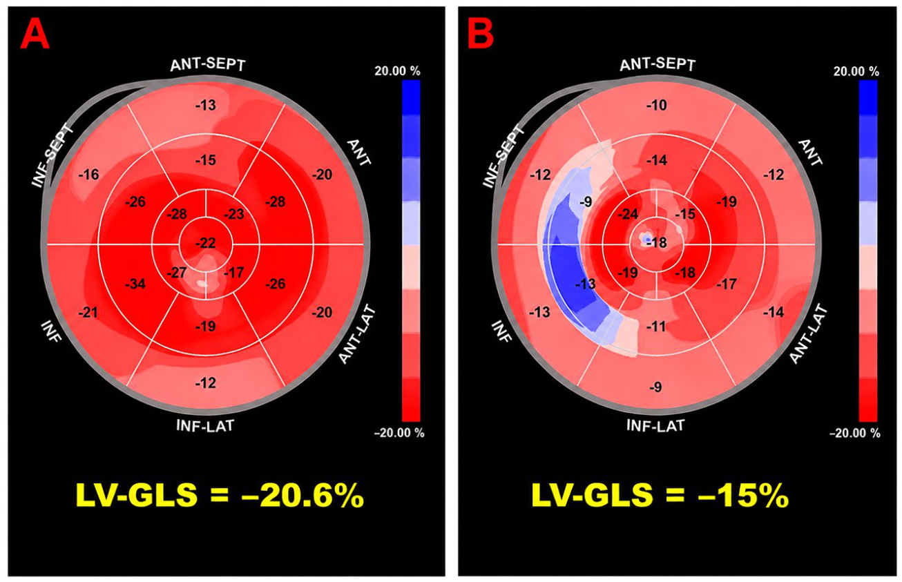
Figure 2.
Association between Charlson Comorbidity Index and cancer therapy-related cardiac dysfunction. CCI, Charlson Comorbidity Index; CTRCD, cancer therapy-related cardiac dysfunction.
Figure 2.
Association between Charlson Comorbidity Index and cancer therapy-related cardiac dysfunction. CCI, Charlson Comorbidity Index; CTRCD, cancer therapy-related cardiac dysfunction.
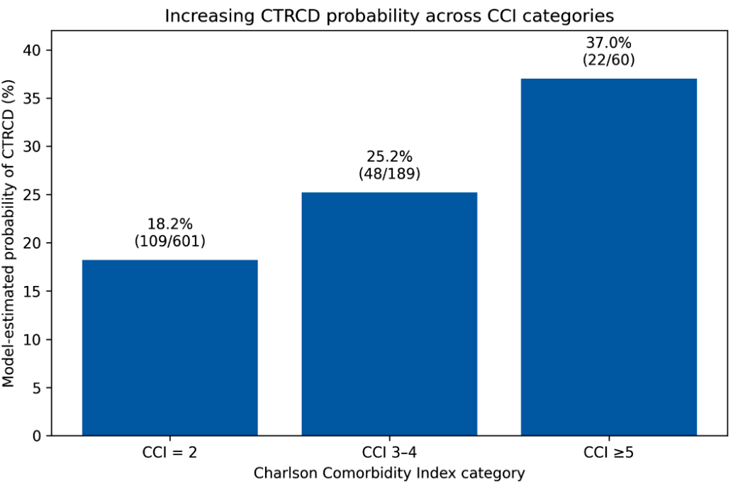
Figure 3.
Association between cumulative anti-HER2 exposure and cancer therapy-related cardiac dysfunction. CTRCD, cancer therapy-related cardiac dysfunction; HER2, human epidermal growth factor receptor 2.
Figure 3.
Association between cumulative anti-HER2 exposure and cancer therapy-related cardiac dysfunction. CTRCD, cancer therapy-related cardiac dysfunction; HER2, human epidermal growth factor receptor 2.
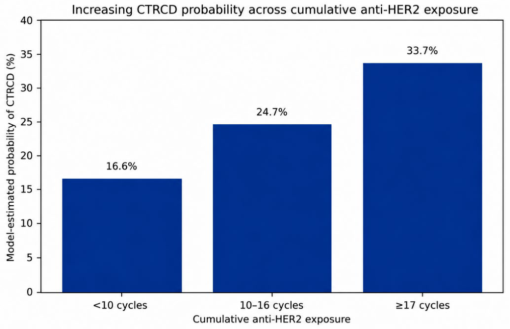
Figure 4.
Proposed framework for dynamic cardiovascular risk assessment and surveillance during HER2-targeted therapy. The framework integrates baseline global comorbidity burden, assessed by the Charlson Comorbidity Index (CCI), with longitudinal evaluation of cumulative anti-HER2 exposure. Increasing comorbidity burden and cumulative treatment exposure may identify patients requiring progressively intensified cardiovascular surveillance, ranging from standard monitoring to closer echocardiographic and biomarker assessment, early cardioprotective strategies, and multidisciplinary cardio-oncology management. This approach emphasizes dynamic reassessment of cardiovascular risk throughout the oncological pathway rather than reliance on baseline risk stratification alone. The proposed framework is hypothesis-generating and requires prospective validation before implementation in routine clinical practice. CCI, Charlson Comorbidity Index; cTn, cardiac troponin; HER2, human epidermal growth factor receptor 2; LV-GLS, left ventricular global longitudinal strain; LVEF, left ventricular ejection fraction; NT-proBNP, N-terminal pro-B-type natriuretic peptide.
Figure 4.
Proposed framework for dynamic cardiovascular risk assessment and surveillance during HER2-targeted therapy. The framework integrates baseline global comorbidity burden, assessed by the Charlson Comorbidity Index (CCI), with longitudinal evaluation of cumulative anti-HER2 exposure. Increasing comorbidity burden and cumulative treatment exposure may identify patients requiring progressively intensified cardiovascular surveillance, ranging from standard monitoring to closer echocardiographic and biomarker assessment, early cardioprotective strategies, and multidisciplinary cardio-oncology management. This approach emphasizes dynamic reassessment of cardiovascular risk throughout the oncological pathway rather than reliance on baseline risk stratification alone. The proposed framework is hypothesis-generating and requires prospective validation before implementation in routine clinical practice. CCI, Charlson Comorbidity Index; cTn, cardiac troponin; HER2, human epidermal growth factor receptor 2; LV-GLS, left ventricular global longitudinal strain; LVEF, left ventricular ejection fraction; NT-proBNP, N-terminal pro-B-type natriuretic peptide.
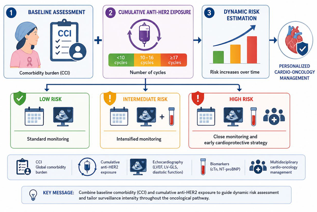

Table 1.
Demographic, anthropometric, and cardiovascular risk profile of the 10-year cohort.
| Variable | 10-year cohort (n = 850) |
| Age at start of cancer therapy, years | 54.1 ± 11.4 |
| Female sex, n (%) | 845 (99.4) |
| Male sex, n (%) | 5 (0.6) |
| Weight, kg | 63.3 ± 15.7 |
| Height, cm | 162.0 ± 6.8 |
| BMI, kg/m² | 23.6 ± 4.7 |
| Hypertension, n (%) | 268 (31.6) |
| Diabetes mellitus, n (%) | 60 (7.0) |
| Dyslipidemia, n (%) | 219 (25.7) |
| Current/former smoking, n (%) | 159 (18.7) |
Data are presented as mean ± SD or n (%). BMI, body mass index; SD, standard deviation.
Table 2.
Pre-existing cardiac disease, comorbidity profile, and Charlson Comorbidity Index distribution in the 10-year cohort.
Table 2.
Pre-existing cardiac disease, comorbidity profile, and Charlson Comorbidity Index distribution in the 10-year cohort.
| Variable | 10-year cohort (n = 850) |
| Pre-existing cardiac disease, n (%) | 273 (32.2) |
| Mitral valve prolapse/billowing ± mitral regurgitation, n (%) | 149 (17.5) |
| LV hypertrophy/hypertensive heart disease, n (%) | 75 (8.8) |
| More-than-mild aortic valve/aortic disease, n (%) | 50 (5.8) |
| Arrhythmia/conduction disorder, n (%) | 35 (4.1) |
| Ischemic/atherosclerotic cardiovascular disease, n (%) | 25 (2.9) |
| Most frequent comorbidities | |
| Osteopenia/osteoporosis, n (%) | 373 (43.9) |
| Thyroid disease, n (%) | 149 (17.5) |
| Hepatic steatosis/MASLD/NAFLD, n (%) | 139 (16.4) |
| Metastatic disease, n (%) | 119 (14.0) |
| Anxiety/depressive disorders, n (%) | 75 (8.8) |
| Venous thromboembolism/thrombosis, n (%) | 65 (7.6) |
| Renal disease/nephrolithiasis, n (%) | 65 (7.6) |
| BRCA1/2 mutation, n (%) | 60 (7.0) |
| Anemia, n (%) | 55 (6.4) |
| Chronic pulmonary disease, n (%) | 55 (6.4) |
| Charlson Comorbidity Index | |
| CCI, median [IQR] | 2 [2,3] |
| CCI = 2, n (%) | 601 (70.8) |
| CCI 3–4, n (%) | 189 (22.2) |
| CCI ≥5, n (%) | 60 (7.0) |
Data are presented as n (%) unless otherwise specified. The Charlson Comorbidity Index summarizes the overall burden of clinically relevant comorbidities and is reported both as median [IQR] and according to predefined categories. Individual comorbidities are not mutually exclusive; therefore, percentages do not sum to 100%. BRCA, breast cancer susceptibility gene; CCI, Charlson Comorbidity Index; IQR, interquartile range; LV, left ventricular; MASLD, metabolic dysfunction-associated steatotic liver disease; NAFLD, non-alcoholic fatty liver disease.
Table 3.
Baseline laboratory characteristics of the 10-year cohort.
| Variable | 10-year cohort (n = 850) |
| Hemoglobin, g/dL | 12.1 ± 1.5 |
| NLR | 2.40 [1.61–3.52] |
| RDW, % | 13.5 [12.9–15.4] |
| Leukocytes, ×10⁹/L | 5.81 ± 2.23 |
| Creatinine, mg/dL | 0.72 ± 0.20 |
| eGFR, mL/min/1.73 m² | 92.8 ± 17.8 |
| AST, U/L | 23 [19–34.5] |
| ALT, U/L | 22 [17–31] |
| Sodium, mEq/L | 141.0 ± 3.3 |
| Potassium, mEq/L | 4.18 ± 0.38 |
| Total bilirubin, mg/dL | 0.60 [0.47–0.80] |
| Calcium, mmol/L | 2.38 [2.27–2.43] |
| CRP, mg/dL* | 0.35 [0.10–3.50] |
| Troponin, ng/mL* | 0.015 [0.003–0.120] |
| NT-proBNP, pg/mL* | 110 [35–1,200] |
Data are presented as mean ± SD or median [IQR], as appropriate. *CRP, troponin, and NT-proBNP values were available only in a subset of patients. ALT, alanine aminotransferase; AST, aspartate aminotransferase; CRP, C-reactive protein; eGFR, estimated glomerular filtration rate; IQR, interquartile range; NLR, neutrophil-to-lymphocyte ratio; NT-proBNP, N-terminal pro-B-type natriuretic peptide; RDW, red cell distribution width; SD, standard deviation.
Table 4.
Tumor characteristics, histological features, and biomarker profile of the 10-year cohort.
| Tumor characteristic | 10-year cohort (n = 850) |
| Tumor laterality | |
| Left breast, n (%) | 413 (48.6) |
| Right breast, n (%) | 368 (43.3) |
| Bilateral breast cancer, n (%) | 45 (5.3) |
| Occult breast primary, n (%) | 15 (1.8) |
| Breast laterality unavailable/unspecified, n (%) | 9 (1.1) |
| Histological type | |
| Invasive ductal/NST carcinoma, n (%) | 716 (84.2) |
| Invasive lobular carcinoma, n (%) | 65 (7.6) |
| Apocrine differentiation, n (%) | 25 (2.9) |
| Metaplastic carcinoma, n (%) | 15 (1.8) |
| Other special breast histology, n (%) | 15 (1.8) |
| Mixed ductal/lobular carcinoma, n (%) | 5 (0.6) |
| Other/unspecified breast histology, n (%) | 9 (1.1) |
| Tumor grading | |
| G1, n (%) | 5 (0.6) |
| G2, n (%) | 229 (26.9) |
| G3/high grade, n (%) | 547 (64.3) |
| Grade unavailable/not classifiable, n (%) | 70 (8.2) |
| Hormone-receptor profile | |
| ER, % | 70 [0–95] |
| ER-positive (≥1%), n (%) | 566 (66.6) |
| PgR, % | 10 [0–70] |
| PgR-positive (≥1%), n (%) | 477 (56.1) |
| HER2 status | |
| HER2-positive/amplified, n (%) | 447 (52.6) |
| HER2-low (IHC 1+ or 2+/ISH−), n (%) | 199 (23.4) |
| HER2-zero/negative, n (%) | 179 (21.1) |
| HER2 unavailable/indeterminate, n (%) | 25 (2.9) |
| Ki-67, % | 32 [22–50] |
Data are presented as n (%) or median [IQR], as appropriate. Percentages may not sum to 100% because of rounding or unavailable/unclassifiable data. ER, estrogen receptor; HER2, human epidermal growth factor receptor 2; IHC, immunohistochemistry; IQR, interquartile range; ISH, in situ hybridization; NST, no special type; PgR, progesterone receptor.
Table 5.
Anticancer treatment characteristics and therapeutic exposures in the 10-year cohort.
| Treatment variable | 10-year cohort (n = 850) |
| Neoadjuvant systemic therapy, n (%) | 249 (29.2) |
| Anthracycline exposure, n (%) | 661 (77.8) |
| AC/EC cycles among exposed, median [IQR] | 4 [4,4] |
| Taxane exposure, n (%) | 775 (91.2) |
| Any anti-HER2 therapy, n (%) | 850 (100) |
| Trastuzumab exposure, n (%) | 700 (82.4) |
| Pertuzumab exposure, n (%) | 174 (20.5) |
| T-DM1 (trastuzumab emtansine) exposure, n (%) | 75 (8.8) |
| T-DXd (trastuzumab deruxtecan) exposure, n (%) | 35 (4.1) |
| Neoadjuvant anti-HER2 therapy, n (%) | 154 (18.1) |
| Neoadjuvant anti-HER2 cycles, median [IQR] | 4 [4–6] |
| Adjuvant anti-HER2 therapy, n (%) | 520 (61.2) |
| Adjuvant anti-HER2 cycles, median [IQR] | 17 [14–17] |
| Metastatic/maintenance anti-HER2 therapy, n (%) | 288 (33.9) |
| Metastatic/maintenance anti-HER2 cycles, median [IQR] | 18 [12–30] |
| Radiotherapy, n (%) | 646 (76.0) |
| Endocrine therapy, n (%) | 502 (59.1) |
| Radiotherapy details | |
| Breast/chest-wall ± regional nodal RT, n (%) | 570 (67.1) |
| Left-sided breast/chest-wall RT, n (%) | 278 (32.7) |
| Regional nodal irradiation, n (%) | 264 (31.1) |
| Metastatic/palliative or stereotactic RT, n (%) | 100 (11.8) |
| Typical locoregional RT dose, Gy | 40–50 |
| Hypofractionated RT (15 fractions), n (%) | 204 (24.0) |
| Boost to tumor bed, n (%) | 139 (16.4) |
| Endocrine therapy details | |
| Aromatase inhibitor-based therapy, n (%) | 438 (51.5) |
| Tamoxifen-based therapy, n (%) | 154 (18.1) |
| LHRH analogue/ovarian suppression, n (%) | 109 (12.8) |
| Fulvestrant exposure, n (%) | 90 (10.6) |
Data are presented as n (%), median [IQR], or range, as appropriate. Treatment categories are not mutually exclusive because patients could receive multiple sequential or concomitant anticancer therapies. Individual anti-HER2 agent categories and treatment settings are not mutually exclusive because patients could receive multiple HER2-targeted agents sequentially or in combination across different phases of the oncological treatment course. Anti-HER2 therapy could be administered in the neoadjuvant, adjuvant, and/or metastatic/maintenance setting. Radiotherapy and endocrine therapy subcategories may overlap. AC, doxorubicin plus cyclophosphamide; CDK4/6, cyclin-dependent kinase 4 and 6; EC, epirubicin plus cyclophosphamide; Gy, gray; HER2, human epidermal growth factor receptor 2; IQR, interquartile range; LHRH, luteinizing hormone-releasing hormone; RT, radiotherapy; T-DM1, trastuzumab emtansine; T-DXd, trastuzumab deruxtecan.
Table 6.
Longitudinal echocardiographic findings, myocardial deformation, and cancer therapy-related cardiac dysfunction in the 10-year cohort.
Table 6.
Longitudinal echocardiographic findings, myocardial deformation, and cancer therapy-related cardiac dysfunction in the 10-year cohort.
| Echocardiographic variable | 10-year cohort (n = 850) |
| Cardiac morphology | |
| Interventricular septal thickness at baseline, mm | 9.6 ± 2.4 |
| LV end-diastolic diameter at baseline, mm | 44.3 ± 5.2 |
| Left atrial volume at baseline, mL | 47 [40–59] |
| LV diastolic function | |
| E/A <1 at baseline, n (%) | 319 (37.5) |
| E/A >1 at baseline, n (%) | 124 (14.6) |
| E/A <1 at final follow-up, n (%) | 423 (49.8) |
| E/A >1 at final follow-up, n (%) | 204 (24.0) |
| E/e′ at baseline | 7.8 [6.7–9.2] |
| E/e′ at final follow-up | 8.0 [7.0–9.5] |
| LV systolic function | |
| LVEF at baseline, % | 65.2 ± 4.7 |
| LVEF nadir, % | 59.2 ± 7.1 |
| LVEF at final follow-up, % | 64.2 ± 5.3 |
| Absolute LVEF decline, percentage points | 5 [0–10] |
| LVEF decline ≥10 percentage points, n (%) | 145 (17.1) |
| LVEF <50% at nadir, n (%) | 50 (5.9) |
| Myocardial deformation | |
| Patients with technically adequate images for longitudinal LV-GLS assessment, n (%) | 750 (88.2) |
| LV-GLS not reliably assessable because of inadequate acoustic window/image quality, n (%) | 100 (11.8) |
| LV-GLS at baseline, % | 19.9 ± 0.8 |
| LV-GLS nadir, % | 18.3 ± 1.8 |
| LV-GLS at final follow-up, % | 19.4 ± 1.2 |
| Absolute change in LV-GLS from baseline to nadir, percentage points | −1.5 [−3.0–0.0] |
| Relative LV-GLS decline >15% from baseline, n (%) | 139 (18.5) |
| Pulmonary artery systolic pressure | |
| PASP at baseline, mmHg | 29 [25–33] |
| PASP at final follow-up, mmHg | 28 [25–32] |
| Cancer therapy-related cardiac dysfunction | |
| CTRCD, n (%) | 179 (21.1) |
| Mild CTRCD, n (%) | 119 (14.0) |
| Moderate CTRCD, n (%) | 40 (4.7) |
| Severe CTRCD, n (%) | 20 (2.4) |
| Clinical heart failure, n (%) | 25 (2.9) |
| Myocardial pattern/territory in CTRCD | |
| Inferior/inferoseptal, predominantly basal or mid-basal involvement, n (%) | 70 (8.2) |
| Global/diffuse LV involvement, n (%) | 25 (2.9) |
| Mixed global + inferior/inferoseptal involvement, n (%) | 15 (1.8) |
| Other/unspecified regional involvement, n (%) | 10 (1.2) |
| CTRCD with myocardial territory not documented, n (%) | 59 (6.9) |
Data are presented as mean ± SD, median [IQR], or n (%), as appropriate. LV-GLS measurements and derived changes refer to patients with technically adequate images available for longitudinal speckle-tracking analysis (n = 750). For ease of interpretation, LV-GLS values are reported as positive absolute values, with lower values indicating worse myocardial deformation. Percentages for CTRCD severity are calculated on the overall cohort. Myocardial patterns describe the predominant distribution of LV involvement among patients developing CTRCD. CTRCD, cancer therapy-related cardiac dysfunction; E/A, ratio of early (E) to late (A) transmitral flow velocity; E/e′, ratio of early transmitral flow velocity to early diastolic mitral annular velocity; IQR, interquartile range; LV, left ventricular; LV-GLS, left ventricular global longitudinal strain; LVEF, left ventricular ejection fraction; PASP, pulmonary artery systolic pressure; SD, standard deviation.
Table 7.
Management of cancer therapy-related cardiac dysfunction and subsequent recovery of left ventricular systolic function in the 10-year cohort.
Table 7.
Management of cancer therapy-related cardiac dysfunction and subsequent recovery of left ventricular systolic function in the 10-year cohort.
| Variable | 10-year cohort (n = 850) |
| CTRCD management | |
| Temporary/permanent interruption of oncological therapy for cardiotoxicity, n (%) | 50 (5.9) |
| Cardioprotective/HF therapy initiated for CTRCD, n (%) | 139 (16.4) |
| ACE-i/ARB/ARNI use for CTRCD, n (%) | 114 (13.4) |
| Beta-blocker use for CTRCD, n (%) | 129 (15.2) |
| Mineralocorticoid receptor antagonist, n (%) | 30 (3.5) |
| SGLT2 inhibitor, n (%) | 5 (0.6) |
| Diuretic therapy for congestion/HF, n (%) | 40 (4.7) |
| LVEF recovery after CTRCD | |
| Complete LVEF recovery, n (%) | 124 (14.6) |
| Partial LVEF recovery, n (%) | 30 (3.5) |
| No documented LVEF recovery, n (%) | 15 (1.8) |
| Recovery status unavailable, n (%) | 10 (1.2) |
| Estimated time to any LVEF recovery, months* | 4 [2–8] |
| Estimated time to complete LVEF recovery, months* | 4 [2–7] |
| Estimated time to partial LVEF recovery, months* | 6 [3–10] |
Data are presented as n (%) or median [IQR], as appropriate. Treatment categories are not mutually exclusive because more than one cardioprotective/HF therapy could be initiated in the same patient. LVEF recovery categories refer to patients who developed CTRCD. *Time-to-recovery estimates were calculated among patients with available temporal information documenting subsequent LVEF recovery. ACE-i, angiotensin-converting enzyme inhibitor; ARB, angiotensin receptor blocker; ARNI, angiotensin receptor–neprilysin inhibitor; CTRCD, cancer therapy-related cardiac dysfunction; HF, heart failure; IQR, interquartile range; LVEF, left ventricular ejection fraction; SGLT2, sodium–glucose cotransporter 2.
Table 8.
Oncological outcomes at the latest available follow-up in the 10-year cohort.
| Oncological outcome | 10-year cohort (n = 850) |
| No evidence of disease / disease-free follow-up, n (%) | 437 (51.4) |
| Metastatic disease with complete/partial response or stable disease, n (%) | 164 (19.3) |
| Metastatic disease with documented progression / palliative course, n (%) | 124 (14.6) |
| Ongoing neoadjuvant/adjuvant curative-intent treatment, n (%) | 75 (8.8) |
| Other/insufficiently classifiable oncological outcome, n (%) | 50 (5.9) |
| Pathological complete response after neoadjuvant therapy, n (%)* | 70 (8.2) |
Data are presented as n (%). Oncological outcome categories refer to the latest available clinical assessment and are mutually exclusive. *Pathological complete response after neoadjuvant therapy represents an additional treatment-response variable and is not mutually exclusive with the main oncological outcome categories; therefore, it is not included in their cumulative percentage. n, number of patients.
Table 9.
Univariable logistic regression analysis of clinical, oncological, treatment-related, and baseline echocardiographic factors associated with CTRCD.
Table 9.
Univariable logistic regression analysis of clinical, oncological, treatment-related, and baseline echocardiographic factors associated with CTRCD.
| Variable | OR | 95% CI | p-value |
| Clinical variables | |||
| Age, per 10 years | 1.12 | 0.88–1.43 | 0.360 |
| BMI, per 5 kg/m² | 1.58 | 0.93–2.70 | 0.090 |
| Hypertension | 1.28 | 0.86–1.91 | 0.225 |
| Diabetes mellitus | 1.34 | 0.68–2.64 | 0.398 |
| Dyslipidemia | 1.18 | 0.79–1.76 | 0.418 |
| Current/former smoking | 1.32 | 0.54–3.25 | 0.544 |
| Charlson Comorbidity Index, per 1 point | 1.32 | 1.10–1.58 | 0.003 |
| Oncological/treatment variables | |||
| Tumor grade, per 1 grade increase | 1.16 | 0.52–2.60 | 0.717 |
| Anthracycline pretreatment (yes vs no) | 1.13 | 0.45–2.85 | 0.790 |
| AC cycles, per cycle | 1.00 | 0.80–1.24 | 0.977 |
| Total anti-HER2 duration, per 5 cycles | 1.37 | 1.09–1.71 | 0.006 |
| Baseline echocardiographic variables | |||
| Baseline LVEF, per 1% decrease | 0.97 | 0.89–1.05 | 0.449 |
| Baseline LV-GLS magnitude, per 1% decrease | 0.67 | 0.39–1.15 | 0.145 |
Odds ratios and corresponding 95% CIs were derived from univariable binary logistic regression analyses with CTRCD as the dependent variable. Continuous variables were modeled according to the increments specified in the table. Statistically significant p-values are shown in bold. AC, anthracycline-containing chemotherapy; BMI, body mass index; CI, confidence interval; CTRCD, cancer therapy-related cardiac dysfunction; HER2, human epidermal growth factor receptor 2; LV-GLS, left ventricular global longitudinal strain; LVEF, left ventricular ejection fraction; OR, odds ratio.
Table 10.
Multivariable logistic regression analysis of factors independently associated with cancer therapy-related cardiac dysfunction.
Table 10.
Multivariable logistic regression analysis of factors independently associated with cancer therapy-related cardiac dysfunction.
| Variable | Adjusted OR | 95% CI | p-value |
| Charlson Comorbidity Index, per 1 point |
1.35 | 1.12–1.63 | 0.002 |
| Total anti-HER2 duration, per 5 cycles |
1.41 | 1.10–1.80 | 0.006 |
Adjusted odds ratios and corresponding 95% CIs were derived from a parsimonious multivariable binary logistic regression model with CTRCD as the dependent variable. CCI was modeled per one-point increase, whereas cumulative anti-HER2 exposure was modeled per five additional treatment cycles. Statistically significant p-values are reported in bold. CCI, Charlson Comorbidity Index; CI, confidence interval; CTRCD, cancer therapy-related cardiac dysfunction; HER2, human epidermal growth factor receptor 2; OR, odds ratio.
Disclaimer/Publisher’s Note: The statements, opinions and data contained in all publications are solely those of the individual author(s) and contributor(s) and not of MDPI and/or the editor(s). MDPI and/or the editor(s) disclaim responsibility for any injury to people or property resulting from any ideas, methods, instructions or products referred to in the content. |
© 2026 by the authors. Licensee MDPI, Basel, Switzerland. This article is an open access article distributed under the terms and conditions of the Creative Commons Attribution (CC BY) license (http://creativecommons.org/licenses/by/4.0/).
Copyright: This open access article is published under a Creative Commons CC BY 4.0 license, which permit the free download, distribution, and reuse, provided that the author and preprint are cited in any reuse.