

Submitted:
01 September 2026
Posted:
02 September 2026
You are already at the latest version
Abstract
Background: Progressive supranuclear palsy (PSP) is a primary four-repeat tauopathy characterized by marked clinical heterogeneity, resulting in substantial diagnostic challenges. Over the past three decades, understanding of PSP has evolved from a classical Richardson syndrome-centered concept to a broader clinicopathological spectrum encompassing multiple motor, cognitive, behavioral, speech-language, and cortical phenotypes. Methods: A narrative review of the literature was performed to examine the historical development of PSP diagnostic frameworks, including the Hauw neuropathologic criteria, NINDS-SPSP criteria, variant PSP (vPSP) framework, Movement Disorder Society PSP (MDS-PSP) criteria, and Maximum Allocation Extinction (MAX) rules. Relevant clinicopathological studies, consensus statements, validation studies, and biomarker investigations were reviewed. Results: The Hauw criteria established the first standardized neuropathological definition of PSP, whereas the NINDS-SPSP criteria provided a highly specific clinical diagnostic framework for classical PSP-Richardson syndrome. Subsequent recognition of PSP-parkinsonism, progressive gait freezing, frontal, speech-language, corticobasal, and cerebellar variants revealed the limitations of phenotype-restricted diagnostic models and led to the development of the vPSP framework. These observations informed the 2017 MDS-PSP criteria, which incorporated multiple phenotypes, four core functional domains, supportive features, and graded levels of diagnostic certainty. The MAX rules subsequently standardized phenotype allocation among patients fulfilling multiple diagnostic categories. Conclusions: Contemporary PSP diagnostic frameworks have substantially improved recognition of atypical and early-stage disease; however, diagnosis remains primarily clinical and lacks definitive ante-mortem pathological confirmation. Emerging MRI, fluid, and tau PET biomarkers may facilitate biomarker-supported and pathology-informed diagnostic paradigms, enabling earlier diagnosis, improved patient stratification, and more effective therapeutic trials.
Keywords:
progressive supranuclear palsy
; PSP
; diagnostic criteria
; movement disorder society criteria
; MDS-PSP
; Richardson syndrome
; variant PSP
; phenotypic heterogeneity
; tauopathy
; biomarkers
1. Introduction
Progressive supranuclear palsy (PSP) is a rare adult-onset neurodegenerative disorder and one of the most common atypical parkinsonian syndromes. Originally described by Steele et al., PSP is characterized by varying combinations of postural instability, ocular motor dysfunction, akinetic-rigid parkinsonism, cognitive impairment, and bulbar symptoms [1]. Neuropathologically, PSP is classified as a primary 4R tauopathy, characterized by the accumulation of hyperphosphorylated tau protein in neurons and glial cells, leading to NFTs, coiled bodies, neuropil threads, and the characteristic tufted astrocytes described in pathologic diagnostic criteria [2]. The disease typically presents after 60 years of age and is associated with substantial morbidity, disability, and reduced quality of life due to progressive gait impairment, recurrent falls, dysphagia, and cognitive decline [3].
Accurate diagnosis of PSP remains challenging because of the frequent dissociation between clinical presentation and underlying pathology. The 1996 NINDS-SPSP criteria provided the first widely accepted clinical diagnostic framework and demonstrated excellent specificity for the classical Richardson syndrome phenotype [4]. However, subsequent clinicopathological studies revealed that many patients with pathologically confirmed PSP initially present with alternative phenotypes, including PSP-P, PSP-PGF, frontal behavioral syndromes, speech and language disorders, and corticobasal syndrome [5]. These observations highlighted the substantial phenotypic heterogeneity of PSP and explained the limited sensitivity of earlier diagnostic criteria, particularly during the initial stages of disease.
Recognition of this broader clinical spectrum led to the development of the vPSP framework by Respondek et al., which demonstrated that Richardson syndrome represents only one manifestation within a diverse phenotypic continuum [6]. These findings subsequently informed the 2017 MDS criteria, which incorporated multiple PSP phenotypes, four core functional domains, and graded levels of diagnostic certainty to improve early diagnostic sensitivity while maintaining acceptable specificity [7]. More recently, the MAX rules were introduced to standardize phenotype assignment in patients fulfilling multiple MDS-PSP categories [8]. This review summarizes the historical evolution of PSP diagnostic frameworks, examines their strengths and limitations, and discusses future directions toward biomarker-supported and pathology-informed diagnosis of PSP.
2. Methods
A narrative literature review was conducted to summarize the historical evolution of diagnostic frameworks for PSP. A comprehensive search of PubMed/Medline, Scopus, and Google Scholar was performed from database inception through September 1, 2026. Search terms included combinations of “progressive supranuclear palsy,” “PSP,” “diagnostic criteria,” “classification,” “neuropathologic criteria,” “NINDS-SPSP,” “Movement Disorder Society criteria,” “MDS-PSP,” “variant PSP,” “PSP-parkinsonism,” “PSP-PGF,” “PSP-CBS,” “PSP-SL,” “phenotype,” and “diagnosis.” Original research articles, consensus statements, clinicopathological studies, diagnostic framework publications, validation studies, and relevant review articles published in English were considered. Particular emphasis was placed on seminal publications that introduced or substantially modified PSP classification and diagnostic approaches. Additional references were identified through manual review of bibliographies from included articles. Publications were screened for relevance to the development, validation, clinical application, strengths, and limitations of PSP diagnostic frameworks. The selected literature was synthesized narratively, with emphasis on the chronological evolution of diagnostic concepts, emerging recognition of phenotypic heterogeneity, and the transition from syndrome-based toward pathology-informed and biomarker-supported diagnostic approaches.
3. Results
Review of the available literature identified five major milestones in the evolution of PSP diagnostic frameworks (Table 1) [4,6,7,8,9]. Over the past three decades, diagnostic concepts have progressively evolved from pathology-centered definitions toward increasingly inclusive clinical classification systems that recognize the marked phenotypic heterogeneity of PSP. Early frameworks emphasized neuropathologic confirmation and the identification of classical clinical manifestations, whereas later approaches incorporated evidence from clinicopathological studies demonstrating that PSP may present with diverse motor, cognitive, behavioral, speech-language, and gait phenotypes. This shift culminated in the development of multidimensional diagnostic models that improved sensitivity for atypical and early-stage presentations while preserving diagnostic specificity. More recent refinements have focused on optimizing phenotype allocation and standardizing classification among patients who fulfill multiple diagnostic categories, reflecting the growing appreciation of PSP as a clinicopathological spectrum rather than a single, uniform clinical syndrome.
4. Historical Development of PSP Diagnostic Frameworks
4.1. Early Clinicopathological Description of PSP
PSP was first described by Steele, Richardson, and Olszewski in 1964 as a distinct neurodegenerative syndrome [1]. Based on a clinicopathological series of nine patients, the authors recognized a unique constellation of neurological features that differed from PD and other movement disorders, establishing what later became known as “Steele-Richardson-Olszewski syndrome [11].” Their seminal description, “Progressive Supranuclear Palsy: A Heterogeneous Degeneration Involving the Brain Stem, Basal Ganglia and Cerebellum With Vertical Gaze and Pseudobulbar Palsy, Nuchal Dystonia and Dementia,” emphasized supranuclear impairment of vertical gaze, particularly downgaze, a staring facial expression, pseudobulbar dysfunction, dysarthria, retrocollis with nuchal dystonia, and progressive gait impairment, while cognitive decline generally remained mild to moderate. Neuropathological examination demonstrated neuronal loss, gliosis, NFTs, granulovacuolar degeneration, and demyelination involving the BG, brainstem, and cerebellar dentate nucleus, with prominent involvement of the subthalamic nucleus, SN, superior colliculi, periaqueductal gray matter, and pontine tegmentum [1].
4.2. Pre-NINDS-SPSP Clinical Criteria for PSP
Prior to the introduction of the NINDS-SPSP criteria in 1996, several clinical frameworks were proposed to facilitate the diagnosis of PSP [10], largely based on expert clinical observations rather than systematic clinicopathological validation (Table 2)[12,13,14]. Early criteria developed by Lees emphasized a progressive nonfamilial disorder characterized by vertical SGP, particularly affecting downgaze, in combination with postural instability, pseudobulbar palsy, parkinsonism, frontal lobe dysfunction, or axial rigidity [12]. Subsequently, Golbe et al. proposed a more structured approach requiring progressive parkinsonism with SGP and additional features such as dysarthria, dysphagia, retrocollis, early falls, poor levodopa responsiveness, and minimal tremor, while incorporating exclusion criteria to distinguish PSP from other atypical parkinsonian syndromes [13]. Blin et al. later introduced separate categories of probable and possible PSP, with probable PSP requiring a highly restrictive constellation of parkinsonism, postural instability, supranuclear ophthalmoplegia, pseudobulbar symptoms, frontal dysfunction, and poor levodopa response, whereas possible PSP allowed diagnosis when most, but not all, characteristic features were present [14]. Although these frameworks established the clinical importance of vertical gaze abnormalities and early postural instability as hallmark manifestations of PSP, their diagnostic performance varied considerably, with stricter criteria providing high specificity but limited sensitivity and less restrictive criteria improving case detection at the expense of diagnostic accuracy. These limitations, together with the lack of consistent neuropathological validation, highlighted the need for a standardized evidence-based diagnostic framework and ultimately led to the development of the NINDS-SPSP criteria.
4.3. Neuropathologic Definition: Hauw Criteria (1994)
A major milestone in the evolution of PSP classification was the publication of the preliminary NINDS neuropathologic criteria by Hauw et al. in 1994, which established the first standardized pathological framework for diagnosing PSP at autopsy [9]. These criteria classified cases as typical, atypical, or combined PSP and made the semiquantitative assessment of NFTs and neuropil threads the cornerstone of diagnosis (Table 3)(Figure 1)[9]. One of the most important innovations was the recognition that tau-positive astrocytic lesions, later widely known as tufted astrocytes [15], supported the diagnosis and represented a distinctive pathological hallmark of PSP. The criteria also emphasized the characteristic distribution of tau pathology, involving the pallidum, subthalamic nucleus, striatum, nucleus basalis of Meynert, superior colliculi, periaqueductal gray matter, red nucleus, pontine tegmentum, raphe nuclei, inferior olives, and dentate nucleus, with relative sparing of the occipital cortex and cerebellar cortex. Furthermore, the framework introduced exclusion criteria to distinguish PSP from competing neurodegenerative disorders, including AD, Lewy body diseases, MSA, Pick disease, CBD, and prion disorders. The major strength of the Hauw criteria was the establishment of a reproducible neuropathological gold standard that improved diagnostic consistency among brain banks and clinicopathological studies, providing a foundation for subsequent validation of clinical criteria. However, the criteria were inherently limited by their dependence on postmortem examination, precluding their use for ante-mortem diagnosis, and by their focus on advanced pathological stages, offering little guidance for early disease detection or the recognition of the full clinical heterogeneity that would later be encompassed by variant PSP and the MDS-PSP framework.
4.4. NINDS-SPSP Criteria (1996)
The NINDS-SPSP criteria, developed by Litvan et al. in 1996, represented the first evidence-based and clinicopathologically validated clinical diagnostic framework for PSP and rapidly became the reference standard for research and clinical practice [4,10]. Using a large series of neuropathologically confirmed PSP and non-PSP neurodegenerative disorders, the authors identified vertical SGP, particularly involving downward gaze, and postural instability with unexplained falls as the clinical features most strongly associated with underlying PSP pathology. The criteria established two levels of diagnostic certainty: possible PSP and probable PSP (Figure 2)(Table 4) [10]. Probable PSP required the presence of both vertical SGP and prominent postural instability with falls occurring within the first year of symptom onset, whereas possible PSP could be diagnosed in patients with either SGP or slowed vertical saccades accompanied by early postural instability and falls. Additional supportive features included symmetrical akinetic-rigid parkinsonism, axial rigidity exceeding limb rigidity, dysarthria, dysphagia, frontal lobe dysfunction, and poor responsiveness to levodopa, while exclusion criteria were designed to eliminate competing diagnoses. Validation against neuropathologic diagnoses demonstrated excellent specificity, ranging from approximately 95% to 100% for probable PSP and from 80% to 93% for possible PSP, with positive predictive values exceeding 75% in most analyses [4,11]. However, sensitivity was substantially lower, particularly during early disease stages, with sensitivity estimates of approximately 24% to 56% at initial clinical evaluation and 39% to 83% later in the disease course [4,10,11]. These limitations reflected the criteria's strong emphasis on the classic Richardson syndrome phenotype, resulting in underrecognition of patients presenting with other variant PSP phenotypes that were subsequently recognized through clinicopathologic studies. Despite these shortcomings, the NINDS-SPSP criteria provided the first robust clinicopathological framework for ante-mortem PSP diagnosis.
5. Recognition of Clinical Heterogeneity
5.1. PSP-P
A major advance in understanding the clinical heterogeneity of PSP came from the clinicopathological study by Williams et al. in 2005, which identified PSP-P as a distinct phenotype accounting for approximately one-third of pathologically confirmed PSP cases [16]. PSP-P typically presents with asymmetric onset, bradykinesia, tremor, non-axial dystonia, and an initial moderate response to levodopa, often leading to an early clinical diagnosis of PD [17]. Patients with PSP-P generally have a longer disease duration and later age at death than those with PSP-RS, while the hallmark ocular motor abnormalities and postural instability tend to emerge later in the disease course [18]. The recognition of PSP-P demonstrated that many patients with pathologically confirmed PSP do not initially exhibit the classic PSP-RS phenotype emphasized by the NINDS-SPSP criteria, helping explain the limited sensitivity of existing diagnostic frameworks. Clinically, the identification of PSP-P broadened the recognized spectrum of PSP, highlighted the need for careful longitudinal assessment of patients with atypical parkinsonism, and provided early evidence that PSP comprises multiple phenotypic presentations rather than a single uniform clinical syndrome.
5.2. PAGF/PSP-PGF
In 2007, Williams et al. further expanded the clinical spectrum of PSP by characterizing PAGF, later incorporated into the PSP-PGF phenotype [19]. PAGF is characterized by the gradual onset of gait initiation failure and prominent FOG, often accompanied by micrographia, hypophonia, and speech freezing, while lacking the classic features of PSP during the early disease stage [20]. Diagnostic criteria included early FOG or speech, absence of limb rigidity and resting tremor, lack of a sustained response to levodopa, and the absence of dementia, supranuclear ophthalmoplegia, or significant cerebrovascular disease during the first 5 years of illness. In a clinicopathological series, six of seven patients fulfilling these criteria had pathologically confirmed PSP, yielding a positive predictive value of 86% for PSP-tau pathology [19]. Compared with PSP-RS, PAGF exhibited a substantially longer disease duration, delayed emergence of falls, gaze palsy, and cognitive impairment, and generally milder tau burden with relative sparing of the pontine base and cerebellar dentate nucleus [21].
5.3. Frontal and Cognitive Variants
Several clinicopathological studies described patients with pathologically confirmed PSP who initially presented with a syndrome resembling bvFTD, characterized by apathy, social withdrawal, loss of initiative, personality change, impaired judgment, perseverative behavior, and executive dysfunction in the absence of early SGP or postural instability [22,23]. More detailed neuropathological investigations subsequently demonstrated that these cognitive and behavioral manifestations were associated with greater involvement of frontal and allocortical regions, including the prefrontal cortex, compared with classical PSP-RS [24,25,26]. Executive dysfunction emerged as a particularly consistent feature, involving impairments in planning, cognitive flexibility, verbal fluency, attention, working memory, and problem-solving, reflecting disruption of frontal-subcortical circuits by PSP-related tau pathology [25,27].
5.4. Speech and Language Variants
Clinicopathological studies demonstrated that some patients with pathologically confirmed PSP initially present with PNFA or AOS, often several years before developing the characteristic features of SGP, postural instability, and falls [28]. Patients with PNFA typically exhibit slow, effortful, and grammatically simplified speech, impaired sentence construction, phonemic errors, and reduced verbal fluency while relatively preserving language comprehension in the early stages of disease [28]. In contrast, AOS is characterized by impaired motor planning of speech, resulting in distorted articulation, segmented speech, abnormal prosody, and difficulty initiating verbal output without a primary language deficit [28]. Subsequent clinicopathological investigations identified PSP pathology as one of the most common underlying substrates of progressive AOS and a recognized cause of PNFA, reflecting involvement of dominant frontal speech and language networks in addition to the characteristic subcortical tau pathology [28,29].
5.5. PSP-CBS
Patients with PSP-CBS typically present with asymmetric parkinsonism, limb rigidity, dystonia, ideomotor apraxia, cortical sensory deficits, myoclonus, or alien limb phenomena, often leading to an initial diagnosis of CBS rather than PSP [30]. Clinicopathological studies subsequently demonstrated that a substantial proportion of patients meeting clinical criteria for CBS harbor PSP pathology at autopsy, highlighting the poor clinicopathological specificity of the syndrome [31,32]. Compared with classical PSP-RS syndrome, PSP-CBS patients often lack early falls and SGP, delaying recognition of the underlying tauopathy and reducing the sensitivity of earlier diagnostic frameworks such as the NINDS-SPSP criteria. Pathologically, these cases exhibit characteristic PSP-type tau pathology despite a CBS presentation, reinforcing the concept that clinical syndromes and underlying neuropathology do not always correspond directly.
5.6. Cerebellar and Other Rare Variants
Patients with PSP-C typically present with gait and limb ataxia, dysarthria, impaired coordination, and postural instability as predominant early manifestations, often resulting in an initial diagnosis of MSA-C, sporadic adult-onset ataxia, or other cerebellar syndromes rather than PSP [33]. In many cases, the characteristic features of PSP, including SGP, axial rigidity, frontal dysfunction, and recurrent falls, emerge only later in the disease course, creating substantial diagnostic uncertainty during the early stages. Clinicopathological studies have demonstrated that these patients harbor typical PSP-Type 4R tau pathology despite their atypical cerebellar presentation, supporting the concept that clinical phenotype is determined by the regional distribution and severity of pathological involvement rather than by the underlying proteinopathy alone [33]. The considerable overlap between PSP-C and MSA-C is particularly important because both disorders may present with progressive gait ataxia and balance impairment; however, the relative absence of prominent autonomic failure and the later development of characteristic PSP ocular motor abnormalities may provide diagnostic clues.
6. vPSP Framework
6.1. Rationale for a New Classification System
By the early 2010s, accumulating clinicopathological evidence demonstrated that a substantial proportion of patients with pathologically confirmed PSP did not present with the PSP-RS phenotype emphasized by the NINDS-SPSP criteria. Studies identifying PSP-P, PSP-PGF, frontal behavioral syndromes, speech-language disorders, CBS, and cerebellar presentations revealed marked clinical heterogeneity and highlighted the limited sensitivity of existing diagnostic frameworks, particularly during the early stages of disease [34]. In addition, many patients failed to satisfy established phenotype definitions because they exhibited overlapping clinical features or evolved from one presentation to another over time. These observations created the need for a more inclusive classification system capable of capturing the broader clinical spectrum of PSP while maintaining its clinicopathological basis.
6.2. Respondek et al. Multicenter Clinicopathological Study
In 2014, Respondek et al. conducted the first large multicenter clinicopathological study examining 100 autopsy-confirmed PSP cases from five international brain banks [6]. The study demonstrated that classical PSP-RS accounted for only 24% of cases. Principal component analysis identified three major clinical constellations consisting of RS-like features (falls and SGP), parkinsonian features (tremor and asymmetry), and frontal-cognitive dysfunction, supporting the existence of multiple clinically meaningful PSP presentations. The authors proposed a predominance-type model based on the principal clinical features during the first two years of disease, allowing more comprehensive characterization of PSP heterogeneity and facilitating earlier recognition of atypical presentations.
6.3. Major Variant PSP Phenotypes
Building on previous clinicopathological observations, the vPSP framework incorporated several major phenotypic presentations of PSP, including PSP-RS, PSP-P, PSP-PGF, PSP-F, PSP-SL, and PSP-CBS (Table 5) [6]. Rather than viewing these syndromes as distinct diseases, the framework conceptualized them as different clinical manifestations of a common underlying PSP tauopathy. Importantly, the model recognized that individual patients may display overlapping features and undergo phenotypic evolution during the disease course, emphasizing a spectrum-based rather than categorical approach to classification. The framework also demonstrated that survival, cognitive impairment, and diagnostic latency differed significantly among phenotypes, highlighting their prognostic relevance.
7. MDS-PSP Criteria (2017)
7.1. Development of the MDS-PSP Criteria
The MDS-PSP criteria were developed in response to the recognized limitations of the NINDS-SPSP framework, which demonstrated excellent specificity for PSP-RS but insufficient sensitivity for early disease and non-Richardson phenotypes. To address these shortcomings, the MDS–endorsed PSP Study Group conducted a comprehensive evidence-based and consensus-driven revision process that incorporated systematic literature review, analysis of more than 200 autopsy-confirmed PSP cases and multiple disease controls, expert panel discussions, and repeated Delphi consensus rounds. The primary objective was to improve recognition of early and vPSP presentations while preserving diagnostic specificity and maintaining a close relationship with the underlying neuropathological definition of PSP as a 4R tauopathy (Figure 3)(Table 6)[7].
7.2. Conceptual Innovations
A major innovation of the MDS-PSP criteria was the transition from a syndrome-based model focused primarily on PSP-RS to a multidimensional framework capable of capturing the broader phenotypic spectrum of PSP. Instead of relying predominantly on early falls and SGP, the new criteria recognized four core functional domains representing the principal manifestations of PSP pathology: ocular motor dysfunction (O), postural instability (P), akinesia (A), and cognitive dysfunction (C) (Table 7) [7]. Within each domain, clinical features were stratified according to their diagnostic value, allowing flexible combinations of findings to generate syndrome-specific diagnoses and varying levels of diagnostic certainty. This approach acknowledged that PSP may initially present through motor, gait, cognitive, behavioral, speech-language, or cortical syndromes rather than a single stereotyped phenotype.
7.2.1. Ocular Motor Domain (O)
The ocular motor domain identifies characteristic abnormalities of vertical eye movement control and includes vertical SGP, slowed vertical saccades, and frequent macro square-wave jerks or eyelid opening apraxia. These features reflect the prominent involvement of midbrain gaze centers and remain among the most specific clinical indicators of underlying PSP pathology.
7.2.2. Postural Instability Domain (P)
The postural instability domain emphasizes early impairment of balance and locomotion, including recurrent unprovoked falls, a tendency to fall during the pull test, and abnormal postural correction responses. Incorporation of graded postural features allowed earlier recognition of patients presenting with predominant gait and balance disturbances before the emergence of classical PSP-RS.
7.2.3. Akinesia Domain (A)
The akinesia domain captures the heterogeneous parkinsonian manifestations of PSP, ranging from progressive gait freezing and axial levodopa-resistant akinetic-rigid syndrome to asymmetric, tremor-predominant, or partially levodopa-responsive parkinsonism. This domain facilitated recognition of PSP-P and PSP-PGF phenotypes that were frequently underdiagnosed by previous criteria.
7.2.4. Cognitive Dysfunction Domain (C)
The cognitive dysfunction domain incorporates frontal behavioral syndromes, speech-language disorders, and corticobasal syndrome presentations. Included features comprise executive dysfunction, apathy, disinhibition, progressive nonfluent aphasia, apraxia of speech, and corticobasal cortical signs. The addition of this domain formally acknowledged the cortical manifestations of PSP and expanded diagnostic applicability beyond traditional movement disorder presentations.
7.3. Levels of Diagnostic Certainty
To accommodate varying clinical presentations and disease stages, the MDS criteria introduced a hierarchical system of diagnostic certainty (Table 8) [7]. Definite PSP remains restricted to neuropathological confirmation. Probable PSP identifies patients with highly specific clinical combinations suitable for clinical trials and biomarker studies. Possible PSP increases sensitivity by capturing broader phenotypic presentations, whereas Suggestive of PSP was introduced as a novel category intended to facilitate recognition of early or incomplete syndromes before full diagnostic criteria are satisfied. This structure represented a major shift toward earlier diagnosis and prospective identification of patients suitable for disease-modifying interventions. Another important innovation was the introduction of the category probable 4R-tauopathy, which includes patients fulfilling criteria for PSP-SL or PSP-CBS. This category acknowledges the strong association of these clinical syndromes with underlying 4R tau pathology while recognizing the difficulty of differentiating PSP from corticobasal degeneration during life.
7.4. Phenotype-Specific Diagnostic Categories
Unlike earlier frameworks, the MDS criteria explicitly incorporated multiple PSP phenotypes under a unified diagnostic system. These included PSP-RS, PSP-P, PSP-PGF, PSP-OM, PSP-PI, PSP-CBS, PSP-F, and PSP-SL. Importantly, the criteria viewed these syndromes as alternative clinical expressions of a common underlying PSP pathology rather than as distinct diseases. This phenotype-based approach reflected the growing recognition that most patients with autopsy-confirmed PSP do not initially present with classical PSP-RS.
8. Phenotype Allocation and MAX Rules
8.1. Challenges of Multiple Phenotype Assignment
Although the 2017 MDS-PSP criteria represented a major advance in recognizing the clinical heterogeneity of PSP, their broad phenotype-inclusive structure introduced a practical challenge: individual patients frequently fulfilled criteria for multiple PSP phenotypes simultaneously. Because the criteria incorporate four clinical domains (O-P-A-C), combinations of findings can generate several overlapping phenotype assignments in a single patient, particularly later in the disease course as additional manifestations emerge. In a neuropathologically confirmed cohort, more than 90% of PSP patients fulfilled criteria for multiple phenotypes, with an average of approximately seven phenotype allocations per patient [8]. This multiplicity complicated clinical classification, hindered standardization across studies, and created challenges for patient stratification in observational studies and therapeutic trials. Furthermore, phenotypic evolution over time meant that patients initially presenting with one syndrome could subsequently develop features characteristic of several others, making consistent diagnostic labeling increasingly difficult.
8.2. Development of Maximum Allocation Extinction Rules
To address these limitations, the MDS-endorsed PSP Study Group developed the MAX rules, a set of hierarchical principles designed to assign a single predominant phenotype to each patient despite the presence of multiple qualifying diagnoses. The MAX framework prioritizes phenotypes according to diagnostic certainty, temporal evolution, and their predictive value for underlying PSP pathology (Figure 4)[8]. First, the phenotype associated with the highest level of diagnostic certainty is retained. Second, the earliest clinically predominant syndrome is favored until another phenotype clearly becomes dominant. Third, phenotypes are ranked hierarchically according to their association with PSP neuropathology, with PSP-RS generally receiving highest priority, followed by PSP-OM and PSP-PI, and subsequently other variant phenotypes. Application of these rules substantially reduced diagnostic multiplicity, yielding a single predominant phenotype in the majority of patients while preserving clinically relevant information about disease evolution.
9. Comparative Analysis
9.1. Evolution of Diagnostic Concepts
The evolution of PSP diagnostic frameworks reflects a progressive shift from pathology-based definitions toward multidimensional clinical classification systems (Table 9) [4,6,7,8,9]. The Hauw criteria established a neuropathological gold standard for PSP diagnosis, whereas the NINDS-SPSP criteria introduced the first clinicopathologically validated ante-mortem diagnostic approach centered on the classical PSP-RS phenotype. Subsequent recognition of multiple non-Richardson presentations led to the development of the vPSP framework, which conceptualized PSP as a spectrum of phenotypes rather than a single clinical syndrome. These observations ultimately informed the MDS-PSP criteria, which integrated phenotypic diversity into a unified diagnostic system through the use of core functional domains and graded levels of diagnostic certainty.
A central challenge in PSP diagnosis has been balancing diagnostic sensitivity and specificity. The NINDS-SPSP criteria demonstrated excellent specificity, particularly for probable PSP, but limited sensitivity during the early disease stages and among variant phenotypes. Recognition of PSP-P, PSP-PGF, PSP-F, PSP-SL, and PSP-CBS highlighted the inability of earlier criteria to identify a substantial proportion of pathologically confirmed cases. By incorporating multiple phenotypic presentations and introducing the "suggestive of PSP" category, the MDS-PSP criteria substantially improved sensitivity while maintaining acceptable specificity. Consequently, modern frameworks provide a better balance between accurate diagnosis and early case detection.
9.2. Remaining Challenges and Future Needs
Despite substantial advances, important challenges remain (Table 10). Definite diagnosis still requires neuropathological confirmation, and considerable overlap persists with CBD, MSA, FTD, and other atypical parkinsonian disorders. Furthermore, phenotype evolution over time may complicate classification even within the MDS-PSP framework, necessitating approaches such as the MAX rules. Future diagnostic systems will likely incorporate imaging, fluid, genetic, and molecular biomarkers to improve early diagnosis, enhance pathological specificity, and facilitate precision medicine approaches in PSP.
10. Emerging Biomarkers and Future Directions
10.1. MRI Biomarkers
Structural MRI has become one of the most extensively investigated imaging modalities for PSP and serves as an important adjunct to clinical diagnosis. Characteristic findings include selective midbrain atrophy resulting in the so-called hummingbird sign on sagittal imaging and the morning glory sign on axial images. Although highly specific in advanced disease, these visual signs have limited sensitivity during early stages and are therefore insufficient as standalone diagnostic markers [35]. The MRPI, calculated from measurements of the midbrain, pons, middle cerebellar peduncle, and superior cerebellar peduncle, reliably distinguishes PSP from PD and MSA [36]. Subsequent refinement led to the development of MRPI 2.0 [37], which incorporates third ventricle width and frontal horn measurements and has shown improved performance for identifying PSP-P and other non-RS phenotypes.
10.2. Fluid Biomarkers
NfL is currently the most consistently validated fluid biomarker in PSP. Elevated CFS and plasma NfL concentrations reflect axonal injury and correlate with disease severity, progression rate, and survival. Although NfL lacks specificity for PSP, it may assist in distinguishing atypical parkinsonian syndromes from PD and serve as a valuable prognostic marker in clinical trials [38]. Because PSP is a primary 4R tauopathy, considerable effort has focused on developing tau-based biomarkers. However, conventional CSF total tau and phosphorylated tau measurements have generally shown limited diagnostic value, partly because they were originally developed for AD [39]. More recently, assays targeting tau fragments, conformations, and 4R tau-specific species have shown promise, although none have yet reached routine clinical implementation [40]. The development of PSP-specific tau biomarkers remains a critical unmet need.
10.3. Tau PET Imaging
Tau PET imaging represents one of the most promising approaches for in vivo identification of PSP pathology. First-generation tracers such as 18F-AV-1451 (flortaucipir) demonstrated increased uptake in the globus pallidus, subthalamic nucleus, and midbrain of PSP patients but were limited by substantial off-target binding and reduced affinity for non-AD tau aggregates [41]. Second-generation tau PET ligands have shown improved binding characteristics and may better differentiate PSP from other neurodegenerative disorders [42]. Nevertheless, challenges remain regarding sensitivity, specificity, and correlation with neuropathological tau burden. At present, tau PET remains primarily a research tool but has the potential to transform diagnosis by enabling direct visualization of PSP-related tau pathology during life.
10.4. Implications for Clinical Trials
The emergence of biomarker-supported diagnosis has important implications for therapeutic development. Historically, reliance on clinical criteria alone resulted in delayed diagnosis and enrollment of heterogeneous patient populations, potentially reducing the ability of trials to detect treatment effects. Earlier and more accurate identification of PSP through biomarkers could facilitate recruitment during stages when neurodegeneration remains potentially modifiable. Biomarkers may also enable patient stratification according to phenotype, pathological burden, and disease progression rate, thereby supporting precision medicine approaches. Furthermore, quantitative imaging and fluid biomarkers could serve as surrogate outcome measures, providing objective assessments of target engagement and disease modification. As disease-modifying therapies targeting tau pathology advance into clinical testing, the integration of biomarkers into diagnostic and trial frameworks will likely become essential for optimizing study design and therapeutic success.
11. Conclusions
The evolution of PSP diagnostic frameworks reflects a broader transformation in the understanding of PSP from a relatively uniform clinicopathological entity into a heterogeneous spectrum of clinical phenotypes linked by a common underlying 4R tauopathy. The Hauw criteria established a neuropathological gold standard for diagnosis, whereas the NINDS-SPSP criteria provided the first clinicopathologically validated ante-mortem framework with excellent specificity for classical PSP-RS. Subsequent clinicopathological studies demonstrated that many patients with pathologically confirmed PSP present with alternative motor, cognitive, behavioral, speech-language, and cortical syndromes, leading to the development of the vPSP framework and fundamentally reshaping concepts of disease classification. These observations directly informed the 2017 MDS-PSP criteria, which incorporated multiple phenotypes, four core functional domains, supportive diagnostic features, and graded levels of diagnostic certainty, substantially improving recognition of atypical and early-stage disease. The later introduction of the MAX rules further standardized phenotype allocation in patients fulfilling multiple diagnostic categories, facilitating consistency across clinical practice and research. Despite these advances, important limitations remain, including persistent clinicopathological overlap with related neurodegenerative disorders, phenotype evolution over time, and the continued reliance on clinical manifestations that often emerge after significant neurodegeneration has occurred. Emerging MRI markers, fluid biomarkers, and tau-targeted molecular imaging hold considerable promise for improving early diagnosis, pathological specificity, patient stratification, and therapeutic trial design. Consequently, future diagnostic paradigms will likely integrate clinical phenotyping with multimodal biomarkers, moving toward biologically informed and pathology-based classification systems capable of supporting precision medicine approaches and disease-modifying therapies in PSP.
Author Contributions
Conceptualization, J.P.R. and A.L.F.C.; methodology, J.P.R. and A.L.F.C.; investigation, J.P.R.; literature search, J.P.R.; data curation, J.P.R.; writing—original draft preparation, J.P.R.; writing—review and editing, J.P.R. and A.L.F.C.; visualization, J.P.R.; supervision, A.L.F.C. All authors have read and agreed to the published version of the manuscript.
Funding
This research received no external funding.
Institutional Review Board Statement
Not applicable.
Informed Consent Statement
Not applicable.
Data Availability Statement
The data supporting the findings discussed in this review are available in the cited references. No new datasets were generated or analyzed during the preparation of this manuscript.
Conflicts of Interest
The authors declare no conflicts of interest.
Abbreviations
The following abbreviations are used in this manuscript:
| AD | Alzheimer’s disease |
| ALS | Amyotrophic lateral sclerosis |
| AOS | Apraxia of speech |
| BG | Basal ganglia |
| bvFTD | behavioral variant frontotemporal dementia |
| CBD | Corticobasal degeneration |
| CBS | Corticobasal syndrome |
| CSF | Cerebrospinal fluid |
| DLB | Dementia with Lewy bodies |
| FOG | Freezing of gait |
| MAX | Maximum Allocation Extinction |
| MDS | Movement Disorder Society |
| MRI | Magnetic resonance imaging |
| MRPI | Magnetic Resonance Parkinsonism Index |
| MSA | Multiple system atrophy |
| MSA-C | MSA with cerebellar features |
| NFT | Neurofibrillary tangle |
| NfL | Neurofilament light chain |
| NINDS | National Institute of Neurological Disorders and Stroke |
| NINDS-SPSP | National Institute of Neurological Disorders and Stroke and Society for PSP |
| OH | Orthostatic hypotension |
| PD | Parkinson’s disease |
| PNFA | Progressive nonfluent aphasia |
| PPA | Primary progressive aphasia |
| PSP | Progressive supranuclear palsy |
| PSP-C | PSP-cerebellar ataxia |
| PSP-CBS | PSP-corticobasal syndrome |
| PSP-F | PSP with frontal behavioral dysfunction |
| PSP-OM | PSP with predominant ocular motor dysfunction |
| PSP-P | PSP-parkinsonism |
| PSP-PAGF | PSP-pure akinesia with gait freezing |
| PSP-PGF | PSP-progressive gait freezing |
| PSP-PI | PSP with predominant postural instability |
| PSP-RS | PSP-Richardson syndrome |
| PSP-SL | PSP with speech-language impairment |
| RS | Richardson syndrome |
| SGP | Supranuclear gaze palsy |
| SN | Substantia nigra |
| vPSP | Variant PSP |
| 4R | Four-repeat |
References
- Steele, J.C.; Richardson, J.C.; Olszewski, J. Progressive Supranuclear Palsy: A Heterogeneous Neurodegenerative Disorder Involving the Brainstem, Basal Ganglia, and Cerebellum with Vertical Gaze Palsy, Pseudobulbar Palsy, Nuchal Dystonia, and Dementia. Arch. Neurol. 1964, 10, 333–359. [Google Scholar] [CrossRef] [PubMed]
- Dickson, D.W.; Rademakers, R.; Hutton, M.L. Progressive Supranuclear Palsy: Pathology and Genetics. Brain Pathol. 2007, 17, 74–82. [Google Scholar] [CrossRef] [PubMed]
- Agarwal, S.; Gilbert, R. Progressive Supranuclear Palsy. In StatPearls; StatPearls Publishing: Treasure Island (FL), 2026. [Google Scholar]
- Litvan, I.; Agid, Y.; Jankovic, J.; Goetz, C.; Brandel, J.P.; Lai, E.C.; Wenning, G.; D’Olhaberriague, L.; Verny, M.; Chaudhuri, K.R.; et al. Accuracy of Clinical Criteria for the Diagnosis of Progressive Supranuclear Palsy (Steele-Richardson-Olszewski Syndrome). Neurology 1996, 46, 922–930. [Google Scholar] [CrossRef] [PubMed]
- Parmera, J.B.; de Oliveira, M.C.B.; Rodrigues, R.D.; Coutinho, A.M. Progressive Supranuclear Palsy and Corticobasal Degeneration: Novel Clinical Concepts and Advances in Biomarkers. Arq. Neuropsiquiatr. 2022, 80, 126–136. [Google Scholar] [CrossRef] [PubMed]
- Respondek, G.; Stamelou, M.; Kurz, C.; Ferguson, L.W.; Rajput, A.; Chiu, W.Z.; van Swieten, J.C.; Troakes, C.; Al Sarraj, S.; Gelpi, E.; et al. The Phenotypic Spectrum of Progressive Supranuclear Palsy: A Retrospective Multicenter Study of 100 Definite Cases. Mov. Disord. 2014, 29, 1758–1766. [Google Scholar] [CrossRef] [PubMed]
- Höglinger, G.U.; Respondek, G.; Stamelou, M.; Kurz, C.; Josephs, K.A.; Lang, A.E.; Mollenhauer, B.; Müller, U.; Nilsson, C.; Whitwell, J.L.; et al. Clinical Diagnosis of Progressive Supranuclear Palsy: The Movement Disorder Society Criteria. Mov. Disord. 2017, 32, 853–864. [Google Scholar] [CrossRef] [PubMed]
- Ali, F.; Botha, H.; Whitwell, J.L.; Josephs, K.A. Utility of the Movement Disorders Society Criteria for Progressive Supranuclear Palsy in Clinical Practice. Mov. Disord. Clin. Pract. 2019, 6, 436–439. [Google Scholar] [CrossRef] [PubMed]
- Hauw, J.J.; Daniel, S.E.; Dickson, D.; Horoupian, D.S.; Jellinger, K.; Lantos, P.L.; McKee, A.; Tabaton, M.; Litvan, I. Preliminary NINDS Neuropathologic Criteria for Steele-Richardson-Olszewski Syndrome (Progressive Supranuclear Palsy). Neurology 1994, 44, 2015–2019. [Google Scholar] [CrossRef] [PubMed]
- Litvan, I.; Agid, Y.; Calne, D.; Campbell, G.; Dubois, B.; Duvoisin, R.C.; Goetz, C.G.; Golbe, L.I.; Grafman, J.; Growdon, J.H.; et al. Clinical Research Criteria for the Diagnosis of Progressive Supranuclear Palsy (Steele-Richardson-Olszewski Syndrome): Report of the NINDS-SPSP International Workshop. Neurology 1996, 47, 1–9. [Google Scholar] [CrossRef] [PubMed]
- Litvan, I.; Mangone, C.A.; McKee, A.; Verny, M.; Parsa, A.; Jellinger, K.; D’Olhaberriague, L.; Chaudhuri, K.R.; Pearce, R.K. Natural History of Progressive Supranuclear Palsy (Steele-Richardson-Olszewski Syndrome) and Clinical Predictors of Survival: A Clinicopathological Study. J. Neurol. Neurosurg. Psychiatry 1996, 60, 615–620. [Google Scholar] [CrossRef] [PubMed]
- Lees, A. Steele-Richardson-Olszewski Syndrome (Progressive Supranuclear Palsy). Mov. Disord. 1987, 272–287. [Google Scholar]
- Golbe, L.I.; Davis, P.H.; Schoenberg, B.S.; Duvoisin, R.C. Prevalence and Natural History of Progressive Supranuclear Palsy. Neurology 1988, 38, 1031–1034. [Google Scholar] [CrossRef] [PubMed]
- Blin, J.; Baron, J.C.; Dubois, B.; Pillon, B.; Cambon, H.; Cambier, J.; Agid, Y. Positron Emission Tomography Study in Progressive Supranuclear Palsy. Brain Hypometabolic Pattern and Clinicometabolic Correlations. Arch. Neurol. 1990, 47, 747–752. [Google Scholar] [CrossRef] [PubMed]
- Dickson, D.W. Neuropathologic Differentiation of Progressive Supranuclear Palsy and Corticobasal Degeneration. J. Neurol. 1999, 246 Suppl 2, II6–15. [Google Scholar] [CrossRef] [PubMed]
- Williams, D.R.; de Silva, R.; Paviour, D.C.; Pittman, A.; Watt, H.C.; Kilford, L.; Holton, J.L.; Revesz, T.; Lees, A.J. Characteristics of Two Distinct Clinical Phenotypes in Pathologically Proven Progressive Supranuclear Palsy: Richardson’s Syndrome and PSP-Parkinsonism. Brain 2005, 128, 1247–1258. [Google Scholar] [CrossRef] [PubMed]
- Necpál, J.; Borsek, M.; Jeleňová, B. “Parkinson’s Disease” on the Way to Progressive Supranuclear Palsy: A Review on PSP-Parkinsonism. Neurol. Sci. 2021, 42, 4927–4936. [Google Scholar] [CrossRef] [PubMed]
- Alster, P.; Madetko, N.; Koziorowski, D.; Friedman, A. Progressive Supranuclear Palsy-Parkinsonism Predominant (PSP-P)-A Clinical Challenge at the Boundaries of PSP and Parkinson’s Disease (PD). Front Neurol. 2020, 11, 180. [Google Scholar] [CrossRef] [PubMed]
- Williams, D.R.; Holton, J.L.; Strand, K.; Revesz, T.; Lees, A.J. Pure Akinesia with Gait Freezing: A Third Clinical Phenotype of Progressive Supranuclear Palsy. Mov. Disord. 2007, 22, 2235–2241. [Google Scholar] [CrossRef] [PubMed]
- Krzosek, P.; Madetko, N.; Migda, A.; Migda, B.; Jaguś, D.; Alster, P. Differential Diagnosis of Rare Subtypes of Progressive Supranuclear Palsy and PSP-Like Syndromes-Infrequent Manifestations of the Most Common Form of Atypical Parkinsonism. Front Aging Neurosci. 2022, 14, 804385. [Google Scholar] [CrossRef] [PubMed]
- Compta, Y.; Valldeoriola, F.; Tolosa, E.; Rey, M.J.; Martí, M.J.; Valls-Solé, J. Long Lasting Pure Freezing of Gait Preceding Progressive Supranuclear Palsy: A Clinicopathological Study. Mov. Disord. 2007, 22, 1954–1958. [Google Scholar] [CrossRef] [PubMed]
- Davis, P.H.; Bergeron, C.; McLachlan, D.R. Atypical Presentation of Progressive Supranuclear Palsy. Ann. Neurol. 1985, 17, 337–343. [Google Scholar] [CrossRef] [PubMed]
- Masliah, E.; Hansen, L.A.; Quijada, S.; DeTeresa, R.; Alford, M.; Kauss, J.; Terry, R. Late Onset Dementia with Argyrophilic Grains and Subcortical Tangles or Atypical Progressive Supranuclear Palsy? Ann. Neurol. 1991, 29, 389–396. [Google Scholar] [CrossRef] [PubMed]
- Braak, H.; Jellinger, K.; Braak, E.; Bohl, J. Allocortical Neurofibrillary Changes in Progressive Supranuclear Palsy. Acta Neuropathol. 1992, 84, 478–483. [Google Scholar] [CrossRef] [PubMed]
- Verny, M.; Duyckaerts, C.; Agid, Y.; Hauw, J.J. The Significance of Cortical Pathology in Progressive Supranuclear Palsy. Clinico-Pathological Data in 10 Cases. Brain 1996, 119 Pt 4, 1123–1136. [Google Scholar] [CrossRef] [PubMed]
- Bergeron, C.; Pollanen, M.S.; Weyer, L.; Lang, A.E. Cortical Degeneration in Progressive Supranuclear Palsy. A Comparison with Cortical-Basal Ganglionic Degeneration. J. Neuropathol. Exp. Neurol. 1997, 56, 726–734. [Google Scholar] [CrossRef]
- Bigio, E.H.; Brown, D.F.; White, C.L., 3rd. Progressive Supranuclear Palsy with Dementia: Cortical Pathology. J. Neuropathol. Exp. Neurol. 1999, 58, 359–364. [Google Scholar] [CrossRef] [PubMed]
- Josephs, K.A.; Duffy, J.R.; Strand, E.A.; Whitwell, J.L.; Layton, K.F.; Parisi, J.E.; Hauser, M.F.; Witte, R.J.; Boeve, B.F.; Knopman, D.S.; et al. Clinicopathological and Imaging Correlates of Progressive Aphasia and Apraxia of Speech. Brain 2006, 129, 1385–1398. [Google Scholar] [CrossRef] [PubMed]
- Ling, H. Clinical Approach to Progressive Supranuclear Palsy. J. Mov. Disord. 2016, 9, 3–13. [Google Scholar] [CrossRef] [PubMed]
- Gibb, W.R.; Luthert, P.J.; Marsden, C.D. Corticobasal Degeneration. Brain 1989, 112 Pt 5, 1171–1192. [Google Scholar] [CrossRef] [PubMed]
- Paulus, W.; Selim, M. Corticonigral Degeneration with Neuronal Achromasia and Basal Neurofibrillary Tangles. Acta Neuropathol. 1990, 81, 89–94. [Google Scholar] [CrossRef] [PubMed]
- Tsuboi, Y.; Josephs, K.A.; Boeve, B.F.; Litvan, I.; Caselli, R.J.; Caviness, J.N.; Uitti, R.J.; Bott, A.D.; Dickson, D.W. Increased Tau Burden in the Cortices of Progressive Supranuclear Palsy Presenting with Corticobasal Syndrome. Mov. Disord. 2005, 20, 982–988. [Google Scholar] [CrossRef] [PubMed]
- Kanazawa, M.; Shimohata, T.; Toyoshima, Y.; Tada, M.; Kakita, A.; Morita, T.; Ozawa, T.; Takahashi, H.; Nishizawa, M. Cerebellar Involvement in Progressive Supranuclear Palsy: A Clinicopathological Study. Mov. Disord. 2009, 24, 1312–1318. [Google Scholar] [CrossRef] [PubMed]
- Ryan, C.; Camargo, C.; Salan, T.; Pallikkuth, S.; Gao, H.; Govind, V. Diagnostic Utility of Biomarkers in Progressive Supranuclear Palsy: Toward a Biotyping Framework. J. Neurol. 2025, 273, 29. [Google Scholar] [CrossRef] [PubMed]
- Sonthalia, N.; Ray, S. The Hummingbird Sign: A Diagnostic Clue for Steele-Richardson-Olszweski Syndrome. BMJ Case Rep. 2012. [Google Scholar] [CrossRef] [PubMed]
- Heim, B.; Krismer, F.; Seppi, K. Differentiating PSP from MSA Using MR Planimetric Measurements: A Systematic Review and Meta-Analysis. J. Neural Transm. 2021, 128, 1497–1505. [Google Scholar] [CrossRef] [PubMed]
- Quattrone, A.; Morelli, M.; Nigro, S.; Quattrone, A.; Vescio, B.; Arabia, G.; Nicoletti, G.; Nisticò, R.; Salsone, M.; Novellino, F.; et al. A New MR Imaging Index for Differentiation of Progressive Supranuclear Palsy-Parkinsonism from Parkinson’s Disease. Park. Relat. Disord. 2018, 54, 3–8. [Google Scholar] [CrossRef] [PubMed]
- Lee, S.; Kim, H.; Kim, R.; Jin, B.; Kim, S.; Ha, S.H.; Shin, J.H.; Jeon, B.; Kim, H.-J.; Lee, J.-Y. Plasma NFL, GFAP, sTREM2, pTau231, and Clinical Severity in Progressive Supranuclear Palsy. J. Neurol. 2025, 273, 50. [Google Scholar] [CrossRef] [PubMed]
- Ishiguro, T.; Kasuga, K. Alzheimer’s Disease-Related Cerebrospinal Fluid Biomarkers in Progressive Supranuclear Palsy. Brain Sci. 2024, 14. [Google Scholar] [CrossRef] [PubMed]
- Shen, X.; Li, H.; Zhang, B.; Li, Y.; Zhu, Z. Tau-Targeted Therapeutic Strategies: Mechanistic Targets, Clinical Pipelines, and Analysis of Failures. Cells 2025, 14. [Google Scholar] [CrossRef] [PubMed]
- Schonhaut, D.R.; McMillan, C.T.; Spina, S.; Dickerson, B.C.; Siderowf, A.; Devous, M.D.S.; Tsai, R.; Winer, J.; Russell, D.S.; Litvan, I.; et al. (18) F-Flortaucipir Tau Positron Emission Tomography Distinguishes Established Progressive Supranuclear Palsy from Controls and Parkinson Disease: A Multicenter Study. Ann. Neurol. 2017, 82, 622–634. [Google Scholar] [CrossRef] [PubMed]
- Dong, C.; Ma, J.-H.; Qiao, H.-W.; Tamagnan, G.D.; Chan, P.; Liu, S.-Y. Tau Conformation, Distribution and PET Imaging Correlations in Progressive Supranuclear Palsy. Transl. Neurodegener. 2026, 15. [Google Scholar] [CrossRef] [PubMed]
Figure 1.
Diagnostic classification algorithm based on the preliminary NINDS neuropathologic criteria for PSP proposed by Hauw et al. (1994).
Figure 1.
Diagnostic classification algorithm based on the preliminary NINDS neuropathologic criteria for PSP proposed by Hauw et al. (1994).
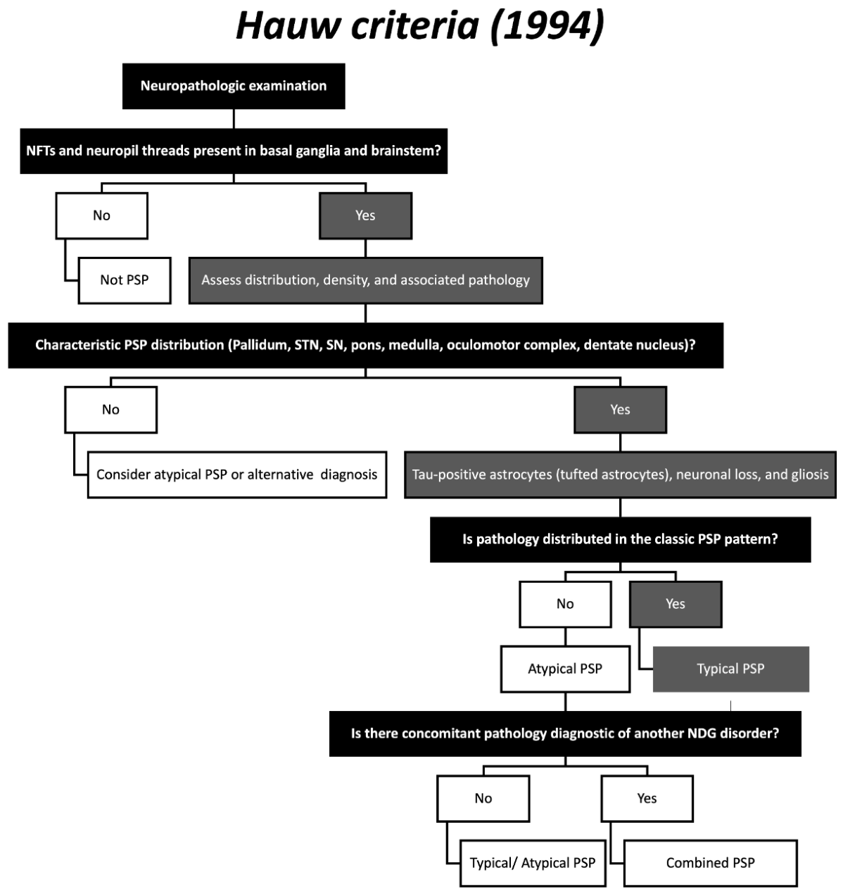
Figure 2.
Simplified clinical diagnostic algorithm based on the 1996 NINDS-SPSP criteria. The term suspected PSP denotes patients with clinical features suggestive of PSP who do not yet meet formal NINDS-SPSP diagnostic thresholds.
Figure 2.
Simplified clinical diagnostic algorithm based on the 1996 NINDS-SPSP criteria. The term suspected PSP denotes patients with clinical features suggestive of PSP who do not yet meet formal NINDS-SPSP diagnostic thresholds.
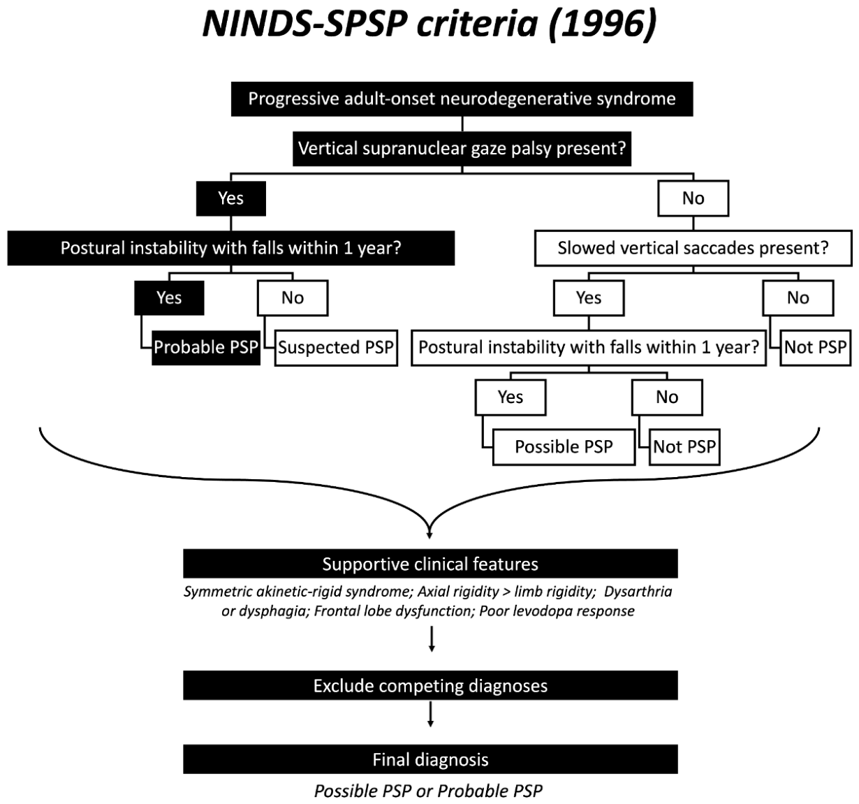
Figure 3.
Simplified diagnostic workflow based on the 2017 MDS criteria for PSP. Following con-firmation of a progressive adult-onset neurodegenerative syndrome and application of inclusion and exclusion criteria, patients are assessed across four core functional domains: ocular motor dysfunction (O), postural instability (P), akinesia (A), and cognitive dys-function (C). Supportive clinical and imaging features may increase diagnostic confidence. Specific combinations of domain features generate phenotype-specific PSP classifications and corresponding levels of diagnostic certainty.
Figure 3.
Simplified diagnostic workflow based on the 2017 MDS criteria for PSP. Following con-firmation of a progressive adult-onset neurodegenerative syndrome and application of inclusion and exclusion criteria, patients are assessed across four core functional domains: ocular motor dysfunction (O), postural instability (P), akinesia (A), and cognitive dys-function (C). Supportive clinical and imaging features may increase diagnostic confidence. Specific combinations of domain features generate phenotype-specific PSP classifications and corresponding levels of diagnostic certainty.
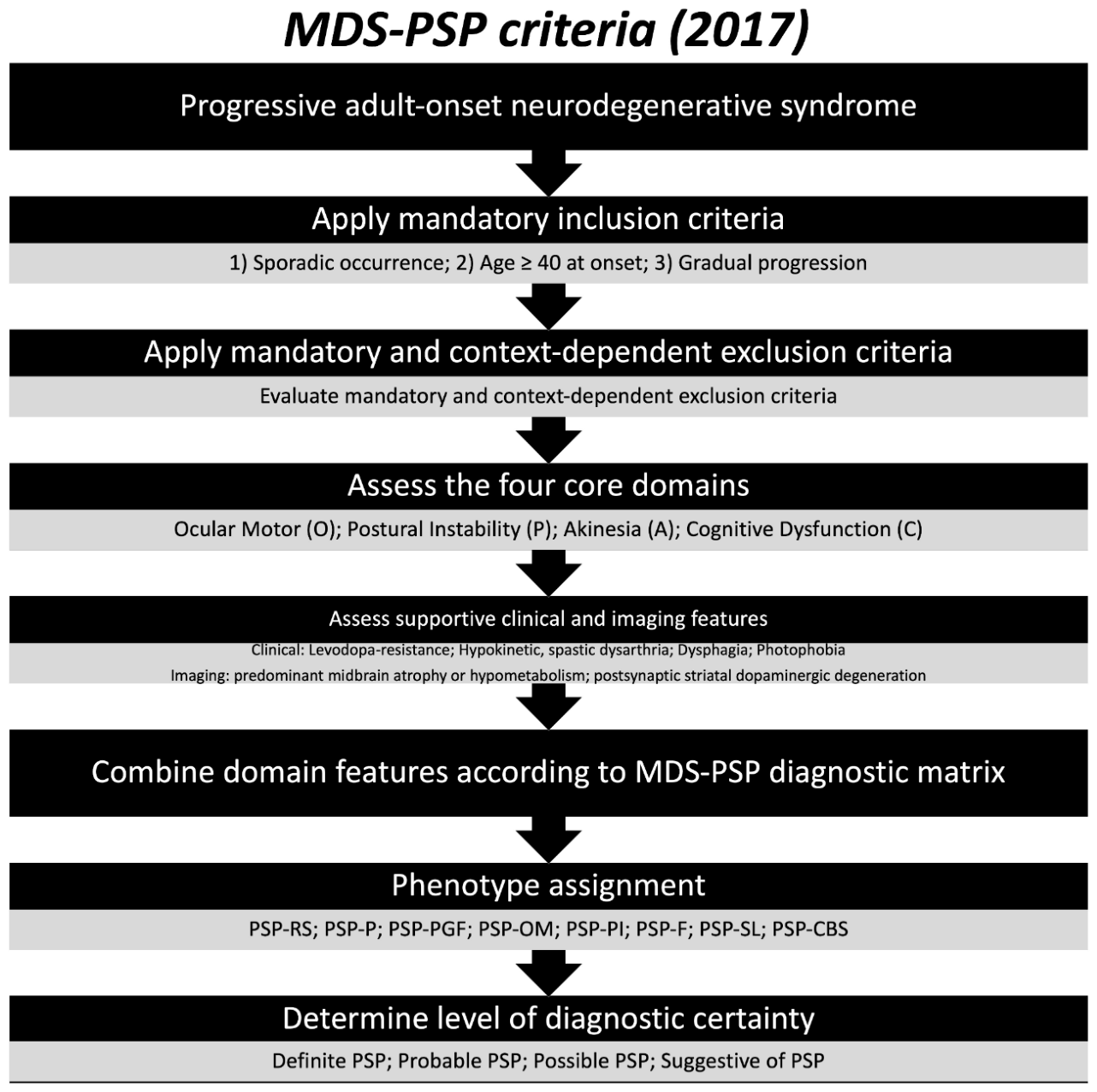
Figure 4.
Application of the MAX Rules for Phenotype Assignment in PSP.
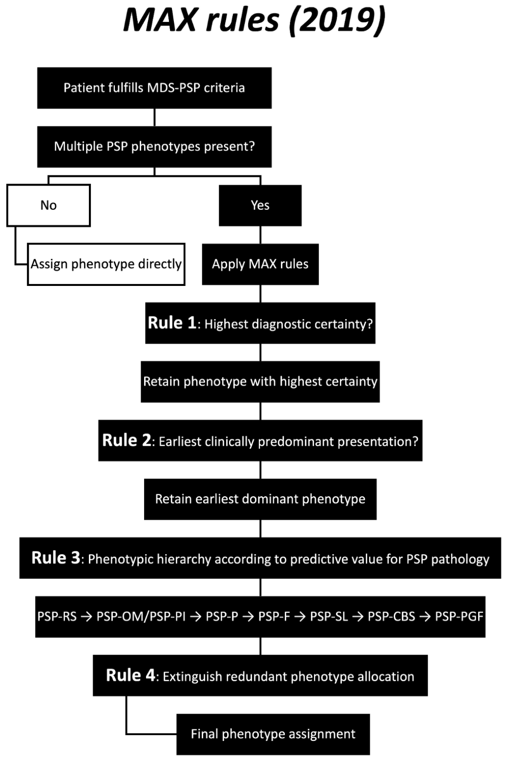

Table 1.
Historical evolution of diagnostic frameworks for PSP.
| Year | Criteria name | Other names | Reference |
|---|---|---|---|
| 1994 | Hauw criteria | Neuropathologic PSP Definition | Hauw et al., 1994 [9] |
| 1996 | NINDS-SPSP | NINDS-SPSP Clinical Diagnostic Criteria; Litvan criteria | Litvan et al., 1996 [10] |
| 2014 | vPSP | Variant PSP framework; Clinicopathological PSP Phenotype Classification | Respondek et al., 2014 [6] |
| 2017 | MDS-PSP | Movement Disorder Society PSP Criteria | Höglinger et al., 2017 [7] |
| 2019 | MAX rules | Maximum Allocation Extinction Rules | Ali et al., 2019 [8] |
Table 2.
Summary of major pre-NINDS-SPSP clinical diagnostic frameworks for PSP.
| Criteria | Mandatory features | Additional features required | Main limitation |
|---|---|---|---|
| Lees et al., 1987 [12] | Progressive nonfamilial disorder; vertical SGP with downgaze impairment | ≥2 of: postural instability/falls, pseudobulbar palsy, bradykinesia-rigidity, frontal lobe signs, axial dystonia/rigidity | No formal exclusion criteria |
| Golbe et al., 1988 [13] | Age >40 years, progressive course, bradykinesia, SGP | ≥3 of: dysarthria/dysphagia, axial rigidity, retrocollis, minimal tremor, early falls/gait disorder, poor levodopa response | More restrictive; may miss atypical presentations |
| Blin et al., 1990 Probable PSP [14] | Progressive disease, disease duration <10 years, absence of focal neurological lesions, postural instability/falls, parkinsonism, vertical gaze palsy, dysarthria or pseudobulbar palsy, frontal lobe dysfunction, poor levodopa response | All features required | Very high specificity but poor sensitivity |
| Blin et al., 1990 Possible PSP [14] | Same features as probable PSP | ≥7 of 9 features required | Improved sensitivity but reduced specificity |
Table 3.
Distinguishing features of the Hauw neuropathologic categories for PSP (1994).
| Feature | Typical PSP | Atypical PSP | Combined PSP |
|---|---|---|---|
| Definition | Classical neuropathologic form of PSP | PSP with an atypical severity or distribution of lesions | PSP coexisting with another neurodegenerative disorder |
| NFTs | Numerous NFTs in characteristic regions | Present but less dense or distributed differently from typical PSP | Characteristic PSP NFTs plus pathology of another disease |
| Neuropil threads | Abundant in affected BG and brainstem regions | Present but with atypical distribution | Present together with lesions of a coexisting disorder |
| Primary regions involved | Globus pallidus, subthalamic nucleus, SN, pons, medulla, oculomotor complex, and dentate nucleus | Same regions affected but with altered severity and/or topography | Typical PSP distribution maintained |
| Cortical involvement | Usually limited, predominantly frontal and precentral cortices | May be more extensive than expected for classic PSP | Variable depending on accompanying disease |
| Tau-positive astrocytes (tufted astrocytes) | Present in affected regions and support diagnosis | Present and support diagnosis | Present |
| Neuronal loss and gliosis | Prominent in involved brainstem and BG structures | Variable | Present together with pathology of the second disorder |
| Associated neurodegenerative pathology | Absent | Absent | Present |
| Examples of concomitant pathology | None | None | Alzheimer disease, Lewy body disease, multiple system atrophy, Pick disease, or corticobasal degeneration |
| Diagnostic interpretation | Definitive neuropathologic PSP with classic distribution | Pathologic variant of PSP | Mixed neurodegenerative pathology including PSP |
Table 4.
NINDS-SPSP clinical diagnostic categories for PSP (1996).
| Domain | Possible PSP | Probable PSP |
|---|---|---|
| Diagnostic certainty | Lower diagnostic certainty, intended to improve case detection | Higher diagnostic certainty, intended to maximize diagnostic accuracy |
| Ocular motor criterion | Vertical SGP or slowing of vertical saccades | Vertical SGP |
| Postural instability criterion | Prominent postural instability with falls | Prominent postural instability with falls within the first year of symptom onset |
| Core diagnostic concept | Presence of one major ocular motor feature combined with early postural instability | Presence of both cardinal PSP features: SGP and early falls |
| Supportive clinical features | Symmetric akinetic-rigid syndrome, axial rigidity > limb rigidity, dysarthria, dysphagia, frontal lobe dysfunction, poor levodopa response | Same supportive features |
| Major exclusion criteria | Cerebellar signs, marked autonomic failure, hallucinations, focal cortical syndromes, severe cerebrovascular disease, and alternative diagnoses explaining symptoms | Same exclusion criteria |
| Primary clinical phenotype captured | Classic PSP-Richardson syndrome and some early cases | Classical PSP-Richardson syndrome |
| Reported specificity | Approximately 80-93% | Approximately 95-100% |
| Major strength | Greater sensitivity for case identification | Excellent specificity and positive predictive value |
| Major limitation | Limited recognition of variant PSP phenotypes | Low sensitivity in early disease and non-Richardson presentations |
Table 5.
Major phenotypes incorporated into the vPSP framework.
| Phenotype | Predominant early presentation | Distinguishing features | Common initial misdiagnosis | Clinical course |
|---|---|---|---|---|
| PSP-RS | Early falls and gait instability | Vertical SGP, axial rigidity, frontal-executive dysfunction, symmetric akinetic-rigid syndrome | Classical PSP | More rapid progression and shorter survival |
| PSP-P | Asymmetric parkinsonism | Tremor, asymmetric onset, moderate initial levodopa responsiveness, delayed falls and gaze palsy | PD | Slower progression and longer survival than PSP-RS |
| PSP-PGF | Progressive gait freezing | Gait initiation failure, FOG, micrographia, hypophonia, absence of early gaze palsy or dementia | Primary progressive freezing gait, PD | Classic PSP features emerge later in the disease course |
| PSP-F | Behavioral and executive dysfunction | Apathy, disinhibition, personality change, loss of insight, impaired executive function | bvFTD | Cognitive and behavioral symptoms dominate the early stage |
| PSP-SL | Speech and language impairment | Progressive nonfluent aphasia, apraxia of speech, effortful speech, agrammatism, impaired speech motor planning | PPA | Motor manifestations of PSP often appear later |
| PSP-CBS | Asymmetric cortical syndrome | Limb apraxia, dystonia, cortical sensory deficits, myoclonus, alien limb phenomena | CBS/CBD | Cortical features precede typical brainstem manifestations |
Table 6.
Exclusion Criteria in the MDS-PSP Framework (2017).
| Type | Category | Exclusion criterion | Alternative diagnosis | Clinical relevance |
|---|---|---|---|---|
| Mandatory | Cognitive | Predominant unexplained episodic memory impairment | AD | PSP typically causes frontal-executive dysfunction rather than a primary amnestic syndrome |
| Autonomic | Predominant unexplained autonomic failure (e.g., severe OH) | MSA, DLB | Marked autonomic dysfunction is uncommon in PSP | |
| Neuropsychiatric | Predominant visual hallucinations or fluctuations in alertness | DLB | Suggests Lewy body pathology rather than PSP | |
| Motor neuron involvement | Combined upper and lower motor neuron signs | ALS or related motor neuron disorders | Not a characteristic feature of PSP | |
| Disease course | Sudden onset, stepwise progression, or rapidly progressive disease | Vascular, autoimmune, metabolic, or prion disorders | Inconsistent with the typical gradual progression of PSP | |
| Cerebellar syndrome | Prominent appendicular ataxia | MSA-cerebellar type or other cerebellar degenerations | Suggests an alternative cerebellar disorder | |
| Structural abnormalities | Severe leukoencephalopathy or relevant structural lesions on neuroimaging | Vascular or secondary parkinsonism | Provides an alternative explanation for symptoms | |
| Context dependent | PSP-CBS evaluation | Positive AD biomarkers (amyloid PET positivity or AD CSF profile) | AD-related corticobasal syndrome | Important when evaluating PSP-CBS presentations |
| Young-onset presentations (<45 years) | Wilson disease, Niemann-Pick disease type C, neuroacanthocytosis, hypoparathyroidism, or neurosyphilis | Metabolic or genetic disorders | Should be excluded in atypically young patients | |
| Rapid progression or atypical features | Prion disease, paraneoplastic encephalitis, Whipple disease, or pathogenic mutations causing FTD, AD, Huntington disease, spinocerebellar ataxias, mitochondrial disorders, and related conditions | Alternative neurodegenerative or systemic disorders | Investigated only when clinical features raise suspicion for these conditions |
Table 7.
Core functional domains of the MDS-PSP criteria (2017).
| Domain | Level 1 (highest diagnostic certainty) | Level 2 | Level 3 |
|---|---|---|---|
| Ocular Motor Dysfunction (O) | O1: Vertical supranuclear gaze palsy | O2: Slow vertical saccades | O3: Frequent macro square-wave jerks or eyelid opening apraxia |
| Postural Instability (P) | P1: Repeated unprovoked falls within 3 years | P2: Tendency to fall on pull test within 3 years | P3: More than two corrective steps on pull test within 3 years |
| Akinesia (A) | A1: Progressive gait freezing within 3 years | A2: Axial, levodopa-resistant akinetic-rigid parkinsonism | A3: Tremor-predominant, asymmetric, or levodopa-responsive parkinsonism |
| Cognitive Dysfunction (C) | C1: Speech/language disorder (progressive apraxia of speech or nonfluent/agrammatic PPA) | C2: Frontal cognitive or behavioral syndrome | C3: Corticobasal syndrome |
Table 8.
Levels of diagnostic certainty in the MDS-PSP criteria (2017).
| Diagnostic category | Required feature combination | Typical phenotype generated | Purpose |
|---|---|---|---|
| Definite PSP | Neuropathologic confirmation of PSP pathology at autopsy | Any PSP phenotype | Gold-standard diagnosis |
| Probable PSP | (O1 or O2) + (P1 or P2) | PSP-RS | Highest ante-mortem diagnostic specificity |
| (O1 or O2) + A1 | PSP-PGF | High-confidence diagnosis | |
| (O1 or O2) + (A2 or A3) | PSP-P | High-confidence diagnosis | |
| (O1 or O2) + C2 | PSP-F | High-confidence diagnosis | |
| Possible PSP | O1 alone | PSP-OM | Increased sensitivity |
| O2 + P3 | PSP-RS | Increased sensitivity | |
| A1 alone | PSP-PGF | Early recognition | |
| (O1 or O2) + C1 | PSP-SL | Also qualifies as probable 4R-tauopathy | |
| (O1 or O2) + C3 | PSP-CBS | Also qualifies as probable 4R-tauopathy | |
| Suggestive of PSP | O2 or O3 | PSP-OM | Early ocular motor presentation |
| P1 or P2 | PSP-PI | Early postural instability presentation | |
| O3 + (P2 or P3) | PSP-RS | Early Richardson syndrome | |
| (A2 or A3) + supportive feature* | PSP-P | Early parkinsonian presentation | |
| C1 alone | PSP-SL | Early speech-language presentation | |
| C2 + (O3 or P3) | PSP-F | Early frontal presentation | |
| C3 alone | PSP-CBS | Early corticobasal presentation | |
| Probable 4R-Tauopathy | Possible PSP-SL or Possible PSP-CBS | PSP-SL, PSP-CBS | Suggests underlying PSP or CBD pathology |
| Note: Diagnostic combinations have been simplified for educational purposes and are adapted from the MDS-PSP diagnostic matrix. | |||
Table 9.
Comparison of major PSP diagnostic frameworks.
| Feature | Hauw Criteria (1994) [9] | NINDS-SPSP (1996) [4] | vPSP Framework (2014) [6] | MDS-PSP (2017) [7] | MAX Rules (2019) [8] |
|---|---|---|---|---|---|
| Primary focus | Neuropathology | Clinical diagnosis | Phenotypic classification | Comprehensive clinical diagnosis | Phenotype allocation |
| Basis of diagnosis | Autopsy | Clinical features | Clinicopathological phenotypes | OPAC domains + phenotypes | Hierarchical phenotype assignment |
| PSP-RS recognition | N/A | Excellent | Included | Included | Included |
| Variant phenotypes | Limited | Poorly recognized | Major focus | Fully incorporated | Manages overlap |
| Early disease detection | No | Limited | Moderate | Improved | N/A |
| Diagnostic certainty levels | No | Possible, Probable | No | Suggestive, Possible, Probable, Definite | Uses existing categories |
| Clinical applicability | Low | Moderate | Moderate | High | High |
| Main limitation | Postmortem only | Low sensitivity | Not a formal diagnostic system | Still clinical rather than biomarker-based | May oversimplify phenotype complexity |
Table 10.
Remaining Gaps in PSP Diagnostic Frameworks and Future Research Priorities.
| Current challenge | Limitation of existing frameworks | Clinical impact | Potential future solution | Research priority |
|---|---|---|---|---|
| Early diagnosis | Most criteria rely on clinical manifestations that often emerge years after disease onset | Delayed diagnosis and missed therapeutic windows | Preclinical and prodromal PSP markers | Identification of at-risk populations |
| Ante-mortem pathological confirmation | Definite PSP still requires neuropathological examination | Diagnostic uncertainty during life | Pathology-specific biomarkers | Development of highly specific in vivo diagnostic tests |
| Differentiation from other 4R tauopathies | Significant overlap with CBD, particularly PSP-CBS and PSP-SL | Diagnostic misclassification and challenges in trial enrollment | Tau isoform-specific biomarkers and advanced molecular imaging | Improving pathological specificity |
| Differentiation from atypical parkinsonism | Clinical overlap with PD, MSA, DLB, and vascular parkinsonism | Delayed or incorrect diagnosis | Multimodal diagnostic algorithms combining clinical, imaging, and fluid biomarkers | Validation in real-world clinical cohorts |
| Phenotypic evolution over time | Patients may transition between diagnostic categories | Inconsistent phenotype classification | Longitudinal phenotype tracking models | Understanding disease progression pathways |
| Rare PSP phenotypes | PSP-C, PSP-PLS, and other uncommon variants remain poorly characterized | Underrecognition and diagnostic uncertainty | International multicenter clinicopathological studies | Refinement of phenotype classification |
| Biomarker integration | Current criteria rely predominantly on clinical findings | Limited diagnostic accuracy in early disease | MRI, fluid, genetic, and PET-based biomarker incorporation | Biomarker-guided diagnostic criteria |
| Disease monitoring | Existing criteria are primarily diagnostic rather than prognostic | Difficulty monitoring progression and treatment response | Quantitative imaging and fluid biomarkers | Development of progression markers |
| Clinical trial enrichment | Phenotypic heterogeneity complicates patient selection | Increased variability in therapeutic studies | Precision medicine approaches and biomarker-based stratification | Improved clinical trial design |
| Standardization across centers | Interobserver variability remains despite MDS and MAX frameworks | Reduced reproducibility across studies | Digital tools and AI-assisted diagnostic platforms | International harmonization of PSP classification |
Disclaimer/Publisher’s Note: The statements, opinions and data contained in all publications are solely those of the individual author(s) and contributor(s) and not of MDPI and/or the editor(s). MDPI and/or the editor(s) disclaim responsibility for any injury to people or property resulting from any ideas, methods, instructions or products referred to in the content. |
© 2026 by the authors. Licensee MDPI, Basel, Switzerland. This article is an open access article distributed under the terms and conditions of the Creative Commons Attribution (CC BY) license (http://creativecommons.org/licenses/by/4.0/).
Copyright: This open access article is published under a Creative Commons CC BY 4.0 license, which permit the free download, distribution, and reuse, provided that the author and preprint are cited in any reuse.