

Submitted:
31 August 2026
Posted:
02 September 2026
You are already at the latest version
Abstract
Acute gastrointestinal bleeding is a serious emergency condition which without prompt and accurate diagnosis can lead to severe hemorrhagic shock and death. The initial diagnosis relies on a clinical assessment of the bleeding site. If symptoms such as hematemesis and melena are present, an esophagogastroduodenoscopy (EGD) is typically the first diagnostic procedure. In cases of massive rectal bleeding, a computed tomography scan with angiography (CTA) is often used to identify the source and cause of the bleeding. multiphasic CTA is the definitive first-line diagnostic standard, enabling prompt diagnosis and crucial pre-procedural planning for endovascular management in massive cystojejunal bleeding.
Keywords:
cystojejunal anastomosis
; computed tomography
; surgery
; bleeding
Figure 1.
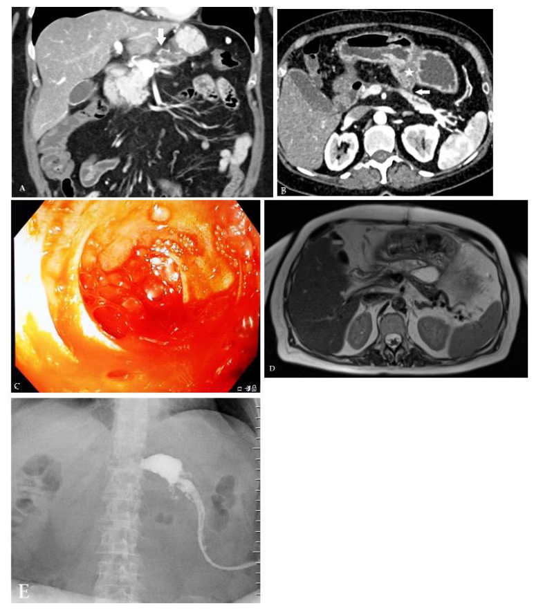
A,B Computed tomography angiography (CTA) – coronal and axial reconstruction showed irregular and wavy contours of juxtaanastomotic arterial blood vessel (arrow) in 65-year-old woman who was admitted to the Emergency Center due to severe rectal bleeding. She had previously undergone surgery for a pancreatic pseudocyst two years ago when CJA was performed, which is of critical importance for this patient. Massive gastrointestinal hemorrhage secondary to internal drainage of a pancreatic pseudocyst via cystojejunostomy represents a rare but life-threatening post-interventional complication. Upon admission, the patient was diagnosed with anemia (Hgb 80g/L) and a abdominopelvic CTA was performed. The results showed no active bleeding and normal findings in the colon, as well as a normal appearance of CJA. However, the juxtaanastomotic arterial blood vessel appeared to have irregular and wavy contours and a “sentinel” hematoma was detected in juxtaanastomotic jejunal loop. A large number of cohort studies and some multicenter studies showed that CTA can diagnose acute gastrointestinal bleeding by determining the optimal bleeding area, suspicious bleeding artery, or intraluminal blood clots with high sensitivity and negative predictive value [1,2]. High pooled sensitivity and negative predictive value with an acceptable effective radiation dose were reported in meta-analyses. Secondly, although there is no optimal bleeding area, such as for the bleeding diverticulum in lower gastrointestinal bleeding, the possibility of finding the focus of bleeding was low when there was no evidence of hematoma in the CTA features [3,4]. Upper endoscopy was then performed, which did not reveal any signs of active bleeding or blood above Treitz's ligament. While peptic ulcer disease and esophageal varices represent the most common causes of acute upper gastrointestinal bleeding, and diverticulosis and vascular malformations predominantly account for lower tract bleeding, CT angiography plays a pivotal role in rapidly detecting active extravasation and defining precise vascular anatomy to guide targeted transcatheter endovascular, endoscopic, or surgical interventions. The preferred treatment of acute gastrointestinal bleeding is usually endoscopic hemostasis, with interventional-radiological transcatheter embolization being used in a smaller percentage of cases. While emergency endoscopic evaluation was compromised by severe luminal clot pooling, CTA offered unobstructed spatial and vascular mapping of the peri-anastomotic territory [5]. If endoscopic findings are negative and there is no clear site of bleeding on CTA, surgery is the only option in cases of hemodynamic instability with active bleeding [6]. In our patient, previous intestinal surgery was a potential site of bleeding, as shown by preoperative CTA [7]. However, embolization was not possible for two reasons: CTA did not show active bleeding, and embolization of the artery at the pseudocystojejunostomy would result in ischemia and an uncontrolled pancreatic fistula. Despite receiving three blood transfusions, the patient's hemoglobin levels dropped to 68g/L and she required emergency surgery. During the surgery, a significant amount of blood was found in the small intestine, especially in juxtaanastomotic jejunal loop, as well as at the CJA. An enterotomy was performed approximately 40 cm distal to the anastomosis, and intraoperative endoscopy revealed active bleeding from the small juxtaanastomotic artery, which could not be treated endoscopically (C). Deanastomosis was performed during the same operation, and the arterial blood vessel was sutured to achieve optimal hemostasis. The patient's postoperative recovery was uneventful, with the only complication being a small pancreatic fistula and fluid collection (D), which was resolved through percutaneous drainage (E). Multiphasic CTA is the definitive first-line diagnostic standard, enabling prompt diagnosis and crucial pre-procedural planning for management in massive cystojejunal bleeding [8,9,10]. Because active extravasation is often not captured, identification of the bleeding site relies heavily on subtle morphological alterations of the juxta-anastomotic arteries (e.g., pseudoaneurysms, caliber irregularities, focal wall disruption, or adjacent inflammatory encasement). It is our belief that the active role of radiologists in critical conditions, and their collaboration with the other critical figures of the emergency, will result in new technological improvements that will place CTA among the ever more successful variants of classical angiography.
Figure 1.
A,B Computed tomography angiography (CTA) – coronal and axial reconstruction showed irregular and wavy contours of juxtaanastomotic arterial blood vessel (arrow) in 65-year-old woman who was admitted to the Emergency Center due to severe rectal bleeding. She had previously undergone surgery for a pancreatic pseudocyst two years ago when CJA was performed, which is of critical importance for this patient. Massive gastrointestinal hemorrhage secondary to internal drainage of a pancreatic pseudocyst via cystojejunostomy represents a rare but life-threatening post-interventional complication. Upon admission, the patient was diagnosed with anemia (Hgb 80g/L) and a abdominopelvic CTA was performed. The results showed no active bleeding and normal findings in the colon, as well as a normal appearance of CJA. However, the juxtaanastomotic arterial blood vessel appeared to have irregular and wavy contours and a “sentinel” hematoma was detected in juxtaanastomotic jejunal loop. A large number of cohort studies and some multicenter studies showed that CTA can diagnose acute gastrointestinal bleeding by determining the optimal bleeding area, suspicious bleeding artery, or intraluminal blood clots with high sensitivity and negative predictive value [1,2]. High pooled sensitivity and negative predictive value with an acceptable effective radiation dose were reported in meta-analyses. Secondly, although there is no optimal bleeding area, such as for the bleeding diverticulum in lower gastrointestinal bleeding, the possibility of finding the focus of bleeding was low when there was no evidence of hematoma in the CTA features [3,4]. Upper endoscopy was then performed, which did not reveal any signs of active bleeding or blood above Treitz's ligament. While peptic ulcer disease and esophageal varices represent the most common causes of acute upper gastrointestinal bleeding, and diverticulosis and vascular malformations predominantly account for lower tract bleeding, CT angiography plays a pivotal role in rapidly detecting active extravasation and defining precise vascular anatomy to guide targeted transcatheter endovascular, endoscopic, or surgical interventions. The preferred treatment of acute gastrointestinal bleeding is usually endoscopic hemostasis, with interventional-radiological transcatheter embolization being used in a smaller percentage of cases. While emergency endoscopic evaluation was compromised by severe luminal clot pooling, CTA offered unobstructed spatial and vascular mapping of the peri-anastomotic territory [5]. If endoscopic findings are negative and there is no clear site of bleeding on CTA, surgery is the only option in cases of hemodynamic instability with active bleeding [6]. In our patient, previous intestinal surgery was a potential site of bleeding, as shown by preoperative CTA [7]. However, embolization was not possible for two reasons: CTA did not show active bleeding, and embolization of the artery at the pseudocystojejunostomy would result in ischemia and an uncontrolled pancreatic fistula. Despite receiving three blood transfusions, the patient's hemoglobin levels dropped to 68g/L and she required emergency surgery. During the surgery, a significant amount of blood was found in the small intestine, especially in juxtaanastomotic jejunal loop, as well as at the CJA. An enterotomy was performed approximately 40 cm distal to the anastomosis, and intraoperative endoscopy revealed active bleeding from the small juxtaanastomotic artery, which could not be treated endoscopically (C). Deanastomosis was performed during the same operation, and the arterial blood vessel was sutured to achieve optimal hemostasis. The patient's postoperative recovery was uneventful, with the only complication being a small pancreatic fistula and fluid collection (D), which was resolved through percutaneous drainage (E). Multiphasic CTA is the definitive first-line diagnostic standard, enabling prompt diagnosis and crucial pre-procedural planning for management in massive cystojejunal bleeding [8,9,10]. Because active extravasation is often not captured, identification of the bleeding site relies heavily on subtle morphological alterations of the juxta-anastomotic arteries (e.g., pseudoaneurysms, caliber irregularities, focal wall disruption, or adjacent inflammatory encasement). It is our belief that the active role of radiologists in critical conditions, and their collaboration with the other critical figures of the emergency, will result in new technological improvements that will place CTA among the ever more successful variants of classical angiography.
Supplementary Materials
The supplementary information can be downloaded at the website of this paper posted on Preprints.org
Author Contributions
Conceptualization, V.D. and V.G.; Methodology, V.J. and M.K.; Software, P.A.; Validation, M.B., S.D. and P.A.; Formal Analysis, J.M.; Investigation, T.T.; Writing – Original Draft Preparation, V.D.; Writing – Review & Editing, V.G.; Visualization, P.A.; Supervision, T.T.
Funding
This research did not receive any specific grant from funding agencies in the public, commercial, or not-for-profit sectors.
Institutional Review Board Statement
Ethical review and approval were waived for this study on the professions of physician and dentist, which states that only a medical experiment (research or therapeutic) requires an ethics committee opinion: Law on Health Care ("Official Gazette of the RS", No. 25/2019), Competence of the Ethics Committee (Articles 155–160).
Informed Consent Statement
Written informed consent has been obtained from the patient to publish this paper.
Data Availability Statement
Data available in a publicly accessible repository.
Acknowledgments
There is no acknowledgements neither financial and material support.
Conflicts of Interest
There is no conflict of interest.
References
- Kim, J.; Lee, I. Role of surgery in gastrointestinal bleeding. Int. J. Gastrointest. Interv. 2018, 7, 136–141. [Google Scholar] [CrossRef]
- Bayoumi, S.S.; Abo Alezz, A.O.; Abdlsed, A.A.; Ali Shabaan, R.R. The Role of CT-Angiography in the Acute Gastrointestinal Bleeding. Zagazig Univ. Med. J. 2024, 30(5), 1832–40. [Google Scholar] [CrossRef]
- Erno, J.; Gregoski, M.J.; Rockey, D.C. Diagnostic utility of CT angiography compared with endoscopy in patients with acute GI hemorrhage. Gastrointest. Endosc. 2024, 99(2), 257–261.e5. [Google Scholar] [CrossRef]
- Spiritos, Z.; Horton, A.; Parish, A.; Niedzwiecki, D.; Wilson, G.; Kim, C.Y.; Wild, D. Clinical Predictors of a Positive Ct Angiogram Study Used for the Evaluation of Acute Gastrointestinal Hemorrhage. Dig. Dis. Sci. 2023, 68(1), 181–6. [Google Scholar] [CrossRef]
- Zerem, E.; Hauser, G.; Loga-Zec, S.; Kunosić, S.; Jovanović, P.; Crnkić, D. Minimally invasive treatment of pancreatic pseudocysts. World J. Gastroenterol. 2015, 21(22), 6850–6860. [Google Scholar] [CrossRef]
- Ono, R.; Tominaga, T.; Nonaka, T.; et al. A case of pancreaticoduodenal artery bleeding after laparoscopic right colectomy requiring open hemostasis. Surg. Case Rep. 2024, 10, 18. [Google Scholar] [CrossRef]
- Ullah, N.; Sharif, G.; Khan, S. M. Post-Operative Complications Among Patients Treated with Cystogastrostomy for Pancreatic Pseudocyst. J. Health Rehabil. Res. 2024, 4(1), 1158–1162. [Google Scholar] [CrossRef]
- Wortman, J.R.; Landman, W.; Fulwadhva, U.P.; Viscomi, S.G.; Sodickson, A.D. CT angiography for acute gastrointestinal bleeding: what the radiologist needs to know. Br. J. Radiol. 2017, 90, 20170076. [Google Scholar] [CrossRef]
- Tiralongo, F.; Signorelli, D.; Gangi, S.; Leanza, C.; Palmucci, S.; Basile, A. Visceral Artery Aneurysms and Pseudoaneurysms: Retrospective Evaluation of Interventional Radiology Management and Long-Term Follow-Up. Diagnostics 2023, 13(3), 481. [Google Scholar] [CrossRef]
- Zhou, C.G.; Lv, W.F.; Lu, D.; Wang, H.B.; Zhu, Y.Q.; Cheng, D.L. Transarterial embolization for massive gastrointestinal hemorrhage following abdominal surgery. World J. Gastroenterol. 2013, 19(40), 6869–6875. [Google Scholar] [CrossRef]
Disclaimer/Publisher’s Note: The statements, opinions and data contained in all publications are solely those of the individual author(s) and contributor(s) and not of MDPI and/or the editor(s). MDPI and/or the editor(s) disclaim responsibility for any injury to people or property resulting from any ideas, methods, instructions or products referred to in the content. |
© 2026 by the authors. Licensee MDPI, Basel, Switzerland. This article is an open access article distributed under the terms and conditions of the Creative Commons Attribution (CC BY) license.
Copyright: This open access article is published under a Creative Commons CC BY 4.0 license, which permit the free download, distribution, and reuse, provided that the author and preprint are cited in any reuse.