Preprint
Article

This version is not peer-reviewed.

Novel Thermophilic Species of Limisphaera Utilize a Wide Range of Carbohydrates and Possess Versatile Energy Conservation Pathways

Submitted:

31 August 2026

Posted:

01 September 2026

You are already at the latest version

Abstract

Two novel facultatively anaerobic thermophilic bacteria, strains 4302-coT and VF2T, were isolated from terrestrial hot springs of Geyser Valley and Lake Baikal region (Russian Federation), respectively. Cells were motile cocci, forming aggregates. Strain 4302-coT grew optimally at 60–65 °C and pH 7.0; strain VF2T – at 55 °C and pH 8.5. They were chemoorganoheterotrophs, utilizing mono-, di- and polysaccharides (cellulose, xylan, xanthan gum, arabinan, gluco- and galactomannan). Growth under anaerobic conditions was observed during fermentation or respiration with sulfur, nitrite, nitrate, ferric citrate or ferrihydrite. Strains 4302-coT and VF2T had genome sizes of 3.94 Mb and 4.32 Mb, with G+C contents of 65.8% and 65.2%, respectively. The genomes of both strains encoded a vast number of carbohydrate-active enzymes, including more than 100 glycoside hydrolases of diverse families and more than 15 polysaccharide lyases, along with enzymes involved in the catabolism of glucose, mannose, galactose, ribose, and rhamnose. Fermentation pathways as well as electron transfer chains of aerobic and anaerobic respiration were also reconstructed. Based on phylogenomic analysis and phenotypic features, the isolates 4302-coT and VF2T were assigned to novel species of the genus Limisphaera, for which the names Limisphaera vallis sp. nov. and Limisphaera sibirica sp. nov. are proposed.

Keywords: 
;  ;  ;  ;  ;  ;  ;  ;  ;  

1. Introduction

The Verrucomicrobiota is a long-known, ubiquitous and ecologically important bacterial phylum. It includes aerobes and anaerobes, as well as chemoorganotrophs, which utilize a wide range of polysaccharides and make up the vast majority of the phylum, and autotrophic methanotrophs, all representatives of which belong to the single family Methylacidiphilaceae. Discovered over 90 years ago, members of Verrucomicrobiota are still underrepresented in pure cultures. However, recently, this phylum has attracted the attention of key groups of microbiologists. In part, this is driven by the potential benefits of Verrucomicrobiota members, namely, the ability to efficiently hydrolyze complex polysaccharides under extreme environmental conditions (low and high pH, elevated salinity, oligotrophic conditions, etc.) [1,2]. Thermophily provides additional advantages for biotechnological applications, including reduced risk of contamination, increase of substances solubility and processes efficiency rates [3,4]. Nevertheless, only a few representatives of thermophilic verrucomicrobes are currently known. For example, methanotrophs of the genus Methylacidiphilum have been isolated from hot springs in Kamchatka, Italy, New Zealand, the Philippines, and the United States, forming a group of moderately thermophilic acidophiles. Among the chemoorganotrophic representatives, Limisphaera ngatamarikiensis, also isolated from a hot spring in New Zealand, is a moderately thermophilic alkalitolerant bacterium [5], and was recently reclassified as the type species of a new order Limisphаerales [6]. We have also described another member of this order, Fontisphaera persica, the type species of the novel family Fontisphaeraceae, isolated from a terrestrial hot spring in the Lake Baikal region [6]. This bacterium was a moderately thermophilic neutrophile, utilizing preferentially mono-, di- and polysaccharides during aerobic growth or fermentation. As far as we know, no other thermophilic verrucomicrobia, besides F. persica and Methylacidiphilum kamchatkense, have been isolated from terrestrial hot springs of Russia. At the same time, other members of Verrucomicrobiota, and in particular of the order Limisphaerales, are found in the hot springs of Lake Baikal region and Kamchatka [6,7].
The aim of this work was to isolate and investigate novel verrucomicrobia from geographically distant thermal springs of Russia. The study focused primarily on hydrolytic representatives capable of respiration using electron acceptors other than oxygen. As a result, two novel thermophilic representatives of the genus Limisphaera, Limisphaera vallis strain 4302-coT and L. sibirica strain VF2T, were successfully obtained and described.

2. Materials and Methods

2.1. Sampling and Isolation

Mixed sample of water and pink peach fouling was collected from a hot spring of Geyser Valley (N54°26′22″ E160°8′38″, pH 7.85 and 61 °C), Kamchatka, Russia, in 2023, in sterile 50 ml vials and transported to the laboratory. 10% (v/v) of inoculum was added to 15 ml Hungate tubes filled with 5 ml aerobic modified Widdel medium [8] supplemented with xyloglucan (1 g l-1) and the tubes were incubated at 65 °C. After 7 days of incubation, the growth of cells with various morphotypes, including numerically dominant small cocci, was observed. After several subsequent transfers at 65 °C on the same medium, followed by serial 10-fold dilutions, the small cocci were isolated into a pure culture 4302-co.
A mixed sample of water, silty sediment and plastic bag fragments suspended in the water was collected from a hot spring (N52°59′14.04′′ E108°18′27.97′′) formed from a borehole at the Goryachinsk thermal water basin, Russia, in 2023, in sterile 50 ml glass vial and transported to the laboratory. An aerobic enrichment culture was obtained from the sample (10% inoculum) with LDPE (10 g l⁻¹) as the sole carbon and energy source. The medium of the following composition (g l-1) was used: NH₄Cl (0.5); MgCl₂·6H₂O (0.165); CaCl₂·2H₂O (0.05); KCl (0.165); KH₂PO₄ (0.25); NaHCO₃ (1.0), 1 ml of a trace element solution [9], 1 ml of a vitamin solution [10]. The stable enrichment was obtained after 7 transfers. To isolate a pure culture 1 ml of the enrichment was inoculated to 10 ml of the same medium supplemented with starch (1 g l-1). Strain VF2 was isolated from an enrichment culture by serial 10-fold dilutions. Strain VF2T was isolated from an enrichment culture obtained on plastic, but in the present work its involvement in plastic degradation was not investigated.
The purity of the strains was verified by cultivation on organic-rich medium containing glucose and yeast extract or peptone (2 g l-1) at various pH (6.0–9.0) and temperatures (37–65 °C) to reveal fast-growing organotrophs as well as by analyzing of the whole genome sequence assemblies.

2.2. Phenotypic and Chemotaxonomic Characterization

Cell morphology and the effects of temperature, pH and NaCl concentration on growth of strains 4302-coT and VF2T were analyzed using above-mentioned mineral media with starch or cellobiose as a substrate, respectively. The spectrum of tested substrates included xanthan gum, locust bean gum, karaya gum, xyloglucan, carboxymethylcellulose (CMC), microcrystalline cellulose (Avicel), dextrin, dextran, starch, chitin, galactan, galactomannan, glucomannan, mannan, arabinan, curdlan, lichenan, levan, pectin, xylan, rhamnogalacturonan, polygalacturonan, monogalacturonate, pachyman, alginate, chondroitin sulfate, agarose, inulin, hyaluronic acid, tragacanth added directly to the mineral medium before autoclaving (4 g l-1). Glucose, fructose, xylose, raffinose, rhamnose, arabinose, mannose, maltose, cellobiose, sucrose, lactose, galactose, ribose, pyruvate, lactate, methanol, yeast extract, beef extract, tryptone and peptone were added after autoclaving to a final concentration of 0.5 or 1 g l-1 from filtered stock solutions. Elemental sulfur (1 g l-1), sodium thiosulfate (10 mM), sulfate (10 mM), sodium nitrite (2 mM), sodium nitrate (10 mM), sodium arsenate (5 mM), sodium selenate (5 mM), ferric citrate (10 mM) and ferrihydrite (50 mM) were tested as possible electron acceptors for anaerobic respiration. Sulfide formation was detected spectrophotometrically according to Trüper and Schlegel [11]. Nitrite and nitrate reduction were monitored by qualitative tests for nitrate/nitrite (Merck, USA). Arsenate reduction was assessed by the formation of characteristic yellow precipitated arsenic sulfide (orpiment). Fe(III) and Fe-citrate reduction were analyzed using the ferrozine method [12]. Autotrophic growth was tested in a H2/CO2 gas phase (4:1, v/v) under anaerobic conditions without electron acceptors and in the presence of elemental sulfur, sodium nitrite or sodium nitrate.
Catalase and oxidase reactions were checked with 3% hydrogen peroxide and N,N,N′,N′-tetramethyl-p-phenylenediamine [13]. For analysis of cellular fatty acids (CFAs) and quinones, the cells of strains 4302-coT and VF2T grown on liquid medium supplemented with starch (1 g l-1) were harvested at the late exponential growth phase. CFAs were determined as described earlier [14] but with a CP-Sil 88 column instead of an HP-5MS column. Isoprenoid quinones were determined as described earlier [15].

2.3. Genome Sequencing, Assembly, Annotation and Phylogenetic Analysis

Genome sequencing and raw read processing were done as described earlier [16] with a single modification: NovaSeq X Plus was used instead of NovaSeq 6000. Assembly of whole-genome sequences was performed as published elsewhere [17]. Completeness and contamination of the genomic assemblies were measured using CheckM v.1.2.2 with the “Bacteria” marker set [18]. The whole genome indices (ANI, AAI and dDDH) were calculated according to Larralde et al., Kim et al., Hölzer and Meier-Kolthoff et al., respectively [19,20,21,22]. To construct a phylogenetic tree, the 120 bacterial single-copy conserved marker genes (bac120) were annotated, concatenated and aligned using GTDB-Tk v2.7.1 [23]. The phylogenetic tree was built using IQ-TREE v.3.0.1 [24].

2.4. Genome Analysis

The genomes of the strains 4302-coT and VF2T were annotated with NCBI Prokaryotic Genome Annotation Pipeline, PGAP, v.6.10 [25].
Genes encoding carbohydrate-active enzymes (CAZymes) were identified using dbCAN v.4 with all available tools [26]. A further BLAST search against the curated Swiss-Prot database [27] was performed for more accurate prediction of putative enzyme activity.
Reconstruction of central carbon metabolism, sugar utilization as well as energy conservation pathways was performed by searching the genes encoding proteins responsible for these processes as described earlier [28]. Proteins from different known iron reducers and Planctopirus limnophila were used as references for the detection of multiheme cytochromes [29] and components of bacterial microcompartments (BMCs) and BMC-associated enzymes [30], respectively.
Phylogenetic analysis of molybdopterin oxidoreductases was performed as follows: sequences belonging to the prokaryotic molybdopterin-containing oxidoreductase family with evidence at the protein level as well as seven putative dissimilatory molybdopterin-containing oxidoreductases encoded in the analyzed genomes of Limisphaera were aligned using Muscle v.5.2 [31]. The alignment was trimmed using trimAL v.1.4.1 [32] with –gt option of 0.85. The tree was constructed in RAxML v. 8.2.12 [33] with the PROTGAMMAILG model and 1000 rapid bootstrap replications as support values. Visualization of the phylogenetic tree was performed using iTOL v.7 [34].

2.5. Biochemical Characterization of the Glycosidase from GH51 Family

2.5.1. In Silico Analysis of GH51 Glycosidases

The domain architecture and the presence of signal peptide were predicted using InterProScan [35] and SignalP v.6. [36]. Phylogenetic analysis of glycoside hydrolases from GH51 family was performed as described earlier [37].

2.5.2. Construction and Cloning of the Lim81

The gene encoding protein MGA4643381.1 of strain 4302-coT (named lim81, with a length of 2190 bp) was amplified using the de novo designed primers (Table S1) and the DNA of Limisphaera sp. 4302-coT as a template. PCR product was purified with the Cleanup Standard Kit (#BC022, Evrogen). Cloning of the lim81 was performed using the aLICator Ligation Independent Cloning and Expression System kit (#K1251, Thermo Scientific) with selection of vector pLATE 51, which contained N-terminal 6xHis-tag and an enterokinase cleavage site (DDDDK^). The ligation independent cloning (LIC) reaction was performed for 5 min at 25 °C, upon which the vector with the insert was directly transformed into E. coli BL21 (DE3) competent cells. This was done, according to the manufacturer instructions, and a 720 bp control PCR fragment was used to assure the efficiency of the LIC reaction. The presence of cloned genes was verified by PCR with DNA from grown E. coli colonies as the template and using specific primers (LIC Forward Sequencing primer 5’-TAATACGACTCACTATAGGG-3’ and LIC Reverse Sequencing primer 5’-GAGCGGATAACAATTTCACACAGG-3’), and confirmed by the following analysis of sequence obtained using Sanger sequencing (Evrogen, Russia).

2.5.3. Gene Expression and Enzyme Purification

For the expression of recombinant protein Lim81, the E. coli BL21 (DE3) strain carrying vector pLATE51::lim81 was grown on in 1.5 l LB medium supplemented with ampicillin, 100 μg ml-1, at 37 °C for 5 hours to an optical density (λ = 600 nm) of 0.5. Gene expression was induced by the addition of 0.5 mM isopropyl β-D-1-thiogalactopyranoside (IPTG). After 16 h of incubating at 20 °C, cells were harvested by centrifugation (9,000×g, 4 °C, 20 min) and resuspended in the following buffer: 50 mM TrisHCl (pH 7.8), 150 mM NaCl, 20 mM imidazole and 0.1% (v/v) protease inhibitor PMSF (DiaM, Russia). Cells were disrupted by sonication using SoniPrep (MSE, UK) at 4 °C and further centrifuged (13,400×g, 4 °C, 30 min). The supernatant, containing soluble protein Lim81, was purified using fast protein liquid chromatograph (FPLC, Äkta Start, Cytiva, USA). The metal affinity chromatography was performed on 1 ml HisTrap HP column (Cytiva, USA). The column was equilibrated by Ni-A buffer (50 mM Tris-HCl (pH 7.8), 0.5 M NaCl, 20 mM imidazole). The supernatant was adjusted and washed with 10 column volumes (CV) of a Ni-A buffer. Protein elution from the column was performed by linear gradient of imidazole from 20 mM to 500 mM using Ni-B buffer (50 mM Tris-HCl pH 7.8, 0.5 M NaCl, 500 mM imidazole), gradient volume was 15 CV. Fractions, containing the Lim81, were desalted and concentrated on ultracentrifuge module VivaSpin with MWCO cutoff of 10 kDa (Sartorius, Germany) at 3,000×g, 4 °C, 15 min.
The presence of the target proteins was confirmed by electrophoresis in a polyacrylamide gel [38]. Protein ladders #22610, #26614, and #26630 (ThermoFischer, USA) as well as G2058 (Servisbio, China) were used as protein standards. Protein concentrations were measured using the Qubit Protein Assay Kit and Bradford method [39].

2.5.4. Biochemical Characterization of Glycosidase Lim81

The endoglucanase activity of different Lim81-containing fractions was visualized on agarose (2% w/v) plates or zymogram gel containing 0.2% CMC as a substrate, hydrolysis zones were detected via Congo red staining [40,41].
Glycoside hydrolase activity was measured using the DNSA assay [42]. Reactions were initiated by adding 100 μl cell extract or purified enzyme solutions to 600 μl buffer (50 mM TrisHCl, 150 mM NaCl, 0.5 mM CaCl2, pH 7.8), preincubated at the respective temperature, and contained containing 0.3% substrate (beta-glucan, lichenan, arabinan, arabinoxylan, xylan, arabinogalactan, galactan, mannan, pachyman, curdlan (all from Megazyme), CMC (Fluka), laminarin (Sigma-Aldrich), agarose (Helicon), and Avicel (Fluka)), followed by incubation at various temperatures and various time (see the results) in Thermo Shaker TS-100C (Biosan, Latvia). The same reaction mixtures without protein were used as the control experiments. Aliquots of 300 μl of reaction mixture were taken at the time points 0, 2.5, 24 h. In each aliquot the reaction was stopped by adding the 3,5-dinitrosalicylic acid (DNS) reagent (1:1, v:v) followed by cooling down to the room temperature. Afterwards, the mixtures were incubated for 15 min at 98 °C, cooled down again to the room temperature and the absorbance at 575 nm was measured with a spectrophotometer SPECTROstar Nano (BMG Labtech, Germany). All measurements were made in triplicate.
Temperature dependence was measured at pH 5.0 with barley beta-glucan as a substrate, using Thermo Shaker TS-100C (Biosan, Latvia) for the temperatures 22-95 °C. The influence of pH on activity of the glycosidase Lim81 was analyzed in the pH range 2.5–10.5 in 50 mM HEPES buffer (pH 2.5–5.0), 50 mM MES buffer (pH 5–6.5), 50 mM MOPS (pH 6.5–7.0), 50 mM HEPES buffer (pH 7.0–7.5), 50 mM TrisHCl (pH 7.5–9.1) and 50 mM CAPS (pH 9.1–10.5) at 70 °C.
The influence of metal ions, denaturing and reducing agents was evaluated by incubation of the reaction mixtures with beta-glucan as a substrate, containing 1-5 mM of each of the following agents: CaCl2, CuCl2, MgCl2, MnCl2, ZnCl2, Cr(SO4)2, dithiothreitol (DTT), Tween 80, urea, Triton-X100 and sodium dodecyl sulfate (SDS). The enzymatic activities were measured using DNSA assay as described above.
One unit (U) of enzyme activity is defined as the amount of enzyme required to release 1 µg ml-1 of the reducing sugars (equivalent to glucose, mannose, galactose or xylose) per minute. Specific activity was calculated as the enzyme activity per milligram of total protein (U mg-1 protein).

3. Results and Discussion

3.1. Phenotypic Properties

Cells of strain 4302-coT were pink-colored cocci, 0.7–1.3 μm in diameter, reproduced by binary fission. They were present singly, in pairs, or in aggregates and were motile in exponential growth phase by means of flagella (Figure 1A). Ultrathin sections of the cells revealed a diderm cell envelope (Figure 1B) and the presence of bacterial microcompartments (BMCs) during the growth on rhamnose (Figure 1C).
Strain 4302-coT grew at 37–70 °C, with an optimum at 60–65 °C, at pH 6.2–9.2, with an optimum at 7.4. The strain grew in the presence of NaCl up to 1.5% yet, the optimal growth was registered without NaCl in the medium. No growth was observed at temperatures ≤30 °C and ≥ 75 °C, at pH 5.9 and below and 9.7 and above, and at NaCl concentrations ≥ 1.9%.
Strain 4302-coT was facultatively anaerobic. Under aerobic conditions without shaking the growth rate was slightly higher than under aerobic conditions with shaking or strictly anaerobic conditions. The strain was oxidase positive and catalase negative.
The novel isolate 4302-coT did not require yeast extract for growth. The following carbohydrates and proteinaceous substrates were utilized under aerobic and anaerobic conditions: glucose, arabinose, mannose, maltose, cellobiose, galactose, rhamnose, lactose, sucrose, xylose, ribose, raffinose, arabinan, dextrin, dextran, starch, xylan, xyloglucan, galactan, microcrystalline cellulose (Avicel), carboxymethylcellulose, lichenan, pachyman, polygalacturonan, rhamnogalacturonan, glucomannan, galactomannan, xanthan gum, locust bean gum, chondroitin sulfate, yeast extract, beef extract, peptone, tryptone. Fructose, alginate, agarose, levan, inulin, pectin, mannan, monogalacturonate, curdlan, hyaluronic acid, tragacanth, karaya gum, and pyruvate were not utilized. Strain 4302-coT did not grow lithoautotrophically under H2/CO2 atmosphere either in the presence or in the absence of electron acceptors (elemental sulfur, nitrate, nitrite). However, it grew organoheterotrophically by anaerobic respiration on cellobiose with sulfur, arsenate, nitrate, nitrite, ferric citrate, or ferrihydrite. Thiosulfate, sulfate and selenate were not used for growth as electron acceptors. The products of cellobiose fermentation were acetate, lactate, ethanol, H2 and CO2. Major cellular fatty acids (>10%) of strain 4302-coT were C16:0, anteiso-C15:0 and iso-C16:0 (Table S2). The only isoprenoid quinone was identified as MK-7.
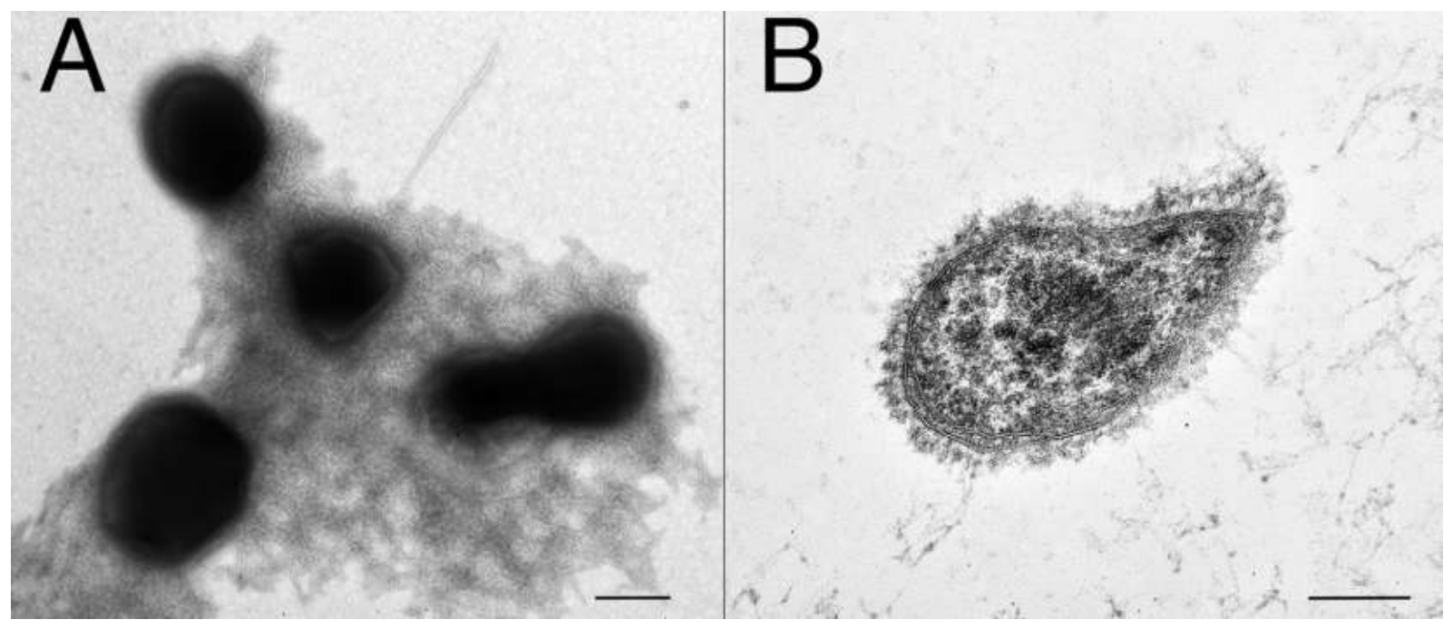
Cells of VF2T were cocci 0.7–1.0 μm in diameter, reproduced by binary fission. Cells appeared singly, or in pairs or triplets, and often formed large aggregates. Aggregated cells were surrounded by a fibrillar matrix. Cells were motile due to a single flagellum (Figure 2A). Ultrathin sections of the cells revealed a diderm cell envelope (Figure 2B).
Strain VF2T grew at 37–70 °C, with an optimum at 55 °C, at pH 6.5–9.5, with an optimum at 8.5. The strain grew in the presence of NaCl up to 1.5% yet, the optimal growth was registered without NaCl in the medium. No growth was observed at temperatures ≤30 °C and ≥ 75 °C, at pH 5.9 and below and 9.7 and above, and at ≥ 1.9% of NaCl.
Strain VF2T was facultatively anaerobic. In aerobic conditions without shaking the growth rate was slightly higher than in strictly anaerobic conditions. The strain was oxidase positive and catalase negative.
The novel isolate VF2T was able to utilize yeast extract, as well as peptone, tryptone, and beef extract. The following carbohydrates were utilized under aerobic conditions: glucose, maltose, lactose, galactose, mannose, xylose, rhamnose, raffinose, fructose, sucrose, cellobiose, arabinose, arabinan, dextran, dextrin, starch, xylan, xyloglucan, galactan, carboxymethylcellulose, microcrystalline cellulose (Avicel), glucomannan, galactomannan, rhamnogalacturonan, xanthan gum, karaya gum, locust bean gum, alginate, pectin, chitin, lichenan pullulan, curdlan, pachyman, polygalacturonan. Agarose and pyruvate were not utilized. Strain VF2T grew anaerobically on glucose, sucrose, maltose, fructose, raffinose, galactose, starch, xylan, xanthan gum and karaya gum, and did not grow anaerobically on pyruvate, lactate or methanol. Strain VF2T did not grow lithoautotrophically under H2/CO2 atmosphere either in the presence or in the absence of electron acceptors (elemental sulfur, nitrate, nitrite). However, it grew organoheterotrophically by anaerobic respiration on sucrose, maltose or starch with elemental sulfur, thiosulfate, nitrate or nitrite, ferric citrate or ferrihydrite, and did not respire on sulfate, arsenate, or selenate. The products of glucose fermentation were acetate, ethanol, H2 and CO2. Major cellular fatty acids (>10%) of strain VF2T were iso-C16:0, anteiso-C15:0 and anteiso-C17:0 (Table S2). The only isoprenoid quinone was identified as MK-7 similar to strain 4302-coT.
Thus, strains 4302-coT and VF2T had several features shared with the only validly published member of the order LimisphaeralesL. ngatamarikiensis strain NGM72.4T. They were motile bacteria with spherical cells occurring singly or in pairs and dividing by binary fission; they had a diderm cell envelope; they were moderate thermophiles that grew optimally in the absence of sodium chloride; they were chemoorganotrophs that grew aerobically on simple and complex sugars and did not grow by means of sulfate respiration; the sole detected quinone was MK-7 (Table 1). On the other hand, a number of features made them different from the closest relative L. ngatamarikiensis. They formed cell aggregates; they were facultative anaerobes able to grow by fermentation and respiration with different electron acceptors, including nitrate; they utilized a large number of polysaccharides (cellulose, xanthan gum, starch etc.), as well as proteinaceous substrates (yeast extract, peptone, etc.), but did not grow on pyruvate. In addition, strains 4302-coT and VF2T also differed from each other in a number of features: pigment composition, pH parameters, and the ability of strain VF2T to grow on pectin, karaya gum and fructose. Finally, all three strains were characterized by a different spectrum of major fatty acids.

3.2. Phylogeny and Analysis of Distribution

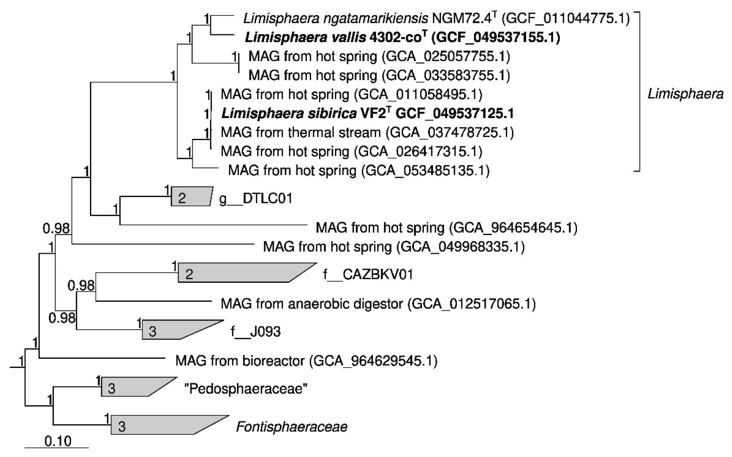
Both strains were first identified by 16S rRNA gene sequences extracted from the genomes. Strains 4302-coT and VF2T have 97.2% similarity to each other and 98.3% and 97.5% similarity to the closest cultured microorganism Limisphaera ngatamarikiensis NGM72.4T, respectively. ANI analysis also showed that genomes of 4302-coT and VF2T strains represent separate species within the genus Limisphaera: comparison of both strains with L. ngatamarikiensis yielded ANI values of 78.2 and 74.7%, respectively, and 74.7% between each other. Other results of the genomic comparison (dDDH and AAI) are given in Table S3. All these results clearly indicate that both strains represent separate species within the genus Limisphaera. Phylogenetic analysis based on 120 bacterial single-copy conserved marker genes [43] confirmed this conclusion (Figure 3). Therefore, based on phylogenomic analysis we propose to assign strains 4302-coT and VF2T to novel species of the genus Limisphaera, L. vallis sp. nov. and L. sibirica sp. nov., respectively.
Species of the genus Limisphaera tend to thrive in terrestrial hot springs, and the species represented by the new strains are no exception. Using the Sandpiper service [44], we were able to reliably (coverage >10x) detect species represented by strain VF2T in 10 publicly available metagenomes. This species was detected in hot springs in Argentina (PRJEB67756), Canada (PRJNA375330), and Malaysia (PRJEB4990). In all cases, its relative abundance did not exceed 1.5%. The species represented by strain 4302-coT was reliably detected in 14 publicly available metagenomes. Eleven of these metagenomes were obtained from hot spring sediments of the Tengchong County, Yunnan Province, China [45], where its relative abundance was up to 1%. It was also detected in Tretyakovsky Hot Spring (Kunashir Island; PRJNA493162), but at a relative abundance of only 0.06%.

3.3. Genome Analysis

The genomes of strains 4302-coT and VF2T were assembled into 37 and 14 contigs with N50 of 222,613 bp and 732,743 bp, respectively. The genome of strain 4302-coT was of 3.94 Mbp in size with G+C proportion of 65.8%, while length of the strain VF2T genome was 4.32 Mbp and G+C content of 65.2%. Both assemblies were of high quality with estimated completeness of 100% and contamination of 1.72%. According to the annotation, the genomes of strains 4302-coT and VF2T contained 2,928 and 2,957 protein-coding genes, respectively. A single rRNA operon was identified in each genome. Additionally, 46 tRNA genes, 4 ncRNA genes and 29/24 pseudogenes were identified in the genomes.
In addition to the genomes of the two novel isolates, the genome of L. ngatamarikiensis and the available metagenome-assembled genomes (MAGs) affiliated with the genus Limisphaera were selected for further analysis.
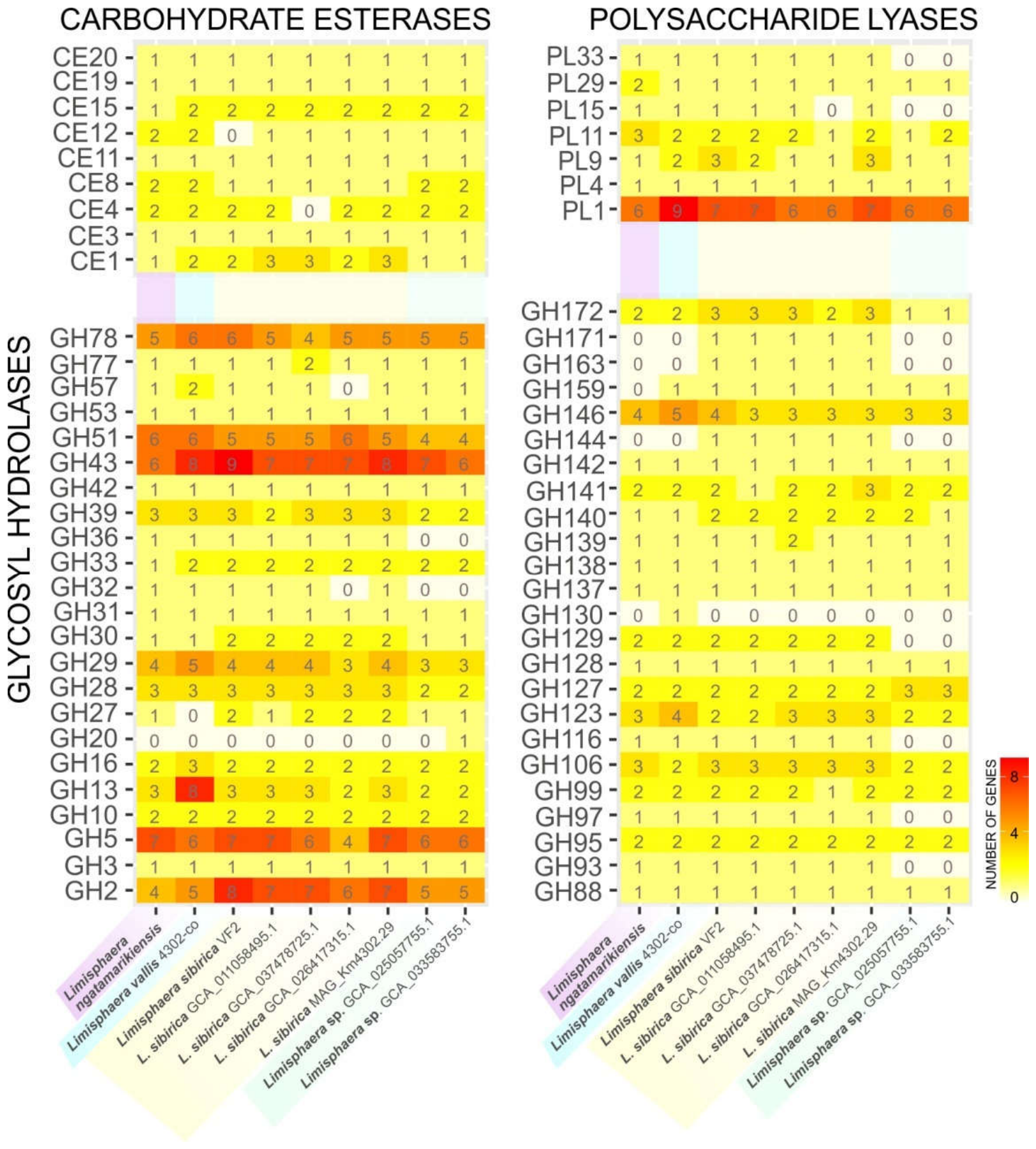
Since novel isolates, as well as all known members of the order Limisphaerales, are capable of utilizing wide range of polysaccharides, the diversity of CAZymes in these bacteria and their functions were estimated. The genomes of the novel strains 4302-coT and VF2T, as well as L. ngatamarikiensis and six MAGs, encoded the vast diversity of CAZymes; more than a hundred genes of encoding glycoside hydrolases (GHs), polysaccharide lyases (PLs), and carbohydrate esterases (CEs) were found in each genome (Figure 4). Glycosidases from GH2 (5-8 genes), GH5 (4-7 genes), GH10 (2 genes), GH43 (6-9 genes), GH51 (4-6 genes), GH78 (4-6 genes) and GH146 (3-5 genes) and some other families were encoded in all genomes. These enzymes are presumably involved in hydrolysis of plant polysaccharides, including xylans, arabinans, and galactans. Endo-acting enzymes are represented in GH5, GH10, GH51, GH53 and GH43 families and are able to hydrolyze cellulose, xylan, arabinan, galactan, gluco- and galactomannans. The GH5 family exhibits a wide range of activities, including endoglucanase, xyloglucanase, endomannanase, and endoxylanase. At the same time, the GH51 encompasses endoglucanases, beta-xylosidases, and α-L-arabinofuranosidases; the latter being involved in hydrolysis of arabinans and arabinoxylans and often also possessing xylanase activity [46]. In the family GH10 only endo-beta-1,4-xylanases were found. The GH43 family includes α-L-arabinofuranosidases, α-L-arabinanases, β-D-xylosidases, and exo-α-1,3-galactanases. Glycosidases from GH2 family are β-galactosidases and/or β-glucuronidases, while enzymes from GH78 family presumably exhibit α-L-rhamnosidase activity. In addition, genes encoding polysaccharide lyases from families PL1, PL4, PL9, PL11 and PL29 were found in all genomes. Enzymes from these families exhibit pectin/pectate lyase (PL1, PL9) and rhamnogalacturonan lyase (PL4, PL9 and PL11) activities, which are essential for degradation of pectins and their component, rhamnogalacturonan (RGN). Carbohydrate esterases, which are required for removing acetyl groups from xylan (CE1, CE3), pectin, and rhamnogalacturonan (CE20), as well as methyl group in pectin (CE8, CE19), were also encoded in all genomes. The presence of a set of CAZymes involved in degradation of hemicelluloses confirms the hydrolytic potential of Limisphaerales representatives and their ecological niche as degraders of plant-derived organic matter.
The genome of L. sibirica VF2T encodes PL1, PL4, PL9 (pectate and rhamnogalacturonan lyases), involved in pectin degradation, as well as PL15, a putative alginate lyase. The genome of L. vallis 4302-coT encodes the majority of PLs, including PL1, PL4, PL9 and PL11 (pectate and RGN lyases), PL29 and PL33 (both chondroitin/hyaluronate lyases), as well as pectin methyl esterases and RGN esterases belonging to CE8, CE19 and CE20 families, respectively. Moreover, both strains possessed beta-xylosidases (GH3, GH5 and GH43), endo-1,4-beta-xylanase (GH10, GH30), endoglucanases (GH5, GH16, GH51, GH128), endo-alpha-1,5-arabinases (GH43) and endo-beta-1,4-galactanases (GH53). Genes of encoding glycoside hydrolases from families GH13 and GH57 presumably active against alpha-glucans (starch, dextrin) were also present in the genomes of strains 4302-coT and VF2T. The presence of numerous and variable CAZyme genes correlates with the wide spectrum of carbohydrate substrates utilized by these strains. This suggests the combined action of several enzymes towards recalciltrant polysaccharides (like cellulose, xanthan gum, and galactomannans) as well as involvement of specific sets of PLs, GHs and CEs in the targeted degradation of acidic polysaccharides like pectin, RGN, and hyaluronate.
Comparison of CAZyme genes between the species (including MAGs) within the genus Limisphaera revealed large common set among different species; however, there were some minor differences were revealed in their distribution (Table S4). Strain 4302-coT lacked genes encoding alpha-galactosidases from the GH27 family, however, it had genes encoding the unique mannooligosaccharide phosphorylase (GH130) and eight genes of GH13 family (compared with 2-3 genes in other genomes). Genome of strain VF2T did not encode rhamnogalacturonan acetylesterase from CE12. On the other hand, strain VF2 T and MAGs belonging to the same species contained genes of endo-β-N-acetylglucosaminidases (GH163), which are involved in N-glycans hydrolysis [47,48], and exo-β-N-acetylmuramidases (GH171). MAGs SKYG106 and SKYGB_hs_bin94 had narrowest repertoires of CAZyme genes with the absence of genes ones encoding beta-fructosidases (GH32), some arabinan-active glycosidases (GH93, GH116), as well as polysaccharide lyases from PL15 and PL33 families. Beta-hexosaminidase from GH20 family was encoded only in MAG SKYGB_hs_bin94. This likely reflects a distinct ecological niche for this species, potentially driven by the limited availability of complex polysaccharides in its habitat.
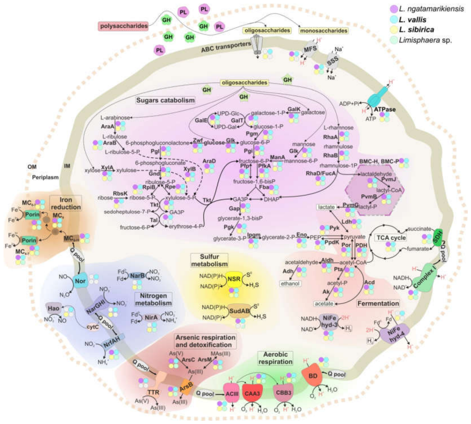
Moreover, Limisphaera representatives can grow on various monosaccharides – products of polysaccharides hydrolysis. Porters belonging to MFS and SSS families as well as ABC transporters from CUT-2 family can be responsible for import of mono-/oligosaccharides into the cells. Surprisingly, all studied genomes contained several genes of IIA subunit of PTS transporter, however, genes encoding other subunits were absent. These proteins may be involved in regulation of carbohydrates metabolism rather than in transport processes [49]. Almost all studied genomes encoded the full set of enzymes of glycolysis for glucose utilization (Figure 5, Table S5a) with the exception of two genomes belonging to uncultivated Limisphaera species, which lacked the pyruvate kinase genes, however, they had genes encoding pyruvate, phosphate dikinase indicating that glycolysis still may be active in these bacteria. Additionally, genes encoding enzymes involved in both oxidative (glucose-6-phosphate 1-dehydrogenase, 6-phosphogluconolactonase and decarboxylating 6-phosphogluconate dehydrogenase) and non-oxidative branches of pentose phosphate pathway were found. Ribose 5-phosphate isomerases in Limisphaera members had additional serine hydroxymethyltransferase domain. BLAST search showed that such unusual enzymes are also present in different verrucomicrobes including extremely acidophilic methanotroph “Ca. Methylacidiphilum infernorum” [50]. Surprisingly, strain VF2T and the related MAGs, as well as species represented by only two MAGs (SKYG106=GCA_025057755.1 and SKYGB_hs_bin94=GCA_033583755.1), did not have transaldolase genes. These enzymes presumably can be functionally replaced with pyrophosphate-dependent phosphofructokinases (produces sedoheptulose-1,7-bis-phosphate from sedoheptulose-7-phosphate) and fructose-1,6-bis-phosphate aldolase (cleaves sedoheptulose-1,7-bis-phosphate to erythrose-4-phospate and dihydroxyacetone phosphate) as it was proposed for some bacteria from Bacillota and Planctomycetota phyla [51,52]. All genomes except L. ngatamarikiensis genome encoded mannose-6-phosphate isomerase required for mannose conversion into glucose. Ribokinase was encoded in all genomes with exception of two MAGs, SKYG106 and SKYGB_hs_bin94. Genes encoding enzymes involved in catabolism of L-arabinose (L-arabinose isomerase, ribulokinase and L-ribulose-5-phosphate 4-epimerase) and galactose (galactose mutarotase, galactokinase, galactose-1-phosphate uridylyltransferase and UDP-glucose 4-epimerase) were identified in all genomes. L-rhamnose is utilized through the action rhamnose isomerase, rhamnulokinase and rhamnulose-1-phophate aldolase (Table S5b); further several reactions associated with BMCs presumably occur as proposed for some representatives of Bacillota [53], Planctomycetota [30,54], Verrucomicrobiota [55] and Bacteroidota [56].
Under anaerobic conditions without external electron acceptor Limisphaera representatives can ferment organic substrates with the formation of several products (Table S5c). Phosphate acetyltransferase and acetate kinase are responsible for acetate production, while L-lactate dehydrogenase – for lactate. Ethanol can be formed due to the presence of putative acetaldehyde dehydrogenase and both iron- and zinc-containing alcohol dehydrogenases. Probably, hydrogen is evolved through the action of [NiFe] hydrogenases of group 3 and 4 but not [FeFe] hydrogenases, which are not encoded in the genomes.
In the presence of electron acceptors in the medium, reducing equivalents are generated in the complete tricarboxylic acid cycle (Table S5a) and are further used in the respective electron transfer chains (ETC). Both NADH:quinone oxidoreductase and succinate dehydrogenase transfer electrons to the quinone pool. Under aerobic conditions several membrane complexes participate in respiration: bd-type quinol oxidase as well as caa3- and cbb3-type terminal cytochrome oxidases (Table S5d). No Limisphaera species possess the cytochrome b/c1 complex which is replaced by alternative complex III as described for Rhodothermus marinus [57]. Under anaerobic conditions representatives of the genus Limisphaera can utilize different electron acceptors other than oxygen. All of them presumably can reduce nitrite due to the presence of dissimilatory nitrite reductase NrfAH (Table S5e). At the same time, genes encoding distant homologs of membrane-bound nitrate reductase NarGHI (Figure S1) and putative hydroxylamine oxidoreductase were found only in genomes of strains 4302-coT, VF2T (as well as in MAGs affiliated to the same species). Nitric oxide reductase was unique feature of strain 4302-coT. Probably, homologs of NAD(P)H sulfur oxidoreductase are responsible for sulfur reduction in Limisphaera during “facilitated” fermentation as was previously proposed for Thermotoga maritima [58], while polysulfide or thiosulfate reductases were not encoded in any of the genomes (Table S5f). In addition, genomes of L. ngatamarikiensis, strain 4302-coT as well as MAGs SKYG106 and SKYGB_hs_bin94 encoded sulfide dehydrogenase SudAB (MAG SKYGB_hs_bin94 had only sudB), which presumably performs NADPH-dependent sulfur reduction in cytoplasm [59]. Genes of the canonical arsenate oxidoreductase complex ArrABC were absent in all analyzed genomes, however, genes encoding another molybdopterin-containing oxidoreductase were found in the genome of L. ngatamarikiensis and strain 4302-coT (Figure S1). Their closest characterized homologs were described as selenate reductase SdrABC [60] and tetrathionate reductase TtrABC [61]. Previously it was proposed that some Ttr-related oxidoreductases may be responsible for reduction of arsenate in bacteria and archaea [62,63,64]. Considering the growth experiments (the ability of strain 4302-coT to grow with arsenate but not with selenate as electron acceptor), we suggest that its Ttr-like enzyme (MGA4643419-21) acts as arsenate reductase. All analyzed Limisphaera-affiliated genomes also contained genes of proteins involved in arsenate detoxification (arsenate reductase ArsC, arsenite methyltransferase ArsM and arsenite efflux permease ArsB). Dissimilatory iron reduction presumably occurs due to the presence of several inner membrane, periplasmic and porin-associated multiheme cytochromes (MHCs) containing from 6 to 11 heme-binding motifs (Table S5g) and encoded in several loci (1-3 genes). Two loci also contained genes of porins belonging to OMR family not related to porins from well-studied Gram-stain-negative iron reducer Shewanella oneidensis (MtrB). Moreover, one porin contains a single N-terminal heme-binding motif as does MtrB in metal-reducing proteobacteria [65]. In another cluster in addition to two inner membrane MHCs, three-subunit efflux transporters of the RND superfamily were encoded. The closest characterized homolog of inner membrane subunit is multidrug resistance protein MdtB, while the outer membrane subunit is relatively close to OpmQ subunit of the pyoverdine exporter, pyoverdine is an iron siderophore specific for Pseudomonas [66]. Probably members of Limisphaera could scavenge pyoverdine via this transporter, further export outside the cells and utilize it for iron chelation in environment, or identified transporter can play some unknown function.
Although some Limisphaerales representatives are considered methanotrophs based on genome analysis [67], methane monooxygenases and lanthanide-dependent enzymes specific for known verrucomicrobial methanotrophs of the order Methylacidiphilales [68], were not encoded in the studied genomes of Limisphaera.

3.4. Biochemical Characterization of Glycosidase Lim81

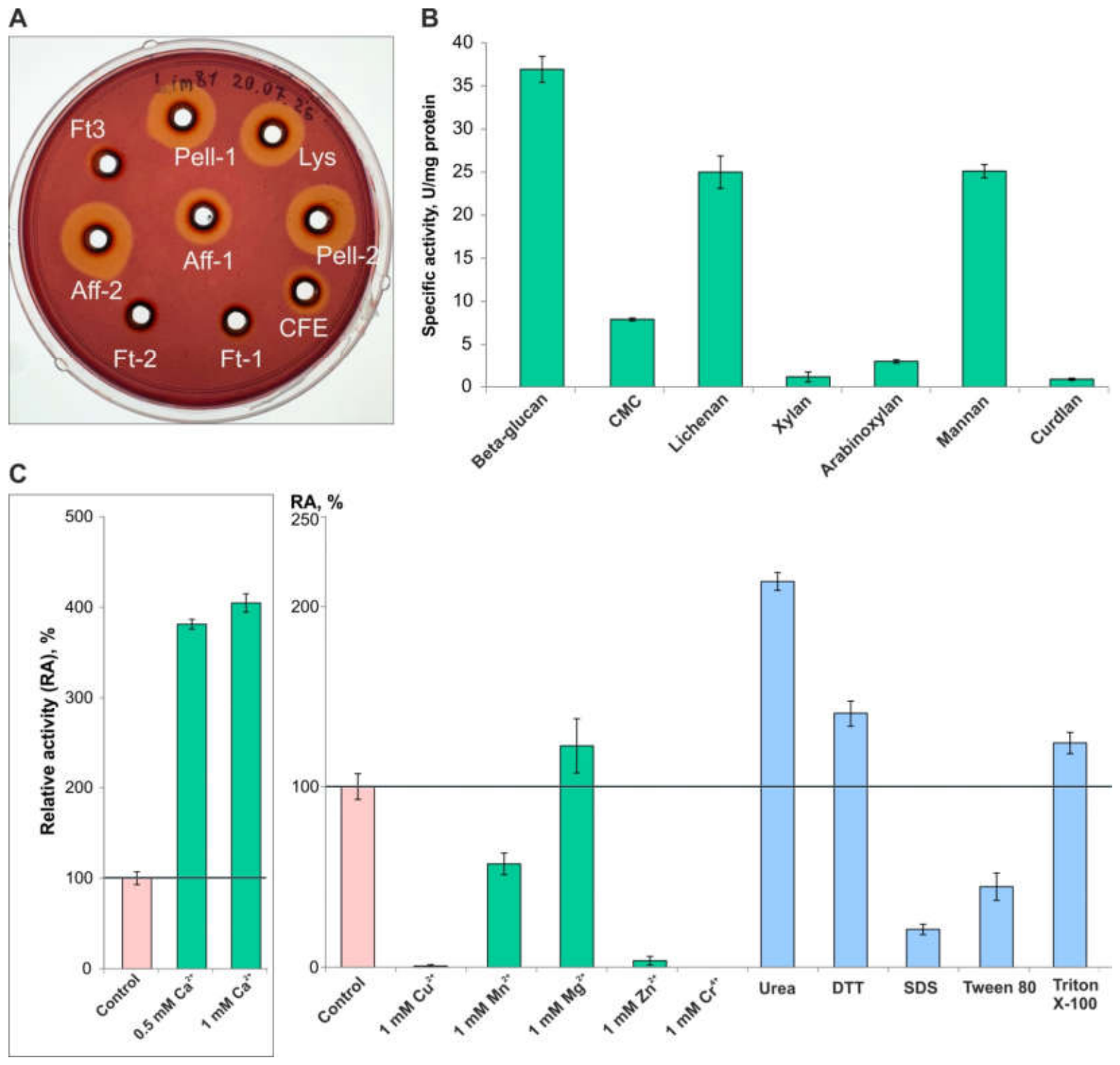
The genomes of both novel isolates each contained five genes encoding glycoside hydrolases of family GH51. Most of these proteins in strains 4302-coT and VF2T are presumably alpha-L-arabinofuranosidases (Figure S2), which is a typical activity for GH51 enzymes. However, MGA4643381.1 (4302-coT) and MGA4578311.1 (VF2T) clustered with several characterized endoglucanases. Since only 7 out of 121 glycosylhydrolases in this family are characterized as endoglucanases, and Limisphaera proteins are relatively distant from these homologs, MGA4643381.1 from Limisphaera sp. 4302-coT was chosen for recombinant expression and further biochemical characterization. This enzyme possessed a Sec-type signal peptide (1-27), a carbohydrate-binding module 8 (32-170) and GH51 family catalytic domain (219-599).
The sequence length of the cloned part of the lim81 was 2190 bp (without signal peptide). Predicted mass of the protein was 80.5 kDa and pI 5.5. After the heterologous expression and purification, the protein about 85 kDa was observed on SDS-PAGE (Figure S3A, S3B). Qualitative analysis of endoglucanase activity revealed the presence of active glycosidase in fractions of cell extract, pellet (containing insoluble proteins) and fractions from FPLC purification (Figure 6A, Figure S3C). The Lim81 was active at pH 5.0 and T 70 °C towards beta-glucan, lichenan, mannan, and CMC with specific activities 36.92, 25.00, 25.10, and 7.91 U mg-1 protein, respectively (Figure 6B). Weak activity was also observed on arabinoxylan, xylan and curdlan (2.96, 1.16 and 0.88 U mg-1 protein). At the same time, no activity was detected towards Avicel, amorphous cellulose, arabinan, arabinogalactan, galactan, pachyman, laminarin, and agarose. This substrate spectrum reflects the phylogenetic position of this enzyme as an endo-1,4(1,3)-glucanase with broad specificity towards β-1,4- linked polysaccharides with arabinose side chains (arabinoxylan). At the same time, the hydrolysis of xylans by two endoglucanases CelVA and CelB from the family GH51 has been shown [69,70]. This suggests that Lim81 endoglucanase may have the ability to hydrolyze the backbone of arabinoxylan. The activity of Lim81 was stimulated fourfold by Ca2+ ions, while Mg2+ slightly increased it. Addition of Cu2+, Zn2+, and Mn2 ions reduced activity fully or slightly (Figure 6C). Addition of urea, DTT, Triton-X100, had no influence on the activity of Lim81, and slightly increased it, when the presence of Tween 80 and SDS inhibited the enzyme. This suggests an influence of small amounts (1-5 mM) of urea as an accelerator of enzyme activity [71].
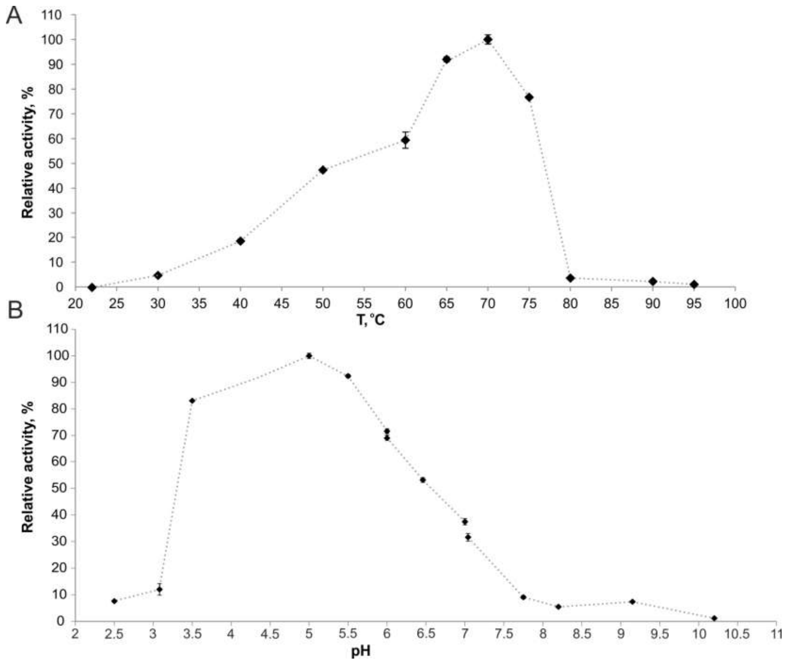
The Lim81 was active towards beta-glucan at temperature range from 30 to 80 °C (at 22 and 85 °C no activity was observed, (Figure 7A) with optimum at 70 °C, and pH range 3.5-8.5 with optimum 5.0 (Figure 7B). It is worth noting that, only four endoglucanases from the family GH51 were characterized to date: CelB, CelVA, CelA4, and Cel51A – three from the representatives of the genus Alicyclobacillus and the last one – from Fibrobacter [69,70,71,72,73]. Also, the GH51-related protein, Fisuc_2081, isolated from F. succinogenes S85, was isolated, possessing weak endoglucanase activity against beta-glucan at 40 °C [73]. All these enzymes possess activity at acidic pH and thermophilic conditions (40-80 °C). Their major activity is endoglucanase activity against CMC and beta-glucan. For CelB, CelVA, and Cel51A weak xylanolytic activity was demonstrated (Table S6).
Among 121 characterized enzymes in the GH51 family only seven are assigned as cellulases/endoglucanases, but two of them were predicted as homologs of GH51 protein (SScel24, Lc-CelC). Information on substrate specificity, stability and characteristics of other GH51 cellulases is very scarce, and our data shed light on the ability and unique properties of these cellulases. Lim81 possessed an unusual ability to hydrolyze both the β-1,4 linkage in various polysaccharides with glucose, xylose and mannose backbone, and the β- 1,3 linkages in curdlan backbone. Future studies will focus on investigating the stability of this enzyme and its slight activation by detergents, while structural refinement and analysis should elucidate the unique mechanisms underlying the properties of this GH51 glycoside hydrolase.

4. Conclusions

In the current study, two strains, 4302-coT and VF2T, were isolated from hot springs of Geyser Valley (Kamchatka) and Baikal rift zone. Based on phylogenomic analysis each strain was affiliated to the separate novel species of the genus Limisphaera (order Limisphaerales of the phylum Verrucomicrobiota). Both growth experiments and genome analysis revealed the strong hydrolytic potential of these bacteria as well as their metabolic flexibility including aerobic and anaerobic respiration with different electron acceptors. The capacity for iron reduction was demonstrated here for the first time for the representatives of the phylum Verrucomicrobiota. Biochemical characterization of the glycosidase Lim81 from strain 4302-coT, which belongs to the family GH51, was performed. These bacteria were detected as minor components of microbiomes in diverse hot springs of Argentina, Canada, Malaysia, China and Russia. Thus, thermophilic verrucomicrobes presumably play crucial role in the processes of aerobic and anaerobic degradation of complex organic matter in terrestrial hot springs and can be promising objects for both the study of unknown physiological features and the discovery of new thermoactive enzymes, especially glycoside hydrolases.

4.1. Description of Limisphaera vallis sp. Nov.

Limisphaera vallis (val’lis. L. gen. n. vallis, of a valley, here referring to the Geyser Valley, Kamchatka, Russian Federation, from where the type strain was isolated).
Cells are cocci, 0.7 - 1.3 μm in diameter. Cells occur singly, in pairs or in aggregates, some of them are motile by means of a single flagellum. Diderm cell envelope. Pink-pigmented under aerobic conditions. Moderate thermophile with a growth temperature range of 37–70 °C and the optimum at 60–65 °C. The pH range for growth is 6.2–9.2 with the optimum at 7.4. Sodium chloride and yeast extract are not required for growth. Facultative anaerobe. Catalase negative and oxidase positive. Chemoorganotrophic. Can utilize glucose, mannose, maltose, cellobiose, arabinose, xylose, galactose, rhamnose, lactose, sucrose, ribose, raffinose, arabinan, dextrin, dextran, starch, xylan, xyloglucan, galactan, microcrystalline cellulose (Avicel), carboxymethylcellulose, lichenan, pachyman, polygalacturonan, rhamnogalacturonan, glucomannan, galactomannan, xanthan gum, locust bean gum, chondroitin sulfate, yeast extract, beef extract, peptone, tryptone. No growth is observed on fructose, alginate, agarose, levan, inulin, pectin, mannan, monogalacturonate, curdlan, hyaluronic acid, karaya gum, tragacanth, pyruvate or H2/CO2. Fermentation products of cellobiose are acetate, lactate, ethanol, hydrogen and carbon dioxide. Can respire with elemental sulfur, arsenate, nitrite, nitrate, ferric citrate, ferrihydrite and oxygen. Major fatty acids are C16:0, anteiso-C15:0, and iso-C16:0. MK-7 is the sole respiratory quinone. The type strain has a genome size of 3.94 Mb and a G+C content of 65.8%. The type strain, 4302-coT =CGMCC 1.18274T =BIM B-2082T =VKM B-3882T=UQM 41853T, was isolated from a terrestrial thermal spring, Geyser Valley, Kamchatka, Russia.

4.2. Description of Limisphaera sibirica sp. Nov.

Limisphaera sibirica (si.bi’ri.ca. N. L. fem. adj. sibirica, originating from Siberia, region in northern Asia, Russian Federation, from where the type strain was isolated).
Cells are cocci, 0.7 - 1.0 μm in diameter. Cells occur singly, in pairs or in aggregates, some of them are motile by means of a single flagellum. Diderm cell envelope. Slightly peach-pigmented in aerobic conditions. Moderate thermophile with a growth temperature range of 37–70 °C and the optimum at 55 °C. The pH range for growth is 6.5–9.5 with the optimum at 8.5. Sodium chloride and yeast extract are not required for growth. Facultative anaerobe. Catalase negative and oxidase positive. Chemoorganotrophic. Can utilize glucose, maltose, lactose, galactose, mannose, xylose, rhamnose, raffinose, fructose, sucrose, cellobiose, arabinose, arabinan, dextran, dextrin, starch, xylan, xyloglucan, galactan, carboxymethylcellulose, Avicel, glucomannan, galactomannan, rhamnogalacturonan, xanthan gum, karaya gum, locust bean gum, pectin, chitin, lichenan, pullulan, curdlan, pachyman, polygalacturonan, yeast extract, beef extract, peptone, tryptone. No growth is observed on agarose, pyruvate or H2/CO2. Fermentation products of glucose are acetate, ethanol, hydrogen and carbon dioxide. Can respire with elemental sulfur, thiosulfate, nitrate or nitrite, ferric citrate, ferrihydrite, but not with sulfate, selenate or arsenate. Major fatty acids are iso-C16:0, anteiso-C15:0 and anteiso-C17:0. MK-7 is the sole respiratory quinone. The genome of the type strain is characterized by the size of 4.32 Mbp and G+C content – 65.2%. The type strain, VF2T=VKM B-3901T = UQM 41924T, was isolated from a terrestrial thermal spring, Goryachinsk, Baikal region, Russia.

4.3. Emended Description of Genus Limisphaera

The description is based on that provided by Anders et al. [5], with the following amendments. The genus contains strictly aerobic and facultatively anaerobic species. The latter oxidize organic substrates with nitrate, nitrite, elemental sulfur, arsenate, ferric citrate or ferrihydrite as electron acceptors or ferment mono-, di- and polysaccharides or proteins. The major fatty acids are C16:0, ai-C15:0, i-C16:0, ai-C17:0, and C18:0. The type species is Limisphaera ngatamarikiensis.

Supplementary Materials

The following supporting information can be downloaded at the website of this paper posted on Preprints.org, Table S1: Primers designed for lim81 gene. Specific sequences are underlined; start- and stop codons are bolded, Table S2: Cellular fatty acids of strains 4302-coT and VF2T, Table S3: Genome to genome comparisons of three species of Limisphaera, Table S4: Glycoside hydrolases, polysaccharide lyases and carbohydrate esterases encoded in the genomes of Limisphaera representatives, Table S5: Enzymes involved in carbon metabolism and energy conservation within Limisphaera representatives, Table S6: Characterized cellulases from GH51 family, Figure S1: Phylogeny of molybdopterin-containing oxidoreductase subunits, Figure S2: Phylogeny of glycoside hydrolases from GH51 family, Figure S3: (A) SDS-PAAGE of fractions, obtained after affinity chromatography, (B) Affinity chromatography process, (C) Hydrolytic activities visualized on CMC-zymogram gel.

Author Contributions

Conceptualization, O.A.P.; methodology, O.A.P., T.G.S., K.S.Z., A.A.N., A.Y.M.; software, A.Y.M., A.G.E.; validation, O.A.P., T.G.S., K.S.Z.; formal analysis, A.Y.M., A.G.E.; investigation, O.A.P., T.G.S., K.S.Z., P.R.K., A.A.K., A.Y.M., A.G.E.; data curation, A.A.K., A.Y.M.; writing—original draft preparation, O.A.P., T.G.S., K.S.Z., A.Y.M., A.G.E.; writing—review and editing, O.A.P., T.G.S., A.A.K.; visualization, K.S.Z., A.A.K., A.Y.M., A.G.E.; funding acquisition, K.S.Z. All authors have read and agreed to the published version of the manuscript.

Funding

The work of K.S.Z. and A.G.E. was supported by the grant of Russian Science Foundation no. 25-24-00393.

Institutional Review Board Statement

Not applicable.

Data Availability Statement

The whole-genome sequences are deposited into GenBank within BioProject PRJNA1222819: JBMVQZ000000000 (4302-co) and JBMVQY000000000 (VF2). The strains were deposited in China General Microbiological Culture Collection Center, Belarusian Collection of Nonpathogenic Microorganisms, All-Russian Collection of Microorganisms and Collection of Unique and Extremophilic Microorganisms under a number 4302-coT=CGMCC 1.18274T =BIM B-2082T =VKM B-3882T=UQM 41853T and VF2T=VKM B-3901T=UQM 41924 T, respectively.

Conflicts of Interest

The authors declare no conflicts of interest.

References

  1. Rakitin, A.L.; Kulichevskaya, I.S.; Beletsky, A.V.; Mardanov, A.V.; Dedysh, S.N.; Ravin, N.V. Verrucomicrobia of the Family Chthoniobacteraceae Participate in Xylan Degradation in Boreal Peat Soils. Microorganisms 2024, 12, 2271. [CrossRef]
  2. Sorokin, D.Y.; Khot, V.; Merkel, A.Y.; Mosier, D.; Bale, N.J.; Koenen, M.; Strous, M. Physiology, functional genomics, and proteomics of Verruconatronum alginivorum gen. nov., sp. nov., the first isolated haloalkaliphile within Verrucomicrobiota, representing a new family, Verruconatronumaceae fam. nov. Appl. Environ. Microbiol. 2026, 92, e0047526. [CrossRef]
  3. DeCastro, M.E.; Rodríguez-Belmonte, E.; González-Siso, M.I. Metagenomics of Thermophiles with a Focus on Discovery of Novel Thermozymes. Front. Microbiol. 2016, 7, 1521. [CrossRef]
  4. Kumar, S.; Dangi, A.K.; Shukla, P.; Baishya, D.; Khare, S.K. Thermozymes: Adaptive strategies and tools for their biotechnological applications. Bioresour. Technol. 2019, 278, 372–382. [CrossRef]
  5. Anders, H.; Power, J.F.; MacKenzie, A.D.; Lagutin, K.; Vyssotski, M.; Hanssen, E.; Moreau, J.W.; Stott, M.B. Limisphaera ngatamarikiensis gen. nov., sp. nov., a thermophilic, pink-pigmented coccus isolated from subaqueous mud of a geothermal hotspring. Int. J. Syst. Evol. Microbiol. 2015, 65, 1114–1121. [CrossRef]
  6. Podosokorskaya, O.A.; Elcheninov, A.G.; Novikov, A.A.; Merkel, A.Y.; Kublanov, I.V. Fontisphaera persica gen. nov., sp. nov., a thermophilic hydrolytic bacterium from a hot spring of Baikal lake region, and proposal of Fontisphaeraceae fam. nov., and Limisphaeraceae fam. nov. within the Limisphaerales ord. nov. (Verrucomicrobiota). Syst. Appl. Microbiol. 2023, 46, 126438. [CrossRef]
  7. Zavarzina D.G.; Klyukina A.A.; Merkel A.Y.; Maslova T.A.; Maslov A.A. Alkalothermophilic iron-reducing bacteria of the Goryachinsk Thermal Water Basin. Microbiology, 2024, 93, 864–875. [CrossRef]
  8. Podosokorskaya, O.A.; Bonch-Osmolovskaya, E.A.; Novikov, A.A.; Kolganova, T.V.; Kublanov, I.V. Ornatilinea apprima gen. nov., sp. nov., a novel cellulolytic representative of class Anaerolineae. Int. J. Syst. Evol. Microbiol. 2013, 63, 86–92. [CrossRef]
  9. Pfennig, N.; Lippert, K.D. Über das Vitamin B12-Bedürfnis phototropher Schwefelbakterien. Archiv. Mikrobiol. 1966, 55, 245–256. [CrossRef]
  10. Wolin, E.A.; Wolin, M.J.; Wolfe, R. S. Formation of methane by bacterial extracts. J. Biol. Chem. 1963, 238, 2882–2886. [CrossRef]
  11. Trüper, H.G.; Schlegel, H.G. Sulphur metabolism in Thiorhodaceae. I. Quantitative measurements on growing cells of Chromatium okenii. Antonie van Leeuwenhoek 1964, 30, 225–238. [CrossRef]
  12. Tugel, J.B.; Hines, M.E.; Jones, G.E. Microbial iron reduction by enrichment cultures isolated from estuarine sediments. Appl. Environ. Microbiol. 1986, 52, 1167–1172.
  13. Smibert, R.M; Krieg, N.R. Phenotypic Characterization. In Methods for General and Molecular Bacteriology; Gerhardt, P.; Murray, R.G.E.; Wood, W.A; Krieg, N.R., Eds.; American Society for Microbiology: Washington DC, USA, 1994; pp. 607-654.
  14. Karnachuk, O.V.; Lukina, A.P.; Avakyan, M.R.; Kadnikov, V.V.; Begmatov, S.; Beletsky, A.V.; Vlasova, K.G.; Novikov, A.A.; Shcherbakova, V.A.; Mardanov, A.V.; Ravin, N.V. Novel thermophilic genera Geochorda gen. nov. and Carboxydochorda gen. nov. from the deep terrestrial subsurface reveal the ecophysiological diversity in the class Limnochordia. Front. Microbiol. 2024, 15, 1441865. [CrossRef]
  15. Frolov, E.N.; Kublanov, I.V.; Toshchakov, S.V.; Samarov, N.I.; Novikov, A.A.; Lebedinsky, A.V.; Bonch-Osmolovskaya, E.A.; Chernyh, N.A. Thermodesulfobium acidiphilum sp. nov., a thermoacidophilic, sulfate-reducing, chemoautotrophic bacterium from a thermal site. Int. J. Syst. Evol. Microbiol. 2017, 67, 1482–1485. [CrossRef]
  16. Podosokorskaya, O.A.; Prokofeva, M.I.; Kuchierskaya, A.A.; Klyukina, A.A.; Elcheninov, A.G. Tenuifilum osseticum sp. nov., a novel thermophilic hydrolytic bacterium within the Tenuifilaceae isolated from a North Ossetian thermal spring, and emended description of the genus Tenuifilum. Syst. Appl. Microbiol. 2025, 48, 126591. [CrossRef]
  17. Zayulina, K.S.; Podosokorskaya, O.A.; Klyukina, A.A.; Panova, T.V.; Novikov, A.A.; Kublanov, I.V.; Bonch-Osmolovskaya, E.A.; Elcheninov, A.G. A Novel Species of the Genus Thermanaerothrix Isolated from a Kamchatka Hot Spring Possesses Hydrolytic Capabilities. Curr. Microbiol. 2024, 81, 293. [CrossRef]
  18. Parks, D.H.; Imelfort, M.; Skennerton, C.T.; Hugenholtz, P.; Tyson, G.W. CheckM: assessing the quality of microbial genomes recovered from isolates, single cells, and metagenomes. Genome Res. 2015, 25, 1043–1055. [CrossRef]
  19. Larralde, M.; Zeller, G.; Carroll, L.M. PyOrthoANI, PyFastANI, and Pyskani: a suite of Python libraries for computation of average nucleotide identity. NAR Genom. Bioinform. 2025, 7, lqaf095. [CrossRef]
  20. Kim, D.; Park, S.; Chun, J. Introducing EzAAI: a pipeline for high throughput calculations of prokaryotic average amino acid identity. J. Microbiol. 2021, 59, 476–480. [CrossRef]
  21. Hölzer M. POCP-nf: an automatic Nextflow pipeline for calculating the percentage of conserved proteins in bacterial taxonomy. Bioinformatics 2024, 40, btae175. [CrossRef]
  22. Meier-Kolthoff, J.P.; Carbasse, J.S.; Peinado-Olarte, R.L.; Göker, M. TYGS and LPSN: a database tandem for fast and reliable genome-based classification and nomenclature of prokaryotes. Nucleic Acids Res. 2022, 50, D801–D807. [CrossRef]
  23. Chaumeil, P.-A.; Mussig, A.J.; Hugenholtz, P.; Parks, D.H. GTDB-Tk v2: memory friendly classification with the genome taxonomy database. Bioinformatics 2022, 38, 5315–5316. [CrossRef]
  24. Wong, T.K.F.; Ly-Trong, N.; Ren, H.; Demotte, P.; Baños, H.; Roger, A.J.; Susko, E.; Bielow, C.; De Maio, N.; Goldman, N.; Hahn, M.W.; Dos Reis, M.; Sy Vinh, L.; Huttley, G.; Lanfear, R.; Minh, B.Q. IQ-TREE 3: Phylogenomic Inference Software using Complex Evolutionary Models. Mol. Biol. Evol. 2026, 43, msag117. [CrossRef]
  25. Tatusova, T.; DiCuccio, M.; Badretdin, A.; Chetvernin, V.; Nawrocki, E.P.; Zaslavsky, L.; Lomsadze, A.; Pruitt, K.D.; Borodovsky, M.; Ostell, J. NCBI prokaryotic genome annotation pipeline. Nucleic Acids Res. 2016, 44, 6614–6624. [CrossRef]
  26. Zheng, J.; Ge, Q.; Yan, Y.; Zhang, X.; Huang, L.; Yin, Y. dbCAN3: automated carbohydrate-active enzyme and substrate annotation. Nucleic Acids Res. 2023, 51, W115–W121. [CrossRef]
  27. Boutet, E.; Lieberherr, D.; Tognolli, M.; Schneider, M.; Bansal, P.; Bridge, A.J.; Poux, S.; Bougueleret, L.; Xenarios, I. UniProtKB/Swiss-Prot, the Manually Annotated Section of the UniProt KnowledgeBase: How to Use the Entry View. Methods Mol. Biol. 2016, 1374, 23–54. [CrossRef]
  28. Podosokorskaya, O.A.; Elcheninov, A.G.; Gavrilov, S.N.; Petrova, N.F.; Klyukina, A.A.; Zavarzina, D.G.; Merkel, A.Y. New Representatives of the Class Ignavibacteria Inhabiting Subsurface Aquifers of Yessentuki Mineral Water Deposit. Water 2023, 15, 3451. [CrossRef]
  29. Gavrilov, S.N.; Zavarzina, D.G.; Elizarov, I.M.; Tikhonova, T.V.; Dergousova, N.I.; Popov, V.O.; Lloyd, J.R.; Knight, D.; El-Naggar, M.Y.; Pirbadian, S.; Leung, K.M.; Robb, F.T.; Zakhartsev, M.V.; Bretschger, O.; Bonch-Osmolovskaya, E.A. Novel Extracellular Electron Transfer Channels in a Gram-Positive Thermophilic Bacterium. Front. Microbiol. 2021, 11, 597818. [CrossRef]
  30. Erbilgin, O.; McDonald, K.L.; Kerfeld, C.A. Characterization of a planctomycetal organelle: a novel bacterial microcompartment for the aerobic degradation of plant saccharides. Appl. Environ. Microbiol. 2014, 80, 2193–2205. [CrossRef]
  31. Edgar, R.C. Muscle5: High-accuracy alignment ensembles enable unbiased assessments of sequence homology and phylogeny. Nat. Commun. 2022, 13, 6968. [CrossRef]
  32. Capella-Gutiérrez, S.; Silla-Martínez, J.M.; Gabaldón, T. trimAl: a tool for automated alignment trimming in large-scale phylogenetic analyses. Bioinformatics 2009, 25, 1972–1973. [CrossRef]
  33. Stamatakis A. RAxML version 8: a tool for phylogenetic analysis and post-analysis of large phylogenies. Bioinformatics 2014, 30, 1312–1313. [CrossRef]
  34. Letunic, I.; Bork, P. Interactive Tree of Life (iTOL) v6: recent updates to the phylogenetic tree display and annotation tool. Nucleic Acids Res. 2024, 52, W78–W82. [CrossRef]
  35. Blum, M.; Hobbs, E.; Florentino, L.; Bateman, A. InterProScan 6: a modern large-scale protein function annotation pipeline. Bioinform. Adv. 2026, 6, vbag141. [CrossRef]
  36. Teufel, F.; Almagro Armenteros, J.J.; Johansen, A.R.; Gíslason, M.H.; Pihl, S.I.; Tsirigos, K.D.; Winther, O.; Brunak, S.; von Heijne, G.; Nielsen, H. SignalP 6.0 predicts all five types of signal peptides using protein language models. Nat. Biotechnol. 2022, 40, 1023–1025. [CrossRef]
  37. Elcheninov, A.G.; Podosokorskaya, O.A.; Zayulina, K.S. Representatives of Melioribacteraceae Are the Source of Glycoside Hydrolases from Under-Explored Families and Families with Various Activities. Microbiology 2025, 94, S30–S34. [CrossRef]
  38. Laemmli U.K. Cleavage of structural proteins during the assembly of the head of bacteriophage T4. Nature 1970, 227, 680–685. [CrossRef]
  39. Bradford M.M. A rapid and sensitive method for the quantitation of microgram quantities of protein utilizing the principle of protein-dye binding. Anal. Biochem. 1976, 72, 248–254. [CrossRef]
  40. Teather, R.M.; Wood, P.J. Use of Congo red-polysaccharide interactions in enumeration and characterization of cellulolytic bacteria from the bovine rumen. Appl. Environ. Microbiol. 1982, 43, 777–780. [CrossRef]
  41. Gavrilov SN, Stracke C, Jensen K, Menzel P, Kallnik V, Slesarev A, Sokolova T, Zayulina K, Bräsen C, Bonch-Osmolovskaya EA, Peng X, Kublanov IV, Siebers B. Isolation and Characterization of the First Xylanolytic Hyperthermophilic Euryarchaeon Thermococcus sp. Strain 2319x1 and Its Unusual Multidomain Glycosidase. Front Microbiol. 2016; 7, 552. [CrossRef]
  42. Miller G.L. Use of Dinitrosalicylic Acid Reagent for Determination of Reducing Sugar. Anal. Chem. 1959, 31, 426–428. [CrossRef]
  43. Parks, D.H.; Chuvochina, M.; Chaumeil, P.A.; Rinke, C.; Mussig, A.J.; Hugenholtz, P. A complete domain-to-species taxonomy for Bacteria and Archaea. Nat. Biotechnol. 2020, 38, 1079–1086. [CrossRef]
  44. Woodcroft, B.J.; Aroney, S.T.N.; Zhao, R.; Cunningham, M.; Mitchell, J.A.M.; Nurdiansyah, R.; Blackall, L.; Tyson, G.W. Comprehensive taxonomic identification of microbial species in metagenomic data using SingleM and Sandpiper. Nat. Biotechnol. 2026, 44, 948–953. [CrossRef]
  45. Liu, L.; Lian, Z.H.; Lv, A.P.; Salam, N.; Zhang, J.C.; Li, M.M.; Sun, W.M.; Tan, S.; Luo, Z.H.; Gao, L.; Yuan, Y.; Ming, Y.Z.; OuYang, Y.T.; Li, Y.X.; Liu, Z.T.; Hu, C.J.; Chen, Y.; Hua, Z.S.; Shu, W.S.; Hedlund, B.P.; Li, W.J.; Jiao, J. Y. Insights into chemoautotrophic traits of a prevalent bacterial phylum CSP1-3, herein Sysuimicrobiota. Nat. Sci. Rev. 2024, 11, nwae378. [CrossRef]
  46. Numan, M.T.; Bhosle, N.B. α-L-Arabinofuranosidases: the potential applications in biotechnology, J. Ind.l Microbiol. Biotechnol. 2006, 33, 247–260. [CrossRef]
  47. Müller, M.; Calvert, M.; Hottmann, I.; Kluj, R.M.; Teufel, T.; Balbuchta, K.; Engelbrecht, A.; Selim, K.A.; Xu, Q.; Borisova, M.; Titz, A.; Mayer, C. The exo-β-N-acetylmuramidase NamZ from Bacillus subtilis is the founding member of a family of exo-lytic peptidoglycan hexosaminidases. J. Biol. Chem. 2021, 296, 100519. [CrossRef]
  48. Bains, R.K.; Nasseri, S.A.; Liu, F.; Wardman, J.F.; Rahfeld, P.; Withers, S.G. Characterization of a new family of 6-sulfo-N-acetylglucosaminidases. J. Biol. Chem. 2023, 299, 105214. [CrossRef]
  49. Dozot, M.; Poncet, S.; Nicolas, C.; Copin, R.; Bouraoui, H.; Mazé, A.; Deutscher, J.; De Bolle, X.; Letesson, J.J. Functional characterization of the incomplete phosphotransferase system (PTS) of the intracellular pathogen Brucella melitensis. PLoS One 2010, 9, e12679. [CrossRef]
  50. Hou, S.; Makarova, K.S.; Saw, J.H.; Senin, P.; Ly, B.V.; Zhou, Z.; Ren, Y.; Wang, J.; Galperin, M.Y.; Omelchenko, M.V.; Wolf, Y.I.; Yutin, N.; Koonin, E.V.; Stott, M.B.; Mountain, B.W.; Crowe, M.A.; Smirnova, A.V.; Dunfield, P.F.; Feng, L.; Wang, L.; Alam, M. Complete genome sequence of the extremely acidophilic methanotroph isolate V4, Methylacidiphilum infernorum, a representative of the bacterial phylum Verrucomicrobia. Biol. Direct 2008, 3, 26. [CrossRef]
  51. Schellenberg, J.J.; Verbeke, T.J.; McQueen, P.; Krokhin, O.V.; Zhang, X.; Alvare, G.; Fristensky, B.; Thallinger, G.G.; Henrissat, B.; Wilkins, J.A.; Levin, D.B.; Sparling, R. Enhanced whole genome sequence and annotation of Clostridium stercorarium DSM8532T using RNA-seq transcriptomics and high-throughput proteomics. BMC Genomics 2014, 15, 567. [CrossRef]
  52. Elcheninov, A.G.; Menzel, P.; Gudbergsdottir, S.R.; Slesarev, A.I.; Kadnikov, V.V.; Krogh, A.; Bonch-Osmolovskaya, E.A.; Peng, X.; Kublanov, I.V. Sugar Metabolism of the First Thermophilic Planctomycete Thermogutta terrifontis: Comparative Genomic and Transcriptomic Approaches. Front. Microbiol. 2017, 8, 2140. [CrossRef]
  53. Petit, E., LaTouf, W.G.; Coppi, M.;V.; Warnick, T.A.; Currie, D.; Romashko, I.; Deshpande, S.; Haas, K.; Alvelo-Maurosa, J.G.; Wardman, C.; Schnell, D.J.; Leschine, S.B.; Blanchard, J.L. Involvement of a bacterial microcompartment in the metabolism of fucose and rhamnose by Clostridium phytofermentans. PLoS One 2013, 8, e54337. [CrossRef]
  54. Dedysh, S.N.; Beletsky, A.V.; Ivanova, A.A.; Kulichevskaya, I.S.; Suzina, N.E.; Philippov, D.A.; Rakitin, A.L.; Mardanov, A.V.; Ravin, N.V. Wide distribution of Phycisphaera-like planctomycetes from WD2101 soil group in peatlands and genome analysis of the first cultivated representative. Environ. Microbiol. 2021, 23, 1510–1526. [CrossRef]
  55. Danilova, O.V.; Salova, V.D.; Oshkin, I.Y.; Naumoff, D.G.; Ivanova, A.A.; Suzina, N.E.; Dedysh, S.N. Isolation of Ultra-Small Opitutaceae-Affiliated Verrucomicrobia from a Methane-Fed Bioreactor. Microorganisms 2025, 13, 1922. [CrossRef]
  56. Podosokorskaya, O.A.; Merkel, A.Y.; Novikov, A.A.; Klyukina, A.A.; Elcheninov, A.G. Pyranulibacter aquaticus gen. nov., sp. nov., the first nitrate-reducing representative of the class Ignavibacteria from an Iturup well. Syst. Appl. Microbiol. 2025, 48, 126639. [CrossRef]
  57. Pereira, M.M.; Refojo, P.N.; Hreggvidsson, G.O.; Hjorleifsdottir, S.; Teixeira, M. The alternative complex III from Rhodothermus marinus - a prototype of a new family of quinol:electron acceptor oxidoreductases. FEBS Lett. 2007, 581, 4831–4835. [CrossRef]
  58. Liang, J.; Huang, H.; Wang, Y.; Li, L.; Yi, J.; Wang, S. A Cytoplasmic NAD(P)H-Dependent Polysulfide Reductase with Thiosulfate Reductase Activity from the Hyperthermophilic Bacterium Thermotoga maritima. Microbiol. Spectr. 2022, 10, e0043622. [CrossRef]
  59. Ma, K.; Adams, M.W. Sulfide dehydrogenase from the hyperthermophilic archaeon Pyrococcus furiosus: a new multifunctional enzyme involved in the reduction of elemental sulfur. J. Bacteriol. 1994,176, 6509–6517. [CrossRef]
  60. Kuroda, M.; Yamashita, M.; Miwa, E.; Imao, K.; Fujimoto, N.; Ono, H.; Nagano, K.; Sei, K.; Ike, M. Molecular cloning and characterization of the srdBCA operon, encoding the respiratory selenate reductase complex, from the selenate-reducing bacterium Bacillus selenatarsenatis SF-1. J. Bacteriol. 2011, 193, 2141–2148. [CrossRef]
  61. Hensel, M.; Hinsley, A.P.; Nikolaus, T.; Sawers, G.; Berks, B.C. The genetic basis of tetrathionate respiration in Salmonella typhimurium. Mol. Microbiol. 1999, 32, 275–287. [CrossRef]
  62. Cozen, A.E.; Weirauch, M.T.; Pollard, K.S.; Bernick, D.L.; Stuart, J.M.; Lowe, T.M. Transcriptional map of respiratory versatility in the hyperthermophilic crenarchaeon Pyrobaculum aerophilum. J. Bacteriol. 2009, 191, 782–794. [CrossRef]
  63. Gavrilov, S.; Podosokorskaya, O.; Alexeev, D.; Merkel, A.; Khomyakova, M.; Muntyan, M.; Altukhov, I.; Butenko, I.; Bonch-Osmolovskaya, E.; Govorun, V.; Kublanov, I. Respiratory Pathways Reconstructed by Multi-Omics Analysis in Melioribacter roseus, Residing in a Deep Thermal Aquifer of the West-Siberian Megabasin. Front. Microbiol. 2017, 8, 1228. [CrossRef]
  64. Muramatsu, F.; Tonomura, M.; Yamada, M.; Kasahara, Y.; Yamamura, S.; Iino, T.; Amachi, S. Possible Involvement of a Tetrathionate Reductase Homolog in Dissimilatory Arsenate Reduction by Anaeromyxobacter sp. Strain PSR-1. Appl. Environ. Microbiol. 2020, 86, e00829-20. [CrossRef]
  65. Wee, S.K.; Burns, J.L.; DiChristina, T.J. Identification of a molecular signature unique to metal-reducing Gammaproteobacteria. FEMS Microbiol. Lett. 2014, 350, 90–99. [CrossRef]
  66. Henríquez, T.; Stein, N.V.; Jung, H. PvdRT-OpmQ and MdtABC-OpmB efflux systems are involved in pyoverdine secretion in Pseudomonas putida KT2440. Environ. Microbiol. Rep. 2019, 11, 98–106. [CrossRef]
  67. Dalcin Martins, P.; de Jong, A.; Lenstra, W.K.; van Helmond, N.A.G.M.; Slomp, C.P.; Jetten, M.S.M.; Welte, C.U.; Rasigraf, O. Enrichment of novel Verrucomicrobia, Bacteroidetes, and Krumholzibacteria in an oxygen-limited methane- and iron-fed bioreactor inoculated with Bothnian Sea sediments. MicrobiologyOpen 2021, 10, e1175. [CrossRef]
  68. Pol, A.; Barends, T.R.; Dietl, A.; Khadem, A.F.; Eygensteyn, J.; Jetten, M.S.; Op den Camp, H.J. Rare earth metals are essential for methanotrophic life in volcanic mudpots. Environ. Microbiol. 2014, 16, 255–264. [CrossRef]
  69. Boyce, A.; Walsh, G. Characterisation of a novel thermostable endoglucanase from Alicyclobacillus vulcanalis of potential application in bioethanol production. Appl. Microbiol. Biotechnol. 2015, 99, 7515–7525. [CrossRef]
  70. Eckert, K.; Schneider, E. A thermoacidophilic endoglucanase (CelB) from Alicyclobacillus acidocaldarius displays high sequence similarity to arabinofuranosidases belonging to family 51 of glycoside hydrolases. Eur. J. Biochem. 2003, 270, 3593–3602. [CrossRef]
  71. Scheerer D, Levy D, Casier R, Riven I, Mazal H, Haran G. Interplay between conformational dynamics and substrate binding regulates enzymatic activity: a single-molecule FRET study. Chem Sci. 2025, 16, 7, 3066-3077. [CrossRef]
  72. Bai, Y.; Wang, J.; Zhang, Z.; Shi, P.; Luo, H.; Huang, H.; Luo, C.; Yao, B. Expression of an extremely acidic beta-1,4-glucanase from thermoacidophilic Alicyclobacillus sp. A4 in Pichia pastoris is improved by truncating the gene sequence. Microb. Cell Fact. 2010, 9, 33. [CrossRef]
  73. Brumm, P.; Mead, D.; Boyum, J.; Drinkwater, C.; Deneke, J.; Gowda, K.; Stevenson, D.; Weimer, P. Functional annotation of Fibrobacter succinogenes S85 carbohydrate active enzymes. Appl. Biochem. Biotechnol. 2011, 163, 649–657. [CrossRef]
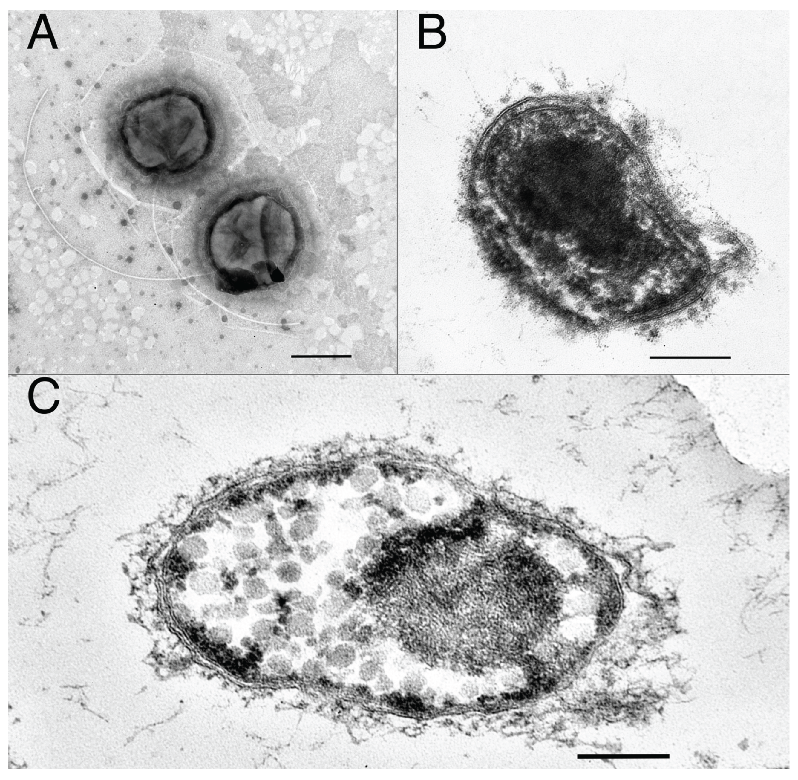
Figure 1. Transmission electron micrograph of the cells of 4302-coT: (A) negatively stained cells with flagella, bar 0.5 µm; (B) thin section of the cell grown on cellobiose, showing diderm (Gram-stain-negative) cell wall, bar 0.2 µm; (C) thin section of the cell grown on rhamnose, showing bacterial microcompartments (BMCs), bar 0.2 µm.
Preprints 231005 g001
Figure 2. Transmission electron micrograph of the cells of VF2T: (A) negatively stained cells with flagellum in fibrillar matrix, bar 0.5 μm; (B) thin section of the cell grown on sucrose, showing diderm (Gram-stain-negative) cell wall, bar 0.2 μm.
Preprints 231005 g002
Figure 3. Phylogenetic placement of Limisphaera vallis 4302-coT and L. sibirica VF2T based on concatenated amino acid sequences of 120 bacterial single-copy conserved marker proteins. The length of the alignment is 20,811 aa. Bootstrap values shown at the nodes. Bar, 0.1 change per position.
Preprints 231005 g003
Figure 4. Distribution of CAZyme genes in the genomes of cultivated and uncultivated representatives of Limisphaera genus.
Preprints 231005 g004
Figure 5. Schematic overview of metabolic processes reconstructed from Limisphaera genomes. Sugar catabolism: Glk – ROK family glucokinase, Pgi – glucose-6-P isomerase, PfkA/Pfp – ATP/pyrophosphate-dependent phosphofructokinase, Fba – class II fructose-bisphosphate aldolase, Gap – glyceraldehyde-3-phosphate dehydrogenase, Pgk – phosphoglycerate kinase, Ipgm – 2,3-bisphosphoglycerate-independent phosphoglycerate mutase, Eno – enolase, Pyk – pyruvate kinase, PpdK – pyruvate, phosphate dikinase, Zwf – glucose-6-phosphate 1-dehydrogenase, Pgl – 6-phosphogluconolactonase, Gnd – 6-phosphogluconate dehydrogenase, RpiB – ribose-5-phosphate isomerase, Rpe – ribulose-phosphate 3-epimerase, Tkt – transketolase, Tal – transaldolase, GalK – galactokinase, GalT - galactose-1-phosphate uridylyltransferase, GalE - UDP-glucose 4-epimerase, Pgm – phosphoglucomutase, XylA – xylose isomerase, XylB – xylulose kinase, ManA – mannose-6-P isomerase, AraA – L-arabinose isomerase, AraB – ribulokinase, AraD – L-ribulose-5-phosphate 4-epimerase, RbsK – ribokinase, RhaA – L-rhamnose isomerase, RhaB – rhamnulokinase, RhaD/FucA – bifunctional rhamnulose-P/fuculose-P aldolase, PvmJ – lactaldehyde dehydrogenase, PvmB – lactyl-P transferase, PvmG – lactate kinase, Por – pyruvate:ferredoxin oxidoreductase complex, PDH – pyruvate dehydrogenase complex. Fermentation: Ldh – lactate dehydrogenase, Pta - phosphate acetyltransferase, Ak – acetate kinase, Acd – ADP-forming acetate--CoA ligase, Aldh – acetaldehyde dehydrogenase, Adh – alcohol dehydrogenase, NiFe hyd – type 3 or type 4 NiFe hydrogenases. Aerobic respiration: ACIII – alternative complex III, BD – bd-type quinol oxidase, CBB3 – cbb3-type cytochrome oxidase, CAA3 – caa3-type cytochrome oxidase. Nitrogen metabolism: NarGHI – dissimilatory nitrate reductase, NrfAH – dissimilatory nitrite reductase, Hao - hydroxylamine oxidoreductase-like enzyme, Nor – nitric oxide reductase, NarB – assimilatory nitrate reductase, NirA – Fd-dependent assimilatory nitrite reductase. Iron reduction: MCx – multiheme cytochrome c with “x” heme-binding motifs. Arsenic respiration and detoxification: TTR – homolog of tetrathionate reductase with arsenate reductase activity, ArsC – cytoplasmic arsenate reductase, ArsM – arsenite methyltransferase, ArsB – arsenite efflux permease. Sulfur metabolism: NSR - NAD(P)H sulfur oxidoreductase, SudAB – sulfide dehydrogenase.
Preprints 231005 g005
Figure 6. Enzymatic activities of GH51 glycosidase Lim81 from strain 4302-coT. (A) Endoglucanase activity towards CMC of fractions with Lim81, plate was stained by 0.1% Congo red. Abbreviations: Lys – cell lysate after sonication, Pell-1, Pell-2 – pellet with insoluble proteins, CFE – cell free extract, Ft1- flow through fraction after application of CFE on HP column (unbinding proteins), Ft2 – fraction of washed from affine column unbound proteins, Aff-1, Aff-2 – fractions with Lim81 protein (according Figure S3_B), Ft3 – fraction of 500 mM imidazole, after column washing. (B) Substrate specificity of Lim81 towards different polysaccharides. Activity was measured at 70 °C and pH 5.0. Error bars represent the standard deviation of three replicates. (C) Influence of metal ions and reducing and denaturing agents on the activity of Lim81. Control – reaction mixture containing beta-glucan without any additives (Me+ or detergents). Relative activity is presented as a percentage of the specific activity of the enzyme in the presence of ions/detergents.
Preprints 231005 g006
Figure 7. Effect of (А) temperature and (В) pH on the specific activity of the recombinant Lim81. Activity was measured against beta-glucan with addition of 0.5 mM Ca2+.
Preprints 231005 g007
Table 1. Phenotypic characteristics differentiating strains 4302-coT, VF2T and Limisphaera ngatamarikiensis strain NGM72.4T [5]. All strains are Gram-stain-negative, motile cocci with monotrichous flagella, reproducing by binary fission, occurring single or in pairs and characterized by: the ability to grow aerobically on glucose, arabinose, sucrose, mannose, xylose, maltose, galactose, cellobiose, dextrin, locust bean gum; the inability to reduce sulfate; presence of MK-7 as a sole respiratory quinone; ND, No data available; +, positive growth or reaction; -, negative growth or reaction.
Characteristic 4302-coT VF2T NGM72.4T
Origin Water/fouling, hot spring, Geyser Valley, Kamchatka, Russia Water/sediment, hot spring, Goryachinsk thermal water basin, Russia Water/clay, hot spring, New Zealand
Cell size, μm 0.7-1.3 0.7-1.0 0.5-0.8
Color pink peach-white pink
Aggregates formation + + -
min/opt/max t 37/60-65/70 37/55/70 45/60-65/71
min/opt/max pH 6.2/7.4/9.2 6.5/8.5/9.5 5.6/8.1-8.4/8.9
min/opt/max NaCl, % 0/0/1.5 0/0/1.5 0/0/0.8
Catalase/oxidase -/+ -/+ +/+
Oxygen tolerance facultative anaerobe facultative anaerobe aerobe and microaerobe
Growth on substrates:
yeast extract + + -
peptone + + -
tryptone + + ND
fructose - + +
starch + + -
cellulose + + -
xanthan gum + + -
pectin - + -
pyruvate - - +
Reduction of:
nitrate + + -
Major fatty acids (>10%) C16 : 0, ai-C15 : 0, i-C16 : 0 i-C16 : 0, ai-C15 : 0, ai-C17 : 0 C16 : 0, ai-C15 : 0, i-C16 : 0, ai-C17 : 0, C18 : 0
G+C% 65.8 65.2 65.6
Genome size, Mb 3.94 4.32 3.91
Disclaimer/Publisher’s Note: The statements, opinions and data contained in all publications are solely those of the individual author(s) and contributor(s) and not of MDPI and/or the editor(s). MDPI and/or the editor(s) disclaim responsibility for any injury to people or property resulting from any ideas, methods, instructions or products referred to in the content.
Copyright: This open access article is published under a Creative Commons CC BY 4.0 license, which permit the free download, distribution, and reuse, provided that the author and preprint are cited in any reuse.