

Submitted:
31 August 2026
Posted:
01 September 2026
You are already at the latest version
Abstract
Research into molecular machinery of eukaryotic unicellular organisms (protists) gives a unique opportunity to unveil mechanisms of intricate cellular processes and complete life cycles at the level of a single cell as a whole organism. Knowledge of protistan molecular biology and life cycle shifts as adaptations to varying physical-chemical features of the environment helps resolve important issues like evaluation of the balance between external abiotic triggers and internal molecular drivers of cells development. Understanding molecular mechanisms of these processes is crucial for prognostic modeling of population growth of the ecologically and economically important protists like toxic species forming harmful algal blooms (HABs) that currently expand worldwide. Here, we review basic features of bloom-forming mixotrophic dinoflagellates as drivers of their excess proliferation and HABs. We unveil key components of molecular machinery involved in life cycle regulation of dinoflagellates and highlight the deficiency/redundancy of nitrogen- and phosphorus-containing substrates as modulators of life cycles, diversity and nutrition strategies, including mixotrophy and kleptoplasty. We discuss linkages between cellular and molecular biology, omics and ecological modeling, since merging research approaches of these disciplines is a promising perspective for future progress of molecular biology within the framework of translational aquatic ecology.
Keywords:
dinoflagellates
; harmful algal blooms
; kleptoplasty
; life cycle
; mathematical modeling
; mixotrophy
; molecular ecology
; nutrients
; transcriptomic analysis
; translational aquatic ecology
1. Introduction
Molecular machinery, including mechanisms maintaining signal transduction and gene regulation networks, provides a solid though flexible basis for development of cells and organisms within the entire biological life hierarchy, ranging from a variety of intracellular processes to complete life cycles. Among all living creatures, eukaryotic unicellular organisms (protists) are of particular importance for molecular ecologists as they render a unique opportunity to study these mechanisms at the level of a single cell as a whole organism. Knowledge of protistan molecular biology, including life cycle shifts as adaptations to varying physical and chemical features of the environment, is of paramount significance. This information helps resolve such sophisticated issues as chaotic population dynamics (Benincà et al., 2008; Medvinsky et al., 2015), enhancing the evaluation of the balance between external triggers (abiotic characteristics) and internal (cellular, molecular) drivers of cells and organisms development (Telesh et al., 2019; Cagle, Roelke, 2024). Comprehensive understanding of molecular mechanisms of these processes is crucial for the adequate assessments and forecasts of population growth of the ecologically and economically important protists and other influential microorganisms. Of particular interest are toxic and potentially toxic microalgae that cause harmful algal blooms (HABs).
Nowadays, HABs are intensifying and expanding not only at the regional level, e.g., as results of the excess of nutrients due to aquaculture development and increased discharges of agricultural waste waters and fertilizers (Hallegraeff et al., 2021; Zhang et al., 2023), but also worldwide (Glibert, 2020; Gobler, 2020; Hou et al., 2022; Glibert et al., 2025; Li et al., 2026). They occur in a variety of marine and continental water basins of different types, from small eutrophic ponds to vast oligotrophic oceanic regions, since many of those areas are strongly affected by climate change and growing anthropogenic eutrophication (Brown et al., 2020; Anderson et al., 2021; Orlova et al., 2022). The devastating effects of HABs on aquatic ecosystems and their biota are well known and have been repeatedly reviewed elsewhere (Landsberg, 2002; Gobler, 2020; Anderson et al., 2021, 2025; Hou et al., 2022; Dai et al., 2023; Skarlato, Telesh, 2024; Telesh et al., 2025a). In brief, usually by the end of a HAB, the decomposition of algal biomass accumulations results in oxygen depletion in the water column and deterioration of the quality of natural waters, which destabilizes the environment and causes irreparable harm to the flora and fauna, fisheries and fish farming, aquaculture, tourism and public health. The latter issues, along with direct toxic effects of some bloom-forming protists on aquatic organisms and humans, are generally listed among the major negative consequences of HABs (Orlova et al., 2022; Rattner et al., 2022; Occhipinti et al., 2025). However, the bloom onset most often remains enigmatic and unpredictable because of a number of impartial reasons. On the one hand, it depends on numerous, variable and mutually dependent environmental factors. On the other hand, the contribution of molecular data on bloom-forming species to disclosing the triggering mechanisms and driving forces of emergence, development, timing, magnitude and decay of HABs are largely unappreciated. A lack of this knowledge is the major reason for the insufficient effectiveness of many current prognostic models of HABs.
One of the key HAB-forming groups of protists are dinoflagellates (Dinoflagellata). A great part of ca. 2,500 known morphological species of these microalgae cause massive population outbreaks, or blooms, also called “red tides”, in coastal waters of seas and oceans. Moreover, out of the 200 reliably known morphological taxa of various marine phytoplankton whose cells produce toxins, more than 105 are dinoflagellates (Hallegraeff et al., 2021). Their toxins and other secondary metabolites cause up to 500,000 human intoxications per year, with an average mortality rate of approximately 1.5% (Wang, 2008). Besides, among microorganisms, dinoflagellates are the only group of plastid-containing protists capable of bioluminescence: nearly 70 species from more than 18 genera of dinoflagellates express bioluminescence, and the majority of them emit a blue-green light (Okolodkov, 2005, 2011; Marcinko et al., 2013; Timsit et al., 2021).
The effective bloom-forming capacities of many dinoflagellates are backed up by their molecular, cellular, physiological and metabolic peculiarities (Hackett et al., 2004; Okolodkov, 2005, 2011; Soyer-Gobillard, 2025). One of these particular features is the capacity for mixotrophic nutrition and kleptoplasty (Cruz, Cartaxana, 2022), which enhances cells population development often leading to HABs (Millette et al., 2023; Skarlato, Telesh, 2024; Telesh et al., 2025a, 2025b). However, little numerical data on the contribution of these specific characteristics to dinoflagellate HABs formation are available so far. Moreover, mathematical models of these devastating events, based on computational approaches, transcriptomic data, laboratory and theoretical experiments, and the analyses of long-term data series, as well as generalizations of the available knowledge on these topics are scarce (Calbet, 2026). Meanwhile, if available, they could have significantly contributed to the development of comprehensive HAB control strategies for environmental management aimed at effective protection of natural aquatic ecosystems and their resources.
In this review article, molecular features of bloom-forming mixotrophic dinoflagellate cells are considered as the basis for their excess proliferation, including the extreme red-tide events. We discuss the deficiency and redundancy of nutrition substrates in water containing nitrogen (N) and phosphorus (P), and their roles for dinoflagellates population growth and diversity studied in the experiments as well as in nature. We also highlight the effects of variations in nutrient concentrations and availability as modulators of life cycles and nutrition strategies (including mixotrophy and kleptoplasty) of HAB-forming planktonic dinoflagellates. In some sections, the article focuses on biology of a model red-tide microalgae species – the globally distributed mixotrophic, potentially toxic armored dinoflagellates Prorocentrum cordatum (Ostenfeld) J.D. Dodge, 1975, also known by its main synonym Prorocentrum minimum (Pavillard) J.Schiller 1933. Here, we review its peculiar molecular features studied in experiments with laboratory cultures. We also summarize the data on cellular and molecular mechanisms that drive the proliferation of natural P. cordatum populations forming blooms and characterize some of their traits in the southwestern coastal waters of the Baltic Sea.
Special attention is paid to the results of the transcriptomic analyses of molecular machinery involved in life cycle regulation of dinoflagellates, and molecular basics of their mixotrophic strategies in the changing environment. In conclusion, we highlight the dire necessity of including molecular data into conceptual and mathematical ecological models, and particularly the models of kleptoplasty in bloom-forming mixotrophic dinoflagellates, for proper evaluating and forecasting HABs. This approach is in the mainstream of the translational aquatic ecology (TAE) – one of the recently emerged scientific disciplines, which capitalizes on smart conversion of the fundamental ecological findings and hot-spot molecular biology discoveries conducive to their practical use in the essential human life activities: biotechnology, aquaculture, fisheries, environmental management, and nature protection (Skarlato et al., 2018). The authors believe that this review could provide hints to enhancing the linkages between cellular and molecular biology, omics, aquatic ecology and ecological modeling, since merging the approaches of these disciplines is a promising perspective for the future studies that will secure the progress of molecular ecology within the TAE framework.
2. Major Roles of Dinoflagellates in Aquatic Ecosystems
Dinoflagellates form an important marine and brackish-water phytoplankton cluster characterized by pronounced taxonomic and functional diversity. They are crucial to aquatic ecosystems due to high abundances, biomass and productivity. Dinoflagellates effectively fulfill nutrient cycling and energy transfer, synthesize specific toxins, implement benthic-pelagic coupling, form harmful red tides, and thus have huge overall ecological significance (Okolodkov, 2005, 2011; Bravo, Figueroa, 2014; Khanaychenko et al., 2019; Deng et al., 2023; Telesh et al., 2025a, 2026). Numerous studies prove that dinoflagellates play a key role in microplankton biodiversity (Hällfors, 2004; Gómez, 2005; Okolodkov, 2005, 2011). These protists account for more than 2500 actual morphological species and ca. 4000 fossil species (Taylor et al., 2008; Hoppenrath, 2017). The maximum taxonomic diversity of dinoflagellates and their highest abundances are usually registered in marine coastal waters and river mouths (Taylor, Pollingher, 1987; Graham, Wilcox, 2000).
Based primarily on molecular-phylogenetic studies of dinoflagellates, cell biology, modern taxonomy and systematics attribute these protists to a large monophyletic group, Alveolata, along with ciliates (Ciliophora) and sporozoa (Apicomplexa) (Cavalier-Smith, 1991; Fast et al., 2002; Adl et al., 2019). Molecular phylogeny proves that dinoflagellates are most closely related to Apicomplexa but not to Ciliophora (Leander, Keeling, 2003). Meanwhile, the evolutionary line of Alveolata belongs to a supergroup SAR, along with Stramenopila and Rhizaria (Burki et al., 2007). The latter supergroup was rather recently expanded to TSAR due to the addition of the phylum Telonemia after new molecular phylogeny data were obtained (Tikhonenkov et al., 2022).
Most peculiar features of the dinoflagellate cell organization comprise specificity of cellular coverings (Morrill, Loeblich, 1983; Netzel, Dürr, 1984; Pozdnyakov, Skarlato, 2012), unusual structure of the nucleus and permanently condensed chromosomes with distinct chromatin architecture (Rizzo, 2003; Golyshev et al., 2018; Wong et al., 2019). They are also characterized by variability and specific organization of ion channels that shed light on cell physiology (Pozdnyakov et al., 2018, 2020, 2021), and complex structure of chloroplasts (Keeling, 2010; Maréchal, 2024). Molecular features of dinoflagellates involve one of the largest genomes among the eukaryotes, extensive gene duplication, peculiar gene organization patterns such as tandem arrays, specific features of splicing, predominance of post-transcriptional and translational regulation over transcriptional control, and other processes (Bachvaroff, Place, 2008; Lin, 2011, 2024; Fukuda, Suzaki, 2015; Berdieva et al., 2026). These microalgae have a complex life cycle with the alternating vegetative and sexual phases that employ various strategies and include dormant immotile stages (Bravo, Figueroa, 2014; Figueroa et al., 2018). The sexual process in the life cycle of dinoflagellates is known and generally regarded as both a survival strategy in the adverse environmental conditions like nutrient deficiency, stressful temperature and salinity regimes, intense grazing, or parasite infection (Agrawal, 2012; Kalinina et al., 2023, 2025), and a proliferation strategy (Lin et al., 2025).
Among functional traits, the ability of dinoflagellates to symbiotic relations with corals is well known (Aranda et al., 2016). In marine and coastal ecosystems, dinoflagellates are crucial being major primary producers along with diatoms (Fensome, Munsterman, 2026). Dinoflagellates are an effective biological “carbon pump” and a means of transporting nutrients within the food web, serving as food for microzooplankton and fish larvae (Winder, Sommer, 2012; Mitra et al., 2014; Khanaychenko et al., 2019). They can form devastating HABs in marine and brackish-water coastal areas worldwide (Heil et al., 2005; Orlova et al., 2014, 2022; Li et al., 2022), including the Arctic seas (Okolodkov, 2005; Silva et al., 2023). In addition, it has been established that the biomass of these protists during the blooming period can be used for thermochemical conversion production of liquid biofuels with its subsequent catalytic purification (Zhou, Hu, 2020). On a global time scale, because of the mass decomposition of algal cells after the cessation of the bloom, thecae of dinoflagellates accumulate in bottom sediments and contribute to the formation of fossil fuels (Okolodkov, Cervantes-Urieta, 2026).
Many dinoflagellates produce structurally diverse toxins that are derived via divergent pathways (Tango et al., 2005; Grattan et al., 2016; Verma et al., 2019; Guillotin, Delcourt, 2022). The main toxin-producing dinoflagellate species in plankton belong to the genera Prorocentrum, Alexandrium, Dinophysis, Gambierdiscus, Ostreopsis and Karenia. The dinoflagellate toxins cause nausea, vomiting, abdominal pain, diarrhea, and neurologic symptoms such as fatigue, myalgias, pruritus, circumoral paresthesias, reversal of hot and cold sensation, psychiatric abnormalities, and memory loss; the neurologic symptoms may persist for months to years (Wang, 2008).
Along with toxins sensu lata, marine dinoflagellates also produce a wide range of other biologically active secondary metabolites, including unique pigments like peridinin (Takaichi, 2011) and specific proteins, although the roles of some of them remain poorly understood so far (Pechkovskaya et al., 2021, 2023; Filatova et al., 2025). As demonstrated for various microalgae, some of these compounds possess antiviral (Carbone et al., 2021), antibacterial (Najdenski et al., 2013), antifungal (Li et al., 2006), and antitumor (Martínez Andrade et al., 2018) properties; therefore, they can be used in pharmacology and food industries (Pulz, Gross, 2004; Borowitzka, 2013). Much attention has been paid to dinoflagellates’ metabolites containing complex compounds with diverse chemical natures (Assunção et al., 2017), including macrolides, cyclic polyethers, spirolides, purine alkaloids, and other components (Kellmann et al., 2010).
Currently, dinoflagellates are being actively used in the search for new natural migrastatic substances that could inhibit cancer cells motility, thereby preventing metastasis. For example, recent studies using the MALDI-TOF technique (Calvano et al., 2015) have identified biologically active secondary metabolites, including pigments chlorophyll a and peridinin, and proteins (chaperones, cytoprotective and antioxidant proteins), produced by the potentially toxic dinoflagellates Prorocentrum cordatum (Filatova et al., 2024, 2025). Specifically, using adherent murine hepatoma MH22a cells as one of the target models it was found that the ethanol extract of P. cordatum, due to its low toxicity and ability to suppress cell adhesion and migration, appeared to be the most promising candidate for further studies as a potential biologically active agent (Filatova et al., 2025).
In aquatic ecosystems, dinoflagellates also facilitate connectivity between the pelagic and benthic communities by producing cysts that accumulate on the bottom but re-inoculate the water column when favorable conditions for excystation are restored (Bravo, Figueroa, 2014; Skarlato et al., 2018; Liu et al., 2020). The life cycle complexity of these protists defines the variability of their dormancy strategies; therefore, duration of the immotile persistence of dinoflagellates in the ecosystem is determined by the type of cysts. The thick-wall long-term dinosporin-containing resting cysts are characterized by a significant period of dormancy, which can reach several months or even years (Bravo, Figueroa, 2014). The thin-wall — pellicle or thecate — temporary cysts do not undergo prolonged dormancy and stay immotile for a few hours or days, although sometimes for months (Bravo et al., 2010; Bravo, Figueroa, 2014; Matantseva et al., 2020). Consequently, the nature of the benthic-pelagic coupling involving HAB-forming dinoflagellates is complex and ambiguous, and these interactions play a dual role in the formation of red tides: first, due to cyst formation and deposition at the bottom and, second, because of the composite, non-linear effects of benthic feeding on nutrients content in water. Concerning the latter, on the one hand, the excretion of available nutrients by organisms in benthos such as bivalves, accompanied by the excystation of dinoflagellates under favorable conditions and their release into plankton, increases the trophic status of a water body and promotes HABs. On the other hand, the elimination of microalgae by these same mollusks during their intense feeding by filtration reduces the population size of bloom-forming species and thus prevents red tides (Telesh et al., 2025a).
Many HAB-forming planktonic dinoflagellates thrive in various aquatic environments and withstand external stressors due to their wide ecological niches, like the cosmopolitan marine mixotrophic species Prorocentrum cordatum, particularly with respect to temperature, pH, salinity, and concentration of basic nutrients (Telesh et al., 2016). Besides, largely due to mixotrophy of many dinoflagellate species (Stoecker et al., 1991, 1997, 2017; Matantseva, Skarlato, 2013), the broad nutrition spectrum is another great ecological advantage of these protists (Jeong et al., 2010, 2021). Mixotrophic dinoflagellates utilize a wide range of organic and inorganic resources available for their growth and reproduction, often changing trophic preferences depending on environmental conditions (Abassi, Ki, 2022; Li et al., 2026). Specifically, by using stable isotope tracers and NanoSIMS, it was shown that individual P. cordatum cells within a population displayed significant heterogeneity in the rates of nutrient uptake and extent of the urea-mediated inhibition (by 30-40%) of the nitrate uptake (Matantseva et al., 2016). Thus, starting to consume urea at a high rate as soon as it appeared in the environment, P. cordatum demonstrated the rates of urea-N uptake that were 1.6-4.1 times higher than those of nitrate-N in P. cordatum population (Matantseva et al., 2016). Moreover, these dinoflagellates also demonstrated the uncoupled assimilation of carbon and nitrogen from urea and the amino acid glycine (Matantseva et al., 2018). The discovered functional heterogeneity of these protist populations, leading to the putative co-existence of various sub-populations with differing nutrition modes, ensures the ecological success of these dinoflagellates and enhances their competitive advantages in stressful environments.
The above features contribute to the proliferation of dinoflagellates in diverse habitats, in water bodies of different types, and the expansion of red tide zones therein (Figure 1). They also allow these protists to compete successfully with closely related species, as demonstrated by the invasion history of P. cordatum in the Baltic Sea and its displacement of the previously dominant congeners among planktonic dinoflagellates therein (Telesh et al., 2016). Thus, knowledge of the nutrition strategies of these flagellates and new data on their ecological niches have already made a significant contribution to HAB research (Telesh, Skarlato, 2022). Specifically, it was shown that different trophic strategies of these microorganisms determine their ability to rapidly proliferate by modulating growth rate and reproductive efficiency, providing competitive advantages, high invasion potential and range expansion (Matantseva et al., 2016, 2018; Telesh et al., 2016, 2021, 2024, 2025a, 2025b, 2026).
Enumeration of peculiar molecular, cellular and physiological features and functional traits of dinoflagellates can be continued, and their list can be much longer. However, we will cease it for now, since the subsequent sections of this review will focus primarily on the essential nutrients and their roles in the molecular machinery of dinoflagellate trophic strategies, reproduction and life cycle regulation. All together, these characteristics secure pronounced adaptability of these protists to climate changes and anthropogenic stresses; enhance their persistence, population growth, and HABs formation.
3. Nitrogen and Phosphorus as Essential Nutrients Modulating Dinoflagellate Cell Physiology
Nitrogen (N) and phosphorus (P) are the two biogenic elements whose availability in the environment constitutes one of the principal prerequisites regulating various aspects of dinoflagellate cell physiology. Nitrogen is a structural component of amino acids, nucleotides, and chlorophyll molecules. The availability of nitrogen limits the productivity of marine microbial communities and plays an essential role in structuring of phytoplankton species composition (Bertrand, Allen, 2012; Moore et al., 2013; Shibata et al., 2015). Marine phytoplankton provide a major proportion of global primary production and modulate biogeochemical cycles by assimilating nitrogen. It is usually assumed that most microorganisms can use dissolved inorganic nitrogen (DIN) in the form of nitrate, nitrite, and ammonium (Zehr, Ward, 2002). Dissolved organic nitrogen (DON) can be a large pool in the oceans and an even larger one in coastal waters. A significant fraction of this pool is composed of amides (including urea); it also includes heterocyclic compounds, proteins and amino acids (Zehr, Ward, 2002). Nitrogen limitation affects metabolic processes in dinoflagellate cells, enhancing glycolysis, the pentose-phosphate pathway, the tricarboxylic acid cycle, and fatty acid metabolism, and altering expression of genes encoding photosystem proteins (Cooper et al., 2016; Hou et al., 2018; H. Li et al., 2021; T. Li et al., 2021; Shi et al., 2021). Nitrogen deficiency activates nitrate/nitrite and urea transporters to increase assimilation of available nitrogen sources (H. Li et al., 2021; Shi et al., 2021).
Phosphorus is a biogenic element, which plays a crucial role in the cells as it is a component of nucleic acids (DNA, RNA), cell membranes (phospholipids), signaling molecules (inositol triphosphate, cAMP), nicotinamide adenine dinucleotide (NAD) and its phosphorylated form (NADP), adenosine triphosphate (ATP, an energy currency), etc. Phosphorylation activates many enzymes necessary for maintaining basic metabolic pathways and regulating the cell cycle. Thus, phosphorus availability has a significant impact on the viability and growth of marine phytoplankton and, consequently, on their primary production in the seas and oceans.
Phosphorus can be available to dinoflagellates in the form of dissolved inorganic phosphate, DIP (primarily orthophosphate [Pi]) and as a variety of dissolved organophosphorus (DOP) compounds such as ATP, cytidine-5-monophosphate, fructose-6-phosphate, glucose-6-phosphate, glycerophosphate, uridine-5-monophosphate, phenyl phosphate, RNA, etc. (Antia et al., 1990; Huang et al., 2005; Oh et al., 2010; Li et al., 2015; Lin et al., 2016). DOP appears to be the main source of this nutrient during algal blooms, when inorganic nutrients are depleted (Huang et al., 2022). Phosphorus deficiency stimulates phosphate uptake and intracellular recycling (Lei, Lu, 2011; Morey et al., 2011; Zhang et al., 2014; Li et al., 2016). For example, proteomic analysis of phosphate starved Karenia mikimotoi revealed overexpression of vacuolar phosphate transporters, the proteins associated with vesicular transport and proteins responsible for lipid and phospholipid degradation (Lei, Lu, 2011). K. mikimotoi and Prorocentrum shikokuense Y.Hada[1] have also been shown to replace membrane phospholipids partially with glycerol glycolipids and betaine lipids (Huang et al., 2023; Li et al., 2024). In freshwater ecosystems, despite the historical “dogma” defining them as strictly phosphorus-limited (Hecky, Kilham, 1988), many systems exhibit co-limitation, where both N and P availability simultaneously constrain primary production (Bracken et al., 2015; Lewis et al., 2020).
Notably, toxin biosynthesis depends on availability of both elements. Primarily, nitrogen is required for the production of saxitoxins, as shown for Alexandrium tamarense and Alexandrium minutum (Béchemin et al., 1999; Chen et al., 2024). The most recent studies indicated that high nitrogen supply enhanced not only saxitoxin biosynthesis, but also toxin release: excess nitrogen promoted sxtA4 gene expression by Alexandrium catenella (Whedon & Kofoid) Balech, with a positive correlation between sxtA4 and total yield (Lian et al., 2026). Nevertheless, the production of okadaic acid (diarrheic shellfish toxin) is modulated by phosphorus level (Chen et al., 2025).
4. Trophic Strategies of Dinoflagellates in the Changing Environment: Mixotrophy and Kleptoplasty as Its Peculiar Constituent
In recent years, researchers have been paying much attention to studying not only the ecological prerequisites of dinoflagellate blooms but also the intracellular drivers and molecular mechanisms that back up their trophic strategies and secure fast population growth often leading to HABs (Soyer-Gobillard, 2025). Mixotrophy is one of the nutrition modes typical for many bloom-forming dinoflagellates that combine autotrophy due to photosynthesis in the light using inorganic nutrient substances with heterotrophy in the forms of osmotrophy and phagotrophy (Stoecker, 1991; Stoecker et al., 1997, 2017; Matantseva, Skarlato, 2013; Mansour, Anestis, 2021). Osmotrophy involves the consumption of dissolved organic compounds by osmosis, and phagotrophy is the ability of cells to engulf, absorb and assimilate solid organic substances, such as microalgae and bacterial cells. Currently, photo-osmo-mixotrophy is considered a generally accepted and widespread nutritional mode of microalgae (Mitra et al., 2023). At the same time, mixotrophy, which includes phagotrophy, is not ubiquitous; moreover, the modes of use of this nutritional strategy also vary significantly among diverse planktonic protists (Mitra et al., 2016, 2023).
The combination of the two strategies, photo-osmo-mixotrophy and photo-phago-mixotrophy, provides high adaptability of protists to fluctuations in the composition and abundance of food resources and, consequently, increases the ecological homogeneity of many unicellular mixotrophic organisms, which provides the basis for their unification within the concept of “mixoplankton” (Flynn et al., 2013, 2019). Mixoplankton (i.e., plankton capable of mixotrophy) are generally characterized by high adaptability to fluctuating environment (Glibert, 2015; Wilken et al., 2020). This is primarily due to the ability of mixoplankton to utilize various alternative resources (Matantseva et al., 2016, 2018), and their broad ecological niches, including a wide range of food substrates (Telesh et al., 2016, 2024; Jeong et al., 2021). As a result, because of the high adaptability and rapid growth of mixoplankton populations, they are often the reason of HABs (Anderson et al., 2019; Carstensen et al., 2020; Glibert, 2020; Hallegraeff et al., 2021; Karlson et al., 2021).
Currently, an increasing number of dinoflagellate species are exhibiting mixotrophic properties, and this enhances their resilience to changing environmental conditions, including shifts in the composition and concentration of nitrogen- and phosphorus-containing compounds or decreased light, which impedes photosynthesis. Specifically, phosphorus deficiency stimulates phagotrophy in some mixotrophic species of dinoflagellates, such as Prorocentrum cordatum and Tripos sp. (the latter was described as Ceratium furca (Ehrenberg) Claparède & Lachmann) (Smalley et al., 2003; Johnson, 2015; Berdieva et al., 2020), and increases expression and activity of cell-surface alkaline phosphatase (Dyhrman, Palenik, 1997; Lin et al., 2012; Ou et al., 2020). A lack of phosphorus in the environment induces the expression of plasma membrane and vacuolar phosphate transporters as well as proteins associated with vesicular transport (Lei, Lu, 2011; Kalinina et al., 2025). Nitrogen depletion can also induce phagotrophy in P. cordatum (Johnson, 2015) and Karlodinium veneficum (D.Ballantine) J.Larsen (described as Gyrodinium galatheanum (Baarud) Taylor in: Li et al., 2000), although to a lesser extent than phosphorus. Activation of endocytosis and phagosome-related pathways has also been detected at the transcriptomic level (H. Li et al., 2021).
Recently, it has been shown that none of the dominant HAB-forming dinoflagellate species in the coastal waters of the southwestern Baltic Sea is purely autotrophic since seven out of ten of these dominants are mixotrophs, while the remaining three species are heterotrophs (Telesh et al., 2026), following the classification by Jeong with co-authors (2021). Mixotrophy involving phagotrophy is observed in all phytoplankton clades, with the exception of diatoms and cyanobacteria (Borics et al., 2021). Moreover, nowadays mixotrophy is considered not just a nutritional strategy, but also a unique life strategy for dinoflagellates and many other protists (Jeong et al., 2021; Chen et al., 2025; Glibert et al., 2025). The environmental triggering of mixotrophy is highly complex and multidimensional (reviewed by Calbet, 2026). Recent transcriptomic analyses have revealed that mixotrophy is not just the synergistic process of “grazing while photosynthesizing”; rather, prey availability can trigger large-scale transcriptional reprogramming that shifts cellular metabolism toward heterotrophic pathways (Lie et al., 2017; Ma et al., 2023; Calbet, 2026). However, some subtle molecular and cellular aspects of mixotrophic metabolism remain poorly understood (Glibert, Mitra, 2022).
Kleptoplasty is one of the less studied though most enigmatic intracellular mechanisms of mixotrophy (Nishitani et al., 2011). The term “kleptoplasty” refers to the processes of acquisition, accumulation, and utilization of kleptoplasts, i.e., unassimilated chloroplasts of microalgae absorbed by heterotroph and mixotroph cells during phagotrophic nutrition (Gast et al., 2007; Rumpho et al., 2007; Kim et al., 2012; Millette et al., 2023). Kleptoplasty is observed in a wide range of not only unicellular organisms such as dinoflagellates, ciliates, and foraminifera (Stoecker et al., 2009; Pillet, Pawlowski, 2013), but also in multicellular invertebrates, for example, sacoglossan sea slugs of the genus Elysia (Rumpho et al., 2007), and some flatworms (Van Steenkiste et al., 2019).
It has been established that kleptoplasts not just persist, but also photosynthesize in the host cell, sometimes for a relatively long period, up to several months (Stamatakis et al., 2017), after which the photosynthesis of kleptoplasts is diminished and finally lost, and/or the “stolen” plastid is digested (Mansour, Anestis, 2021). For example, in Nusuttodinium aeruginosum (F.Stein) Y.Takano & T.Horiguchi, kleptoplasts can be inherited by the daughter cells through five generations, and can even be found in the resting cysts (Onuma, Horiguchi, 2015). Along with their own plastids, kleptoplasts in mixotrophs serve as additional “reactors of photosynthesis” (Skarlato, Telesh, 2024; Telesh et al., 2025b). For the majority of photophago-mixotrophic protists, however, the question of how great the contribution of functioning kleptoplasts to the overall photosynthesis and cell growth is – remains open.
Dinoflagellates obtain kleptoplasts from prey algae such as various representatives of cryptophytes (Li et al., 2000), haptophytes (Berge et al., 2008), chlorophytes (Strom, Buskey, 1993), prasinophytes (Berge et al., 2008), raphidophytes (Nakamura et al., 1995), diatoms (Menden-Deuer et al., 2005; Yoo et al., 2009), and other dinoflagellates (Tillmann, 2004; Adolf et al., 2006). The pathway of plastid acquisition can be intricate. Dinophysis acuminata Claparède & Lachmann acquires plastids of Geminigera/Teleaulax cryptophyte from the captured ciliate Myrionecta rubra (Lohmann) Jankowski (Wisecaver, Hackett, 2010). As a result, the two outer plastid membranes, the cryptophyte nucleus and the nucleomorph, are lost. At the same time, the nuclear genome of D. acuminata contains five plastid genes from multiple algal lineages, only one of which was derived from a cryptophyte, and this implies instability of the photosynthetic system in these dinoflagellates (Wisecaver, Hackett, 2010). Meanwhile, the Antarctic Ross Sea dinoflagellate appears to retain its ancestral secondary plastid and obtains temporary kleptoplasts stolen from haptophyte prey partitioning functions between the two plastid’ systems (Hehenberger et al., 2019). Moreover, this dinoflagellate has acquired nuclear genes by horizontal gene transfer (HGT) from outside the kleptoplast lineage; however, those HGT-genes encode kleptoplast-targeted proteins. Their expression is not strongly changed by environmental conditions and many of them are shared with related dinoflagellates with fully integrated plastids. Such a pattern suggests that the protein targeting system had developed before the plastid became permanently fixed (Hehenberger et al., 2019).
In this regard, a separate issue is the fate and physiological role of captured plastids in a mixotrophic dinoflagellate cell. Laboratory experiments showed that, for example, for Prorocentrum spp. (P. micans Ehrenberg, P. cordatum), the cryptophyte microalgae are most often the optimum prey in terms of cell size and consumption rate (Jeong et al., 2010). The results of the long-term Baltic Sea database analyses support this outcome by demonstrating the co-existence of P. cordatum and P. micans with cryptophyte species like Hemiselmis virescens Droop or Teleaulax sp. in plankton, resulting in strong and statistically significant (p < 0.05) positive correlations between the abundance of Prorocentrum spp. and the cryptophytes (Table 1).
In the meantime, neither the laboratory studies of the dinoflagellates feeding rates nor the field data on their co-occurrence with certain prey objects can provide information on the efficiency of kleptoplasty, while the experimental investigations of this specific and delicate intracellular process are problematic yet. Thus, in most cases, the real contribution of kleptoplasty to dinoflagellate cells growth remains unknown; therefore, this phenomenon has not been taken into account in the majority of available mathematical models of the growth and population dynamics of mixotrophic organisms (Flynn, Mitra, 2009; Mitra et al., 2014; Chen et al., 2025). The only exception known so far is a mathematical model of kleptoplasty based on the fundamental J. Monod model of microorganisms growth (Monod, 1949), a series of theoretical experiments (“thought experiments”), and a set of differential equations for a simple biological system, which consisted of one mixotrophic protist cell, one inorganic resource, and one organic resource (Telesh et al., 2025b). The model was developed and tested using experimental data on the feeding of dinoflagellates Prorocentrum cordatum on the cryptophyte microalgae Teleaulax sp. (Jeong et al., 2010, 2021), taking into account their reproduction rates and morphometry (Heil et al., 2005). This study also allowed the authors to suggest an original kleptoplasty index (k) – a new ecological indicator reflecting the quantitative contribution of kleptoplasty as an important intracellular process to HABs formation. In particular, the model has shown that kleptoplasty of the mixotrophic P. cordatum using Teleaulax sp. as food can increase the cell division rate by up to 40%, promoting rapid population growth of these dinoflagellates and the subsequent algal bloom (Telesh et al., 2025b).
In the future modeling studies, the assessment of the role of kleptoplasty in the formation of dinoflagellate HABs can be correlated with the major cellular parameters and molecular mechanisms that determine the functioning of these protists. Thus, further research into molecular basics of kleptoplasty as one of the key mixotrophic nutrition mechanisms affecting dinoflagellate HABs can be a future prospect of molecular ecology, which could help understand algal blooms and contribute to prognostic mathematical modeling of HABs.
5. Effects of Nitrogen Redundancy on the Expression of N-Transporters’ Genes, Transcriptional Responses, Diversity, and Dinoflagellates Population Growth
The abundance, composition and proportions of different inorganic and organic nutrients as food substrates in the environment play a crucial role in mixoplankton development and proliferation. The presence of different forms of nitrogen is mandatory for various cellular metabolic processes of dinoflagellate cells that secure their maintenance, growth, and reproduction (see also Section 6). For example, experimental results suggest that mixotrophic dinoflagellates Prorocentrum cordatum are sensitive to the proportions of N-containing substrates and can adopt the most effective strategy for assimilation of available nitrogen sources in the changing environment, demonstrating response to shifts in their composition (Matantseva et al., 2016, 2018; Berdieva et al., 2021a; Pechkovskaya et al., 2020; Kalinina et al., 2023). It was discovered that the expression levels of the genes dur3 and nrt2 encoding the transporters of urea and nitrate, respectively, in the NO3-grown cultures depended on the added nitrogen sources being the lowest in the presence of ammonium (Pechkovskaya et al., 2020).
Studies of the transcriptional responses of P. cordatum to available N-containing substrates allowed estimating the influence of NO3−, NH4+ and urea on the transcription levels, as well as on the expression of urea and nitrate transporter genes in these dinoflagellates. Specifically, it was shown that dur3 expression levels were downregulated after the supplementation of additional N sources and were 1.7–2.6-fold lower than in the nitrate-grown culture, while the nrt2 expression levels decreased 1.9-fold in the presence of NH4+ (Pechkovskaya et al., 2020). Total RNA and DNA synthesis rates, estimated by the analysis of incorporation of 3H-thymidine and 3H-uridine in batch and continuous cultures, have demonstrated that the addition of N compounds did not affect the DNA synthesis rates, while the transcription levels increased up to 12.5-fold after the excessive N supplementation (Pechkovskaya et al., 2020). These investigations of various nitrogen sources as biomarkers of dinoflagellate proliferation due to their differentiated impact on expression of dur3 and nrt2 genes and modification of transcription rates in the mixotrophic P. cordatum allowed concluding that the knowledge on nitrogen redundancy, diversity and proportions of N-substrates in the environment is of paramount importance. Incorporation of these data into ecological models could increase the precision of the future HAB forecasts under global N pollution and eutrophication.
Meanwhile, along with different nutrient sources, natural systems are characterized by fluctuating light, episodic nutrient pulses, variable prey communities, and shifting thermal gradients; therefore, the kinetics of trophic switching in mixoplankton under such dynamic conditions and their effects on cells proliferation remain poorly characterized (Calbet, 2026). Besides, inter- and intra-population heterogeneity of cells as well as the ambiguity of dinoflagellates’ behavior under various stresses (Matantseva et al., 2020; Pechkovskaya et al., 2021) and their peculiar life cycles (Kalinina et al., 2018, 2020, 2023; Berdieva et al., 2020, 2021a,b) hamper the usage of the available data on dinoflagellate cell biology for prognostic mathematical modeling of their HABs. Furthermore, the molecular mechanisms linking environmental sensing to the coordinated regulation of phototrophic and heterotrophic machinery are only just starting to be mapped (Calbet, 2026).
Nevertheless, certain advances in unveiling the linkage of some environmental prerequisites and fine cellular-molecular mechanisms underlying dinoflagellate blooms have been achieved in recent decades. For example, it has been shown that the concentration of total nitrogen (TN) in water affects development and growth of protist cells as well as the overall phytoplankton composition, abundance and distribution; thus, TN can be considered an anthropogenic stress marker (Rönnberg, Bonsdorf, 2004; Howarth, 2008; Schiewer, 2008; Mekonnen, Hoekstra, 2015; Zhang et al., 2023). Recently, it has also been demonstrated that dinoflagellates with the widest TN-niche limits (along with broad niches for total phosphorus, water temperature and salinity) have the strongest potential for most frequent bloom events in the fluctuating coastal environments (Telesh et al., 2024). The latter study suggested a conceptual model for merging the bloom-forming capacity of dinoflagellates with the width of their TN niches (Figure 1), and this approach allowed coming closer to prediction of red tides.
Broad range of organic and inorganic resources used by mixoplankton underpins coexistence of quite a large number of mixotrophic dinoflagellates in space and time, as demonstrated by the data from the Baltic coastal waters (Telesh et al., 2016, 2021, 2024, 2025b). Meanwhile, our recent research hypothesis, suggesting those coastal regions with TN concentrations that are optimal for bloom-forming dinoflagellates to host greater taxonomic diversity of these protists, compared to the areas with non-optimum TN content, unexpectedly was not supported by the analyses of the 82 dinoflagellate species in the 44 years-long database from the SW Baltic Sea (Telesh et al., 2026). Instead, surprisingly, the highest species richness of these planktonic protists was observed at relatively low eutrophication level (TN < 15 µmol/L). Moreover, the study has demonstrated that the diversity of dominant bloom-forming dinoflagellate species decreased gradually from low to moderate, high, and very high eutrophication levels, with the minimum number of species in the extremely eutrophic waters at TN > 45 µmol/L (Telesh et al., 2026). These results disagree with the theoretically expected species-richness maximum at the intermediate TN concentrations, as predicted by the intermediate disturbance hypothesis (IDH), which postulates that maximum species richness should be expected at moderately stressed conditions (Connell, 1978). A possible though speculative explanation of this discrepancy could involve the linkage between the dinoflagellate growth rates and TN disturbance frequencies rather than magnitude (Gaedecke, Sommer, 1986; Weider, 1992). As shown earlier, usually a species diversity maximum is observed when disturbance frequencies are equal to a three-generation cycle (Lampert, Sommer, 2007). For dinoflagellates at nutrient-saturated (i.e., optimum) conditions, the growth rates between ca. 0.3–0.7 day-1 are known from laboratory experiments (Jia et al., 2019), which is in accord with field observations (Baek et al., 2008) and the knowledge that at suboptimal conditions, the algal growth rates decrease. Consequently, this will shift the optimum disturbance pattern from ca. 4–10 days to much longer periods, allowing for other disturbance factors than TN to serve as stressors affecting the dinoflagellates competition pattern. In turn, slower growth of the superior competitor generally favors the enhanced survival time of the inferior ones. In such a case, the relation between these parameters could secure different competition patterns at the optimal and suboptimal TN conditions, promoting higher species richness of mixotrophic dinoflagellates at suboptimal TN concentrations due to lower growth rates and, therefore, less effective competition for nutrients (Telesh et al., 2026).
Thus, similarly to the ambiguous effects of benthic-pelagic coupling involving HAB-forming mixotrophic dinoflagellates (Telesh et al., 2025a), the effects of nitrogen redundancy on dinoflagellate diversity is also nonlinear and enigmatic. This fact demonstrates that nutrient-driven regulation of biodiversity and trophic strategies, including mixotrophy, is a dynamic, multi-faceted stoichiometric optimization, which is shaped by cells assimilation efficiency, functional architecture of aquatic assemblages, and the complex ever-changing reality of the marine environment (Calbet, 2026). Therefore, the role of TN excess and overall nitrogen redundancy in dinoflagellate cells growth, gene expression, transcriptional responses, and population development of these mixoplankters requires further investigations. Prospective studies could include testing the applicability of Tilman’s resource competition theory (RCT; Tilman, 1977, 1980; Zhang, Becks, 2025), aiming to unveil the unexpected response of dinoflagellate biomass to N-availability, as described above. Previously, the applicability of RCT has been shown by both theoretical and practical approaches (e.g., Smayda, Reynolds, 2003; Bi et al., 2021), at least for the competitive relations between dinoflagellates and diatoms. However, with respect to nitrogen as a co-limiting resource, the competition of diatoms and dinoflagellates with cyanobacteria still needs to be unravelled. The existing knowledge refers to several potential mechanisms ranging from vertical migration (e.g., Fisher et al., 2013) to allelopathic interactions (e.g., Vardi et al., 2002). However, a comprehensive study, which also takes into account the variable and complex species-specific trophic strategies (Smayda, Reynolds, 2003), is still lacking.
6. Nitrogen and Phosphorus Deficiency as Major Stressors Modulating Life Cycles of Dinoflagellates
Nitrogen and phosphorus deficiency limits cell proliferation and can lead to cell cycle arrest, as shown for various dinoflagellate species (e.g., Blanco, 1995; Zhang et al., 2014; Li et al., 2016; Zhao et al., 2017; Chen et al., 2024). Particularly, phosphorus deficiency leads to cell cycle arrest in G1/G0 phase, although the cells continue to grow and increase in size (Lei, Lu, 2011; Zhang et al., 2014; Li et al., 2016). At the molecular level, the lack of nutrients suppresses cell-cycle regulators such as G1/S-specific cyclin and cyclin-dependent kinases (Shi et al., 2017; H. Li et al., 2021). In Alexandrium catenella, phosphorus deficiency reduces expression of calcium-dependent protein kinase (CPK), which plays a crucial role in signal transduction and cell division in plants. In contrast, fizzy/cell division cycle 20-related protein (FDC) is upregulated; it acts as a negative regulator of the cell cycle, inhibiting cell division by regulating the anaphase-promoting complex (APC) and maintaining the required levels of mitotic proteins (Zhang et al., 2014). More importantly, nitrogen and phosphorus depletion serves as a notable signal that initiates life-cycle transitions promoting long-term population survival.
Phosphorus deficiency can trigger the transfer to sexual phase in complex life cycles of diverse marine dinoflagellates (Supplementary Materials, Table S1). Among those species are Prorocentrum cordatum, Lingulaulax polyedra (syn. Lingulodinium polyedra), Gymnodinium nolleri, Protoceratium reticulatum, and Alexandrium minutum (Figueroa, Bravo, 2005; Figueroa et al., 2006c; Figueroa et al., 2011; Salgado et al., 2017; Kalinina et al., 2023). These processes included gamete formation and fusion, planozygote formation and encystment.
Life cycle transition has been well described in the clonal culture of P. cordatum, where two weeks of phosphate deprivation led to increase in the proportion of planozygotes with relative nuclear DNA content 2C and 4C (Kalinina et al., 2023). Transcriptomic analysis at this stage revealed increase in the expression of six homologs of meiosis-associated genes, that are rad51, spo11, RAD21/REC8-like protein coding gene, exo1, msh4 и msh5, responsible for meiotic recombination and double-strands break DNA reparation (Kalinina et al., 2025). The addition of full medium containing sufficient amounts of all the nutrients stimulated cell divisions and transition to vegetative 1C phase (Kalinina et al., 2023).
Nitrogen limitation also serves as a nutrient-related trigger of transition to the sexual phase (Table S1). In laboratory experiments, cultivation under nitrogen deficiency induced sexuality in various marine dinoflagellates — Alexandrium taylori, Karenia brevis, Kryptoperidinium triquetrum (syn. K. foliaceum), Levanderina fissa (syn. Gyrodinium uncatenum), and Scrippsiella lachrymosa (Anderson et al., 1985; Figueroa et al., 2006a; Figueroa et al., 2009; Persson et al., 2013; Persson et al., 2021). In Lingulaulax polyedra, planozygotes encysted in N-depleted medium, as in P-depleted conditions, although nitrogen deficiency yielded more temporary cysts while phosphorus deficiency produced a greater yield of resting cysts (Figueroa, Bravo, 2005). Gymnodinium catenatum showed a similar switch in response to nitrogen deficiency (as well as phosphorus deficiency), but only this condition, like replete medium, allowed most planozygotes to undergo division, and more cysts to successfully germinate and retain viability, despite frequent formation of faulty cyst walls (Figueroa et al., 2006b).
Notably, available literature on freshwater species mostly reports the effects of nitrogen deficiency on reproductive strategy that are consistent with some of the more complex ideas about nutrient regulation in freshwater ecosystems. Several studies describe Peridinium species induced to enter the sexual process, including encystment of planozygotes (Pfiester, 1975, 1977, 1984; Sako et al., 1984; Chapman, Pfester, 1995) (Table S1).
Transcriptomic data reflecting reproductive strategy transitions in response to nitrogen limitations in dinoflagellates also remain sparsely discussed. Nevertheless, Alexandrium minutum has been shown to upregulate some genes coding for meiosis-associated proteins under nitrogen deficiency, for example, homologs of the double-strand break repair protein MRE11, topoisomerase II, DNA mismatch repair protein MLH3, and about two dozen homologs of the RNA-binding protein MEI2 (Akbar et al., 2023). MEI2-like proteins are abundant in dinoflagellates and appear to form two putative functional sets: one related to the sexual phase and another associated with encystment of vegetative cells (reviewed by Berdieva et al., 2026). Two mei2-like genes also altered their expression in nitrogen-starved cells of Amphidinium carterae Hulburt: one was upregulated and the other slightly downregulated (Lauritano et al., 2017).
Nutrient deficiency can specifically trigger the direct transition from vegetative growth to resting cyst formation since cysts can be formed not only as a dormant stage in zygote development, but also as survival strategy when conditions for the vegetative phase are unfavorable. Resting cysts are protected against viruses, predators, and parasites. Nitrogen limitation effect has been shown in some marine armored and naked dinoflagellates, including Alexandrium minutum (syn. A. lusitanicum), A. taylori, Gymnodinium corollarium, Lingulaulax polyedra, and Scrippsiella acuminata (syn. S. trochoidea) (Blanco, 1995; Figueroa et al., 2006a; Kremp et al., 2009; Yue et al., 2022). A slight effect was reported for the peridinialean Ensiculifera sp. (Blanco, 1995). Karenia mikimotoi forms cysts referred to as temporary; however, the presented micrographs do not allow confident identification of their type (Zhao et al., 2017). In freshwater species, nutrient depletion can also stimulate encystment (Chapman, Pfester, 1995). In P-depleted conditions, encystment has been shown in Alexandrium taylori, Lingulaulax polyedra and Gymnodinium catenatum. In the two latter species, phosphorus depletion has a stronger effect on cyst production than nitrogen limitation does (Blanco, 1995; Figueroa et al., 2006a, b), while in Scrippsiella acuminata and Alexandrium minutum, the opposite was true (Blanco, 1995; Yue et al., 2022). Thus, the nutrient deficiency effects cannot be generalized across taxa. Yue et al. (2022) also showed that the life-cycle transition was consistently accompanied by altered expression of cell-cycle regulators, including cyclin B and cyclin-dependent kinase 1; expression of these genes in resting cysts remained moderate but significantly lower than that in fast-growing vegetative cells.
It is noteworthy that cysts must possess sufficient ATP reserves to germinate. For example, S. acuminata cysts can take up phosphate from the environment (Rengefors et al., 1996). This enables dinoflagellate cysts to accumulate phosphorus from benthic sources rich in Pi for subsequent use during germination and ensuing growth.
As for genes potentially involved in meiosis, the increased expression of dmc1, spo11, and some genes encoding MEI2-like proteins were found in dormant cysts of S. acuminata (Deng et al., 2017). At the same time, several mei2-like genes were downregulated. Nevertheless, the phase status — vegetative or sexual — has not been entirely clear for cysts in that case (Deng et al., 2017). Similar genes, namely spo11, dmc1, and also hop2, mnd1, msh4 and numerous mei2 homologs, are expressed at an elevated level during germination of S. acuminata dormant cysts formed in culture system, i.e., during the process when meiosis reliably occurs (Lin et al., 2022).
Simultaneous nitrogen and phosphorus deficiency can also trigger life cycle transitions, as expected. In some cases, combined nitrogen and phosphorus limitation produces the same effect as single-nutrient limitation. This has been described in Parvodinium cunningtonii, Peridinium cinctum, and Scrippsiella acuminata (Sako et al., 1984; Grigorszky et al., 2006; Yue et al., 2022) (Table S1). In other cases, combined deficiency can enhance the impact, for example, gamete fusion and planozygote encystment occurred in a larger proportion in -P-N medium than under N- or P-only deficient conditions in Gymnodinium catenatum (Figueroa et al., 2006b). The overview of nutrient deficiency-driven transitions between dinoflagellate life-cycle stages and associated changes in the expression of genes involved in meiotic process is presented in Figure 2.
Culture aging without direct altering in the medium composition also acts as conditions triggering life cycle switches. Although in this case it is more accurate to consider a combination of factors affecting the cell population, one of them will most likely be a deficiency of nutrients. Gamete pairs were most easily found in older cultures of Pfiesteria piscicida Steidinger & J.M.Burkholder (Parrow, Burkholder, 2004). Mating gametes occurred in the late exponential or stationary cultures of Prorocentrum cordatum, Prorocentrum donghaiense D.Lu and Karenia mikimotoii (Berdieva et al., 2020; Liu et al., 2020; Hu et al., 2022).
However, transition in the life cycle is a very complex process, which is regulated not only by simple nutrient deprivation but also by multiple physical and chemical factors, such as salinity, temperature and light intensity; and this effect was well demonstrated on Alexandrium minutum (Figueroa et al., 2011; Sixto et al., 2024). Decreased salinity and increased temperature together with phosphorus deprivation promoted planozygote formation and subsequent resting cysts production. In addition, specific irradiance level was shown to be an important factor promoting resting cysts production together with nutrient deficiency. For A. minutum, the optimal light intensity was 150 μE m−2 s−1. Higher irradiances reduced the total encystment yield (Sixto et al., 2024). The low nitrogen in combination with high irradiance had a significant impact on Peridinium cinctum (Chapman, Pfester, 1995). Specifically, in P. cinctum culture, only the highest temperature tested (22 °C) induced higher cyst yield under phosphorus deprivation compared with the control (full medium) (Grigorszky et al., 2006). Cell density is another important factor, which increases the probability of gamete mating. Moreover, presence of appropriate genetically diverse mating partners is a crucial factor for heterothallic species (Figueroa et al., 2011; Sixto et al., 2024).
7. Field Transcriptomic Evidence of Dinoflagellate Life Cycle Switching
Field transcriptomics is now intended to give direct, though still taxonomically specific, evidence that dinoflagellate life-cycle transitions leave a measurable gene-expression footprint in natural populations. In part, these footprints can be primarily used to detect and predict bloom development. Increased expression of genes associated with meiosis, such as dmc1, spo11, mnd1, msh6, rad21, rad51, exo1, mns1, smc3, smc2, and smc1, was demonstrated in metatranscriptome analysis of samples taken from natural populations of Prorocentrum shikokuense during the bloom (Lin et al., 2022). In Karenia mikimotoi bloom, the genes rad54, msh2, rad21, spo11, dmc1, mnd1, smc3 were upregulated. This observation became the basis of the “sex-for-proliferation” hypothesis, suggesting that sexual reproduction can facilitate population expansion or bloom maintenance without encystment (Lin et al., 2022). A recent study confirmed this hypothesis by showing the increase in gamete fusion and meiotic divisions (not encystment) during Dinophysis acuminata bloom peak (Sung-Clarke et al., 2026).
Yu et al. (2023) argued that bloom development and collapse of Karenia longicanalis Z.B.Yang, I.J.Hodgkiss & Gert Hansen were governed by multiple processes, especially energy and nutrient acquisition on the one hand and grazing, microbial attack, and community turnover – on the other. They have also identified 270 bloom-associated genes that included cell proliferation genes comprising those regulating sexual reproduction and cell division cycle (Yu et al., 2023). This shows the environmental context in which reproductive switching becomes visible: these are not isolated expression events in culture, but parts of a whole-community bloom trajectory. Three years of metatranscriptome analysis of Alexandrium minutum blooms showed that the species displayed a very specific expression pattern during natural blooms, with lower expression of photosynthesis and central-metabolism transcripts than the surrounding community (Metegnier et al., 2020). Moreover, despite the correlation of expression patterns with concentration of nutrients (phosphorus, nitrogen, silicate), genes coding for proteins involved in nutrient uptake and metabolism did not display extensive alterations (Metegnier et al., 2020).
Deng et al. (2025) used a dinoflagellate-specific DinoSL mRNA “hook” to analyze sediment-buried cyst assemblages and found that most major metabolic and regulatory pathways remained transcriptionally active, except photosynthesis; they also implicated autophagy and abscisic acid/gibberellin signaling in cyst dormancy and germination. That makes the sediment bank look less like a silent reservoir and more like a transcriptionally regulated dormant stage. Related sediment studies found Hsp90 and small heat-shock-protein transcripts in field-collected or buried cysts (Deng et al., 2021).
Culture-based studies make the field patterns easier to interpret because they identify the transcript modules that track encystment and dormancy. In Scrippsiella acuminata, encystment and cyst dormancy were associated with strong reprogramming of photosynthesis, active catabolism, meiotic recombination genes, MEI2-like genes, and abscisic-acid metabolism (Deng et al., 2017). In the later study, it has been demonstrated that energy storage dominated the immature-cyst stage, energy consumption dominated germination, and “genetic information processing” marked S. acuminata resting cyst (Wu et al., 2022). In Prorocentrum cordatum, cold-induced temporary cysts showed repression of calcium and protein kinase signaling, and some specific RNA-binding proteins, particularly proteins containing RNA-binding pentatricopeptide repeats and several MEI2-like proteins that have been determined as relevant to non-sexual cyst formation (Berdieva et al., 2025). These data still need to be summarized and processed to identify genes that could serve as molecular cues of processes occurring in natural populations. Such attempts have been repeatedly made for a long time. For example, Coyne and Cary (2005) showed that specific cyst transcripts could be detected both in laboratory cultures and directly in natural estuarine sediments by RT-PCR, supporting mRNA as a proxy for viable cysts.
8. Outlook: Molecular Biology Tools and Ecological Modeling Secure the Progress of Translational Aquatic Ecology
The expansion of HABs as a consequence of the enhanced inflow of anthropogenic nitrogen and phosphorus to aquatic ecosystems is one of the most crucial environmental problems, which causes detrimental economic consequences (Howarth, 2008; Gobler, 2020; Hallegraeff et al., 2021; Karlson et al., 2021; Orlova et al., 2022; Dai et al., 2023; Li et al., 2026; Telesh et al., 2026). Extensive studies of dinoflagellate blooms over the past decades have allowed identifying several important milestones on the way toward predicting red tides. These steps include conceptual and mathematical modeling based on the knowledge of molecular characteristics (e.g., regulation of cell cycle shifts and nutrients acquisition modes), individual cellular features (such as kleptoplasty efficiency, ability to form cysts and specific cysts dynamics), and major autecology characteristics (intra-population heterogeneity, auto-/heterotrophy balance, ecological niche sizes, etc.) of common HAB-forming dinoflagellate species (Figure 3).
Consideration of molecular mechanisms regulating life cycles, cysts production and excystment in these protists is essential for the development of basic ecological theories and practices. Among the latter, the theoretical basics of the classical intermediate disturbance hypothesis (Connell, 1978) and the resource competition theory (Tilman, 1977), as well as the newly suggested concepts like the protistan species maximum for the zone of critical salinity 5–8 (Telesh et al., 2011) and the mixoplankton concept (Flynn et al., 2019), are of particular importance for HABs modeling. The hypothesis describing the “sex-for-proliferation” scenario (as an alternative to “sex-for-encystment” concept) proposed by Lin with co-authors (2022) also expands the understanding of bloom development strategies. Genomics, transcriptomics, and molecular phylogeny data can substantially contribute to the progress of chaos theory (Benincà et al., 2008; Telesh et al., 2019; Cagle, Roelke, 2024) and secure the improvement of translational aquatic ecology in general (Skarlato et al., 2018). Coupling of the achievements of these theoretical and experimental studies with omics data and mathematical modeling is a basic prerequisite for the adequate comprehension and interpretation of the complex, nonlinear biotic interactions within aquatic communities and ecosystems.
Modeling studies currently span multiple levels of abstraction (Glibert et al., 2010; Horemans et al., 2023; Adamovich et al., 2025; Telesh et al., 2025b; Calbet, 2026). Trait-based models predict elevated trophic energy transfer efficiency, larger average organism size, and altered vertical carbon flux when mixotrophy is properly parameterized (Flynn, Mitra, 2009; Ward, Follows, 2016). Empirical habitat models can forecast bloom probability of dinoflagellates from certain physical and biogeochemical predictors (Horemans et al., 2023). The recent kleptoplasty model proposes the criterion for quantifying the contribution of plastids, retained by dinoflagellates during mixotrophy, to their photosynthesis, cells growth, and the autotrophy–heterotrophy balance (Telesh et al., 2025b).
Until recently, molecular studies of bloom-forming microorganisms and ecological research of the reasons and triggers, onset conditions, drivers of processing, causes of decay, and ecological consequences of algal blooms have been progressing largely in parallel. There has been little or no interference in their methodologies, although the studies aimed at resolving similar tasks like assessment of possible consequences of HABs. Now it has become possible to take an effort of merging the approaches of these disciplines within the framework of translational aquatic ecology, which implies close linkage of cellular biology, molecular ecology and omics with the classical ecological theories and practices (Skarlato et al., 2018). Application of the TAE strategy could help produce effective forecasts and thus come closer to combatting the devastating effects of HABs that enhance worldwide.
In this review, we attempted to unveil the linkages between the results of cellular and molecular biology studies of HAB-forming mixotrophic dinoflagellates with assessment of nutrient availability as a major modulator of their trophic strategies, diversity and life cycle regulation. In particular, it was shown that field transcriptomics likely provides direct evidence that dinoflagellate life-cycle transitions leave measurable gene-expression footprints in natural populations. These footprints, in turn, can be used in mathematical models to detect and predict the enhanced protist population growth and development leading to HABs. Likewise, a new ecological indicator, the kleptoplasty index, was developed because of conceptual and mathematical modeling, laboratory investigations of dinoflagellates physiology, and theoretical experiments. Coupling of these findings allowed assessing the quantitative contribution of kleptoplasty, an important intracellular process, to overall photosynthesis, cells proliferation, and finally – to population growth and the consequent HAB formation. The results highlighted in this review indicate that mathematical modeling can provide the primary opportunity to merge the newest outcomes of different disciplines – molecular biology and omics, protist cell morphology and physiology, field and laboratory experimental aquatic ecology, etc. – and thus secure a promising perspective for the mutually interlinked progress of these topical research directions.
Supplementary Materials
The following supporting information can be downloaded at the website of this paper posted on Preprints.org.
Author Contributions
Conceptualization, S.O.S., I.V.T., M.A.B., V.O.K. and H.S.; writing—original draft preparation, I.V.T., M.A.B. and V.O.K.; writing—review and editing, I.V.T., H.S., M.A.B., V.O.K. and S.O.S.; visualization, I.V.T., M.A.B. and V.O.K.; project administration, S.O.S.; funding acquisition, S.O.S. All authors have read and agreed to the published version of the manuscript.
Funding
This research was funded by the Russian Science Foundation Project # 22-14-00056-Π (S.O.S., M.A.B., and I.V.T.).
Institutional Review Board Statement
Not applicable.
Informed Consent Statement
Not applicable.
Data Availability Statement
No new data were created it this study.
Acknowledgments
The team of the Effective Language Tutoring Services is gratefully acknowledged for the English language check of the manuscript.
Conflicts of Interest
The authors declare no conflicts of interest. The funders had no role in the design of the study; in the collection, analyses, or interpretation of data; in the writing of the manuscript; or in the decision to publish the results.
References
- Abassi, S.; Ki, J.-S. Increased nitrate concentration differentially affects cell growth and expression of nitrate transporter and other nitrogen-related genes in the harmful dinoflagellate Prorocentrum minimum. Chemosphere 2022, 288(2), 132526. [Google Scholar] [CrossRef]
- Adamovich, B.V.; Nurieva, N.I.; Medvinsky, A.B.; et al. Production Capacity of Phytoplankton Under Different Trophic Conditions in the Naroch Lakes, Belarus. Environ. Proc. 2025, 12, 34. [Google Scholar] [CrossRef]
- Adl, S.M.; Bass, D.; Lane, C.E.; Lukeš, J.; Schoch, C.L.; et al. Revisions to the Classification, Nomenclature, and Diversity of Eukaryotes. J. Eukaryot. Microbiol. 2019, 66(1), 4–119. [Google Scholar] [CrossRef]
- Adolf, J.E.; Stoecker, D.K.; Harding, L.W. The balance of autotrophy and heterotrophy during mixotrophic growth of Karlodinium micrum (Dinophyceae). J. Plankton Res. 2006, 28, 737–751. [Google Scholar] [CrossRef]
- Agrawal, S.C. Factors controlling induction of reproduction in algae – review. Folia Microbiol. 2012, 57, 387–407. [Google Scholar] [CrossRef]
- Akbar, M.A.; Mohd Yusof, N.Y.; Usup, G.; Ahmad, A.; Baharum, S.N.; Bunawan, H. Nutrient deficiencies impact on the cellular and metabolic responses of saxitoxin producing Alexandrium minutum: a transcriptomic perspective. Mar. Drugs 2023, 21, 497. [Google Scholar] [CrossRef]
- Anderson, C.R.; Berdalet, E.; Kudela, R.M.; et al. Scaling up from regional case studies to a global Harmful Algal Bloom observing system. Front. Mar. Sci. 2019, 6, 250. [Google Scholar] [CrossRef]
- Anderson, D.M.; Kulis, D.M.; Binder, B.J. Sexuality and cyst formation in the dinoflagellate Gonyaulax tamarensis: cyst yield in batch cultures. J. Phycol. 1984, 20, 418–425. [Google Scholar] [CrossRef]
- Anderson, D.M.; Coats, D.W.; Tyler, M.A. Encystment of the dinoflagellate Gyrodinium uncatenum : temperature and nutrient effects. J. Phycol. 1985, 21, 200–206. [Google Scholar] [CrossRef]
- Anderson, D.M.; Fensin, E.; Gobler, C.J.; et al. Marine harmful algal blooms (HABs) in the United States: History, current status and future trends. Har. Alg. 2021, 102, 101975. [Google Scholar] [CrossRef]
- Anderson, D.M.; Wells, M.L.; Trainer, V.L.; et al. Controlling harmful algal blooms (HABs) in marine waters: Review of current status and future prospects. Har. Alg. 2025, 150, 102989. [Google Scholar] [CrossRef]
- Antia, A.N.; Carpenter, E.J.; Chang, J. Species-specific phytoplankton growth rates via diel DNA synthesis cycles. III. Accuracy of growth rate measurements in the dinoflagellate Prorocentrum minimum. Mar. Ecol. Prog. Ser. 1990, 63, 273–279. [Google Scholar] [CrossRef]
- Assunção, J.; Guedes, A.C.; Malcata, F.X. Biotechnological and pharmacological applications of biotoxins and other bioactive molecules from dinoflagellates. Mar. Drugs 2017, 15(12), 393. [Google Scholar] [CrossRef]
- Bachvaroff, T.R.; Place, A.R. From stop to start: tandem gene arrangement, copy number and trans-splicing sites in the dinoflagellate Amphidinium carterae. PLoS ONE 2008, 3, e2929. [Google Scholar] [CrossRef]
- Béchemin, C.; Grzebyk, D.; Hachame, F.; Hummert, C.; Maestrini, S.Y. Effect of different nitrogen/phosphorus nutrient ratios on the toxin content in Alexandrium minutum. Aquat. Microb. Ecol. 1999, 20, 157–165. [Google Scholar] [CrossRef]
- Benincà, E.; Huisman, J.; Heerkloss, R.; et al. Chaos in a long-term experiment with a plankton community. Nature 2008, 451, 822. [Google Scholar] [CrossRef]
- Berdieva, M.; Fel, A.; Kalinina, V.; Skarlato, S.; Matantseva, O. Induced phagotrophy in the mixotrophic dinoflagellate Prorocentrum cordatum: exploring the role of cytoskeleton in prey ingestion. Protistology 2020, 14(3), 178–185. [Google Scholar] [CrossRef]
- Berdieva, M.; Kalinina, V.; Fel, A.; Skarlato, S. Heterotrophy in dinoflagellates: components of endocytosis molecular machinery in Prorocentrum cordatum and Amphidinium carterae transcriptomes. Protistology 2021a, 15(4), 284–293. [Google Scholar] [CrossRef]
- Berdieva, M.; Pozdnyakov, I.; Kalinina, V.; Skarlato, S. Putative meiotic toolkit in the dinoflagellate Prorocentrum cordatum: additional evidence for sexual process from transcriptome. J. Eukaryot. Microbiol. 2021b, 68(3), e12845. [Google Scholar] [CrossRef]
- Berdieva, M.; Safonov, P.; Palii, O.; Prilutsky, M.; Matantseva, O.; Skarlato, S. Transcriptomic analysis of cold-induced temporary cysts in marine dinoflagellate Prorocentrum cordatum. Int. J. Mol. Sci. 2025, 26, 5432. [Google Scholar] [CrossRef]
- Berdieva, M.; Safonov, P.; Skarlato, S. RNA-binding proteins in dinoflagellates. Int. J. Mol. Sci. 2026, 27, 462. [Google Scholar] [CrossRef]
- Berge, T.; Hansen, P.J.; Moestrup, Ø. Feeding mechanism, prey specificity and growth in light and dark of the plastidic dinoflagellate Karlodinium armiger. Aquat. Microb. Ecol. 2008, 50, 279–288. [Google Scholar] [CrossRef]
- Bertrand, E.M.; Allen, A.E. Influence of vitamin B auxotrophy on nitrogen metabolism in eukaryotic phytoplankton. Front. Microbiol. 2012, 3, 1–16. [Google Scholar] [CrossRef]
- Bi, R.; Cao, Z.; Ismar-Rebitz, S.M.H.; Sommer, U.; Zhang, H.; Ding, Y.; Zhao, M. Responses of Marine Diatom-Dinoflagellate Competition to Multiple Environmental Drivers: Abundance, Elemental, and Biochemical Aspects. Front. Microbiol. 2021, 12, 731786. [Google Scholar] [CrossRef]
- Binder, B.J.; Anderson, D.M. Physiological and environmental control of germination in Scrippsiella trochoidea (Dinophyceae) resting cysts. J. Phycol. 1987, 23, 99–107. [Google Scholar] [CrossRef]
- Blackburn, S.I.; Hallegraeff, G.M.; Bolch, C.J. Vegetative reproduction and sexual life cycle of the toxic dinoflagellate Gymnodinium catenatum from Tasmania, Australia. J. Phycol. 1989, 25, 577–590. [Google Scholar] [CrossRef]
- Blanco, J. Cyst production in four species of neritic dinoflagellates. J. Plankton Res. 1995, 17, 165–182. [Google Scholar] [CrossRef]
- Borics, G.; Abonyi, A.; Salmaso, N.; Ptacnik, R. Freshwater phytoplankton diversity: models, drivers and implications for ecosystem properties. Hydrobiologia 2021, 848, 53–75. [Google Scholar] [CrossRef]
- Borowitzka, M.A. High-value products from microalgae—their development and commercialisation. J. Appl. Phycol. 2013, 25, 743–756. [Google Scholar] [CrossRef]
- Bracken, M.E.S.; Hillebrand, H.; Borer, E.T.; Seabloom, E.W.; Cebrian, J.; et al. Signatures of nutrient limitation and co-limitation: responses of autotroph internal nutrient concentrations to nitrogen and phosphorus additions. Oikos 2015, 124, 113–121. [Google Scholar] [CrossRef]
- Bravo, I.; Figueroa, R. Towards an ecological understanding of dinoflagellate cyst functions. Microorganisms 2014, 2, 11–32. [Google Scholar] [CrossRef]
- Bravo, I.; Figueroa, R.; Garcés, E.; Fraga, S.; Massanet, A. The intricacies of dinoflagellate pellicle cysts: The example of Alexandrium minutum cysts from a bloom-recurrent area (Bay of Baiona, NW Spain). Deep. Res. Part II Top. Stud. Oceanogr. 2010, 57, 166–174. [Google Scholar] [CrossRef]
- Bravo, I.; Figueroa, R.I. Towards an ecological understanding of dinoflagellate cyst functions. Microorganisms 2014, 2, 11–32. [Google Scholar] [CrossRef]
- Brown, A.R.; Lilley, M.; Shutler, J.; et al. Assessing risks and mitigating impacts of harmful algal blooms on mariculture and marine fisheries. Rev. Aquacult. 2020, 12(3), 1663–1688. [Google Scholar] [CrossRef]
- Burki, F.; Shalchian-Tabrizi, K.; Minge, M.; Skjæveland, Å.; Nikolaev, S.I.; Jakobsen, K.S.; Pawlowski, J. Phylogenomics reshuffles the eukaryotic supergroups. PLoS ONE 2007, 2(8), e790. [Google Scholar] [CrossRef]
- Cagle, S.E.; Roelke, D.L. Chaotic mixotroph dynamics arise with nutrient loading: Implications for mixotrophy as a harmful bloom forming mechanism. Ecol. Model. 2024, 492, 110714. [Google Scholar] [CrossRef]
- Calbet, A. Environmental triggers and modulators of mixotrophy in aquatic protists. Har. Alg. 2026, 158, 103172. [Google Scholar] [CrossRef]
- Calvano, C.D.; Ventura, G.; Cataldi, T.R.; Palmisano, F. Improvement of chlorophyll identification in foodstuffs by MALDI ToF/ToF mass spectrometry using 1,5-diaminonaphthalene electron transfer secondary reaction matrix. Anal. Bioanal. Chem. 2015, 407, 6369–6379. [Google Scholar] [CrossRef]
- Carbone, D.A.; Pellone, P.; Lubritto, C.; Ciniglia, C. Evaluation of microalgae antiviral activity and their bioactive compounds. Antibiotics 2021, 10(6), 746. [Google Scholar] [CrossRef]
- Carstensen, J.; Conley, D.J.; Almroth-Rosell, E.; et al. Factors regulating the coastal nutrient filter in the Baltic Sea. Ambio 2020, 49, 1194–1210. [Google Scholar] [CrossRef]
- Cavalier-Smith, T. Cell diversification in heterotrophic flagellates. In The Biology of free-living heterotrophic flagellates; Clarendon Press: Oxford, 1991; pp. P. 113–131. [Google Scholar] [CrossRef]
- Chapman, A.D.; Pfester, L.A. The effects of temperature, irradiance, and nitrogen on the encystment and growth of the freshwater dinoflagellates Peridinium cinctum and P. willei in culture (Dinophyceae). J. Phycol. 1995, 31, 355–359. [Google Scholar] [CrossRef]
- Chen, H.; Huang, Z.; Guan, W.; Huang, K.; Cui, L.; Zhang, H.; Lu, S. Phosphorus rather than nitrogen driving biosynthesis of diarrhetic shellfish toxins in Prorocentrum caipirignum via ATP. Har. Alg. 2025, 145, 102842. [Google Scholar] [CrossRef]
- Chen, T.; Zhang, H.; Dong, W.; Bu, K.; Chen, X. Toxin production and transcriptomic response to nitrate concentrations in the toxic dinoflagellate Alexandrium tamarense. Mar. Environ. Res. 2024, 198, 106550. [Google Scholar] [CrossRef]
- Chen, Y.; Li, M.; Glibert, P.; et al. A modeling investigation into the ecological role of mixotrophy in Karenia brevis blooms on the West Florida Shelf. Har. Alg. 2025, 150, 102979. [Google Scholar] [CrossRef]
- Connell, J.H. Diversity in Tropical Rain Forests and Coral Reefs. Science 1978, 199, 1302–310. [Google Scholar] [CrossRef]
- Cooper, J.T.; Sinclair, G.A.; Wawrik, B. Transcriptome analysis of Scrippsiella trochoidea CCMP 3099 reveals physiological changes related to nitrate depletion. Front. Microbiol. 2016, 7, 1–19. [Google Scholar] [CrossRef]
- Coyne, K.J.; Cary, C.S. Molecular approaches to the investigation of viable dinoflagellate cysts in natural sediments from estuarine environments. J. Eukaryot. Microbiol. 2005, 52, 90–94. [Google Scholar] [CrossRef]
- Cruz, S.; Cartaxana, P. Kleptoplasty: Getting away with stolen chloroplasts. PLoS Biol. 2022, 20, e3001857. [Google Scholar] [CrossRef]
- Dai, Y.; Yang, S.; Zhao, D.; et al. Coastal phytoplankton blooms expand and intensify in the 21st century. Nature 2023, 615, 280–284. [Google Scholar] [CrossRef]
- Deng, Y.; Hu, Z.; Shang, L.; Peng, Q.; Tang, Y.Z. Transcriptomic analyses of Scrippsiella trochoidea reveals processes regulating encystment and dormancy in the life cycle of a dinoflagellate, with a particular attention to the role of abscisic acid. Front. Microbiol. 2017, 8, 1–19. [Google Scholar] [CrossRef]
- Deng, Y.; Li, F.; Hu, Z.; Yue, C.; Tang, Y.Z. Expression patterns of the heat shock protein 90 (Hsp90) gene suggest its possible involvement in maintaining the dormancy of dinoflagellate resting cysts. Int. J. Mol. Sci. 2021, 22, 1–18. [Google Scholar] [CrossRef]
- Deng, Y.; Wang, K.; Hu, Z.; et al. Toxic and non-toxic dinoflagellates host distinct bacterial communities in their phycospheres. Commun. Earth Environ. 2023, 4, 263. [Google Scholar] [CrossRef]
- Deng, Y.; Yue, C.; Yang, H.; Li, F.; Hu, Z.; Shang, L.; Chai, Z.; Lin, S.; Tang, Y.Z. Broad active metabolic pathways, autophagy, and antagonistic hormones regulate dinoflagellate cyst dormancy in marine sediments. Sci. Adv. 2025, 11, 1–13. [Google Scholar] [CrossRef]
- Dyhrman, S.T.; Palenik, B.P. The identification and purification of a cell-surface alkaline phosphatase from the dinoflagellate Prorocentrum minimum (Dinophyceae). J. Phycol. 1997, 33, 602–612. [Google Scholar] [CrossRef]
- Fast, N.M.; Xue, L.; Bingham, S.; Keeling, P.J. Re-examining alveolate evolution using multiple protein molecular phylogenies. J. Eukaryot. Microbiol. 2002, 49(1), 30–37. [Google Scholar] [CrossRef]
- Fensome, R.A.; Munsterman, D.K. Dinoflagellates. In Fossils and Earth Time; Elsevier, 2026; pp. P. 167–187. [Google Scholar] [CrossRef]
- Figueroa, R.I.; Bravo, I. Sexual reproduction and two different encystment strategies of Lingulodinium polyedrum (Dinophyceae) in culture. J. Phycol. 2005, 41, 370–379. [Google Scholar] [CrossRef]
- Figueroa, R.I.; Garcés, E.; Bravo, I. Comparative study of the life cycles of Alexandrium tamutum and Alexandrium minutum (Gonyaulacales, Dinophyceae) in culture. J. Phycol. 2007, 43, 1039–1053. [Google Scholar] [CrossRef]
- Figueroa, R.I.; Bravo, I.; Fraga, S.; Garcés, E.; Llaveria, G. The life history and cell cycle of Kryptoperidinium foliaceum, a dinoflagellate with two eukaryotic nuclei. Protist 2009, 160, 285–300. [Google Scholar] [CrossRef]
- Figueroa, R.I.; Bravo, I.; Garcés, E. Multiple routes of sexuality in Alexandrium taylori (Dinophyceae) in culture. J. Phycol. 2006a, 42, 1028–1039. [Google Scholar] [CrossRef]
- Figueroa, R.I.; Bravo, I.; Garcés, E.; Ramilo, I. Nuclear features and effect of nutrients on Gymnodinium catenatum (Dinophyceae) sexual stages. J. Phycol. 2006b, 42, 67–77. [Google Scholar] [CrossRef]
- Figueroa, R.I.; Estrada, M.; Garcés, E. Life histories of microalgal species causing harmful blooms: haploids, diploids and the relevance of benthic stages. Har. Alg. 2018, 73, 44–57. [Google Scholar] [CrossRef]
- Figueroa, R.I.; Rengefors, K.; Bravo, I. Effects of parental factors and meiosis on sexual offspring of Gymnodinium nolleri (Dinophyceae). J. Phycol. 2006c, 42, 350–362. [Google Scholar] [CrossRef]
- Figueroa, R.I.; Vázquez, J.A.; Massanet, A.; Murado, M.A.; Bravo, I. Interactive effects of salinity and temperature on planozygote and cyst formation of Alexandrium minutum (Dinophyceae) in culture. J. Phycol. 2011, 47, 13–24. [Google Scholar] [CrossRef]
- Filatova, N.A.; Skarlato, S.O.; Pechkovskaya, S.A. The effect of biologically active compounds of dinoflagellates Prorocentrum cordatum on proliferation and motility of the transformed CT26 cells. Protistology 2024, 18(1), 60–71. [Google Scholar] [CrossRef]
- Filatova, N.A.; Mittenberg, A.G.; Skarlato, S.O.; Pechkovskaya, S.A. MALDI TOF-TOF mass spectrometry as a methodological strategy to characterize and quantify aqueous and ethanol extracts with antitumor activity from potentially toxic dinoflagellates Prorocentrum cordatum. Protistology 2025, 19(2), 141–156. [Google Scholar] [CrossRef]
- Fisher, J.; James, C.S.; Moore, V.L.; Moss, B. Dinophyta characterise nitrogen scarcity more strongly than Cyanobacteria in moderately deep lakes. Acta Protozool. 2013, 52(3), 203–216. [Google Scholar] [CrossRef]
- Flynn, K.J.; Mitra, A. Building the “perfect beast”: modelling mixotrophic plankton. J. Plankton Res. 2009, 31(9), 965–992. [Google Scholar] [CrossRef]
- Flynn, K.J.; Mitra, A.; Anestis, K.; et al. Mixotrophic protists and a new paradigm for marine ecology: where does plankton research go now? J. Plankton Res. 2019, 41(4), 375–391. [Google Scholar] [CrossRef]
- Flynn, K.J.; Stoecker, D.K.; Mitra, A.; et al. Misuse of the phytoplankton-zooplankton dichotomy: the need to assign organisms as mixotrophs within plankton functional types. J. Plankton Res. 2013, 35, 3–11. [Google Scholar] [CrossRef]
- Fukuda, Y.; Suzaki, T. Unusual features of dinokaryon, the enigmatic nucleus of dinoflagellates. In Marine Protists; Springer Japan: Tokyo, 2015; pp. P. 23–45. [Google Scholar] [CrossRef]
- Gaedecke, A.; Sommer, U. The influence of the frequency of periodic disturbances on the maintenance of phytoplankton diversity. Oecologia 1986, 71, 25–28. [Google Scholar] [CrossRef]
- Gast, R.J.; Moran, D.M.; Dennett, M.R.; Caron, D.A. Kleptoplasty in an Antarctic dinoflagellate: caught in evolutionary transition? Environ. Microbiol. 2007, 9, 39–45. [Google Scholar] [CrossRef]
- Glibert, P.M. Harmful algae at the complex nexus of eutrophication and climate change. Har. Alg. 2020, 91, 101583. [Google Scholar] [CrossRef]
- Glibert, P.M. More than propagule pressure: successful invading algae have physiological adaptations suitable to anthropogenically changing nutrient environments. Aquat. Ecosyst. Health Manag. 2015, 18(3), 334–341. [Google Scholar] [CrossRef]
- Glibert, P.M.; Allen, J.I.; Bouwman, A.F.; et al. Modeling of HABs and eutrophication: status, advances, challenges. J. Mar. Syst. 2010, 83(3–4), 262–275. [Google Scholar] [CrossRef]
- Glibert, P.M.; Heil, C.A.; Li, M. More sustained, more severe blooms and shifting monthly patterns of the toxigenic dinoflagellate Karenia brevis on the West Florida Shelf. Har. Alg. 2025, 150, 102967. [Google Scholar] [CrossRef]
- Glibert, P.M.; Mitra, A. From webs, loops, shunts, and pumps to microbial multitasking: Evolving concepts of marine microbial ecology, the mixoplankton paradigm, and implications for a future ocean. Limnol. Oceanogr. 2022, 67(3), 585–597. [Google Scholar] [CrossRef]
- Gobler, C.J. Climate change and harmful algal blooms: insights and perspective. Har. Alg. 2020, 91, 101731. [Google Scholar] [CrossRef]
- Golyshev, S.; Berdieva, M.; Musinova, Ya.; Sheval, E.; Skarlato, S. Ultrastructural organization of the chromatin elements in chromosomes of the dinoflagellate Prorocentrum minimum. Protistology 2018, 12(4), 163–172. [Google Scholar] [CrossRef]
- Gómez, F. A list of free-living dinoflagellate species in the world’s oceans. Acta Botan. Croat. 2005, 64(1), 129–212. [Google Scholar]
- Graham, L.K.; Wilcox, L.W. The origin of alternation of generations in land plants: a focus on matrotrophy and hexose transport. Philos. Trans. R. Soc. Lond. Ser. B Biol. Sci. 2000, 355(1398), 757–767. [Google Scholar] [CrossRef]
- Grattan, L.M.; Holobaugh, S.; Morris, J.G., Jr.; et al. Harmful algal blooms and public health. Har. Alg. 2016, 57(B), 2–8. [Google Scholar] [CrossRef]
- Grigorszky, I.; Kiss, K.T.; Béres, V.; Bácsi, I.; M-Hamvas, M.; et al. The effects of temperature, nitrogen, and phosphorus on the encystment of Peridinium cinctum, Stein (Dinophyta). Hydrobiologia 2006, 563, 527–535. [Google Scholar] [CrossRef]
- Guillotin, S.; Delcourt, N. Marine neurotoxins’ effects on environmental and human health: an OMICS overview. Mar. Drugs 2022, 20, 18. [Google Scholar] [CrossRef]
- Hackett, J.D.; Anderson, D.M.; Erdner, D.L.; Bhattacharya, D. Dinoflagellates: a remarkable evolutionary experiment. Am. J. Bot. 2004, 91(10), 1523–1534. [Google Scholar] [CrossRef]
- Hallegraeff, G.M.; Anderson, D.M.; Belin, C.; et al. Perceived global increase in algal blooms is attributable to intensified monitoring and emerging bloom impacts. Commun. Earth Environ. 2021, 2, 117. [Google Scholar] [CrossRef]
- Hällfors, G. Checklist of Baltic Sea phytoplankton species (including some heterotrophic protistan groups). Balt. Sea Environ. Proc. 2004, 95, 1–210. [Google Scholar]
- Hecky, R.E.; Kilham, P. Nutrient limitation of phytoplankton in freshwater and marine environments: a review of recent evidence on the effects of enrichment. Limnol. Oceanogr. 1988, 33, 796–822. [Google Scholar] [CrossRef]
- Hehenberger, E.; Gast, R.J.; Keeling, P.J. A kleptoplastidic dinoflagellate and the tipping point between transient and fully integrated plastid endosymbiosis. Proc. Natl. Acad. Sci. U. S. A. 2019, 116, 17934–17942. [Google Scholar] [CrossRef]
- Heil, C.A.; Glibert, P.M.; Fan, C. Prorocentrum minimum (Pavillard) Schiller – a review of a harmful algal bloom species of growing worldwide importance. Har. Alg. 2005, 4(3), 449–470. [Google Scholar] [CrossRef]
- Hoppenrath, M. Dinoflagellate taxonomy—a review and proposal of a revised classification. Mar. Biodiv. 2017, 47(2), 381–403. [Google Scholar] [CrossRef]
- Horemans, D.M.L.; Friedrichs, M.A.M.; St-Laurent, P.; et al. Forecasting Prorocentrum minimum blooms in the Chesapeake Bay using empirical habitat models. Front. Mar. Sci. 2023, 10, 1127649. [Google Scholar] [CrossRef]
- Hou, D.; Mao, X.; Gu, S.; Li, H.; Liu, J.; Yang, W. Systems-level analysis of metabolic mechanism following nitrogen limitation in benthic dinoflagellate Prorocentrum lima. Algal Res. 2018, 33, 389–398. [Google Scholar] [CrossRef]
- Hou, X.; Feng, L.; Dai, Y.; et al. Global mapping reveals increase in lacustrine algal blooms over the past decade. Nat. Geosci. 2022, 15(2), 130–134. [Google Scholar] [CrossRef]
- Howarth, R.W. Coastal nitrogen pollution: A review of sources and trends globally and regionally. Har. Alg. 2008, 8, 14–20. [Google Scholar] [CrossRef]
- Hu, Z.; Liu, Y.; Deng, Y.; Tang, Y.Z. The notorious harmful algal blooms-forming dinoflagellate Prorocentrum donghaiense produces sexual resting cysts, which widely distribute along the coastal marine sediment of China. Front. Mar. Sci. 2022, 9. [Google Scholar] [CrossRef]
- Huang, B.; Ou, L.; Hong, H.; Luo, H.; Wang, D. Bioavailability of dissolved organic phosphorus compounds to typical harmful dinoflagellate Prorocentrum donghaiense Lu. Mar. Pollut. Bull. 2005, 51, 838–844. [Google Scholar] [CrossRef]
- Huang, K.; Wang, Z.; Tan, J.; Wang, D.; Dai, X.; Cen, J.; Ou, L.; Lu, S. Phosphomonoesterase and phosphodiesterase activities and their regulation during dinoflagellate blooms under different external phosphate conditions. Mar. Ecol. Prog. Ser. 2022, 698, 41–54. [Google Scholar] [CrossRef]
- Huang, X.-L.; Zhuang, Y.-Q.; Xiong, Y.-Y.; Li, D.-W.; Ou, L.-J. Efficient modulation of cellular phosphorus components in response to phosphorus deficiency in the dinoflagellate Karenia mikimotoi. Appl. Environ. Microbiol. 2023, 89, e00867-23. [Google Scholar] [CrossRef]
- Jeong, H.J.; Kang, H.C.; Lim, A.S.; et al. Feeding diverse prey as an excellent strategy of mixotrophic dinoflagellates for global dominance. Sci. Adv. 2021, 7(2), 4214. [Google Scholar] [CrossRef]
- Jeong, H.J.; Yoo, D.Y.; Kim, J.S.; et al. Growth, feeding and ecological roles of the mixotrophic and heterotrophic dinoflagellates in marine planktonic food webs. Ocean Sci. J. 2010, 45(2), 65–91. [Google Scholar] [CrossRef]
- Jerney, J.; Ahonen, S.A.; Hakanen, P.; Suikkanen, S.; Kremp, A. Generalist life cycle aids persistence of Alexandrium ostenfeldii (Dinophyceae) in seasonal coastal habitats of the Baltic Sea. J. Phycol. 2019, 55(6), 1226–1238. [Google Scholar] [CrossRef]
- Johnson, M.D. Inducible mixotrophy in the dinoflagellate Prorocentrum minimum. J. Eukaryot. Microbiol. 2015, 62, 431–443. [Google Scholar] [CrossRef]
- Kalinina, V.; Berdieva, M.; Aksenov, N.; Skarlato, S. Phosphorus deficiency induces sexual reproduction in the dinoflagellate Prorocentrum cordatum. Sci. Rep. 2023, 13(1), 14191. [Google Scholar] [CrossRef]
- Kalinina, V.; Berdieva, M.; Matantseva, O. The role of the cytoskeleton in the ecdysis of the dinoflagellate Prorocentrum minimum. Protistology 2020, 14(1), 38–44. [Google Scholar] [CrossRef]
- Kalinina, V.; Matantseva, O.; Berdieva, M.; Skarlato, S. Trophic strategies in dinoflagellates: how nutrients pass through the amphiesma. Protistology 2018, 12(1), 3–11. [Google Scholar] [CrossRef]
- Kalinina, V.; Palii, O.; Safonov, P.; Skarlato, S.; Berdieva, M. Transcriptomic response of marine dinoflagellate Prorocentrum cordatum to phosphorus deficiency. Sci. Rep. 2025, 15, 18797. [Google Scholar] [CrossRef]
- Karlson, B.; Andersen, P.; Arneborg, L.; et al. Harmful algal blooms and their effects in coastal seas of Northern Europe. Har. Alg. 2021, 102, 101989. [Google Scholar] [CrossRef]
- Keeling, P.J. The endosymbiotic origin, diversification and fate of plastids. Philos. Trans. R. Soc. Lond. B Biol. Sci. 2010, 365(1541), 729–748. [Google Scholar] [CrossRef]
- Kellman, R.; Stüken, A.; Orr, R.J.; Svendsen, H.M.; et al. Biosynthesis and molecular genetics of polyketides in marine dinoflagellates. Mar. Drugs 2010, 8(4), 1011–1048. [Google Scholar] [CrossRef]
- Khanaychenko, A.N.; Telesh, I.V.; Skarlato, S.O. Bloom-forming potentially toxic dinoflagellates Prorocentrum cordatum in marine plankton food webs. Protistology 2019, 13(3), 95–125. [Google Scholar] [CrossRef]
- Kim, M.; Kim, S.S.; Yih, W.; Park, M. The marine dinoflagellate genus Dinophysis can retain plastids of multiple algal origins at the same time. Har. Alg. 2012, 13, 105–111. [Google Scholar] [CrossRef]
- Kremp, A.; Rengefors, K.; Montresorc, M. Species specific encystment patterns in three baltic cold-water dinoflagellates: the role of multiple cues in resting cyst formation. Limnol. Oceanogr. 2009, 54, 1125–1138. [Google Scholar] [CrossRef]
- Landsberg, J.H. The effects of harmful algal blooms on aquatic organisms. Rev. Fish. Sci. 2002, 10, 113–390. [Google Scholar] [CrossRef]
- Lauritano, C.; De Luca, D.; Ferrarini, A.; Avanzato, C.; Minio, A.; Esposito, F.; Ianora, A. De novo transcriptome of the cosmopolitan dinoflagellate Amphidinium carterae to identify enzymes with biotechnological potential. Sci. Rep. 2017, 7, 1–12. [Google Scholar] [CrossRef]
- Leander, B.S.; Keeling, P.J. Morphostasis in alveolate evolution. Trends Ecol. Evol. 2003, 18(8), 395–402. [Google Scholar] [CrossRef]
- Lei, Q.Y.; Lu, S.H. Molecular ecological responses of the dinoflagellate Karenia mikimotoi to phosphate stress. Har. Alg. 2011, 12, 39–45. [Google Scholar] [CrossRef]
- Lewis, A.S.L.; Kim, B.S.; Edwards, H.L.; Wander, H.L.; Garfield, C.M.; et al. Prevalence of phytoplankton limitation by both nitrogen and phosphorus related to nutrient stoichiometry, land use, and primary producer biomass across the northeastern United States. Inl. Waters 2020, 10, 42–50. [Google Scholar] [CrossRef]
- Li, X.C.; Jacob, M.R.; Ding, Y.; Agarwal, A.K.; et al. Capisterones A and B, which enhance fluconazole activity in Saccharomyces cerevisiae, from the marine green alga Penicillus capitatus. J. Nat. Prod. 2006, 69(4), 542–546. [Google Scholar] [CrossRef]
- Li, A.; Stoecker, D.K.; Coats, D.W. Mixotrophy in Gyrodinium galatheanum (Dinophyceae): grazing responses to light intensity and inorganic nutrients. J. Phycol. 2000, 36, 33–45. [Google Scholar] [CrossRef]
- Li, C.; Wang, Y.; Jiang, N.; et al. Effects of nutrient forms on the bloom dynamics and phytoplankton community associated with Prorocentrum cordatum. Har. Alg. 2026, 153, 103061. [Google Scholar] [CrossRef]
- Li, D.W.; Tan, J.Z.; Li, Z.F.; Ou, L.J. Membrane lipid remodeling and autophagy to cope with phosphorus deficiency in the dinoflagellate Prorocentrum shikokuense. Chemosphere 2024, 349, 140844. [Google Scholar] [CrossRef]
- Li, H.; Li, L.; Yu, L.; Yang, X.; Shi, X.; Wang, J.; Li, J.; Lin, S. Transcriptome profiling reveals versatile dissolved organic nitrogen utilization, mixotrophy, and N conservation in the dinoflagellate Prorocentrum shikokuense under N deficiency. Sci. Total Environ. 2021, 763. [Google Scholar] [CrossRef]
- Li, M.; Chen, Y.; Zhang, F.; et al. A three-dimensional mixotrophic model of Karlodinium veneficum blooms for a eutrophic estuary. Har. Alg. 2022, 113, 102203. [Google Scholar] [CrossRef]
- Li, M.; Li, L.; Shi, X.; Lin, L.; Lin, S. Effects of phosphorus deficiency and adenosine 5′-triphosphate (ATP) on growth and cell cycle of the dinoflagellate Prorocentrum donghaiense. Har. Alg. 2015, 47, 35–41. [Google Scholar] [CrossRef]
- Li, M.; Shi, X.; Guo, C.; Lin, S. Phosphorus deficiency inhibits cell division but not growth in the dinoflagellate Amphidinium carterae. Front. Microbiol. 2016, 7, 1–11. [Google Scholar] [CrossRef]
- Li, T.; Chen, X.; Lin, S. Physiological and transcriptomic responses to N-deficiency and ammonium: nitrate shift in Fugacium kawagutii (Symbiodiniaceae). Sci. Total Environ. 2021, 753, 141906. [Google Scholar] [CrossRef]
- Lian, Z.; Xu, C.; Guo, L.; Li, J.; Huang, H.; Yu, R.-C. Toxin partitioning and sxtA4 expression in Alexandrium catenella under excess nitrogen supply. Har. Alg. 2026, 158, 103159. [Google Scholar] [CrossRef]
- Lie, A.A.Y.; Liu, Z.; Terrado, R.; Tatters, A.O.; et al. Effect of light and prey availability on gene expression of Ochromonas sp. BMC Genom. 2017, 18, 163. [Google Scholar] [CrossRef]
- Lin, S. Genomic understanding of dinoflagellates. Res. Microbiol. 2011, 162(6), 551–569. [Google Scholar] [CrossRef]
- Lin, S.; Litaker, R.W.; Sunda, W.G. Phosphorus physiological ecology and molecular mechanisms in marine phytoplankton. J. Phycol. 2016, 52, 10–36. [Google Scholar] [CrossRef]
- Lin, S.; Yu, L.; Wu, X.; Li, M.; Zhang, Y.; Luo, H.; Li, H.; Li, T.; Li, L. Active meiosis during dinoflagellate blooms: a ‘sex for proliferation’ hypothesis. Har. Alg. 2022, 118, 102307. [Google Scholar] [CrossRef]
- Lin, X.; Zhang, H.; Huang, B.; Lin, S. Alkaline phosphatase gene sequence characteristics and transcriptional regulation by phosphate limitation in Karenia brevis (Dinophyceae). Har. Alg. 2012, 17, 14–24. [Google Scholar] [CrossRef]
- Liu, Y.; Hu, Z.; Deng, Y.; Tang, Y.Z. Evidence for production of sexual resting cysts by the toxic dinoflagellate Karenia mikimotoi in clonal cultures and marine sediments. J. Phycol. 2020, 56, 121–134. [Google Scholar] [CrossRef]
- Ma, M.; Li, H.; Wang, C.; Li, T.; Wang, J.; Yuan, H.; et al. A comparative study reveals the relative importance of prokaryotic and eukaryotic proton pump rhodopsins in a subtropical marginal sea. ISME Commun. 2023, 3(1), 79. [Google Scholar] [CrossRef]
- Mansour, J.S.; Anestis, K. Eco-evolutionary perspectives on Mixoplankton. Front. Mar. Sci. 2021, 8, 666160. [Google Scholar] [CrossRef]
- Maréchal, E. (Ed.) Plastids. Methods and Protocols; Methods in Molecular Biology; Springer Protocols: Humana New York, NY, 2024. [Google Scholar] [CrossRef]
- Marcinko, C.L.; Painter, S.C.; Martin, A.P.; Allen, J.T. A review of the measurement and modelling of dinoflagellate bioluminescence. Prog. Oceanogr. 2013, 109, 117–129. [Google Scholar] [CrossRef]
- Martínez Andrade, K.A.; Lauritano, C.; Romano, G.; Ianora, A. Marine microalgae with anti-cancer properties. Mar. Drugs 2018, 16(5), 165. [Google Scholar] [CrossRef]
- Matantseva, O.; Berdieva, M.; Kalinina, V.; Pozdnyakov, I.; Pechkovskaya, S.; Skarlato, S. Stressor-induced ecdysis and thecate cyst formation in the armoured dinoflagellates Prorocentrum cordatum. Sci. Rep. 2020, 10, 18322. [Google Scholar] [CrossRef]
- Matantseva, O.; Pozdnyakov, I.; Voss, M.; Liskow, I.; Skarlato, S. The uncoupled assimilation of carbon and nitrogen from urea and glycine by the bloom-forming dinoflagellate Prorocentrum minimum. Protist 2018, 169(5), 603–614. [Google Scholar] [CrossRef]
- Matantseva, O.; Skarlato, S.; Vogts, A.; et al. Superposition of individual activities: urea-mediated suppression of nitrate uptake in the dinoflagellate Prorocentrum minimum revealed at the population and single-cell levels. Front. Microbiol. 2016, 7, 1310. [Google Scholar] [CrossRef]
- Matantseva, O.V.; Skarlato, S.O. Mixotrophy in microorganisms: Ecological and cytophysiological aspects. J. Evol. Biochem. Physiol. 2013, 49, 377–388. [Google Scholar] [CrossRef]
- Medvinsky, A.B.; Adamovich, B.V.; Chakraborty, A.; et al. Chaos far away from the edge of chaos: a recurrence quantification analysis of plankton time series. Ecol. Complex. 2015, 23, 61–67. [Google Scholar] [CrossRef]
- Medvinsky, A.B.; Nurieva, N.I.; Adamovich, B.V.; et al. Direct input of monitoring data into a mechanistic ecological model as a way to identify the phytoplankton growth-rate response to temperature variations. Sci. Rep. 2023, 13, 10124. [Google Scholar] [CrossRef]
- Mekonnen, M.M.; Hoekstra, A.Y. Global Gray Water Footprint and Water Pollution Levels Related to Anthropogenic Nitrogen Loads to Fresh Water. Environ. Sci. Technol. 2015, 49, 12860–12868. [Google Scholar] [CrossRef]
- Menden-Deuer, S.; Lessard, E.J.; Satterberg, J.; Grünbaum, D. Growth rates and starvation survival of three species of the pallium-feeding, thecate dinoflagellate genus Protoperidinium. Aquat. Microb. Ecol. 2005, 41(2), 145–152. [Google Scholar] [CrossRef]
- Metegnier, G.; Paulino, S.; Ramond, P.; Siano, R.; Sourisseau, M.; Destombe, C.; Le Gac, M. Species specific gene expression dynamics during harmful algal blooms. Sci. Rep. 2020, 10, 1–14. [Google Scholar] [CrossRef]
- Millette, N.C.; Gast, R.J.; Luo, J.Y.; et al. Mixoplankton and mixotrophy: future research priorities. J. Plankton Res. 2023, 45(4), 576–596. [Google Scholar] [CrossRef]
- Mitra, A.; Caron, D.A.; Faure, E.; et al. The Mixoplankton Database – diversity of photo-phagotrophic plankton in form, function and distribution across the global ocean. J. Eukaryot. Microbiol. 2023, 70(4), e12972. [Google Scholar] [CrossRef]
- Mitra, A.; Flynn, K.J.; Burkholder, J.M.; et al. The role of mixotrophic protists in the biological carbon pump. Biogeosciences 2014, 11, 995–1005. [Google Scholar] [CrossRef]
- Mitra, A.; Flynn, K.J.; Tillmann, U.; et al. Defining planktonic protist functional groups on mechanisms for energy and nutrient acquisition; incorporation of diverse mixotrophic strategies. Protist 2016, 167, 106–120. [Google Scholar] [CrossRef]
- Moore, C.M.; Mills, M.M.; Arrigo, K.R.; Berman-Frank, I.; Bopp, L.; et al. Processes and patterns of oceanic nutrient limitation. Nat. Geosci. 2013, 6, 701–710. [Google Scholar] [CrossRef]
- Morey, J.S.; Monroe, E.A.; Kinney, A.L.; Beal, M.; Johnson, J.G.; Hitchcock, G.L.; Van Dolah, F.M. Transcriptomic response of the red tide dinoflagellate, Karenia brevis, to nitrogen and phosphorus depletion and addition. BMC Genom. 2011, 12, 346. [Google Scholar] [CrossRef]
- Morrill, L.C.; Loeblich, A.R., III. Ultrastructure of the dinoflagellate amphiesma. Int. Rev. Cytol. 1983, 82, 151–180. [Google Scholar] [CrossRef]
- Najdenski, H.M.; Gigova, L.G.; Iliev, I.I.; Pilarski, P.S.; et al. Antibacterial and antifungal activities of selected microalgae and cyanobacteria. Int. J. Food Sci. Technol. 2013, 48(7), 1533–1540. [Google Scholar] [CrossRef]
- Nagai, S.; Matsuyama, Y.; Oh, S.-J.; Itakura, S. Effect of nutrients and temperature on encystment of the toxic dinoflagellate Alexandrium tamarense (Dinophyceae) isolated from Hiroshima Bay, Japan. Plankton Biol. Ecol. 2004, 51, 103–109. [Google Scholar]
- Nakamura, R.L.; McKendree, W.L., Jr.; Hirsch, R.E.; Sedbrook, J.C.; et al. Expression of an Arabidopsis potassium channel gene in guard cells. Plant Physiol. 1995, 109(2), 371–374. [Google Scholar] [CrossRef]
- Netzel, H.; Dürr, G. Dinoflagellate cell cortex. In Dinoflagellates; Spector, D.L., Ed.; Academic Press: Orlando, FL, USA, 1984; pp. Pp. 43–105. [Google Scholar]
- Nishitani, G.; Nagai, S.; Hayakawa, S.; Kosaka, Y.; et al. Multiple plastids collected by the dinoflagellate Dinophysis mitra through kleptoplastidy. Appl. Environ. Microbiol. 2011, 78(3), 813–821. [Google Scholar] [CrossRef]
- Occhipinti, G.; Solidoro, C.; Grimaudo, R.; Valenti, D.; Lazzari, P. Plankton Communities Behave Chaotically Under Seasonal or Stochastic Temperature Forcings. Ecol. Evol. 2025, 15(8), e71930. [Google Scholar] [CrossRef]
- Oh, S.J.; Kwon, H.K.; Noh, I.H.; Yang, H.S. Dissolved organic phosphorus utilization and alkaline phosphatase activity of the dinoflagellate Gymnodinium impudicum isolated from the South sea of Korea. Ocean Sci. J. 2010, 45, 171–178. [Google Scholar] [CrossRef]
- Okolodkov, Yu.B. The global distributional patterns of toxic, bloom dinoflagellates recorded from the Eurasian Arctic. Har. Alg. 2005, 4(2), 351–369. [Google Scholar] [CrossRef]
- Okolodkov, Yu.B. Dinoflagellata. In Protists. Guide book in Zoology.; KMK: St. Petersburg, Moscow, 2011; pp. Pp. 7–94. [Google Scholar]
- Okolodkov, Y.B.; Cervantes-Urieta, V.A. Algal blooms, dinoflagellates and petroleum resources. Geol. Earth Mar. Sci. 2026, 8(1), 1–7. [Google Scholar] [CrossRef]
- Olli, K.; Anderson, D.M. High encystment success of the dinoflagellate Scrippsiella cf. lachrymosa in culture experiments. J. Phycol. 2002, 38, 145–156. [Google Scholar] [CrossRef]
- Onuma, R.; Horiguchi, T. Kleptochloroplast enlargement, karyoklepty and the distribution of the cryptomonad nucleus in Nusuttodinium (= Gymnodinium) aeruginosum (Dinophyceae). Protist 2015, 166, 177–195. [Google Scholar] [CrossRef]
- Orlova, T.Yu.; Alexanin, A.I.; Lepskaya, E.V.; et al. A massive bloom of Karenia species (Dinophyceae) off the Kamchatka coast, Russia, in the fall of 2020. Har. Alg. 2022, 120, 102337. [Google Scholar] [CrossRef]
- Orlova, T.Yu.; Konovalova, G.V.; Stonik, I.V.; et al. Harmful algal blooms on the eastern coast of Russia. PICES Sci. Rep. 2014, 47, 41–58. [Google Scholar]
- Ou, L.; Qin, X.; Shi, X.; Feng, Q.; Zhang, S.; Lu, S.; Qi, Y. Alkaline phosphatase activities and regulation in three harmful Prorocentrum species from the coastal waters of the East China Sea. Microb. Ecol. 2020, 79, 459–471. [Google Scholar] [CrossRef]
- Parrow, M.W.; Burkholder, J.A.M. The sexual life cycles of Pfiesteria piscicida and cryptoperidiniopsoids (Dinophyceae). J. Phycol. 2004, 40, 664–673. [Google Scholar] [CrossRef]
- Pechkovskaya, S.A.; Knyazev, N.A.; Matantseva, O.V.; et al. Dur3 and nrt2 genes in the bloom-forming dinoflagellate Prorocentrum minimum: Transcriptional responses to available nitrogen sources. Chemosphere 2020, 241, 125083. [Google Scholar] [CrossRef]
- Pechkovskaya, S.A.; Knyazev, N.A.; Skarlato, S.O.; Filatova, N.A. Day and night regulation of the HO-1/HSP32 synthesis in the harmful dinoflagellate Prorocentrum minimum: response to salinity stress. J. Experim. Mar. Biol. Ecol. 2021, 539, 151545. [Google Scholar] [CrossRef]
- Pechkovskaya, S.A.; Skarlato, S.O.; Filatova, N.A. Phylogenetic analysis of the amino acid sequences of peridinin-containing protein complexes LHC from dinoflagellate transcriptomes, with emphasis on Prorocentrum cordatum (Ostenfeld) Dodge, 1975. Protistology 2023, 17(1), 30–37. [Google Scholar] [CrossRef]
- Persson, A.; Smith, B.C.; Alix, J.H.; Li, Y.; Holohan, B.A.; Wikfors, G.H. Differences in specific mass density between dinoflagellate life stages and relevance to accumulation by hydrodynamic processes. J. Phycol. 2021, 57, 1492–1503. [Google Scholar] [CrossRef]
- Persson, A.; Smith, B.C.; Morton, S.; Shuler, A.; Wikfors, G.H. Sexual life stages and temperature dependent morphological changes allow cryptic occurrence of the Florida red tide dinoflagellate Karenia brevis. Har. Alg. 2013, 30, 1–9. [Google Scholar] [CrossRef]
- Pfiester, L.A. Sexual reproduction of Peridinium cinctum f. ovoplanum (Dinophyceae). J. Phycol. 1975, 11, 259–265. [Google Scholar] [CrossRef]
- Pfiester, L.A. Sexual reproduction of Peridinium gatunense (Dinophyceae). J. Phycol. 1977, 13, 92–95. [Google Scholar] [CrossRef]
- Pfiester, L.A. Sexual reproduction. In Dinoflagellates; Spector, D.L., Ed.; Academic Press, 1984; Vol. 318, pp. Pp. 181–199. [Google Scholar]
- Pillet, L.; Pawlowski, J. Transcriptome analysis of foraminiferan Elphidium margaritaceum questions the role of gene transfer in kleptoplastidy. Mol. Biol. Evol. 2013, 30, 66–69. [Google Scholar] [CrossRef]
- Pozdnyakov, I.; Skarlato, S. Dinoflagellate amphiesma at different stages of the life cycle. Protistology 2012, 7(2), 108–115. [Google Scholar]
- Pozdnyakov, I.; Matantseva, O.; Skarlato, S. Diversity and evolution of four-domain voltage-gated cation channels of eukaryotes and their ancestral functional determinants. Sci. Rep. 2018, 8, 3539. [Google Scholar] [CrossRef]
- Pozdnyakov, I.; Matantseva, O.; Skarlato, S. Consensus channelome of dinoflagellates revealed by transcriptomic analysis sheds light on their physiology. Algae 2021, 36(4), 315–326. [Google Scholar] [CrossRef]
- Pozdnyakov, I.; Safonov, P.; Skarlato, S. Diversity of voltage-gated potassium channels and cyclic nucleotide-binding domain-containing channels in eukaryotes. Sci. Rep. 2020, 10, 17758. [Google Scholar] [CrossRef]
- Pulz, O.; Gross, W. Valuable products from biotechnology of microalgae. Appl. Microbiol. Biotechnol. 2004, 65, 635–648. [Google Scholar] [CrossRef]
- Rattner, B.A.; Wazniak, C.E.; Lankton, J.S.; et al. Review of harmful algal bloom effects on birds with implications for avian wildlife in the Chesapeake Bay region. Har. Alg. 2022, 120, 102319. [Google Scholar] [CrossRef]
- Rengefors, K.; Anderson, D.M.; Pettersson, K. Phosphorus uptake by resting cysts of the marine dinoflagellate Scrippsiella trochoidea. J. Plankton Res. 1996, 18, 1753–1765. [Google Scholar] [CrossRef]
- Rizzo, P.J. Those amazing dinoflagellate chromosomes. Cell Res. 2003, 13(4), 215–217. [Google Scholar] [CrossRef]
- Rönnberg, C.; Bonsdorf, E. Baltic Sea eutrophication: area-specific consequences. Hydrobiologia 2004, 514, 227–241. [Google Scholar] [CrossRef]
- Rumpho, M.E.; Dastoor, F.P.; Manhart, J.R.; Lee, J. The Kleptoplast. In The structure and function of plastids. Advances in Photosynthesis and Respiration; Wise, R.R., Hoober, J.K., Eds.; Springer: Dordrecht, 2007; Vol. 23. [Google Scholar] [CrossRef]
- Sagert, S.; Rieling, T.; Eggert, A.; Schubert, H. Development of a phytoplankton indicator system for the ecological assessment of brackish coastal waters (German Baltic Sea coast). Hydrobiologia 2008, 611(1), 91–103. [Google Scholar] [CrossRef]
- Sako, Y.; Ishida, Y.; Kadota, H.; Hata, Y. Sexual reproduction and cyst formation in the freshwater dinoflagellate Peridinium cunningtonii. Nippon Suisan Gakkaishi 1984, 50, 743–750. [Google Scholar] [CrossRef]
- Salgado, P.; Figueroa, R.I.; Ramilo, I.; Bravo, I. The life history of the toxic marine dinoflagellate Protoceratium reticulatum (Gonyaulacales) in culture. Har. Alg. 2017, 68, 67–81. [Google Scholar] [CrossRef]
- Schiewer, U. Introduction. In Ecology of Baltic Coastal Waters;Ecological Studies; Schiewer, U., Ed.; Springer: Berlin, Heidelberg, 2008; Vol. 197, pp. 1–22. [Google Scholar] [CrossRef]
- Shi, X.; Lin, X.; Li, L.; Li, M.; Palenik, B.; Lin, S. Transcriptomic and microRNAomic profiling reveals multi-faceted mechanisms to cope with phosphate stress in a dinoflagellate. ISME J. 2017, 11, 2209–2218. [Google Scholar] [CrossRef]
- Shi, X.; Xiao, Y.; Liu, L.; Xie, Y.; Ma, R.; Chen, J. Transcriptome responses of the dinoflagellate Karenia mikimotoi driven by nitrogen deficiency. Har. Alg. 2021, 103, 101977. [Google Scholar] [CrossRef]
- Shibata, H.; Branquinho, C.; McDowell, W.H.; et al. Consequence of altered nitrogen cycles in the coupled human and ecological system under changing climate: The need for long-term and site-based research. AMBIO 2015, 44, 178–193. [Google Scholar] [CrossRef]
- Silva, E.; Counillon, F.; Brajard, J.; et al. Forecasting harmful algae blooms: Application to Dinophysis acuminata in northern Norway. Har. Alg. 2023, 126, 102442. [Google Scholar] [CrossRef]
- Sixto, M.; Riobó, P.; Rodríguez, F.; Díaz, P.A.; Figueroa, R.I. Climate change stressors, phosphate limitation, and high irradiation interact to increase Alexandrium minutum toxicity and modulate encystment rates. Microorganisms 2024, 12, 1480. [Google Scholar] [CrossRef]
- Skarlato, S.O.; Telesh, I.V. Kleptoplastidy as a principal driver of mixotrophy in bloom-forming dinoflagellates. Protistology 2024, 18(4), 286–292. [Google Scholar] [CrossRef]
- Skarlato, S.O.; Telesh, I.V.; Matantseva, O.V.; et al. Studies of bloom-forming dinoflagellates Prorocentrum minimum in fluctuating environment: contribution to aquatic ecology, cell biology and invasion theory. Protistology 2018, 12(3), 113–157. [Google Scholar] [CrossRef]
- Smalley, G.W.; Coats, D.W.; Stoecker, D.K. Feeding in the mixotrophic dinoflagellate Ceratium furca is influenced by intracellular nutrient concentrations. Mar. Ecol. Prog. Ser. 2003, 262, 137–151. [Google Scholar] [CrossRef]
- Smayda, T.J.; Reynolds, C.S. Strategies of marine dinoflagellate survival and some rules of assembly. J. Sea Res. 2003, 49(2), 95–106. [Google Scholar] [CrossRef]
- Soyer-Gobillard, M.-O. Some insights into the inventiveness of dinoflagellates: Coming back to the cell biology of these protists. Microorganisms 2025, 13, 969. [Google Scholar] [CrossRef]
- Stamatakis, K.; Vayenos, D.; Kotakis, C.; et al. The extraordinary longevity of kleptoplasts derived from the Ross Sea haptophyte Phaeocystis antarctica within dinoflagellate host cells relates to the diminished role of the oxygen-evolving Photosystem II and to supplementary light harvesting by mycosporinelike amino acid/s. Biochim. Biophys. Acta. 2017, 1858, 189–195. [Google Scholar] [CrossRef]
- Stoecker, D. Mixotrophy in Marine Planktonic Ciliates: Physiological and Ecological Aspects of Plastid-Retention by Oligotrichs. In Protozoa and Their Role in Marine Processes. NATO ASI Series; Reid, P.C., Turley, C.M., Burkill, P.H., Eds.; Springer: Berlin, Heidelberg, 1991; Vol. 25. [Google Scholar] [CrossRef]
- Stoecker, D.K.; Johnson, M.D.; Devargas, C.; Not, F. Acquired phototrophy in aquatic protists. Aquat. Microb. Ecol. 2009, 57, 279–310. [Google Scholar] [CrossRef]
- Stoecker, D.K.; Hansen, P.J.; Caron, D.A.; Mitra, A. Mixotrophy in the marine plankton. Ann. Rev. Mar. Sci. 2017, 9, 311–335. [Google Scholar] [CrossRef]
- Stoecker, D.K.; Li, A.; Coats, D.W.; et al. Mixotrophy in the dinoflagellate Prorocentrum minimum. Mar. Ecol. Prog. Ser. 1997, 152, 1–12. [Google Scholar] [CrossRef]
- Strom, S.L.; Buskey, E.J. Feeding, growth, and behavior of the thecate heterotrophic dinoflagellate Oblea rotunda. Limnol. Oceanogr. 1993, 38, 965–977. [Google Scholar] [CrossRef]
- Sung-Clarke, S.; Ayache, N.; Zhang, W.; Tong, M.; Smith, J.L.; Brosnahan, M. Rapid sexual reproduction intensifies blooms of a mixotrophic dinoflagellate. Limnol. Oceanogr. 2026, 71. [Google Scholar] [CrossRef]
- Takaichi, S. Carotenoids in algae: Distributions, biosynthesis and functions. Mar. Drugs 2011, 9, 1101–1118. [Google Scholar] [CrossRef]
- Tango, P.J.; Magnien, R.; Butler, W.; Luckett, C.; Luckenbach, M.; et al. Impacts and potential effects due to Prorocentrum minimum blooms in Chesapeake Bay. Har. Alg. 2005, 4, 525–531. [Google Scholar] [CrossRef]
- Taylor, F.J.R.; Hoppenrath, M.; Saldarriaga, J.F. Dinoflagellate diversity and distribution. Biodiv. Conserv. 2008, 17(2), 407–418. [Google Scholar] [CrossRef]
- Taylor, F.J.R.; Pollingher, U. Ecology of dinoflagellates. The biology of dinoflagellates. Bot. Monogr. 1987, 21, 399–529. [Google Scholar]
- Telesh, I.V.; Skarlato, S.O. Harmful blooms of the potentially toxic dinoflagellates in the Baltic Sea: ecological, cellular and molecular background. Russ. J. Ecol. 2022, 53(6), 464–477. [Google Scholar] [CrossRef]
- Telesh, I.V.; Naumenko, E.N.; Skarlato, S.O. Challenges and prospects of modern planktology: new insights into dinoflagellate bloom-formation mechanisms. Protistology 2025a, 19(4), 273–282. [Google Scholar] [CrossRef]
- Telesh, I.V.; Rodin, G.J.; Schubert, H.; Skarlato, S.O. Modeling Unveils How Kleptoplastidy Affects Mixotrophy Boosting Algal Blooms. Biology 2025b, 14(7), 900. [Google Scholar] [CrossRef]
- Telesh, I.V.; Schubert, H.; Joehnk, K.D.; Heerkloss, R.; Schumann, R.; et al. Chaos theory discloses triggers and drivers of plankton dynamics in stable environment. Sci. Rep. 2019, 9, 20351. [Google Scholar] [CrossRef]
- Telesh, I.V.; Schubert, H.; Skarlato, S.O. Revisiting Remane’s concept: evidence for high plankton diversity and a protistan species maximum in the horohalinicum of the Baltic Sea. Mar. Ecol. Prog. Ser. 2011, 421, 1–11. [Google Scholar] [CrossRef]
- Telesh, I.V.; Schubert, H.; Skarlato, S.O. Ecological niche partitioning of the invasive dinoflagellate Prorocentrum minimum and its native congeners in the Baltic Sea. Har. Alg. 2016, 59, 100–111. [Google Scholar] [CrossRef]
- Telesh, I.; Schubert, H.; Skarlato, S. Abiotic stability promotes dinoflagellate blooms in marine coastal ecosystems. Estuar. Coast. Shelf Sci. 2021, 251, 107239. [Google Scholar] [CrossRef]
- Telesh, I.; Schubert, H.; Skarlato, S. Wide ecological niches ensure frequent harmful dinoflagellate blooms. Heliyon 2024, 10(4), e26495. [Google Scholar] [CrossRef]
- Telesh, I.V.; Schubert, H.; Skarlato, S.O. Total Nitrogen Shapes Diversity of Bloom-Forming Dinoflagellates in the Baltic Coastal Waters. Biology 2026, 15(1), 48. [Google Scholar] [CrossRef]
- Tikhonenkov, D.V.; Jamy, M.; Borodina, A.S.; Belyaev, A.O.; et al. On the origin of TSAR: morphology, diversity and phylogeny of Telonemia. Open Biol. 2022, 12, 210325. [Google Scholar] [CrossRef]
- Tillmann, U. Interactions between Planktonic Microalgae and Protozoan Grazers. J. Eukaryot. Microbiol. 2004, 51, 156–168. [Google Scholar] [CrossRef]
- Tilman, D. Resource Competition between Plankton Algae: An Experimental and Theoretical Approach. Ecology 1977, 58, 338–348. [Google Scholar] [CrossRef]
- Tilman, D. Resources: A Graphical-Mechanistic Approach to Competition and Predation. Am. Nat. 1980, 116(3), 362–393. [Google Scholar] [CrossRef]
- Timsit, Y.; Lescot, M.; Valiadi, M.; Not, F. Bioluminescence and photoreception in unicellular organisms: Light-signalling in a bio-communication perspective. Int. J. Mol. Sci. 2021, 22(21), 11311. [Google Scholar] [CrossRef]
- Van Steenkiste, N.W.L.; Stephenson, I.; Herranz, M.; Husnik, F.; Keeling, P.J.; Leander, B.S. A new case of kleptoplasty in animals: marine flatworms steal functional plastids from diatoms. Sci. Adv. 2019, 5, eaaw4337. [Google Scholar] [CrossRef]
- Vardi, A.; Schatz, D.; Beeri, K.; Motro, U.; Sukenik, A.; Levine, A.; Kaplan, A. Dinoflagellate-Cyanobacterium communication may determine the composition of phytoplankton assemblage in a mesotrophic Lake. Curr. Biol. 2002, 12(10), 1767–1772. [Google Scholar] [CrossRef]
- Verma, A.; Kohli, G.S.; Harwood, D.T.; Ralph, P.J.; Murray, S.A. Transcriptomic investigation into polyketide toxin synthesis in Ostreopsis (Dinophyceae) species. Environ. Microbiol. 2019, 21(11), 4196–4211. [Google Scholar] [CrossRef]
- Wang, D.Z. Neurotoxins from marine dinoflagellates: a brief review. Mar. Drugs 2008, 6, 349–371. [Google Scholar] [CrossRef]
- Weider, L.J. Disturbance, competition and maintenance of clonal diversity in. Daphnia Pulex J. Evol. Biol. 1992, 5, 505–522. [Google Scholar] [CrossRef]
- Wilken, S.; Choi, C.J.; Worden, A.Z. Contrasting mixotrophic lifestyles reveal different ecological niches in two closely related marine protists. J. Phycol. 2020, 56, 52–67. [Google Scholar] [CrossRef]
- Winder, M.; Sommer, U. Phytoplankton response to a changing climate. Hydrobiologia 2012, 698, 5–16. [Google Scholar] [CrossRef]
- Wisecaver, J.H.; Hackett, J.D. Transcriptome analysis reveals nuclear-encoded proteins for the maintenance of temporary plastids in the dinoflagellate Dinophysis acuminata. BMC Genom. 2010, 11. [Google Scholar] [CrossRef]
- Wong, J.T.Y. Architectural organization of dinoflagellate liquid crystalline chromosomes. Microorgamisms 2019, 7(2), 27. [Google Scholar] [CrossRef]
- Wu, X.; Li, L.; Lin, S. Energy metabolism and genetic information processing mark major transitions in the life history of Scrippsiella acuminata (Dinophyceae). Har. Alg. 2022, 116, 102248. [Google Scholar] [CrossRef]
- Yoo, Y.D.; Jeong, H.J.; Kim, M.S.; Kang, N.S.; et al. Feeding by phototrophic red-tide dinoflagellates on the ubiquitous marine diatom Skeletonema costatum. J. Eukaryot. Microbiol. 2009, 56, 413–420. [Google Scholar] [CrossRef]
- Yu, L.; Li, T.; Li, H.; Ma, M.; Li, L.; Lin, S. In situ molecular ecological analyses illuminate distinct factors regulating formation and demise of a harmful dinoflagellate bloom. Microbiol. Spectr. 2023, 11, 1–18. [Google Scholar] [CrossRef]
- Yue, C.; Chai, Z.; Hu, Z.; Shang, L.; Deng, Y.; Tang, Y.Z. Deficiency of nitrogen but not phosphorus triggers the life cycle transition of the dinoflagellate Scrippsiella acuminata from vegetative growth to resting cyst formation. Har. Alg. 2022, 118, 102312. [Google Scholar] [CrossRef]
- Zehr, J.P.; Ward, B.B. Nitrogen cycling in the ocean: new perspectives on processes and paradigms. Appl. Environ. Microbiol. 2002, 68, 1015–1024. [Google Scholar] [CrossRef]
- Zhang, Z.; Becks, L. Mechanistic prediction of community composition across resource conditions and species richness. Nat. Comm. 2025, 16, 9962. [Google Scholar] [CrossRef]
- Zhang, C.; Lin, S.; Huang, L.; Lu, W.; Li, M.; Liu, S. Suppression subtraction hybridization analysis revealed regulation of some cell cycle and toxin genes in Alexandrium catenella by phosphate limitation. Har. Alg. 2014, 39, 26–39. [Google Scholar] [CrossRef]
- Zhang, Z.; Deng, C.; Dong, L.; Zou, T.; et al. Evaluating the anthropogenic nitrogen emissions to water using a hybrid approach in a city cluster: Insights into historical evolution, attribution, and mitigation potential. Sci. Total Environ. 2023, 855, 158500. [Google Scholar] [CrossRef]
- Zhao, Y.; Tang, X.; Zhao, X.; Wang, Y. Effect of various nitrogen conditions on population growth, temporary cysts and cellular biochemical compositions of Karenia mikimotoi. PLoS ONE 2017, 12, e0171996. [Google Scholar] [CrossRef]
- Zhou, Y.; Hu, C. Catalytic thermochemical conversion of algae and upgrading of algal oil for the production of high-grade liquid fuel: a review. Catalysts 2020, 10, 145. [Google Scholar] [CrossRef]
Figure 1.
Conceptual basics of inter-relations between bloom frequency of mixotrophic dinoflagellates and their ecological niche sizes, external triggers and internal HAB drivers. A: Schematic illustration of the crucial role of mixotrophy and kleptoplasty of dinoflagellates in the interplay of nutrients content and abiotic conditions in the environment as external triggers, on the one hand, and cellular characteristics as internal drivers of HABs – on the other hand (based on the data from: Telesh et al., 2025a). B: Scheme of a conventional photo-phago-mixotrophic dinoflagellate cell structure (modified considerably from: Skarlato, Telesh, 2024). C: Schematic representation of bloom frequency dependence on the ecological niche width of the dominant dinoflagellate species in the Baltic Sea. Dinoflagellates: Tripos furca (Ehrenberg) F.Gómez (syn. Ceratium furca), Tripos lineatus (Ehrenberg) F.Gómez (syn. Ceratium lineatum), Nematopsides vigilans (S.M.Marshall) Greuet (syn. Proterythropsis vigilans), Dinophysis norvegica Claparède & Lachmann, P. brevipes – Protoperidinium brevipes (Paulsen) Balech, Prorocentrum micans Ehrenberg, Oxytoxum lohmannii Tillmann & Gottschling (syn. Amphidinium crassum). The species are grouped according to the niche limits for water temperature, salinity, total nitrogen and total phosphorus concentrations (based on the data from: Telesh et al., 2024).
Figure 1.
Conceptual basics of inter-relations between bloom frequency of mixotrophic dinoflagellates and their ecological niche sizes, external triggers and internal HAB drivers. A: Schematic illustration of the crucial role of mixotrophy and kleptoplasty of dinoflagellates in the interplay of nutrients content and abiotic conditions in the environment as external triggers, on the one hand, and cellular characteristics as internal drivers of HABs – on the other hand (based on the data from: Telesh et al., 2025a). B: Scheme of a conventional photo-phago-mixotrophic dinoflagellate cell structure (modified considerably from: Skarlato, Telesh, 2024). C: Schematic representation of bloom frequency dependence on the ecological niche width of the dominant dinoflagellate species in the Baltic Sea. Dinoflagellates: Tripos furca (Ehrenberg) F.Gómez (syn. Ceratium furca), Tripos lineatus (Ehrenberg) F.Gómez (syn. Ceratium lineatum), Nematopsides vigilans (S.M.Marshall) Greuet (syn. Proterythropsis vigilans), Dinophysis norvegica Claparède & Lachmann, P. brevipes – Protoperidinium brevipes (Paulsen) Balech, Prorocentrum micans Ehrenberg, Oxytoxum lohmannii Tillmann & Gottschling (syn. Amphidinium crassum). The species are grouped according to the niche limits for water temperature, salinity, total nitrogen and total phosphorus concentrations (based on the data from: Telesh et al., 2024).
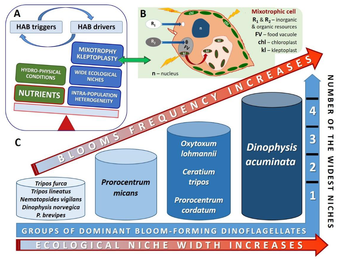
Figure 2.
Schematic representation of nutrient deficiency-induced transitions between dinoflagellate life-cycle stages and associated changes in the expression of meiosis- and recombination-associated genes, including meiosis-associated and encystment-associated mei2-like genes. Nitrogen and/or phosphorus deficiency (-N, -P) can promote the formation of temporary or resting cysts or induce the transition from vegetative cells toward gamete formation and fusion, followed by planozygote and hypnozygote formation. Under nutrient-replete conditions (+N, +P), hypnozygotes germinate and give rise to planomeiocytes, which undergo meiotic division to produce haploid cells. Changes in the expression of selected genes are indicated by upward (↑) or downward (↓) arrows.
Figure 2.
Schematic representation of nutrient deficiency-induced transitions between dinoflagellate life-cycle stages and associated changes in the expression of meiosis- and recombination-associated genes, including meiosis-associated and encystment-associated mei2-like genes. Nitrogen and/or phosphorus deficiency (-N, -P) can promote the formation of temporary or resting cysts or induce the transition from vegetative cells toward gamete formation and fusion, followed by planozygote and hypnozygote formation. Under nutrient-replete conditions (+N, +P), hypnozygotes germinate and give rise to planomeiocytes, which undergo meiotic division to produce haploid cells. Changes in the expression of selected genes are indicated by upward (↑) or downward (↓) arrows.
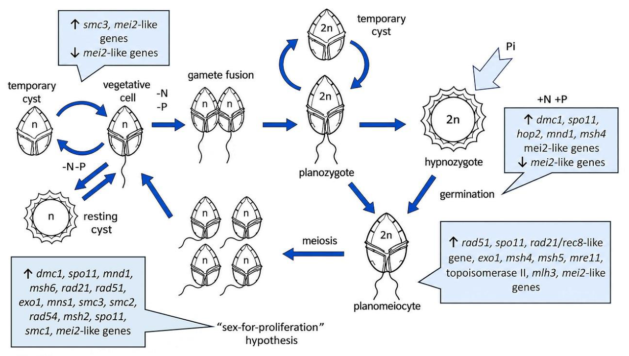
Figure 3.
Basic milestones (concepts and theories, species autecology, molecular and cellular data) on the way toward forecasting red tides of mixotrophic dinoflagellates, and some prerequisites of the future effective HAB modeling.
Figure 3.
Basic milestones (concepts and theories, species autecology, molecular and cellular data) on the way toward forecasting red tides of mixotrophic dinoflagellates, and some prerequisites of the future effective HAB modeling.
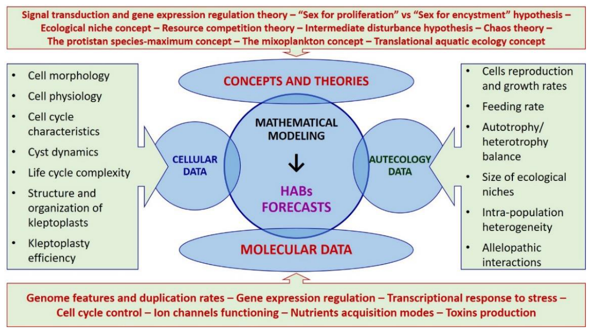

Table 1.
Statistically significant (p < 0.05) correlations between the abundances (cells/mL) of dinoflagellates Prorocentrum cordatum and their prey, the cryptophyte microalgae Hemiselmis virescens, in the SW Baltic coastal waters, with outliers included (outlier: ALARM) and excluded (outlier: 0). “Outlier level” was calculated as “3 x SD + mean”. SD – standard deviation, m – slope, SE– standard error, F – the F-test of overall significance, n – number of analyzed datasets. Based on the data from the 44 years-long phytoplankton database described earlier (Sagert et al., 2008; Telesh et al., 2016, 2021).
Table 1.
Statistically significant (p < 0.05) correlations between the abundances (cells/mL) of dinoflagellates Prorocentrum cordatum and their prey, the cryptophyte microalgae Hemiselmis virescens, in the SW Baltic coastal waters, with outliers included (outlier: ALARM) and excluded (outlier: 0). “Outlier level” was calculated as “3 x SD + mean”. SD – standard deviation, m – slope, SE– standard error, F – the F-test of overall significance, n – number of analyzed datasets. Based on the data from the 44 years-long phytoplankton database described earlier (Sagert et al., 2008; Telesh et al., 2016, 2021).
| Outliers included | Outliers excluded | |||
| Species | Prorocentrum cordatum | Hemiselmis virescens | P. cordatum | H. virescens |
| Outlier | ALARM | ALARM | 0 | 0 |
| Max abundance | 1497 | 2453 | 323 | 1286 |
| Mean abundance | 108.59 | 328.01 | 21.81 | 195.20 |
| SD | 366.17 | 668.43 | 80.45 | 395.90 |
| Outlier level | 1207.09 | 2333.32 | - | - |
| m | 0.478 | 0.120 | ||
| SE | 0.069 | 0.044 | ||
| R2 | 0.76 | 0.35 | ||
| F | target 4.45 | 47.92 | target 4.49 | 7.56 |
| n | 17 | 17 | 16 | 16 |
| 1 | The authors’ names of the species are given when first mentioned in the text only for the species that are not indicated in the Supplementary Table S1 (except for Prorocentrum cordatum and its synonym, P. minimum). |
Disclaimer/Publisher’s Note: The statements, opinions and data contained in all publications are solely those of the individual author(s) and contributor(s) and not of MDPI and/or the editor(s). MDPI and/or the editor(s) disclaim responsibility for any injury to people or property resulting from any ideas, methods, instructions or products referred to in the content. |
© 2026 by the authors. Licensee MDPI, Basel, Switzerland. This article is an open access article distributed under the terms and conditions of the Creative Commons Attribution (CC BY) license.
Copyright: This open access article is published under a Creative Commons CC BY 4.0 license, which permit the free download, distribution, and reuse, provided that the author and preprint are cited in any reuse.