
Submitted:
30 August 2026
Posted:
31 August 2026
You are already at the latest version
Abstract
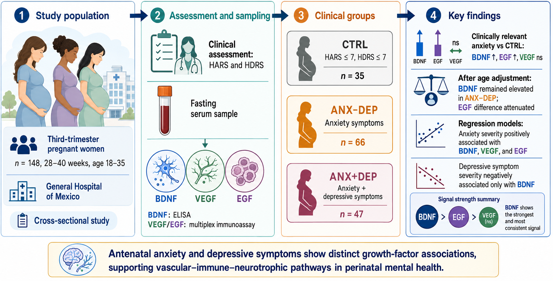
Antenatal anxiety and depression are common and frequently comorbid, affecting maternal and offspring outcomes; however, their underlying biological correlates remain incompletely understood. This cross-sectional study examined whether peripheral levels of brain-derived neurotrophic factor (BDNF), vascular endothelial growth factor (VEGF), and epidermal growth factor (EGF) were associated with anxiety and depressive symptoms during late pregnancy. A total of 148 women with uncomplicated second and third-trimester pregnancies was assessed using the Hamilton Anxiety Rating Scale and Hamilton Depression Rating Scale. Serum BDNF levels were quantified by ELISA, whereas VEGF and EGF were measured using a multiplex bead-based immunoassay. Based on scale scores, participants were classified as controls, women with anxiety without depressive symptoms, or women with anxiety and depressive symptoms. In the between-group comparisons, women with clinically significant anxiety exhibited higher BDNF and EGF levels than controls, whereas differences in VEGF levels did not reach statistical significance. Regression analyses indicated that anxiety severity was positively associated with BDNF, VEGF, and EGF levels, whereas depressive symptom severity was negatively associated with BDNF levels. These findings suggest distinct associations of antenatal anxiety and depressive symptoms with circulating growth factors, particularly BDNF, and warrant further investigation of vascular–immune–neurotrophic pathways potentially involved in perinatal mental health.
Keywords:
vascular factors
; BDNF
; vascular-neuroimmune pathways
; anxiety
; depression
; pregnancy
1. Introduction
Anxiety, along with depression, represents one of the most prevalent and clinically significant psychiatric conditions experienced during the perinatal period [1,2,3]. Prevalence estimates indicate that around 20% to 29% of women experience at least one anxiety disorder during pregnancy or the postpartum period [4,5].
Although anxiety and depressive symptoms can be common and transient during pregnancy [6], if they persist and are untreated, they may become clinically significant, negatively affecting quality of life and impairing daily functioning of the pregnant woman [7,8]. More importantly, both disorders not only negatively impinge upon maternal well-being but also extend beyond delivery, leading to adverse obstetric, neonatal, and developmental outcomes, including poorer social-emotional, cognitive, language, and adaptive functioning in offspring [9,10,11,12]. Despite growing recognition of the detrimental role of depressive and particularly anxiety symptoms during the perinatal period, the psychobiological mechanisms underlying their manifestation remain incompletely understood.
Current evidence supports a multifactorial model in which neuroendocrine, immune-inflammatory, placental-related processes [13], and psychosocial factors [14] interact throughout pregnancy and even beyond the postpartum period. Particularly during pregnancy, substantial remodeling events occur, characterized by dynamic adaptations in immune tolerance, hematopoiesis, angiogenesis, endothelial function, and placental vascular development [15,16]. Overall, they constitute the physiological frame in which perinatal depression and anxiety may be manifested.
Most biomedical research on perinatal mental health has investigated classical inflammatory cytokines as potential biomarkers of related diseases or disorders [17,18,19]. Regarding depressive symptoms, a recent systematic review highlighted the role of inflammatory dysregulation, with the most recurrent altered signals across pregnancy and the postpartum period being IL-6, IL-8, CRP, and TNF-α [20]. However, for perinatal anxiety, other distinctive immune profiles have been detected, suggesting that anxiety-related biology may not be fully reducible to depressive symptomatology [21].
In any case, the roles of many other circulating molecules traditionally classified as growth factors, angiogenic factors, or those related to endothelial function have received comparatively less attention. Notably, some of these proteins—primarily recognized as factors involved in proliferation, differentiation, and tissue repair—constitute a plausible biological interface linking perinatal affective/anxiety symptoms with placental, vascular, and neurodevelopmental processes [22]. Some findings suggest that disruptions in growth-factor signaling indicate both nonspecific inflammatory activity and impaired vascular and neurotrophic adaptation during pregnancy and the postpartum period.
In particular, Brain-Derived Neurotrophic Factor (BDNF) has been proposed as a biologically significant molecule during pregnancy, positioned at the intersection of placental function, fetal development, maternal metabolism, and stress-related neuroendocrine regulation [23]. BDNF protein and its receptor TrkB are expressed in the placenta and fetal tissues, where they participate in processes like stimulation and survival of trophoblast and the implantation of the blastocyst, essential events in subsequent placental development and fetal growth [24]. Moreover, alterations in the BDNF/TrkB system have been described in pregnancy-related complications such as preeclampsia, gestational diabetes, intrauterine growth restriction, and preterm delivery [25]. Collectively, these findings indicate that, beyond its established role in the development and function of the nervous system, BDNF can also be a mediator of feto-placental development and pregnancy adaptation [26,27]. On the other hand, aside from its function as a neurotrophic factor, the protein has also been implicated in mood regulation during pregnancy. Therefore, reduced BDNF levels have often been linked to maternal perinatal depression, either in early pregnancy following antepartum depression [28], Post traumatic stress disorder with comorbid depression [29], or in postpartum depression [30,31,32,33]. However, much less is known about its relation to anxiety; nonetheless, anxious and depressive symptoms are frequently comorbid [4,6].
Vascular endothelial growth factor A (VEGF-A) is another molecule candidate due to its established roles in endothelial cell survival, regulation of the blood–brain barrier, and vascular development [16]. The expression of vascular endothelial growth factor (VEGF) and its receptors VEGFR-1 (Flt-1) and VEGFR-2 (KDR) play critical roles in promoting normal placental angiogenesis [34]. A longitudinal study indicated that serum VEGF-A concentrations decrease during the peripartum, a decline particularly pronounced in women experiencing antenatal and postpartum depression [35]. However, the relationship between VEGF-A and perinatal anxiety remains insufficiently characterized.
Epidermal growth factor (EGF) may also be particularly relevant during the perinatal period, when significant placental and vascular adaptations occur. Accumulating evidence suggests that human trophoblast survival and invasive capacity are linked to intercellular signaling by peptides related to epidermal growth factor (EGF). Peptide members of the EGF signaling system induce downstream signaling by binding to receptor tyrosine kinases of the human EGF receptor (EGFR)/ERBB family [36]. The placenta has the highest expression levels of the EGF receptor protein in the human body [37]. However, the role of EGF signaling system remains comparatively unexplored in relation to perinatal depression and anxiety [38].
Accordingly, the present study aimed to examine serum levels of these distinct peripheral growth factors in relation to depression and anxiety symptoms during pregnancy. By analyzing these biologically interconnected systems, this work aims to advance the understanding of the vascular-immune-neurotrophic pathways underlying affective vulnerability during pregnancy and the postpartum period.
2. Methods
2.1. Design of the Study
The present cross-sectional analytical study was conducted in the Department of Gynecology and Obstetrics at the General Hospital of Mexico (HGM, Dr. Eduardo Liceaga, Mexico City) from 2015 to 2017. The study was reviewed and approved by the hospital research and bioethical committees in 2014 (project ID: D1/14/112/04/072, 2014-2017).
Participants were women aged 18-35 years with uncomplicated second and third-trimester pregnancies (28-40 weeks’ gestation) who met the inclusion criteria and, after being informed of the purpose of the study and the required clinical and laboratory procedures, voluntarily agreed to participate. Most participants resided in Mexico City or the surrounding states.
After hospital admission, women were examined to collect physical, clinical, and obstetric information. Data gathered included sociodemographic (marital status, education level, and working status), anthropometric (height, weight, and body mass index [BMI]), obstetric variables (maternal age, parity, and mode of delivery) and pregnancy status.
Those individuals consuming psychotropic medication or illicit substances of abuse, with psychiatric disorders (other than major depression and anxiety diagnosed before or during enrollment of patients), obstetric pathologies, acute or chronic infections, and other medical conditions (i.e., neurological, metabolic, cardiovascular, degenerative, neuroendocrine, immune-related disorders), including assisted conception, were eliminated from participation to the study at entry.
A total of 148 women in the third trimester of their pregnancy were included in the present report. Participants showed no alcohol consumption, smoking, drug use, or abuse. They were not medicated during the initial medical interviews and blood sampling.
Upon entry, participants underwent second- and third-trimester blood and urine tests, a complete lipid profile, a steroid hormone profile, a thyroid study, and fetal ultrasound. Laboratory tests were approved by the bioethical committee (Bioethics approval document No. CI/223/14, 2014) and performed at the hospital’s Central Laboratory.
Elimination criteria included: Patients with incomplete questionnaires, absent or incomplete laboratory tests, inconsistencies in the psychiatric evaluation of mood disorders, missing regular appointments, or abandonment of the protocol.
Women without a history of acute or chronic obstetric complications, acute or chronic illnesses, or neuropsychiatric pathology (other than mood- or anxiety-related complaints) within the past six months were referred to the Psychiatry Department for psychological counseling and psychiatric evaluation.
No associated mental pathologies (i.e., bipolar disorder, schizophrenia, psychosis, and neuropsychiatric disorder, including uncontrolled compulsive eating and obsessive disorders) were identified based on the diagnostic criteria defined in the Diagnostic and Statistical Manual of Mental Disorders (4th ed., text rev.; American Psychiatric Association, 2000) [39]. Mood or affective symptoms were assessed by clinical psychologists applying the hetero-reported Hamilton Depression Rating Scale (HDRS) [40] and the Hamilton Anxiety Rating Scale (HARS) [41].
Upon completing the clinician-rated scales, all participants (fasting for 8- 12 h) were referred to the clinical laboratory for blood sampling and quantification of serum analytes.
2.2. Blood Sampling
Blood sampling was carried out between 7:00 and 9:00 am., under aseptic conditions. Six mL of venous blood was collected in sterile 13 x 100/ Vacutainer BD Hemogard Tubes containing Clot Activator/Polymer Gel for serum separation (Becton & Dickinson 367977, USA). Samples were cooled on ice and allowed to clot for 1 h before serum separation. Serum fraction was obtained by centrifugation at 1600 × g for 15 min and aliquoted and stored at -80 °C until testing.
VEGF AND EGF were determined using the immunoassay/multiplex bead array, LEGENDplex™ Human Growth Factor Panel (13-plex), (Cat. 740180, BioLegend, USA), following manufacturer’s instructions. FACS Aria III flow cytometer (BD, USA) and the LEGENDplexTM Data Analysis Software v 7.0 (Biolegend, CA, USA) were used to determine serum levels of both growth factors. The lower limits of detection of factors assayed in the study were: EGF (2.6pg/mL), and VEGF (11.5pg/mL). The intra-assay coefficients were ≤8.3%, and the inter-assay coefficients of variation were <12.9%.
2.3. Quantification of BDNF in Serum
Serum BDNF concentrations were measured using a colorimetric enzyme immunoassay (ChemiKine Brain Derived Neurotrophic Factor Sandwich ELISA Kit, Cat. CYT 306, Sigma-Aldrich, USA. Intra- and inter-assay variation coefficients were <10%, respectively. All assays were performed in a double-blind fashion, without knowledge of the clinical status of the sample analyzed.
2.4. Statistical Analysis
Clinical and gestational features of participants are described as mean ± SD, while gestational-week periods and sociodemographic characteristics are reported as percentages (%). For continuous variables, Tukey-adjusted pairwise comparisons were applied. Categorical variables were compared using Pearson’s chi-square test.
Pearson’s or Spearman’s rank correlations were computed to assess associations among biochemical parameters, psychometric scores, and clinical variables collected from the tested groups.
An unpaired t-test with Welch’s correction was used to detect differences in the mean serum levels of vascular and neurotrophic growth factors between the anxious-related symptomatic pregnant population and the healthy control group.
ANOVA with the post hoc Tukey test was used to assess differences in blood levels of vascular and neurotrophic growth factors, clinical variables, and psychometric scores among the study groups. ANOVA with Games-Howell adjustment was applied when variances were not equal, after Levene’s test.
The Friedman test and the chi-square test were used to assess differences in clinical and demographic parameters among pregnant women.
General linear models were constructed using multiple linear regression to analyze whether scores of the anxiety (HARS) and depression (HDRS) were predictors of serum BDNF, VEGF, or EGF concentrations. To reduce the influence of heteroscedasticity and non-normal residuals, standard errors were estimated using robust procedures, and confidence intervals were estimated using bias-corrected and accelerated bootstrap with 10,000 resamples.
For all statistical analyses, the p-value was set at <0.05. All analyses were performed using GraphPad Prism 7 (GraphPad Software Inc., USA) and SPSS software v.27 (Armonk, NY: IBM Corp) or JAMOVI 2.7.31.0
3. Results
At the time of assessments (third trimester of their pregnancy), participants had a mean age of 25.2 ± 6.0 years and a mean gestational age of 34.8 ± 4.0 weeks. Mean body weight was 67.2 ± 10 kg, with a mean body mass index (BMI) of 27.8 ± 3.7 kg/m². Most participants were either cohabiting (39.2%) or divorced (26.4%), had completed either middle (33.1%) or high school (32.4%), and were primarily engaged in home labor (43%).
3.1. Clinical Groups
Since anxiety and depressive symptoms were the focus of the present analyses, participants were categorized according to the Hamilton Anxiety (HARS) and Depression (HDRS) Rating Scale scores. Pregnant women with both HARS and HDRS scores in the non-clinical range (≤7) were defined as the control group (CTRL; N = 35) and used as the reference category for further analyses; whereas all those meeting the threshold for at least mild anxiety symptoms (HARS ≥8) were classified into a global anxiety group (ANX; N = 113; HARS range: 16-39). The ANX group was subsequently subdivided according to depressive symptom burden, yielding an anxiety-only subgroup with HDRS scores ≤7 (ANX -DEP; n = 66; HDRS range: 6-7) and an anxiety and depressive high-symptom subgroup (ANX +DEP; n = 47; HDRS range: 20-51). HARS and HDRS were moderately and positively correlated (Spearman’s ρ = .555, p < .001), revealing the frequent parallel manifestation of anxiety and depressive symptoms.
As shown, Table 1 summarizes the anthropometric and clinical characteristics of the study groups. Table 2 describes the sociodemographic characteristics of recruited pregnant population.
Distribution of gestational- three-week periods were compared using Pearson’s chi-square test.
A one-way ANOVA showed no significant between-group differences in body weight or BMI; however, age differed significantly across groups (F (2, 145) = 6.86, p = .001, η² = .07), with women belonging to CTRL group exhibiting a higher mean age than either ANX-DEP and ANX+DEP groups (Tukey post hoc ANX -DEP group, b = −4.41, SE = 1.20, t = −3.69, p < .001, and the ANX + DEP group, b = −3.24, SE = 1.28, t = −2.54, p = .012.
Although the mean of gestational age at assessment between-groups of comparison did not reach statistical significance (F (2, 145) = 2.81, p = .064 η² = .07), the distribution of evaluations across gestational-week categories was not uniform among groups: χ² (8, N = 148) = 26.21, p = .001, Cramer’s V = 0.30. Specifically, the ANX -DEP group was represented by mothers evaluated at later gestational weeks as compared to CTRL and ANX +DEP groups.
Regarding sociodemographic characteristics, no significant between-group differences were observed in either educational level or employment status, although marital status showed a significant difference across groups (χ2 = 9.72; p = 0.02) (Table 2).
3.2. Serum Growth Factor Levels Across Gestational Weeks During the Third Trimester of Pregnancy
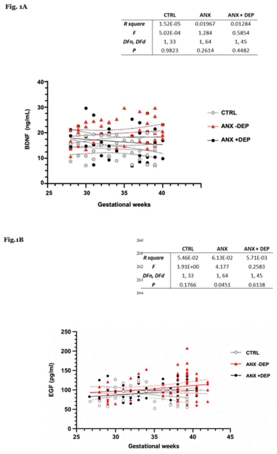
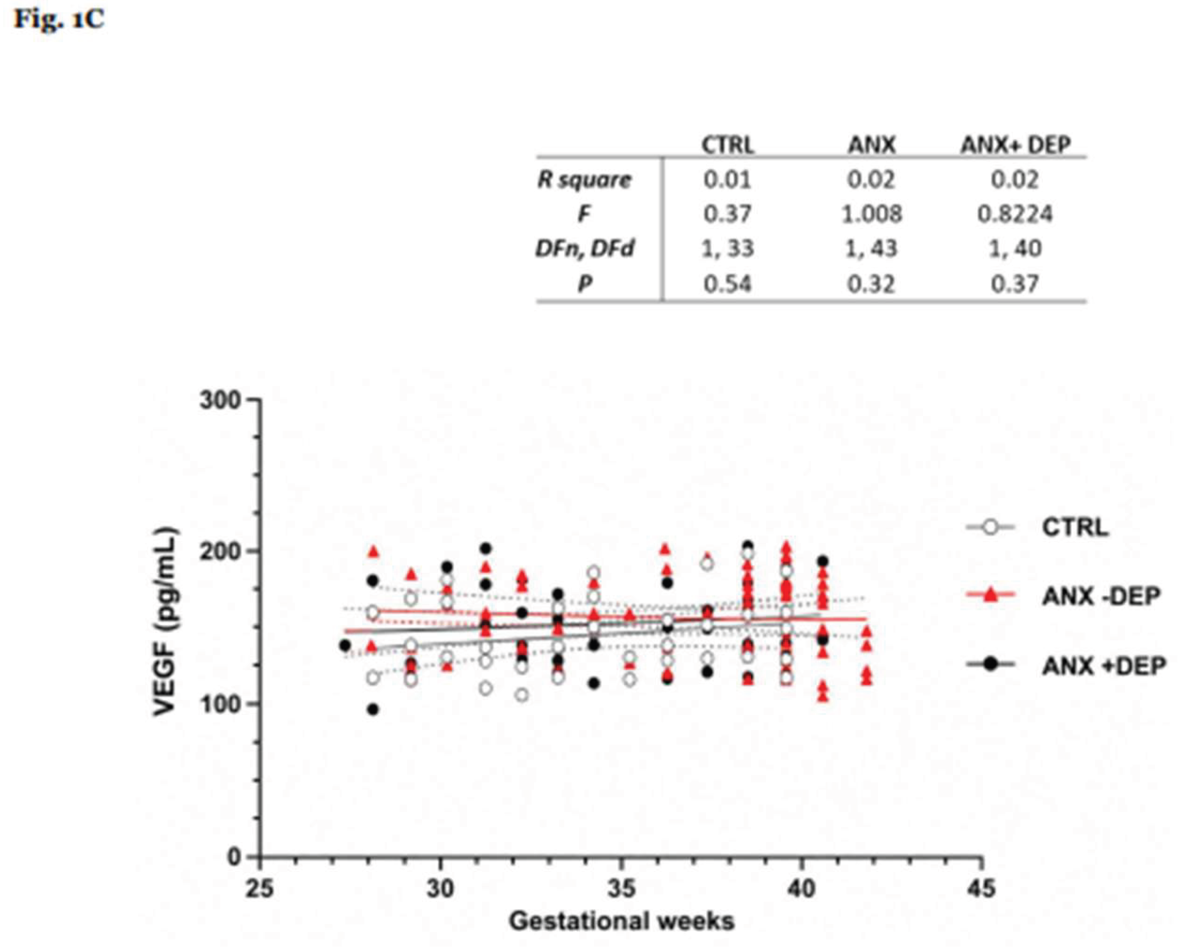
Overall, the serum levels of three growth factors analyzed throughout the third trimester of pregnancy did not show important differences in their distribution, either as a whole or between groups, except for EGF in the ANX-DEP group, in which the linear regression slope did reach statistical significance (R square = 6.13E-2, F(1,64) = 4.18; P = 0.045).
Figure 1.
A–C shows scatter plots depicting serum levels of growth factors in relation to gestational weeks, stratified by group.
Figure 1.
A–C shows scatter plots depicting serum levels of growth factors in relation to gestational weeks, stratified by group.
3.3. Serum Levels of BDNF, VEGF and EGF in the Third Trimester of Gestation in Function of Anxiety
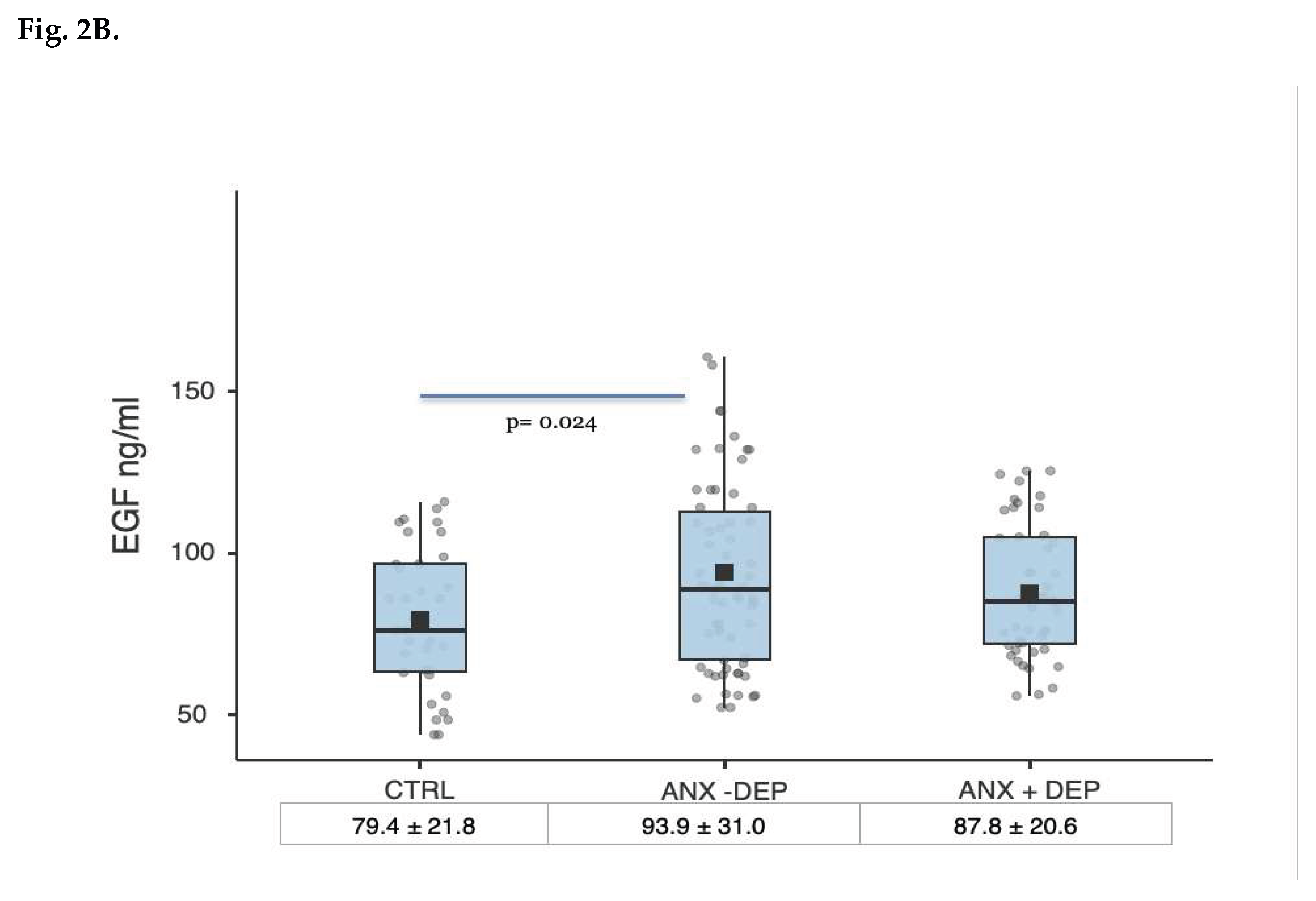
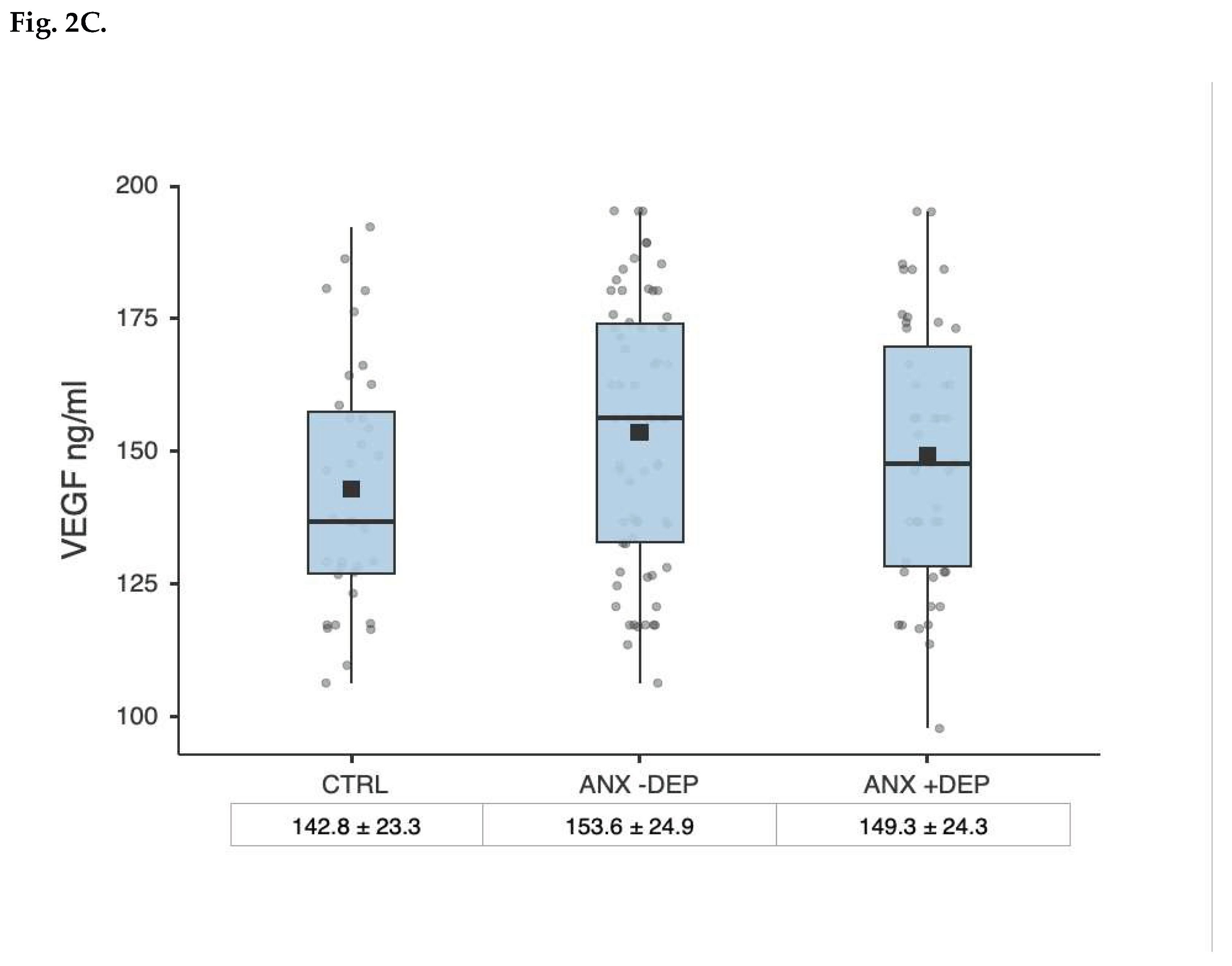
Pregnant women showing anxious symptoms of clinical relevance, irrespective of the presence of depressive symptoms, showed higher circulating levels of BDNF, EGF, and VEGF as compared with the control group. The differences were statistically significant for BDNF and EGF, and marginal for VEFG (Table 3).
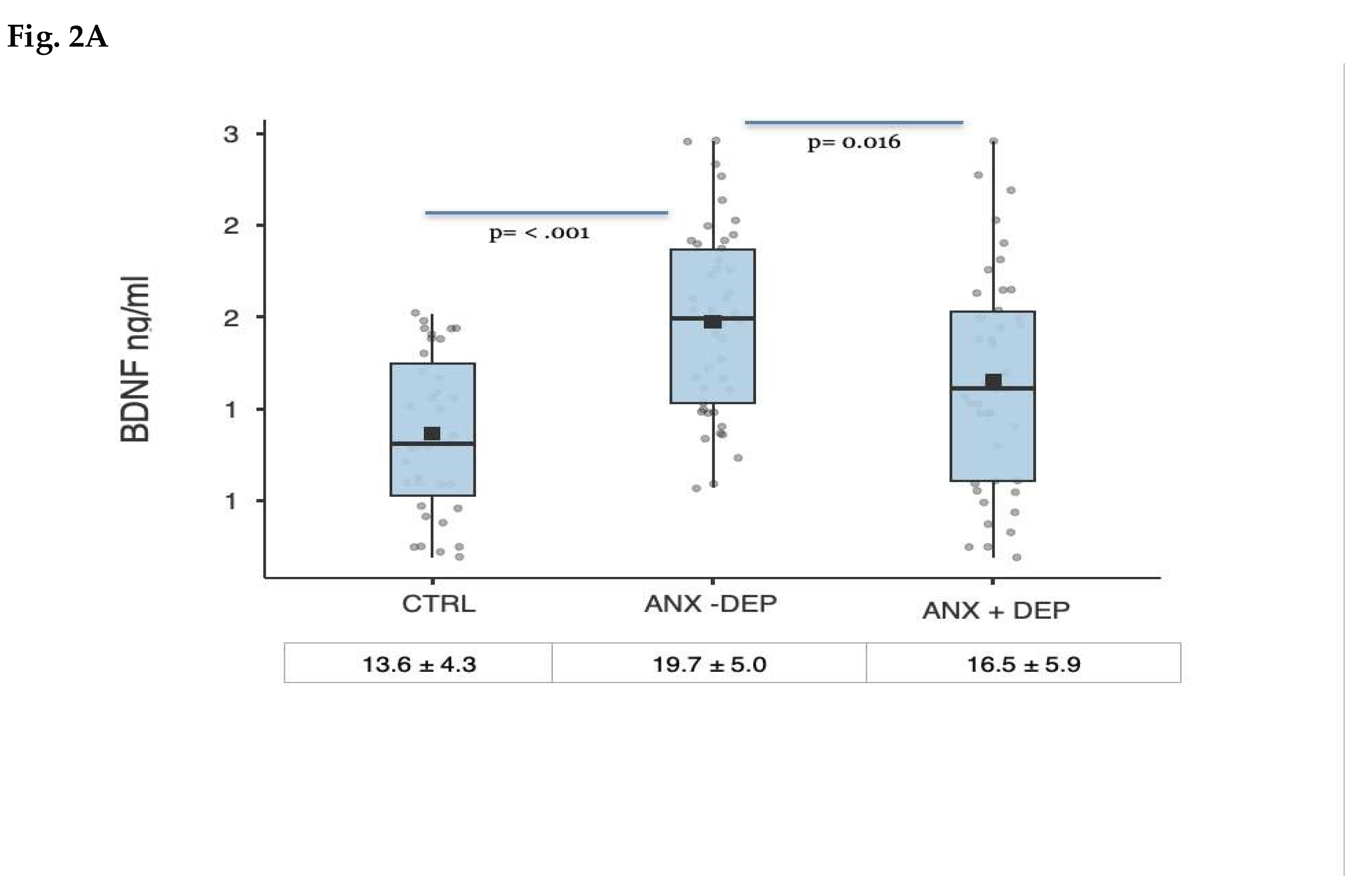
In the case of BDNF, peripheral levels differed significantly across the three categories evaluated, with a significant main effect of the diagnostic group: One-factor ANOVA (F(2, 119) = 13.9, p < 0.001). The effect was maintained after controlling for age: ANCOVA F (3, 118) = 9,7 p < .001, partial η² = .158.
Post-hoc comparisons, using Tukey’s HSD test, indicated that BDNF levels were significantly higher in the ANX -DEP group with respect to CTRL group, mean difference = -5.7, SE = 1.2, t(118) = -4.7, p < .001, Cohen’s d = 1.10, 95% CI [0.61, 1.59]. No significant difference was observed between the CTRL and ANX +DEP groups, mean difference = -2.6, SE = 1.2, t(118) = -2.11, p = .092.
BDNF levels were also significantly higher in the ANX -DEP group than in the ANX +DEP group, mean difference = 3.1, SE = 1.1, t(118) = 2.8, p = .016, Cohen’s d = 0.60, 95% CI [0.17, 1.04].
Figure 2A-C depict the peripheral serum levels of BDBF, EGF and VEGF among the study groups.
Similarly, in the case of EGF, serum levels of this factor were unequal among the groups: One-factor ANOVA (F(2, 145) = 3.6, p = 0.03). The main effect of the diagnostic group was maintained after controlling for age: ANCOVA F(3, 144) = 3.14 p = 0.027, partial η² = .031.
Tukey-adjusted post hoc comparisons showed a significant mean difference between the ANX -DEP and CTRL group: mean differences= -14.5 ± 5.4, t(145) = -2.66, p = .024, but not for the ANX +DEP, mean difference = -8.4 ± 5.8, t(145) = -1.44, p = .32.
In the same line, no statistically significant differences were found between the serum levels of VEGF among the stud groups under a two-tailed hypothesis.: One-factor ANCOVA (F(3, 144) = 1.9, p = .14
3.4. Regression Lineal Analyses
A general linear model was used to examine the association between anxiety symptom severity and serum levels of BDNF, EGF and VEGF.
BDNF: A general linear model was used to examine the association between anxiety symptom severity and serum BDNF levels. The overall model was statistically significant, F(1, 120) = 13.70, p < .001, explaining 10.2% of the variance in BDNF levels, adjusted R² = .095. HARS scores were positively associated with serum BDNF levels, B = 0.163, robust SE = 0.038, 95% bootstrap CI [0.087, 0.238], β = .320, t(120) = 4.241, p < .001.
When HDRS scores were included, the overall model remained statistically significant, F(2, 119) = 11.7, p < .001, with an important increase in the explained variance, R² = .164, adjusted R² = .153.
HARS scores were positively associated with serum BDNF levels, B = 0.23, SE = 0.043, 95% CI [0.151, 0.326], β = .452, t(119) = 4.57, p < .001; partial effect size η²p = .126. Interestingly, depressive symptoms scores were negatively and significantly associated with BDNF, B = −0.132, SE = 0.046, 95% CI [−0.222, −0.036], β = −.281, t(119) = −2.81, p = .006; partial effect size effect, η²p = .069.
EGF: The model examining the association between anxiety symptom severity with serum EGF levels was statistically significant, F(1, 146) = 7.36, p = 0.007, explaining 4.8% of the variance (adjusted R² = .041).
HARS scores were positively associated with serum EGF levels, B = 0.509, SE = 0.192, 95% CI [0.144, 0.894], β = .217, t(146) = 2.66, p =.009
The overall model was still statistically significant when weeks of gestation was included as a covariable, F(3, 145) = 5.07, p =.007, with a small increase in the explained variance, R² = .065, adjusted R² = .052.
HARS scores maintained their positive association with serum EGF concentrations, B = 0.413, SE = 0.231, 95% CI [0.057, 0.963], β = .178, t(145) = 2.11, p = .036; partial effect size η²p = .018
VEGF: The overall model was statistically significant, F(1, 146) = 5.51, p = .02, explaining 3.6% of the variance, adjusted R² = .030. HARS scores were positively associated with serum VEGF concentrations, B = 0.412, SE = 0.170 95% CI [0.031, 0.344], β = .191, t(146) = 2.417, p =0.017.
When age was included as a covariable, the overall model was still statistically significant, F(2, 145) = 3.06, p = .04, explaining 4.3% of the variance (adjusted R² = .030). HARS scores were positively and significantly associated with VEGF levels, B = 0.364, SE = 0.197, 95% CI [0.01, 0.88], β = .168, t(145) = 2.48, p = .043; ηp² = .027.
4. Discussion
The present work investigated the relationship between the expression of depressive and anxiety symptoms in expectant mothers and circulating antenatal levels of BDNF, VEGF, and EGF; three protein factors involved in critical physiological events underlying normal vascular adaptations during pregnancy, and dysregulation has been associated with placental dysfunctions and negative perinatal outcomes (see e.g. BDNF: [22]; VEGF:[34]; EGF: [41].
BDNF, in particular, exerts diverse physiological effects that support an overlapping role in both reproductive biology and mood regulation [42]. A body of research has examined its relation to psychiatric conditions during the perinatal stage; however, most studies have focused on depression, while considerably less attention has been given to its association with maternal anxiety, despite the noteworthy co-occurrence of depressive and anxiety symptoms during pregnancy [12].
Anxiety of clinical relevance during pregnancy has been linked to several adverse postpartum outcomes, like increased odds of preterm birth, low birth weight, or a reduction in breastfeeding [10,43]. Moreover, it is associated with the behavioral expression of negative attitudes during motherhood, altering the maternal-fetal quality of attachment [11,44]
It is worth mentioning the high prevalence of clinically significant anxiety symptoms detected in our sample, a feature of concern, which can be attributed to the population studied consisting of women attending one of the major public health hospitals in Mexico City, which primarily serves a segment of the population lacking social security coverage, with low educational attainments, and often unemployed. As a matter of comparison in Latin America, a substantially higher prevalence of antenatal anxiety and depressive symptoms among Nicaraguan women from rural areas attending public hospitals or community health centers was reported, relative to Dutch pregnant women using the health service system of this developed country [45]. Similarly, an elevated prevalence of anxiety either as state (59.5%) or trait (45.3%) was reported in women attending a gineco-obstetric clinic in Brazil [46]. Diverse risk factors, including a history of psychological or psychiatric problems, educational level, employment status, or marital problems, among many others, have been postulated to explain these important differences [14]. Certainly, further research is warranted.
4.1. BDNF
In the case of BDNF, literature reporting their serum/plasma levels in the perinatal period is extensive and heterogeneous [47], mostly referring to associated obstetric conditions like preeclampsia, gestational diabetes, or premature delivery [48,49,50].
Circulating BDNF during pregnancy has been reported to be lower than in non-pregnant women [51,52,53]. Nonetheless, data regarding its concentration throughout pregnancy are imprecise. For instance, while a sustained decline of the neurotrophin from the first through third trimesters, and a subsequent increase at postpartum was reported [53]; another study detected a slight increase of the protein just during middle and late pregnancy [54]; while another report identified that its levels were generally stable over the period analyzed, with a sustained decline only in relation to preeclampsia [48]. In any case, we did not identify substantial changes in its circulating levels along the third trimester explored.
When the whole sample of pregnant women was categorized by severity of mood and anxiety symptoms, it was evident that the circulating levels of BDNF were significantly elevated in those experiencing anxious symptomatology, with differences especially noticeable in those women not manifesting depressive symptoms. Linear regression analysis supported this finding, showing that HARS scores positively predicted serum BDNF concentrations, accounting for 12.6% of the residual variance after controlling for putative confounders.
Nonetheless, the physiological significance of the relationship between anxiety symptom severity and serum BDNF levels is challenging to interpret given the limited availability of comparable data. A systematic review and meta-analysis of all studies published by 2013 in relation to BDNF circulating levels and anxiety disorders found that protein levels were generally decreased among affected individuals (55). However, the reduction was not consistently seen across the various anxiety disorders examined, being largely attributable to the significantly lowered levels found in OCD patients. Besides, BDNF levels in cord blood samples from newborns of women diagnosed with generalized anxiety disorder were reported to be lower compared with samples from infants of healthy women [56]; however, this finding was not replicated by other researchers [57]. In the study most closely related to the present research [47], Dingsdale et al. initially found no overall correlation between maternal BDNF levels and State-Trait Anxiety Inventory scores in their cohort. Still, when stratified by fetal sex, they observed a significant positive association among mothers of male infants. Therefore, the anxiety symptoms seem to represent a factor of moderate effect size relative to the other predictors.
As cited above, most of the research addressing the relationship between BDNF and psychiatric/psychological ailments in the antenatal stage has centered on clinical depression or exacerbated depressive symptomatology. For example, Fung et al. reported that women with serum BDNF levels in the lowest three quartiles during early pregnancy had a 1.6-fold increased risk of antepartum depression compared to those in the highest quartile [28]. Christian et al. identified an inverse relationship between serum BDNF and depressive symptoms in the third trimester [54]. Similarly, Zhang et al. found in Chinese women that their serum BDNF levels throughout pregnancy were negatively correlated with PHQ-9 scores and a reduced risk of prenatal depressive symptoms [58]. A recent meta-analysis concluded that experimental data support reduced BDNF protein levels in both antepartum and postpartum depression [59]. However, other studies have not observed this negative association [33,51,60].
Our finding of a statistical significant reduction in BDNF levels in the subgroup of women with anxiety who also expressed depressive symptoms compared with those with no manifesting affective symptoms of clinical relevance (although no statistical different respect to controls), can be interpreted in function of hierarchical lineal regression in which the inclusion of an HDRS scores in the model change dramatically the regression coefficients, disclosing instead a negative relation for the depressive symptoms, while maintaining the positive association for anxiety symptoms. suggesting that both symptom types exert a mixed influence on the neurotrophin levels in the frame of the last trimester of pregnancy.
4.2. EGF and VEGF
As stated, we also identified slightly higher levels of both growth factors in the group of women showing anxiety symptoms but not showing depressive symptoms. At the same time, HARS scores were positive predictors of their serum concentrations.
Both proteins play critical roles in promoting normal placental angiogenesis and nutrient transport, processes especially relevant during the perinatal period, when the placenta and vasculature undergo significant adaptations. Imbalances in VEGF and EGF levels and their receptors have been associated with placental dysfunction and clinical conditions such as gestational diabetes mellitus and eclampsia [34,61,62,63]. In particular, elevated VEGF has been linked to increased placental vascularization, which may enhance nutrient availability to the fetus and promote overgrowth. In this context, Mirabelli et al. reported that, as with well-established altered metabolic conditions such as maternal hyperglycemia, serum VEGF and EGF levels were also positive predictors of accelerated fetal growth [41].
Limited data regarding EGF and VEGF in the context of anxiety disorders are available. Roknuzzaman et al. reported elevated serum EGF levels in General anxiety disorder (GAD) patients compared to healthy subjects, an increase that was also positively associated with GAD severity, as measured by the GAD-7 [64]. However, to our knowledge, the relationship of these growth factors and perinatal depression or anxiety had not been examined.
Nonetheless, it is worth mentioning some studies that investigated their association in the context of psychosocial stress-related illnesses. Increased baseline plasma EGF or VEGF levels were identified in Swedish women exhibiting either important signs of occupational stress or during the period of an extended sick leave as a consequence of having experienced an affective or stress-related mental disorder following chronic stress exposure [65]. Similarly, women with stress-related exhaustion disorder exhibited significantly elevated plasma VEGF and EGF levels compared to healthy women, a pattern even noted after 24 months of follow-up [66]. In contrast, Sjörs Dahlman et al. detected markedly low concentrations of EGF, VEGF, and BDNF in subjects who experienced stress-related exhaustion disorder compared to controls. However, they also counterintuitively found that anxiety symptoms (as measured by the Hospital Anxiety and Depression Scale) moderately correlated with the three molecules, whereas no correlation was seen with symptoms of depression or burnout [67].
Several limitations of the present analysis may affect the interpretation of the results. First, as noted, the sample included a cohort of Mexican women with particular socioeconomic features that are not representative of the broader population of pregnant women. Second, the severity of anxiety symptoms was assessed at a single point in time during a specific stage of pregnancy. Moreover, we used a clinical scale with questions targeting general worries, fears, and somatic concerns; however, pregnancy-specific concerns like fear of childbirth or worries about bearing a handicapped child are not included [12,14,68]. Moreover, statistical power analyses indicate that the observed variation in the levels of the analyzed molecules in relation to anxiety symptoms is modest at most for BDNF and even smaller for the other growth factors. Consequently, a substantial portion of the variance likely arises from unmeasured factors or variables not included in the analysis. Future research with longitudinal designs, multiple and more specific assessments, and verification procedures is warranted.
Author Contributions
PLG and CCF designed and conducted the present clinical study. PLG wrote the initial body of the manuscript and CCF designed and wrote the final version of the manuscript. ICA reviewed the paper and made amendments to some sections. MFR evaluated the final scores posterior to application of the clinician-rated scale scores (HARS, HDRS) used in the study. MPMR applied the psychometric battery to patients enrolled in the study. FMCC facilitated the study at the General Hospital of Mexico (HGM), Eduardo Liecaga. FMCC and BFL conducted medical history and obstetric records in pregnant subjects before their enrollment into the study. Moreover, MPMR and BFL collected the sociodemographic and anthropometric data from patients included in the clinical protocol. PLG conducted the blood sampling of patients at 4 °C at the main central lab in the General Hospital of Mexico (HGM). PLG performed the lab- processing of serum samples at the Experimental Medical Research Unit (UNAM) at the HGM, and kept them into cryogenic vials at -80 °C until further use. Both BFL and PLG uploaded the medical records, clinical data and psychometric evaluations into specific clinical software. IMH was responsible for preparation, quantification and analysis of vascular trophic factors using a immunoassay/multiplex bead array and FACS Aria III flow cytometer. APM was responsible for determining BDNF from plasma fractions in pregnant subjects under standard ELISA procedures. APM and CCF were responsible for carrying out the final statistical clinical and biological factors described herein. All authors read and approved the final manuscript. All authors read the final version of the manuscript.
Funding
The present study was funded by the National Institute of Perinatology (project No. 212250-3000-10916-01-16) and FOSSIS/CONACyT Project No. 272458.
Institutional Review Board Statement
PLG received the approval for the clinical study from the head of the Ethical and Research Committee of General Hospital of Mexico (Hospital General de Mexico, Dr. Eduardo, Mexico City) with the file number, Project No. DI/14/1 12/04/072.
Informed Consent Statement
Written informed consent was obtained from all participants recruited for the present study.
Data Availability Statement
All datasets used or analyzed during the current study are available from the corresponding author upon reasonable request.
Conflicts of Interest
The authors declare that they have no competing financial interests that could influence the publishing of the final version of the manuscript.
References
- Ross, L. E., & McLean, L. M. (2006). Anxiety disorders during pregnancy and the postpartum period: A systematic review. The Journal of Clinical Psychiatry, 67(8), 1285–1298. [CrossRef]
- Anniverno, R., Bramante, A., Mencacci, C., & Durbano, F. (2013). Anxiety Disorders in Pregnancy and the Postpartum Period. In New Insights into Anxiety Disorders. InTech. [CrossRef]
- Astbury, L., Pinnington, D. M., Milgrom, J., & Bei, B. (2025). The longitudinal trajectory of depression and anxiety across the perinatal period. Journal of affective disorders, 370, 1–8. [CrossRef]
- Andersson, L., Sundström-Poromaa, I., Wulff, M., Aström, M., & Bixo, M. (2006). Depression and anxiety during pregnancy and six months postpartum: A follow-up study. Acta Obstetricia et Gynecologica Scandinavica, 85(8), 937–944. [CrossRef]
- Fawcett, E. J., Fairbrother, N., Cox, M. L., White, I. R., & Fawcett, J. M. (2019). The prevalence of anxiety disorders during pregnancy and the postpartum period: A multivariate Bayesian meta-analysis. The Journal of Clinical Psychiatry, 80(4), 18r12527. [CrossRef]
- Heron, J., O’Connor, T. G., Evans, J., Golding, J., Glover, V., & ALSPAC Study Team (2004). The course of anxiety and depression through pregnancy and the postpartum in a community sample. Journal of affective disorders, 80(1), 65–73. [CrossRef]
- Silverwood, V. A., Bullock, L., Turner, K., Chew-Graham, C. A., & Kingstone, T. (2022). The approach to managing perinatal anxiety: A mini-review.Frontiers in Psychiatry, 13, 1022459. [CrossRef]
- Silverwood, V., Bullock, L., Jordan, J., Turner, K., Chew-Graham, C. A., Kingstone, T., & Dawson, S. (2023). Non-pharmacological interventions for the management of perinatal anxiety in primary care: A meta-review of systematic reviews. BJGP Open, 7(3), BJGPO.2023.0022. [CrossRef]
- Field, T., Diego, M., Hernandez-Reif, M., Schanberg, S., Kuhn, C., Yando, R., & Bendell, D. (2003). Pregnancy anxiety and comorbid depression and anger: Effects on the fetus and neonate. Depression and Anxiety, 17, 140–151. [CrossRef]
- Grigoriadis, S., Graves, L., Peer, M., Mamisashvili, L., Tomlinson, G., Vigod, S. N., Dennis, C. L., Steiner, M., Brown, C., Cheung, A., Dawson, H., Rector, N. A., Guenette, M., & Richter, M. (2018). Maternal anxiety during pregnancy and the association with adverse perinatal outcomes: Systematic review and meta-analysis.The Journal of Clinical Psychiatry, 79(5), 17r12011. [CrossRef]
- Grigoriadis, S., Graves, L., Peer, M., Mamisashvili, L., Tomlinson, G., Vigod, S. N., Dennis, C. L., Steiner, M., Brown, C., Cheung, A., Dawson, H., Rector, N. A., Guenette, M., & Richter, M. (2019). A systematic review and meta-analysis of the effects of antenatal anxiety on postpartum outcomes. Archives of Women’s Mental Health, 22(5), 543–556. [CrossRef]
- Araji, S., Griffin, A., Dixon, L., Spencer, S. K., Peavie, C., & Wallace, K. (2020). An overview of maternal anxiety during pregnancy and the post-partum period. Journal of Mental Health & Clinical Psychology, 4(4), 47–56.
- O’Brien, K., & Wang, Y. (2023). The Placenta: A Maternofetal Interface. Annual review of nutrition, 43, 301–325. [CrossRef]
- Jimènez-Barragan, M., Falguera-Puig, G., Curto-Garcia, J. J., Monistrol, O., Coll-Navarro, E., Tarragó-Grima, M., Ezquerro-Rodriguez, O., Ruiz, A. C., Codina-Capella, L., Urquizu, X., & Pino Gutierrez, A. D. (2024). Prevalence of anxiety and depression and their associated risk factors throughout pregnancy and postpartum: a prospective cross-sectional descriptive multicentred study. BMC pregnancy and childbirth, 24(1), 500. [CrossRef]
- Sato, Y., Fujiwara, H., & Konishi, I. (2011). Mechanism of maternal vascular remodeling during human pregnancy. Reproductive medicine and biology, 11(1), 27–36. [CrossRef]
- Esposito, M., Paulesu, L., & Mandalà, M. (2025). The role of placental hormones and metabolites in modulating uterine circulation in physiological and pathological pregnancies. Frontiers in endocrinology, 16, 1637570. [CrossRef]
- Bränn, E., Skalkidou, A., Schwarz, J., Papadopoulos, F. C., Sundström Poromaa, I., & Fransson, E. (2022). Longitudinal assessment of inflammatory markers in the peripartum period by depressive symptom trajectory groups. Brain, behavior, & immunity - health, 22, 100468. [CrossRef]
- Moore, T. A., & Case, A. J. (2022). Cytokine levels throughout the perinatal period. The journal of maternal-fetal & neonatal medicine : the official journal of the European Association of Perinatal Medicine, the Federation of Asia and Oceania Perinatal Societies, the International Society of Perinatal Obstetricians, 35(25), 5513–5519. [CrossRef]
- Jarmund, A. H., Giskeødegård, G. F., Ryssdal, M., Steinkjer, B., Stokkeland, L. M. T., Madssen, T. S., Stafne, S. N., Stridsklev, S., Moholdt, T., Heimstad, R., Vanky, E., & Iversen, A. C. (2021). Cytokine Patterns in Maternal Serum From First Trimester to Term and Beyond. Frontiers in immunology, 12, 752660. [CrossRef]
- Silva-Fernandes, A., Conde, A., Marques, M., Caparros-Gonzalez, R. A., Fransson, E., Mesquita, A. R., Figueiredo, B., & Skalkidou, A. (2024). Inflammatory biomarkers and perinatal depression: A systematic review. PloS one, 19(5), e0280612. [CrossRef]
- Mancuso, R. A., Ross, K. M., Accortt, E., Coussons-Read, M., Okun, M. L., Irwin, J., Carroll, J., Hobel, C. J., & Schetter, C. D. (2024). Prenatal mood and anxiety disorders and associated cytokine changes. Journal of affective disorders, 347, 635–644. [CrossRef]
- Dhobale M. (2014). Neurotrophins: Role in adverse pregnancy outcome. International journal of developmental neuroscience : the official journal of the International Society for Developmental Neuroscience, 37, 8–14. [CrossRef]
- D’Angelo, A., Ceccanti, M., Petrella, C., Greco, A., Tirassa, P., Rosso, P., Ralli, M., Ferraguti, G., Fiore, M., & Messina, M. P. (2020). Role of neurotrophins in pregnancy, delivery and postpartum. European journal of obstetrics, gynecology, and reproductive biology, 247, 32–41. [CrossRef]
- Kawamura, K., Kawamura, N., Sato, W., Fukuda, J., Kumagai, J., & Tanaka, T. (2009). Brain-derived neurotrophic factor promotes implantation and subsequent placental development by stimulating trophoblast cell growth and survival. Endocrinology, 150(8), 3774–3782. [CrossRef]
- Mayeur, S., Silhol, M., Moitrot, E., Barbaux, S., Breton, C., Gabory, A., Vaiman, D., Dutriez-Casteloot, I., Fajardy, I., Vambergue, A., Tapia-Arancibia, L., Bastide, B., Storme, L., Junien, C., Vieau, D., & Lesage, J. (2010). Placental BDNF/TrkB signaling system is modulated by fetal growth disturbances in rat and human. Placenta, 31(9), 785–791. [CrossRef]
- Sahay, A., Kale, A., & Joshi, S. (2020). Role of neurotrophins in pregnancy and offspring brain development. Neuropeptides, 83, 102075. [CrossRef]
- Mayeur, S., Lukaszewski, M. A., Breton, C., Storme, L., Vieau, D., & Lesage, J. (2011). Do neurotrophins regulate the feto-placental development?. Medical hypotheses, 76(5), 726–728. [CrossRef]
- Fung, J., Gelaye, B., Zhong, Q. Y., Rondon, M. B., Sanchez, S. E., Barrios, Y. V., Hevner, K., Qiu, C., & Williams, M. A. (2015). Association of decreased serum brain-derived neurotrophic factor (BDNF) concentrations in early pregnancy with antepartum depression. BMC psychiatry, 15, 43. [CrossRef]
- Yang, N., Gelaye, B., Zhong, Q., Rondon, M. B., Sanchez, S. E., & Williams, M. A. (2016). Serum brain-derived neurotrophic factor (BDNF) concentrations in pregnant women with post-traumatic stress disorder and comorbid depression. Archives of women’s mental health, 19(6), 979–986. [CrossRef]
- Gao, X., Wang, J., Yao, H., Cai, Y., & Cheng, R. (2016). Serum BDNF concentration after delivery is associated with development of postpartum depression: A 3-month follow up study. Journal of affective disorders, 200, 25–30. [CrossRef]
- Lee, Y., Kim, K. H., Lee, B. H., & Kim, Y. K. (2021). Plasma level of brain-derived neurotrophic factor (BDNF) in patients with postpartum depression. Progress in neuro-psychopharmacology & biological psychiatry, 109, 110245. [CrossRef]
- Gazal, M., Motta, L.S., Wiener, C.D. et al. Brain-Derived Neurotrophic Factor in Post-Partum Depressive Mothers. Neurochem Res 37, 583–587 (2012). [CrossRef]
- Kittel-Schneider, S., Davidova, P., Kalok, M., Essel, C., Ahmed, F. B., Kingeter, Y., Matentzoglu, M., Leutritz, A. L., Kersken, K., Koreny, C., Weber, H., Kollert, L., McNeill, R. V., Reif, A., Bahlmann, F., & Trautmann-Villalba, P. (2022). A pilot study of multilevel analysis of BDNF in paternal and maternal perinatal depression. Archives of Women’s Mental Health, 25(1), 237–249. [CrossRef]
- Pietro, L., Daher, S., Rudge, M. V., Calderon, I. M., Damasceno, D. C., Sinzato, Y. K., Bandeira, C., & Bevilacqua, E. (2010). Vascular endothelial growth factor (VEGF) and VEGF-receptor expression in placenta of hyperglycemic pregnant women. Placenta, 31(9), 770–780. [CrossRef]
- Bränn, E., Skalkidou, A., Schwarz, J., Papadopoulos, F. C., Sundström Poromaa, I., & Fransson, E. (2022). Longitudinal assessment of inflammatory markers in the peripartum period by depressive symptom trajectory groups. Brain, behavior, & immunity - health, 22, 100468. [CrossRef]
- Armant, D. R., Fritz, R., Kilburn, B. A., Kim, Y. M., Nien, J. K., Maihle, N. J., Romero, R., & Leach, R. E. (2015). Reduced expression of the epidermal growth factor signaling system in preeclampsia. Placenta, 36(3), 270–278. [CrossRef]
- https://www.proteinatlas.org/ENSG00000146648-EGFR?
- Waye, A. A., Moeller, J., & Veiga-Lopez, A. (2025). Epidermal growth factor receptor in placental health and disease: pathways, dysfunction, and chemical disruption. Toxicological sciences : an official journal of the Society of Toxicology, 205(1), 11–27. [CrossRef]
- Hamilton, M. (1959). The assessment of anxiety states by rating. British Journal of Medical Psychology, 32(1), 50–55. [CrossRef]
- Hamilton, M. (1960). A rating scale for depression. Journal of Neurology, Neurosurgery & Psychiatry, 23(1), 56–62. [CrossRef]
- Maria Mirabelli 1 2, Marta Greco 1 3, Stefano Iuliano 1, Francesco Dragone 1 3, Eusebio Chiefari 1 2, Daniela Foti 3 4, Antonio Brunetti, J Clin Transl Endocrinol. 2025 Jun 20:41:100404. eCollection 2025 Sep. [CrossRef]
- Singh, S., Fereshetyan, K., Shorter, S., Paliokha, R., Dremencov, E., Yenkoyan, K., & Ovsepian, S. V. (2023). Brain-derived neurotrophic factor (BDNF) in perinatal depression: Side show or pivotal factor?. Drug discovery today, 28(2), 103467. [CrossRef]
- Shao, S., Yan, S., Zhu, P., Hao, J., Zhu, B., & Tao, F. (2022). Persistent pregnancy-related anxiety reduces breastfeeding exclusiveness and duration: A prospective cohort study. Breastfeeding Medicine, 17(7), 577–583. [CrossRef]
- Surkan, P. J., Park, S., Sheng, Z., Zaidi, A., Atif, N., Osborne, L. M., Rahman, A., & Malik, A. (2025). Effects of a Prenatal Anxiety Randomized Controlled Trial Intervention on Infant Development in Pakistan. Academic pediatrics, 25(1), 102551. [CrossRef]
- Verbeek, T., Arjadi, R., Vendrik, J. J., Burger, H., & Berger, M. Y. (2015). Anxiety and depression during pregnancy in Central America: a cross-sectional study among pregnant women in the developing country Nicaragua. BMC psychiatry, 15, 292. [CrossRef]
- Faisal-Cury, A., & Rossi Menezes, P. (2007). Prevalence of anxiety and depression during pregnancy in a private setting sample. Archives of Women’s Mental Health, 10(1), 25–32. [CrossRef]
- Dingsdale, H., Nan, X., Garay, S. M., Mueller, A., Sumption, L. A., Chacón-Fernández, P., Martinez-Garay, I., Ghevaert, C., Barde, Y. A., & John, R. M. (2021). The placenta protects the fetal circulation from anxiety-driven elevations in maternal serum levels of brain-derived neurotrophic factor. Translational psychiatry, 11(1), 62. [CrossRef]
- D’Souza, V., Patil, V., Pisal, H., Randhir, K., Joshi, A., Mehendale, S., Wagh, G., Gupte, S., & Joshi, S. (2014). Levels of brain derived neurotrophic factors across gestation in women with preeclampsia. International journal of developmental neuroscience : the official journal of the International Society for Developmental Neuroscience, 37, 36–40. [CrossRef]
- Lamadé, E. K., Pedraz-Petrozzi, B., Lindner, O., Meininger, P., Coenen, M., Witt, S. H., Rietschel, M., Dukal, H., Gilles, M., Wudy, S. A., Hellweg, R., & Deuschle, M. (2024). Stress during pregnancy and fetal serum BDNF in cord blood at birth. Psychoneuroendocrinology, 165, 107035. [CrossRef]
- Nokay, E., Kale, İ., & Eskin, D. (2026). Evaluation of serum brain-derived neurotrophic factor in hyperemesis gravidarum. The journal of maternal-fetal & neonatal medicine : the official journal of the European Association of Perinatal Medicine, the Federation of Asia and Oceania Perinatal Societies, the International Society of Perinatal Obstetricians, 39(1), 2703898. [CrossRef]
- Lommatzsch, M., Hornych, K., Zingler, C., Schuff-Werner, P., Höppner, J., & Virchow, J. C. (2006). Maternal serum concentrations of BDNF and depression in the perinatal period. Psychoneuroendocrinology, 31(3), 388–394. [CrossRef]
- Kim, D. R., Sammel, M., et al. (2012). Brain derived neurotrophic factor is altered in human pregnancy. Clinical Neuropsychiatry, 9(6), 207–211.
- Garcés, M. F., Sanchez, E., Torres-Sierra, A. L., Ruíz-Parra, A. I., Angel-Müller, E., Alzate, J. P., Sánchez, Á. Y., Gomez, M. A., Romero, X. C., Castañeda, Z. E., Sanchez-Rebordelo, E., Diéguez, C., Nogueiras, R., & Caminos, J. E. (2014). Brain-derived neurotrophic factor is expressed in rat and human placenta and its serum levels are similarly regulated throughout pregnancy in both species. Clinical endocrinology, 81(1), 141–151. [CrossRef]
- Christian, L. M., Mitchell, A. M., Gillespie, S. L., & Palettas, M. (2016). Serum brain-derived neurotrophic factor (BDNF) across pregnancy and postpartum: Associations with race, depressive symptoms, and low birth weight. Psychoneuroendocrinology, 74, 69–76. [CrossRef]
- Suliman, S., Hemmings, S. M., & Seedat, S. (2013). Brain-Derived Neurotrophic Factor (BDNF) protein levels in anxiety disorders: systematic review and meta-regression analysis. Frontiers in integrative neuroscience, 7, 55. [CrossRef]
- Uguz, F., Sonmez, E. O., Sahingoz, M., Gokmen, Z., Basaran, M., Gezginc, K., Sonmez, G., Kaya, N., Erdem, S. S., Cicekler, H., & Tasyurek, E. (2013). Maternal generalized anxiety disorder during pregnancy and fetal brain development: A comparative study on cord blood brain-derived neurotrophic factor levels. Journal of Psychosomatic Research, 75(4), 346–350. [CrossRef]
- Akbaba, N., Annagür, B. B., Annagür, A., Akbulut, H., Akyürek, F., & Çelık, Ç. (2018). Neurotrophins and neuroinflammation in fetuses exposed to maternal depression and anxiety disorders during pregnancy: A comparative study on cord blood. Archives of Women’s Mental Health, 21(1), 105–111. [CrossRef]
- Zhang, Y. R., Liu, Y. P., Wu, X. M., Yan, Y., Lou, Y. F., & Ni, J. (2025). Association of brain-derived neurotrophic factor levels at different trimesters and new-onset depressive symptom in the third trimester among pregnant women: a longitudinal study. Frontiers in psychiatry, 16, 1618041. [CrossRef]
- Jafarabady, K., Shafiee, A., Bahri, R. A., Rajai Firouzabadi, S., Mohammadi, I., & Amini, M. J. (2024). Brain-derived neurotrophic factor levels in perinatal depression: A systematic review and meta-analysis. Acta psychiatrica Scandinavica, 150(5), 308–319. [CrossRef]
- Pawluski, J. L., Brain, U., Hammond, G. L., & Oberlander, T. F. (2019). Selective serotonin reuptake inhibitor effects on neural biomarkers of perinatal depression. Archives of Women’s Mental Health, 22(3), 431–435. [CrossRef]
- Fujita, Y., Kurachi, H., Morishige, K., Amemiya, K., Terakawa, N., Miyake, A., & Tanizawa, O. (1991). Decrease in epidermal growth factor receptor and its messenger ribonucleic acid levels in intrauterine growth-retarded and diabetes mellitus-complicated pregnancies. The Journal of clinical endocrinology and metabolism, 72(6), 1340–1345. [CrossRef]
- Loukovaara, M., Leinonen, P., Teramo, K., Andersson, S., Alfthan, H., & Stenman, U. H. (2004). Diabetic pregnancy associated with increased epidermal growth factor in cord serum at term. Obstetrics and gynecology, 103(2), 240–244. [CrossRef]
- Bolatai, A., He, Y., & Wu, N. (2022). Vascular endothelial growth factor and its receptors regulation in gestational diabetes mellitus and eclampsia. Journal of translational medicine, 20(1), 400. [CrossRef]
- Roknuzzaman, A. S. M., Qusar, M. S., Shahriar, M., Ashraful Islam, S. M., & Islam, M. R. (2024). Increased serum EGF but not SDF-1 levels are associated with the pathophysiology and development of generalized anxiety disorder. Scientific reports, 14(1), 27409. [CrossRef]
- Asberg, M., Nygren, A., Leopardi, R., Rylander, G., Peterson, U., Wilczek, L., Källmén, H., Ekstedt, M., Akerstedt, T., Lekander, M., & Ekman, R. (2009). Novel biochemical markers of psychosocial stress in women. PloS one, 4(1), e3590. [CrossRef]
- Wallensten, J., Åsberg, M., Nygren, Å., Szulkin, R., Wallén, H., Mobarrez, F., & Nager, A. (2016). Possible Biomarkers of Chronic Stress Induced Exhaustion - A Longitudinal Study. PloS one, 11(5), e0153924. [CrossRef]
- Sjörs Dahlman, A., Blennow, K., Zetterberg, H., Glise, K., & Jonsdottir, I. H. (2019). Growth factors and neurotrophins in patients with stress-related exhaustion disorder. Psychoneuroendocrinology, 109, 104415. [CrossRef]
- Nilsson, C., Hessman, E., Sjöblom, H., Dencker, A., Jangsten, E., Mollberg, M., Patel, H., Sparud-Lundin, C., Wigert, H., & Begley, C. (2018). Definitions, measurements and prevalence of fear of childbirth: a systematic review. BMC pregnancy and childbirth, 18(1), 28. [CrossRef]

Table 1.
Clinical and gestational characteristics of participants by study group.
|
CTRL (n = 35) |
ANX-DEP (n = 66) |
ANX + DEP (n = 47) |
||
|
Clinical characteristics, mean (SD) | ||||
| Gestational age, weeks | 33.9 (3.6) | 35.7 (4.1) | 34.3 (3.9) | |
| Weight, kg | 67.5 (9.8) | 67.0 (8.9) | 67.2 (11.6) | |
| BMI, kg/m² | 27.9 (3.8) | 27.9 (3.6) | 27.4 (3.9) | |
| HARS score | 3.1 (0.6) | 26.0 (6.3)*** | 26.5 (5.7)*** | |
| HDRS score | 5.1 (0.9) | 5.9 (1.0) | 30.1 (7.8)*** | ANX-DEP vs ANX + DEP *** |
|
Gestational- three-week period |
n (%) | |||
| 27–29 | 8 (22.8) | 11 (16.7) | 6 (12.8) | |
| 30–32 | 14 (40.0) | 12 (18.2) | 9 (19.1) | |
| 33–35 | 10 (28.6) | 9 (13.6) | 12 (25.5) | |
| 36–38 | 3 (8.6) | 18 (27.3) | 16 (34.1) | |
| 39–41 | 0 (0.0) | 16 (24.2) | 4 (8.5) | |
| χ²(4) = 9.72 | ||||
| p= .020 | ||||
CTRL = control; ANX-DEP = anxiety, no depression; ANX + DEP = anxiety and depression; BMI = body mass index; HARS = Hamilton Anxiety Rating Scale; HDRS = Hamilton Depression Rating Scale. For continuous variables, p values after Tukey-adjusted pairwise comparisons are shown as follows: * p<0.05, **p<0.01, ***p<0.001.
Table 2.
Sociodemographic characteristics of participants by study group.
| Characteristic | CTRL (n = 35) | ANX -DEP (n = 66) | ANX + DEP (n = 47) |
| Marital status, n (%) | |||
| Never married | 6 (17.1) | 11 (16.7) | 8 (17.0) |
| Married | 5 (14.3) | 9 (13.6) | 12 (25.5) |
| Divorced | 11 (31.4) | 18 (27.3) | 10 (21.3) |
| Cohabiting | 13 (37.2) | 28 (42.4) | 17 (36.2) |
| χ² (3) = 9.9, p=.02 | |||
| Education level, n (%) | |||
| Elementary school | 3 (8.6) | 6 (9.1) | 3 (6.4) |
| Middle school | 12 (34.3) | 18 (27.3) | 19 (40.4) |
| High school | 10 (28.6) | 23 (34.8) | 15 (32.0) |
| Bachelor’s degree | 7 (20.0) | 10 (15.1) | 5 (10.6) |
| Postgraduate | 1 (2.8) | 4 (6.1) | 3 (6.4) |
| Technician degree | 2 (5.7) | 5 (7.6) | 2 (4.2) |
| χ² (5) =5.6, p=.13 | |||
| Employment status, n (%) | |||
| Employed | 10 (28.6) | 8 (12.1) | 12 (25.6) |
| Unemployed | 3 (8.6) | 6 (9.0) | 2 (4.2) |
| Home labor | 13 (37.1) | 32 (48.5) | 18 (38.3) |
| Commerce | 6 (17.1) | 11 (16.7) | 13 (27.7) |
| Profession | 3 (8.6) | 7 (10.7) | 2 (4.2) |
| Other | 0 (0.0) | 2 (3.0) | 0 (0.0) |
| χ² (5) = 4.1, p= 0.25 |
Table 3.
Serum levels of neurotrophic and angiogenic factors.
|
CTRL (n = 35) |
ANX (n = 123) |
Pairwise comparisons | CTRL vs ANX | |
| Welch’s t-test: | p value | |||
| BDNF | 13.6 ± 4.3 | 18.2 ± 5.7 | t(81.9) = −4.55 | <0.001 |
| EGF | 79.4 ± 21.8 | 91.3± 27.2 | t(69.8) = −2.66 | 0.01 |
| VEGF | 142.8 ± 23.3 | 152.1 ± 24.7 | t(59.6) = −1.98 | 0.052 |
When ANX group was divided between those with or without depression symptoms, the comparisons were as follows: Figure 1 A-C show serum levels of circulating growth factors analyzed across the three clinical categories evaluated.
Disclaimer/Publisher’s Note: The statements, opinions and data contained in all publications are solely those of the individual author(s) and contributor(s) and not of MDPI and/or the editor(s). MDPI and/or the editor(s) disclaim responsibility for any injury to people or property resulting from any ideas, methods, instructions or products referred to in the content. |
© 2026 by the authors. Licensee MDPI, Basel, Switzerland. This article is an open access article distributed under the terms and conditions of the Creative Commons Attribution (CC BY) license (http://creativecommons.org/licenses/by/4.0/).
Copyright: This open access article is published under a Creative Commons CC BY 4.0 license, which permit the free download, distribution, and reuse, provided that the author and preprint are cited in any reuse.