

Submitted:
30 August 2026
Posted:
31 August 2026
You are already at the latest version
Abstract
Cheyne-Stokes-Respiration (CSR) manifests as a crescendo-decrescendo breathing pattern with alternating central events and is a common finding during sleep in patients chronic heart failure. Despite its high prevalence, well-defined diagnostic criteria and specific treatment options, the pathophysiology of CSR is not completely understood. Besides its occurrence in chronic heart failure, studies have shown an increasing likelihood with other cardiovascular disorders like atrial fibrillation or pulmonary hypertension and report an association with trigger factors like cortical arousals, fluid shifts and oxygen desaturations. In fact, hypoxemia tends to be neglected in patients with chronic heart failure due to an increased compensatory respiratory drive but is the most consistent finding in nocturnal CSR under conditions of high altitude. Therefore, based on the assumption of two parallel chemoreceptive systems to detect changes in arterial oxygen and carbon dioxide, the present work hypothesizes that the additional respiratory drive by hypoxemia (“hypoxic drive”) leads to an excess exspiration of the main controlled variable CO2 which destabilizes the respiratory feedback loop. Based on the current knowledge about CSR in the context of chronic heart failure and hypobaric hypoxemia as well as clinical observations, a pathophysiological concept on nocturnal CSR is outlined. The essential features of this theory include recurrent episodes of hypoxemia detected by peripheral chemoreceptors causing a temporarily increased respiratory drive which destabilizes respiration due to a time delay between peripheral sensation of pO2 and central chemoreception of pCO2. This pathophysiological theory explains the treatment efficacy of supplemental oxygen and may also apply to other clinical scenarios like metabolic acidosis in chronic kidney disease in which the temporal delay between the peripheral sensation of pH and the central detection of pCO2 may result in nocturnal periodic breathing.
Keywords:
Cheyne-Stokes-Respiration
; periodic breathing
; hypoxemia
; chronic heart failure
Current Explanations
Cheyne-Stokes-Respiration with central sleep hypopnea or apnea according to the American Association of Sleep Medicine (AASM) criteria is scored if at least five central events separating a crescendo-decrescendo breathing pattern are recorded over a minimum of 2 hours recording time [1].
Most studies trying to explain the pathophysiology of Cheyne-Stokes-Respiration (Periodic Breathing) refer to clinical observations associated with its occurrence. Chronic heart failure is the most common clinical scenario in which CSR is described and a vast number of studies have investigated the risk factors for developing nocturnal CSR [2]. Left ventricular myocardial dysfunction is strongly related to CSR and the likelihood of its occurrence increases with the severity of contractile impairment [3]. The resulting reduction of cardiac output causes a prolonged circulation time [4] which may be correlated with the duration of the crescendo-decrescendo cycle length. The relationship between reduced cardiac output and the occurrence of CSR is substantiated by the observation of ventilatory stabilization by increasing the heart rate in patients with pacemakers [5]. Another factor contributing to the occurrence of CSR is pulmonary congestion which may be aggravated by nocturnal fluid retention. The activation of pulmonary stretch receptors by perialveolar fluid accumulation can act as a breathing stimulus resulting in hyperventilation [6]. Cardiovascular instabilities may also lead to autonomous and cortical arousals inducing periods of hyperventilation.
The overreaction of the respiratory system resulting in hypocapnia and consecutive hypoventilation is sometimes explained with alterations in chemoreceptor sensitivity and the apneic threshold [7].
Another setting to observe nocturnal CSR is hypobaric hypoxia under high altitude conditions [8]. In contrast to patients with chronic heart failure, CSR at high altitude is not related to reduced cardiac output or nightly arousals. Rather, hypobaric conditions and low partial pressure of oxygen induce hyperventilation to meet the metabolic oxygen demand. The stimulus to cause hyperventilation under high altitude conditions is low arterial partial pressure of oxygen which acts as an additional respiratory drive increasing breathing minute volume. With the onset of sleep, cortical control over ventilation diminishes while arterial partial pressure of carbon dioxide remains the main respiratory stimulus and the main controlled variable by respiration. This hypoxemic drive leads to ventilation beyond the necessities to stabilize arterial carbon dioxide which results in hypocapnia inducing repeated episodes of hypopnea or apnea.
Clinical Observations
The two presented scenarios which can lead to nocturnal CSR differ significantly in individual and external factors contributing to the occurrence of altered respiration. However, both entities share the common factor of hypoxemia which is essential for the development of CSR and is oftentimes overlooked in routine clinical practice. Hypoxemia indicating long-term oxygen treatment defined as arterial partial pressure of oxygen ≤ 55 mmHg at rest or oxygen saturation ≤ 88% is rather rarely observed in patients with chronic heart failure as increased minute volume ventilation compensates for the oxygen deficit [9]. The increase of minute breathing volume due to hypoxemia as an additional breathing stimulus is sometimes referred to as the “hypoxic (hypoxemic) drive” which adds to the main regulating and controlled variable of arterial pCO2 [10]. This explains the regular observation of hypocapnia in chronic heart failure which has been described as a risk factor for the development of nocturnal CSR [11]. In fact, applying a correction formula which accounts for excess ventilation due to “hypoxic (hypoxemic) drive” can reveal the oxygen demand in individuals with heart failure as exemplified in the section below [12].
Hyperventilation is intended to correct for the reduced circulatory volume and impaired diffusion capacity of oxygen whereas an excess of the relatively stable metabolic CO2 is exspired. This can lead to periodic breathing during wakefulness if cardiac impairment is severe or stress tests cause a significant divergence between oxygen demand and supply.
The association between a lack of oxygen in chronic heart failure and hypobaric hypoxia as the main factor determining the occurrence of nocturnal CSR is further substantiated by the effect of oxygen treatment which regularly resolves periodic breathing [13].
Another View on Cheyne-Stokes-Respiration (Periodic Breathing)
Based on clinical observations in patients with chronic heart failure a comprehensive theory to explain the occurrence of nocturnal CSR is outlined.
To demonstrate the sequential order of findings in chronic heart failure ultimately manifesting as CSR, an illustration of the neuro-cardiopulmonary processes is shown in Figure 1.
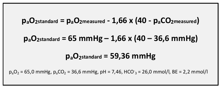
The theory hypothesizes that the development of CSR begins with a lack of oxygen, measured as reduced paO2 and SpO2, which is compensated by an increased minute breathing volume resulting in arterial hypocapnia. From own clinical observations, a lowered paCO2 or sometimes even day-time hypocapnia, is the most consistent finding in patients with chronic heart failure developing nocturnal CSR. The subtle oxygen deficit can be revealed by a formula which corrects for hyperventilation and is applied for a patient who presented with day-time hypocapnia and nocturnal CSR (Figure 2).
In both patients with chronic heart failure and conditions of hypobaric hypoxia, hypoxemia can occur immediately after sleep onset when cortical control over respiration is reduced and a shift towards a stronger reliance on chemoreception results (1). Peripheral chemoreceptors in the aortic arch and carotid bifurcation can detect changes of arterial pCO2, pO2 and pH, while central chemoreceptors in the retrotrapezoid nucleus (RTN) are restricted to sense changes of paCO2 (Figure 1) [14]. Peripheral and central chemoreceptors differ with regards to the temporal delay of detection and the relative weight with which they can modify respiratory drive [15]. Peripheral chemoreceptors are essential for the detection of paO2 and pH changes and work with a delay of 5-10 seconds (2a), while central chemoreceptors are the main integration hub to regulate paCO2 by a minimal latency of 20 seconds seconds (2b) [16,17]. The sensory input from peripheral chemoreceptors is integrated in the Nucleus tractus solitarius (NTS). Sensory information from both central and peripheral chemoreceptors converges onto neurons in the more ventral parts of the Medulla oblongata including the (pre-) Boetzinger complex [18,19]. These cells include early-inspiratory and post-inspiratory neurons with reciprocal inhibition resulting in an oscillating activity pattern which is enhanced by sensory input from chemoreceptors (3) [20]. The additional hypoxemic respiratory stimulus (“hypoxic drive”) results in increased phrenical activation which translates into a higher minute breathing volume illustrated in a leftward shift of the breathing minute volume-/paCO2 -curve referred to as “controller gain” (4) [21]. The increased minute breathing volume exceeds the ventilatory demand to stabilize arterial pCO2. The adaptation to reduced arterial paCO2 is delayed due to the latency of the central chemoreceptive feedback loop for CO2. This may result in a reduction of paCO2 below the apneic threshold causing a temporary cessation of respiratory drive. A low tidal volume leads to ventilatory inefficiency due to alveolar hypoventilation causing an increase of paCO2 (5) and a drop of paO2 which is again detected with a temporal delay between peripheral and central chemoreceptors.
Discussion
The outlined pathophysiological mechanisms highlight the importance of a comprehensive approach including cardiopulmonary dynamics and their effect on respiration through sensorimotor integration. The presented pathophysiological concept suggests that the integration of hypoxemic sensory input from peripheral chemoreceptors acts as an additional respiratory drive altering paCO2 as the controlled variable which is sensed with a time-gap by central chemoreceptors. This results in ventilatory instability manifesting as nocturnal periodic breathing with or without central apnea.
The inverse correlation between the increasing prevalence of CSR with decreasing left ventricular function in chronic heart failure is well-described [2,3]. Comparably, an increasing occurrence of CSR with lower partial pressure of oxygen under high altitude conditions is observed [22]. The shared pathomechanism in both entities is an increasing oxygen demand which is compensated by an increase in breathing minute volume. The respiratory compensation for an insufficient oxygen supply as a starting point of CSR is substantiated by the therapeutic efficacy of supplemental oxygen in both entities [13].
While many studies have described the association between alterations in blood gases, particularly hypocapnia, and the occurrence of CSR, the temporal differences between peripheral and central chemoreceptors which add up to alter respiratory drive have been neglected. Theoretical concepts oftentimes emphasize the cardiopulmonary interactions without integrating neuronal processes which enable the dynamic regulation of blood gases by respiration as a feedback control system.
The outlined pathophysiological theory indicates that CSR in chronic heart failure and high altitude share the common factor of hypoxemia which has been described as a symptom but not as the main mechanism underlying the development of CSR.
The temporal delay between peripheral paO2 and central paCO2 chemoreception is a new aspect in the pathophysiology of CSR which may be essential to explain the inability of the respiratory system to stabilize paCO2 as the controlled variable. Further research may investigate if delayed sensory feedback loops may also apply to metabolic alterations, e.g., acidosis in chronic kidney disease, and whether these may also result in periodic breathing. Clinicians may particularly consider patients with day-time hypocapnia for sleep study to assess for the occurrence of nocturnal CSR.
Funding
The author declares to have no financial interest or personal relationship that could have influenced the work presented in this manuscript. The author did not receive any financial support that could have biased the results.
Abbreviations
| AASM | American Association of Sleep Medicine |
| CSR | Cheyne-Stokes-Respiration |
| NTS | Nucleus tractus solitarius |
| RG | Respiratory Group |
| RTN | Retrotrapezoid Nucleus |
References
- Rudrappa M, Modi P, Bollu PC. Cheyne Stokes Respirations. PubMed. Treasure Island (FL): StatPearls Publishing; 2023. Available from: https://www.ncbi.nlm.nih.gov/books/NBK448165/.
- Bradley, T.D.; Floras, J.S. Sleep apnea and heart failure: Part II: central sleep apnea. Circ. U. S. 2003, Vol. 107, 1822–6. Available online: https://pubmed.ncbi.nlm.nih.gov/12682029/.
- Sin, D.D.; Fitzgerald, F.; Parker, J.D.; Newton, G.; Floras, J.S.; Bradley, T.D. Risk Factors for Central and Obstructive Sleep Apnea in 450 Men And Women with Congestive Heart Failure. Am. J. Respir. Crit. Care Med. 1999, 160(4), 1101–6. [Google Scholar] [CrossRef] [PubMed]
- Hall, M.J.; Xie, A.; Rutherford, R.; Ando, S.; Floras, J.S.; Bradley, T. D. Cycle length of periodic breathing in patients with and without heart failure. American journal of respiratory and critical care medicine; England, 1996; Vol. 154, pp. 376–81. Available online: https://pubmed.ncbi.nlm.nih.gov/8756809/.
- Garrigue, S.; Bordier, P.; Jaïs, P.; Shah, D.C.; Hocini, M.; Raherison, C.; et al. Benefit of Atrial Pacing in Sleep Apnea Syndrome. N. Engl. J. Med. 2002, 346(6), 404–12. [Google Scholar] [CrossRef] [PubMed]
- Solin, P.; Bergin, P.; Richardson, M.; Kaye, D.M.; Walters, E.H.; Naughton, M.T. Influence of pulmonary capillary wedge pressure on central apnea in heart failure. Circ. U. S. 1999, Vol. 99, 1574–9. Available online: https://pubmed.ncbi.nlm.nih.gov/10096933/. [CrossRef] [PubMed]
- Lorenzi-Filho, G.; Azevedo, E.R.; Parker, J. D.; Bradley, T. D. Relationship of carbon dioxide tension in arterial blood to pulmonary wedge pressure in heart failure. The European respiratory journal; England, 2002; Vol. 19, pp. 37–40. Available online: https://pubmed.ncbi.nlm.nih.gov/11843325/.
- Pramsohler, S.; Schilz, R.; Patzak, A.; Rausch, L.; Netzer, N.C. Periodic breathing in healthy young adults in normobaric hypoxia equivalent to 3500 m, 4500 m, and 5500 m altitude. Sleep Breath. Schlaf Atmung. Ger. 2019, Vol. 23, 703–9. Available online: https://pubmed.ncbi.nlm.nih.gov/30972693/. [CrossRef] [PubMed]
- National Coverage Determination—Home Use of Oxygen (240.2). Cms.gov. Available online: https://www.cms.gov/medicare-coverage-database/view/ncd.aspx?NCDId=169&ncdver=1&DocID=240.2&SearchType=Advanced&bc=IAAAABAAAAAA&.
- Guntheroth, W.G. Cheyne-Stokes respiration: hypoxia plus a deep breath that interrupts hypoxic drive, initiating cyclic breathing. Med. Hypotheses. U. S. 2011, Vol. 77, 714–6. Available online: https://pubmed.ncbi.nlm.nih.gov/21824731/. [CrossRef] [PubMed]
- Hanly, P.; Zuberi, N.; Gray, R. Pathogenesis of Cheyne-Stokes respiration in patients with congestive heart failure. Relationship to arterial PCO2. Chest. U. S. 1993, Vol. 104, 1079–84. Available online: https://pubmed.ncbi.nlm.nih.gov/8404170/.
- Diekmann, M.; Smidt, U. Berechnung eines Standard-PaO2 in Analogie zum Standard-Bikarbonat. Atemw Lungenkr 1984, 10, 248–260. [Google Scholar]
- Staniforth, A.; Kinnear, M.; Starling, C.; Hetmanski, D.J.; Cowley, A.P. Effect of oxygen on sleep quality, cognitive function and sympathetic activity in patients with chronic heart failure and Cheyne–Stokes respiration. 1998, 19(6), 922–8. [Google Scholar] [CrossRef] [PubMed]
- Guyenet, P.G.; Bayliss, D.A. Neural Control of Breathing and CO2 Homeostasis. Neuron 2015, 87(5), 946–61. Available online: https://www.ncbi.nlm.nih.gov/pmc/articles/PMC4559867/. [CrossRef] [PubMed]
- López-Barneo, J.; Ortega-Sáenz, P.; Pardal, R.; Pascual, A.; Piruat, J.I.; Durán, R.; et al. Oxygen Sensing in the Carotid Body. Ann. N. Y. Acad. Sci. 2009, 1177(1), 119–31. [Google Scholar] [CrossRef] [PubMed]
- Kryger, M.H.; Roth, T.; Dement, W.C. Principles and practice of sleep medicine; Elsevier: Philadelphia, Pa, 2017. [Google Scholar]
- Duffin, J. The role of the central chemoreceptors: A modeling perspective. Respir. Physiol. Neurobiol. 2010, 173(3), 230–43. [Google Scholar] [CrossRef] [PubMed]
- Krohn, F.; Novello, M.; van der Giessen, R.S.; De Zeeuw, C.I.; Pel, J.J.; Bosman, L.W. The integrated brain network that controls respiration; Chin, J., Ed.; eLife, 8 Mar 2023; Volume 12, p. e83654. Available online: https://elifesciences.org/articles/83654.
- Molkov, Y.I.; Rubin, J.E.; Rybak, I.A.; Smith, J.C. Computational models of the neural control of breathing; Wiley Interdisciplinary Reviews: Systems Biology and Medicine, 23 Dec 2016; Volume 9, 2, p. e1371. [Google Scholar]
- Anderson, T.M.; Ramirez, J.M. Respiratory rhythm generation: triple oscillator hypothesis. F1000Research 2017, 6, 139. Available online: https://f1000research.com/articles/6-139. [CrossRef] [PubMed]
- Chowdhuri, S.; Badr, M.S. Control of Ventilation in Health and Disease. Chest 2017, 151(4), 917–29. [Google Scholar] [CrossRef] [PubMed]
- Wickramasinghe, H.; Anholm, J.D. Sleep and breathing at high altitude. Sleep Breath 1999, 3, 89–102. [Google Scholar] [CrossRef] [PubMed]
Figure 1.
Schematic illustration of the main steps involved in the pathophysiology of CSR. (1) Hypoxemia (low paO2 or SpO2) is detected by peripheral chemoreceptors with a latency of 1-5 seconds (2a), while changes in paCO2 are sensed by central chemoreceptors after a minimum duration of 20 seconds (2b). Sensory input from blood gas alterations is integrated in the dorsal respiratory group and is transmitted to the ventral Medulla oblongata (3). The activity of an oscillatory network including early-inspiratory and post-inspiratory neurons is enhanced in case of a hypoxemic stimulation (“hypoxic drive”) which results in an increased breathing minute volume (4). The drop of paCO2 due to hypoxemic hyperventilation is detected with a time delay by central chemoreceptors and may fall below the apneic threshold. Low tidal volumes associated with alveolar hypoventilation (5) causes an increasing paCO2 and a drop of oxygen levels which may result in another episode of hypoxemic hyperventilation.
Figure 1.
Schematic illustration of the main steps involved in the pathophysiology of CSR. (1) Hypoxemia (low paO2 or SpO2) is detected by peripheral chemoreceptors with a latency of 1-5 seconds (2a), while changes in paCO2 are sensed by central chemoreceptors after a minimum duration of 20 seconds (2b). Sensory input from blood gas alterations is integrated in the dorsal respiratory group and is transmitted to the ventral Medulla oblongata (3). The activity of an oscillatory network including early-inspiratory and post-inspiratory neurons is enhanced in case of a hypoxemic stimulation (“hypoxic drive”) which results in an increased breathing minute volume (4). The drop of paCO2 due to hypoxemic hyperventilation is detected with a time delay by central chemoreceptors and may fall below the apneic threshold. Low tidal volumes associated with alveolar hypoventilation (5) causes an increasing paCO2 and a drop of oxygen levels which may result in another episode of hypoxemic hyperventilation.
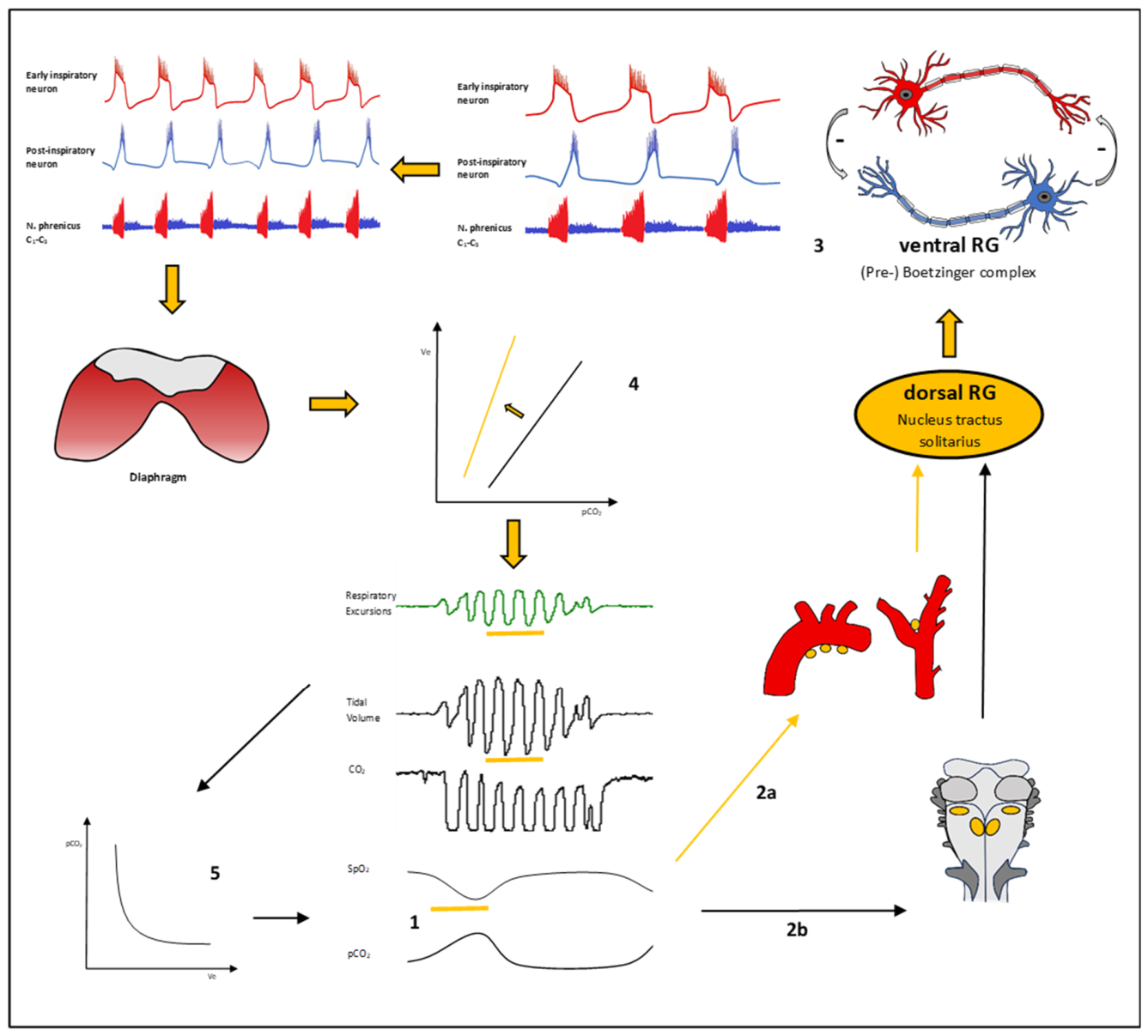
Figure 2.
Formula to calculate standard paO2 exemplified for a patient with chronic heart failure and nocturnal CSR [12].
Figure 2.
Formula to calculate standard paO2 exemplified for a patient with chronic heart failure and nocturnal CSR [12].
Disclaimer/Publisher’s Note: The statements, opinions and data contained in all publications are solely those of the individual author(s) and contributor(s) and not of MDPI and/or the editor(s). MDPI and/or the editor(s) disclaim responsibility for any injury to people or property resulting from any ideas, methods, instructions or products referred to in the content. |
© 2026 by the authors. Licensee MDPI, Basel, Switzerland. This article is an open access article distributed under the terms and conditions of the Creative Commons Attribution (CC BY) license (http://creativecommons.org/licenses/by/4.0/).
Copyright: This open access article is published under a Creative Commons CC BY 4.0 license, which permit the free download, distribution, and reuse, provided that the author and preprint are cited in any reuse.