

Submitted:
30 August 2026
Posted:
31 August 2026
You are already at the latest version
Abstract
Background/Objectives: The arcade of Frohse (AF) is a potential site of compression of the deep branch of the radial nerve (DBRN). However, its direct spatial relationship with the nerve has not been systematically classified using reproducible macroscopic criteria. This study aimed to develop a macroscopic classification of the AF–DBRN relationship and to examine its morphometric correlates. Methods: Forty-two formalin-fixed upper limbs from 29 donors were examined. The AF–DBRN relationship was classified as type 0 (no direct contact), type 1 (direct contact without visible nerve deformation), or type 2 (direct contact with macroscopic DBRN deformation). Two observers independently classified all speci-mens. DBRN cross-sectional area (CSA) and the mediolateral-to-anteroposterior dimen-sion ratio (ML/AP ratio) were assessed at three levels relative to the AF. A cen-tral-to-proximal CSA ratio was calculated in 36 limbs. Results: Type 0 was identified in 12 limbs (28.6%), type 1 in 14 (33.3%), and type 2 in 16 (38.1%). Interobserver agreement was 92.9% (unweighted Cohen’s κ = 0.892; linearly weighted κ = 0.918). The cen-tral-to-proximal CSA ratio differed among relationship types (H(2) = 9.9; nominal p = 0.007), with the highest median observed in type 2. AF dimensions and position were not associated with relationship type. Conclusions: The proposed macroscopic classification demonstrated high interobserver agreement and provides a reproducible framework that complements conventional morphometric assessment of the AF and DBRN. It should be regarded as an anatomical rather than diagnostic classification but may support standar-dized reporting and future anatomical, imaging, biomechanical, and clinical studies.
Keywords:
arcade of Frohse
; cadaveric study
; deep branch of the radial nerve
; morphometry
; peripheral nerve entrapment
; radial tunnel
; supinator muscle
1. Introduction
Some anatomical structures are clinically important not because of their size, but because of their close relationship to adjacent nerves or vessels. One such structure is the arcade of Frohse (AF), which forms the proximal border of the superficial layer of the supinator muscle (SM). The AF lies in close proximity to the deep branch of the radial nerve (DBRN). Owing to this relationship, the AF is commonly considered a potential site of DBRN compression and is relevant to the clinical anatomy of the proximal forearm and to compressive neuropathies involving the DBRN [1,2].
Near the cubital fossa, the radial nerve (RN) divides into the superficial branch of the radial nerve (SBRN) and the DBRN. The DBRN then enters the SM and passes between its superficial and deep layers. After emerging from the distal border of the SM, the nerve is generally referred to as the posterior interosseous nerve (PIN), which may therefore be regarded as the distal continuation of the DBRN [2,3]. However, the terms DBRN and PIN are not used consistently in the literature on radial tunnel syndrome and compressive neuropathies involving the SM [2,3]. In the present study, the term DBRN was used consistently for the portion of the nerve evaluated in relation to the AF.
The AF was described by Frohse and Fränkel in 1908 as a normal anatomical component of the SM [2]. Its possible clinical relevance received greater attention during the second half of the twentieth century. According to the historical account presented by Ozturk et al. [2], Kopell and Thompson suggested in 1963 that the DBRN could be compressed at the level of the AF, whereas Capener subsequently emphasized the susceptibility of the nerve to compression in this region. Spinner [4] later associated the AF with PIN palsy. Since then, the AF has been investigated both as a normal component of the SM and as one of the most frequently described potential sites of DBRN compression [1,5,6].
Nevertheless, the presence of the AF alone does not indicate that compression of the DBRN is occurring. Because the AF is a normal anatomical structure, its potential clinical significance depends on its specific spatial relationship with the nerve rather than on its presence alone. Consequently, describing the AF in isolation provides only limited information about its potential interaction with the DBRN. Particularly relevant features may include whether the AF is in direct contact with the DBRN and whether such contact is accompanied by visible macroscopic deformation of the nerve. Local anatomical conditions within the radial tunnel and changes occurring during forearm movement may also influence this relationship [1,2,7].
In the proximal forearm, the DBRN is also related to several other structures that may alter its course or represent potential sites of compression. In addition to the AF, proposed compression sites within the radial tunnel include fibrous bands, the radial recurrent vessels, the medial border of the extensor carpi radialis brevis muscle, and the distal border of the SM [1,3,6]. Consequently, evaluation of the morphology and dimensions of the AF alone may be insufficient to characterize its relationship with the DBRN.
Despite numerous anatomical investigations of the AF, its relationship with the DBRN has not been systematically classified using reproducible macroscopic criteria. Previous studies have focused primarily on the prevalence, tissue composition, dimensions, and topographic position of the AF relative to landmarks of the proximal forearm [2,5,7]. Although these investigations have substantially improved our understanding of AF anatomy, they do not fully characterize its direct relationship with the DBRN. Furthermore, it remains unclear whether relationship patterns defined by direct contact and visible nerve deformation are associated with objective DBRN morphometric parameters, including cross-sectional area (CSA), the mediolateral-to-anteroposterior dimension ratio (ML/AP ratio), and the central-to-proximal CSA ratio.
Establishing a standardized anatomical framework for describing AF–DBRN relationships may improve the consistency of future anatomical investigations and facilitate comparisons across morphometric, imaging, and clinical studies.
The primary aim of this study was therefore to propose and apply a macroscopic classification of the AF–DBRN relationship based on direct contact between the AF and DBRN and the presence or absence of visible nerve deformation. Interobserver agreement in assigning the relationship types was also assessed. The secondary aims were to characterize the morphology and position of the AF, evaluate DBRN CSA and the ML/AP ratio at measurement levels defined relative to the AF, calculate the central-to-proximal CSA ratio, and determine whether the AF–DBRN relationship type was associated with DBRN morphometric parameters, AF dimensions and position, side, or donor sex.
2. Materials and Methods
2.1. Study Design
An observational anatomical and morphometric study was conducted using formalin-fixed specimens. The primary component of the study was the macroscopic assessment and classification of the AF–DBRN relationship. Selected morphometric parameters of the AF and DBRN and the position of the AF relative to anatomical landmarks of the forearm were also evaluated.
2.2. Specimens
The study included 42 upper limbs from 29 donors. Bilateral upper limbs were available from 13 donors, whereas one upper limb was available from each of the remaining 16 donors. The material comprised 20 right and 22 left limbs, including 28 limbs from male donors and 14 limbs from female donors. Donor age at death ranged from 68 to 93 years.
The specimens were obtained from the Department of Human Anatomy, Faculty of Medical Sciences in Katowice, Medical University of Silesia, Katowice, Poland, through its institutional body donation program, which has been in operation since 2003. All donors were recorded as Caucasian in the available documentation.
Upper limbs without macroscopically visible pathological changes in the cubital fossa or proximal forearm were included. Limbs showing evidence of previous trauma, surgery, neoplastic lesions, osteoarticular deformity, or other abnormalities that could alter the normal topography of the examined region were excluded. This exclusion criterion did not apply to localized DBRN deformation assessed as part of the AF–DBRN relationship classification.
All upper limbs available during the study period that met the inclusion criteria were examined. Owing to the exploratory nature of the study and the limited availability of anatomical material, no formal sample-size calculation was performed.
2.3. Fixation and Storage
The specimens were fixed by injection with a 20% formalin solution and subsequently stored in tanks containing a 4%–5% formalin solution. All specimens remained in the storage solution for at least 12 months before examination.
2.4. Anatomical Dissection
Some specimens had previously been used for educational purposes; however, the cubital fossa and proximal forearm containing the AF and DBRN had not previously been dissected or otherwise disturbed. The cubital fossa and proximal forearm were dissected according to a standardized protocol. The RN, SBRN, DBRN, SM, and AF were exposed (Figure 1).
All specimens were examined with the elbow extended and the forearm positioned midway between pronation and supination. This position was obtained by placing the hand in the sagittal plane with the thumb directed upward. Wrist position was not otherwise standardized. Before measurement, only the amount of adipose and loose connective tissue necessary to expose the AF, DBRN, and other relevant structures was removed. Dissection was performed carefully to preserve the continuity of the examined structures and avoid altering their anatomical relationships.
2.5. Morphometric Measurements
After identification of the AF, its proximal and distal borders were defined. The proximal border was defined as the most proximal point of the structure forming the arcade, whereas the distal border was defined as its most distal point. The central point of the AF was defined as the intersection of the AF with a line drawn perpendicular to the segment connecting its proximal and distal borders and passing through the midpoint of that segment. This point corresponded to the thickness point described by Ozturk et al. [2].
All measurements were performed using a QC–Quality Control digital caliper (catalog no. IW-WEI-000-001) with a measurement range of 0–150 mm and a stated accuracy of 0.02 mm. Each parameter was measured twice by the same investigator (W.B.D.) using the same measurement protocol, with the specimen maintained in the same position. The arithmetic mean of the two measurements was recorded for statistical analysis. The individual replicate measurements were not retained as separate observations. Particular care was taken to avoid mechanical compression of the tissues, especially the DBRN, because such compression could alter the shape or dimensions of the nerve.
AF length was measured along the proximal-to-distal axis, and AF width was measured along the medial-to-lateral axis. AF thickness was measured at three levels: the proximal border, central point, and distal border.
Topographic distances were measured linearly between the lateral epicondyle (LE) of the humerus and the proximal border of the AF, between the LE and the distal border of the AF, and between the RN bifurcation and the proximal border of the AF.
When the proximal border of the AF was located at the level of the reference point defined by the LE, the distance between the LE and the proximal border of the AF was recorded as 0 mm. In such cases, the distance between the LE and the distal border of the AF corresponded to AF length.
DBRN parameters were assessed at three measurement levels: 1 cm proximal to the proximal border of the AF, at the central point of the AF, and at the distal border of the AF. Measurements obtained at the proximal border of the AF concerned the arcade itself, whereas the proximal DBRN measurement level was located 1 cm proximal to the proximal border of the AF. These locations were treated as separate measurement sites.
At each DBRN measurement level, two perpendicular dimensions of the nerve were recorded: the anteroposterior dimension (AP) and the mediolateral dimension (ML). Measurements were obtained in a plane oriented as nearly perpendicular as possible to the longitudinal axis of the nerve.
When the DBRN had not yet separated from the RN trunk at the level located 1 cm proximal to the proximal border of the AF, or when it could not be measured reliably as a separate structure, the measurement was recorded as not available (NA). Such observations were not treated as zero values and were not used to calculate parameters dependent on the proximal measurement.
The anatomical landmarks, method used to determine the central point of the AF, and selected DBRN measurement levels are shown in Figure 2.
The estimated CSA of the DBRN was calculated from the AP and ML dimensions, assuming an elliptical nerve cross-section:
CSA = (π/4) × AP × ML
The ML/AP ratio was calculated as:
ML/AP ratio = ML / AP
The ML/AP ratio represented the proportion between two anatomically defined nerve dimensions. It was not calculated as the conventional ratio of the larger dimension to the smaller dimension; therefore, its value also depended on the orientation of the nerve within the measurement plane.
The central-to-proximal CSA ratio was calculated as:
Central-to-proximal CSA ratio = CSA at the central point of the AF / CSA 1 cm proximal to the proximal border of the AF
A value of 1 indicated equal DBRN CSA at the two measurement levels. A value greater than 1 indicated a larger CSA at the central point of the AF than at the proximal reference level, whereas a value below 1 indicated a smaller CSA at the central point. The central-to-proximal CSA ratio was a relative morphometric parameter and did not directly measure the force, pressure, or degree of compression acting on the nerve.
The point located 1 cm proximal to the proximal border of the AF was selected as a standardized reference level for all specimens. This level was not assumed to be unaffected by other structures within the radial tunnel or to represent a normal control site. The central point of the AF was selected as a standardized measurement site within the arcade. Comparison of these two levels allowed assessment of the relative change in DBRN CSA within the same limb.
The distal border of the AF was analyzed as a separate measurement level but was not included in the calculation of the central-to-proximal CSA ratio because it could also reflect further changes in DBRN shape associated with the course of the nerve through the SM. Mean CSA across multiple measurement levels was not used because averaging could reduce the contribution of a localized change in CSA at the level of the AF.
The central-to-proximal CSA ratio was calculated only when reliable CSA measurements were available at both the central and proximal measurement levels.
Forearm length was measured as the distance between the LE and the radial styloid process. The AF position index was calculated as:
AF position index = distance from the LE to the proximal border of the AF / forearm length
2.6. Qualitative Assessment and Classification
The macroscopic anatomical relationship between the AF and DBRN was assessed with the specimen maintained in the standardized position. Classification was performed after complete exposure of both structures and before DBRN morphometric measurements were obtained. Contact or deformation was not induced by pressing, elevating, or deliberately displacing the AF or DBRN during assessment.
Direct contact between the AF and DBRN was defined as the absence of a visible space separating the two structures at the site of their relationship. No contact was recorded when a visible space remained between the AF and DBRN.
Macroscopic DBRN deformation was defined as a localized alteration in the contour of the nerve occurring directly at the site of contact with the AF and differing from the contour of adjacent nerve segments. Deformation could include flattening, indentation, localized narrowing, or another clearly visible alteration in shape. The natural curvature of the nerve, minor surface irregularities, and changes occurring outside the site of contact with the AF were not considered AF-related deformation.
The AF–DBRN relationship was classified independently by two investigators (W.B.D. and T.L.). Both observers assessed all 42 limbs using the criteria described above and were unaware of the classification assigned by the other observer. In cases of disagreement, the specimens were jointly reassessed, and the final relationship type was determined by consensus. The consensus classification was used in subsequent analyses.
2.7. Data Quality Control
Before statistical analysis, the measurement dataset was checked for accuracy. Measurement units and side designations were verified, and raw measurements were compared with the parameters calculated from them. Extreme values were additionally checked against the source measurements and recalculated from the corresponding AP and ML dimensions. Observations were not excluded solely because of extreme values. Each such observation was evaluated for a potential recording error, incorrect side designation, or anatomical variability.
2.8. Statistical Analysis
Statistical analyses were performed using jamovi, version 2.7. The individual upper limb was the primary unit of analysis. Bilateral upper limbs were available from 13 donors. Direct comparisons between the right and left sides within this subgroup were performed using the Wilcoxon signed-rank test, with p values adjusted using the Holm method. In the remaining analyses, each limb was treated as a separate anatomical observation. Dependence between bilateral limbs from the same donor was not directly modeled; therefore, inferential analyses involving AF–DBRN relationship type, donor sex, and morphometric parameters were considered exploratory.
Quantitative variables are presented as the mean, standard deviation, median, first and third quartiles, minimum, and maximum. Qualitative variables are presented as counts and percentages.
The distributions of quantitative variables were assessed using the Shapiro–Wilk test and graphical examination of histograms and quantile–quantile plots. Because most DBRN parameters, particularly CSA, the ML/AP ratio, and the central-to-proximal CSA ratio, did not meet the assumption of normality, nonparametric tests were used for most comparative analyses.
Interobserver agreement for the AF–DBRN relationship classification was assessed using the initial independent classifications assigned by the two observers before joint reassessment of discordant cases. Percentage agreement, unweighted Cohen’s kappa, and linearly weighted Cohen’s kappa were calculated. The weighted coefficient was used because types 0–2 represented ordered categories. A weight of 1 was assigned to complete agreement, 0.5 to disagreement between adjacent types, and 0 to disagreement between types 0 and 2. Ninety-five percent confidence intervals for the unweighted and linearly weighted Cohen’s kappa coefficients were estimated using percentile bootstrap resampling with 10,000 repetitions. Resampling was performed at the level of paired observer ratings. The final consensus classification was used in all other analyses.
Quantitative parameters were compared among the three AF–DBRN relationship types using the Kruskal–Wallis test. When the global test result was significant, two-sided pairwise comparisons were performed using the Mann–Whitney U test for independent samples with continuity correction. The p values obtained for the three pairwise comparisons were adjusted using the Holm method. Pairwise analyses were performed using the Rj Editor module within jamovi.
DBRN CSA and the ML/AP ratio were compared among the three measurement levels in the 36 limbs for which measurements were available at all three sites. Differences were assessed using the Friedman test. When the global test result was significant, pairwise comparisons were performed using the Wilcoxon signed-rank test with Holm adjustment. All comparisons included the same 36 complete cases.
Associations between AF dimensions and position and the central-to-proximal CSA ratio were evaluated using Spearman’s rank correlation coefficient. The p values for this set of correlations were adjusted using the Holm method. Associations between AF–DBRN relationship type and side or donor sex were evaluated using Fisher’s exact test.
Missing data were not imputed. When a parameter could not be determined for anatomical reasons, it was not treated as zero. Derived parameters, including CSA, the ML/AP ratio, the central-to-proximal CSA ratio, and the AF position index, were calculated only when all values required for their calculation were available.
Statistical significance was set at p < 0.05. Analyses examining associations between AF–DBRN relationship type and donor sex, side, and morphometric parameters were exploratory. No common adjustment was applied across all global tests comparing parameters among AF–DBRN relationship types; therefore, the corresponding p values should be interpreted as nominal. Holm adjustment was applied to pairwise comparisons performed after a significant global test and to the other predefined sets of comparisons described above.
2.9. Ethical Considerations
All donors provided written informed consent during their lifetime for the use of their bodies for scientific and educational purposes. The study was conducted in accordance with the procedures governing the institutional body donation program. Under institutional regulations, separate approval by a bioethics committee was not required for this cadaveric anatomical study. Donor data were anonymized, and the study followed the ethical principles governing the use of human cadaveric material in anatomical research [8].
3. Results
3.1. Study Material and Distribution of AF–DBRN Relationship Types
Type 0 was identified in 12 limbs (28.6%). Type 1 was identified in 14 limbs (33.3%), whereas type 2 was identified in 16 limbs (38.1%).
Direct contact between the AF and DBRN, comprising types 1 and 2, was present in 30 of 42 limbs (71.4%). Macroscopic DBRN deformation corresponding to type 2 was observed in 16 of 42 limbs (38.1%). The characteristics of the study material and the distribution of AF–DBRN relationship types are presented in Table 2.
3.2. Interobserver Agreement for the AF–DBRN Relationship Classification
The two observers assigned the same AF–DBRN relationship type in 39 of 42 cases, corresponding to an overall agreement of 92.9%. The unweighted Cohen’s kappa coefficient was κ = 0.892 (95% bootstrap CI: 0.750–1.000), and the linearly weighted Cohen’s kappa coefficient was κw = 0.918 (95% bootstrap CI: 0.810–1.000), indicating high interobserver agreement.
Disagreement occurred in three cases, all involving differentiation between types 1 and 2. No disagreement involved type 0. Following joint reassessment, the final classification of all three discordant cases was established by consensus. In each case, the consensus classification corresponded to the initial assessment of observer 1 (W.B.D.). The consensus classification was used in all subsequent analyses. Interobserver agreement results are presented in Table 3.
3.3. AF Morphometry and Topography
Mean AF length was 15.50 mm, with a median of 14.93 mm. Mean AF width was 11.61 mm, with a median of 10.63 mm.
AF thickness was assessed at the proximal border, central point, and distal border. Mean thickness was 0.86 mm at the proximal border, 0.83 mm at the central point, and 0.77 mm at the distal border. The corresponding median values were 0.64, 0.78, and 0.63 mm, respectively.
Topographic distances were assessed relative to the LE and RN bifurcation. The mean distance from the LE to the proximal border of the AF was 21.90 mm, with a median of 22.88 mm, whereas the mean distance from the LE to the distal border of the AF was 33.89 mm, with a median of 32.58 mm. The mean distance between the RN bifurcation and the proximal border of the AF was 27.42 mm, with a median of 27.05 mm.
Mean forearm length was 236.30 mm, with a median of 234.73 mm. The mean AF position index was 0.09, with a median of 0.10. Complete descriptive data for AF morphometry and topography are presented in Table 4.
3.4. DBRN Morphometry
DBRN CSA was assessed at three measurement levels: 1 cm proximal to the proximal border of the AF, at the central point of the AF, and at the distal border of the AF. Proximal measurements were available for 36 limbs because, in six cases, the DBRN had not yet separated clearly from the RN trunk at this level. These missing measurements were not treated as zero values.
Mean DBRN CSA was 3.22 mm² at the proximal measurement level, 4.21 mm² at the central point of the AF, and 5.45 mm² at the distal border of the AF. The corresponding median values were 2.43, 2.56, and 3.12 mm², respectively.
The ML/AP ratio was also assessed at the three measurement levels. Its mean value was 1.73 at the proximal level, 1.96 at the central point of the AF, and 2.15 at the distal border of the AF. The corresponding median values were 1.41, 1.69, and 1.96, respectively.
The central-to-proximal CSA ratio was available for 36 limbs. Its mean value was 1.26, with a median of 1.17. Complete descriptive data for DBRN CSA, the ML/AP ratio, and the central-to-proximal CSA ratio are presented in Table 5.
3.5. Changes in DBRN CSA and ML/AP Ratio Across Measurement Levels
Changes in DBRN CSA and the ML/AP ratio across the three measurement levels were analyzed in 36 limbs for which complete measurements were available 1 cm proximal to the proximal border of the AF, at the central point of the AF, and at the distal border of the AF.
DBRN CSA differed significantly among the three measurement levels, χ2 (2) = 12.7, p = 0.002. The highest CSA values were observed at the distal border of the AF. In pairwise comparisons performed using the Wilcoxon signed-rank test with Holm adjustment, CSA at the distal border of the AF differed significantly from CSA measured 1 cm proximal to the proximal border of the AF (p < 0.001) and from CSA measured at the central point of the AF (p < 0.001). The difference between CSA measured 1 cm proximal to the proximal border of the AF and CSA measured at the central point of the AF was not statistically significant (p = 0.084) (Figure 4).
The DBRN ML/AP ratio also differed significantly among the three measurement levels, χ2 (2) = 8.4, p = 0.015. Pairwise comparisons using the Wilcoxon signed-rank test with Holm adjustment demonstrated a significant difference between the ML/AP ratio measured 1 cm proximal to the proximal border of the AF and that measured at the distal border of the AF (p = 0.039). No significant difference was observed between the proximal measurement level and the central point of the AF (p = 0.338) or between the central point and distal border of the AF (p = 0.123) (Figure 5). The global test results and significant post hoc comparisons are presented in Table 6.
3.6. Central-to-Proximal CSA Ratio According to AF–DBRN Relationship Type
The analysis of the central-to-proximal CSA ratio included 36 limbs for which DBRN CSA measurements were available at the central point of the AF and 1 cm proximal to the proximal border of the AF. The analysis included 10 type 0, 11 type 1, and 15 type 2 cases. The median central-to-proximal CSA ratio was 1.01 (Q1–Q3: 0.63–1.20) for type 0, 0.88 (Q1–Q3: 0.73–1.10) for type 1, and 1.65 (Q1–Q3: 1.18–2.12) for type 2.
In the exploratory analysis, the central-to-proximal CSA ratio differed among AF–DBRN relationship types, H(2) = 9.9, nominal p = 0.007. In two-sided pairwise comparisons performed using the Mann–Whitney U test with continuity correction and Holm adjustment, type 2 differed significantly from type 0 (U = 31, p = 0.032) and type 1 (U = 29, p = 0.018). No significant difference was observed between types 0 and 1 (U = 60, p = 0.751). The ratio of DBRN CSA measured at the central point of the AF to CSA measured 1 cm proximal to the proximal border of the AF was higher in type 2 than in types 0 and 1. The distribution of the central-to-proximal CSA ratio according to AF–DBRN relationship type is shown in Figure 6.
3.7. CSA, ML/AP Ratio, and AF Morphometry According to AF–DBRN Relationship Type
Unlike the central-to-proximal CSA ratio, individual DBRN CSA values at the three measurement levels and ML/AP ratio values measured at the central point and distal border of the AF did not differ significantly among AF–DBRN relationship types.
DBRN CSA measured 1 cm proximal to the proximal border of the AF did not differ significantly among the relationship types. The median was 2.30 mm² (Q1–Q3: 1.15–2.77 mm²) for type 0, 3.37 mm² (Q1–Q3: 2.19–4.56 mm²) for type 1, and 1.75 mm² (Q1–Q3: 1.22–3.71 mm²) for type 2, H(2) = 1.8, nominal p = 0.411. This analysis included 36 limbs with an available proximal measurement.
CSA measured at the central point of the AF did not differ significantly among relationship types, H(2) = 2.5, nominal p = 0.291, nor did CSA measured at the distal border of the AF, H(2) = 1.4, nominal p = 0.505.
No significant differences were observed in the ML/AP ratio measured at the central point of the AF, H(2) = 1.9, nominal p = 0.394, or at the distal border of the AF, H(2) = 0.18, nominal p = 0.913.
AF length, width, and thickness did not differ significantly among AF–DBRN relationship types. The position of the AF also did not differ significantly among the three types. No significant differences were found in the distance from the LE to the proximal or distal border of the AF, the distance from the RN bifurcation to the proximal border of the AF, or the AF position index. The complete results are presented in Table 7.
3.8. Association of AF–DBRN Relationship Type with Side and Donor Sex
In the exploratory analysis, AF–DBRN relationship type was not significantly associated with side, p = 0.296. Among left limbs, type 0 was identified in 4 limbs, type 1 in 9 limbs, and type 2 in 9 limbs. Among right limbs, type 0 was identified in 8 limbs, type 1 in 5 limbs, and type 2 in 7 limbs.
In the exploratory limb-level analysis, the distribution of AF–DBRN relationship types differed according to donor sex, with a nominal p value of 0.018. Among limbs from female donors, type 0 was identified in 8 limbs, type 1 in 2 limbs, and type 2 in 4 limbs. Among limbs from male donors, type 0 was identified in 4 limbs, type 1 in 12 limbs, and type 2 in 12 limbs. Because donor sex is a donor-level characteristic and bilateral limbs were available from 13 donors, the observations were not fully independent. This result should therefore be regarded as preliminary and should not be interpreted as evidence of a confirmed sex-related difference. The results are presented in Table 8.
3.9. Correlations Between AF Morphometry and the Central-to-Proximal CSA Ratio
No significant correlations were observed between the central-to-proximal CSA ratio and AF dimensions or position. After Holm adjustment, none of the analyzed correlations reached statistical significance. All Spearman correlation coefficients indicated weak associations. Detailed results are presented in Table 9.
3.10. Comparison of Left and Right Limbs in Bilateral Specimens
Left and right limbs were compared in the subgroup of 13 donors for whom bilateral specimens were available. After Holm adjustment, no significant side-to-side differences were observed in any of the analyzed AF or DBRN morphometric or topographic parameters. Detailed results are presented in Table S1.
4. Discussion
4.1. Main Findings and Anatomical Significance
The principal contribution of this study is the development of a macroscopic classification of the AF–DBRN relationship based on two directly observable features: direct contact between the structures and visible deformation of the nerve. Direct contact was observed in most specimens, whereas macroscopic deformation occurred less frequently, indicating that simple apposition of the AF and DBRN is not equivalent to alteration of the nerve contour. This distinction is anatomically relevant because it separates contact without visible deformation from a configuration in which contact is accompanied by a localized change in DBRN shape.
The classification demonstrated high interobserver agreement. In the exploratory analysis, type 2 was associated with a higher central-to-proximal CSA ratio than types 0 and 1, whereas individual CSA values, ML/AP ratios, and AF dimensions and position did not differ among relationship types. These findings suggest that assessment of the direct AF–DBRN relationship provides information that is not captured by isolated measurements of either structure. Nevertheless, type 2 should be regarded as a macroscopic anatomical configuration rather than evidence of pathological nerve compression.
4.2. Interobserver Agreement and the Need for Further Validation
All disagreements involved differentiation between types 1 and 2, whereas no disagreement occurred for type 0. Thus, the presence or absence of direct contact was identified more consistently than subtle alterations in DBRN contour.
Distinguishing direct contact without deformation from slight flattening, indentation, or alteration of the DBRN contour may be difficult. Although the distinction between types 1 and 2 reflects a central criterion of the proposed classification, the threshold between no visible deformation and a minor change in nerve shape requires further refinement. More detailed descriptive criteria and standardized reference photographs may improve consistency in future studies.
The present agreement estimates were based on two observers from a single institution who evaluated the same anatomical material. Intraobserver agreement was not assessed by repeated classification of the same specimens after a predefined interval. Accordingly, the findings demonstrate high interobserver agreement within the present dataset but do not constitute complete validation of the classification.
4.3. The AF–DBRN Relationship Classification as a Complement to AF Morphometry
Previous anatomical studies have focused primarily on AF tissue composition, shape, dimensions, and position. Although these parameters are important for characterizing the arcade, they do not establish whether the AF is in direct contact with the DBRN or whether such contact is accompanied by visible nerve deformation. Previous reports have likewise indicated that tendinous AF morphology alone is insufficient to infer a compressive effect on the DBRN [1,9,10].
In the present study, AF length, width, thickness, and position did not differ among AF–DBRN relationship types and were not significantly correlated with the central-to-proximal CSA ratio. These findings provide no evidence that AF size or topographic position alone determines direct contact or visible DBRN deformation. The proposed classification therefore complements, rather than replaces, conventional morphometric assessment by describing the spatial relationship between both structures in an individual specimen.
4.4. DBRN CSA, ML/AP ratio, and Interpretation of the Central-to-Proximal CSA Ratio
Individual DBRN CSA values measured 1 cm proximal to the proximal border of the AF, at the central point of the AF, and at its distal border did not differ significantly among AF–DBRN relationship types. Similarly, no significant differences among relationship types were identified in the ML/AP ratio measured at the central point or distal border of the AF. These findings indicate that the relationship types were not distinguished by isolated measurements of nerve size or shape at individual levels. However, the absence of statistically significant differences should not be interpreted as evidence of equivalence, as the analyses may have been limited by substantial interindividual variability, small subgroup sizes, and the estimation of CSA from two linear dimensions.
The ML/AP ratio represented the mediolateral dimension divided by the anteroposterior dimension of the DBRN. Because it was calculated using two anatomically defined axes rather than as the larger dimension divided by the smaller dimension, its value depended on both nerve shape and orientation within the measurement plane. It should therefore be interpreted as a measure of the proportion between the two recorded dimensions rather than as a direct index of nerve flattening. A localized alteration in nerve contour may also occur without a reduction in CSA if a decrease in one dimension is accompanied by an increase in the perpendicular dimension.
The central-to-proximal CSA ratio provided a within-limb comparison between CSA at the central point of the AF and CSA at a standardized reference level located 1 cm proximal to its proximal border. This approach reduced, but did not eliminate, the influence of interindividual differences in overall nerve size. The proximal level was selected for methodological consistency and was not assumed to represent an unaffected control site. The distal border was not included in the ratio because the DBRN continues between the layers of the SM at this level, and distal CSA may therefore reflect additional changes associated with the intramuscular course of the nerve. Consistent with this consideration, distal CSA was significantly greater than CSA at both the proximal and central measurement levels, although the present study could not determine the cause of this difference.
Type 2 was associated with a higher central-to-proximal CSA ratio than types 0 and 1. Thus, when direct AF–DBRN contact was accompanied by visible nerve deformation, CSA at the central point of the AF was greater relative to the proximal reference level. This finding may reflect a larger central CSA, a smaller proximal CSA, or a combination of both and does not identify the underlying mechanism. Changes in nerve caliber and shape near the AF have been described both in patients with PIN syndrome and in asymptomatic individuals, indicating that morphometric alteration alone cannot be regarded as evidence of pathological compression [11,12,13,14]. More recently, Basavaraj et al. [15] used dynamic high-resolution ultrasonography in a prospective case-control study of patients with radial tunnel syndrome and matched healthy volunteers. They demonstrated position-dependent differences in PIN dimensions, including a larger CSA proximal to the AF during forearm pronation in symptomatic patients, and identified the AF as the most common site of compression during surgical decompression. These findings support the relevance of dynamic, level-specific nerve assessment but are not directly equivalent to the present cadaveric classification, which is based on macroscopic contact and deformation rather than clinical diagnosis or in vivo nerve dimensions. The present association was exploratory and requires confirmation in an independent dataset. The central-to-proximal CSA ratio should therefore be interpreted as a relative morphometric parameter; it does not directly quantify pressure or compressive force and cannot establish a diagnosis of compressive neuropathy.
4.5. AF Morphometry and Topography in the Context of Previous Studies
The AF in the present study had a width comparable to the pooled estimate reported by Benes et al. [5], whereas its mean length was shorter and its central thickness was greater. Differences between the present findings and previously reported values may reflect variation in specimen characteristics, fixation and storage conditions, dissection technique, limb position, measurement methodology, and natural anatomical variability. Direct comparison of morphometric values across studies should therefore be undertaken cautiously, particularly when anatomical reference points or measurement definitions differ.
AF length and width were assessed using an approach broadly comparable to that used in previous morphometric studies. The central point of the AF was defined as the intersection of the arcade with a line perpendicular to the segment connecting its proximal and distal borders and passing through the midpoint of that segment. This point corresponded to the thickness point described by Ozturk et al. [2]. By contrast, thickness measurements obtained at the proximal and distal borders represented additional assessment levels and should not be regarded as direct equivalents of the central thickness measurement. Although AF thickness decreased descriptively in the distal direction, no formal statistical comparison was performed among the three thickness levels; this pattern should therefore be interpreted as descriptive only.
Topographic assessment showed substantial variability in the position of the AF relative to the lateral epicondyle and RN bifurcation. Comparison with previous studies is limited by differences in the selected endpoints. Ozturk et al. [2] measured the distance from the lateral epicondyle to the thickness point of the AF and calculated a corresponding ratio relative to forearm length, whereas the present study used the proximal border of the AF as the primary reference point. Consequently, the lower AF position index observed in the present material may partly reflect the use of a more proximal endpoint and should not be interpreted as evidence that the AF was anatomically located more proximally. These findings emphasize the need for precise and standardized definitions of anatomical landmarks, as the expression “distance from the lateral epicondyle to the AF” may refer to different portions of the arcade and may therefore produce non-equivalent measurements across studies.
4.6. Side
No significant association was identified between AF–DBRN relationship type and side. Similarly, among the 13 bilateral donor pairs, no significant left–right differences were observed after Holm adjustment in the analyzed morphometric or topographic parameters of the AF and DBRN. The findings therefore provide no evidence of a clear side-related pattern in the AF–DBRN relationship. However, the absence of statistically significant differences should not be interpreted as proof of complete symmetry because weaker side-to-side differences may have remained undetected in the limited number of bilateral pairs.
Ceri et al. [11], in a bilateral ultrasonographic study of 30 asymptomatic individuals, found no significant side-related differences in the anteroposterior nerve dimension and no effect of limb dominance at any of the three levels assessed relative to the AF; all p values exceeded 0.10. Babaei-Ghazani et al. [16], who examined 65 individuals and 130 nerves, likewise reported no significant difference in DBRN CSA between dominant and nondominant limbs (p = 0.620) and proposed 0.35 mm² as the upper normal limit for side-to-side CSA difference. However, these studies evaluated nerve dimensions rather than the qualitative AF–DBRN relationship and therefore provide only an indirect reference.
4.7. Donor Sex
In the exploratory limb-level analysis, the distribution of AF–DBRN relationship types differed according to donor sex. This finding should be interpreted with considerable caution. The analysis was performed at the limb level, whereas sex was a donor-level characteristic, and bilateral limbs were available from 13 donors. Consequently, some donors contributed two observations, and the observations were not fully independent. Group sizes were also small, and the sex-related analysis was one of several exploratory comparisons. The finding should therefore be regarded as preliminary and requires confirmation in a larger dataset analyzed at the donor level.
Previous ultrasonographic studies have not demonstrated significant sex-related differences in the size of the examined nerve segment. Ceri et al. [11] found no significant differences between male and female participants in the anteroposterior nerve dimension at any of the three levels assessed relative to the AF; all p values were at least 0.369. Babaei-Ghazani et al. [16] reported a mean DBRN CSA of 0.98 ± 0.23 mm² in men and 0.93 ± 0.22 mm² in women, with no statistically significant difference (p = 0.200). These findings are not directly comparable with those of the present study because they concerned absolute nerve dimensions rather than AF–DBRN relationship type.
4.8. Potential Clinical, Imaging, and Surgical Relevance
The proposed classification should currently be regarded primarily as an anatomical framework for describing the AF–DBRN relationship rather than as a clinically validated classification. Its clinical, imaging, and surgical utility has not yet been established.
In the clinical and imaging literature, the term PIN is sometimes applied to the nerve segment located adjacent to the AF or passing through the SM. Findings from such studies may be relevant to the interpretation of the present results when they concern the same anatomical level. Comparisons should therefore be based on the actual location of the evaluated nerve segment rather than solely on the terminology used by individual authors [11,12,13,14,15,16].
Type 2 denotes direct contact between the AF and DBRN accompanied by visible nerve deformation. It is not equivalent to a diagnosis of DBRN compressive neuropathy, radial tunnel syndrome, or PIN syndrome. The classification describes only the anatomical relationship observed in a standardized specimen position. Clinical diagnosis requires assessment of symptoms, physical examination findings, and, where appropriate, electrodiagnostic or imaging studies.
High-resolution ultrasonography may be particularly useful for further evaluation of the proposed classification because it allows assessment of nerve course, shape, caliber, and echotexture in the region of the AF and SM [11,12,13,14,15,16].
From a surgical perspective, the classification may eventually facilitate standardized documentation of the AF–DBRN relationship during anatomical dissection or intraoperative assessment, including the presence of direct contact and visible deformation at the contact site. Recent work has also demonstrated the feasibility of ultrasound-guided percutaneous release at the AF. Marès et al. [17] validated a fully ultrasound-guided release technique in 10 fresh-frozen cadaveric specimens and reported preliminary clinical experience in five patients, supporting the anatomical feasibility of minimally invasive ultrasound-guided decompression while emphasizing the need for further clinical validation. However, the proposed classification should not be used as an independent basis for selecting patients for surgical decompression.
The AF is not the only structure that may affect the DBRN. Other proposed sites of compression within the radial tunnel include fibrous bands near the radiocapitellar joint, the radial recurrent vessels, the border of the extensor carpi radialis brevis muscle, and the distal border of the SM [1,6,10,18]. Assessment of the AF–DBRN relationship should therefore form part of a broader evaluation of the entire nerve course and should not replace examination of other potential sites of compression.
4.9. Limitations
Several limitations should be considered when interpreting the present findings.
The study was conducted using anatomical specimens fixed by formalin injection and subsequently stored in formalin solution for an extended period. Fixation and prolonged storage may alter tissue shape, elasticity, mechanical properties, and dimensions. Accordingly, the morphometric values obtained may not fully reflect conditions in fresh tissue or in vivo.
All specimens were evaluated in a single standardized limb position. The study did not assess the AF–DBRN relationship during forearm pronation and supination, at different elbow positions, or during tension of the SM. It therefore remains unknown whether direct contact or visible DBRN deformation would change during movement.
Anatomical dissection required exposure of the AF, RN, RN bifurcation, and DBRN. This involved removal of portions of adipose tissue, loose connective tissue, and fascial structures. Although care was taken to preserve the anatomical relationships, specimen manipulation may have affected the relative position of the structures, nerve contour, or some measurements.
The classification was performed by two observers from a single institution. External validation involving investigators from other centers was not performed, and intraobserver agreement was not assessed by repeating classification after a predefined interval. The greatest potential for subjective interpretation involved differentiation between types 1 and 2.
Morphometric measurements were performed by one investigator. Each parameter was measured twice, and the arithmetic mean was used in the analyses; however, the individual replicate measurements were not retained as separate observations, and formal intraobserver repeatability could therefore not be assessed. The investigator who performed the measurements also participated in the AF–DBRN relationship classification and was aware of the assigned relationship type. An influence of classification knowledge on the measurement process cannot therefore be excluded.
DBRN CSA was calculated from the AP and ML dimensions under the assumption of an elliptical nerve cross-section. The resulting values were therefore estimates and may have been less accurate when the nerve had an irregular contour, was visibly deformed, or was not measured exactly perpendicular to its longitudinal axis.
The central-to-proximal CSA ratio was a relative parameter based on the ratio between CSA at the central point of the AF and CSA measured 1 cm proximal to the proximal border of the AF. It described the relationship between two estimated CSA values obtained at different measurement levels but did not directly measure force or pressure acting on the nerve.
In six limbs, DBRN CSA could not be determined 1 cm proximal to the proximal border of the AF because the DBRN had not yet clearly separated from the RN trunk at that level. Consequently, the central-to-proximal CSA ratio and analyses involving all three DBRN measurement levels were based on 36 rather than 42 limbs. This smaller sample reduced the precision of the estimates and the ability to detect weaker associations. The missing proximal measurements resulted from RN bifurcation anatomy rather than random data loss. The 36 limbs included in these analyses may therefore have differed in nerve topography from the six limbs without an available proximal measurement.
The study was based on macroscopic and morphometric assessment. No histological analysis of the AF or DBRN was performed. It was therefore not possible to determine whether AF tissue composition was associated with AF–DBRN relationship type or whether macroscopic nerve deformation was accompanied by changes in the nerve sheaths, fascicles, or other microscopic structures.
Bilateral upper limbs were included from 13 donors. Dependence between the right and left limbs was accounted for in direct side-to-side comparisons using paired analyses; however, each limb was treated as a separate observation in the remaining analyses. Observations from bilateral specimens may therefore not have been fully independent, potentially affecting p values and the precision of estimates in comparisons among AF–DBRN relationship types, correlation analyses, and the analysis involving donor sex. The effective number of independent observations may have been smaller than the number of analyzed limbs. Accordingly, these inferential results should be interpreted as exploratory.
The overall sample size was limited, and subgroup sizes were small after stratification by AF–DBRN relationship type, donor sex, and side. This may have reduced the ability to detect weaker associations and may have produced unstable estimates. In addition, several exploratory analyses were performed, and no common correction was applied across all global tests. The findings concerning the central-to-proximal CSA ratio and donor sex should therefore be considered preliminary and require confirmation in an independent dataset.
Potential donor-related factors such as limb dominance, occupation, physical activity level, neurological disease, musculoskeletal disease, and previous upper-limb symptoms were not evaluated. These factors may have influenced DBRN and AF morphology, but their contribution could not be assessed in the available material.
The specimens originated from a single institution and included only donors aged 68–93 years who were recorded as Caucasian in the available documentation. These characteristics limit the generalizability of the findings to younger and more diverse populations. External validation should therefore include larger and more demographically diverse anatomical series.
4.10. Directions for Future Research
The present findings suggest several priorities for further investigation.
First, larger anatomical studies with adequately sized and more balanced groups are needed. Statistical analyses should account for the inclusion of bilateral limbs from some donors. This would allow more robust evaluation of the effects of donor sex, side, and interindividual variability on the AF–DBRN relationship while reducing the influence of within-donor dependence.
Second, the proposed classification requires external validation. Future studies should include independent specimen series, observers from other institutions, and investigators who were not involved in developing the classification criteria. Particular attention should be given to differentiation between types 1 and 2 because all disagreements in the present study involved these categories. Intraobserver agreement should also be evaluated by repeating specimen classification after a predefined interval. Standardized reference photographs and more detailed criteria for DBRN deformation may improve reproducibility.
Third, studies combining macroscopic assessment with histological evaluation of the AF and DBRN would be particularly valuable. Such work could determine whether AF tissue composition is associated with direct DBRN contact, visible nerve deformation, or changes in DBRN CSA and ML/AP ratio. Histological assessment of the DBRN could also establish whether type 2 is accompanied by alterations in the nerve sheaths or fascicular architecture. Different histological forms of the AF have been described, but their direct relationship with the spatial AF–DBRN relationship remains unclear.
Fourth, the qualitative classification could be supplemented by quantitative assessment of DBRN deformation. Standardized photography, three-dimensional scanning, or digital image analysis could be used to measure the length of AF–DBRN contact, indentation depth, and changes in nerve shape and dimensions at the contact site. This would help determine whether types 0–2 also represent increasing degrees of DBRN deformation.
Fifth, dynamic studies are needed to evaluate the AF–DBRN relationship during pronation and supination, at different elbow positions, and during simulated SM tension. Such studies could determine whether the relationship type observed in a single standardized position changes during movement. Biomechanical studies should additionally include direct measurement of pressure acting on the DBRN near the AF to determine whether visible nerve deformation is accompanied by increased local pressure.
Sixth, further in vivo imaging studies, particularly those using high-resolution dynamic ultrasonography, are warranted. Basavaraj et al. [15] have demonstrated that position-dependent changes in PIN dimensions can be detected in patients with radial tunnel syndrome compared with healthy controls. Future studies should determine whether the AF–DBRN relationship types proposed here can be identified reliably in vivo, whether the classification is reproducible among examiners, and whether these relationship types provide information beyond conventional nerve-dimension measurements. The greatest clinical value would come from studies combining imaging with clinical assessment, electrodiagnostic testing, intraoperative observations, and treatment outcomes. Such studies could determine whether type 2 is associated with symptoms, operative findings, or response to treatment.
Finally, the association between type 2 and a higher central-to-proximal CSA ratio should be evaluated in a larger, independent dataset. The repeatability of this ratio and its distribution in anatomical and in vivo populations should also be investigated. Any clinically relevant reference ranges should be established only through appropriately designed in vivo studies.
5. Conclusions
This study proposes, to our knowledge, the first macroscopic classification of the AF–DBRN relationship based on direct anatomical contact and visible nerve deformation. The proposed classification demonstrated high interobserver agreement and provides a reproducible framework that complements conventional morphometric assessment of the AF and DBRN. Type 2 was associated with a higher central-to-proximal CSA ratio in the exploratory analysis, whereas AF dimensions and position were not associated with relationship type. The classification should currently be regarded as an anatomical framework rather than a diagnostic tool, but it may facilitate standardized reporting and future anatomical, imaging, biomechanical, and clinical investigations of proximal radial nerve entrapment. External validation in independent and more diverse datasets is required.
Supplementary Materials
The following supporting information can be downloaded at: Preprints.org, Table S1: Comparison of left and right limbs in 13 bilateral donor pairs.
Author Contributions
Conceptualization, T.L. and W.B.D.; methodology, W.B.D., L.S. and K.S.; formal analysis, W.B.D.; investigation, T.L., W.B.D., K.K. and W.Z.; resources, T.L.; data curation, W.B.D.; writing—original draft preparation, W.B.D.; writing—review and editing, T.L., W.B.D., K.K., L.S., W.Z., K.S. and W.W.; visualization, T.L. and W.B.D.; supervision, T.L. and W.B.D.; project administration, T.L. and W.B.D. All authors have read and agreed to the published version of the manuscript.
Funding
This research received no external funding.
Institutional Review Board Statement
Ethical review and approval were not required for this cadaveric anatomical study under the institutional regulations governing the Conscious Body Donation Program of the Department of Human Anatomy, Medical University of Silesia, Katowice, Poland. The study was conducted in accordance with the procedures of the institutional body donation program, and all donor data were anonymized.
Informed Consent Statement
Written informed consent was obtained from all donors during their lifetime for the use of their bodies for scientific and educational purposes.
Data Availability Statement
The data supporting the findings of this study are available from the corresponding author upon reasonable request.
Acknowledgments
The authors express their sincere gratitude to the donors and their families, whose generous contribution made this study possible. Their selfless decision to donate their bodies to science has provided invaluable opportunities to advance anatomical knowledge and improve clinical practice.
Conflicts of Interest
The authors declare no conflicts of interest.
Abbreviations
The following abbreviations are used in this manuscript:
| AF | Arcade of Frohse |
| AP | Anteroposterior dimension |
| CSA | Cross-sectional area |
| DBRN | Deep branch of the radial nerve |
| LE | Lateral epicondyle |
| ML | Mediolateral dimension |
| ML/AP ratio | Mediolateral-to-anteroposterior dimension ratio |
| NA | Not available |
| PIN | Posterior interosseous nerve |
| RN | Radial nerve |
| SBRN | Superficial branch of the radial nerve |
| SM | Supinator muscle |
References
- Clavert, P.; Lutz, J.C.; Adam, P.; Wolfram-Gabel, R.; Liverneaux, P.; Kahn, J.L. Frohse's arcade is not the exclusive compression site of the radial nerve in its tunnel. Orthop. Traumatol. Surg. Res. 2009, 95, 114–118. [Google Scholar] [CrossRef] [PubMed]
- Ozturk, A.; Kutlu, C.; Taskara, N.; Kale, A.C.; Bayraktar, B.; Cecen, A. Anatomic and morphometric study of the arcade of Frohse in cadavers. Surg. Radiol. Anat. 2005, 27, 171–175. [Google Scholar] [CrossRef] [PubMed]
- Węgiel, A.; Karauda, P.; Zielinska, N.; Tubbs, R.S.; Olewnik, Ł. Radial nerve compression: anatomical perspective and clinical consequences. Neurosurg. Rev. 2023, 46, 53. [Google Scholar] [CrossRef] [PubMed]
- Spinner, M. The arcade of Frohse and its relationship to posterior interosseous nerve paralysis. J. Bone Jt. Surg. Br. 1968, 50-B, 809–812. [Google Scholar] [CrossRef]
- Benes, M.; Kachlik, D.; Kunc, V.; Kunc, V. The arcade of Frohse: a systematic review and meta-analysis. Surg. Radiol. Anat. 2021, 43, 703–711. [Google Scholar] [CrossRef] [PubMed]
- Riffaud, L.; Morandi, X.; Godey, B.; Brassier, G.; Guegan, Y.; Darnault, P.; Scarabin, J.M. Anatomic bases for the compression and neurolysis of the deep branch of the radial nerve in the radial tunnel. Surg. Radiol. Anat. 1999, 21, 229–233. [Google Scholar] [CrossRef] [PubMed]
- Caetano, E.B.; Vieira, L.A.; Sabongi Neto, J.J.; Caetano, M.B.F.; Sabongi, R.G.; Nakamichi, Y.D.C. Anatomical study of radial tunnel and its clinical implications in compressive syndromes. Rev. Bras. Ortop. (Sao Paulo) 2020, 55, 27–32. [Google Scholar] [CrossRef] [PubMed]
- Iwanaga, J.; Singh, V.; Takeda, S.; Ogeng'o, J.; Kim, H.-J.; Moryś, J.; Ravi, K.S.; Ribatti, D.; Trainor, P.A.; Sañudo, J.R.; Apaydin, N.; Sharma, A.; Smith, H.F.; Walocha, J.A.; Hegazy, A.M.S.; Duparc, F.; Paulsen, F.; Del Sol, M.; Adds, P.; Louryan, S.; Fazan, V.P.S.; Boddeti, R.K.; Tubbs, R.S. Standardized statement for the ethical use of human cadaveric tissues in anatomy research papers: Recommendations from Anatomical Journal Editors-in-Chief. Clin. Anat. 2022, 35, 526–528. [Google Scholar] [CrossRef] [PubMed]
- Debouck, C.; Rooze, M. The arcade of Fröhse: an anatomic study. Surg. Radiol. Anat. 1995, 17, 245–248. [Google Scholar] [CrossRef] [PubMed]
- Prasartritha, T.; Liupolvanish, P.; Rojanakit, A. A study of the posterior interosseous nerve (PIN) and the radial tunnel in 30 Thai cadavers. J. Hand Surg. Am. 1993, 18, 107–112. [Google Scholar] [CrossRef] [PubMed]
- Ceri, T.; Podda, A.; Behr, J.; Brumpt, E.; Alilet, M.; Aubry, S. Posterior interosseous nerve of the elbow at the arcade of Frohse: ultrasound appearance in asymptomatic subjects. Diagn. Interv. Imaging 2019, 100, 521–525. [Google Scholar] [CrossRef] [PubMed]
- Djurdjevic, T.; Loizides, A.; Löscher, W.; Gruber, H.; Plaikner, M.; Peer, S. High resolution ultrasound in posterior interosseous nerve syndrome. Muscle Nerve 2014, 49, 35–39. [Google Scholar] [CrossRef] [PubMed]
- Kim, Y.; Ha, D.H.; Lee, S.M. Ultrasonographic findings of posterior interosseous nerve syndrome. Ultrasonography 2017, 36, 363–369. [Google Scholar] [CrossRef] [PubMed]
- Raeburn, K.; Burns, D.; Hage, R.; Tubbs, R.S.; Loukas, M. Cross-sectional sonographic assessment of the posterior interosseous nerve. Surg. Radiol. Anat. 2015, 37, 1155–1160. [Google Scholar] [CrossRef] [PubMed]
- Basavaraj, S.; Raju, V.; Paraj, D.; Bhat, A.K.; Acharya, A. Role of high-resolution dynamic ultrasonography in the evaluation of posterior interosseous nerve compression at radial tunnel: a prospective case-control study. J. Hand Surg. Eur. online ahead of print. Vol. 2026. [CrossRef] [PubMed]
- Babaei-Ghazani, A.; Roomizadeh, P.; Sanaei, G.; Najarzadeh-Mehdikhani, S.; Habibi, K.; Nikmanzar, S.; Kheyrollah, Y. Ultrasonographic reference values for the deep branch of the radial nerve at the arcade of Frohse. J. Ultrasound 2018, 21, 225–231. [Google Scholar] [CrossRef] [PubMed]
- Marès, O.; Ferreira, J.; Pradel, S.; Martinel, V.; Chaves, C.; Apard, T. Radial nerve at Frohse's arcade: a new technique of release under total ultrasound guidance. First experience. Hand Surg. Rehabil. 2025, 44S, 102089. [Google Scholar] [CrossRef] [PubMed]
- Konjengbam, M.; Elangbam, J. Radial nerve in the radial tunnel: anatomic sites of entrapment neuropathy. Clin. Anat. 2004, 17, 21–25. [Google Scholar] [CrossRef] [PubMed]
Figure 1.
Anatomical specimen of the proximal forearm demonstrating the relationship between the arcade of Frohse (AF) and the deep branch of the radial nerve (DBRN). The radial nerve bifurcates into the superficial branch of the radial nerve (SBRN) and the DBRN before the latter enters the supinator muscle. The white arrows indicate the course of the AF, corresponding to the proximal border of the superficial layer of the supinator muscle (SLSM). The deep layer of the supinator muscle (DLSM) is also shown. Surrounding tissues were gently retracted to improve visualization of the anatomical structures.
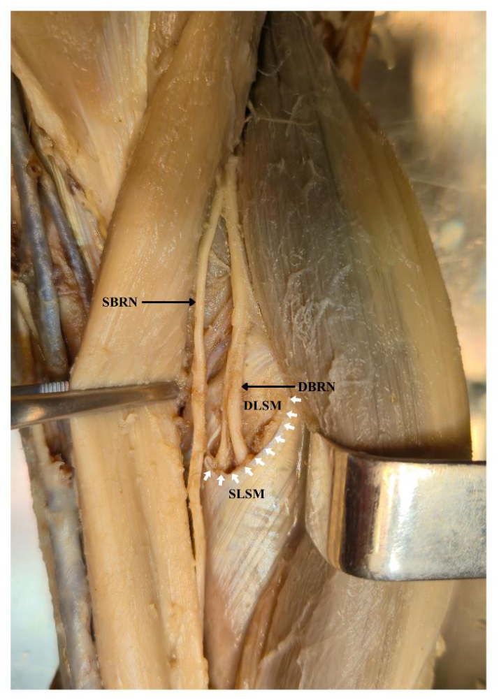
Figure 2.
Schematic representation of the anatomical landmarks and selected measurement levels used in the study. (A) Topography of the radial nerve and its branches in the proximal forearm and their relationship with the arcade of Frohse and supinator muscle. (B) AF reference points and selected DBRN measurement levels. PB-AF and DB-AF indicate the proximal and distal borders of the AF, respectively. CP-AF indicates the point at which the AF intersects a line drawn perpendicular to the segment connecting PB-AF and DB-AF and passing through the midpoint of that segment. C-DBRN and D-DBRN indicate DBRN measurement sites at the levels of CP-AF and DB-AF, respectively. The proximal measurement level was located 1 cm proximal to PB-AF. In the illustrated variant, the DBRN had not yet separated from the RN trunk at this level; therefore, the proximal measurement was unavailable. Purple markers indicate the reference and measurement levels, and dashed lines indicate the construction used to determine CP-AF. The D-DBRN marker indicates the projected location of the nerve beneath the superficial layer of the supinator muscle. The schematic is not drawn to scale. AF, arcade of Frohse; BRM, brachioradialis muscle; DBRN, deep branch of the radial nerve; DLSM, deep layer of the supinator muscle; LE, lateral epicondyle; PIN, posterior interosseous nerve; RN, radial nerve; RSP, radial styloid process; SBRN, superficial branch of the radial nerve; SLSM, superficial layer of the supinator muscle.
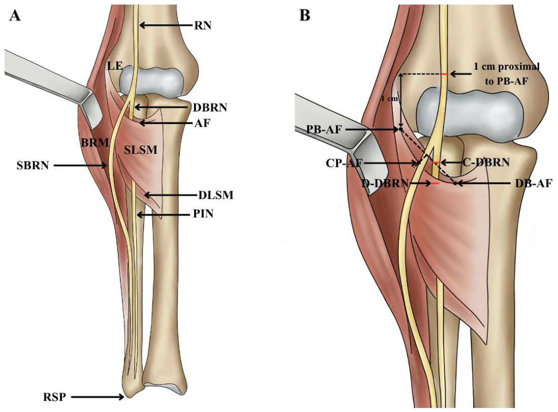
Figure 3.
Representative examples of the macroscopic relationship between the arcade of Frohse (AF) and the deep branch of the radial nerve (DBRN). (A) Type 0: no direct contact between the AF and DBRN, with a visible space separating the structures. (B) Type 1: direct contact between the AF and DBRN without visible macroscopic nerve deformation. (C) Type 2: direct contact between the AF and DBRN with localized macroscopic deformation of the DBRN at the level of the AF. Black arrows indicate the AF, the asterisk indicates the visible space between the AF and DBRN, and the white arrow indicates the site of DBRN deformation. DBRN, deep branch of the radial nerve; SBRN, superficial branch of the radial nerve. Adjacent structures, including the SBRN, were gently displaced with forceps solely to improve visualization of the AF and DBRN.
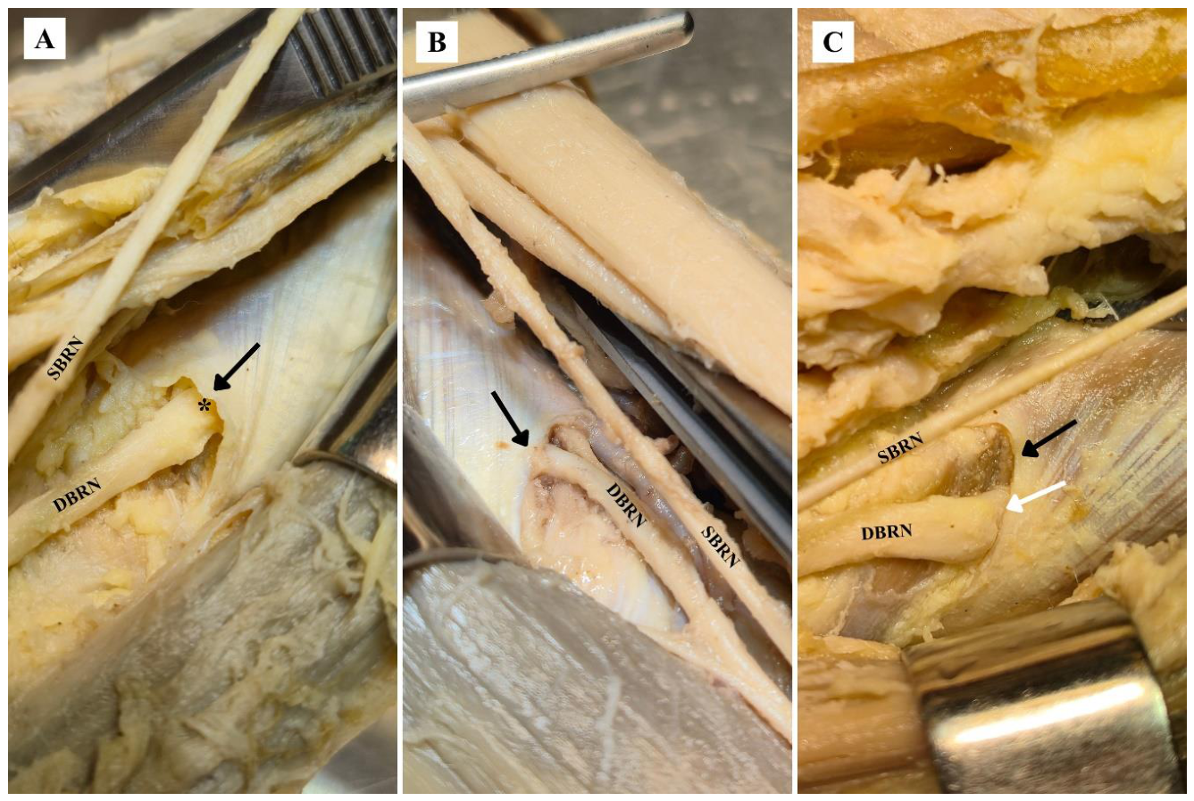
Figure 4.
DBRN cross-sectional area (CSA) measured 1 cm proximal to the proximal border of the AF, at the central point of the AF, and at the distal border of the AF. The plot includes 36 limbs for which measurements were available at all three levels. CSA measured at the distal border of the AF differed significantly from CSA measured 1 cm proximal to the proximal border of the AF and from CSA measured at the central point of the AF. AF, arcade of Frohse; DBRN, deep branch of the radial nerve.
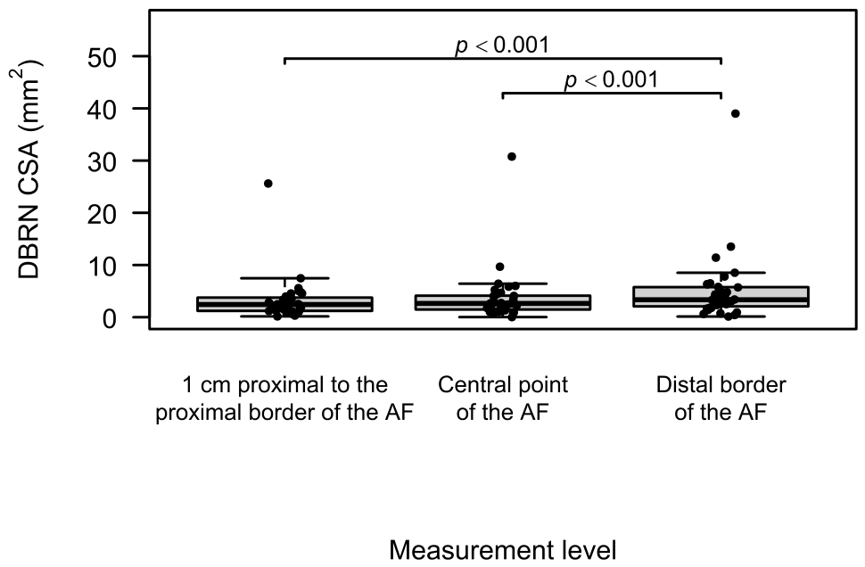
Figure 5.
DBRN mediolateral-to-anteroposterior dimension ratio (ML/AP ratio) measured 1 cm proximal to the proximal border of the AF, at the central point of the AF, and at the distal border of the AF. The plot includes 36 limbs for which measurements were available at all three levels. A significant difference was observed between the ML/AP ratio measured 1 cm proximal to the proximal border of the AF and that measured at the distal border of the AF. AF, arcade of Frohse; DBRN, deep branch of the radial nerve.
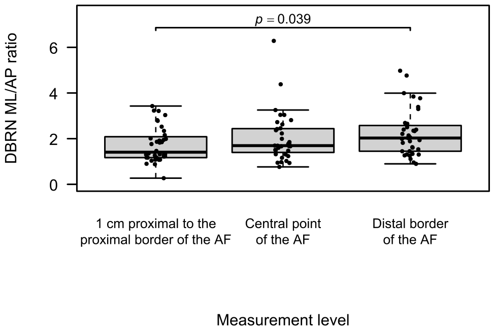
Figure 6.
Central-to-proximal DBRN cross-sectional area ratio according to AF–DBRN relationship type. The plot includes 36 limbs: 10 type 0, 11 type 1, and 15 type 2 cases. The median ratio was higher in type 2. In pairwise comparisons performed using the Mann–Whitney U test with Holm adjustment, type 2 differed significantly from types 0 and 1, whereas no significant difference was observed between types 0 and 1. AF, arcade of Frohse; DBRN, deep branch of the radial nerve.
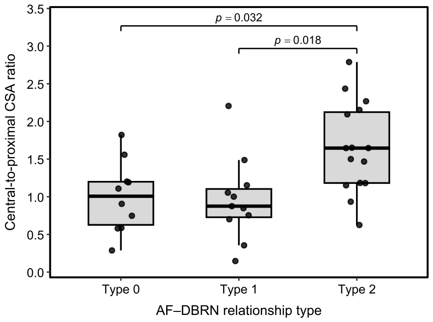

Table 1.
Macroscopic classification of the AF–DBRN relationship.
| Relationship type | Macroscopic criterion | Anatomical description |
| Type 0 | No direct contact between the AF and DBRN | A visible space separates the AF and DBRN |
| Type 1 | Direct contact between the AF and DBRN without visible nerve deformation | The AF is in contact with the DBRN, but the nerve contour at the contact site remains continuous and does not clearly differ from that of adjacent segments. No localized flattening, indentation, or other evident deformation is present |
| Type 2 | Direct contact between the AF and DBRN with visible macroscopic nerve deformation | The AF is in contact with the DBRN, and a localized alteration in nerve contour is visible directly at the contact site. The alteration differs from adjacent nerve segments and may include flattening, indentation, narrowing, or another evident deformation |
Abbreviations: AF, arcade of Frohse; DBRN, deep branch of the radial nerve.
Table 2.
Characteristics of the study material and distribution of AF–DBRN relationship types.
| Variable | Category | n | % |
| Side | Right | 20 | 47.6 |
| Left | 22 | 52.4 | |
| Donor sex | Male | 28 | 66.7 |
| Female | 14 | 33.3 | |
| AF–DBRN relationship type | Type 0 | 12 | 28.6 |
| Type 1 | 14 | 33.3 | |
| Type 2 | 16 | 38.1 |
Abbreviations: AF, arcade of Frohse; DBRN, deep branch of the radial nerve; n, number of limbs.
Table 3.
Interobserver agreement for the AF–DBRN relationship classification.
| Observer 1 classification | Observer 2: Type 0 | Observer 2: Type 1 | Observer 2: Type 2 | Total |
| Type 0 | 12 | 0 | 0 | 12 |
| Type 1 | 0 | 13 | 1 | 14 |
| Type 2 | 0 | 2 | 14 | 16 |
| Total | 12 | 15 | 15 | 42 |
| Overall agreement was 92.9% (39/42); κ = 0.892 (95% bootstrap CI: 0.750–1.000); κw = 0.918 (95% bootstrap CI: 0.810–1.000). | ||||
Abbreviations: AF, arcade of Frohse; DBRN, deep branch of the radial nerve; CI, confidence interval; κ, unweighted Cohen’s kappa; κw, linearly weighted Cohen’s kappa.
Table 4.
Morphometric and topographic parameters of the AF.
| Parameter | n | Mean | SD | Median | Q1 | Q3 | Min | Max |
| AF length | 42 | 15.50 | 5.67 | 14.93 | 11.65 | 18.21 | 6.08 | 31.59 |
| AF width | 42 | 11.61 | 3.78 | 10.63 | 8.69 | 13.85 | 5.47 | 23.82 |
| AF thickness, proximal border | 42 | 0.86 | 0.46 | 0.64 | 0.53 | 1.09 | 0.31 | 1.96 |
| AF thickness, central point | 42 | 0.83 | 0.34 | 0.78 | 0.57 | 1.06 | 0.20 | 1.72 |
| AF thickness, distal border | 42 | 0.77 | 0.46 | 0.63 | 0.43 | 1.12 | 0.11 | 2.01 |
| LE to proximal border of AF | 42 | 21.90 | 10.98 | 22.88 | 16.47 | 29.53 | 0.00 | 42.22 |
| LE to distal border of AF | 42 | 33.89 | 10.15 | 32.58 | 25.35 | 42.22 | 13.47 | 59.71 |
| RN bifurcation to proximal border of AF | 42 | 27.42 | 14.79 | 27.05 | 15.36 | 35.53 | 0.81 | 55.97 |
| Forearm length | 42 | 236.30 | 17.09 | 234.73 | 222.79 | 251.95 | 204.73 | 262.58 |
| AF position index | 42 | 0.09 | 0.05 | 0.10 | 0.07 | 0.13 | 0.00 | 0.17 |
Abbreviations: AF, arcade of Frohse; LE, lateral epicondyle; RN, radial nerve; SD, standard deviation; Q1, first quartile; Q3, third quartile; Min, minimum; Max, maximum. All linear measurements are presented in millimeters. The AF position index is dimensionless.
Table 5.
Morphometric parameters of the DBRN.
| Parameter | n | Mean | SD | Median | Q1 | Q3 | Min | Max |
| DBRN CSA, 1 cm proximal to the proximal border of the AF | 36 | 3.22 | 4.18 | 2.43 | 1.23 | 3.69 | 0.16 | 25.62 |
| DBRN CSA, central point of the AF | 42 | 4.21 | 7.00 | 2.56 | 1.23 | 4.09 | 0.02 | 36.83 |
| DBRN CSA, distal border of the AF | 42 | 5.45 | 8.34 | 3.12 | 1.79 | 5.63 | 0.13 | 41.15 |
| DBRN ML/AP ratio, 1 cm proximal to the proximal border of the AF | 36 | 1.73 | 0.77 | 1.41 | 1.17 | 2.05 | 0.27 | 3.43 |
| DBRN ML/AP ratio, central point of the AF | 42 | 1.96 | 1.01 | 1.69 | 1.33 | 2.41 | 0.76 | 6.28 |
| DBRN ML/AP ratio, distal border of the AF | 42 | 2.15 | 1.01 | 1.96 | 1.36 | 2.47 | 0.90 | 4.98 |
| Central-to-proximal CSA ratio | 36 | 1.26 | 0.63 | 1.17 | 0.82 | 1.65 | 0.15 | 2.79 |
Abbreviations: AF, arcade of Frohse; DBRN, deep branch of the radial nerve; CSA, cross-sectional area; ML/AP ratio, mediolateral-to-anteroposterior dimension ratio; SD, standard deviation; Q1, first quartile; Q3, third quartile; Min, minimum; Max, maximum. CSA values are presented in mm². The ML/AP ratio and central-to-proximal CSA ratio are dimensionless. The level located 1 cm proximal to the proximal border of the AF refers exclusively to DBRN measurements and is distinct from measurements obtained at the proximal border of the AF itself. The table presents all available observations for each measurement level. Comparisons of CSA and the ML/AP ratio among the three measurement levels were performed separately using the 36 limbs with complete measurements at all three levels.
Table 6.
Comparison of DBRN CSA and ML/AP ratio among the three measurement levels.
| Parameter | Test | Result | Significant post hoc comparisons |
| CSA | Friedman | χ2 (2) = 12.7, p = 0.002 | Distal border of the AF vs 1 cm proximal to the proximal border of the AF: p < 0.001; distal border of the AF vs central point of the AF: p < 0.001 |
| ML/AP ratio | Friedman | χ2 (2) = 8.4, p = 0.015 | Distal border of the AF vs 1 cm proximal to the proximal border of the AF: p = 0.039 |
Abbreviations: AF, arcade of Frohse; DBRN, deep branch of the radial nerve; CSA, cross-sectional area; ML/AP ratio, mediolateral-to-anteroposterior dimension ratio; χ2, Friedman test statistic. Analyses included 36 limbs with complete measurements at all three levels. Pairwise comparisons were performed using the Wilcoxon signed-rank test, and p values were adjusted using the Holm method.
Table 7.
Associations between AF–DBRN relationship type and selected quantitative parameters.
| Parameter | Test | Result | Interpretation |
| Central-to-proximal CSA ratio | Kruskal–Wallis | H(2) = 9.9; p = 0.007 |
Nominally significant difference |
| DBRN CSA, 1 cm proximal to the proximal border of the AF | Kruskal–Wallis | H(2) = 1.8; p = 0.411 |
No significant difference |
| DBRN CSA, central point of the AF | Kruskal–Wallis | H(2) = 2.5; p = 0.291 |
No significant difference |
| DBRN CSA, distal border of the AF | Kruskal–Wallis | H(2) = 1.4; p = 0.505 |
No significant difference |
| DBRN ML/AP ratio, central point of the AF | Kruskal–Wallis | H(2) = 1.9; p = 0.394 |
No significant difference |
| DBRN ML/AP ratio, distal border of the AF | Kruskal–Wallis | H(2) = 0.18; p = 0.913 |
No significant difference |
| AF length | Kruskal–Wallis | H(2) = 0.95; p = 0.623 |
No significant difference |
| AF width | Kruskal–Wallis | H(2) = 0.18; p = 0.914 |
No significant difference |
| AF thickness, proximal border | Kruskal–Wallis | H(2) = 0.49; p = 0.781 |
No significant difference |
| AF thickness, central point | Kruskal–Wallis | H(2) = 0.53; p = 0.766 |
No significant difference |
| AF thickness, distal border | Kruskal–Wallis | H(2) = 0.35; p = 0.839 |
No significant difference |
| LE to proximal border of the AF | Kruskal–Wallis | H(2) = 1.5; p = 0.476 |
No significant difference |
| LE to distal border of the AF | Kruskal–Wallis | H(2) = 0.92; p = 0.631 |
No significant difference |
| RN bifurcation to proximal border of the AF | Kruskal–Wallis | H(2) = 0.22; p = 0.894 |
No significant difference |
| AF position index | Kruskal–Wallis | H(2) = 2.2; p = 0.329 |
No significant difference |
Abbreviations: AF, arcade of Frohse; DBRN, deep branch of the radial nerve; CSA, cross-sectional area; ML/AP ratio, mediolateral-to-anteroposterior dimension ratio; LE, lateral epicondyle; RN, radial nerve; H, Kruskal–Wallis test statistic. CSA values are presented in mm². The ML/AP ratio, central-to-proximal CSA ratio, and AF position index are dimensionless. The analyses were exploratory. The p values for the global tests were not adjusted collectively for multiple testing and should therefore be interpreted as nominal. When the Kruskal–Wallis test was significant, pairwise comparisons were performed, and the resulting p values were adjusted using the Holm method. The analysis of DBRN CSA measured 1 cm proximal to the proximal border of the AF included 36 limbs.
Table 8.
Associations between AF–DBRN relationship type and side or donor sex.
| Variable | Category | Type 0 | Type 1 | Type 2 | Test | p |
|
Side |
Left | 4 | 9 | 9 |
Fisher’s exact test |
0.296 |
| Right | 8 | 5 | 7 | |||
|
Donor sex |
Female | 8 | 2 | 4 |
Fisher’s exact test |
0.018 |
| Male | 4 | 12 | 12 |
Abbreviations: AF, arcade of Frohse; DBRN, deep branch of the radial nerve. Associations were assessed using Fisher’s exact test. The analyses were exploratory, and the p values should be interpreted as nominal. Because donor sex is a donor-level characteristic and bilateral specimens were included, the sex-related analysis should be interpreted cautiously.
Table 9.
Correlations between AF dimensions and position and the central-to-proximal CSA ratio.
| Variable | n | Spearman’s ρ | Unadjusted p | Holm-adjusted p |
| AF length | 36 | 0.220 | 0.198 | 0.988 |
| AF width | 36 | -0.112 | 0.516 | 1.000 |
| AF thickness, proximal border | 36 | -0.240 | 0.159 | 0.952 |
| AF thickness, central point | 36 | -0.118 | 0.492 | 1.000 |
| AF thickness, distal border | 36 | -0.067 | 0.696 | 1.000 |
| LE to proximal border of the AF | 36 | -0.273 | 0.107 | 0.856 |
| LE to distal border of the AF | 36 | -0.309 | 0.067 | 0.601 |
| RN bifurcation to proximal border of the AF | 36 | -0.153 | 0.373 | 1.000 |
| AF position index | 36 | -0.254 | 0.134 | 0.941 |
Abbreviations: AF, arcade of Frohse; LE, lateral epicondyle; RN, radial nerve; ρ, Spearman’s rank correlation coefficient. The central-to-proximal CSA ratio and AF position index are dimensionless.
Disclaimer/Publisher’s Note: The statements, opinions and data contained in all publications are solely those of the individual author(s) and contributor(s) and not of MDPI and/or the editor(s). MDPI and/or the editor(s) disclaim responsibility for any injury to people or property resulting from any ideas, methods, instructions or products referred to in the content. |
© 2026 by the authors. Licensee MDPI, Basel, Switzerland. This article is an open access article distributed under the terms and conditions of the Creative Commons Attribution (CC BY) license.
Copyright: This open access article is published under a Creative Commons CC BY 4.0 license, which permit the free download, distribution, and reuse, provided that the author and preprint are cited in any reuse.