

Submitted:
28 August 2026
Posted:
31 August 2026
You are already at the latest version
Abstract
Background/Objectives: Infectious diseases remain a major burden on global public health, exacerbated by the serious threat of antimicrobial resistance and the limitations of conventional pharmaceutical formulations. This review aims to analyze the current state of 3D printing technology in the development of drug delivery systems tailored to the treatment of infectious diseases, emphasizing its capabilities across various routes of administration, current challenges, and potential clinical prospects. Methods: A literature search was conducted in ScienceDirect for research articles published between 2020 and May 2026 using the terms “antibiotic” AND “3D printing” AND “drug delivery.” Retrieved articles were selected, filtered, and categorized according to their route of administration. Results: Additive manufacturing allows for precise control of dosage, geometry, and release kinetics, facilitating personalized treatments for infectious diseases. Semi-solid extrusion has become the dominant technique due to its compatibility with various biomaterials and thermolabile active pharmaceutical ingredients. Topical applications represent the most advanced area, integrating multifunctional properties such as stimuli response and wound healing. Furthermore, 3D printing has demonstrated significant efficacy in expanding delivery platforms for vaginal, ocular, and oral routes. Conclusions: Pharmaceutical 3D printing is evolving from a manufacturing tool for personalized dosage forms to an advanced platform for designing multifunctional therapeutic systems. Overcoming current regulatory, clinical validation, and manufacturing challenges will be crucial to translating these innovations into routine clinical practice to combat infectious diseases and antimicrobial resistance.
Keywords:
3D printing
; drug delivery systems
; infectious diseases
; antimicrobial resistance
; personalized medicine
; semi-solid extrusion
; targeted delivery
1. Introduction
Infectious diseases are those caused by pathogenic microorganisms, such as bacteria, viruses, parasites, or fungi, and can be transmitted, directly or indirectly, from one person to another. According to the World Health Organization (WHO) [1], these diseases can be grouped into three categories: diseases associated with high mortality rates; diseases that impose a heavy burden of disability on populations; and diseases that, due to their rapid and unpredictable spread, can have serious global repercussions.
Despite significant efforts and advances in prevention, diagnosis, and treatment, infectious diseases continue to generate a high health burden, especially in low- and middle-income countries (Figure 1).
Furthermore, the growing resistance to antibiotics has become one of the main threats to global public health. Contributing factors include the misuse and overuse of these drugs, a limited understanding of the true extent of the problem, the lack of effective surveillance systems, deficiencies in infection prevention and control programs, and the widespread use of antimicrobials in animal production for human consumption. Consequently, it is estimated that, if current trends continue, antimicrobial resistance could cause up to 10 million deaths annually by 2050 and reduce global gross domestic product by between 2% and 3.5% [2]. Considering this scenario, the WHO highlights in its Resolution WHA51.17 [3] the urgent need to develop more effective, safe and patient-friendly therapeutic strategies that can optimize drug administration and help mitigate the advance of antimicrobial resistance.
Currently, most drug treatments for infectious diseases are administered via conventional pharmaceutical forms, primarily orally or parenterally. However, many active pharmaceutical ingredients (APIs) have biopharmaceutical limitations that can compromise their therapeutic efficacy. Among these, low aqueous solubility is one of the main challenges in modern pharmaceutical development. In fact, it is estimated that approximately 70% of new chemical entities identified during the drug discovery process exhibit poor water solubility, while nearly 40% of currently marketed immediate-release oral medications are classified as practically insoluble (<100 μg/mL) [4] .
The solubility of a drug plays a fundamental role in its dissolution and absorption, especially in pharmaceutical forms where the time available for dissolution is limited. Consequently, low solubility usually results into a small amount of drug available for absorption. However, the bioavailability of an API does not depend solely on its solubility, but also on other factors such as the rate of dissolution, permeability across biological membranes, susceptibility to efflux mechanisms, and first-pass metabolism [5].
These limitations can generate high variability in therapeutic response, lead to the need for higher doses or more frequent administrations and negatively affect adherence to treatment. In the context of infectious diseases, these difficulties can lead to subtherapeutic drug concentrations, promote treatment failure, and indirectly contribute to the development and spread of antimicrobial resistance.
Beyond these biopharmaceutical limitations, the lack of dose individualization remains a major limitation of current pharmacotherapy. Routine clinical practice relies on standardized dosages, often without adequately considering individual variations such as body weight, age, renal or hepatic function, the presence of comorbidities, or drug interactions. This standardization can lead to therapeutic ineffectiveness in some patients while causing drug toxicity in others. This problem is exacerbated in pediatric and geriatric populations, where physiological heterogeneity is greater and where, in many cases, commercially available formulations adapted to specific dosing needs are unavailable. Similarly, some chronic infectious diseases require prolonged treatments tailored to the patient’s characteristics, further highlighting the limitations of conventional dosing regimens [6].
An additional challenge lies in the need to treat infections that require combination therapies, whether to enhance efficacy, reduce the individual toxicity of each drug, or decrease the risk of resistance [7]. Such is the case with tuberculosis, which requires the simultaneous administration of several agents, or Helicobacter pylori infection, usually treated with combinations of antibiotics and gastric antisecretory agents [8]. However, manufacturing pharmaceutical dosage forms that integrate multiple APIs with differentiated release profiles is a complex process, as there may be physicochemical incompatibilities between the compounds, as well as different requirements regarding the rate and site of drug release [9].
Given this scenario, 3D printing applied to the pharmaceutical field emerges as a promising technology to overcome these challenges. Thanks to the precise manipulation of geometry, porosity, and layer arrangement, structures can be generated capable of releasing the API immediately, continuously, or in a pulsatile manner, as required by the treatment [10]. This potential is particularly valuable in the treatment of infectious diseases, where the plasma concentration of the drug must be maintained within a therapeutic range for prolonged periods to achieve its effectiveness (Figure 2).
The most widespread 3D printing techniques applied to the pharmaceutical field are based on the controlled extrusion of a molten or semi-solid material through a nozzle. Two main variants stand out: fused deposition modeling (FDM) and semi-solid extrusion (SSE). FDM uses a polymer filament previously obtained through hot melt extrusion (HME), which is melted and deposited. This process requires polymers capable of generating stable filaments with suitable mechanical properties for handling, as well as ensuring the thermal stability of the API during the high temperatures of HME. In contrast, SSE printing uses semi-solid formulations, generally gels or pastes, which are dispensed from a reservoir or syringe at relatively low temperatures. While this strategy avoids thermal exposure, it often requires post-processing steps such as drying, crosslinking, or photocuring to achieve the required mechanical and functional properties [10].
Another 3D printing technology is photopolymerization printing, based on the selective polymerization of a photosensitive liquid resin, which allows for obtaining pharmaceutical dosage forms with high resolution and precision. Among the most relevant modalities are stereolithography (SLA) and digital light processing (DLP). In SLA printing, a light beam scans the resin surface point by point, causing localized photopolymerization of the monomer, thus achieving layer-by-layer printing. In contrast, the DLP technique uses a projected light source that simultaneously polymerizes each complete layer of the resin, allowing for significantly shorter manufacturing times compared to SLA, although with a slight reduction in resolution. The materials used in these technologies are mainly modified acrylic or methacrylic resins, as well as photopolymerizable biopolymers designed for biomedical applications. In some cases, modified polymers such as gelatin have been developed to be suitable for this technology [11]. In the pharmaceutical field, photopolymerization offers the advantage of generating drug delivery systems with complex architectures and high structural precision, although it is limited by the need for biocompatible monomers and the potential photodegradation of the API.
In this context, 3D printing technology is emerging as an innovative approach to overcome the biopharmaceutical and dosing challenges presented by conventional therapies. Therefore, the main objective of this work is to review recent advances in the development and application of 3D drug delivery systems for the treatment of infectious diseases, analyzing the main manufacturing techniques (such as FDM and SSE), the polymers used, and the challenges associated with the design of customized or combined dosage forms. The impact of these strategies on optimizing the therapeutic profile and mitigating antimicrobial resistance is also discussed.
2. Search Design
The literature search was conducted in ScienceDirect, one of the most relevant databases in the scientific field, using the search terms “antibiotic” AND “3D printing” AND “drug delivery”. The search was filtered to include only “Research articles” published between 2020 and May 2026, yielding a total of 715 articles (Figure 3).
In a second stage, the titles and abstracts of the retrieved articles were analyzed, and those that met any of the following exclusion criteria were discarded: review articles erroneously classified as research articles, studies that did not produce a defined pharmaceutical form, studies whose main objective was not the treatment of infections or diseases, and studies in which the technique used to obtain the pharmaceutical form did not correspond to an additive manufacturing technique.
The remaining articles were then reviewed in full, and the extracted data were classified according to the route of administration.
Finally, the collected information was reorganized and synthesized in a narrative format, resulting in this review.
3. 3D-printed systems for the treatment of infectious diseases according to the route of administration
Table 1 shows a compilation of all the works selected in this article, presenting their main characteristics. The following subsections detail recent advances, formulation strategies, and therapeutic outcomes of 3D-printed delivery systems for the treatment of infectious disease, categorized according to topical, vaginal, ocular, and oral routes of administration.
3.1. Topical
A key aspect of 3D printing applied to topical systems is its versatility in customizing drug delivery. While commercial dressings have standardized and inflexible compositions, this technology allows for modulating both the dose and the location of the API within the material. In this regard, Jia Heng Teoh et al. [12] demonstrated the feasibility of manufacturing multilayer dressings capable of releasing different drugs in staggered time intervals. To achieve this, they synthesized chitosan inks mixed with lidocaine and levofloxacin (LEVO) to print dressings with diverse designs and evaluate their effect on release rates and profiles. The results showed that 3D printing made it possible to adjust drug doses and release rates by co-loading drugs in different positions and varying the thickness of the layers, optimizing therapeutic adherence and reducing material waste.
Another relevant approach focuses on the development of bilayer dressings that mimic the epidermis and dermis, combining electrospun membranes with 3D-printed hydrogels. These systems not only create a physical barrier against bacterial infections but also maintain a moist and porous microenvironment that promotes cell proliferation and healing. The incorporation of antibiotics and growth factors into these structures enabled efficient drug release, accompanied by significant antibacterial activity and adequate in vivo biocompatibility, reinforcing their potential for clinical applications [13].
Wang et al. prepared carboxymethyl cellulose/ε-polylysine hydrogels, with which they designed dressings by 3D printing with regenerative properties and significant antimicrobial activity, demonstrating the possibility of combining antimicrobial functionality with stimulation of healing in a single device [14]. Additionally, Maghsoudi et al. developed alginate and gelatin scaffolds with modified zeolitic amylate structures, which not only improved mechanical and degradation properties, but also showed activity against Escherichia coli and Staphylococcus aureus [15].
The versatility of this technology has also enabled the development of multifunctional dressings through the incorporation of nanoparticles (NPs). An example is the dressing formulated by Sharareh Shahroudi et al. [16], in which the addition of cerium oxide NPs to chitosan structures, coated with antibiotic-loaded alginate, conferred simultaneous antibacterial and antioxidant properties. This approach made it possible to neutralize reactive oxygen species and accelerate chronic wound healing, while maintaining adequate cellular biocompatibility.
An additional advantage of incorporating these nanomaterials is their ability to modulate the response to external stimuli. For example, Dilruba Baykara et al. [17] demonstrated that incorporating bismuth ferrite NPs into 3D-printed chitosan scaffolds enabled the controlled release of amoxicillin in response to electrical stimulation, providing greater control over drug administration and opening up the possibility of dynamically adjustable therapies during the healing process.
Another interesting approach was conducted by Huang et al., who formulated a crosslinked alginate gel incorporating bacitracin or selenium NPs and added calcium phosphate NPs to confer sensitivity to pH variations. In this way, they obtained devices capable of releasing the APIs more rapidly under the elevated pH conditions associated with bacterial infections, increasing the efficacy of the treatment in case of infection [18]. Rivero Berti et al. [19] developed biopolymeric systems of alginate and carboxymethyl chitosan loaded with octenidine using 3D extrusion printing followed by ionic crosslinking. Their aim was to obtain dressings with pH-sensitive release, capable of modulating the delivery of the antiseptic agent according to the wound microenvironment. The release profiles showed a pH-dependent response, with cumulative percentages of octenidine released after seven days ranging from 20% to 85%. The lowest release rates were recorded at pH 5.5, while release increased under near-neutral conditions, characteristic of many chronic or infected wounds. From a therapeutic perspective, this intelligent response would allow for increased local availability of the antiseptic when the wound presents pathological conditions and reduced release in healthy or healing tissue. Finally, all formulations exhibited antimicrobial activity against Staphylococcus aureus, one of the main opportunistic pathogens associated with chronic wounds and burns. López-Vidal et al. offered a different perspective by evaluating the antiviral activity of alginate patches loaded with acyclovir nanocrystals against human papillomavirus. These devices released the entire drug within 4 h and showed no cytotoxicity after concentration by centrifugation, demonstrating that the safety and efficacy of the system depend directly on the ink formulation [20].
Faced with the growing challenge of antimicrobial resistance, the integration of natural compounds into different therapeutic systems is becoming increasingly relevant. In this regard, the incorporation of Quercus infectoria extracts into dressings manufactured using three techniques—3D printing, electrospinning, and film casting—was evaluated [21]. The resulting wound dressings exhibited a significant antibacterial effect, especially against Gram-positive bacteria, demonstrating the potential of plant-derived products as an alternative to reduce the overuse of antibiotics in topical treatments. On the other hand, Ali Bidaki et al. studied the encapsulation of Barijeh extracts in niosomes incorporated into 3D printed chitosan-alginate hydrogels, observing not only antibacterial and antibiofilm activity against Pseudomonas aeruginosa and S. aureus, but also a significant improvement in tissue regeneration in vivo, with a reduction in inflammation and an increase in collagen deposition. El-Habashy et al. [23] developed a bioactive hydrogel scaffold using 3D extrusion printing, composed of pectin, chitosan, and collagen peptides, and incorporating kiwifruit-derived carbon dots and bee-propolis for infected wound healing. This three-dimensional platform demonstrated excellent antibiofilm capacity against methicillin-resistant S. aureus in vitro and, in in vivo models, achieved a notable bacterial reduction, accelerated healing, and significantly decreased inflammatory markers.
In another line of research, coaxial printing carried out by Fratini et al. [24] allowed the combination of hydroxyethylcellulose gels with thyme oil liposomes and alginate gels with cellulose nanocrystals. Although release from the liposomes was slow, an outer layer with free oil provided immediate release, achieving efficacy against Pseudomonas aeruginosa for up to 48 hours, although activity declined after that time, indicating the need for periodic replacement of the device. Similarly, in an interesting study, 3D-printed dressings were developed using SSE technology with zein and ethylcellulose as polymer matrices. Two zein inks and two ethylcellulose inks, loaded with either LEVO or curcumin (CUR), were formulated and printed on two-layer grid structures. The filaments containing one drug or the other were alternated, resulting in two types of final dressings with identical pharmacological compositions but different polymer matrices. In vitro release studies showed that the type of polymer used did not produce significant differences in the release profiles of CUR and LEVO. Antimicrobial assays confirmed the antibacterial effectiveness of CUR, LEVO, and their combination against S. aureus [25].
Along the same lines, hydrogel dressings based on chitosan and halloysite nanotubes (HNTs) were developed for rapid bleeding control and infected wound healing, taking advantage of the intermolecular interactions generated by hydrogen bonds and electrostatic attractions between the two components. The hydrogels were loaded with LEVO, resulting in systems with hemostatic and antibacterial properties that can be adapted to different geometries via 3D printing. Incorporation of HNTs significantly enhanced the hydrogel’s mechanical strength, shape retention, and sustained antibacterial activity [26].
On the other hand, Brites et al. created 3D-printed gelatin patches with Manuka honey as an antibacterial agent. These were printed in different layer configurations to demonstrate their activity against Gram-negative and Gram-positive bacteria, showing the potential of 3D printing to customize the layer arrangement and, consequently, the therapeutic properties [27].
Another innovative approach offered by 3D printing that has garnered significant attention in the treatment of microbial infections is the implementation of microneedles, due to their minimal invasiveness, lack of pain, and ease of administration. In a study by Kawre et al. [28], microneedles inspired by the mouth structure of mosquitoes were manufactured using SLA mediated by vat photopolymerization, assisted by 3D printing technology, for the transdermal delivery of ceftriaxone, an antibiotic with low oral bioavailability and instability in the gastrointestinal tract. The device incorporated a reservoir of ceftriaxone solution, which was released through holes in the needles upon insertion into the skin. Ex vivo studies in porcine skin showed complete drug release within 18 hours, primarily via cutaneous diffusion.
Similarly, microneedles were designed for the transdermal administration of rifampicin, a drug limited by its high hepatotoxicity when administered orally [29]. In this case, drug permeation through porcine skin reached 100% in approximately 14 hours. Another strategy developed by Erkus et al. [30] involved directly loading the drug onto methacrylate gelatin ink. This allowed for the printing of microneedles using DLP technology, resulting in structures that incorporated the API and enabled the sustained release of amoxicillin. The system showed efficacy against S. aureus and E. coli; however, cell toxicity assays were not performed.
Taken together, these studies demonstrate that 3D printing is a highly versatile platform for developing topical dressings and devices capable of integrating antibacterial, antioxidant, and regenerative properties into customized systems. The incorporation of microneedles adds an innovative dimension, allowing for the direct and controlled delivery of drugs through the skin barrier, increasing bioavailability and reducing side effects.
3.2. Vaginal
Bacterial vaginosis is the most common vaginal infection in women, followed by candidiasis [52]. Conventional formulations administered via this route—such as solutions, emulsions, creams, suppositories, films, and tablets—have shown some efficacy; however, their limited residence time at the site of administration often results in irregular drug release. Furthermore, the need for frequent administration may cause discomfort and negatively impact treatment adherence.
3D printing has opened new possibilities for the manufacture of intravaginal rings designed to overcome these limitations. Tiboni et al. [31] developed thermoplastic polyurethane rings loaded with clotrimazole (CTZ) using 3D printing, achieving complete inhibition of Candida albicans in simulated vaginal fluid after five days of testing. Similarly, Moroni et al. [32] formulated ethylene-vinyl acetate rings with either bifonazole or CTZ, observing that the former provided a more prolonged release. In both cases, the amount of antifungal agent released exceeded the minimum inhibitory concentration against C. albicans, highlighting the potential of 3D-printed rings as an effective platform for the treatment of vaginal fungal infections.
Chiappa et al. [33] proposed an innovative design of reservoir-like vaginal rings intended for the prolonged release of metronidazole (MTZ), a drug used as first-line therapy for bacterial vaginosis. Hollow ring structures were printed from a soft thermoplastic elastomer and subsequently filled with drug-loaded, in situ crosslinking hydrogel formulations based on alginate. Three design configurations and nine filling formulations were evaluated, finding that the release profile depended on the drug’s solubility and the available contact surface between the hydrogel and the vaginal fluid, determined by both the internal and external architecture of the device.
Expanding the range of 3D-printed intravaginal devices, Utomo et al. [34] designed intravaginal devices in the form of meshes and bilayer discs. One of the layers, composed of polycaprolactone in different molecular weight proportions, was loaded with MTZ, while the other, made of methyl vinyl ether and maleic anhydride copolymer, provided mucoadhesive properties. The results showed that the discs offered adjustable release profiles between 3 and 9 days depending on the polymer composition, while the meshes showed no significant differences. In all cases, the MTZ-loaded discs inhibited the growth of Gardnerella vaginalis.
Another approach was carried out by Herold et al. [35], who developed 3D-printed cylindrical silicone scaffolds loaded with MTZ, evaluating the influence of different curing conditions. After 14 days in simulated vaginal fluid, these devices maintained adequate compressive and tensile mechanical properties, confirming their viability for intravaginal applications. Furthermore, the authors proposed the future integration of probiotic bacteria and antibiotics within a single platform as an innovative strategy to address the growing challenge of antimicrobial resistance.
An interesting study was conducted by El Turk et al. [36], who incorporated the bactericidal properties of gold NPs into 3D-printed ovules based on a mixture of 2-hydroxyethyl methacrylate and polyethylene glycol (PEG) diacrylate for treating vaginal bacterial infections. The stability and behavior of the ovules was examined through mechanical, surface, water content, and degradation tests. The results showed the viability of the developed ovules in reducing the bacterium P. aeruginosa.
The studies analyzed demonstrate that 3D printing allows for the development of intravaginal devices with mechanical and pharmacotechnical properties suitable for treating infections. Key advantages include more controlled and prolonged release of the API, increased mucoadhesion, and the ability to combine multiple drugs in a single system, potentially leading to improved treatment adherence. However, to date, most studies have been limited to in vitro or simulated environments, highlighting the need for further preclinical and clinical evaluations to validate their therapeutic application.
3.3. Ocular
Microbial keratitis is a severe corneal inflammation caused by various types of microbes, which can lead to severe visual impairment without appropriate treatment. Commercially available eye drops for this treatment have limited bioavailability, as only 1 to 5% of the API penetrates the intraocular tissues. This limitation often requires frequent dosing, which may negatively impact treatment adherence. Although ointments can prolong contact time with the ocular surface, their use is often associated with adverse effects such as blurred vision, tearing, or irritation [53]. These challenges have encouraged the search of alternative drug delivery strategies, among which 3D printing has emerged as a promising approach to develop customized ophthalmic systems with improved drug release profiles and therapeutic performance.
To improve therapeutic efficacy, Alzahrani et al. [37] developed 3D-printed hydroxypropyl cellulose (HPC) ocular inserts loaded with ciprofloxacin. They demonstrated that modifying printing parameters, such as thickness and infill density, allowed for modulation of the drug release rate. The resulting system exhibited biocompatible, biodegradable, and mucoadhesive properties, achieving sustained ciprofloxacin release for 24 hours. The physicochemical stability of the inserts was tested for at least 3 months, showing satisfactory results. These findings confirm the ability of 3D printing to generate customized ophthalmic formulations capable of reducing administration frequency and improving bioavailability.
Another promising approach is that of Tagami et al. [38], who developed lyophilized ophthalmic patches with different geometries by 3D printing of hydroxypropyl methylcellulose (HPMC) inks loaded with LEVO. Although in vitro release of the drug, evaluated in Franz cells, was completed within 60 to 120 minutes, the authors note that in vivo, the time could be longer due to the limited volume of the conjunctival sac. These tear-soluble patches constitute a mucoadhesive system that could be applied several times a day or even before going to sleep, providing comfort and efficacy. Furthermore, the researchers demonstrated the possibility of directly loading multiple drugs from commercial eye drop formulations, highlighting the versatility of 3D printing in creating personalized ophthalmic therapies.
The integration of encapsulation technologies with additive manufacturing represents a promising strategy to further enhance ocular drug delivery. In this regard, Duman et al. [39] developed an ocular insert with moxifloxacin encapsulated in liposomes to improve stability and corneal permeation. The geometry obtained through printing allowed control of the exposed surface area and the release profile, achieving prolonged residence times and sustained concentrations compared to conventional formulations.
On the other hand, Kothari et al. [40] developed contact lens-like patches by photopolymerizing 3D-printed structures of hyaluronic acid and polyvinyl alcohol (PVA) hydrogels loaded with tobramycin. These patches combined high transparency, mucoadhesive properties, and sustained antibiotic release, thereby reducing the frequency of application and potentially enhancing therapeutic compliance compared with conventional eye drops.
Ophthalmic applications of 3D printing have also been enhanced by the possibility of incorporating advanced nanomaterials. Kailasam et al. [41] focused their research on fungal keratitis, developing a biomimetic corneal patch with stabilized amphotericin B within a 3D-printed polymer scaffold based on HPMC and chitosan. The system preserved the antifungal activity of the drug, reduced its aggregation, and improved local release, while decreasing the toxicity associated with systemic treatment.
These investigations, taken together, demonstrate how the convergence of bioprinting, functional polymers, and nanotechnology is transforming ophthalmic therapy. 3D-printed inserts, patches, and hybrid systems offer sustained and targeted drug release, improve ocular bioavailability, and reduce adverse effects. Furthermore, the ability to design devices tailored to the specific severity and needs of each patient paves the way for precision ophthalmology. Thus, 3D printing is establishing itself as a versatile and promising platform for treating ocular infections, with a direct impact on treatment adherence, reducing antimicrobial resistance, and optimizing clinical outcomes.
3.4. Oral
3D printing applied to oral administration is one of the most promising areas within personalized drug delivery, especially in vulnerable populations such as pediatric and geriatric patients. Conventional formulations often present limitations related to taste, rigid dosing, and lack of adaptability, factors that negatively impact therapeutic adherence. In this regard, Stoops et al. [42] developed chewable tablets of sulfamethoxazole and trimethoprim for pediatric patients, demonstrating that the printed structures significantly improved palatability and maintained dissolution profiles comparable to commercial products, suggesting bioequivalence and greater acceptance by children. Furthermore, the inks used retained their stability and printability for more than three months, reinforcing their viability for small-scale production of personalized clinical batches.
A limitation in the oral administration is the low solubility of some antibiotics. In this context, Fratini et al. [43] developed an ink based on an oil/water emulsion loaded with azithromycin, using carrageenan as a thickening agent, obtaining pharmaceutical dosage forms with geometries attractive to pediatric patients. The inks exhibited rheological properties suitable for 3D printing, facilitating extrusion and maintaining the printed shape. Furthermore, the printed dosage forms showed good physical stability during storage, high weight reproducibility, and met the dosage uniformity requirements established by the European Pharmacopoeia. Finally, the formulations demonstrated antimicrobial activity against E. coli and S. aureus.
As a strategy to improve the solubility of albendazole, López-Vidal et al. [44] developed oral tablets using 3D melt solidification printing (3D-MESO-PP), incorporating nanocrystals of the drug. The printing inks were formulated using a mixture of PEG 1500 and propylene glycol, achieving up to 50% w/w ABZ nanocrystals, a considerably higher loading than that typically obtained using conventional tablet manufacturing methods. From a biopharmaceutical perspective, the printed tablets showed a higher dissolution rate than that obtained with nanocrystals administered in hard gelatin capsules, demonstrating that the combination of nanocrystallization and 3D printing is a promising strategy for improving the dissolution of poorly soluble drugs without compromising the stability of the API. Along the same lines, customized formulations of benznidazole (BNZ) were developed using 3D-MESO-PP printing technology for the treatment of Chagas disease, employing PEG 1500 as the printing matrix [45]. To improve the poor aqueous solubility of BNZ, an interpolyelectrolyte complex (IPEC) composed of chitosan and pectin was incorporated. This strategy successfully produced printed systems with customized doses of 25, 50, and 100 mg of BNZ. The incorporation of IPEC reduced the drug’s crystallinity, improving its processability and significantly increasing the dissolution rate, releasing more than 85% of the BNZ within the first 90 minutes, demonstrating the potential of using IPEC complexes in personalized therapies.
A further advantage of 3D printing lies in its ability to integrate distinct drug-release kinetics within a single dosage form. These biphasic-release systems have garnered significant attention because they can provide an initial rapid release to achieve therapeutic drug levels, followed by sustained release to maintain efficacy over time. In this regard, Fang et al. [46] developed 3D-printed tablets loaded with ofloxacin, prepared using common excipients such as HPMC and lactose, that integrated an immediate-release portion and a sustained-release portion. By varying the ratio and spatial arrangement of these two portions, the authors were able to modulate the drug-release profile. These systems not only showed adequate mechanical strength but also gastric buoyancy for up to 12 hours, expanding the possibilities for designing individualized therapies for infections that require precise pharmacokinetic control.
The ability to generate multilayer tablets has also been extended to the treatment of more complex infectious diseases, such as tuberculosis. An example of this is the work developed by Ghanizadeh Tabriz et al. [47] who fabricated 3D-printed bilayer tablets in which isoniazid, incorporated into a HPC matrix, was released under acidic gastric conditions, while rifampicin, contained in hypromellose acetate succinate, was released under intestinal conditions. 3D printing allowed modulation of the filler density and coating layers, controlling the release rate and preventing both the degradation of rifampicin in the acidic environment and drug-drug interactions. This approach represents a key advance toward highly effective personalized therapies.
In addition, innovative systems for treating gastric infections caused by Helicobacter pylori, the main cause of gastritis, peptic ulcers, and gastric cancer, have been explored. Conventional treatment regimens rely on multidose therapy with various antibiotics, proton pump inhibitors, and bismuth compounds, which often lead to adverse effects and promote antimicrobial resistance. To overcome these limitations, Xie et al. [48] designed a 3D-printed magnetic microbullet with microneedles at one end, capable of anchoring to the mucosa and resisting gastric emptying. The device, loaded with clarithromycin and with external magnetic guidance, achieved sustained local release and complete eradication of H. pylori with a single dose in preclinical trials. Complementarily, Chen et al. [49] developed a floating core-shell system loaded with clarithromycin, whose core contained microairbags that expanded upon generating CO2, ensuring more than 10 hours of gastric buoyancy. This design allowed for the maintenance of therapeutic concentrations in the stomach, improving efficacy against the bacteria.
In another study, orodispersible PVA tablets loaded with fluconazole were developed using FDM [50]. The authors obtained printable filaments with fluconazole contents ranging from 10% to 70% (w/w), from which tablets were manufactured. The disintegration times of these tablets were less than 3 minutes, meeting the specifications established by the European Pharmacopoeia for orodispersible dosage forms. Furthermore, the study demonstrated that the internal geometry of the tablet significantly influences its disintegration behavior, as more porous structures facilitated water penetration and accelerated dissolution of the system. All printed tablets released over 95% of the fluconazole within 30 minutes. Moreover, the printouts remained stable for two weeks. These results demonstrate the feasibility of obtaining high-dose dosage forms using FDM, while maintaining both adequate processability and the critical quality properties of the final product.
Selective laser sintering (SLS) is one of the least explored 3D printing technologies for manufacturing pharmaceutical dosage forms because exposure to high-energy lasers can compromise the stability of APIs. However, incorporating photoabsorbing dyes into drug-polymer mixtures allows them to absorb laser energy and promote the sintering process while minimizing drug degradation. In this context, Choudhury et al. [51] developed SLS-printed oral tablets using dapsone as a model drug, an agent with antibacterial and anti-inflammatory activity, poly(1-vinylpyrrolidone-co-vinyl acetate) as the polymer matrix, and ADS830AT as a near-infrared absorbing dye. The results demonstrated that increasing the laser power and reducing the spacing between passes promoted greater particle fusion, resulting in tablets with more homogeneous surfaces, higher density, and improved mechanical properties. Furthermore, a progressive decrease in the crystallinity of dapsone was observed when modifying the printing parameters, which demonstrates the potential of SLS technology for obtaining amorphous solid dispersions and, consequently, for improving the solubility and bioavailability of poorly soluble drugs.
All these studies evidence that 3D printing offers innovative solutions for the oral administration of antibiotics and antimicrobials. The ability to modulate release based on pH, incorporate multiple APIs into a single tablet, improve palatability, and design local and prolonged-release systems reinforces its potential in personalized medicine. Beyond pharmaceutical optimization, these advances point to a substantial improvement in therapeutic adherence, a reduction in bacterial resistance, and clinical efficacy in highly prevalent infectious diseases.
4. Future perspectives
3D printing has evolved from a tool for manufacturing personalized therapies to a platform capable of developing increasingly sophisticated drug delivery systems for the treatment of infectious diseases. The ability to control dosage, geometry, and release profiles has enabled the design of devices adapted to different routes of administration, incorporating multifunctional properties such as antimicrobial activity, regenerative capacity, sensitivity to stimuli, and combined delivery of multiple therapeutic agents. These advances position additive manufacturing as one of the most promising technologies for the development of next-generation anti-infective therapies.
However, for this potential to translate into clinical practice, future research should focus on a more comprehensive validation of these platforms. While most studies demonstrate adequate printability, stability, and pharmacotechnical performance, few systematically compare these devices with conventionally available formulations in terms of therapeutic efficacy, safety, treatment adherence, and cost-effectiveness. In this regard, it will be essential to move from proof-of-concept studies to preclinical and clinical studies that establish the true therapeutic benefit of these technologies.
Beyond validating its clinical efficacy, another major opportunity for 3D printing lies in consolidating the potential of personalized medicine. Although numerous studies demonstrate the technological capacity to manufacture formulations with variable doses or geometries, the integration of patient-specific clinical information remains limited. In this context, the incorporation of computational modeling tools, artificial intelligence, and clinical decision support systems will allow the design of devices capable of simultaneously adapting the dose, composition, and release kinetics according to the physiological and therapeutic characteristics of each individual. This approach would not only optimize the efficacy and safety of antimicrobial treatments but would also promote more precise administration of these drugs, reducing the waste of raw materials and the unnecessary release of antimicrobials into the environment. From a One Health perspective, this approach could contribute to reducing the selective pressure responsible for the emergence and spread of resistant microorganisms, one of the main global health challenges highlighted by the United Nations in the Political Declaration of the High-Level Meeting on Antimicrobial Resistance.
Moreover, the evolution of 3D printing will likely be geared towards the development of increasingly intelligent and multifunctional therapeutic systems. While platforms sensitive to pH, electrical stimuli, or capable of combining multiple APIs have already been described, the next generation of devices could integrate mechanisms for simultaneous response to more than one signal from the infectious microenvironment, such as changes in pH, temperature, inflammation, or bacterial load, enabling dynamic and adaptive release of the API. The convergence of advanced biomaterials, nanotechnology, artificial intelligence, and additive manufacturing opens a particularly promising scenario for the design of more effective, safe, and truly personalized anti-infective therapies.
Figure 4 shows a SWOT analysis of the use of 3D printing for the development of pharmaceutical formulations for the treatment of infectious diseases.
Taken together, the available evidence suggests that the main challenge is no longer demonstrating that 3D printing can manufacture delivery systems for infectious diseases, but rather establishing how these platforms can be safely, reproducibly, and cost-effectively incorporated into clinical practice. Overcoming this transition from proof of concept to clinical implementation will likely be the next major milestone in the evolution of pharmaceutical 3D printing and in the development of innovative strategies to address infectious diseases and the growing problem of antimicrobial resistance.
5. Conclusions
The literature compiled in this review demonstrates that 3D printing has become established as a versatile platform for developing drug delivery systems tailored to the treatment of infectious diseases. Its ability to simultaneously adjust dosage, geometry, matrix composition, and release profiles has enabled the design of formulations adapted to various routes of administration, significantly expanding therapeutic possibilities compared to conventional pharmaceutical forms.
Among the available techniques, SSE has become the predominant technology. This is largely due to its compatibility with a wide range of biomaterials and APIs, the simplicity of ink preparation, and the ability to process formulations at low temperatures, thus preserving the stability of thermolabile drugs. At the same time, topical applications currently represent the most developed field, facilitated by the ease of manufacturing customized devices that seamlessly integrate antimicrobial, regenerative, antioxidant, and response to stimuli functionalities into a single therapeutic platform.
The collective analysis of recent studies also reveals a fundamental conceptual evolution in this field. While early research focused primarily on fabricating platforms capable of providing a controlled release of antibiotics, recent efforts are moving toward multifunctional systems that integrate NPs, natural compounds, probiotics, growth factors, and physiological response mechanisms. This transition highlights a paradigm shift: pharmaceutical 3D printing is evolving from a simple manufacturing tool for customized dosage forms to a comprehensive platform for the rational engineering of therapeutic microenvironments.
However, despite the remarkable scientific progress observed in recent years, most of the available evidence is limited to in vitro and in vivo preclinical studies, underscoring the urgent need for robust clinical validation, standardization of manufacturing processes, and the establishment of clear regulatory frameworks. Overcoming these translational hurdles will be essential to accelerating the integration of 3D printing into clinical practice and fully realizing its potential to deliver more effective, personalized, and sustainable anti-infective therapies.
Author Contributions
Conceptualization, S.N.C., C.A.B.N. and A.G.C.; methodology, S.N.C., C.A.B.N. and C.E.L.; formal analysis, S.N.C., C.A.B.N., C.E.L., E.C.A. and M.V.; data curation, S.N.C., C.A.B.N. and C.E.L.; writing—original draft preparation, S.N.C. and C.A.B.N.; writing—review and editing, A.G.C.; visualization, S.N.C. and C.A.B.N.; supervision, A.G.C.; project administration, A.G.C., S.D.P. and J.M.B. All authors have read and agreed to the published version of the manuscript.
Funding
This research received no external funding.
Institutional Review Board Statement
Not applicable.
Informed Consent Statement
Not applicable.
Data Availability Statement
Not applicable.
Acknowledgments
Not applicable.
Conflicts of Interest
The authors declare no conflicts of interest.
Abbreviations
The following abbreviations are used in this manuscript:
| 3D-MESO-PP | 3D melt solidification printing process |
| API | Active pharmaceutical ingredient |
| BNZ | Benznidazole |
| CTZ | Clotrimazole |
| CUR | Curcumin |
| DLP | Digital light processing |
| FDM | Fuses deposition modeling |
| HME | Hot melt extrusion |
| HNT | Holloysite nanotubes |
| HPC | Hydroxypropyl cellulose |
| HPMC | Hydroxypropyl methylcellulose |
| IPEC | Interpolyelectrolyte complex |
| LEVO | Levofloxacin |
| MTZ | Metronidazole |
| NP | Nanoparticle |
| PEG | Plyethylene glycol |
| PVA | Polyvinyl acetate |
| SLA | Stereolithography |
| SLS | Selective laser sintering |
| SSE | Semi-solid extrusion |
| WHO | World health organization |
References
- World Health Organization. Infectious diseases. Available online: https://www.emro.who.int/health-topics/infectious-diseases/ (accessed on 07/31/2026).
- WHO Regional Office for the Eastern Mediterranean. Health and well-being profile of the Eastern Mediterranean Region: an overview of the health situation in the Region and its countries in 2019; World Health Organization: Cairo, Egypt, 2020. [Google Scholar]
- World Health Organization. Emerging and other communicable diseases: antimicrobial resistance; Word Health Organization: Geneva, Switzerland, 1998. [Google Scholar]
- Kawabata, Y.; Wada, K.; Nakatani, M.; Yamada, S.; Onoue, S. Formulation design for poorly water-soluble drugs based on biopharmaceutics classification system: Basic approaches and practical applications. Int. J. Pharm. 2011, 420, 1–10. [Google Scholar] [CrossRef] [PubMed]
- Bhalani, D.V.; Nutan, B.; Kumar, A.; Singh Chandel, A.K. Bioavailability Enhancement Techniques for Poorly Aqueous Soluble Drugs and Therapeutics. Biomedicines 2022, 10, 2055. [Google Scholar] [CrossRef] [PubMed]
- Polasek, T.M.; Peck, R.W. Beyond Population-Level Targets for Drug Concentrations: Precision Dosing Needs Individual-Level Targets that Include Superior Biomarkers of Drug Responses. Clin. Pharmacol. Ther. 2024, 116, 602–612. [Google Scholar] [CrossRef] [PubMed]
- Zhu, M.; Tse, M.W.; Weller, J.; Chen, J.; Blainey, P.C. The future of antibiotics begins with discovering new combinations. Ann. N.Y. Acad. Sci. 2021, 1496, 82–96. [Google Scholar] [CrossRef] [PubMed]
- Godavarthy, P.K.; Puli, C. From Antibiotic Resistance to Antibiotic Renaissance: A New Era in Helicobacter pylori Treatment. Cureus 2023, 15, e36041. [Google Scholar] [CrossRef] [PubMed]
- Janczura, M.; Sip, S.; Cielecka-Piontek, J. The Development of Innovative Dosage Forms of the Fixed-Dose Combination of Active Pharmaceutical Ingredients. Pharmaceutics 2022, 14, 834. [Google Scholar] [CrossRef] [PubMed]
- Paccione, N.; Guarnizo-Herrero, V.; Ramalingam, M.; Larrarte, E.; Pedraz, J.L. Application of 3D printing on the design and development of pharmaceutical oral dosage forms. J. Control. Release 2024, 373, 463–480. [Google Scholar] [CrossRef] [PubMed]
- Fagotto-Clavijo, R.; Lodoso-Torrecilla, I.; Diez-Escudero, A.; Ginebra, M.-P. Strategic advances in Vat Photopolymerization for 3D printing of calcium phosphate-based bone scaffolds: A review. Bioact. Mater. 2025, 52, 719–752. [Google Scholar] [CrossRef] [PubMed]
- Teoh, J.H.; Tay, S.M.; Fuh, J.; Wang, C.-H. Fabricating scalable, personalized wound dressings with customizable drug loadings via 3D printing. J. Control. Release 2022, 341, 80–94. [Google Scholar] [CrossRef] [PubMed]
- Song, Y.; Hu, Q.; Liu, S.; Wang, Y.; Zhang, H.; Chen, J.; Yao, G. Electrospinning/3D printing drug-loaded antibacterial polycaprolactone nanofiber/sodium alginate-gelatin hydrogel bilayer scaffold for skin wound repair. Int. J. Biol. Macromol. 2024, 275, 129705. [Google Scholar] [CrossRef] [PubMed]
- Wang, X.; Qi, J.; Zhang, W.; Pu, Y.; Yang, R.; Wang, P.; Liu, S.; Tan, X.; Chi, B. 3D-printed antioxidant antibacterial carboxymethyl cellulose/ε-polylysine hydrogel promoted skin wound repair. Int. J. Biol. Macromol. 2021, 187, 91–104. [Google Scholar] [CrossRef] [PubMed]
- Maghsoudi, M.A.F.; Aghdam, R.M.; Asbagh, R.A.; Moghaddaszadeh, A.; Ghaee, A.; Tafti, S.M.A.; Foroutani, L.; Tafti, S.H.A. 3D-printing of alginate/gelatin scaffold loading tannic acid@ZIF-8 for wound healing: In vitro and in vivo studies. Int. J. Biol. Macromol. 2024, 265, 130744. [Google Scholar] [CrossRef] [PubMed]
- Shahroudi, S.; Parvinnasab, A.; Salahinejad, E.; Abdi, S.; Rajabi, S.; Tayebi, L. Efficacy of 3D-printed chitosan-cerium oxide dressings coated with vancomycin-loaded alginate for chronic wounds management. Carbohyd. Polym. 2025, 349, 123036. [Google Scholar] [CrossRef] [PubMed]
- Baykara, D.; Pilavci, E.; Ulag, S.; Valentine Okoro, O.; Nie, L.; Shavandi, A.; Ceren Koyuncu, A.; Bingol Ozakpinar, O.; Eroglu, M.; Gunduz, O. In vitro electrically controlled amoxicillin release from 3D-printed chitosan/bismuth ferrite scaffolds. Eur. Polym. J. 2023, 193, 112105. [Google Scholar] [CrossRef]
- Huang, T.; Sun, Z.; Heath, D.E.; O’Brien-Simpson, N.; O’Connor, A.J. 3D printed and smart alginate wound dressings with pH-responsive drug and nanoparticle release. Chem. Eng. J. 2024, 492, 152117. [Google Scholar] [CrossRef]
- Rivero Berti, I.; Horue, M.; Boztepe, T.; Calderón, M.; Mengatto, L.; Gehring, S.; Katz, S.; Islan, G.; Karp, F. 3D-printed biopolymer matrices for the vehiculization and controlled release of octenidine in wound antibiotic therapy. J. Drug Deliv. Sci. Tec. 2025, 114, 107558. [Google Scholar] [CrossRef]
- Lopez-Vidal, L.; Juskaite, K.; Fandino, O.; G. Fuster, M.; Sangalli, M.; Adhami, M.; Wang, J.; Palma, S.D.; Volpe-Zanutto, F.; Larrañeta, E.; Paredes, A.J. 3D-printed acyclovir nanocrystals: An advanced approach to herpes simplex treatment. Appl. Mater. Today 2025, 44, 102695. [Google Scholar] [CrossRef]
- Aydın, S.T.; Demirhan, I.; Şengör, M. Quercus infectoria gall loaded patches for wound dressing: A comparison of fabrication methods. J. Herb. Med. 2022, 36, 100605. [Google Scholar] [CrossRef]
- Bidaki, A.; Rezaei, N.; Kazemi, S.; Ali, S.N.; Ziaei, S.; Moeinzadeh, A.; Hosseini, F.; Noorbazargan, H.; Farmani, A.R.; Ren, Q. 3D printed bioengineered scaffold containing chitosan, alginate, and Barijeh-loaded niosomes enabled efficient antibiofilm activity and wound healing. Int. J. Biol. Macromol. 2025, 311, 143743. [Google Scholar] [CrossRef] [PubMed]
- El-Habashy, S.E.; El-Kamel, A.H.; Ashour, A.A.; Shehat, M.G.; Elbadry, O.A.; Bakr, B.A.; Heikal, L.; El-Moslemany, R.M. 3D-printed polysaccharide/collagen scaffolds co-integrated with carbon dots and propolis for infected wound healing. J. Drug Deliv. Sci. Tec. 2025, 114, 107424. [Google Scholar] [CrossRef]
- Fratini, C.; Weaver, E.; Moroni, S.; Irwin, R.; Dallal Bashi, Y.H.; Uddin, S.; Casettari, L.; Wylie, M.P.; Lamprou, D.A. Combining microfluidics and coaxial 3D-bioprinting for the manufacturing of diabetic wound healing dressings. Biomater. Adv. 2023, 153, 213557. [Google Scholar] [CrossRef] [PubMed]
- Wongso, B.J.; Hartrianti, P.; Adhami, M.; Febriani, E.; Chandra, C.V.; Anginan, T.B.R.A.; Salim, E.A.; Domínguez-Robles, J.; Donnelly, R.F.; Larrañeta, E.; Anjani, Q.K. Formulation and evaluation of 3D-printed dressings loaded with curcumin and levofloxacin for wound healing application. Mater. Today Adv. 2025, 28, 100649. [Google Scholar] [CrossRef]
- Chen, X.; Zhou, Y.; Zhou, S.; Zhang, D.; Zeng, L.; Zhou, C.; Liu, M. 3D printing of chitosan hydrogel reinforced with tubular nanoclay for hemostasis and infected wound healing. Bioact. Mater. 2025, 54, 404–422. [Google Scholar] [CrossRef] [PubMed]
- Brites, A.; Ferreira, M.; Bom, S.; Grenho, L.; Claudio, R.; Gomes, P.S.; Fernandes, M.H.; Marto, J.; Santos, C. Fabrication of antibacterial and biocompatible 3D printed Manuka-Gelatin based patch for wound healing applications. Int. J. Pharm. 2023, 632, 122541. [Google Scholar] [CrossRef] [PubMed]
- Kawre, S.; Suryavanshi, P.; Lalchandani, D.S.; Deka, M.K.; Kumar Porwal, P.; Kaity, S.; Roy, S.; Banerjee, S. Bioinspired labrum-shaped stereolithography (SLA) assisted 3D printed hollow microneedles (HMNs) for effectual delivery of ceftriaxone sodium. Eur. Polym. J. 2024, 204, 112702. [Google Scholar] [CrossRef]
- Yadav, V.; Sharma, P.K.; Murty, U.S.; Mohan, N.H.; Thomas, R.; Dwivedy, S.K.; Banerjee, S. 3D printed hollow microneedles array using stereolithography for efficient transdermal delivery of rifampicin. Int. J. Pharm. 2021, 605, 120815. [Google Scholar] [CrossRef] [PubMed]
- Erkus, H.; Bedir, T.; Kaya, E.; Tinaz, G.B.; Gunduz, O.; Chifiriuc, M.-C.; Ustundag, C.B. Innovative transdermal drug delivery system based on amoxicillin-loaded gelatin methacryloyl microneedles obtained by 3D printing. Materialia 2023, 27, 101700. [Google Scholar] [CrossRef]
- Tiboni, M.; Campana, R.; Frangipani, E.; Casettari, L. 3D printed clotrimazole intravaginal ring for the treatment of recurrent vaginal candidiasis. Int. J. Pharm. 2021, 596, 120290. [Google Scholar] [CrossRef] [PubMed]
- Moroni, S.; Bischi, F.; Aluigi, A.; Campana, R.; Tiboni, M.; Casettari, L. 3D printing fabrication of Ethylene-Vinyl Acetate (EVA) based intravaginal rings for antifungal therapy. J. Drug Deliv. Sci. Tec. 2023, 84, 104469. [Google Scholar] [CrossRef]
- Chiappa, A.; Fusari, A.; Uboldi, M.; Petrini, P.; Melocchi, A.; Vangosa, F.B.; Zema, L. 3D printed reservoir-like vaginal rings for antibiotic delivery. Int. J. Pharm. 2025, 671, 125217. [Google Scholar] [CrossRef] [PubMed]
- Utomo, E.; Domínguez-Robles, J.; Anjani, Q.K.; Picco, C.J.; Korelidou, A.; Magee, E.; Donnelly, R.F.; Larrañeta, E. Development of 3D-printed vaginal devices containing metronidazole for alternative bacterial vaginosis treatment. Int. J. Pharm. X 2023, 5, 100142. [Google Scholar] [CrossRef] [PubMed]
- Herold, S.E.; Kyser, A.J.; Orr, M.G.; Mahmoud, M.Y.; Lewis, W.G.; Lewis, A.L.; Steinbach-Rankins, J.M.; Frieboes, H.B. Release kinetics of metronidazole from 3D printed silicone scaffolds for sustained application to the female reproductive tract. Biomed. Eng. Adv. 2023, 5, 100078. [Google Scholar] [CrossRef] [PubMed]
- El Turk, S.; Dhaiban, S.; Park, S.; Sajini, A.; Butt, H. Vat photopolymerization of nanocomposite ovules for bacterial vaginosis. Mater. Des. 2026, 261, 115286. [Google Scholar] [CrossRef]
- Alzahrani, A.; Youssef, A.A.A.; Nyavanandi, D.; Tripathi, S.; Bandari, S.; Majumdar, S.; Repka, M.A. Design and optimization of ciprofloxacin hydrochloride biodegradable 3D printed ocular inserts: Full factorial design and in-vitro and ex-vivo evaluations: Part II. Int. J. Pharm. 2023, 631, 122533. [Google Scholar] [CrossRef] [PubMed]
- Tagami, T.; Goto, E.; Kida, R.; Hirose, K.; Noda, T.; Ozeki, T. Lyophilized ophthalmologic patches as novel corneal drug formulations using a semi-solid extrusion 3D printer. Int. J. Pharm. 2022, 617, 121448. [Google Scholar] [CrossRef] [PubMed]
- Duman, G.; Yıldır, İ.; Macit, M.; Genç, E.; Sümer, E.; Kale, S.; Deniz, İ. Development and evaluation of 3D-printed ocular insert containing liposomal moxifloxacin. J. Drug Deliv. Sci. Tec. 2024, 92, 105353. [Google Scholar] [CrossRef]
- Kothari, P.P.; Ch, S.; Padaga, S.G.; Biswas, S. Tobramycin-laden 3D-printed UV-cured hyaluronic acid-PVA-based contact lens-like patches for improved antibiofilm activity and corneal healing in bacterial keratitis. Int. J. Biol. Macromol. 2025, 319, 145307. [Google Scholar] [CrossRef] [PubMed]
- Kailasam, V.; Hiremath, M.S.; Sudharsan, P.; Nagarjuna, V.; Garg, P.; Nirmal, J. Stability enhancement of Amphotericin B using 3D printed biomimetic polymeric corneal patch to treat fungal infections. Int. J. Pharm. 2025, 670, 125149. [Google Scholar] [CrossRef] [PubMed]
- Stoops, M.; Do, B.; Ramos, S.; Tan, B.X.; Sheng Chua, N.Y.; Mazet, R.; Guiblin, N.; Michelet, A.; Flynn, S.; Abbou, S.; et al. Clinical implementation of a paediatric 3D-printed combination of Sulfamethoxazole and Trimethoprim. Int. J. Pharm. 2025, 676, 125581. [Google Scholar] [CrossRef] [PubMed]
- Fratini, C.; Imbriano, A.; D’Abbrunzo, I.; Bigucci, F.; Tiboni, M.; Parolin, C.; Abruzzo, A.; Hasa, D.; Pagano, C.; Casettari, L. 3D printing of azithromycin loaded gummies for paediatric patients using a carrageenan-based thermoresponsive system. Carbohydr. Polym. Technol. Appl. 2026, 13, 101096. [Google Scholar] [CrossRef]
- Lopez-Vidal, L.; Real, J.P.; Real, D.A.; Camacho, N.; Kogan, M.J.; Paredes, A.J.; Palma, S.D. Nanocrystal-based 3D-printed tablets: Semi-solid extrusion using melting solidification printing process (MESO-PP) for oral administration of poorly soluble drugs. Int. J. Pharm. 2022, 611, 121311. [Google Scholar] [CrossRef] [PubMed]
- Magi, M.S.; Lopez-Vidal, L.; Rega, P.; Ibarra, M.; Palma, S.D.; Jimenez Kairuz, A.; Real, J.P. 3D printed benznidazole tablets based on an interpolyelectrolyte complex by melting solidification printing process (MESO-PP): An innovative strategy for personalized treatment of Chagas disease. Int. J. Pharm. 2024, 662, 124476. [Google Scholar] [CrossRef] [PubMed]
- Fang, D.; Guan, Q.; Wang, X.; Pan, H. Exploration and preparation of ofloxacin biphasic tablets via semi-solid extrusion technology. J. Drug Deliv. Sci. Tec. 2024, 96, 105737. [Google Scholar] [CrossRef]
- Ghanizadeh Tabriz, A.; Nandi, U.; Hurt, A.P.; Hui, H.-W.; Karki, S.; Gong, Y.; Kumar, S.; Douroumis, D. 3D printed bilayer tablet with dual controlled drug release for tuberculosis treatment. Int. J. Pharm. 2021, 593, 120147. [Google Scholar] [CrossRef] [PubMed]
- Xie, H.; Liu, D.; Shen, J.; Yan, W.; Wei, M.; Sun, Y.; Fang, Y.; Yuan, B.; Deng, P.; Jin, Y. Single-dose oral administration of drug-loaded magnetic 3D-printed microbullets for eradication of Helicobacter pylori. Asian J. Pharm. Sci. 2025, 20, 101013. [Google Scholar] [CrossRef] [PubMed]
- Chen, P.; Liu, J.; Zhang, K.; Huang, D.; Huang, S.; Xie, Q.; Yang, F.; Huang, J.; Fang, D.; Huang, Z.; et al. Preparation of clarithromycin floating core-shell systems (CSS) using multi-nozzle semi-solid extrusion-based 3D printing. Int. J. Pharm. 2021, 605, 120837. [Google Scholar] [CrossRef] [PubMed]
- Pyteraf, J.; Jamróz, W.; Kurek, M.; Bąk, U.; Loskot, J.; Kramarczyk, D.; Paluch, M.; Jachowicz, R. Preparation and advanced characterization of highly drug-loaded, 3D printed orodispersible tablets containing fluconazole. Int. J. Pharm. 2023, 630, 122444. [Google Scholar] [CrossRef] [PubMed]
- Choudhury, D.; Ponneganti, S.; Radhakrishnanand, P.; Murty, U.S.; Banerjee, S. Selective laser sintering additive manufacturing of solid oral dosage form: Effect of laser power and hatch spacing on the physico-technical behaviour of sintered printlets. App. Mater. Today 2023, 35, 101943. [Google Scholar] [CrossRef]
- Osmałek, T.; Froelich, A.; Jadach, B.; Tatarek, A.; Gadziński, P.; Falana, A.; Gralińska, K.; Ekert, M.; Puri, V.; Wrotyńska-Barczyńska, J.; Michniak-Kohn, B. Recent Advances in Polymer-Based Vaginal Drug Delivery Systems. Pharmaceutics 2021, 13, 884. [Google Scholar] [CrossRef] [PubMed]
- Youssef, A.A.A.; Cai, C.; Dudhipala, N.; Majumdar, S. Design of Topical Ocular Ciprofloxacin Nanoemulsion for the Management of Bacterial Keratitis. Pharmaceuticals 2021, 14. [Google Scholar] [CrossRef] [PubMed]
Figure 1.
Number of deaths caused by infectious diseases according to world regions. Data source: Global Burden of Disease Study 2023 (http://ihmeuw.org/7l9f).
Figure 1.
Number of deaths caused by infectious diseases according to world regions. Data source: Global Burden of Disease Study 2023 (http://ihmeuw.org/7l9f).
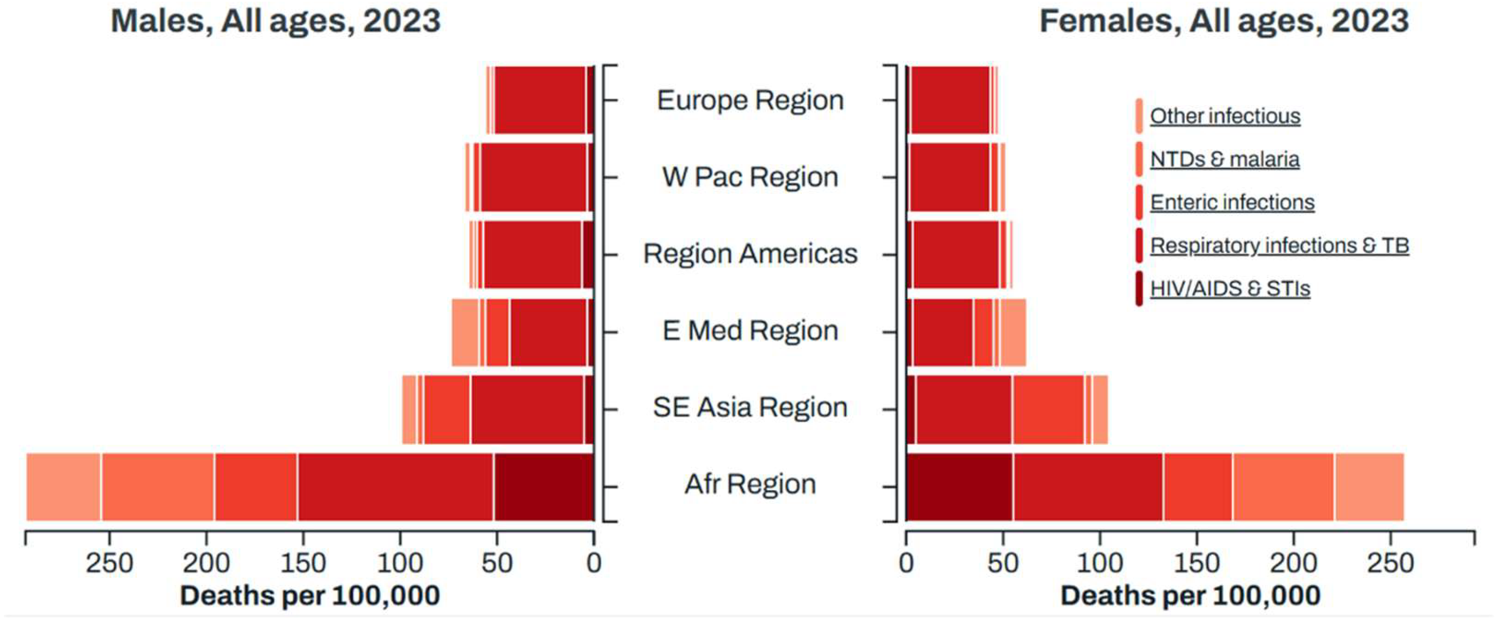
Figure 2.
The contribution of pharmaceutical 3D printing to antimicrobial management.
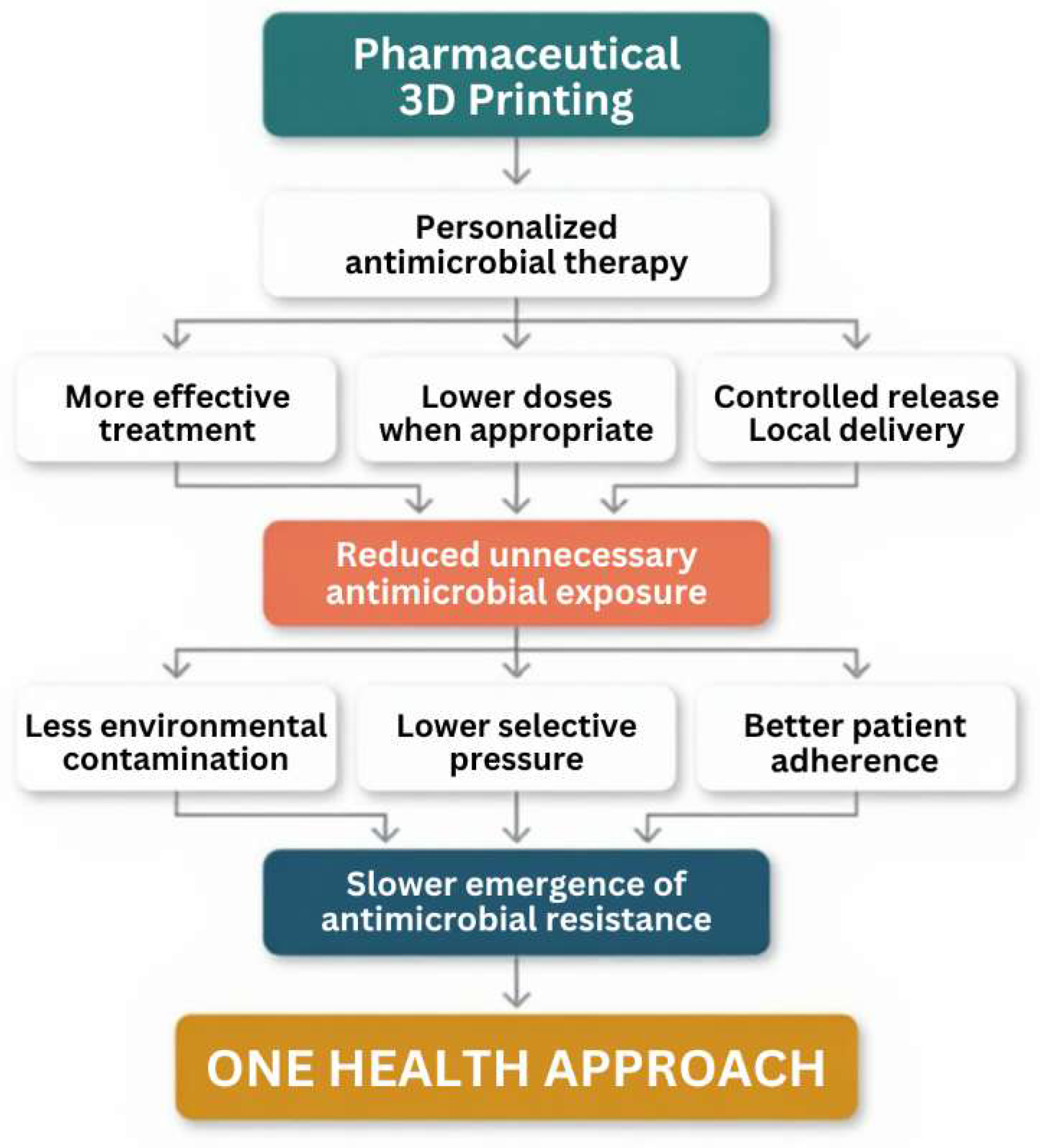
Figure 3.
Evolution of the number of published research articles per year (2020–2026) related to 3D printing and antibiotic drug delivery.
Figure 3.
Evolution of the number of published research articles per year (2020–2026) related to 3D printing and antibiotic drug delivery.
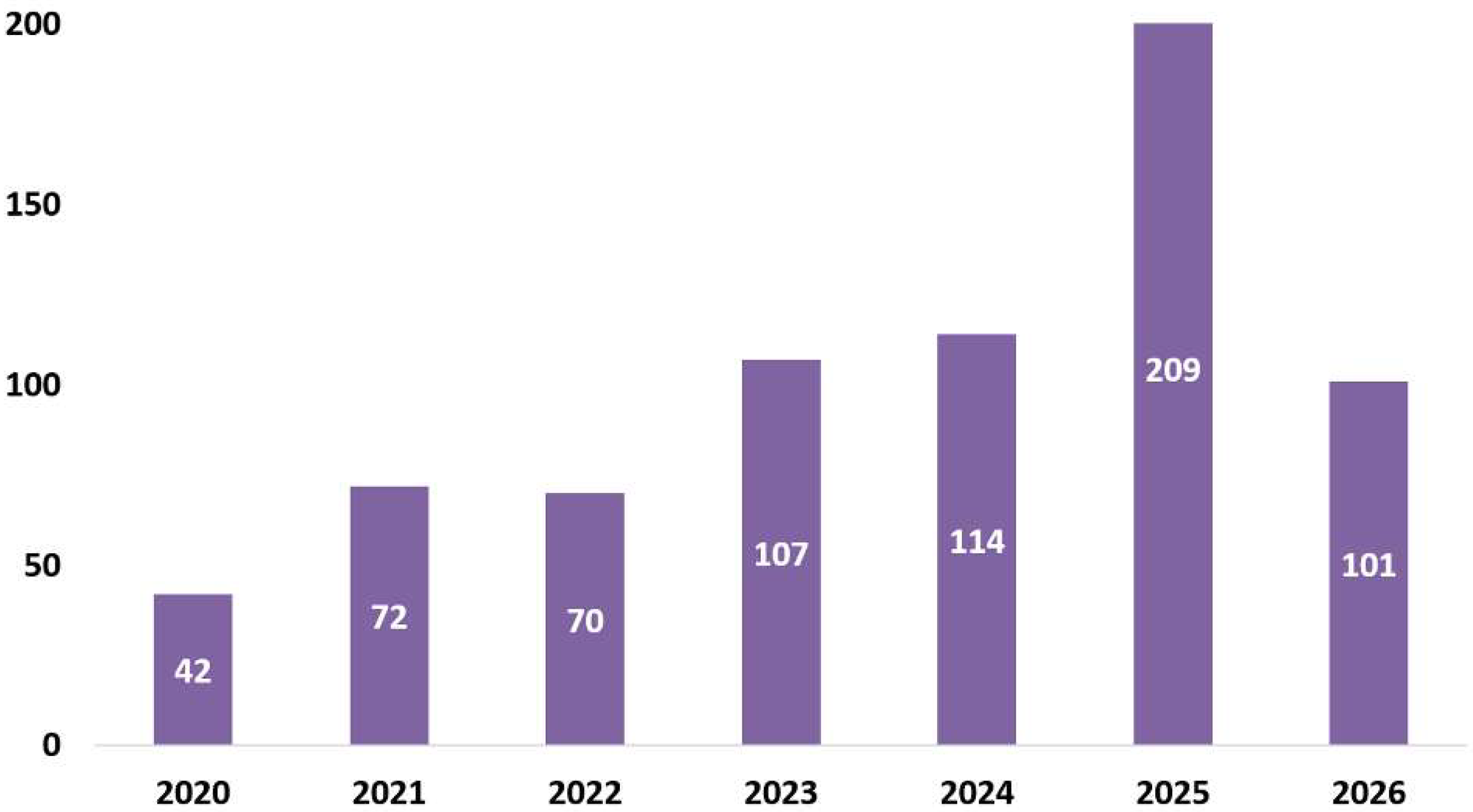
Figure 4.
SWOT analysis pf 3D printing in the treatment of infectious diseases.
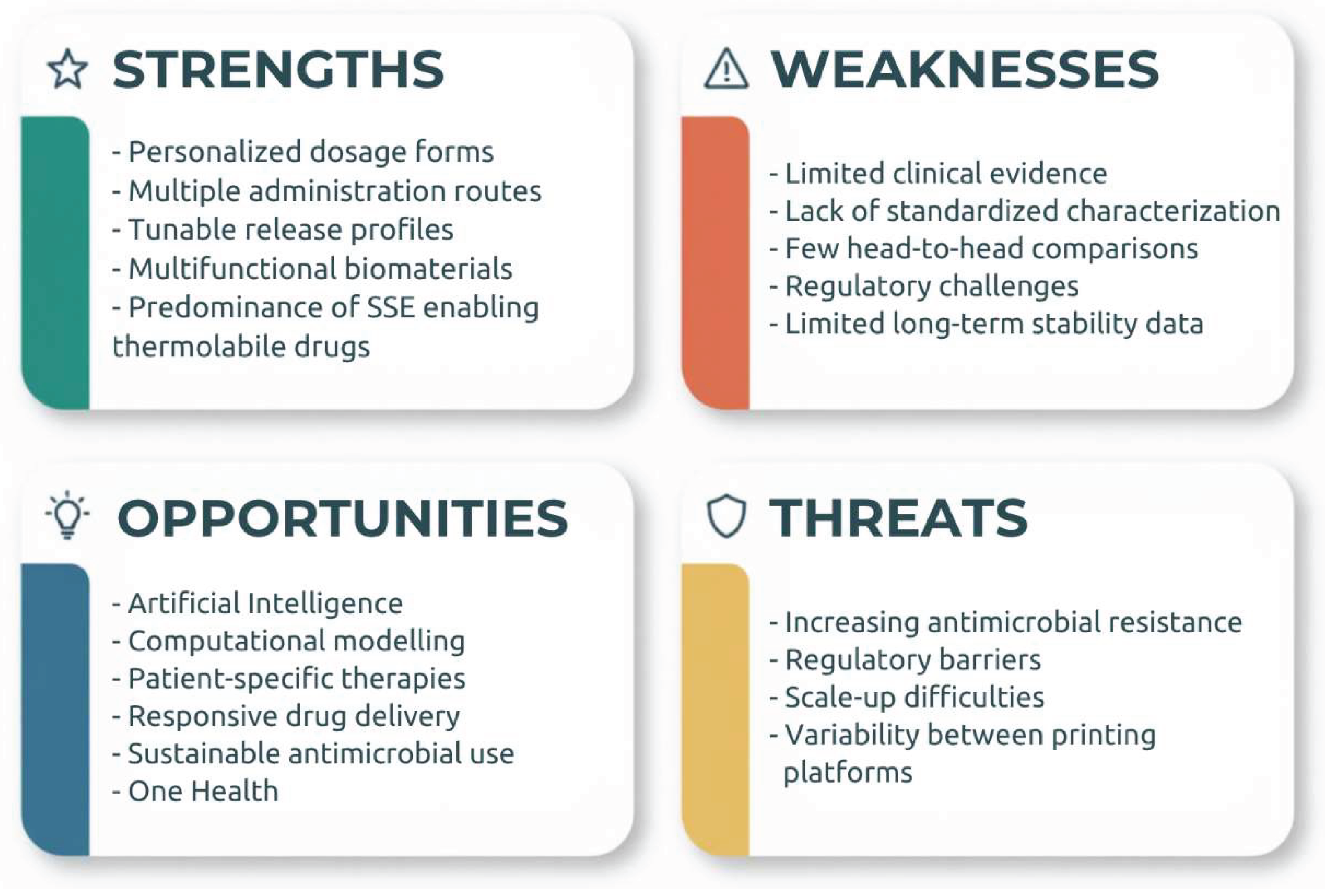

Table 1.
Lates advances in 3D printing to improve treatment against infectious diseases from 2020 to 2026.
Table 1.
Lates advances in 3D printing to improve treatment against infectious diseases from 2020 to 2026.
| Active ingredient | Matrix Compounds | Treatment target | Dosage form | 3D-Printing technique | Study stage | Ref |
|---|---|---|---|---|---|---|
| Lidocaine, Levofloxacin | Chitosan methacrylate | Wound infection | Wound dressing | SSE | In vitro | [12] |
| Amoxicillin | Poly(caprolactone); sodium alginate; gelatin | Wound infection | Bilayer scaffold | SSE | In vitro and in vivo | [13] |
| ε-Polylysine | Glycidyl methacrylate; Carboxymethylcellulose | Wound infection | Wound dressing | SLA | In vivo | [14] |
| Tannic Acid@Zif-8 | Alginate; gelatin | Wound infection | Scaffold | SSE | In vitro and in vivo | [15] |
| Vancomycin; Cerium Oxide Np | Chitosan; alginate | Chronic wound infection | Scaffold | SSE | In vitro | [16] |
| Amoxicillin; Bismuth Ferrite Np | Chitosan | Wound infection | Scaffold | SSE | In vitro | [17] |
| Bacitracin; Selenium Np; Calcium Phosphate Np | Alginate | Bacterial infection | Wound dressing | SSE | In vitro | [18] |
| Octenidine | Alginate; carboxymethyl chitosan | Chronic and acute wounds | Wound dressing | SSE | In vivo | [19] |
| Aciclovir Nanocrystal | Alginate | Herpes simplex virus | Patch | SSE | Ex vivo and in vivo | [20] |
| Quercus Infectoria | Polyvinyl alcohol | Wound infection | Patch | SSE | In vitro | [21] |
| Barijeh niosome | Chitosan; alginate | Wound infection | Scaffold | SSE | In vitro and in vivo | [22] |
| Carbon Dots; Propolis | Pectin; chitosan; collagen | Wound infection | Scaffold | SSE | In vitro and in vivo | [23] |
| Thyme Oil Liposomes | Hydroxyethyl cellulose, alginate, cellulose nanocrystals | Diabetic foot ulcers | Scaffolds | SSE | In vitro | [24] |
| Curcumin; Levofloxacin | Ethyl cellulose; corn protein (zein) | Antibacterial | Bilayer wound dressing | SSE | In vitro and in vivo | [25] |
| Levofloxacin | Chitosan; nanoclay | Wound infection | Scaffolds | SSE | In vitro and in vivo | [26] |
| Manuka Honey | Gelatin | Wound infection | Patch | SSE | In vitro and in vivo | [27] |
| Ceftriaxone | Photocurable resin | Non-specified | Microneedle | SLA | Ex vivo | [28] |
| Rifampicin | Dental resin | Non-specified | Microneedle | SLA | In vivo | [29] |
| Amoxicillin | Methacrylate gelatin | Wound infection | Microneedle | DLP | In vitro | [30] |
| Clotrimazole | Thermoplastic polyurethane | Vulvovaginal candidiasis | Intravaginal ring | FDM | In vitro | [31] |
| Clotrimazole; Bifonazole | Ethylene vinyl acetate | Vulvovaginal candidiasis | Intravaginal ring | FDM | In vitro | [32] |
| Metronidazole | Ethylene vinyl acetate; alginate | Bacterial vaginosis | Intravaginal ring | SSE | In vitro | [33] |
| Metronidazole | Poly(caprolactone); methyl vinyl ether-maleic anhydride copolymer | Bacterial vaginosis | Intravaginal device | SSE | In vitro | [34] |
| Metronidazole | Silicone | Bacterial vaginosis | Cylinder | SSE | In vitro | [35] |
| Gold Nanoparticles | 2-hydroxyethyl methacrylate; polyethylene glycol diacrylate | Bacterial vaginosis | Vaginal ovule | SLA | In vitro | [36] |
| Ciprofloxacin | Hydroxypropyl cellulose | Bacterial keratitis | Ocular insert | FDM | In vitro and ex vivo | [37] |
| Levofloxacin | HPMC | Anti-microbial | Patch | SSE | In vitro | [38] |
| Moxifloxacin | Alginate; soy lecithin liposomes | Bacterial keratitis | Ocular insert | SSE | In vivo | [39] |
| Tobramycin | polyvinyl alcohol methacrylate; hyaluronic acid methacrylate | Bacterial keratitis | Patch | SSE | In vivo | [40] |
| Amphotericin B | HPMC; chitosan | Fungal keratitis | Patch | SSE | Ex vivo and in vivo | [41] |
| Sulfamethoxazole; Trimethoprim | Pregelatinized starch; gelatine | Non-specified | Chewable tablets | SSE | In vitro | [42] |
| Azithromycin | Carrageenan and bentonite nanoclay | Non-specified | Chewable gummies | SSE | In vitro | [43] |
| Albendazole nano crystals | PEG; propylene glycol | Parasitic infections | Oral tablet | SSE | In vitro | [44] |
| Benznidazole | PEG; chitosan; pectin | Chagas disease | Oral tablet | SSE | In vitro | [45] |
| Ofloxacin | HPMC | Non-specified | Oral tablet | SSE | In vitro | [46] |
| Isoniazid; Rifampicin |
HPC; hypromellose acetate succinate | Tuberculosis | Bilayer oral tablet | FDM | In vitro | [47] |
| Clarithromycin | Dental resin | Helicobacter pylori infections | Magnetic microbullet | SSE | In vitro and in vivo | [48] |
| Clarithromycin | HPMC, Poloxamer 188; Poly(vinylpyrrolidone) | Helicobacter pylori infections | Oral floating core-shell tablet | FDM | In vitro | [49] |
| Fluconazole | Polyvinyl alcohol | Oral cavity infections | Orodispersible tablet | FDM | In vitro | [50] |
| Dapsone | Poly(1-vinylpyrrolidone-co-vinyl acetate) | Leprosy | Oral tablet | SLS | In vitro and in vivo | [51] |
HPMC: hydroxypropyl methylcellulose, Np: nanoparticle, SSE: semisolid extrusion, SLA: stereolithography, FDM: fused deposition modeling, SLS: selective laser sintering, DLP: digital light processing.
Disclaimer/Publisher’s Note: The statements, opinions and data contained in all publications are solely those of the individual author(s) and contributor(s) and not of MDPI and/or the editor(s). MDPI and/or the editor(s) disclaim responsibility for any injury to people or property resulting from any ideas, methods, instructions or products referred to in the content. |
© 2026 by the authors. Licensee MDPI, Basel, Switzerland. This article is an open access article distributed under the terms and conditions of the Creative Commons Attribution (CC BY) license (http://creativecommons.org/licenses/by/4.0/).
Copyright: This open access article is published under a Creative Commons CC BY 4.0 license, which permit the free download, distribution, and reuse, provided that the author and preprint are cited in any reuse.