

Submitted:
28 August 2026
Posted:
31 August 2026
You are already at the latest version
Abstract
Atypical optic neuritis (ON) is an important diagnostic gateway to aquaporin-4 immunoglobulin G-positive neuromyelitis optica spectrum disorder (AQP4-IgG-positive NMOSD) and myelin oligodendrocyte glycoprotein antibody-associated disease (MOGAD), but the same clinical warning features may also occur in vascular, hereditary, infectious, intracranial-pressure, toxic, nutritional, inflammatory, or compressive mimics. This narrative review translates contemporary neuro-ophthalmic and neuroimmunology evidence into a Gulf Cooperation Council (GCC)-adapted, ophthalmology-initiated diagnostic and referral framework. The framework separates first-contact clinical and fundoscopic activation features from post-investigation MRI, OCT, serological, and recovery findings, while emergency features determine same-day escalation independently of any numerical score. Core elements include a mandatory mimic checkpoint, phenotype-driven serum AQP4-IgG and MOG-IgG testing using validated cell-based assays, dedicated MRI of the brain and orbits, OCT documentation, visual evoked potentials when relevant to multiple-sclerosis dissemination-in-space assessment, structured referral communication, and early registry capture. GCC-specific considerations include the regional burden of vascular and metabolic disease, hereditary optic neuropathies, Behçet disease, idiopathic intracranial hypertension, tuberculosis and ethambutol toxicity, post-bariatric nutritional deficiency, neuroretinitis, and cross-sector or cross-border discontinuity of care. The framework incorporates the 2024 McDonald criteria, in which the optic nerve is a fifth anatomical location for dissemination in space. It is intended as a literature-informed implementation aid rather than a diagnostic criterion, consensus statement, or validated triage rule. Prospective Bahrain-led and multicountry validation should quantify sensitivity, specificity, over-referral, inter-rater reliability, time to dedicated imaging, antibody sampling and plasma-exchange escalation, visual outcomes, and implementation feasibility.
Keywords:
atypical optic neuritis
; neuromyelitis optica spectrum disorder
; myelin oligodendrocyte glycoprotein antibody-associated disease
; neuro-ophthalmology
; red-flag framework
; diagnostic pathway
; gulf cooperation council
; misdiagnosis
Key Clinical Messages
- Atypical ON should trigger two parallel questions: could this be NMOSD/MOGAD, and could this be a mimic in which premature immunotherapy may cause harm or delay the correct diagnosis?
- First-contact clinical and fundoscopic red flags should activate targeted investigation; MRI, serology, OCT and recovery features belong to post-investigation reassessment and should not determine retrospectively whether imaging or antibody testing was ordered.
- Emergency features determine same-day escalation directly and are not assigned numerical points.
- Serum AQP4-IgG and MOG-IgG testing should be phenotype-driven and performed with validated cell-based assays, with assay platform, qualitative strength/titre, specimen timing and treatment exposure documented.
- Dedicated MRI of the brain and orbits, OCT and - when relevant to MS diagnosis - VEP should be interpreted with the complete clinical phenotype and active exclusion of important mimics.
- The proposed GCC red-flag framework is an unvalidated implementation aid; predictive weights, thresholds and decision rules should be derived prospectively rather than asserted before validation.
Introduction
Optic neuritis occupies a central position in neuro-ophthalmology because it is a common cause of acute or subacute visual loss, a frequent first manifestation of inflammatory demyelinating disease, and a diagnostic crossroads at which early decisions may influence long-term visual and neurological outcomes. The classic description of typical demyelinating ON remains useful for a young adult with unilateral painful visual loss, a compatible examination, substantial recovery and an MS-consistent brain MRI. Contemporary neuroimmunology, however, has established that ON is a syndrome rather than a single disease entity [1,2,3,4,5].
The clinician who first evaluates an inflamed optic nerve may be encountering the earliest manifestation of MS, AQP4-IgG-positive NMOSD, MOGAD, post-infectious demyelination, sarcoidosis, Behçet disease, infection, ischaemic optic neuropathy, IIH, hereditary or mitochondrial optic neuropathy, malignancy, or compressive, infiltrative, toxic or nutritional disease [1,4,5,6,7,8,9,10,11,12,13,14,15,16,47,48,63,64,65,66,67,68,69,70]. The distinction is clinically consequential. NMOSD and MOGAD differ from MS in immunopathogenesis, attack severity, relapse risk, recovery pattern and treatment selection, while false-positive or phenotype-discordant antibody interpretation can create the opposite harm: durable diagnostic labelling and unnecessary immunotherapy in a patient with a mimic [4,5,6,7,8,9,10,11,12,13,14,15,16,17,18,19,29,30,31,32,33,34].
AQP4-IgG-positive NMOSD is an astrocytopathy characterised by severe inflammatory attacks involving the optic nerves, spinal cord, area postrema, brainstem, diencephalon and other central nervous system regions [4,6,7,9,10]. Disability is predominantly attack-related, making early recognition and rapid escalation of severe attacks important. MOGAD is a distinct antibody-associated demyelinating disease that commonly presents with ON, myelitis, ADEM-like illness, brainstem or cerebellar syndromes and cortical encephalitis [5,8,11,12,13,14,15,16,48]. MOGAD-associated ON often includes marked disc oedema, bilateral involvement, robust corticosteroid responsiveness and, in a subgroup, recurrence or steroid dependence [13,14,15,16,47,48].
The diagnostic landscape has also changed for MS. Historical ON and clinically isolated syndrome literature established the importance of phenotype, MRI and longitudinal risk assessment [2,3,49,50]. The 2024 revisions of the McDonald criteria, published in 2025, recognise the optic nerve as a fifth anatomical location for dissemination in space; companion recommendations define how MRI, OCT and VEP can support optic-nerve involvement when there is no better explanation [59,60,61]. A contemporary GCC ON pathway must therefore improve recognition of NMOSD and MOGAD without weakening appropriate MS diagnosis.
The GCC setting gives this diagnostic gateway particular practical relevance. Patients may enter care through private ophthalmology, emergency medicine, optometry, paediatrics, general neurology or tertiary neuroimmunology services. Dedicated orbital MRI protocols and specialised antibody assays may not be applied consistently, and continuity can be disrupted when patients move between sectors or countries. GCC-Stat reported that non-national workers constituted the largest share of the regional labour market in 2024, underscoring the practical value of portable documentation and an explicitly identified follow-up owner [76]. These are implementation considerations that require country-specific adaptation rather than assumptions of uniformity across the six GCC health systems.
The high regional burden of diabetes, obesity, hypertension, dyslipidaemia and metabolic syndrome further complicates first-contact assessment, because ischaemic optic neuropathy, diabetic papillopathy, retinal vascular disease, and obesity-related intracranial-pressure or nutritional disorders can overlap clinically with inflammatory ON and materially influence both diagnostic interpretation and management [56,57,58,66,68].
The novelty of this review lies in translating contemporary ON, NMOSD, MOGAD and MS evidence into an integrated, ophthalmology-initiated GCC diagnostic and referral framework. Its distinctive contribution is the linkage of first-contact red-flag recognition with a mandatory mimic checkpoint, urgency-based escalation, dedicated orbital MRI, phenotype-driven AQP4-IgG/MOG-IgG testing, post-investigation reassessment, structured cross-sector communication and early registry capture within a single regional pathway. By addressing both delayed recognition of antibody-mediated disease and the opposing risk of overdiagnosis from mimics or low-positive serology, the framework extends beyond diagnostic criteria toward a practical and testable model for safer first-contact care, continuity of referral and future GCC-wide validation.
Narrative Review Approach and Framework Development
A focused narrative literature search was undertaken in PubMed/MEDLINE and supplemented by reference-list searching and review of current regional and international guideline sources through 27 August 2026. Search concepts combined optic neuritis with NMOSD, AQP4-IgG, MOGAD, MOG-IgG, MRI, OCT, VEP, antibody assay, relapse, corticosteroid dependence, MS diagnostic criteria, regional mimics and the names of GCC countries. Priority was given to international diagnostic criteria, assay-comparison studies, disease-specific MRI and OCT studies, treatment evidence relevant to acute ON, and peer-reviewed regional data. Because the purpose was a practice-focused narrative synthesis rather than a systematic review, no protocol registration, duplicate screening, formal risk-of-bias assessment or meta-analysis was performed. The review was structured with attention to the quality domains described in the Scale for the Assessment of Narrative Review Articles (SANRA) [62], without representing SANRA as a mandatory reporting guideline.
The framework was developed iteratively by the authors from the literature and their clinical experience in neurology, neuroimmunology and GCC referral practice. No Delphi process, patient-level derivation cohort or statistical weighting procedure was used. Accordingly, the framework does not use an additive score. It separates: (1) first-contact activation features available before targeted imaging; (2) emergency features that determine urgency directly; and (3) post-investigation features available only after dedicated MRI, serology, OCT or longitudinal follow-up. Predictive weights and thresholds are explicitly reserved for prospective derivation and validation.
AI-assisted technology disclosure: OpenAI ChatGPT was used during manuscript preparation for language refinement, organisation, formatting and preliminary visual-layout support. It was not used as an author, source of evidence or substitute for clinical judgement. All scientific content, clinical interpretations, references, tables, figures and final wording were reviewed, verified and approved by the authors, who take full responsibility for the manuscript.
Clinical Spectrum: Typical and Atypical Optic Neuritis
ON usually presents with acute or subacute visual loss, impaired colour vision, reduced contrast sensitivity, a central or diffuse visual-field defect and a RAPD when involvement is unilateral or asymmetric. Pain with eye movement is common but not universal. The optic disc may be normal in retrobulbar disease or swollen in papillitis. Diagnosis should integrate phenotype, ophthalmic examination, MRI, retinal imaging, serology and systemic context rather than rely on symptoms alone [1].
Typical demyelinating ON generally affects one eye, evolves over several days, reaches its nadir within approximately one to two weeks and begins to improve over subsequent weeks. The disc is often normal or only mildly swollen; orbital MRI commonly shows a relatively short segment of optic-nerve enhancement. In contemporary MS assessment, the optic nerve can itself contribute to dissemination in space when involvement is established using accepted MRI, OCT or VEP methods and no better explanation exists [59,60,61].
Atypical ON departs from this pattern and should prompt expanded investigation. High-yield first-contact features include severe visual loss, bilateral simultaneous or rapidly sequential involvement, recurrence, relapse during corticosteroid taper or steroid dependence, marked disc oedema, paediatric onset and accompanying spinal cord, area postrema, brainstem or encephalopathic features. Long-segment or posterior optic-nerve enhancement, chiasmal or optic-tract involvement, optic-sheath or perineural enhancement, an atypical brain MRI, phenotype-discordant serology and unexpectedly poor recovery become important during post-investigation reassessment [1,4,5,13,14,15,16,20,21,35,36,47].

Table 1.
Typical and atypical optic neuritis: practical distinguishing features.
| Domain | Typical demyelinating optic neuritis | Atypical optic neuritis requiring expanded work-up |
| Laterality | Usually unilateral | Bilateral simultaneous, rapidly sequential, or recurrent alternating attacks |
| Visual severity | Mild to moderate loss is common; substantial recovery is expected | Severe nadir vision, including VA ≤0.1 (6/60; 20/200; 1.0 logMAR), counting fingers, hand movements, or worse |
| Pain | Pain with eye movement is common | Severe persistent orbital pain; absent pain plus other atypical features; or pain suggesting perineuritis |
| Disc appearance | Normal disc or mild swelling | Marked disc oedema, haemorrhage, exudate, macular star, pseudo-oedema or features discordant with typical ON |
| Clinical course | Subacute onset with improvement over weeks | Recurrence, relapse during taper/steroid dependence, or recovery discordant with the expected course |
| Optic-nerve MRI | Usually shorter-segment, unilateral involvement | Long-segment, posterior, chiasmal, optic-tract or sheath/perineural involvement |
| Brain/spinal MRI | May show MS-typical lesions | Normal/non-MS brain pattern despite severe ON; area postrema, brainstem, hypothalamic, ADEM-like or LETM lesions |
| Immediate implication | Contemporary MS risk/DIS assessment and follow-up | Activate the atypical pathway, targeted AQP4-IgG/MOG-IgG testing, dedicated MRI, mimic checkpoint and urgency-based referral |
Note: No single clinical, imaging or ophthalmic feature is pathognomonic for a specific ON aetiology. Distinction between typical demyelinating and atypical ON should therefore integrate the overall phenotype, attack severity and laterality, MRI and OCT findings, serological results, recovery trajectory, and systematic exclusion of important inflammatory, infectious, vascular, hereditary, compressive and toxic-nutritional mimics [1,4,5,15,16,20,21,47,59,60,61].
The GCC Context and Regional Evidence
Regional evidence is clinically important but heterogeneous. The Arabian Gulf NMOSD registry remains the most informative multicentre dataset. It retrospectively analysed 144 patients from 15 centres in five GCC countries; 64.3% were reported as AQP4-IgG positive and ON was the most frequent presentation at 48.6% [37]. The study spans different centres and testing eras, so serological proportions should be interpreted in the context of assay heterogeneity rather than as a uniform regional testing estimate.
MS cohorts provide additional disease-specific evidence that ON is a common route into demyelinating-disease care. A published Bahraini conference abstract reported ON in 36.6% of 287 patients with MS [43]. In Qatar, ON accounted for 28 of 135 documented first clinical presentations (20.8%) [42]. A Saudi university-hospital cohort reported ON as the first presentation in 53 of 190 patients (27.9%) [44]. In Oman, 49 of 170 patients with MS had ON (28.8%); among those affected, 14 presented initially with ON, 21 developed it later and 14 had both initial and later attacks [45]. These datasets use different denominators and ascertainment methods and should not be pooled.
The Kuwaiti new-onset ON cohort is particularly relevant because it begins with the clinical syndrome rather than an established demyelinating diagnosis. Among 71 patients seen between 2012 and 2022, 38 (53.5%) received a final diagnosis of MS, 30 (42.3%) remained classified as idiopathic ON, two (2.8%) had NMOSD and one (1.4%) had MOGAD [46]. Antibody-testing practice changed over the study period, with broader AQP4/MOG testing in later years; the small antibody-mediated counts should therefore not be interpreted as population incidence. A 2024 UAE single-centre study of 34 patients (27 NMOSD and 7 MOGAD) provides additional disease-specific radiological and laboratory observations, but not a directly comparable ON-entry denominator [38].
Table 2.
Selected GCC evidence on optic neuritis as a demyelinating-disease entry phenotype.
| Setting | Design / study period | n | Optic neuritis finding | Interpretation |
| Arabian Gulf | Retrospective multicentre NMOSD registry; period not uniformly reported | 144 analysed | ON most frequent presentation (48.6%); AQP4-IgG positive 64.3% | Direct NMOSD evidence; centre/assay-era heterogeneity should be acknowledged |
| Bahrain | National MS registry; 2011-2013; published conference abstract | 287 | ON reported in 36.6% | Useful historical signal but lower evidentiary weight than a full peer-reviewed cohort |
| Qatar | National/hospital-based MS cohort; 2010 ascertainment | 154 total; presentation data n=135 | 28/135 first presentations (20.8%) were ON | Supports eye-to-neurology pathway for first demyelinating events |
| Saudi Arabia | University-hospital cross-sectional MS cohort | 190 | 53/190 first presentations (27.9%) were ON | ON and myelitis were the most frequent first presentations |
| Oman | Retrospective tertiary-hospital MS cohort; 1991-2019 | 170 | 49/170 (28.8%) had ON; 14/49 initial, 21/49 later, 14/49 both | Demonstrates both entry-presentation and later-course relevance |
| Kuwait | Retrospective new-onset ON cohort; 2012-2022 | 71 | MS 38 (53.5%); idiopathic ON 30 (42.3%); NMOSD 2 (2.8%); MOGAD 1 (1.4%) | Most directly relevant GCC ON-to-final-diagnosis cohort; biomarker testing evolved over time |
| United Arab Emirates | Single-centre NMOSD/MOGAD study; published 2024 | 34 (27 NMOSD; 7 MOGAD) | Radiological and laboratory differentiation reported; no directly comparable ON-entry denominator | Supports regional imaging/serology differentiation; not directly comparable with MS cohorts |
Note: Regional studies vary substantially in design, sampling frame, study period, denominator, case definition and availability of contemporary AQP4-IgG/MOG-IgG testing. Accordingly, reported ON proportions should be interpreted within their original study context and should not be pooled, directly ranked or treated as population-level estimates [37,38,42,43,44,45,46].
GCC Atypical Optic Neuritis Red-Flag Framework
The framework is organised around clinical timing rather than arithmetic. First-contact features are deliberately limited to information available at the initial eye-clinic, emergency or neurology encounter. Their purpose is to identify patients in whom targeted imaging, serology and specialist communication should not be delayed. Emergency features determine timing directly and do not require a score. Post-investigation features are applied only after dedicated MRI, OCT, serology or longitudinal assessment becomes available.
The framework is an implementation aid, not a diagnostic criterion or validated triage rule. A patient may require testing or referral even when no listed feature is present if clinical concern remains high. Conversely, a red flag does not establish antibody-mediated disease: it should trigger a mimic checkpoint before or alongside immunological investigation.
Table 3.
GCC atypical optic neuritis red-flag framework: first-contact activation, emergency escalation and post-investigation reassessment.
Table 3.
GCC atypical optic neuritis red-flag framework: first-contact activation, emergency escalation and post-investigation reassessment.
| Phase | Feature | Operational definition | Clinical implication |
| First-contact activation | Severe nadir visual loss | VA ≤0.1 decimal, equivalent to 6/60, 20/200 or 1.0 logMAR; counting fingers/hand movements/worse are more severe | Activate atypical work-up; emergency assessment if rapidly progressive or bilateral |
| First-contact activation | Bilateral simultaneous or rapidly sequential ON | Both eyes affected concurrently, or fellow-eye attack within 30 days | Prompt AQP4-IgG/MOG-IgG and dedicated MRI; exclude IIH, hereditary and toxic causes |
| First-contact activation | Recurrent ON | Documented previous inflammatory ON in either eye, separated from the current attack by clinical recovery or a distinct new attack | Specialist review; verify prior diagnosis, imaging and antibody-testing history |
| First-contact activation | Relapse during corticosteroid taper / steroid dependence | New or recurrent visual/inflammatory symptoms during dose reduction or soon after withdrawal, with objective evidence when possible | Urgent reassessment for MOGAD, CRION and mimics; avoid repeated empirical tapers without classification |
| First-contact activation | Marked optic disc oedema or atypical disc/retinal findings | Disc swelling clearly greater than mild ON-related swelling, especially with haemorrhage, exudate, macular star or bilateral symmetry; document with fundus photography when possible | MOGAD remains possible, but actively exclude IIH, neuroretinitis, ischaemia and inflammatory/infectious causes |
| First-contact activation | Paediatric or ADEM-like presentation | ON in a child/adolescent, especially bilateral or with encephalopathy/polyfocal symptoms | Early paediatric neurology/neuroimmunology assessment and MOG-IgG testing |
| Emergency escalation | Severe bilateral/rapidly progressive visual loss; chiasmal/tract syndrome; LETM; area postrema/brainstem syndrome; encephalopathy | Any feature indicating high risk of major neurological/visual disability or a sight-/life-threatening mimic | Same-day emergency/neuroimmunology pathway; timing is independent of any score |
| Post-investigation reassessment | Long-segment, posterior, chiasmal/tract or perineural enhancement | Dedicated orbital MRI demonstrates lesion extent/location or sheath involvement | Raises concern for NMOSD/MOGAD/perineuritis; interpret with phenotype and serology |
| Post-investigation reassessment | Brain MRI atypical for MS or normal despite severe ON | No MS-typical explanation for severe ON, or an alternative CNS pattern | Reassess NMOSD/MOGAD and mimics; apply 2024 MS criteria only when no better explanation exists |
| Post-investigation reassessment | Unexpected recovery trajectory | Persistent severe deficit or recovery discordant with the expected phenotype; 4-6 weeks is a reassessment point, not a binary diagnostic threshold | Review diagnosis, imaging timing, treatment adequacy and alternative causes rather than treating a fixed time point as a score |
| Post-investigation reassessment | Low-positive or discordant MOG-IgG | Low/weak positive result without a strongly compatible phenotype or supportive MRI | Do not diagnose MOGAD on serology alone; confirm assay type, supporting features and the need for repeat testing |
Note: The GCC atypical ON red-flag framework is an author-developed, literature-informed implementation aid rather than a validated diagnostic or triage instrument. Standardised VA notation, fundus photography, OCT acquisition and reporting, and consistent operational definitions should be used to improve reproducibility across clinicians and centres. No individual feature is disease-specific, and each should be interpreted within the complete clinical phenotype, imaging pattern, serological context, recovery trajectory and active exclusion of mimics. Prospective validation should assess diagnostic performance, inter-rater reliability of each framework item, feasibility, over-referral and generalisability across GCC populations and healthcare settings [1,4,5,13,14,15,16,17,18,19,20,21,35,36,47,59,60,61,63,64,65,66,67,68,69,70].
Ophthalmology-Initiated Diagnostic and Referral Pathway
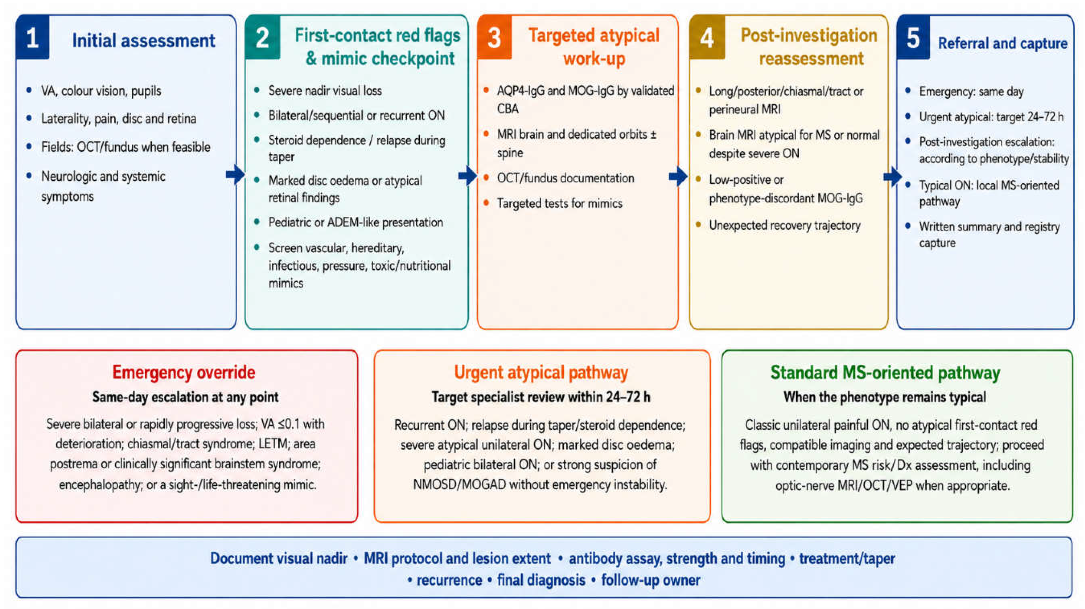
The initial assessment should document best-corrected VA, colour vision, contrast sensitivity when available, pupils and RAPD, ocular-motility pain, visual fields, optic-disc appearance, intraocular inflammation, retinal findings and systemic symptoms. Fundus photography is particularly valuable when disc oedema, haemorrhage, exudate, pallor, pseudo-oedema or retinal pathology is present. OCT should be obtained at baseline when feasible, recognising that acute disc oedema can transiently elevate pRNFL thickness [1,23,24,25,26].
Before a patient is routed into an antibody-mediated pathway, a brief mimic checkpoint should address age, tempo, pain, visual-field pattern, disc and retinal appearance, medication exposure, headache/pulsatile tinnitus/transient visual obscurations, systemic inflammatory or infectious symptoms, bariatric/nutritional history and family or maternal-line visual loss. A red flag is therefore a reason to investigate, not a reason to immunosuppress.
A patient with a classic unilateral painful syndrome, compatible orbital imaging and expected recovery may follow a standard MS-oriented pathway. Under the 2024 McDonald framework, the optic nerve can contribute to dissemination in space when involvement is demonstrated using appropriate MRI, OCT or VEP methods and alternative explanations have been excluded [59,60,61]. Patients older than 50 years or with substantial vascular comorbidity require particular attention to specificity and mimics [59,60].
Emergency assessment is appropriate for severe bilateral or rapidly progressive visual loss, VA ≤0.1 with rapid deterioration, chiasmal/optic-tract disease, LETM, area postrema or clinically significant brainstem syndrome, encephalopathy, or a sight- or life-threatening mimic. Urgent review within approximately 24-72 hours is reasonable for recurrent ON, relapse during taper/steroid dependence, severe unilateral atypical disease, marked disc oedema, paediatric bilateral ON, or strong clinical suspicion of NMOSD/MOGAD without an emergency feature. Post-investigation escalation should be determined by phenotype and clinical stability rather than by a numerical score. These are pragmatic operational targets rather than validated outcome thresholds.
Table 4.
Ophthalmology-initiated GCC atypical optic neuritis referral pathways: clinical triggers, core actions and suggested timing.
Table 4.
Ophthalmology-initiated GCC atypical optic neuritis referral pathways: clinical triggers, core actions and suggested timing.
| Pathway | Clinical trigger | Core actions | Suggested timing |
| Emergency | Severe bilateral or rapidly progressive loss; VA ≤0.1 with deterioration; chiasmal/tract disease; LETM; area postrema/brainstem syndrome; encephalopathy; sight-/life-threatening mimic | Dedicated brain/orbit MRI ± spine; targeted serology; mimic-directed emergency testing; acute-treatment escalation | Same day / emergency |
| Urgent atypical | Recurrent ON; relapse during taper/steroid dependence; severe atypical unilateral ON; marked disc oedema; paediatric bilateral ON; high clinical suspicion without emergency instability | AQP4-IgG/MOG-IgG using validated CBA; dedicated MRI; OCT/fundus; targeted mimic tests; specialist review | Target 24-72 h |
| Post-investigation escalation | Long/posterior/chiasmal/perineural MRI; discordant recovery; low-positive serology; unresolved mimic-versus-inflammatory question | Expert MRI/serology review; repeat/confirm assay if indicated; refine diagnosis and treatment plan | According to phenotype and stability |
| Standard MS-oriented | Classic unilateral painful ON, no atypical first-contact red flags, expected trajectory | 2024 McDonald DIS assessment; MRI and, where appropriate, OCT/VEP under quality-controlled criteria | Local standard pathway |
Note: This author-developed referral framework is intended to support timely, standardised escalation rather than replace clinical judgement. Emergency transfer should never be delayed by documentation requirements. Supplementary Material S1A provides a one-page critical-minimum urgent referral sheet; S1B provides an extended red-flag and registry data form for diagnostic clarification and longitudinal follow-up. Referral timing should be determined by clinical severity, associated neurological features, imaging findings, serological context and the need to exclude sight- or life-threatening mimics [1,4,5,17,18,19,20,21,27,28,35,36,39,40,47,59,60,61,63,64,65,66,67,68,69,70].
Illustrative inflammatory scenario: A 32-year-old woman presents with bilateral simultaneous ON, counting-fingers vision, marked disc oedema and relapse during a rapid corticosteroid taper. The first-contact framework activates the atypical pathway immediately, and severe bilateral loss determines same-day escalation. Dedicated MRI, AQP4-IgG/MOG-IgG testing, OCT documentation and specialist review proceed without waiting for a numerical score. Final diagnosis remains dependent on serology, imaging, clinical course and exclusion of mimics.
Illustrative mimic scenario: A young man develops painless sequential central visual loss over several weeks with pseudo-oedematous discs, no optic-nerve enhancement and a maternal history of visual loss. Although severity and bilateral sequential involvement are red flags, the mimic checkpoint redirects the work-up toward LHON and other hereditary optic neuropathies rather than empirical immunotherapy [63].
Figure 1.
GCC atypical optic neuritis diagnostic and referral pathway. The framework separates first-contact assessment, first-contact red flags and mimic screening, targeted atypical investigation, post-investigation reassessment, and referral/registry capture. Emergency features override all other timing categories and trigger same-day escalation. No numerical score is used. Author-developed pathway informed by contemporary ON, NMOSD, MOGAD, MS, imaging, serological and differential-diagnosis evidence [1,4,5,17,18,19,20,21,27,28,35,36,37,38,39,40,47,59,60,61,63,64,65,66,67,68,69,70].
Figure 1.
GCC atypical optic neuritis diagnostic and referral pathway. The framework separates first-contact assessment, first-contact red flags and mimic screening, targeted atypical investigation, post-investigation reassessment, and referral/registry capture. Emergency features override all other timing categories and trigger same-day escalation. No numerical score is used. Author-developed pathway informed by contemporary ON, NMOSD, MOGAD, MS, imaging, serological and differential-diagnosis evidence [1,4,5,17,18,19,20,21,27,28,35,36,37,38,39,40,47,59,60,61,63,64,65,66,67,68,69,70].
Diagnostic Toolbox and Differential Diagnosis
MRI
MRI is central to classification. A routine brain MRI may miss clinically important optic-nerve disease if dedicated orbital sequences are not obtained. The minimum protocol for atypical ON should include contrast-enhanced MRI of the brain and orbits unless contraindicated, fat-suppressed T2 or STIR orbital sequences, fat-suppressed post-contrast T1 orbital sequences, and axial and coronal views through the optic nerves. Reports should describe lesion length, laterality, anterior or posterior location, chiasmal and optic-tract extension, and optic-nerve sheath or perineural enhancement [1,20,21,22].
The 2024 MAGNIMS-CMSC-NAIMS recommendations strengthen the case for dedicated optic-nerve imaging within modern MS diagnosis while emphasising differential diagnosis, particularly in older patients and those with vascular comorbidity [60]. Typical MS-associated ON often involves a shorter optic-nerve segment. AQP4-IgG-positive NMOSD more often affects the posterior nerve, chiasm or tract, whereas MOGAD frequently affects the anterior optic nerve, may be bilateral and may show perioptic/sheath enhancement. These are probabilistic patterns rather than disease-specific findings [15,16,20,21,47].
OCT, VEP and Visual Function
OCT provides quantitative structural information. Relevant measures include pRNFL thickness, macular GCIPL or GCL-IPL thickness, scan quality, device and normative database, inter-eye asymmetry and longitudinal change. Acute disc oedema may transiently increase pRNFL thickness, particularly in MOGAD, so macular ganglion-cell analysis and repeat imaging after the acute phase are important [16,23,24,25,26]. OCT cannot establish antibody status.
In a retrospective comparison of acute MOGAD-associated ON with MS-associated ON, a pRNFL cut-off of approximately 118 μm yielded 74% sensitivity and 82% specificity for MOGAD [51]. This threshold should not be used as a stand-alone rule; it was derived from a specific cohort and has not been validated against all optic neuropathies, AQP4-IgG-positive NMOSD, OCT platforms, ethnic backgrounds or imaging time points.
For MS dissemination-in-space assessment, OCT inter-eye differences and VEP answer a different diagnostic question. The 2024 companion recommendations identify a pRNFL inter-eye difference ≥6 μm or GCIPL inter-eye difference ≥4 μm as supportive of optic-nerve injury when rigorous quality control is met and no better explanation exists; delayed VEP latency can likewise support demyelinating optic-nerve injury [61]. These thresholds should not be presented as discriminators of NMOSD or MOGAD.
Serology: Assay Selection and Avoiding Overdiagnosis
Serum AQP4-IgG and MOG-IgG should be tested using validated cell-based assays when the phenotype is compatible. Reports should document serum versus CSF, live-cell versus fixed-cell methodology when known, qualitative strength or titre, laboratory, and date relative to relapse and treatment exposure [17,18,19]. For MOG-IgG, live-cell assays generally show stronger inter-laboratory agreement for clearly positive and negative samples than fixed commercial assays, while low-positive results are substantially less reproducible [19].
Testing should therefore be phenotype-driven rather than indiscriminate. A low-positive MOG-IgG result in a clinically incompatible presentation should not by itself establish MOGAD. The 2023 diagnostic criteria require supporting clinical or MRI features when antibody strength is low or not clearly positive [5]. Specialist review should consider assay platform, pre-test probability, treatment timing and whether repeat testing is appropriate. This safeguard is essential because a pathway that appropriately increases case finding can also increase incidental or low-specificity positives.
In seronegative NMOSD, the 2015 criteria require a more stringent combination of core clinical characteristics and MRI features; a first isolated ON episode alone is insufficient for a seronegative NMOSD diagnosis [4]. Structured follow-up and repeat phenotyping therefore remain important even when initial antibody testing is negative.
Regional Mimics and Diagnostic Exit Ramps
Vascular and metabolic mimics: the GCC has a substantial burden of diabetes, obesity, hypertension, dyslipidaemia and metabolic syndrome [56,57,58]. These conditions increase the relevance of non-arteritic anterior ischaemic optic neuropathy, diabetic papillopathy and retinal vascular disease but should not automatically exclude NMOSD, MOGAD or another inflammatory optic neuropathy. Age, tempo, pain, visual-field pattern, disc and retinal appearance, dedicated MRI, OCT, inflammatory markers and metabolic control should be integrated. The 2024 MS diagnostic recommendations also emphasise additional specificity safeguards in older patients and those with vascular comorbidity [59,60].
Hereditary and mitochondrial optic neuropathies: LHON classically causes painless subacute central visual loss that becomes bilateral, often sequentially, and may initially show pseudo-oedematous discs [63]. Consanguinity, which remains common in parts of the Arab region, is more directly relevant to autosomal-recessive optic neuropathies and nuclear mitochondrial disorders than to maternally inherited LHON itself [64]. A maternal-line pedigree, painless progression over weeks to months, absent optic-nerve enhancement and characteristic disc findings should redirect evaluation toward mitochondrial and nuclear genetic testing rather than immunotherapy.
Behçet disease, IIH and neuroretinitis. Behçet disease can produce uveitis, optic neuropathy, cerebral venous thrombosis with papilloedema, and brainstem or mesodiencephalic neurological disease that can mimic inflammatory demyelination [65]. IIH is particularly important in patients with bilateral disc oedema, headache, transient visual obscurations or pulsatile tinnitus; preserved central acuity, enlarged blind spots and symmetric papilloedema without a RAPD should prompt intracranial-pressure evaluation rather than an ON label [66]. Neuroretinitis should be considered when disc oedema is accompanied by a macular star, with targeted evaluation for Bartonella, syphilis, tuberculosis and other causes [69].
Tuberculosis, ethambutol and nutritional/toxic optic neuropathy: exposure history is important in a region with substantial population mobility [76]. Ethambutol toxicity is typically bilateral, painless and subacute, making a medication history a high-yield diagnostic step [67]. Bariatric surgery and malabsorption can lead to thiamine, vitamin B12 or copper deficiency with optic neuropathy; nutritional history belongs in the GCC diagnostic checklist given the regional obesity burden and use of bariatric interventions [56,57,58,68]. Linezolid, amiodarone and other toxic exposures should also be considered when clinically relevant.
CRION (Chronic relapsing inflammatory optic neuropathy) is a steroid-responsive, steroid-dependent relapsing optic neuropathy defined after exclusion of alternative causes [70]. Contemporary evaluation should include MOG-IgG testing because some historical CRION phenotypes overlap with MOGAD. The term should therefore be used descriptively and cautiously rather than as a substitute for full aetiological evaluation.
Figure 2.
MRI and OCT patterns relevant to atypical optic neuritis evaluation and contemporary MS assessment. Panels a-h depict supportive MRI/OCT patterns that may prompt expanded evaluation but are not diagnostic in isolation. Panel i shows the OCT inter-eye thresholds used to support optic-nerve involvement for MS dissemination in space (GCIPL ≥4 μm or pRNFL ≥6 μm) and is included only for contemporary MS assessment; these thresholds are not NMOSD/MOGAD discriminators. All findings should be interpreted with phenotype, timing, serology strength, prior attacks and active exclusion of mimics. Schematic synthesis based on published MRI and OCT characteristics of MS-associated, AQP4-IgG-positive NMOSD and MOGAD ON [15,16,20,21,23,24,25,26,47,51,61]. Abbreviations: AQP4-IgG, aquaporin-4 immunoglobulin G; CNS, central nervous system; DIS, dissemination in space; GCIPL, ganglion cell–inner plexiform layer; LETM, longitudinally extensive transverse myelitis; MOGAD, myelin oligodendrocyte glycoprotein antibody-associated disease; MRI, magnetic resonance imaging; MS, multiple sclerosis; NMOSD, neuromyelitis optica spectrum disorder; OCT, optical coherence tomography; pRNFL, peripapillary retinal nerve fiber layer.
Figure 2.
MRI and OCT patterns relevant to atypical optic neuritis evaluation and contemporary MS assessment. Panels a-h depict supportive MRI/OCT patterns that may prompt expanded evaluation but are not diagnostic in isolation. Panel i shows the OCT inter-eye thresholds used to support optic-nerve involvement for MS dissemination in space (GCIPL ≥4 μm or pRNFL ≥6 μm) and is included only for contemporary MS assessment; these thresholds are not NMOSD/MOGAD discriminators. All findings should be interpreted with phenotype, timing, serology strength, prior attacks and active exclusion of mimics. Schematic synthesis based on published MRI and OCT characteristics of MS-associated, AQP4-IgG-positive NMOSD and MOGAD ON [15,16,20,21,23,24,25,26,47,51,61]. Abbreviations: AQP4-IgG, aquaporin-4 immunoglobulin G; CNS, central nervous system; DIS, dissemination in space; GCIPL, ganglion cell–inner plexiform layer; LETM, longitudinally extensive transverse myelitis; MOGAD, myelin oligodendrocyte glycoprotein antibody-associated disease; MRI, magnetic resonance imaging; MS, multiple sclerosis; NMOSD, neuromyelitis optica spectrum disorder; OCT, optical coherence tomography; pRNFL, peripapillary retinal nerve fiber layer.
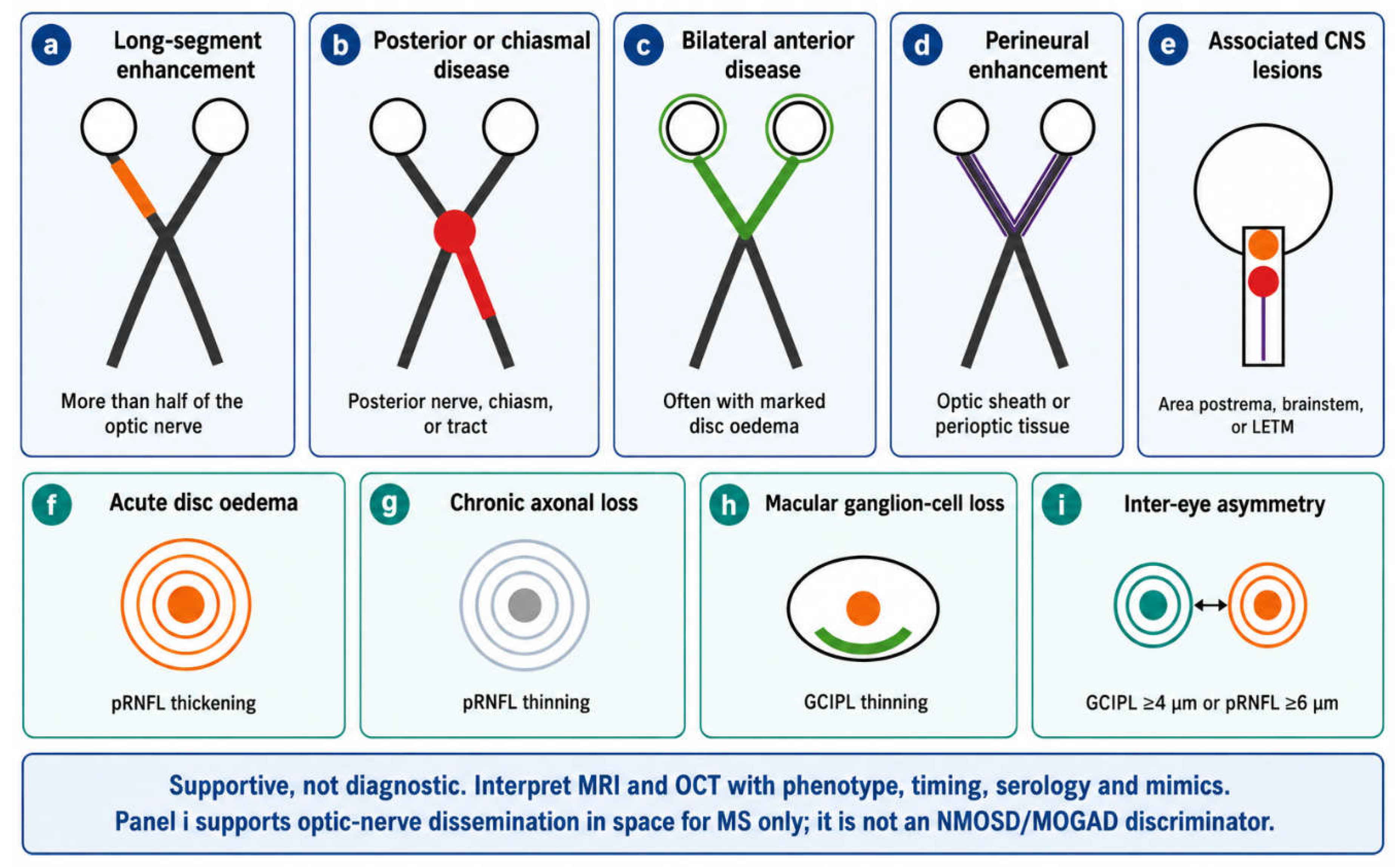
Table 5.
Diagnostic toolbox and key differential diagnoses in atypical optic neuritis: high-yield clues and management implications.
Table 5.
Diagnostic toolbox and key differential diagnoses in atypical optic neuritis: high-yield clues and management implications.
| Domain / diagnosis | High-yield clues | Interpretation or management implication |
| MRI of orbits | Fat-suppressed T2/STIR and post-contrast T1; document length, laterality, anterior/posterior location, chiasm/tract and sheath | Long, bilateral, posterior, chiasmal, tract or perineural patterns support expanded NMOSD/MOGAD/perineuritis evaluation |
| MRI brain/spine | Assess MS-typical lesions plus area postrema, brainstem, diencephalon, ADEM-like lesions, LETM and alternative pathology | Apply 2024 MS criteria when appropriate; normal brain MRI does not exclude NMOSD/MOGAD |
| OCT/VEP | Baseline pRNFL/GCIPL, scan quality/device/normative database; VEP if MS optic-nerve DIS assessment is relevant | Documents structure/function; MS DIS thresholds are not antibody-disease discriminators |
| AQP4-IgG-positive NMOSD | Severe ON, poor recovery, posterior/chiasmal disease, LETM, area postrema syndrome | Urgent attack treatment and relapse-prevention planning; avoid MS misclassification |
| MOGAD | Bilateral ON, marked disc oedema, anterior/perineural enhancement, paediatric onset, relapse during taper | Interpret MOG-IgG strength and assay method in phenotype context; avoid overdiagnosis from low-positive results |
| MS-associated ON | Unilateral painful ON, expected recovery, short-segment enhancement, MS-typical lesions | Assess under 2024 McDonald criteria including optic-nerve DIS when appropriate |
| LHON / hereditary | Painless sequential bilateral central loss, maternal/family history, pseudo-oedema, absent enhancement | Genetic/mitochondrial evaluation; avoid empirical immunotherapy |
| IIH | Headache, transient obscurations, pulsatile tinnitus, bilateral papilloedema, enlarged blind spot | Assess intracranial pressure and venous imaging; do not equate bilateral disc oedema with bilateral ON |
| Behçet / neuro-Behçet | Uveitis, oral/genital ulcers, CVT/papilloedema, brainstem or mesodiencephalic disease; relevant regional inflammatory/vasculitic mimic | Systemic/neurological evaluation; distinguish inflammatory vasculitic disease from NMOSD |
| Infectious / neuroretinitis | Macular star, exposure risk, fever/systemic clues, TB/syphilis/Bartonella risk | Targeted infectious work-up before immunosuppression |
| Toxic/nutritional | Ethambutol/linezolid/amiodarone exposure, bariatric surgery, malabsorption, B12/copper/thiamine risk | Medication and nutritional correction; targeted laboratory testing |
| Ischaemic/compressive/infiltrative | Older age/vascular risk, painless or progressive course, orbital signs, cancer history | Redirect to vascular, orbital/neurosurgical or oncological pathways as appropriate |
Note: The listed features support differential diagnosis and should not be interpreted as disease-specific or diagnostic in isolation. Clinical phenotype, tempo of visual loss, ophthalmic examination, MRI pattern, OCT/VEP findings, serology, treatment exposure, systemic context and exclusion of alternative causes should be integrated before assigning a final diagnosis. A low-positive or phenotype-discordant MOG-IgG result should not establish MOGAD without appropriate supporting clinical or MRI features. OCT and VEP thresholds used to demonstrate optic-nerve involvement under the 2024 McDonald criteria address MS dissemination in space and should not be used as discriminators of NMOSD or MOGAD. Sight- or life-threatening infectious, vascular, compressive, intracranial-pressure or inflammatory mimics require appropriate urgent evaluation before immunotherapy [1,4,5,15,16,17,18,19,20,21,22,23,24,25,26,47,51,56,57,58,59,60,61,62,63,64,65,66,67,68,69,70].
Prognostic and Therapeutic Implications
The central clinical risk in atypical ON is delayed classification and delayed escalation of a severe inflammatory attack. AQP4-IgG-positive NMOSD can cause profound residual visual disability after a single attack. High-dose corticosteroids are commonly used acutely, but severe or corticosteroid-refractory attacks may require early PLEX; retrospective apheresis data support better outcomes with earlier escalation [27,28,39,40]. For this reason, time from presentation to a PLEX eligibility/escalation decision should be treated as a key process outcome of the proposed GCC pathway, alongside time to dedicated orbital MRI and antibody sampling.
Long-term relapse prevention in AQP4-IgG-positive NMOSD now includes disease-specific evidence for complement inhibition and B-cell/interleukin-6 pathway therapies, including eculizumab, inebilizumab, satralizumab and ravulizumab; rituximab also has randomised evidence and remains widely used internationally [29,71,72,73,74]. Selection depends on serostatus, comorbidity, pregnancy plans, infection risk, access and local reimbursement. The present paper is a diagnostic/referral framework rather than a treatment guideline, and therapeutic choice should follow specialist and regional guidance [39,40].
MOGAD-associated ON often responds well to corticosteroids, but relapse during taper or after withdrawal is an important warning pattern. Observational evidence supports several maintenance approaches in relapsing disease; maintenance IVIG has been associated with reduced relapse frequency in adults, and paediatric consensus also includes IVIG among first-line maintenance options [30,52,53,75]. These data should not be converted into a universal dosing protocol. Pregnancy and postpartum status should be documented because they can influence relapse risk and treatment selection in NMOSD, with detailed management individualised by neuroimmunology and obstetric teams [40].
Misclassification as MS can expose patients with NMOSD to ineffective or potentially harmful treatment and delay disease-specific relapse prevention. Interferon beta and natalizumab have been associated with poor outcomes in NMOSD [31,32]. The reverse error also matters: a low-positive MOG-IgG in an incompatible phenotype can lead to unnecessary long-term immunotherapy and durable diagnostic labelling. The pathway therefore treats specificity safeguards as a core clinical outcome rather than an afterthought.
Implementation in GCC Practice
Implementation should be adaptable rather than uniform. Potential barriers include incomplete orbital MRI protocols, delayed or outsourced antibody testing, inconsistent referral communication, variable access to neuro-ophthalmology and neuroimmunology, public-private fragmentation, and discontinuity when patients relocate or change coverage. The large non-national component of GCC labour markets makes portable documentation and an explicit responsible follow-up service particularly important [76]. These are proposed service challenges whose prevalence and impact should be measured prospectively rather than assumed to be identical across countries.
Practical responses include standardised MRI order sets, preferred laboratories with documented live/fixed CBA methodology, an electronic first-contact red-flag template, a one-page urgent referral sheet, joint ophthalmology-neurology education and clear emergency escalation routes. In paediatric services, bilateral ON, ADEM-like presentations, myelitis or relapse during taper should trigger early MOG-IgG testing and paediatric neuroimmunology review [5,35,36,41,53]. Patients with incomplete recovery should be offered low-vision rehabilitation, occupational or educational support, and relevant driving and workplace counselling. The principal implementation challenges, potential clinical consequences and proposed GCC-adapted responses are summarised in Table 6 and reproduced in operational form in Supplementary Material S1C.
Registry Interoperability and Research Priorities: Opportunities for Alignment with MENACTRIMS and the Arabian Gulf NMOSD Registry
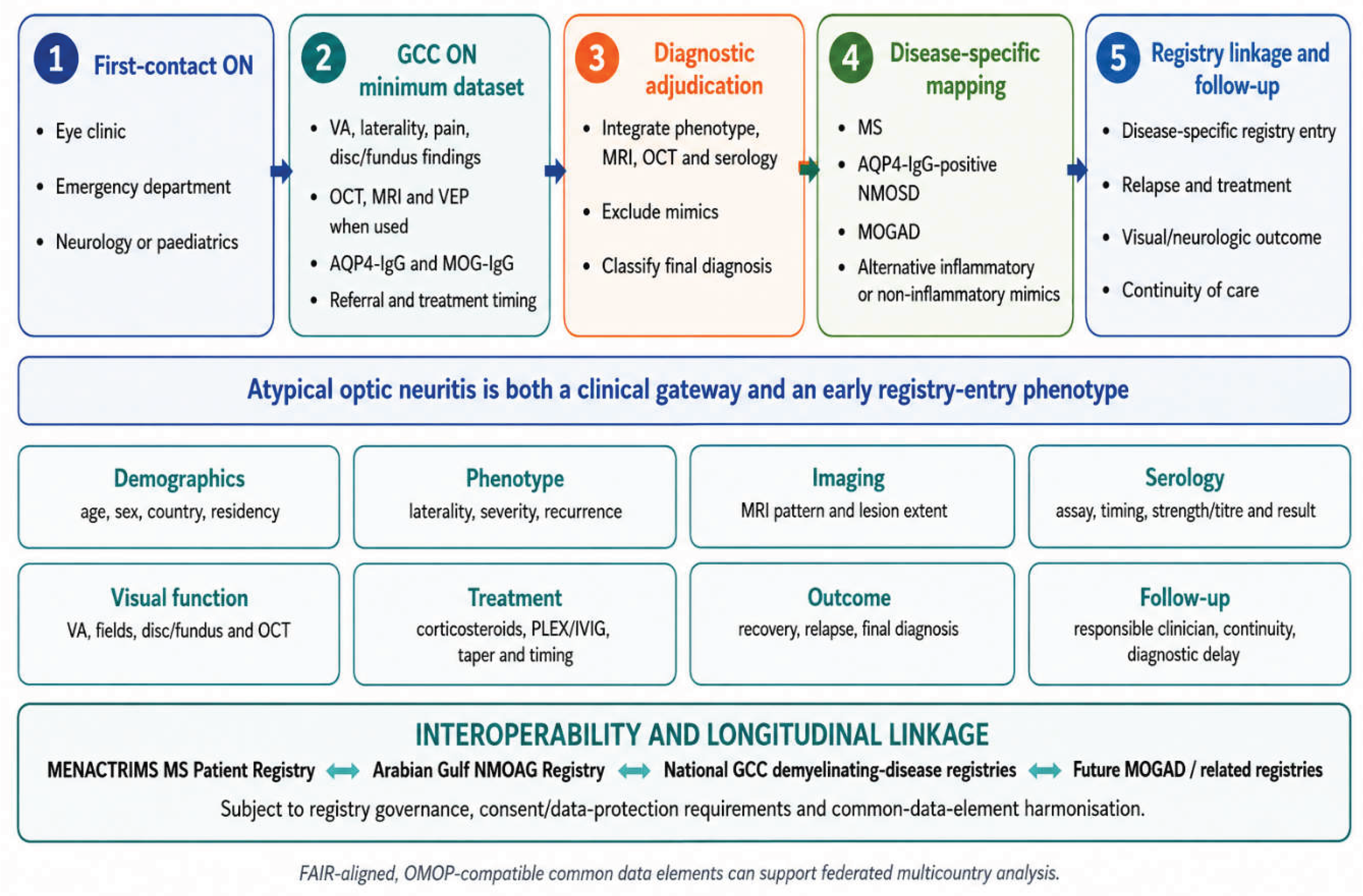
Atypical ON can serve as an early phenotype-capture event within existing or future GCC demyelinating-disease registries. Rather than establishing a parallel stand-alone registry, the proposed GCC optic neuritis module should be developed as an interoperable phenotype-entry dataset capable of linkage, subject to appropriate governance, consent, data-protection requirements and data-sharing agreements, with existing regional and national demyelinating-disease registries. Particular opportunities include harmonisation with the MENACTRIMS MS Patient Registry and the established Arabian Gulf NMOSD (NMOAG) registry described by Shosha et al. [37,77]. The ON module would complement rather than duplicate these resources by capturing the clinical encounter before final disease classification, thereby preserving information from the diagnostic gateway that may otherwise be lost once patients are subsequently categorised as having MS, AQP4-IgG-positive NMOSD, MOGAD, another inflammatory disorder or a non-inflammatory mimic.
The minimum ON dataset should include demographics and care pathway; onset date, laterality, pain, visual nadir, optic-disc and retinal findings, recurrence and associated neurological symptoms; MRI, OCT and VEP findings when used; AQP4-IgG and MOG-IgG assay methodology, qualitative strength or titre, specimen timing and treatment exposure; acute treatment and corticosteroid taper; referral interval; time to dedicated orbital MRI and antibody sampling; time to PLEX eligibility or escalation decision; visual recovery; final adjudicated diagnosis; relapse status; and continuity of follow-up. Following diagnostic adjudication, relevant variables could map to MS-, NMOSD-, MOGAD- or other disease-specific datasets while the ON module retains its distinctive first-contact and diagnostic-trajectory variables. Harmonised definitions and common data elements, aligned where feasible with FAIR principles and OMOP-compatible structures, could facilitate longitudinal linkage and federated multicountry analysis without unrestricted sharing of identifiable patient-level data and while allowing participating registries and institutions to retain their existing governance, custodianship and data ownership [54,55].
Prospective validation should enrol consecutive first and recurrent ON presentations, initially through a Bahrain-led pilot and, preferably, with multicountry GCC participation to improve event capture, external validity and representation across different healthcare settings. Participants should be followed for at least 12 months to permit final diagnostic ascertainment and capture early relapses and recovery trajectories. The reference diagnosis should be adjudicated by a blinded expert panel using contemporary diagnostic criteria, including the 2024 McDonald criteria for MS, the 2015 NMOSD criteria and the 2023 MOGAD criteria, together with predefined criteria for important alternative diagnoses [4,5,59]. The validation phase should also test the feasibility of harmonising the ON dataset with existing regional registry structures and determine the completeness, portability and reproducibility of core variables across participating centres.
The primary diagnostic-performance outcome should be the sensitivity of the first-contact framework for an adjudicated final diagnosis of AQP4-IgG-positive NMOSD or MOGAD. A co-primary safety outcome should be the over-referral rate, defined prospectively as the proportion of patients routed into the atypical inflammatory pathway whose adjudicated final diagnosis is MS without an atypical inflammatory disorder or a non-inflammatory mimic. Secondary outcomes should include specificity, negative predictive value, missed-case rate, time to dedicated orbital MRI, time to antibody sampling, time to PLEX eligibility or escalation decision, visual outcomes at six and twelve months, longitudinal OCT change, relapse during corticosteroid taper or after withdrawal, exposure to inappropriate MS-directed therapy, diagnostic reclassification, resource use, referral completion, registry completeness and continuity of follow-up. Inter-rater reliability should be assessed for each first-contact framework feature using kappa or another appropriate agreement statistic. A formal sample-size calculation should be prespecified in the validation protocol using anticipated regional prevalence of NMOSD and MOGAD among ON presentations and the desired confidence-interval precision for sensitivity and over-referral.
Figure 3.
Proposed GCC optic neuritis registry minimum-dataset architecture (Designed to complement, not duplicate, existing regional registries). Atypical optic neuritis (ON) is positioned as an early phenotype-entry event progressing from first contact through minimum-dataset capture and diagnostic adjudication to disease-specific mapping and registry linkage. The module is intended to complement, not duplicate, existing regional registries. Core domains include demographics, phenotype, imaging, serology, visual function, treatment, outcome and follow-up. Potential linkage includes the MENACTRIMS MS Patient Registry, Arabian Gulf NMOAG Registry, national GCC demyelinating-disease registries and future MOGAD-related registries, subject to governance and common-data-element harmonisation. Author-developed architecture informed by regional ON/demyelinating-disease evidence, FAIR principles and OMOP-compatible data harmonisation [37,46,54,55,76]. Abbreviations: AQP4-IgG, aquaporin-4 immunoglobulin G; FAIR, Findable, Accessible, Interoperable, and Reusable; GCC, Gulf Cooperation Council; IVIG, intravenous immunoglobulin; MENACTRIMS, Middle East North Africa Committee for Treatment and Research in Multiple Sclerosis; MOGAD, myelin oligodendrocyte glycoprotein antibody-associated disease; MOG-IgG, myelin oligodendrocyte glycoprotein immunoglobulin G; MRI, magnetic resonance imaging; MS, multiple sclerosis; NMOAG, Neuromyelitis Optica in Arabian Gulf; NMOSD, neuromyelitis optica spectrum disorder; OCT, optical coherence tomography; OMOP, Observational Medical Outcomes Partnership; PLEX, plasma exchange; VA, visual acuity.
Figure 3.
Proposed GCC optic neuritis registry minimum-dataset architecture (Designed to complement, not duplicate, existing regional registries). Atypical optic neuritis (ON) is positioned as an early phenotype-entry event progressing from first contact through minimum-dataset capture and diagnostic adjudication to disease-specific mapping and registry linkage. The module is intended to complement, not duplicate, existing regional registries. Core domains include demographics, phenotype, imaging, serology, visual function, treatment, outcome and follow-up. Potential linkage includes the MENACTRIMS MS Patient Registry, Arabian Gulf NMOAG Registry, national GCC demyelinating-disease registries and future MOGAD-related registries, subject to governance and common-data-element harmonisation. Author-developed architecture informed by regional ON/demyelinating-disease evidence, FAIR principles and OMOP-compatible data harmonisation [37,46,54,55,76]. Abbreviations: AQP4-IgG, aquaporin-4 immunoglobulin G; FAIR, Findable, Accessible, Interoperable, and Reusable; GCC, Gulf Cooperation Council; IVIG, intravenous immunoglobulin; MENACTRIMS, Middle East North Africa Committee for Treatment and Research in Multiple Sclerosis; MOGAD, myelin oligodendrocyte glycoprotein antibody-associated disease; MOG-IgG, myelin oligodendrocyte glycoprotein immunoglobulin G; MRI, magnetic resonance imaging; MS, multiple sclerosis; NMOAG, Neuromyelitis Optica in Arabian Gulf; NMOSD, neuromyelitis optica spectrum disorder; OCT, optical coherence tomography; OMOP, Observational Medical Outcomes Partnership; PLEX, plasma exchange; VA, visual acuity.
Strengths and Limitations
This review has several strengths. It integrates contemporary evidence across neuro-ophthalmology, NMOSD, MOGAD, MS, MRI, OCT, serology, acute-treatment escalation and important mimics into a single ophthalmology-initiated GCC framework. Rather than proposing another disease-specific diagnostic algorithm, it links first-contact recognition, mimic exclusion, urgency-based referral, dedicated imaging and antibody testing, post-investigation reassessment, structured cross-sector communication and early registry capture. The framework incorporates the 2024 McDonald criteria and explicitly addresses the competing risks of under-recognising antibody-mediated disease and overdiagnosing MOGAD or NMOSD through nonspecific clinical features or low-positive serology. A further strength is the deliberate separation of first-contact clinical triggers from MRI-, serology-, OCT- and recovery-dependent findings, thereby avoiding temporal circularity and false precision from an unvalidated additive score. Regional evidence is presented with study-specific denominators and methodological caveats rather than pooled across heterogeneous datasets, and the proposed pathway is accompanied by operational definitions, implementation strategies, referral tools, a registry-ready dataset and a prespecified direction for prospective validation.
Important limitations remain. This is a narrative review and clinical practice perspective rather than a systematic review, formal guideline or consensus statement; it therefore does not include a registered protocol, duplicate screening, formal risk-of-bias assessment or Delphi-based consensus methodology. No new patient cohort is presented, and the red-flag framework, escalation intervals and operational definitions have not yet been derived or validated using patient-level data. Their performance may vary across age groups, examiner experience, MRI/OCT platforms, laboratory methods and GCC healthcare settings.
Regional evidence is limited and heterogeneous. Some observations derive from MS cohorts, a published conference abstract or single-centre studies rather than prospective ophthalmology-initiated NMOSD/MOGAD cohorts. The Kuwaiti ON-to-final-diagnosis cohort is particularly relevant but small, and biomarker testing evolved during its 2012-2022 study period [46]. No prospective multicentre GCC ON cohort was identified through 27 August 2026 that combined contemporary AQP4-IgG/MOG-IgG assays, standardised MRI, OCT/VEP quality control and longitudinal diagnostic adjudication; this absence statement is itself constrained by the narrative-search design.
Finally, prioritising diagnostic safety may increase investigation and referral. Over-referral, inter-rater reliability, low-positive MOG-IgG interpretation, and reproducibility of features such as marked disc oedema and recovery trajectory therefore require prospective study. The approximately 118 μm acute pRNFL threshold remains cohort-specific and should not be generalised across devices or diseases [51]. The proposed pathway is intended to support recognition, investigation, referral and future validation; it does not replace specialist judgement or prescribe individualised acute or maintenance therapy.
Conclusion
Atypical ON is a clinically important and time-sensitive entry point for the recognition of NMOSD and MOGAD in the GCC, but the same red flags that improve sensitivity can also capture hereditary, vascular, intracranial-pressure, infectious, toxic, nutritional, inflammatory and compressive mimics. A safe regional pathway must therefore combine rapid escalation with disciplined specificity. The proposed GCC framework separates first-contact clinical triggers from post-investigation findings, avoids unvalidated numerical weights, and makes the mimic checkpoint and emergency escalation hierarchy explicit.
At the first encounter, severe or bilateral/sequential visual loss, recurrence, steroid dependence, marked disc oedema, paediatric presentations and associated neurological syndromes should prompt targeted AQP4-IgG/MOG-IgG testing, dedicated MRI of the brain and orbits, careful fundus/OCT documentation and appropriately timed specialist review. MRI, serology and recovery features then refine concern rather than retrospectively determining whether the initial work-up should have been ordered. Low-positive MOG-IgG results, assay methodology and alternative explanations must be actively considered to reduce overdiagnosis as testing expands.
The framework also aligns the standard ON pathway with the 2024 McDonald criteria, in which the optic nerve can contribute to dissemination in space, and links the clinical encounter to a concise referral tool and registry-ready dataset. A Bahrain-led prospective pilot followed by multicountry validation should derive rather than assume predictive weights, quantify sensitivity and over-referral, measure inter-rater reliability, and determine whether earlier imaging, serology and PLEX escalation translate into better visual and diagnostic outcomes. In its present form, the proposal is best viewed as a testable GCC implementation framework: practical enough for first-contact care, cautious enough to avoid false precision, and structured to generate the regional evidence required for future validated decision tools.
Supplementary information
Supplementary Material S1 contains (A) a one-page critical-minimum urgent referral sheet, (B) an extended red-flag and registry data form with operational definitions, and (C) the GCC implementation-challenges table.
Ethics approval and consent to participate
Not applicable. This narrative review and clinical practice perspective does not report research involving human participants, animals, identifiable patient data or biological materials.
Consent for publication
Not applicable.
Data availability
No datasets were generated or analysed during the current study. All source literature is cited in the manuscript.
Funding
The authors received no specific funding for this work.
Author Contributions
IAA conceptualised the review, developed the GCC-focused diagnostic pathway and red-flag framework, drafted the initial manuscript, developed the ON registry module, prepared the tables and figures, and critically revised the manuscript for important intellectual content. AISA contributed clinical neurology and neuroimmunology expertise, critically reviewed the GCC-adapted diagnostic and referral framework, evaluated the implementation and validation strategy, and contributed to substantive revision of the manuscript. BAM contributed expertise in neuroimmunology and multiple sclerosis, critically reviewed the diagnostic pathway, red-flag framework, assay and referral sections, and revised the manuscript for clinical and scientific accuracy. All authors reviewed and approved the final manuscript and agree to be accountable for the accuracy and integrity of the work.
Conflicts of Interest
Isa Ahmed Alsharoqi is a founding member of MENACTRIMS. MENACTRIMS had no role in the conception, evidence selection, preparation, or submission of this manuscript. The authors declare no financial conflicts of interest and no other relevant non-financial conflicts of interest.
Abbreviations
ADEM, acute disseminated encephalomyelitis; AQP4-IgG, aquaporin-4 immunoglobulin G; CBA, cell-based assay; CRION, chronic relapsing inflammatory optic neuropathy; GCC, Gulf Cooperation Council; GCIPL, ganglion cell-inner plexiform layer; IIH, idiopathic intracranial hypertension; LETM, longitudinally extensive transverse myelitis; LHON, Leber hereditary optic neuropathy; MOGAD, myelin oligodendrocyte glycoprotein antibody-associated disease; MOG-IgG, myelin oligodendrocyte glycoprotein immunoglobulin G; MRI, magnetic resonance imaging; MS, multiple sclerosis; NMOSD, neuromyelitis optica spectrum disorder; OCT, optical coherence tomography; ON, optic neuritis; PLEX, plasma exchange; pRNFL, peripapillary retinal nerve fibre layer; RAPD, relative afferent pupillary defect; VA, visual acuity; VEP, visual evoked potential.
References
- Petzold, A.; Fraser, C.L.; Abegg, M.; Alroughani, R.; Alshowaeir, D.; Alvarenga, R.; et al. Diagnosis and classification of optic neuritis. Lancet Neurol. 2022, 21(12), 1120–1134. [Google Scholar] [CrossRef] [PubMed]
- Beck, R.W.; Cleary, P.A.; Anderson, M.M., Jr.; Keltner, J.L.; Shults, W.T.; Kaufman, D.I.; et al. A randomized, controlled trial of corticosteroids in the treatment of acute optic neuritis. N Engl. J. Med. 1992, 326(9), 581–588. [Google Scholar] [CrossRef] [PubMed]
- Optic Neuritis Study Group. The 5-year risk of MS after optic neuritis: experience of the Optic Neuritis Treatment Trial. Neurology 1997, 49(5), 1404–1413. [Google Scholar] [CrossRef] [PubMed]
- Wingerchuk, D.M.; Banwell, B.; Bennett, J.L.; Cabre, P.; Carroll, W.; Chitnis, T.; et al. International consensus diagnostic criteria for neuromyelitis optica spectrum disorders. Neurology 2015, 85(2), 177–189. [Google Scholar] [CrossRef] [PubMed]
- Banwell, B.; Bennett, J.L.; Marignier, R.; Kim, H.J.; Brilot, F.; Flanagan, E.P.; et al. Diagnosis of myelin oligodendrocyte glycoprotein antibody-associated disease: International MOGAD Panel proposed criteria. Lancet Neurol. 2023, 22(3), 268–282. [Google Scholar] [CrossRef] [PubMed]
- Lennon, V.A.; Wingerchuk, D.M.; Kryzer, T.J.; Pittock, S.J.; Lucchinetti, C.F.; Fujihara, K.; et al. A serum autoantibody marker of neuromyelitis optica: distinction from multiple sclerosis. Lancet 2004, 364(9451), 2106–2112. [Google Scholar] [CrossRef] [PubMed]
- Jarius, S.; Wildemann, B. AQP4 antibodies in neuromyelitis optica: diagnostic and pathogenetic relevance. Nat. Rev. Neurol. 2010, 6(7), 383–392. [Google Scholar] [CrossRef] [PubMed]
- Reindl, M.; Waters, P. Myelin oligodendrocyte glycoprotein antibodies in neurological disease. Nat. Rev. Neurol. 2019, 15(2), 89–102. [Google Scholar] [CrossRef] [PubMed]
- Jarius, S.; Paul, F.; Weinshenker, B.G.; Levy, M.; Kim, H.J.; Wildemann, B. Neuromyelitis optica. Nat. Rev. Dis. Primers 2020, 6(1), 85. [Google Scholar] [CrossRef] [PubMed]
- Flanagan, E.P. Neuromyelitis optica spectrum disorder and other non-multiple sclerosis central nervous system inflammatory diseases. Continuum (Minneap Minn) 2019, 25(3), 815–844. [Google Scholar] [CrossRef] [PubMed]
- Jarius, S.; Ruprecht, K.; Kleiter, I.; Borisow, N.; Asgari, N.; Pitarokoili, K.; et al. MOG-IgG in NMO and related disorders: a multicenter study of 50 patients. Part 2: epidemiology, clinical presentation, radiological and laboratory features, treatment responses, and long-term outcome. J. Neuroinflammation 2016, 13(1), 280. [Google Scholar] [CrossRef] [PubMed]
- Jarius, S.; Paul, F.; Aktas, O.; Asgari, N.; Dale, R.C.; de Seze, J.; et al. MOG encephalomyelitis: international recommendations on diagnosis and antibody testing. J. Neuroinflammation 2018, 15(1), 134. [Google Scholar] [CrossRef] [PubMed]
- Jurynczyk, M.; Messina, S.; Woodhall, M.R.; Raza, N.; Everett, R.; Roca-Fernandez, A.; et al. Clinical presentation and prognosis in MOG-antibody disease: a UK study. Brain 2017, 140(12), 3128–3138. [Google Scholar] [CrossRef] [PubMed]
- Ramanathan, S.; Mohammad, S.; Tantsis, E.; Nguyen, T.K.; Merheb, V.; Fung, V.S.C.; et al. Clinical course, therapeutic responses and outcomes in relapsing MOG antibody-associated demyelination. J. Neurol. Neurosurg. Psychiatry 2018, 89(2), 127–137. [Google Scholar] [CrossRef] [PubMed]
- Chen, J.J.; Flanagan, E.P.; Jitprapaikulsan, J.; Lopez-Chiriboga, A.S.; Fryer, J.P.; Leavitt, J.A.; et al. Myelin oligodendrocyte glycoprotein antibody-positive optic neuritis: clinical characteristics, radiologic clues, and outcome. Am. J. Ophthalmol. 2018, 195, 8–15. [Google Scholar] [CrossRef] [PubMed]
- Sechi, E.; Cacciaguerra, L.; Chen, J.J.; Mariotto, S.; Fadda, G.; Dinoto, A.; et al. Myelin oligodendrocyte glycoprotein antibody-associated disease: a review. JAMA Neurol. 2022, 79(12), 1260–1273. [Google Scholar] [CrossRef]
- Waters, P.J.; McKeon, A.; Leite, M.I.; Rajasekharan, S.; Lennon, V.A.; Villalobos, A.; et al. Serologic diagnosis of NMO: a multicenter comparison of aquaporin-4-IgG assays. Neurology 2012, 78(9), 665–671. [Google Scholar] [CrossRef] [PubMed]
- Waters, P.; Woodhall, M.; O’Connor, K.C.; Reindl, M.; Lang, B.; Sato, D.K.; et al. MOG cell-based assay detects non-MS patients with inflammatory neurologic disease. Neurol. Neuroimmunol. Neuroinflamm 2015, 2(3), e89. [Google Scholar] [CrossRef] [PubMed]
- Reindl, M.; Schanda, K.; Woodhall, M.; Tea, F.; Ramanathan, S.; Sagen, J.; et al. International multicenter examination of MOG antibody assays. Neurol. Neuroimmunol. Neuroinflamm 2020, 7(2), e674. [Google Scholar] [CrossRef] [PubMed]
- Kim, H.J.; Paul, F.; Lana-Peixoto, M.A.; Tenembaum, S.; Asgari, N.; Palace, J.; et al. MRI characteristics of neuromyelitis optica spectrum disorder: an international update. Neurology 2015, 84(11), 1165–1173. [Google Scholar] [CrossRef] [PubMed]
- Ramanathan, S.; Prelog, K.; Barnes, E.H.; Tantsis, E.M.; Reddel, S.W.; Henderson, A.P.D.; et al. Radiological differentiation of optic neuritis with MOG antibodies, AQP4 antibodies, and multiple sclerosis. Mult. Scler. 2016, 22(4), 470–482. [Google Scholar] [CrossRef] [PubMed]
- Filippi, M.; Preziosa, P.; Banwell, B.L.; Barkhof, F.; Ciccarelli, O.; De Stefano, N.; et al. Assessment of lesions on magnetic resonance imaging in multiple sclerosis: practical guidelines. Brain 2019, 142(7), 1858–1875. [Google Scholar] [CrossRef] [PubMed]
- Syc, S.B.; Saidha, S.; Newsome, S.D.; Ratchford, J.N.; Levy, M.; Ford, E.; et al. Optical coherence tomography segmentation reveals ganglion cell layer pathology after optic neuritis. Brain 2012, 135 Pt 2, 521–533. [Google Scholar] [CrossRef] [PubMed]
- Bennett, J.L.; de Seze, J.; Lana-Peixoto, M.; Palace, J.; Waldman, A.; Schippling, S.; et al. Neuromyelitis optica and multiple sclerosis: seeing differences through optical coherence tomography. Mult. Scler. 2015, 21(6), 678–688. [Google Scholar] [CrossRef] [PubMed]
- Martinez-Lapiscina, E.H.; Arnow, S.; Wilson, J.A.; Saidha, S.; Preiningerova, J.L.; Oberwahrenbrock, T.; et al. Retinal thickness measured with optical coherence tomography and risk of disability worsening in multiple sclerosis: a cohort study. Lancet Neurol. 2016, 15(6), 574–584. [Google Scholar] [CrossRef] [PubMed]
- Costello, F. The afferent visual pathway: designing a structural-functional paradigm of multiple sclerosis. ISRN Neurol. 2013, 2013, 134858. [Google Scholar] [CrossRef] [PubMed]
- Weinshenker, B.G.; O’Brien, P.C.; Petterson, T.M.; Noseworthy, J.H.; Lucchinetti, C.F.; Dodick, D.W.; et al. A randomized trial of plasma exchange in acute CNS inflammatory demyelinating disease. Ann. Neurol. 1999, 46(6), 878–886. [Google Scholar] [CrossRef]
- Kleiter, I.; Gahlen, A.; Borisow, N.; Fischer, K.; Wernecke, K.D.; Wegner, B.; et al. Apheresis therapies for NMOSD attacks: a retrospective study of 207 therapeutic interventions. Neurol. Neuroimmunol. Neuroinflamm 2018, 5(6), e504. [Google Scholar] [CrossRef] [PubMed]
- Pittock, S.J.; Berthele, A.; Fujihara, K.; Kim, H.J.; Levy, M.; Palace, J.; et al. Eculizumab in aquaporin-4-positive neuromyelitis optica spectrum disorder. N Engl. J. Med. 2019, 381(7), 614–625. [Google Scholar] [CrossRef] [PubMed]
- Chen, J.J.; Flanagan, E.P.; Bhatti, M.T.; Jitprapaikulsan, J.; Dubey, D.; Lopez-Chiriboga, A.S.; et al. Steroid-sparing maintenance immunotherapy for MOG-IgG associated disorder. Neurology 2020, 95(2), e111–e120. [Google Scholar] [CrossRef] [PubMed]
- Palace, J.; Leite, M.I.; Nairne, A.; Vincent, A. Interferon beta treatment in neuromyelitis optica: increase in relapses and aquaporin-4 antibody titers. Arch. Neurol. 2010, 67(8), 1016–1017. [Google Scholar] [CrossRef] [PubMed]
- Kleiter, I.; Hellwig, K.; Berthele, A.; Kumpfel, T.; Linker, R.A.; Hartung, H.P.; et al. Failure of natalizumab to prevent relapses in neuromyelitis optica. Arch. Neurol. 2012, 69(2), 239–245. [Google Scholar] [CrossRef]
- Solomon, A.J.; Bourdette, D.N.; Cross, A.H.; Applebee, A.; Skidd, P.M.; Howard, D.B.; et al. The contemporary spectrum of multiple sclerosis misdiagnosis. Neurology 2016, 87(13), 1393–1399. [Google Scholar] [CrossRef] [PubMed]
- Thompson, A.J.; Banwell, B.L.; Barkhof, F.; Carroll, W.M.; Coetzee, T.; Comi, G.; et al. Diagnosis of multiple sclerosis: 2017 revisions of the McDonald criteria. Lancet Neurol. 2018, 17(2), 162–173. [Google Scholar] [CrossRef] [PubMed]
- Hacohen, Y.; Wong, Y.Y.M.; Lechner, C.; Jurynczyk, M.; Wright, S.; Konuskan, B.; et al. Disease course and treatment responses in children with relapsing MOG antibody-associated disease. JAMA Neurol. 2018, 75(4), 478–487. [Google Scholar] [CrossRef] [PubMed]
- Fadda, G.; Armangue, T.; Hacohen, Y.; Chitnis, T.; Banwell, B. Paediatric multiple sclerosis and antibody-associated demyelination: clinical, imaging, and biological considerations. Lancet Neurol. 2021, 20(2), 136–149. [Google Scholar] [CrossRef]
- Shosha, E.; Qadi, N.; Inshasi, J.; Shami, R.; Al-Asmi, A.; Al-Hashel, J.; et al. Neuromyelitis optica spectrum disorders in Arabian Gulf: establishment and initial characterization of a patient registry. Mult. Scler. Relat. Disord. 2020, 38, 101448. [Google Scholar] [CrossRef] [PubMed]
- Alzarooni, H.; Inshasi, J.; Alawadhi, A.; Giacomini, P. MOGAD and NMOSD: insights on patients’ radiological and laboratory findings from a single UAE center. Front Neurol. 2024, 15, 1480723. [Google Scholar] [CrossRef] [PubMed]
- Shosha, E.; Aljarallah, S.A.; Al Fugham, N.; Al-Jedai, A.H.; Al Luqmani, M.M.; Al Malik, Y.M.; et al. Saudi consensus recommendations on the management of neuromyelitis optica spectrum disorders. Mult. Scler. Relat. Disord. 2022, 66, 104062. [Google Scholar] [CrossRef] [PubMed]
- Yamout, B.; Gouider, R.; Al-Roughani, R.; Aljarallah, S.; Shalaby, N.; Al-Khabouri, J.; et al. Consensus recommendations for the diagnosis and treatment of neuromyelitis optica spectrum disorders: the MENACTRIMS guidelines. CNS Drugs 2026, 40(3), 283–303. [Google Scholar] [CrossRef] [PubMed]
- Malalla, H.; Khalil, A. A case of pediatric neuromyelitis optica spectrum disorder: first case reported in Kingdom of Bahrain. Bahrain Med. Bull. 2021, 43(2), 528–530. [Google Scholar]
- Deleu, D.; Mir, D.; Al Tabouki, A.; Mesraoua, R.; Mesraoua, B.; Akhtar, N.; et al. Prevalence, demographics and clinical characteristics of multiple sclerosis in Qatar. Mult. Scler. 2013, 19(6), 816–819. [Google Scholar] [CrossRef] [PubMed]
- Alsharoqi, I.; Alsaffar, M.F.; Almukhtar, B.; Abdulla, F.; Aljishi, A. P056. Prevalence, demographics and clinical features of multiple sclerosis in Bahrain. Mult. Scler. Relat. Disord. 2014, 3(6), 761. [Google Scholar] [CrossRef]
- Nazish, S.; Shahid, R.; Zafar, A.; Alshamrani, F.; Al Sulaiman, A.; Alabdali, M.; et al. Clinical presentations and phenotypic spectrum of multiple sclerosis at a university hospital in Saudi Arabia. J. Clin. Neurol. 2018, 14(3), 359–365. [Google Scholar] [CrossRef] [PubMed]
- Alkharusi, F.; Sabt, B.; Al-Mujaini, A.S. Incidence of optic neuritis among Omani patients with multiple sclerosis at the Sultan Qaboos University Hospital, Muscat, Oman. Sultan Qaboos Univ. Med. J. 2023, 23(4), 472–478. [Google Scholar] [CrossRef] [PubMed]
- Behbehani, R.; Ali, A.; Alakool, A.; Farouk, S.; Alroughani, R. The clinical profile of new-onset optic neuritis in Arabs: a tertiary center experience in Kuwait. Heliyon 2024, 10(13), e33985. [Google Scholar] [CrossRef] [PubMed]
- Moheb, N.; Chen, J.J. The neuro-ophthalmological manifestations of NMOSD and MOGAD: a comprehensive review. Eye (Lond) 2023, 37(12), 2391–2398. [Google Scholar] [CrossRef] [PubMed]
- Wynford-Thomas, R.; Jacob, A.; Tomassini, V. Neurological update: MOG antibody disease. J. Neurol. 2019, 266(5), 1280–1286. [Google Scholar] [CrossRef] [PubMed]
- Brownlee, W.J.; Hardy, T.A.; Fazekas, F.; Miller, D.H. Diagnosis of multiple sclerosis: progress and challenges. Lancet 2017, 389(10076), 1336–1346. [Google Scholar] [CrossRef] [PubMed]
- Miller, D.H.; Chard, D.T.; Ciccarelli, O. Clinically isolated syndromes. Lancet Neurol. 2012, 11(2), 157–169. [Google Scholar] [CrossRef] [PubMed]
- Chen, J.J.; Sotirchos, E.S.; Henderson, A.D.; Vasileiou, E.S.; Flanagan, E.P.; Bhatti, M.T.; et al. RNFL thickening in acute optic neuritis from MOGAD versus multiple sclerosis. Mult. Scler. Relat. Disord. 2022, 58, 103525. [Google Scholar] [CrossRef] [PubMed]
- Trewin, B.P.; Dale, R.C.; Qiu, J.; Chu, M.; Jeyakumar, N.; Dela Cruz, F.; et al. Oral corticosteroid dosage and taper duration at onset in myelin oligodendrocyte glycoprotein antibody-associated disease influences time to first relapse. J. Neurol. Neurosurg. Psychiatry 2024, 95(11), 1054–1063. [Google Scholar] [CrossRef] [PubMed]
- Bruijstens, A.L.; Wendel, E.M.; Lechner, C.; Bartels, F.; Finke, C.; Breu, M.; et al. E.U. paediatric MOG consortium consensus: Part 5 - treatment of paediatric myelin oligodendrocyte glycoprotein antibody-associated disorders. Eur. J. Paediatr. Neurol. 2020, 29, 41–53. [Google Scholar] [CrossRef] [PubMed]
- Wilkinson, M.D.; Dumontier, M.; Aalbersberg, I.J.J.; Appleton, G.; Axton, M.; Baak, A.; et al. The FAIR Guiding Principles for scientific data management and stewardship. Sci. Data 2016, 3, 160018. [Google Scholar] [CrossRef] [PubMed]
- Hripcsak, G.; Duke, J.D.; Shah, N.H.; Reich, C.G.; Huser, V.; Schuemie, M.J.; et al. Observational Health Data Sciences and Informatics (OHDSI): opportunities for observational researchers. Stud. Health Technol. Inform. 2015, 216, 574–578. [Google Scholar] [CrossRef]
- Alharbi, N.S.; Almutairi, R.; Jones, S.; Al-Daghri, N.; Khunti, K.; de Lusignan, S. Trends in the prevalence of type 2 diabetes mellitus and obesity in the Arabian Gulf States: systematic review and meta-analysis. Diabetes Res. Clin. Pract. 2014, 106(2), e30–e33. [Google Scholar] [CrossRef] [PubMed]
- AlAbdulKader, A.M.; Tuwairqi, K.; Rao, G. Obesity and cardiovascular risk in the Arab Gulf States. Curr. Cardiovasc Risk Rep. 2020, 14, 7. [Google Scholar] [CrossRef]
- Shin, S.; Jee, H. Prevalence of metabolic syndrome in the Gulf Cooperation Council countries: meta-analysis of cross-sectional studies. J. Exerc Rehabil. 2020, 16(1), 27–35. [Google Scholar] [CrossRef] [PubMed]
- Montalban, X.; Lebrun-Frenay, C.; Oh, J.; Arrambide, G.; Moccia, M.; Amato, M.P.; et al. Diagnosis of multiple sclerosis: 2024 revisions of the McDonald criteria. Lancet Neurol. 2025, 24(10), 850–865. [Google Scholar] [CrossRef] [PubMed]
- Barkhof, F.; Reich, D.S.; Oh, J.; Rocca, M.A.; Li, D.K.B.; Sati, P.; et al. 2024 MAGNIMS-CMSC-NAIMS consensus recommendations on the use of MRI for the diagnosis of multiple sclerosis. Lancet Neurol. 2025, 24(10), 866–879. [Google Scholar] [CrossRef] [PubMed]
- Saidha, S.; Green, A.J.; Leocani, L.; Vidal-Jordana, A.; Kenney, R.C.; Bsteh, G.; et al. The use of optical coherence tomography and visual evoked potentials in the 2024 McDonald diagnostic criteria for multiple sclerosis. Lancet Neurol. 2025, 24(10), 880–892. [Google Scholar] [CrossRef] [PubMed]
- Baethge, C.; Goldbeck-Wood, S.; Mertens, S. SANRA - a scale for the quality assessment of narrative review articles. Res. Integr. Peer Rev. 2019, 4, 5. [Google Scholar] [CrossRef] [PubMed]
- Chen, B.S.; Yu-Wai-Man, P.; Newman, N.J. Developments in the diagnosis and management of Leber hereditary optic neuropathy. Curr. Neurol. Neurosci. Rep. 2022, 22(12), 881–892. [Google Scholar] [CrossRef] [PubMed]
- Al-Gazali, L.; Hamamy, H.; Al-Arrayad, S. Genetic disorders in the Arab world. BMJ 2006, 333(7573), 831–834. [Google Scholar] [CrossRef] [PubMed]
- Kalra, S.; Silman, A.; Akman-Demir, G.; Bohlega, S.; Borhani-Haghighi, A.; Constantinescu, C.S.; et al. Diagnosis and management of Neuro-Behçet’s disease: international consensus recommendations. J. Neurol. 2014, 261(9), 1662–1676. [Google Scholar] [CrossRef] [PubMed]
- Ahmad, S.R.; Moss, H.E. Update on the diagnosis and treatment of idiopathic intracranial hypertension. Semin Neurol. 2019, 39(6), 682–691. [Google Scholar] [CrossRef] [PubMed]
- Saxena, R.; Phuljhele, S.; Prakash, A.; Lodha, R.; Singh, D.; Karna, S.; et al. Ethambutol toxicity: expert panel consensus for the primary prevention, diagnosis and management of ethambutol-induced optic neuropathy. Indian J. Ophthalmol. 2021, 69(12), 3734–3739. [Google Scholar] [CrossRef] [PubMed]
- Moss, H.E. Bariatric surgery and the neuro-ophthalmologist. J. Neuroophthalmol. 2016, 36(1), 78–84. [Google Scholar] [CrossRef] [PubMed]
- Shi, J.; Danesh-Meyer, H.V.; Bhatti, M.T. Neuroretinitis: a comprehensive review. Eye (Lond) 2025, 39(2), 238–250. [Google Scholar] [CrossRef] [PubMed]
- Petzold, A.; Plant, G.T. Chronic relapsing inflammatory optic neuropathy: a systematic review of 122 cases reported. J. Neurol. 2014, 261(1), 17–26. [Google Scholar] [CrossRef] [PubMed]
- Cree, B.A.C.; Bennett, J.L.; Kim, H.J.; Weinshenker, B.G.; Pittock, S.J.; Wingerchuk, D.M.; et al. Inebilizumab for the treatment of neuromyelitis optica spectrum disorder (N-MOmentum): a double-blind, randomised placebo-controlled phase 2/3 trial. Lancet 2019, 394(10206), 1352–1363. [Google Scholar] [CrossRef] [PubMed]
- Traboulsee, A.; Greenberg, B.M.; Bennett, J.L.; Szczechowski, L.; Fox, E.; Shkrobot, S.; et al. Safety and efficacy of satralizumab monotherapy in neuromyelitis optica spectrum disorder: a randomised, double-blind, multicentre, placebo-controlled phase 3 trial. Lancet Neurol. 2020, 19(5), 402–412. [Google Scholar] [CrossRef] [PubMed]
- Pittock, S.J.; Barnett, M.; Bennett, J.L.; Berthele, A.; de Seze, J.; Levy, M.; et al. Ravulizumab in aquaporin-4-positive neuromyelitis optica spectrum disorder. Ann. Neurol. 2023, 93(6), 1053–1068. [Google Scholar] [CrossRef] [PubMed]
- Tahara, M.; Oeda, T.; Okada, K.; Kiriyama, T.; Ochi, K.; Maruyama, H.; et al. Safety and efficacy of rituximab in neuromyelitis optica spectrum disorders (RIN-1 study): a multicentre, randomised, double-blind, placebo-controlled trial. Lancet Neurol. 2020, 19(4), 298–306. [Google Scholar] [CrossRef] [PubMed]
- Chen, J.J.; Huda, S.; Hacohen, Y.; Levy, M.; Lotan, I.; Wilf-Yarkoni, A.; et al. Association of maintenance intravenous immunoglobulin with prevention of relapse in adult myelin oligodendrocyte glycoprotein antibody-associated disease. JAMA Neurol. 2022, 79(5), 518–525. [Google Scholar] [CrossRef] [PubMed]
- GCC Statistical Center. 2024 Economic Performance Outlook for the Gulf Cooperation Council; GCC-Stat: Muscat, Oman, 2025; Available online: https://gccstat.org/images/gccstat/docman/publications/Economic_performancee.pdf.
- Middle East North Africa Committee for Treatment and Research in Multiple Sclerosis (MENACTRIMS). MS Patient Registry [Internet]; Available from; MENACTRIMS: Amman, Jordan, 14 [ Jan 2025; MENACTRIMS MS Patient Registry. [Google Scholar]
Table 6.
GCC implementation challenges, potential consequences and proposed responses for atypical optic neuritis care.
Table 6.
GCC implementation challenges, potential consequences and proposed responses for atypical optic neuritis care.
| Implementation challenge | Potential consequence | Proposed response |
| Public-private or cross-border fragmentation | Incomplete transfer of visual nadir, imaging, serology, treatment timing and follow-up responsibility | Use a structured one-page referral summary, patient-held documentation and an explicitly identified follow-up clinician/service |
| Variable use of dedicated orbital MRI protocols | Missed lesion length, posterior/chiasmal disease, optic-tract involvement or perineural enhancement | Implement standardised contrast-enhanced brain-and-orbit MRI order sets with fat-suppressed orbital sequences |
| Delayed or outsourced AQP4-IgG/MOG-IgG testing | Delayed classification, repeated empirical corticosteroid treatment or uncertainty about assay performance | Establish preferred CBA laboratories; document live- versus fixed-cell methodology, specimen timing, treatment exposure and MOG-IgG strength/titre |
| Low-positive or phenotype-discordant MOG-IgG results | MOGAD overdiagnosis, unnecessary immunotherapy and persistent diagnostic labelling | Interpret serology according to phenotype and MRI findings; obtain specialist review and repeat/confirm testing when appropriate |
| Variable access to neuro-ophthalmology and neuroimmunology expertise | Delayed specialist assessment, attack-treatment escalation or PLEX eligibility decisions | Define emergency, urgent and post-investigation referral pathways with locally agreed escalation routes |
| Regional mimics insufficiently considered at first contact | LHON/hereditary optic neuropathy, IIH, Behçet disease, infection, neuroretinitis, ischaemia or toxic/nutritional disease may be misclassified as inflammatory ON | Incorporate a mandatory mimic checkpoint covering phenotype, disc/retinal findings, medication and nutritional history, systemic features and targeted investigations |
| Paediatric MOGAD under-recognition | Bilateral or ADEM-like ON may be managed as nonspecific post-infectious demyelination | Treat paediatric bilateral ON and ADEM-like presentations as high-priority triggers for MOG-IgG testing and paediatric neuroimmunology review |
| Recurrent ON treated episodically | Repeated attacks and corticosteroid courses without diagnostic closure | Require structured reassessment, review of previous imaging and serology, assay-quality confirmation and specialist evaluation |
| Inconsistent visual-function and OCT documentation | Difficulty comparing attack severity, recovery and cumulative structural injury across institutions | Standardise VA notation, fundus photography, OCT acquisition, scan-quality documentation, device information and longitudinal comparison |
| No early ON registry capture | Loss of the first clinical presentation, diagnostic-delay interval, treatment-escalation timing and continuity-of-care information | Add a minimum ON module to existing or future demyelinating-disease registries and link first-contact data to longitudinal outcomes |
Note: These implementation domains represent system-level considerations rather than uniform deficiencies across GCC countries or healthcare sectors. Proposed responses are intended as adaptable, locally configurable strategies to strengthen diagnostic continuity, timely specialist escalation, assay quality, imaging standardisation, mimic recognition and registry capture. Their application should be tailored to national and institutional resources, referral architecture, laboratory and imaging capacity, specialist availability, funding mechanisms, patient mobility and data-governance requirements, with prospective evaluation of feasibility, uptake and clinical impact [17,18,19,37,38,39,40,46,54,55,56,57,58,63,64,65,66,67,68,69,70,76].
Disclaimer/Publisher’s Note: The statements, opinions and data contained in all publications are solely those of the individual author(s) and contributor(s) and not of MDPI and/or the editor(s). MDPI and/or the editor(s) disclaim responsibility for any injury to people or property resulting from any ideas, methods, instructions or products referred to in the content. |
© 2026 by the authors. Licensee MDPI, Basel, Switzerland. This article is an open access article distributed under the terms and conditions of the Creative Commons Attribution (CC BY) license (http://creativecommons.org/licenses/by/4.0/).
Copyright: This open access article is published under a Creative Commons CC BY 4.0 license, which permit the free download, distribution, and reuse, provided that the author and preprint are cited in any reuse.