

Submitted:
28 August 2026
Posted:
31 August 2026
You are already at the latest version
Abstract
Amyotrophic lateral sclerosis (ALS) is a progressive neurodegenerative disease characterized by the loss of mo-tor neurons and widespread neural dysfunction. Loss of serotonin (5-HT) signaling and TAR DNA-binding protein 43 (TDP-43) dysfunction have both been identified as contributing factors to disease progression, although their rela-tionship has yet to be fully defined. The objective of this review was to assess whether any mechanistic links exist between serotonergic dysfunction and TDP-43 proteinopathy in ALS. A scoping review was conducted in accordance with Preferred Reporting Items for Systematic Reviews and Meta-Analyses extension for Scoping Reviews (PRISMA-ScR) guidelines. Databases were queried for peer-reviewed literature concerning ALS pathophysiology, 5-HT, and TDP-43 (1946 to January 2026). Articles were screened and data independently extracted by two reviewers, which were subsequently synthesized qualitatively (n=14). Current literature suggests that serotonergic dysfunction can be identified early in ALS pathology, indicated by decreased 5-HT concentration, degeneration of raphe projections, and changes in receptor activity. TDP-43 proteinopathy is a defining characteristic of ALS progression, characterized by cytoplasmic mislocalization and aggregation of TDP-43. Additionally, studies have identified bidirectional links between these dysfunctions: TDP-43 impairs serotonergic transmission and serotonin deficiency may drive patholog-ical TDP-43 aggregation. Many mechanisms affected by both serotonergic and TDP-43 dysfunction are associated with disease progression including excitotoxicity, metabolic disturbances, and network hyperexcitability. Despite this, there is a lack of direct experimental evidence elucidating connections between these pathologies. Serotonergic dysfunction and TDP-43 proteinopathy may be linked by shared mechanisms in ALS, though specific causality has not been identified. Further research should aim to clarify the connection between these processes and discover therapeutic targets.
Keywords:
amyotrophic lateral sclerosis
; serotonin
; serotonergic dysfunction
; TAR DNA-binding protein 43
; protein aggregation
1. Introduction
1.1. Rationale
Amyotrophic lateral sclerosis (ALS), also known as Lou Gehrig’s disease, is a progressive neurodegenerative disorder characterized by the degeneration of upper and lower motor neurons, leading to muscle weakness, atrophy, paralysis, and ultimately respiratory failure [1]. Disease onset is heterogeneous, although it is most commonly classified as limb- or bulbar-onset, and only rarely as respiratory-onset ALS. Although traditionally considered a motor disorder, up to 50% of patients exhibit cognitive and behavioral impairments, including executive dysfunction, aphasia, and mood disturbances, indicating the involvement of non-motor brain regions [2,3]. ALS pathogenesis remains poorly understood. Approximately 10% to 15% of cases are familial (fALS), associated with mutations in genes such as C9orf72, SOD1, TARDBP, and FUS [4], while the majority are sporadic (sALS), with no single clearly defined monogenic cause. Environmental factors, including toxin exposure, military service, smoking, and certain occupational exposures, have also been implicated in ALS risk, although their mechanisms remain unclear. Neurodegeneration in ALS may extend beyond the motor cortex to regions other than frontotemporal lobes, such as the basal ganglia, overlapping with patterns observed in Huntington’s and Parkinson’s diseases [5].
A central and increasingly well-defined feature of ALS pathology is protein aggregation, particularly involving the 43-kDa TAR DNA-binding protein (TDP-43). TDP-43 proteinopathy is observed in both fALS and sALS and is thought to “bridge the divide” between these forms of the disease. The few notable exceptions include SOD1- and FUS-linked ALS, in whichTDP-43 proteinopathy appears to be absent [6]. Similar to other neurodegenerative disorders, ALS may involve prion-like spread of misfolded proteins [7]. TDP-43 is tightly regulated, with both depletion and overexpression inducing toxicity [8]. Critically, its mislocalization from the nucleus to the cytoplasm is strongly associated with neuronal toxicity and cell death, with cytoplasmic abnormal TDP-43 serving as a key predictor of toxicity [9]. Despite this, the mechanisms driving TDP-43 mislocalization, aggregation, and propagation remain unclear.
Excitotoxicity is another major contributor to ALS pathology. Glutamate, the primary excitatory neurotransmitter in the central nervous system (CNS), activates N-methyl-D-aspartate (NMDA) and α-amino-3-hydroxy-5-methyl-4-isoxazolepropionic acid (AMPA) receptors to regulate neuronal firing. However, excessive or dysregulated glutamatergic signaling can lead to toxic intracellular calcium accumulation and cell death. This occurs through either elevated extracellular glutamate (“classical” excitotoxicity) or increased neuronal sensitivity to normal glutamate levels (“slow” excitotoxicity)[10]. Given the degeneration of both cortical and spinal motor neurons, excitotoxicity is thought to play a broad role in ALS. Therapeutic support for this hypothesis comes from the success of riluzole, which reduces glutamate toxicity via enhanced glutamate clearance from synaptic clefts [11]. Additionally, dextromethorphan/quinidine (DM/Q), marketed in the United States as Nuedexta®, is a treatment for pseudobulbar affect (PBA), which occurs not infrequently in ALS. The active ingredient, DM has been found to act as a noncompetitive NMDA receptor antagonist, thereby reducing glutamatergic firing and potentially dampening glutamate-mediated excitotoxicity [12]. However, other treatments, such as edaravone (Radicava®), act independently of glutamate (potentially as free radical scavengers), suggesting that excitotoxicity is only one component of a multifactorial disease process [13].
In addition to glutamatergic dysfunction, serotonergic signaling has emerged as a potential contributor to ALS pathogenesis. Serotonin (5-HT) interacts with multiple neurotransmitter systems, including GABAergic, glutamatergic, and dopaminergic pathways [14,15]. Serotonergic projections in the CNS originate primarily from the raphe nuclei (RN) in the brainstem and extend widely to regions involved in motor control, cognition, and emotional regulation, including the cortex, striatum, substantia nigra, and ventral tegmental area [16]. Dysfunction in these pathways may contribute to both motor and cognitive symptoms in ALS and further exacerbate excitotoxic damage by disrupting the balance between excitatory and inhibitory signaling [17]. Notably, extensive pathology in lower RN has been reported in ALS, while the upper RN appears to be only minimally affected by TDP-43 pathology [17]. Taken together, these observations raise the possibility of a modulatory relationship between the serotonergic system and TDP-43 pathology in ALS.
1.2. Objectives
This potential relationship between serotonergic dysfunction and TDP-43 proteinopathy in ALS remains poorly understood. While each of these factors has been independently implicated in disease onset and progression, it is unclear whether, or to what extent, they interact as part of a unified pathogenic mechanism. The relationship between serotonergic signaling and TDP-43 mislocalization and aggregation has not yet been systematically examined.
The primary objective of this study was to conduct a scoping review of the existing literature to evaluate potential mechanistic links between 5-HT and TDP-43 abnormalities in ALS. This included assessing whether serotonergic dysfunction may influence TDP-43 pathology directly or indirectly through modulation of excitotoxic pathways, neuronal hyperexcitability, neuroinflammation, and broader neurotransmitter imbalances.
A secondary objective was to identify common molecular or cellular pathways that may link these processes, with particular attention to calcium signaling, neuronal excitability, and protein homeostasis. By synthesizing findings across genetic, molecular, and systems-level studies, this review aimed to clarify whether these mechanisms converge on common pathogenic processes.
This work also sought to identify gaps in the current literature and highlight promising directions for future experimental research. A clearer understanding of the interaction between serotonergic signaling and TDP-43 pathology may also contribute to the identification of novel therapeutic targets for ALS, particularly those aimed at modulating neurotransmitter systems or preventing protein mislocalization and aggregation.
2. Methods
In this paper, we performed a scoping review to examine the existing literature on the relationship between 5-HT, TDP-43, and the etiology and pathogenesis of ALS. The protocol for this scoping review was developed in accordance with the Preferred Reporting Items for Systematic Reviews and Meta-Analyses extension for Scoping Reviews (PRISMA-ScR) checklist (see Supplementary File 1).
2.1. Eligibility Criteria
The authors developed predefined inclusion and exclusion criteria, which were reviewed by the corresponding author. To be included in this review, documents were required to include a substantial explanation of the heritability, etiology and pathophysiological mechanisms underlying ALS. Additionally, documents were required to include, at minimum, a section discussing 5-HT, the serotonergic system, and/or any 5-HT precursors and their interaction with ALS. Finally, documents were required to mention of TDP-43’s role in ALS and/or how TDP-43 interacts with 5-HT or the serotonergic system in ALS.
Papers were included if they were peer-reviewed, published between 1946 and 2026, written in English, and met all three criteria developed by the authors. Papers were excluded if they did not meet all three predefined criteria, or if one or more criteria were mentioned without substantive discussion or sufficient detail to permit meaningful interpretation.
2.2. Information Sources
To identify potentially relevant documents, search strategies were developed in collaboration with an experienced Dentistry-Medicine reference librarian and refined over several meetings. The final search strategy utilized controlled vocabulary (MeSH and Emtree) terms related to ALS, 5-HT, and TDP-43. The full search strategy is provided in Supplementary File 2.
Using the finalized search strategy, the following databases were searched from 1946 to January 28, 2026: Ovid MEDLINE(R) ALL, Embase, EBM Reviews - Cochrane Database of Systematic Reviews, EBM Reviews - Cochrane Central Register of Controlled Trials, Health and Psychosocial Instruments, EBM Reviews - Cochrane Methodology Register, EBM Reviews - Database of Abstracts of Reviews of Effects, EBM Reviews - Health Technology Assessment, EBM Reviews - NHS Economic Evaluation Database, EBM Reviews - ACP Journal Club, EBM Reviews - Cochrane Clinical Answers, BIOSIS Previews, and JBI Best Practice. These databases primarily captured peer-reviewed literature, with limited inclusion of grey literature (e.g. conference abstracts and reports, occasional preprints), but a systematic search of grey literature sources was not conducted. This OVID search was supplemented by manual review of seminal authors’ bibliographies and by screening relevant papers. Final search results were imported into Covidence systematic review software (Veritas Health Innovation, Melbourne, Australia, available at www.covidence.org) for screening. Titles and abstracts were independently screened by two reviewers, followed by full-text assessment of potentially eligible studies. The study selection and screening process were conducted in accordance with PRISMA-ScR guidelines. Screening was performed independently by two reviewers, with discrepancies resolved through discussion.
2.3. Data Charting
Data charting was performed by two reviewers working independently and blinded to each other’s input using Covidence systematic review software. A custom template was used to extract information from the 14 included studies. Following individual data charting, the two reviewers compared their extracted data, and any discrepancies were resolved through discussion until consensus was reached. Extracted data included general study information (e.g., title, study ID, lead author contact details, and date of publication), study characteristics (e.g., aim of study/research question, study design, participant information), as well as results and outcomes related to each of our criteria (e.g. 5-HT relationship with ALS, TDP-43 relationship with ALS, 5-HT relationship with TDP-43). A final version of the charting form is provided in Supplementary File 3.
2.4. Synthesis
The included studies were grouped according to the primary thematic relationship emphasized within each paper (e.g., 5-HT and ALS, TDP-43 and ALS, or 5-HT and TDP-43). Within each group, we summarized study characteristics and broad findings and identified patterns and differences. The evidence was synthesized qualitatively and presented in narrative form.
3. Results
Across the included studies, evidence suggested potential relationships among 5-HT, TDP-43 prion-like propagation, and ALS clinical features. These findings support the need for further research into the connections among 5-HT, TDP-43 propagation, and ALS phenotypes, which may help identify future therapeutic targets.
3.1. Selection of Sources of Evidence
The electronic database search yielded a total of 46 records, as outlined in Figure 1. An additional 8 records were identified through snowball searching, for a total of 54 records. These records were imported into Covidence, where 6 duplicates were identified automatically and 2 manually, leaving 46 unique records for screening. Title and abstract screening resulted in the exclusion of 10 records. The remaining 36 records were sought for retrieval and eligibility assessment. Three full-text records could not be retrieved. An additional 19 records were excluded due to insufficient discussion of 5-HT in ALS (n = 10), insufficient discussion of TDP-43 in ALS (n = 3), wrong population (n = 5), and preprint superseded by published version (n = 1). The remaining 14 studies were included in this review.
3.2. Characteristics of Sources of Evidence
Across the 14 included sources of evidence, study designs included experimental animal studies, randomized and non-randomized analyses, and review literature. Among the experimental studies, transgenic mouse models were used most frequently, followed by Caenorhabditis elegans models, to elucidate specific cellular and mechanistic aspects of ALS in relation to serotonergic dysfunction and TDP-43 pathology. Review articles synthesized the existing literature on serotonergic signaling, including specific 5-HT receptor-mediated mechanisms, cortical hyperexcitability, and potential therapeutic targets in ALS.
Included sources were published between 2012 and 2026, with more than half (57%) published between 2021 and 2026, reflecting growing research interest in this topic. Most papers originated from France (43%) and China (21%). Detailed characteristics of each source are presented in Table 1.
3.3. Results of Individual Sources of Evidence
The key findings from each study, presented in Table A1 (Appendix A), provide a comprehensive overview of the current evidence base.
3.4. Synthesis of Results
The synthesis of results was organized into three thematic groups: (1) the relationship between ALS and 5-HT, (2) the role of TDP-43 in ALS pathogenesis, and (3) the interaction between serotonergic mechanisms and TDP-43 in ALS. Findings across the 14 included studies were summarized within each theme to identify patterns, areas of convergence, and discrepancies in the literature. These papers identified distinct serotonergic alterations in ALS across multiple levels of neurobiological organization, including cellular, circuit, and systems levels.
4. Discussion
The findings of this review suggest that the relationship between serotonergic dysfunction and TDP-43 pathology may help elucidate the complex pathophysiology underlying ALS. Further research is needed to clarify the links between serotonergic signaling and TDP-43 proteinopathy and to determine whether these pathways may represent viable therapeutic targets in ALS.
4.1. Summary of Evidence
4.1.1. ALS and 5-HT
Available evidence supports an association between 5-HT and ALS, although the nature of this relationship remains incompletely defined. A consistent finding was the early and widespread impairment of central serotonergic systems, which appears to precede or accompany key pathological features of ALS rather than simply arise as a downstream consequence of motor neuron degeneration [18,19,20]. Twelve of the 14 analyzed papers supported the presence of serotonergic impairment in ALS, while the remaining two described serotonergic system alterations without confirming a role in disease progression.
One of three most consistent observations was a reduction in 5-HT levels and serotonergic innervation in both ALS patients and animal models [17,19,21,22]. Reduced platelet 5-HT levels in a subset of patients, coupled with a positive correlation with survival, suggest that serotonergic dysfunction may carry prognostic [17,18,22]. Because plasma 5-HT cannot cross the blood–brain barrier, these peripheral findings may reflect dysfunction within the CNS [20,23]. This interpretation is supported by experimental data showing degeneration of serotonergic neurons in the RN, together with loss of their projections to the hippocampus and hypothalamus in the forebrain and to the spinal cord [19,20,21,23]. Importantly, in SOD1 mice, loss of serotonergic function is found to occur presymptomatically, suggesting that this dysfunction may contribute early in ALS pathogenesis [18,19,22].
5-HT plays a key role in modulating motor neuron excitability, with alterations in the serotonergic system contributing to hyperexcitability and spasticity [18,20]. Several authors [18,19,20] generally agree that loss of serotonergic input leads to compensatory changes, including upregulation and constitutive activation of 5-HT2B/2C receptors, which in turn increase motor neuron excitability [19,20]. Although this contributes to spasticity, El Oussini et al. [24] note it may also represent a compensatory mechanism, as reducing serotonergic-driven spasticity can worsen motor performance and accelerate disease progression in ALS models [18,21]. This highlights 5-HT’s multifaceted role, suggesting it may exert both protective and pathological effects.
5-HT may also be relevant to ALS through its relationship with glutamate-mediated excitotoxicity. Serotonergic denervation may reduce inhibitory control over glutamate release via presynaptic receptors, thereby exacerbating excitotoxic damage [17,22]. Brunet et al. [20] also highlight how 5HT directly modulates persistent inward currents and calcium conductances in motor neurons, thereby contributing to sustained excitability. Together, these mechanisms position serotonergic dysfunction as a potential drive of excitatory-inhibitory imbalance observed in ALS.
A substantial body of pharmacological research has attempted to clarify 5-HT’s role in ALS, but findings remain mixed. Jiang et al. [25] used three different 5-HT receptor antagonists to evaluate the effects of different receptor subtypes on disease progression. Interestingly, all three drug classes reduced body weight in transgenic mice but did not accelerate disease onset or shorten survival. These findings suggest a potential protective role for 5-HT in disease progression. Arnoux and Dupuis [18] noted that administration of 5-hydroxytryptophan improves survival and locomotor function in SOD1 mouse models, suggesting similar benefits from enhancing serotonergic tone and function. In contrast, selective serotonin reuptake inhibitors (SSRIs), including fluoxetine and paroxetine, can exacerbate disease progression and increase cortical excitability, particularly when administered early in development [18,20] is important to note that these differing outcomes may be related to timing of administration, specifically neonatal rather than adult treatment in transgenic mice. Arnoux and Dupuis [18] also noted that monoamine oxidase-B inhibition with rasagiline has shown limited but suggestive clinical benefit, potentially through modulation of 5-HT metabolism. Altogether, these mixed pharmaceutical findings suggest that timing, dosage, and mechanism of serotonergic modulation may all be important. Further studies controlling for these factors may help identify future therapeutic targets, both presymptomatically and after symptom onset.
Only recently have different 5-HT receptor subtypes been investigated as exerting distinct effects in ALS, and this area warrants further study. 5-HT receptor subtype 2B (5-HT2B) appears to mediate some of the protective effects described by El Oussini et al. [24] 5-HT depletion leads to overexpression of 5-HT2B receptors and subsequent constitutive receptor activity during the development of spasticity [19], suggesting a compensatory mechanism that may help preserve motor function [24]. El Oussini et al. [24] build on this by demonstrating that 5-HT2B upregulation in ALS transgenic mouse models promotes microglial survival and slows disease progression, while its ablation significantly accelerates progression. In contrast, 5-HT2A receptor signaling in astrocytes contributes to neuroinflammation and oxidative stress, particularly in later disease stages, where its inhibition improves motor neuron survival and extends lifespan in mouse models [26]. Jiang et al. [25] also evaluated the effects of receptor subtypes 2, 3, and 4, finding that inhibitors of 5-HT3 reduced body weight in transgenic mice, whereas inhibition of 5-HT2 and 5-HT4 worsened motor function. These differing results underscore the importance of further research into specific 5-HT receptor subtypes, some of which may be protective whereas others may exacerbate disease progression.
Serotonergic dysfunction also extends beyond motor systems to influence metabolic and hypothalamic pathways. Reduced hypothalamic 5-HT levels and compensatory upregulation of 5-HT2C receptors disrupt melanocortin signaling, contributing to altered feeding behavior and hypermetabolism in ALS models [23]. Given that weight loss is associated with poorer prognosis, these findings highlight a broader systemic role for 5-HT in ALS progression.
Recent research supports a potentially protective role of 5-HT in disease progression, as the receptor antagonists studied in Jiang et al. [25] worsened several motor and disease-related features in transgenic mouse models of ALS. This aligns with clinical observations that higher 5-HT levels are associated with reduced mortality risk [22]. However, the precise role of 5-HT remains complex, as both insufficient and excessive, or mistimed, serotonergic signaling may be detrimental.
Taken together, these findings support a model in which ALS involves an early, progressive disruption of serotonergic systems, contributing to motor dysfunction, neuroinflammation, metabolic changes, and excitatory-inhibitory imbalances. 5-HT appears to exert both protective and pathological effects depending on receptor subtype, disease stage, and cell type, which likely explains the variability experimental and review-based results. Further work should prioritize targeted, receptor-specific investigation across different stages of disease in order to clarify the therapeutic potential of the serotonergic system in ALS.
4.1.2. ALS and TDP-43
TDP-43 is a well-established pathological hallmark of several neurodegenerative diseases, including ALS and frontotemporal dementia (FTD). Compared with the emerging and more complex association between ALS and serotonergic dysfunction, the involvement of TDP-43 in ALS is relatively well established. Accordingly, our analysis focused primarily on whether the findings reported in our studies aligned with the broader body of literature. Overall, the extracted data were largely consistent with prior work.
TDP-43, encoded by the TARDBP gene, is a DNA/RNA-binding protein that plays a critical role in RNA metabolism, including transcription, splicing, and transport [27]. In ALS, pathological alterations in TDP-43 are observed in approximately 97% of cases, regardless of whether mutations in the TARDBP gene are present [18]. These alterations include mislocalization of TDP-43 from the nucleus to the cytoplasm, where it forms aggregates. This mislocalization leads both to loss of critical nuclear function and gain of toxic cytoplasmic function, contributing to widespread cellular dysfunction, including oxidative stress and impaired RNA processing [18,27]. Abnormal cytoplasmic TDP-43 is also believed to spread between cells through a prion-like process of seeding and propagation [7].
Beyond aggregation, several studies highlighted the broad metabolic and cellular consequences of TDP-43 dysfunction, much of which stems from loss of its essential nuclear functions. Disruption of RNA homeostasis, altered gene expression, and downstream effects on protein synthesis and cellular maintenance pathways collectively contribute to neuronal vulnerability and degeneration [18]. These mechanisms reinforce the central role of TDP-43 as a driver of cellular pathology in multiple regions of the nervous system in ALS, most prominently within motor-related areas.
Taken together, these findings reaffirm that TDP-43 proteinopathy is a unifying feature of ALS, characterized by largely consistent molecular, cellular, and anatomical patterns across studies. Although the core mechanisms of TDP-43 dysfunction are relatively well-established, its presence in regions associated with broader neuromodulatory systems highlights important avenues for further investigation, particularly in understanding how canonical ALS pathology may intersect with less-explored systems such as 5-HT.
4.1.3. TDP-43 and 5-HT in ALS
Emerging evidence suggests that serotonergic dysfunction and TDP-43 proteinopathy are mechanistically interconnected in a complex and potentially bidirectional manner. Although this relationship remains incompletely defined, the studies reviewed here support a model in which TDP-43 pathology disrupts serotonergic signalling early in disease progression, while serotonergic dysfunction may in turn influence the propagation and severity of TDP-43 pathology.
Central to this interpretation is the possibility that TDP-43 pathology impairs serotonergic function at a cellular and synaptic level before overt neuronal loss occurs. In C. elegans models, expression of human TDP-43 variants produced graded impairments of 5-HT-dependent behaviours, with cytoplasmically mislocalized TDP-43 causing the most severe deficits [28]. These phenotypes suggest that TDP-43 disrupts serotonergic signaling through functional impairment, such as altered neurotransmitter synthesis and release, or receptor signaling, rather than via complete degeneration of serotonergic neurons. This aligns with previous observations that TDP-43 may compromise neuromodulatory systems, including 5-HT, before neuronal death occurs [29]. Brunet et al. [20] further support this interpretation by describing the effects of TDP-43 pathology on inhibitory synaptic transmission, which may also be linked to altered serotonergic function and destabilized motor circuits. At the systems level, these disruptions likely contribute to network dysfunction and imbalances between excitation and inhibition, a hallmark of ALS. Given 5-HT’s central role in modulating both excitatory and inhibitory inputs, it is reasonable to suspect that serotonergic dysfunction may amplify these effects. Indeed, Koopman et al. [30] demonstrated that motor circuit imbalance underlies TDP-43-induced paralysis, with altered neuromodulatory input contributing to reduced functional output. The authors noted potential serotonergic influence on cholinergic neurons, which may suppress their activity. Furthermore, this paper [30], highlighted how cellular changes caused by TDP-43 toxicity can render normal serotonergic signalling maladaptive. In their model, circuit dysfunction begins with TDP-43-induced degeneration of GABAergic neurons, disrupting motor circuit balance, while serotonergic signalling continues to suppress cholinergic motor neuron activity. In a balanced circuit, this serotonergic inhibition is necessary to regulate motor output; however, in a dysfunctional circuit, this suppression becomes detrimental and produces subsequent deficits in locomotion.
It is also important to consider that this interaction may operate in the reverse direction. Pharmacological evidence suggests that disruption of serotonergic signaling can exacerbate TDP-43 pathology. In ALS mouse models, administration of 5-HT receptor antagonists increased TDP-43 expression, promoted its cytoplasmic mislocalization, and increased the number of TDP-43-positive cells [25]. Using transgenic (Tg) and wild-type (WT) mice, Jiang et al. [25] examined the effects of granisetron, piboserod, and ritanserin, antagonists for 5-HT3, 5-HT4, and 5-HT2 receptors, respectively. Antagonism of each receptor subtype consistently worsened TDP-43 pathology, increasing TDP-43 expression, accumulation, and cytoplasmic mislocalization, while also promoting neuroinflammation and neuronal loss; however, these treatments did not accelerate disease progression or shorten survival in Tg mice. Altogether, these findings support the idea that 5-HT may play a protective or stabilizing role in maintaining TDP-43 homeostasis, and that loss of serotonergic tone may accelerate pathological aggregation processes [25].
This relationship is further complicated by cell-specific differences in vulnerability and pathology. In addition, while TDP-43 pathology is most commonly associated with motor systems, evidence indicates that its distribution extends beyond classical motor circuits. Dentel et al. [19] reported the presence of TDP-43 aggregates in regions with dense serotonergic innervation, such as the raphe magnus and gigantocellular nuclei. However, there is also evidence that serotonergic neurons themselves may be relatively resistant to typical TDP-43 aggregation. Vermeiren et al. (2018) demonstrated that the upper RN which give rise to diffuse cortical projections, appear to be relatively spared from TDP-43 pathology in ALS, raising the possibility that specific serotonergic populations may exert protective effects or possess intrinsic resistance to protein aggregation. El Oussini et al. [21] similarly reported a lack of TDP-43 inclusions within serotonergic neurons, suggesting that these cells may possess unique mechanisms for clearing aggregation-prone proteins. Potential explanations for these discrepancies include regional differences within the RN, as well as differences among the models studied. Taken together, this evidence raises the possibility that serotonergic dysfunction in ALS arises more from functional impairment and circuit-level disruption than from intracellular aggregate toxicity within serotonergic neurons themselves.
The spatial propagation of TDP-43 pathology may also relate to this interpretation. Multiple studies support a prion-like spreading mechanism, in which misfolded and mislocalized TDP-43 propagates through neural networks over time [17,22]. Early environmental exposures may also trigger long-term neurodevelopmental vulnerabilities; for instance, neonatal exposure to the neurotoxin BMAA has been shown to produce persistent monoaminergic dysregulation and neuropathological changes characteristic of neurodegenerative disease [31]. Degeneration of serotonergic projections, or early deposition of pathological proteins within their target regions, may contribute to widespread monoaminergic imbalance, affecting not only motor function but also behavioural and cognitive domains [17]. There is reason to believe this prion-like propagation and serotonergic dysfunction are related, although the exact mechanism remains unresolved. As noted by Vercruysse et al. [23], 5-HT-related deficits, such as disruptions in melanocortin signaling, may influence, or be influenced by, the spread of TDP-43 aggregates, although the paucity of comparable research highlights an important gap in the literature.
In their discussion of melanocortin signaling, emerging work by Vercruysse et al. [17] points to a potential role of the gut–brain axis in linking these processes. Phosphorylated TDP-43 aggregates have been detected in the gastrointestinal tract before symptom onset, suggesting that ALS pathology may begin in the enteric nervous system and spread to the CNS [22]. Given that 5-HT is a key mediator of the gut-brain-microbiome axis, this raises the intriguing possibility that peripheral serotonergic signaling may influence early TDP-43 pathology or its propagation. If confirmed, such a mechanism would further support a systemic, rather than purely central, view of ALS pathogenesis.
Taken together, these multifaceted findings support a model in which TDP-43 proteinopathy and serotonergic dysfunction are interconnected across multiple levels of disease progression. TDP-43 may disrupt serotonergic signaling early through synaptic and molecular mechanisms, thereby contributing to circuit dysfunction and hyperexcitability. Conversely, serotonergic deficits may exacerbate TDP-43 aggregation and propagation, either by removing protective influences or by altering cellular homeostasis. The apparent resistance of serotonergic neurons to protein aggregation, despite functional impairment, further underscores the complexity of this relationship.
Overall, this potential bidirectional relationship highlights the need to move beyond single-pathway explanations of ALS and instead adopt a more integrated framework in which protein aggregation, neuromodulatory dysfunction, and network-level disturbances converge to drive disease progression. Future research should prioritize clarification of the mechanistic links between TDP-43 and 5-HT, particularly in relation to protein propagation, synaptic regulation, and the gut-brain axis, in order to better inform the development of targeted and temporally precise therapeutic strategies.
4.2. Limitations
As a scoping review, this study has several limitations. It was not designed to provide definitive or causal conclusions, but rather to synthesize the existing evidence and identify gaps in a complex and underexplored area. Although data extraction was conducted by two independent reviewers, the small number of reviewers introduces a risk of selection and interpretation bias. In addition, the limited inclusion of grey literature may contribute to publication bias, as unpublished or negative findings are likely underrepresented. There is also potential for search and selection bias related to database limitations, keyword choices, and possible exclusion of relevant studies. Moreover, this review did not include a formal critical appraisal of included sources of evidence (PRISMA-ScR Item 12). Although omission of critical appraisal is consistent with PRISMA-ScR guidance for scoping reviews, the lack of quality assessment limits the ability to evaluate the methodological rigor and risk of bias of the included studies. Considerable heterogeneity across studies, including differences in experimental models (human, mouse, and C. elegans), methodologies, and outcome measures, limits direct comparison and may reduce generalizability. This is particularly relevant because many mechanistic insights are derived from animal models, which may not fully reflect human ALS pathology. Finally, the relative scarcity of studies directly examining interactions between 5-HT and TDP-43 necessitates reliance on indirect evidence, which may introduce interpretive assumptions when proposing mechanistic links.
5. Conclusions
Although complex and multifaceted, this review highlights a potential relationship between TDP-43 proteinopathy and serotonergic dysfunction in ALS. Further experimental research is needed to clarify the nature and directionality of this relationship, particularly with careful consideration of disease stage and model system (e.g. C. elegans, rodent, or human patient). A more precise understanding of the relationship between 5-HT and TDP-43 may help identify novel therapeutic targets, potentially including the gut-brain axis or more specific 5-HT receptor subtypes (e.g., 5-HT2B).
Supplementary Materials
The following supporting information can be downloaded at the website of this paper posted on Preprints.org.
Author Contributions
Conceptualization, L.L.H., M.M.P., E.P.P.; methodology, L.L.H., M.M.P., E.P.P.; investigation, L.L.H., M.M.P.; data curation, L.L.H., M.M.P.; writing—original draft preparation, L.L.H., M.M.P.; writing—review and editing, L.L.H., E.P.P.; supervision, E.P.P.; project administration, E.P.P.; funding acquisition, E.P.P.; All authors have read and agreed to the published version of the manuscript.
Funding
This research received no external funding.
Institutional Review Board Statement
Not applicable.
Informed Consent Statement
Not applicable.
Data Availability Statement
Data sharing is not applicable to this article as no new primary data were created or analyzed in this study. All data and search strategies supporting the findings of this review are included within the manuscript and its Appendix.
Acknowledgments
During the preparation of this manuscript/study, the author(s) used Covidence systematic review software (Veritas Health Innovation, Melbourne, Australia, available at www.covidence.org) for the purposes of title and abstract screening, full-text eligibility assessment, and study selection. The authors have reviewed and edited the output and take full responsibility for the content of this publication.
Conflicts of Interest
The authors declare no conflicts of interest. The funders had no role in the design of the study; in the collection, analyses, or interpretation of data; in the writing of the manuscript; or in the decision to publish the results.
Appendix A

Table A1.
Results of individual sources of evidence.
| Author & Year | Ref. | Key Findings |
| Arnoux & Dupuis (2021) | [18] | Presented evidence for the dual role of serotonergic signaling in ALS disease progression, emphasizing that not all 5-HT receptor subtypes are uniformly beneficial or detrimental. They noted that 5-HT2A receptors expressed on pyramidal cells (including motor neurons) may be responsible for motor neurons’ sensitivity to glutamate-mediated excitation and potentially contribute to excitotoxic mechanisms implicated in ALS. In contrast, they presented evidence supporting a protective role of the 5-HT2B receptor and its predominant expression on microglia and mononuclear phagocytes, where it may modulate neuroinflammation and slow disease progression. |
| Brunet et al. (2020) | [20] | This review focused on the cortical hyperexcitability present in early-stage ALS, proposing that it may support a cortical origin of ALS and related neurodegenerative diseases. They presented evidence that the decrease in cortical 5-HT, as seen in ALS models, may be accompanied by the upregulation of constitutively active serotonergic receptors, which may contribute to increased cortical excitability. Additionally, the authors presented evidence linking cortical hyperexcitability with increased TDP-43 immunoreactivity, thus offering an indirect association between 5-HT mediated neuronal excitation and TDP-43 pathology. |
| Dentel et al. (2013) | [19] | Authors countered the traditional view that ALS associated spasticity arises solely as a result of upper motor neuron (corticofugal pathway) degeneration and propose that 5-HT-producing cells and their projections to the spinal cord and hippocampus deteriorate significantly in both human patients and animal models. Using a mouse model, the researchers discovered that 5-HT levels drop prior to the appearance of motor symptoms, leading to a compensatory increase in 5-HT2B receptor expression thus leading to spasticity. Additionally, they introduce experimental evidence showing that inverse agonists for the 5-HT2B/2C receptors effectively eliminated spasticity in mouse models. Moreover, they suggest that medications like cyproheptadine should undergo rigorous trials but caution that treating spasticity may worsen motor function, as seen in patients with spinal cord injury. |
| El Oussini et al. (2016) | [24] | Identified the 5-HT2B receptor not only as being previously implicated in the development of spasticity, but also, of having a role as a disease modifier. They presented evidence that functional 5-HT2B receptors may actually be protective against mononuclear phagocyte degeneration. They provided further experimental evidence by ablating 5-HT2B receptors in SOD1(G86R) mice which reduced their survival by 30%. They suggested that the dramatically reduced survival time was due to the effect of the ablation on mononuclear phagocytes. In a human ALS cohort, the researchers identified genetic variation associated with higher HTR2B expression (HTR2B encodes the 5-HT2B receptor), was associated with greater preservation of Iba1 positive cells (microglia and macrophages) and increased survival time, supporting a protective role for 5-HT2B signaling in ALS progression. |
| El Oussini et al. (2017) | [21] | Demonstrated that descending bulbospinal neuron degeneration is necessary for the development of spasticity in ALS. They used a conditional SOD1 mouse model and showed that selectively preventing mutant SOD1 expression in 5-HT neurons preserved spinal serotonergic innervation, prevented constitutive 5-HT2B/C receptor activity, and abolished spasticity. However, ‘rescuing’ serotonergic neurons worsened motor function and accelerated paralysis onset, suggesting that spasticity may partially compensate for motor deficits. Additionally, consistent with previous observations of a lack of TDP-43 aggregates in serotonergic neurons from ALS patients, the SOD1 mouse models also exhibited very few SOD1-positive aggregates in these neurons, suggesting that serotonergic neurons may have distinct clearance mechanisms that protect against protein aggregation. |
| Jiang et al. (2023) | [25] | Using SOD1-G93A transgenic (ALS) mice and wild-type (WT) mice, researchers administered three different 5-HT receptor antagonists: granisetron, piboserod, and ritanserin, which target 5-HT3, 5-HT4, and 5-HT2 receptors, respectively. They found that each antagonist exacerbated ALS-related pathology and symptoms. Granisetron significantly reduced body weight in transgenic mice, while piboserod and ritanserin worsened motor function. Importantly, all three antagonists increased the expression of TDP-43 and SOD1 proteins and accelerated the mislocalization of TDP-43 from the nucleus to the cytoplasm. The authors suggested that 5-HT deficiency may contribute to the pathogenesis of ALS by altering the expression and distribution of TDP-43 and SOD1, as well as by promoting glial activation |
| Koopman et al. (2024) | [30] | Utilized a C. elegans model expressing human TDP-43 and found that TDP-43 caused degeneration of GABAergic neurons and impaired cholinergic motor neuron function, resulting in an imbalanced motor circuit. Using a phenomics profile, they identified 5-HT receptor signaling as a major pathway contributing to the behavioural and locomotor defects observed in TDP-43-expressing worms. Pharmacological inhibition of 5-HT receptors with mianserin or methiothepin improved acetylcholine-dependent neurotransmission and restored movement, whereas exogenous 5-HT exacerbated paralysis. 5-HT receptor signaling is highlighted as a maladaptive contributor to motor circuit dysfunction, whereby its normally inhibitory role becomes detrimental when the circuit is compromised by TDP-43 toxicity. |
| Lacour et al. (2026) | [28] | Developed a serotonergic neuron-specific C. elegans model expressing cytoplasmic human TDP-43 to investigate the effects of TDP-43 pathology on serotonergic function. They found that mislocalized TDP-43 caused early behavioural and locomotor impairments despite the absence of overt neurodegeneration or neuronal loss. These findings suggest that TDP-43 pathology can disrupt serotonergic neuron function before structural degeneration occurs. This supports the idea that early serotonergic dysfunction may contribute to disease-related symptoms independently of neurodegeneration. |
| Loh et al. (2024) | [29] | Noted that roughly 90% of 5-HT is produced by gut enterochromaffin cells, that gut microbiota can affect hippocampal 5-HT, and that increased intestinal 5-HT can reshape gut bacterial colonization in a bidirectional host–microbial loop. Changes in gut bacteria are said to potentially weaken the intestinal barrier and blood-brain barrier, increase inflammation, and worsen diseases including ALS. TDP-43 was discussed in this paper as a key biomarker of disease progression, consistent with previous research, and ALS iPSCs carrying the TDP-43 G298S mutation showed rescued mitochondrial respiratory defects following nicotinamide treatment. |
| Lu et al. (2024) | [26] | Demonstrated that in hSOD1G93A ALS mice, spinal astrocytes showed increased 5-HT2A receptor (5HTR2A) expression. Pharmacological (desloratadine) or genetic inhibition of 5HTR2A delayed symptom onset, prolonged survival, reduced motor neuron loss, enhanced autophagy, and decreased oxidative stress and neuroinflammation. This suggests that excessive 5-HT2A receptor signaling contributes to ALS pathology and that 5HTR2A antagonism is neuroprotective. |
| Scott & Downing (2018) | [31] | Examined the long-term effects of a single neonatal exposure to β-N-methylamino-L-alanine (BMAA) in a rat model of ALS/parkinsonism-dementia complex (ALS/PDC). They found persistent alterations in serotonergic and dopaminergic signaling, together with neuropathological changes characteristic of neurodegenerative disease, including spinal cord TDP-43 pathology. The authors proposed that disruption of 5-HT and dopamine signaling during critical developmental periods may increase vulnerability to later neurodegeneration by altering neuronal development and circuit formation. The coexistence of serotonergic abnormalities and TDP-43 pathology suggests that early neurotransmitter dysregulation may contribute to pathways associated with ALS-related neurodegeneration. The study suggests that early serotonergic dysfunction may represent an upstream factor contributing to neurodegenerative mechanisms associated with TDP-43 pathology. |
| Vercruysse et al. (2016) | [23] | Investigated alterations in the hypothalamic melanocortin pathway in ALS using SOD1 and TDP-43 mouse models, as well as ALS patient samples. They found reduced hypothalamic 5-HT levels, loss of serotonergic innervation in the arcuate nucleus, and increased 5-HT2C receptor expression, suggesting compensatory responses to diminished serotonergic input. Because 5-HT normally stimulates proopiomelanocortin (POMC) signaling through 5-HT2C receptors, this serotonergic deficit disrupted melanocortin-mediated regulation of feeding behavior. Treatment with fluoxetine restored POMC expression and normalized feeding abnormalities, supporting a functional role for 5-HT in these metabolic disturbances. Similar alterations were observed in both SOD1 and TDP-43 models, suggesting that hypothalamic serotonergic dysfunction may represent a common feature of ALS pathology independent of the underlying genetic cause. |
| Vermeiren et al. (2018) | [17] | Reviewed evidence of serotonergic dysfunction in ALS and found reduced 5-HT levels, altered 5-HT receptor expression, degeneration of serotonergic pathways, and associations between platelet 5-HT levels and patient survival. They proposed that loss of serotonergic inhibition, particularly through 5-HT1B receptors, may increase glutamate release and exacerbate excitotoxic motor neuron damage. The review also highlighted neuroprotective roles of 5-HT through glial 5-HT1A and 5-HT2B receptor signaling and discussed the relative sparing of upper RN from TDP-43 pathology. Overall, serotonergic dysfunction was proposed to contribute to ALS progression through excitotoxicity, metabolic dysregulation, and impaired neuroprotection, although no direct mechanism linking 5-HT to TDP-43 aggregation was identified. |
| Yang et al.(2023) | [22] | Highlighted evidence indicating that serotonergic dysfunction is a common feature of ALS. Reduced 5-HT and tryptophan levels, altered 5-HT receptor expression, and degeneration of serotonergic pathways were associated with disease severity and progression. Proposed mechanisms include increased glutamate-mediated excitotoxicity, altered motor neuron excitability, and dysregulated neuroinflammation. The review also discussed emerging links between the gut-brain axis, TDP-43 pathology, and serotonergic signaling, suggesting potential interactions that warrant further investigation. |
References
- Al-Chalabi, A.; Hardiman, O. The epidemiology of ALS: A conspiracy of genes, environment and time. Nat. Rev. Neurol. 2013, 9, 617–628. [Google Scholar] [CrossRef]
- Phukan, J.; Pender, N.P.; Hardiman, O. Cognitive impairment in amyotrophic lateral sclerosis. Lancet Neurol. 2007, 6, 994–1003. [Google Scholar] [CrossRef] [PubMed]
- Goldstein, L.H.; Abrahams, S. Changes in cognition and behaviour in amyotrophic lateral sclerosis: Nature of impairment and implications for assessment. Lancet Neurol. 2013, 12, 368–380. [Google Scholar] [CrossRef] [PubMed]
- Chia, R.; Chiò, A.; Traynor, B.J. Novel genes associated with amyotrophic lateral sclerosis: Diagnostic and clinical implications. Lancet Neurol. 2018, 17, 94–102. [Google Scholar] [CrossRef] [PubMed]
- Bede, P.; Elamin, M.; Byrne, S.; McLaughlin, R.L.; Kenna, K.; Vajda, A.; Pender, N.; Bradley, D.G.; Hardiman, O. Basal ganglia involvement in amyotrophic lateral sclerosis. Neurology 2013, 81, 2107–2115. [Google Scholar] [CrossRef] [PubMed]
- Suk, T.R.; Rousseaux, M.W.C. The role of TDP-43 mislocalization in amyotrophic lateral sclerosis. Mol. Neurodegener. 2020, 15, 29. [Google Scholar] [CrossRef] [PubMed]
- Brettschneider, J.; Arai, K.; Del Tredici, K.; Toledo, J.B.; Robinson, J.L.; Lee, E.B.; Kuwabara, S.; Shibuya, K.; Irwin, D.J.; Fang, L.; et al. TDP-43 pathology and neuronal loss in amyotrophic lateral sclerosis spinal cord. Acta Neuropathol. 2014, 128, 423–437. [Google Scholar] [CrossRef] [PubMed]
- Dinh, T.T.H.; Imura, C.; Shiokawa, M.; Ayabe, S.; Yoshiki, A.; Inoue, H.; Amano, T. A partial deletion of the Tardbp 3’UTR affects TDP-43 regulation and leads to motor dysfunction in mice. Exp. Anim. 2025, 75, 40–49. [Google Scholar] [CrossRef] [PubMed]
- Barmada, S.J.; Skibinski, G.; Korb, E.; Rao, E.J.; Wu, J.Y.; Finkbeiner, S. Cytoplasmic mislocalization of TDP-43 is toxic to neurons and enhanced by a mutation associated with familial amyotrophic lateral sclerosis. J. Neurosci. 2010, 30, 639–649. [Google Scholar] [CrossRef] [PubMed]
- Van Den Bosch, L.; Van Damme, P.; Bogaert, E.; Robberecht, W. The role of excitotoxicity in the pathogenesis of amyotrophic lateral sclerosis. Biochim. Biophys. Acta Mol. Basis Dis. 2006, 1762, 1068–1082. [Google Scholar] [CrossRef] [PubMed]
- Dall’Igna, O.P.; Bobermin, L.D.; Souza, D.O.; Quincozes-Santos, A. Riluzole increases glutamate uptake by cultured C6 astroglial cells. Int. J. Dev. Neurosci. 2013, 31, 482–486. [Google Scholar] [CrossRef] [PubMed]
- Spittel, S.; Grehl, T.; Weydt, P.; Kettemann, D.; Fabian, R.; Rödiger, A.; Smesny, U.; Steinbach, R.; Ilse, B.; Weyen, U.; et al. Dextromethorphan/quinidine (DMQ) for reducing bulbar symptoms in amyotrophic lateral sclerosis - Assessment of treatment experience in a multicenter study. Amyotroph. Lateral Scler. Front. Degener. 2026, 27, 185–197. [Google Scholar] [CrossRef] [PubMed]
- Wei, Y.; Zhong, S.; Yang, H.; Wang, X.; Lv, B.; Bian, Y.; Pei, Y.; Xu, C.; Zhao, Q.; Wu, Y.; et al. Current therapy in amyotrophic lateral sclerosis (ALS): A review on past and future therapeutic strategies. Eur. J. Med. Chem. 2024, 272, 116496. [Google Scholar] [CrossRef] [PubMed]
- Ciranna, L. Serotonin as a modulator of glutamate- and GABA-mediated neurotransmission: Implications in physiological functions and in pathology. Curr. Neuropharmacol. 2006, 4, 101–114. [Google Scholar] [CrossRef] [PubMed]
- Courtiol, E.; Menezes, E.C.; Teixeira, C.M. Serotonergic regulation of the dopaminergic system: Implications for reward-related functions. Neurosci. Biobehav. Rev. 2021, 128, 282–293. [Google Scholar] [CrossRef] [PubMed]
- Berger, M.; Gray, J.A.; Roth, B.L. The expanded biology of serotonin. Annu. Rev. Med. 2009, 60, 355–366. [Google Scholar] [CrossRef] [PubMed]
- Vermeiren, Y.; Janssens, J.; Van Dam, D.; De Deyn, P.P. Serotonergic dysfunction in amyotrophic lateral sclerosis and Parkinson’s disease: Similar mechanisms, dissimilar outcomes. Front. Neurosci. 2018, 12, 185. [Google Scholar] [CrossRef] [PubMed]
- Arnoux, A.; Dupuis, L. Serotonin and the 5HT2B Receptor in Amyotrophic Lateral Sclerosis. In 5HT2B Receptors: From Molecular Biology to Clinical Applications; Maroteaux, L., Monassier, L., Eds.; Springer International Publishing: Cham, Switzerland, 2021; pp. 367–386. [Google Scholar] [CrossRef]
- Dentel, C.; Palamiuc, L.; Henriques, A.; Lannes, B.; Spreux-Varoquaux, O.; Gutknecht, L.; René, F.; Echaniz-Laguna, A.; Gonzalez de Aguilar, J.-L.; Lesch, K.P.; et al. Degeneration of serotonergic neurons in amyotrophic lateral sclerosis: A link to spasticity. Brain 2013, 136, 483–493. [Google Scholar] [CrossRef] [PubMed]
- Brunet, A.; Stuart-Lopez, G.; Burg, T.; Scekic-Zahirovic, J.; Rouaux, C. Cortical circuit dysfunction as a potential driver of amyotrophic lateral sclerosis. Front. Neurosci. 2020, 14, 363. [Google Scholar] [CrossRef] [PubMed]
- El Oussini, H.; Scekic-Zahirovic, J.; Vercruysse, P.; Marques, C.; Dirrig-Grosch, S.; Dieterlé, S.; Picchiarelli, G.; Sinniger, J.; Rouaux, C.; Dupuis, L. Degeneration of serotonin neurons triggers spasticity in amyotrophic lateral sclerosis. Ann. Neurol. 2017, 82, 444–456. [Google Scholar] [CrossRef] [PubMed]
- Yang, L.; Cheng, Y.; Zhu, Y.; Cui, L.; Li, X. The serotonergic system and amyotrophic lateral sclerosis: A review of current evidence. Cell. Mol. Neurobiol. 2023, 43, 2387–2414. [Google Scholar] [CrossRef] [PubMed]
- Vercruysse, P.; Sinniger, J.; El Oussini, H.; Scekic-Zahirovic, J.; Dieterlé, S.; Dengler, R.; Meyer, T.; Zierz, S.; Kassubek, J.; Fischer, W.; et al. Alterations in the hypothalamic melanocortin pathway in amyotrophic lateral sclerosis. Brain 2016, 139, 1106–1122. [Google Scholar] [CrossRef] [PubMed]
- El Oussini, H.; Bayer, H.; Scekic-Zahirovic, J.; Vercruysse, P.; Sinniger, J.; Dirrig-Grosch, S.; Dieterlé, S.; Echaniz-Laguna, A.; Larmet, Y.; Müller, K.; et al. Serotonin 2B receptor slows disease progression and prevents degeneration of spinal cord mononuclear phagocytes in amyotrophic lateral sclerosis. Acta Neuropathol. 2016, 131, 465–480. [Google Scholar] [CrossRef] [PubMed]
- Jiang, S.-S.; Gong, M.-N.; Rao, W.; Chai, W.; Chen, W.-Z.; Zhang, X.; Nie, H.-B.; Xu, R.-S. 5-Hydroxytryptamine: A potential therapeutic target in amyotrophic lateral sclerosis. Neural Regen. Res. 2023, 18, 2047–2055. [Google Scholar] [CrossRef] [PubMed]
- Lu, J.; He, A.; Jin, Z.; Zhang, M.; Li, Z.; Zhou, F.; Ma, L.; Jin, H.; Wang, J.; Shen, X. Desloratadine alleviates ALS-like pathology in hSOD1G93A mice via targeting 5HTR2A on activated spinal astrocytes. Acta Pharmacol. Sin. 2024, 45, 926–944. [Google Scholar] [CrossRef] [PubMed]
- Prasad, A.; Bharathi, V.; Sivalingam, V.; Girdhar, A.; Patel, B.K. Molecular mechanisms of TDP-43 misfolding and pathology in amyotrophic lateral sclerosis. Front. Mol. Neurosci. 2019, 12, 25. [Google Scholar] [CrossRef] [PubMed]
- Lacour, A.; Vassallu, F.; Romussi, S.; Rayes, D.; Igaz, L.M. Cytoplasmic TDP-43 leads to early behavioral impairments without neurodegeneration in a serotonergic neuron-specific C. elegans model. Sci. Rep. 2026, 16, 1234. [Google Scholar] [CrossRef] [PubMed]
- Loh, J.S.; Mak, W.Q.; Tan, L.K.S.; Ng, C.X.; Chan, H.H.; Yeow, S.H.; Foo, J.B.; Ong, Y.S.; How, C.W.; Khaw, K.Y. Microbiota–gut–brain axis and its therapeutic applications in neurodegenerative diseases. Signal Transduct. Target. Ther. 2024, 9, 163. [Google Scholar] [CrossRef] [PubMed]
- Koopman, M.; Güngördü, L.; Janssen, L.; Seinstra, R.I.; Richmond, J.E.; Okerlund, N.; Wardenaar, R.; Islam, P.; Hogewerf, W.; Brown, A.E.X.; et al. Rebalancing the motor circuit restores movement in a Caenorhabditis elegans model for TDP-43 toxicity. Cell Rep. 2024, 43, 114204. [Google Scholar] [CrossRef] [PubMed]
- Scott, L.L.; Downing, T.G. A single neonatal exposure to BMAA in a rat model produces neuropathology consistent with neurodegenerative diseases. Toxins 2018, 10, 22. [Google Scholar] [CrossRef] [PubMed]
Figure 1.
PRISMA 2020 flow diagram of study selection. Adapted from Page MJ, et al. BMJ 2021;372:n71. doi: 10.1136/bmj.n71. This work is licensed under CC BY 4.0. To view a copy of this license, visit https://creativecommons.org/licenses/by/4.0/.
Figure 1.
PRISMA 2020 flow diagram of study selection. Adapted from Page MJ, et al. BMJ 2021;372:n71. doi: 10.1136/bmj.n71. This work is licensed under CC BY 4.0. To view a copy of this license, visit https://creativecommons.org/licenses/by/4.0/.
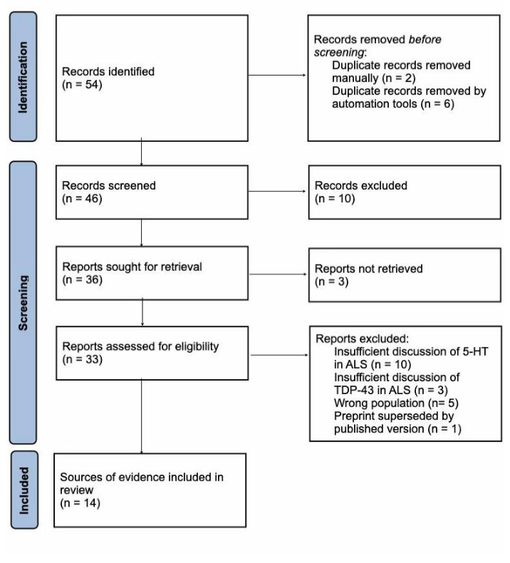
Table 1.
Characteristics of included sources of evidence.
| Author & Year | Ref | Location | Study Design | Model/ Population | Main Focus | |
| Arnoux & Dupuis (2021) | [18] | France | Book chapter | N/A | Synthesize current knowledge of the 5HTR2B receptor in ALS | |
| Brunet et al. (2020) | [22] | France | Review | N/A | Cellular and molecular origins of cortical hyperexcitability in ALS | |
| Dentel et al. (2013) | [19] | France | Non-randomized experimental animal study | Mouse ALS model | Linking serotonergic neuron degeneration in ALS to spasticity | |
| El Oussini et al. (2016) | [24] | France | Non-randomized experimental animal study | Mouse ALS model | The 5HTR2b receptor’s effects on ALS disease progression | |
| El Oussini et al. (2017) | [21] | France | Non-randomized experimental animal study | Mouse ALS model | Providing evidence for brainstem 5-HT neurons’ role in spasticity | |
| Jiang et al. (2023) | [25] | China | Non-randomized experimental animal study | Mouse ALS model | The relationship between ALS and 5HT, using 5-HT receptor antagonists | |
| Koopman et al. (2024) | [30] | Netherlands | Non-randomized experimental study | C. elegans model | Biological mechanisms for disease-related phenotypes in ALS | |
| Lacour et al. (2026) | [28] | Argentina | Non-randomized experimental study | C. elegans model | Potential graded severity of TDP-43 effects in 5-HT neuron-specific C. elegans | |
| Loh et al. (2024) | [29] | Malaysia | Review | N/A | The role of the microbiota-gut-brain axis in neurodegenerative diseases | |
| Lu et al. (2024) | [26] | China | Non-randomized experimental animal study | Mouse ALS model | Therapeutic effects of desloratadine (DLT), a 5HTR2a antagonist | |
| Scott & .Downing (2017) | [31] | South Africa | Non-randomized experimental animal study | Mouse ALS model | BMAA exposure and neurodegeneration relevant to ALS | |
| Vercruysse et al. (2016) | [23] | France | Secondary analysis of RCT and non-randomized experimental animal study | N/A | Hypothalamic melanocortin pathway alterations in ALS | |
| Vermeiren et al. (2018) | [17] | Belgium | Review | N/A | Serotonergic dysfunction in ALS and Parkinson’s Disease (PD) | |
| Yang et al. (2023) | [22] | China | Review | N/A | Summarizing current knowledge on serotonergic alterations in ALS | |
Regarding the financial sponsorship of the included literature, public funding accounted for 92.9% (13/14) of the analyzed studies, while one study (7.1%) did not report a funding source. No industry-sponsored or non-sponsored papers were identified.
Disclaimer/Publisher’s Note: The statements, opinions and data contained in all publications are solely those of the individual author(s) and contributor(s) and not of MDPI and/or the editor(s). MDPI and/or the editor(s) disclaim responsibility for any injury to people or property resulting from any ideas, methods, instructions or products referred to in the content. |
© 2026 by the authors. Licensee MDPI, Basel, Switzerland. This article is an open access article distributed under the terms and conditions of the Creative Commons Attribution (CC BY) license.
Copyright: This open access article is published under a Creative Commons CC BY 4.0 license, which permit the free download, distribution, and reuse, provided that the author and preprint are cited in any reuse.