Preprint
Article

This version is not peer-reviewed.

Antibacterial and Anti-Biofilm Activities of Essential Oils Against Etiological Agents of Bovine Mastitis

Submitted:

28 August 2026

Posted:

31 August 2026

You are already at the latest version

Abstract

Background: The inflammation of the mammary gland, known as bovine mastitis, is a major challenge for the dairy cattle industry globally. The main organisms responsible for mastitis are bacteria, classified as either contagious, such as Staphylococcus aureus, Streptococcus dysgalactiae, and Streptococcus agalactiae; or environmental, primarily Streptococcus spp. (e.g. Streptococcus uberis) and coliform species. Even though the emergence of antibiotic-resistant strains of pathogens poses a serious threat, antibiotics are still the primary treatment for mastitis. It is therefore necessary to explore alternative therapies, such as essential oils (EOs). This in vitro research intended to evaluate the efficacy of EOs against etiological agents of bovine mastitis, as an alternative or complementary treatment to conventional antibiotics. Methods: The bactericidal activity of eleven essential oils and two blends was tested on five strains causing bovine mastitis: Str. agalactiae, Str. dysgalactiae, Str. uberis, Sta. aureus, and Staphylococcus epidermidis. An assessment was carried out of both the reduction of biofilm formation and the inhibition of pre-formed biofilm against biofilm-formative strains of Sta. aureus and Sta. epidermitis. Results: Origanum vulgare and Cinnamomum zeylanicum resulted to be the EOs with the highest activity against the above-mentioned bacteria, inhibiting both bacterial growth and biofilm formation. Conclusions: This study emphasises the relevance of investigating natural products and supports the hypothesis of safe use of EOs to reduce bacterial infections and provide an effective alternative to antibiotics in complementary therapy. Moreover, to avoid treatment failure, the combined use of available antibiotics and natural compounds should also be considered.

Keywords: 
;  ;  ;  ;  

1. Introduction

Bovine mastitis is an inflammatory condition affecting the parenchyma of the mammary gland caused by microbial infections or physical trauma , resulting in alterations in the bacteriological, physical, and chemical characteristics of milk [1]. Because of reduced milk quality and production , cattle mastitis ranks as the leading cause of economic losses and represents one of the primary concerns for the dairy industry worldwide [2,3,4]. Bovine mastitis results from a combination of factors related to the animal, the environment, and pathogens; it can be divided into three classes depending on the severity of inflammation: clinical, sub-clinical, and chronic mastitis [5,6]. Clinical mastitis is obvious and easily identifiable due to noticeable milk or udder abnormalities and common symptoms (fever and loss of appetite) [7]. In subclinical mastitis, instead, no obvious signs are observed, but there is a reduction in milk production and an alteration in its composition [8]. Chronic mastitis refers to inflammation that persists for months, accompanied by clinical relapses.
Organisms such as bacteria, fungi and viruses invading the teat canal are considered the main cause of bovine mastitis [9]. Bacteria are the most common organisms responsible for mastitis and, depending on their origin, the infection can be divided into two classes: contagious and environmental [10]. Contagious pathogens, such as Staphylococcus aureus, Mycoplasma spp., and Streptococcus agalactiae, usually reside on the udder and the teat, colonising and proliferating within the mammary duct [11]. This can lead to subclinical infections, usually accompanied by an increase in somatic cell count (SCC). Environmental pathogens, on the other hand, do not belong to the mammary gland microbiota; once inside the mammary gland, they reproduce, trigger the host immune response and are eliminated quickly [12]. The environmental pathogens mainly include coliform species (e.g. E. coli, Klebsiella spp., Enterobacter spp.), Streptococcus spp. (e.g. Streptococcus uberis), Pseudomonas spp., etc [13].
Bacteria responsible for mastitis, such as Str. uberis and Sta. aureus, can also form biofilm [14]. Biofilm matrices consist of microbial cells, polysaccharides, water and other extracellular products, providing a protective environment ideal for bacterial proliferation [15]. Consequently, bacteria become more resistant to antibiotics and host defences. Therefore, the production of biofilm may result in challenges when treating recurrent infections [16], allowing bacteria to adapt to adverse environments [17].
A prompt and precise identification of the pathogens responsible, given their wide range, is essential both for controlling the spread of the disease (contagious organism) and for the correct selection of antibiotics for therapeutic purposes [7].
Antibiotics, such as penicillin, ampicillin, tetracycline, gentamicin, etc., are the main treatment for mastitis; these can be administered intramammary, intramuscularly or via intravenous infusion [18]. Ideal treatment duration should be long enough to clear subclinical mastitis yet short enough to prevent antibiotic resistance; the best time to treat mastitis is the dry period, with no milk production [19]. Despite antibiotics still being the main therapeutic approach, their effectiveness is limited, and bacterial clearance is often less than 60% [20], with the additional challenge of the emergence of antibiotic-resistant strains becoming a major issue in in antibiotic treatment [21].
In order to prevent the spread of antimicrobial resistance, the Regulation (EU) 2019/6 of the European Parliament and of the Council of 11 December 2018 on veterinary medicinal products and repealing Directive 2001/82/EC, stipulates that “Antimicrobial medicinal products shall not be used for prophylaxis other than in exceptional cases [..]” [22].
The ability of microorganisms to survive or proliferate in the presence of a concentration of an antimicrobial agent (i.e. an antibiotic) normally effective in inhibiting or killing microorganisms of the same species is defined as antimicrobial resistance (AMR) [23]. Systematic use of antibiotics exerts selective pressure on both commensal bacteria, like the gut or skin microbiota, and pathogenic bacteria, which can lead to the dominance of antibiotic-resistant bacteria and the rise of resistant genes. Nowadays, AMR has a serious impact on society, both economically and in terms of public health [24], being a widespread and increasing phenomenon, and representing one of the most difficult global challenges.
AMR is far more complex in animals than in humans, and demands an even more careful and aware use of antibiotics [24]. The administration of antibiotics to food-producing animals, in particular, can leave residues in milk and in edible tissues [25]. It is therefore necessary to seek alternatives based on natural products such as essential oils obtained from medicinal plants, particularly when dealing with multi-drug-resistant bacteria [26].
Plant extracts, including essential oils (EOs), have a long history of use in traditional medicine across the world, and their application in the treatment of bovine mastitis is of growing interest. Several EOs with various biological properties are already being used in this area, although few in vivo studies report on their efficacy, pharmacokinetics and pharmacodynamics [27]. EOs’ antibacterial effects result from the loss of ions and the leakage of cellular components, caused by increased membrane permeability, reduced membrane potential, ATP depletion and disruption of proton pumps, resulting in cell lysis and cell death [28,29,30]. It is harder for microorganisms to develop resistance to essential oils, as they contain a wide variety of components. Furthermore, essential oils can exert antibacterial activity on both resistant and non-resistant pathogens [31,32]. Essential oils and their derived components have attracted growing interest, in recent years, thanks to their encouraging biological activities, as potential candidates for combating bacterial infections and reducing AMR [33,34,35]. Hence, it is crucial to understand the mechanism underlying the phytochemical action through rigorous experimental investigations for the development of new antimicrobial agents [36].
Given these promising results, this research aimed at evaluating the opportunity to use essential oils in the treatment of bovine mastitis as a potential alternative to conventional antibiotics. Essential oils, thank to their additional antioxidant and antinflammatory capabilities, would also provide additional beneficial effects in the inflammatory status of bovine mastitis.
The objective was to evaluate the in vitro bactericidal effect of eleven essential oils and two blends towards five strains causing bovine mastitis: Str. dysgalactiae, Str. uberis, Str. agalactiae, Sta. aureus and Sta. epidermidis. In addition, their antibiofilm activity was assessed against biofilm-forming strains of Sta. aureus and Sta. epidermitis.

2. Results and Discussion

2.1. Chemical Characterization of Essential Oil (GC-FID Analysis)

The chemical characterizations of each EO considered in the present study are reported in the Supplemental materials, from Table S1 to Table S13. Compounds are ranked by elution order, and the values are expressed as the percentage peak area relative to the total composition of each EO, as determined by GC-FID analysis.

2.2. Minimum Inhibitory Concentration (MIC) and Minimum Bactericidal Concentration (MBC)

The results of the antimicrobial screening (MIC and MBC), of the eleven essential oils and the two blends are given for each strain (Str. agalactiae, Str. dysgalactiae, Str. uberis, Sta. aureus and Sta. epidermidis) in Table 1, Table 2, Table 3, Table 4 and Table 5. Table 6 highlights the MIC of all tested EOs by comparing them for the various microorganisms.
The MIC values of the essential oils varied between <0.001% and >3.125% v/v (Table 6). In general, the effect was greater against bacteria of the genus Streptococcus than against those of the genus Staphylococcus. Str. dysgalactiae was the most sensitive strain to the treatment with the EOs, while Sta. epidermidis was the most resistant one, being sensitive just to C. zeylanicum, at the tested concentrations (from 3,125% to 0.001% v/v).
The EOs of Origanum vulgare, Cinnamommum zeylanicum, Thymus vulgaris, and Blend BR proved to be the most effective against Str. agalactiae, Str. dysgalactiae and Str. Uberis, with MIC and MBC values ranging from <0.001% to 0.390% v/v, and from <0.001% to 0.780% v/v, respectively. Against Sta. aureus and Sta. epidermidis, the lowest MICs occurred with Cinnamommum zeylanicum: 0.098% and 0.195% v/v, respectively. Among the EOs, Eucalyptus globosum and Citrus limon exhibited the lowest antibacterial activity towards all pathogens; MBC resulted >3.125% v/v.
Citrus limon showed no antibacterial activity, despite the presence of limonene (57,55%) that showed antibacterial activity against Str. uberis [37]. Limonene, indeed, such as thymol, can alter the composition of the fatty acids in cell membranes, while also affecting their respiration and permeability [38].
1.8-cineole, the main component (93,10%), of Eucalyptus globosum, is able to disintegrate the cell membrane and reduce and concentrate the cytoplasm, causing damage to the structure of Sta. aureus [33]. Nevertheless, the EO of E. globosum did not demonstrate antibacterial property against the tested strains.
The composition of the EO of Melaleuca leucadendrom highlithed as major component the 1,8-cineole (67,85%). As for Eucalyptus globosum, this EO showed moderate activity just against Str. dysgalactiae (0.780% v/v), suggesting that the destabilization in the membrane induced by 1,8-cineole might not be enough, alone, to inhibit the growth of the tested strains. It has been demonstrated in two studies that an essential oil from the Melaleuca genus, rich in 1,8-cineole, has a synergistic effect with rifaximin [39] and erythromycin [33] against Sta. aureus.
Melaleuca alternifolia, known as tea tree oil (TTO), showed weak activity against Streptococcus agalactiae (1.560% v/v), moderate activity against Str. dysgalactiae and Str. uberis (0.780% v/v), and it was not active against the strains of Staphylococcus spp at the tested concentrations (>3.125% v/v). In total, 80–90% of the oil is represented by the monoterpenes terpinen-4-ol, α-terpinene, 1,8-cineole, p-cymene, α-terpineol, α-pinene, terpinolene, limonene, and sabinene. The most abundant (41,21%) of these is terpinen-4-ol, which contributes significantly to the antimicrobial properties of the oil [40].
These results do not contradict the data reported from previous studies on the antibacterial effect of TTO against Streptococcus spp and Staphylococcus spp, since the concentrations tested in our study are lower [41,42].
Rosmarinum officinalis showed good antibacterial activity against Str. dysgalactiae (0.195% v/v). The EO has both antimicrobial and antioxidant attributes, which are primarily associated with monoterpenes, like 1,8-cineole (34.29%), pinene (33.84%), camphor (19.35%), borneol, limonene, camphene, and myrcene [43]. Contrary to the results of [44], in our study, R. officinalis did not appear to be active against most of the pathogenic bacteria.
Menthol (36,43%) is a major component of Mentha piperita L. (peppermint), playing a role in the herb’s antioxidant and antimicrobial effects [45]. M. piperita EO exhibited low MIC values against the Streptococcus strains, while it was not active against the Staphylococcus strains, confirming the results obtained by Tomanić et al. (2022) [46], who highlighted higher MIC values of the EO against Staphylococcus spp compared to Streptococcus spp.
Both the EOs of Lavandula angustifolia and Lavandula hybrida showed a composition rich in linalool (37.32% and 33.33%, respectively) and linalyl acetate (30.64% and 32.20%, respectively). These two predominant compounds are considered responsible for the antimicrobial activity of these plants [47].
Thymus vulgaris was very effective against the strains belonging to the genus Streptococcus, with MIC values from 0.195% v/v to 0.390% v/v. The importance of the high presence of thymol (46,41%) and p-cymene (19,89%), within the essential oils, has been confirmed for the antibacterial properties of the genus Thymus [48,49,50]. Both thymol and carvacrol exert their antimicrobial activity by inducing changes in the structure and function of the cytoplasmic membrane that affect both the external and internal membranes. Thymol may additionally interfere with membrane proteins and intracellular targets. This interaction alters the membrane’s permeability and leads to the release of K+ ions and ATP [51]. Thymol has been shown to be highly effective against isolates from mastitis cows [41,52,53,54].
Origanum vulgare and C. zeylanicum, were the only essential oils to be active against Staphylococcus aureus. In many studies O. vulgare has been shown to be active toward major mastitis pathogens such as Sta. aureus, E. coli, Streptococcus spp., Str. dysgalactiae, and Str. uberis, along with methicillin-resistant Staphylococcus aureus (MRSA) [54,55,56]. The phenolic monoterpenoid carvacrol (79.29%) affects microbial cells by causing damage to the structure and function of their membranes, leading to enhanced membrane fluidity and permeability [57].
Cinnamomum zeylanicum (CZ) EO resulted the most active among the tested EOs. The EO was effective against all the tested strains, especially against Str. agalactiae and Str. uberis, with MIC values of 0.050% v/v and <0.001% v/v respectively (Table 6). This antibacterial property could be attributed to its characteristic secondary metabolites such as trans-cinnamaldehyde (79,03%), cinnamyl acetate, caryophyllene oxide, and β-caryophyllene [58]. These results agree with the literature in vitro studies that demonstrates the bactericidal effects of Cinnamomum EO against bovine mastitis pathogenic isolates [59,60], especially against Gram+ [61].
Therefore, O. vulgare and C. zeylanicum may be regarded as promising candidates in the development of antibacterial treatments for antibiotic-resistant bacterial infections.

2.3. Biofilm

The anti-biofilm assay was carried out against a strain of Staphylococcus aureus (ATCC 6538) and a strain of Staphylococcus epidermidis (DSMZ 20044). The assay was conducted to evaluate the efficacy of the essential oils in preventing cell attachment (anti-adhesion), at 0 h (T0), and in inhibiting the growth of pre-formed biofilms, at 24 h (T24). The crystal violet assay quantifies the entire biofilm biomass, including extracellular polymeric substances formed during biofilm growth that bind or attach to the bacterial cells.
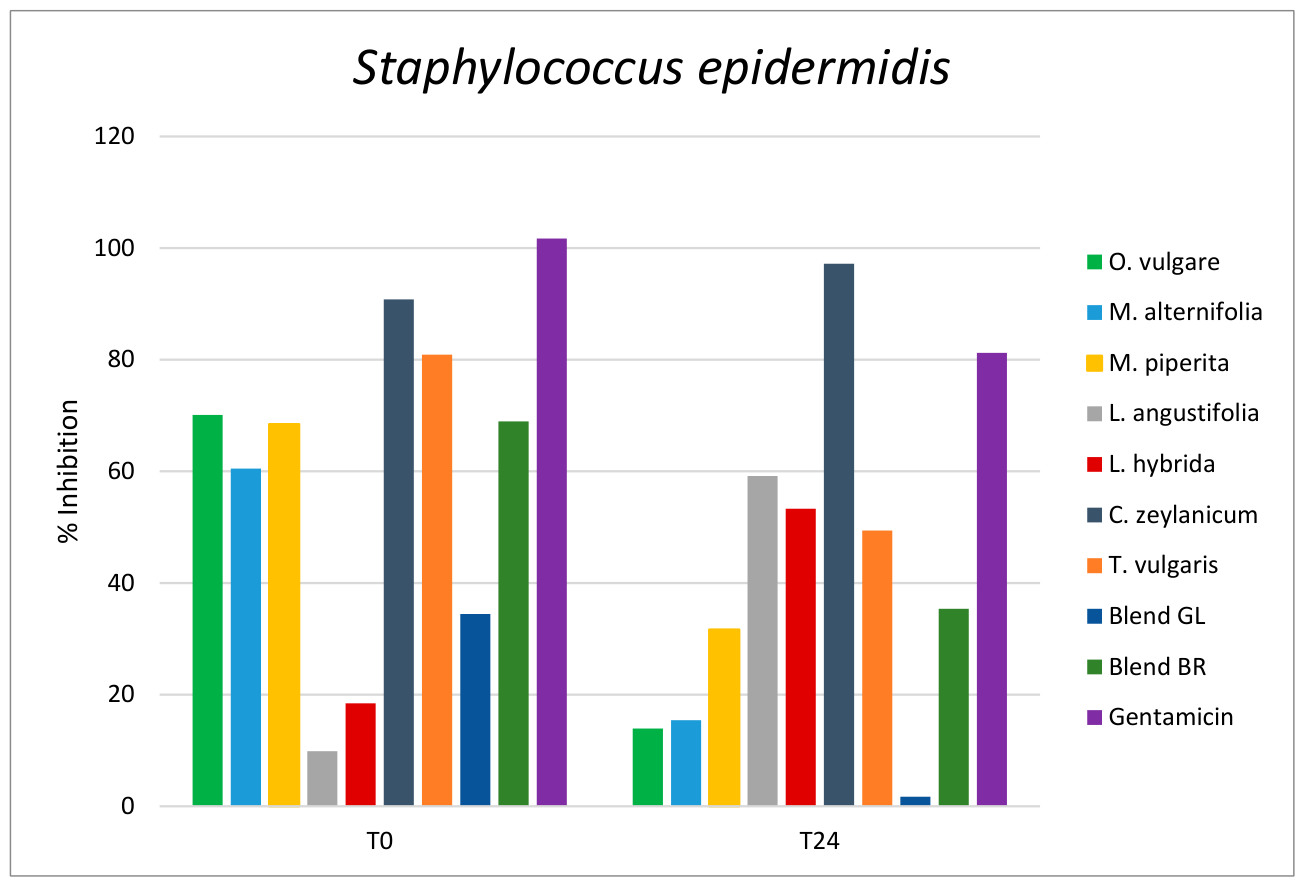
The result regarding the percentage (%) of biofilm inhibition of the three different tested concentration (0.78, 0.39 and 0.195% v/v, μL/mL) of each essential oils are reported in the Supplemental materials, from Figure S1 to Figure S20. The effects of the seven EOs and the two blends on the development and adhesion of biofilms of the tested mastitis etiological agents are illustrated in Figure 1 and Figure 2. All essential oils with an inhibition rate greater than 50 % were classified as having strong antibiofilm activity.
C. zeylanicum EO revealed a very good activity against both strains at both T0 and T24. The strong antibacterial and anti-biofilm properties of cinnamon oil are definitely due to its high cinnamaldehyde level, as shown by Budri et al. (2015) [59], who found that cinnamaldehyde had a lower MIC than Cinnamomum zeylanicum, and it greatly decreased biofilm formation. C. zeylanicum was further evaluated against biofilm formation by Str. agalactiae, where the EO was able to exert potent inhibitory activity [62].
Worth noting that Cinnamomum EO was the one of most effectives despite the reduced concentrations tested, from 0.098% to 0.39% v/v, namely half of the tested concentrations of the other EOs.
Sta. aureus and Sta. epidermidis are two well-known biofilm producers and are commonly associated with intramammary infections, leading to chronic bovine mastitis. It is well established that staphylococcal isolates are the primary cause of biofilm-associated infections [63]. Specifically, the formation of biofilms by Sta. aureus strains and their multidrug-resistance profile increase the risk of chemotherapeutic failure [64].
The anti-biofilm effect of different essential oils against pathogenic isolates from bovine mastitis has been extensively studied, evaluating both the reduction in biofilm formation and the inhibition of pre-formed biofilm [37,59,65,66,67,68,69].
However, published literature showed no prior researches on the antibiofilm potential of L. angustifolia, L. hybrida, M. piperita, Origanum vulgare and Thymus vulgaris against bovine mastitis-causing organisms.
In this study, at time zero (0 h), at concentration of 0.39% v/v (except for CZ, tested at 0.195%), all essential oils showed strong inhibition of biofilm formation above 50% against the tested organism Sta. aureus, except the Blend GL (45.1%) (Figure 1). Indeed, most of the EOs had good to moderate inhibition on biofilm formation at time zero against the strains tested. In general, the extracts were more effective at preventing biofilm formation (with better activity at T0) than at destroying pre-formed biofilm (T24) (Figure 1 and Figure 2).
M. piperita, O. vulgare, C. zeylanicum and T. vulgaris essential oils had the best biofilm inhibition ranging from 97.67% to 102.61% at time zero for Sta. aureus.
All tested essential oil, except for L. angustifolia and L. hybrida, and Blend GL exerted good activity in the reduction (>50%) of development of biofilm, at T0, against Sta. epidermidis. The best activity was showed by T. vulgaris and C. zeylanicum, with inhibition values of 80.91% and 90.81% respectively.
On 24 h-old pre-formed biofilm, Blend GL, and essential oils of O. vulgare and M. alternifolia, had weak antibiofilm activity (1.70% to 15.40%) against Sta. epidermidis, whereas Blend BR and EO of M. piperita had moderate activity (31.67% to 35.38%).
Blend GL resulted to be less active against both the tested strains compared to the majority of the essential oil, while Blend BR demonstrated good activity, especially against Sta. aureus, even higher than the gentamicin control. These findings show how essential oils, mixed together, can behave sometimes synergistically with each other, sometimes not, resulting in mixtures with more or less antibacterial activity.
The lowest concentration (0.195% v/v) of L. angustifolia essential oils increased biofilm growth of Sta. aureus on 24 h pre-formed biofilm, as well as the essential of M. alternifolia and Blend GL enhanced the growth of pre-formed biofilm of Sta. epidermidis. These results find support in some studies where an increase in biofilm growth has been shown after the treatment of pre-formed biofilm with both EOs and their components [70,71].
Indeed, after exposure to low concentration of Origanum vulgare EO, it has been observed an increasing number of sessile cells in mature biofilm of Sta. aureus [72]. According to the Authors, the stimulating effect of the oil occurs at concentrations of phenolic compounds below the MIC, as a reaction to stress conditions, thereby enhancing the ability of the microorganism to form biofilms [73].
Overall, this study revealed that essential oils from O. vulgare and C. zeylanicum exhibited the strongest activity against bacteria and biofilms. Our results are consistent with the literature, as various classes of bioactive secondary metabolites, like carvacrol and trans-cinnamaldehyde, terpenoids and flavonoids have been isolated from these species [57,58]. Given the pharmacological potential of trans-cinnamaldehyde and carvacrol, these molecules and other classes of metabolites contained in the EOs of C. zeylanicum and O. vulgare might be responsible for the observed activity. These findings are consistent with previously published results regarding the antimicrobial and antibiofilm activities of Cinnamomum [59,62].
Several Authors have suggested that treatment failure due to antimicrobial resistance could be addressed by combining available antibiotics with other natural products, such as EOs [67,74,75,76]. The EOs tested may enhance antimicrobial susceptibility rates and help reverse antimicrobial resistance; furthermore, the efficacy of antibiotics may be restored when used in conjunction with EOs.

4. Materials and Methods

4.1. Essential Oils

Eleven pure essential oils and two blends were used for the study. In particular, Origanum vulgare, Melaleuca alternifolia, Melaleuca leucadendrom, Eucalyptus globosum, Mentha piperita, Lavandula angustifolia, Lavandula hybrida, Cinnamommum zeylanicum, Rosmarinum officinalis, Citrus limon, Thymus vulgaris, and two undisclosed blends GL and BR (two confidential solutions under patent processing, containing nine mixtures of the above mentioned EOs) dispersed in the surfactant Alkamuls (Glyceryl polyethyleneglycol ricinoleate, CEE additive cod. E484), were provided by APA-CT (Forlì, FC, Italy). All EOs and the blends were used as emulsions.

4.2. Chemical Characterization of Essential Oil (GC-FID Analysis)

The chemical analysis of all the EOs were obtained from the suppliers (Tables S1-S13). The chromatographic characterization of EOs was carried out using a gas chromatograph (GC) with a flame ionization detector (FID), following the protocol validated by Truzzi et al. (2025) [77].
Qualitative analysis and the semi-quantitative characterization of EOs were performed using an Agilent Technologies (Agilent Technologies, Milan, Italy) HP-5 cross-linked 5% phenyl–95% methyl polysiloxane (25 m × 0.2 mm i.d., 0.5 μm film thickness) capillary column on a 7820 gas chromatograph linked with a 5975C network mass spectrometer.
Compounds were recognized by matching the retention times of the chromatographic peaks with those of authentic reference standards analyzed under identical conditions and by matching the linear retention indices (LRIs).
Semi-quantitative data were derived from the mean of two analyses and calculated as the relative percentage amount of each analyte; specifically, the values were reported as the percentage peak area relative in comparison to the overall composition of each EO, as determined by GC-FID analysis.

4.3. Essential Oils Emulsions

The EOs emulsions were created using Alkamus, a surfactant (Glyceryl polyethyleneglycol ricinoleate cod. E484), approved for use in animal feed, as follows. The stock emulsion was prepared with the EO and Alkamus, in a proportion of 1:3; both were placed in an Eppendorf tube and vortexed for approx. 1 min; then, the growth medium, Muller Hinton (MH), or Brain Heart Infusion (BHI), was added and vortexed again for at least 3 min, after which a milky to opaque emulsion was obtained. Subsequently the stock solution was diluted with growth medium obtaining a concentration of EO of 6,25% v/v.

4.4. Microdilution Assay

Antibacterial activity of each of the essential oils and the two blends was assayed against Streptococcus agalactiae (DSMZ 2134), Streptococcus dysgalactiae (DSMZ 20662), Streptococcus uberis (DSMZ 20569), Staphylococcus aureus (ATCC 6538) and Staphylococcus epidermidis (DSMZ 20044).
The minimum inhibitory concentration (MIC) and the minimum bactericidal concentration (MBC) of EOs were evaluated with the microbroth dilution assay using 96-well microplates, as described by the European Committee on Antimicrobial Susceptibility Testing (EUCAST) international guidelines [78], slightly modified.
The MIC is defined as the lowest concentration that completely inhibits the growth of a given organism compared with the growth in the negative control (substance-free), while the MBC is defined as the lowest concentration resulting in the death of 99.9% or more of the initial inoculum [79].
The growth medium used for both Staphylococccus aureus and Staphylococcus epidermidis was Mueller Hinton (MH, MERCK, Darmstadt, Germany), while the medium used for Streptococcus agalactiae, Streptococcus dysgalactiae, and Streptococcus uberis was Brain Heart Infusion (BHI, Sigma-Aldrich, Saint Louis, MO, USA).
Bacterial cultures, grown overnight in MH broth or BHI broth, were diluted to McFarland standard No 0.5, and adjusted in order to have 5 × 10⁵ CFU/mL of bacterial suspension in each well. The MIC test was performed on a 96-well microtiter plate: aliquots of 50 μL of EO emulsion, prepared in MH or BHI medium, were added to 50 μL of bacterial suspension made in the same medium. EO’s effectiveness was tested using two-fold scalar dilutions of concentrations between 3,125% (31,25 mL/L) to 0.001% v/v (0.01 mL/L).
MH broth, added with and without 9,38% Alkamus, was used as positive control. The microplates were sealed with an adhesive film (to avoid dispersion of the EO more volatile components) and incubated at 37 °C for 24 h. After incubation, the bacterial growth was evaluated spectrophotometrically at 620 nm (Multiskan EX, Thermo Fisher Scientific Oy, Vaanta, Finland).
To assess MBC values, 10 μL were taken from each well showing no microbial growth and seeded onto growth medium MH agars. After 24 h of incubation, CFUs were counted to assess cell viability.
Each assay was performed in triplicate and repeated twice.

4.5. Inhibition of Biofilm Growth (Biomass Production) and Development

Staphylococcus aureus ATCC 6538 and Sta. epidermidis DSMZ 20044 strains were used for determining inhibition of bacterial biofilm. Brain Heart Infusion (BHI) agar plates were prepared and used for overnight culture of strains at 37 °C without shaking.
Freshly prepared BHI broth, in 10 mL sterile tubes, was inoculated with bacteria from the BHI agar plates and incubated overnight for 24 h at 37 °C, with shaking. The overnight bacterial culture was then diluted, after the culture media was used to blank, with sterile distilled water to 0.5 McFarland standard (1.5 X 10⁸ CFU/ml) and then further dilute 1:200 in the culture media BHI broth to obtain an inoculum of 0.75 × 106 CFU/mL.
To study the activity of the essential oils and the two blends on the biofilm formation, natural compounds were evaluated at a concentration range from 0.78% (except Cinnamonum zeylanicum, CZ, 0.39%) v/v to 0.195% (CZ 0.098%) v/v.

4.5.1. Effect on Bacterial Adhesion (T0)

The modified protocol described by Kırmusaoğlu and Kaşıkçı (2020) [80] was followed.
An aliquot of 100 µl of diluted culture were added to the wells of individually labelled flat-bottomed 96-well microtitre plates, apart from the wells designed for blanks (media blank, samples and positive control blank). 100 µl of samples (essential oils and blends) and control were added immediately after to determine the inhibition of cell attachment (T0). Control wells were included in the plate: negative control (culture + media (BHI)), positive control (culture +BHI+ antibiotic), sample control (sample + BHI), antibiotic control (antibiotic + BHI) and media control (BHI) for each test batch. Gentamicin (Sigma-Aldrich, Saint Louis, MO, USA) was used as positive control, at concentrations from 1 mg/mL to 0,25 mg mL.
Plates were then covered with an adhesive film (to avoid dispersion of the EO more volatile components) and incubate for 24 hours at 37 °C, without shaking, to allow cell attachment and biofilm development. The modified crystal violet staining method, described below (4.5.3), was used to quantify the biomass.

4.5.2. Effect on Pre-Formed Biofilm (T24)

The ability of the EOs to prevent further biofilm development or destruction of pre-formed biofilms was explored using the protocol described by O’Toole and Kolter (1998) [81] and modified by Sandasi et al. (2010) [82]. 100 µl diluted culture of Staphylococcus aureus or Sta. epidermidis were added to an individually labelled flat-bottomed 96-well microtiter plate and incubated at 37 °C for 24 h (irreversible attachment phase) without agitation for the formation of a multilayer biofilm. After 24 h (T24), 100 µl aliquots of essential oils and of blends GL and BR, at the different concentrations, were then added to the wells and the plates were sealed and additionally incubated at 37 °C for 24 h. The controls were the same as the previous paragraph: negative control, positive control, sample control, antibiotic control and media control.
Biofilm biomass was assessed using the modified crystal violet staining assay as described below.

4.5.3. Crystal Violet Staining (CVS) Procedure

The attached biofilm mass was indirectly assessed by the modified crystal violet assay [82]. After incubating with the treatment, the wells were carefully emptied and gently washed three times with sterile distilled water to remove any unattached cells. Then the plates were air-dried and subsequently oven-dried at 45 °C for 60 min to fix the adherent cells. The sessile cells were then stained with 100 µl of 0.1% crystal violet solution (CV, Sigma-Aldrich, Saint Louis, MO, USA) for 15 minutes at room temperature while covered with the microtitre lid. The plate was gently washed five times with sterile distilled water to remove any excess stain and unabsorbed stain.
Thereafter, the biofilm biomass was assessed semi-quantitatively by re-solubilizing the crystal violet stain bound to the adherent cells with 150 µl of 100% ethanol. The absorbance of the destaining solution was read at 570 nm using a microplate reader (Infinite F50, TECAN, Salzburg, Austria). The mean optical density (OD570nm) of the sample was determined, and the percentage inhibition of biofilm was calculated using the equation below:
% I n h i b i t i o n = ( O D n e g a t i v e c o n t r o l O D m e d i a c o n t r o l ) ( O D t e s t O D e x t r a c t c o n t r o l ) ( O D n e g a t i v e c o n t r o l O D m e d i a c o n t r o l ) * 100
Both tests, at T0 and T24, were conducted in triplicate and repeated twice.
Microsoft Excel was used to plot sample concentrations against percentage inhibitions.

5. Conclusions

This research activity was conducted with the aim of evaluating the antibacterial efficacy of some essential oils (alone or blends) against bacterial strains responsible for bovine mastitis. The ultimate goal was to identify useful tools for the prevention and control of this disease that would not increase the already widespread phenomenon of antibiotic resistance.
The current scenario of antimicrobial, and especially antibiotic resistance, is indeed extremely worrying and the scientific community is investing time and effort in finding new solutions. Veterinary medicine, and farm animals in particular, have always been considered among the major players in the development of resistance due to the large-scale, and sometimes inappropriate, use of antibiotics. Such molecules are crucial in the treatment of specific diseases, so it is extremely important to “preserve” their effectiveness as much as possible. Moreover, the new European Union Regulations greatly reduce the possibility of preventive antibiotic use in animals and extremely limit the antibiotic categories allowed in Veterinary medicine [83,84,85]. For this reason, it is necessary to find alternatives, especially in the treatment of those diseases caused by resistant bacteria.
The essential oils chosen and tested were used alone and in combination with each other within two blends to evaluate possible synergistic effects. The antimicrobial and antibiofilm activities of EOs against Sta. epidermidis and Sta. aureus, the predominant contagious pathogens causing bovine mastitis, were evaluated and our results showed good bacterial and biofilm inhibiting activities of the investigated EOs, especially of Origanum vulgare and Cinnamomum zeylanicum.
The study highlights the importance of evaluating natural products to strengthen the hypothesis of safe EOs applications for reducing bacterial contamination as effective antibiotic alternatives in complementary therapy. Furthermore, treatment failure due to antimicrobial resistance could be limited by using combinations of the available antibiotics and other natural compound such as EOs.

Supplementary Materials

The following supporting information can be downloaded at the website of this paper posted on Preprints.org.

Author Contributions

Conceptualization, A.C. and A.Z.; methodology, A.C., D.S., M.M. and P.M.; formal analysis, A.C. and D.S.; investigation, A.C. D.S. and M.M.; resources, P.M. and M.S.; data curation, A.C., D.S. and M.M.; writing—original draft preparation, A.C.; writing—review and editing, A.C., D.S., M.M., P.M., M.S. and A.Z.; visualization, A.C.; supervision, P.M., M.S. and A.Z.; project administration, P.M. and A.Z. All authors have read and agreed to the published version of the manuscript.

Funding

This research received no external funding.

Institutional Review Board Statement

Not applicable.

Data Availability Statement

The original contributions presented in this study are included in the article and Supplementary Material. Further inquiries can be directed to the corresponding authors.

Acknowledgments

The authors are grateful to APA-CT SRL for providing the essential oils, the blends and the surfactant.

Conflicts of Interest

The authors declare no conflicts of interest.

Abbreviations

The following abbreviations are used in this manuscript:
EO Essential oil
AMR Antimicrobial resistance
CZ Cinnamomum zeylanicum
MIC Minimum Inhibitory Concentration
MBC Minimum Bactericidal Concentration

References

  1. Gomes, F.; Henriques, M. Control of Bovine Mastitis: Old and Recent Therapeutic Approaches. Curr. Microbiol. 2016, 72, 377–382. [Google Scholar] [CrossRef] [PubMed]
  2. Motaung, T.E.; Petrovski, K.R.; Petzer, I.-M.; Thekisoe, O.; Tsilo, T.J. Importance of Bovine Mastitis in Africa. Anim. Health Res. Rev. 2017, 18, 58–69. [Google Scholar] [CrossRef] [PubMed]
  3. Aghamohammadi, M.; Haine, D.; Kelton, D.F.; Barkema, H.W.; Hogeveen, H.; Keefe, G.P.; Dufour, S. Herd-Level Mastitis-Associated Costs on Canadian Dairy Farms. Front. Vet. Sci. 2018, 5. [Google Scholar] [CrossRef] [PubMed]
  4. Sathiyabarathi, M.; Jeyakumar, S.; Manimaran, A.; Jayaprakash, G.; Pushpadass, H.A.; Sivaram, M.; Ramesha, K.P.; Das, D.N.; Kataktalware, M.A.; Prakash, M.A.; et al. Infrared Thermography: A Potential Noninvasive Tool to Monitor Udder Health Status in Dairy Cows. Vet. World 2016, 9, 1075–1081. [Google Scholar] [CrossRef] [PubMed]
  5. Campos, B.; Pickering, A.C.; Rocha, L.S.; Aguilar, A.P.; Fabres-Klein, M.H.; de Oliveira Mendes, T.A.; Fitzgerald, J.R.; de Oliveira Barros Ribon, A. Diversity and Pathogenesis of Staphylococcus Aureus from Bovine Mastitis: Current Understanding and Future Perspectives. BMC Vet. Res. 2022, 18, 115. [Google Scholar] [CrossRef] [PubMed]
  6. Gonçalves, J.L.; Kamphuis, C.; Martins, C.M.M.R.; Barreiro, J.R.; Tomazi, T.; Gameiro, A.H.; Hogeveen, H.; dos Santos, M.V. Bovine Subclinical Mastitis Reduces Milk Yield and Economic Return. Livest. Sci. 2018, 210, 25–32. [Google Scholar] [CrossRef]
  7. Sharun, K.; Dhama, K.; Tiwari, R.; Gugjoo, M.B.; Iqbal Yatoo, Mohd.; Patel, S.K.; Pathak, M.; Karthik, K.; Khurana, S.K.; Singh, R.; et al. Advances in Therapeutic and Managemental Approaches of Bovine Mastitis: A Comprehensive Review. Vet. Q. 2021, 41, 107–136. [Google Scholar] [CrossRef] [PubMed]
  8. Abebe, R.; Hatiya, H.; Abera, M.; Megersa, B.; Asmare, K. Bovine Mastitis: Prevalence, Risk Factors and Isolation of Staphylococcus Aureus in Dairy Herds at Hawassa Milk Shed, South Ethiopia. BMC Vet. Res. 2016, 12, 270. [Google Scholar] [CrossRef] [PubMed]
  9. Tiwari, J.G.; Babra, C.; Tiwari, H.; Williams, V.; De Wet, S.; Gibson, J.; Paxman, A.; Morgan, E.; Costantino, P.; Sunagar, R. Trends in Therapeutic and Prevention Strategies for Management of Bovine Mastitis: An Overview. J. Vaccines Vaccin. 2013, 4, 1–11. [Google Scholar]
  10. Lakew, B.T.; Fayera, T.; Ali, Y.M. Risk Factors for Bovine Mastitis with the Isolation and Identification of Streptococcus Agalactiae from Farms in and around Haramaya District, Eastern Ethiopia. Trop. Anim. Health Prod. 2019, 51, 1507–1513. [Google Scholar] [CrossRef] [PubMed]
  11. Kibebew, K. Bovine Mastitis: A Review of Causes and Epidemiological Point of View. J. Biol. Agric. Healthc. 2017, 7, 1–14. [Google Scholar]
  12. Bradley, A.J. Bovine Mastitis: An Evolving Disease. Vet. J. 2002, 164, 116–128. [Google Scholar] [CrossRef] [PubMed]
  13. Bogni, C.; Odierno, L.; Raspanti, C.; Giraudo, J.; Larriestra, A.; Reinoso, E.; Lasagno, M.; Ferrari, M.; Ducrós, E.; Frigerio, C. War against Mastitis: Current Concepts on Controlling Bovine Mastitis Pathogens. Sci. Against Microb. Pathog. Commun. Curr. Res. Technol. Adv. 2011, 483–494. [Google Scholar]
  14. Reinoso, E.B. Bovine Mastitis Caused by Streptococcus Uberis. In Virulence Factors and Biofilm; 2017. [Google Scholar]
  15. Gilmore, K.S.; Srinivas, P.; Akins, D.R.; Hatter, K.L.; Gilmore, M.S. Growth, Development, and Gene Expression in a Persistent Streptococcus Gordonii Biofilm. Infect. Immun. 2003, 71, 4759–4766. [Google Scholar] [CrossRef] [PubMed]
  16. Sutherland, I.W. The Biofilm Matrix – an Immobilized but Dynamic Microbial Environment. Trends Microbiol. 2001, 9, 222–227. [Google Scholar] [CrossRef] [PubMed]
  17. Melchior, M.B.; Vaarkamp, H.; Fink-Gremmels, J. Biofilms: A Role in Recurrent Mastitis Infections? Vet. J. 2006, 171, 398–407. [Google Scholar] [CrossRef] [PubMed]
  18. Hossain, M.K.; Paul, S.; Hossain, M.M.; Islam, M.R.; Alam, M.G.S. Bovine Mastitis and Its Therapeutic Strategy Doing Antibiotic Sensitivity Test. Austin J. Vet. Sci. Anim. Husb. 2017, 4, 1030. [Google Scholar] [CrossRef]
  19. Biggs, A. Update on Dry Cow Therapy 1. Antibiotic v Non-Antibiotic Approaches. In Pract. 2017, 39, 328–333. [Google Scholar] [CrossRef]
  20. Hillerton, J.E.; Kliem, K.E. Effective Treatment of Streptococcus Uberis Clinical Mastitis to Minimize the Use of Antibiotics. J. Dairy Sci. 2002, 85, 1009–1014. [Google Scholar] [CrossRef] [PubMed]
  21. Fair, R.J.; Tor, Y. Antibiotics and Bacterial Resistance in the 21st Century. Perspect. Med. Chem. 2014, 6, 25–64. [Google Scholar] [CrossRef] [PubMed]
  22. Regulation (EU) 2019/6 of the European Parliament and of the Council of 11 December 2018 on Veterinary Medicinal Products and Repealing Directive 2001/82/EC (Text with EEA Relevance). 2018; Vol. 004.
  23. Regulation (EU) 2019/6 of the European Parliament and of the Council of 11 December 2018 on Veterinary Medicinal Products and Repealing Directive 2001/82/EC (Text with EEA Relevance). 2018; Vol. 004.
  24. Caneschi, A.; Bardhi, A.; Barbarossa, A.; Zaghini, A. The Use of Antibiotics and Antimicrobial Resistance in Veterinary Medicine, a Complex Phenomenon: A Narrative Review. Antibiotics 2023, 12, 487. [Google Scholar] [CrossRef] [PubMed]
  25. Subirats, J.; Domingues, A.; Topp, E. Does Dietary Consumption of Antibiotics by Humans Promote Antibiotic Resistance in the Gut Microbiome? J. Food Prot. 2019, 82, 1636–1642. [Google Scholar] [CrossRef] [PubMed]
  26. Yang, W.-T.; Ke, C.-Y.; Wu, W.-T.; Lee, R.-P.; Tseng, Y.-H. Effective Treatment of Bovine Mastitis with Intramammary Infusion of Angelica Dahurica and Rheum Officinale Extracts. Evid. Based Complement Altern. Med. 2019, 2019, 7242705. [Google Scholar] [CrossRef] [PubMed]
  27. Kher, M.N.; Sheth, N.R.; Bhatt, V.D. In Vitro Antibacterial Evaluation of Terminalia Chebula as an Alternative of Antibiotics against Bovine Subclinical Mastitis. Anim. Biotechnol. 2019, 30, 151–158. [Google Scholar] [CrossRef] [PubMed]
  28. Oussalah, M.; Caillet, S.; Lacroix, M. Mechanism of Action of Spanish Oregano, Chinese Cinnamon, and Savory Essential Oils against Cell Membranes and Walls of Escherichia Coli O157:H7 and Listeria Monocytogenes. J. Food Prot. 2006, 69, 1046–1055. [Google Scholar] [CrossRef] [PubMed]
  29. Turina, A. del V.; Nolan, M.V.; Zygadlo, J.A.; Perillo, M.A. Natural Terpenes: Self-Assembly and Membrane Partitioning. Biophys. Chem. 2006, 122, 101–113. [Google Scholar] [CrossRef] [PubMed]
  30. Saad, N.Y.; Muller, C.D.; Lobstein, A. Major Bioactivities and Mechanism of Action of Essential Oils and Their Components. Flavour Fragr. J. 2013, 28, 269–279. [Google Scholar] [CrossRef]
  31. Mittal, R.; Rana, A.; Jaitak, V. Essential Oils: An Impending Substitute of Synthetic Antimicrobial Agents to Overcome Antimicrobial Resistance. Curr. Drug Targets 2018, 19. [Google Scholar] [CrossRef] [PubMed]
  32. Lorenzi, V.; Muselli, A.; Bernardini, A.F.; Berti, L.; Pagès, J.-M.; Amaral, L.; Bolla, J.-M. Geraniol Restores Antibiotic Activities against Multidrug-Resistant Isolates from Gram-Negative Species. Antimicrob. Agents Chemother. 2009, 53, 2209–2211. [Google Scholar] [CrossRef] [PubMed]
  33. Buldain, D.; Gortari Castillo, L.; Buchamer, A.V.; Bandoni, A.; Marchetti, L.; Mestorino, N. In Vitro Synergistic Interaction between Melaleuca Armillaris Essential Oil and Erythromycin against Staphylococcus Aureus Isolated from Dairy Cows. Front Vet. Sci. 2022, 9, 1005616. [Google Scholar] [CrossRef] [PubMed]
  34. Arbab, S.; Ullah, H.; Bano, I.; Li, K.; Ul Hassan, I.; Wang, W.; Qadeer, A.; Zhang, J. Evaluation of in Vitro Antibacterial Effect of Essential Oil and Some Herbal Plant Extract Used against Mastitis Pathogens. Vet. Med. Sci. 2022, 8, 2655–2661. [Google Scholar] [CrossRef] [PubMed]
  35. Tariq, S.; Wani, S.; Rasool, W.; Shafi, K.; Bhat, M.A.; Prabhakar, A.; Shalla, A.H.; Rather, M.A. A Comprehensive Review of the Antibacterial, Antifungal and Antiviral Potential of Essential Oils and Their Chemical Constituents against Drug-Resistant Microbial Pathogens. Microb. Pathog. 2019, 134, 103580. [Google Scholar] [CrossRef] [PubMed]
  36. Jubair, N.; Rajagopal, M.; Chinnappan, S.; Abdullah, N.B.; Fatima, A. Review on the Antibacterial Mechanism of Plant-Derived Compounds against Multidrug-Resistant Bacteria (MDR). Evid. Based Complement Altern. Med. 2021, 2021, 3663315. [Google Scholar] [CrossRef] [PubMed]
  37. Montironi, I.D.; Cariddi, L.N.; Reinoso, E.B. Evaluation of the Antimicrobial Efficacy of Minthostachys Verticillata Essential Oil and Limonene against Streptococcus Uberis Strains Isolated from Bovine Mastitis. Rev. Argent. De Microbiol. 2016, 48, 210–216. [Google Scholar] [CrossRef] [PubMed]
  38. Nardoni, S.; Pisseri, F.; Pistelli, L.; Najar, B.; Luini, M.; Mancianti, F. In Vitro Activity of 30 Essential Oils against Bovine Clinical Isolates of Prototheca Zopfii and Prototheca Blaschkeae. Vet. Sci. 2018, 5, 45. [Google Scholar] [CrossRef] [PubMed]
  39. Buldain, D.; Gortari Castillo, L.; Buchamer, A.V.; Aliverti, F.; Bandoni, A.; Marchetti, L.; Mestorino, N. Melaleuca Armillaris Essential Oil in Combination With Rifaximin Against Staphylococcus Aureus Isolated of Dairy Cows. Front. Vet. Sci. 2020, 7. [Google Scholar] [CrossRef] [PubMed]
  40. Sharifi-Rad, M.; Varoni, E.M.; Iriti, M.; Martorell, M.; Setzer, W.N.; Del Mar Contreras, M.; Salehi, B.; Soltani-Nejad, A.; Rajabi, S.; Tajbakhsh, M.; et al. Carvacrol and Human Health: A Comprehensive Review. Phytother Res. 2018, 32, 1675–1687. [Google Scholar] [CrossRef] [PubMed]
  41. Corona-Gómez, L.; Hernández-Andrade, L.; Mendoza-Elvira, S.; Ricardo-González, D.I.; Pérez-Gerardo, M.D.; Quintanar-Guerrero, D. Effect of the in Vitro Activity of a Component Derived from Thymus Vulgaris Essential Oil and Tilmicosin on Different Strains of Staphylococcus Sp, Streptococcus Sp and Escherichia Coli Isolated from Bovine Mastitis. J. Essent. Oil Res. 2022, 34, 270–277. [Google Scholar] [CrossRef]
  42. Taga, I.; Lan, C.Q.; Altosaar, I. Plant Essential Oils and Mastitis Disease: Their Potential Inhibitory Effects on Pro-Inflammatory Cytokine Production in Response to Bacteria Related Inflammation. Nat. Product. Commun. 2012, 7, 1934578X1200700534. [Google Scholar] [CrossRef]
  43. Borges, R.S.; Ortiz, B.L.S.; Pereira, A.C.M.; Keita, H.; Carvalho, J.C.T. Rosmarinus Officinalis Essential Oil: A Review of Its Phytochemistry, Anti-Inflammatory Activity, and Mechanisms of Action Involved. J. Ethnopharmacol. 2019, 229, 29–45. [Google Scholar] [CrossRef] [PubMed]
  44. Pașca, C.; Mărghitaș, L.A.; Dezmirean, D.; Bobiș, O.; Bonta, V.; Mărgăoan, R.; Chirilă, F.; Fit, N. The Assessment of the Antibacterial Activity of Some Plant Extracts on Normal and Pathogenic Microflora from Milk. Sci. Pap. Anim. Sci. Biotechnol. 2015, 48, 166–172. [Google Scholar]
  45. Mahendran, G.; Rahman, L.-U. Ethnomedicinal, Phytochemical and Pharmacological Updates on Peppermint (Mentha × Piperita L.)—A Review. Phyther. Res. 2020, 34, 2088–2139. [Google Scholar] [CrossRef] [PubMed]
  46. Tomanić, D.; Božin, B.; Čabarkapa, I.; Kladar, N.; Radinović, M.; Maletić, M.; Kovačević, Z. Chemical Composition, Antioxidant and Antibacterial Activity of Two Different Essential Oils Against Mastitis Associated Pathogens. Acta Vet. 2022, 72, 45–58. [Google Scholar] [CrossRef]
  47. Dobros, N.; Zawada, K.D.; Paradowska, K. Phytochemical Profiling, Antioxidant and Anti-Inflammatory Activity of Plants Belonging to the Lavandula Genus. Molecules 2023, 28, 256. [Google Scholar] [CrossRef] [PubMed]
  48. Kovačević, Z.; Radinović, M.; Čabarkapa, I.; Kladar, N.; Božin, B. Natural Agents against Bovine Mastitis Pathogens. Antibiotics 2021, 10, 205. [Google Scholar] [CrossRef] [PubMed]
  49. Khodaei Motlagh, M.; Kazemi, M.; KHALTABADI, F.A.H.; YAHYAEI, M.; REZAEI, M.; DE RENSIS, F.; TADDEI, S. Antibacterial Effect of Medicinal Plant Essence (Thymus Vulgaris) on Major Bacterial Mastitis Pathogen in Vitro. 2014. [Google Scholar]
  50. Szweda, P.; Zalewska, M.; Pilch, J.; Kot, B.; Milewski, S. Essential Oils as Potential Anti-Staphylococcal Agents. Acta Vet. (Beograd) 2018, 68, 95–107. [Google Scholar] [CrossRef]
  51. Nazzaro, F.; Fratianni, F.; De Martino, L.; Coppola, R.; De Feo, V. Effect of Essential Oils on Pathogenic Bacteria. Pharmaceuticals 2013, 6, 1451–1474. [Google Scholar] [CrossRef] [PubMed]
  52. Corona-Gómez, L.; Hernández-Andrade, L.; Mendoza-Elvira, S.; Suazo, F.M.; Ricardo-González, D.I.; Quintanar-Guerrero, D. In Vitro Antimicrobial Effect of Essential Tea Tree Oil(Melaleuca Alternifolia), Thymol, and Carvacrol on Microorganisms Isolated from Cases of Bovine Clinical Mastitis. Int. J. Vet. Sci. Med. 2022, 10, 72–79. [Google Scholar] [CrossRef] [PubMed]
  53. Rani, S.; Verma, S.; Singh, H.; Ram, C. Antibacterial Activity and Mechanism of Essential Oils in Combination with Medium-Chain Fatty Acids against Predominant Bovine Mastitis Pathogens. Lett. Appl. Microbiol. 2022, 74, 959–969. [Google Scholar] [CrossRef] [PubMed]
  54. Dal Pozzo, M.; Santurio, D.F.; Rossatto, L.; Vargas, A.C.; Alves, S.H.; Loreto, E.S.; Viegas, J. Activity of Essential Oils from Spices against Staphylococcus Spp. Isolated from Bovine Mastitis. Arq. Bras. Med. Vet. Zootec. 2011, 63, 1229–1232. [Google Scholar] [CrossRef]
  55. Kovačević, Z.; Kladar, N.; Čabarkapa, I.; Radinović, M.; Maletić, M.; Erdeljan, M.; Božin, B. New Perspective of Origanum Vulgare L. and Satureja Montana L. Essential Oils as Bovine Mastitis Treatment Alternatives. Antibiotics 2021, 10, 1460. [Google Scholar] [CrossRef] [PubMed]
  56. Choi, J.-Y.; Damte, D.; Lee, S.-J.; Kim, J.-C.; Park, S.-C. Antimicrobial Activity of Lemongrass and Oregano Essential Oil against Standard Antibiotic Resistant Staphylococcus Aureus and Field Isolates from Chronic Mastitis Cow. Int. J. Phytomed. 2012, 4, 134. [Google Scholar]
  57. Di Pasqua, R.; Betts, G.; Hoskins, N.; Edwards, M.; Ercolini, D.; Mauriello, G. Membrane Toxicity of Antimicrobial Compounds from Essential Oils. J. Agric. Food Chem. 2007, 55, 4863–4870. [Google Scholar] [CrossRef] [PubMed]
  58. Rao, P.V.; Gan, S.H. Cinnamon: A Multifaceted Medicinal Plant. Evid.-Based Complement. Altern. Med. 2014, 2014, e642942. [Google Scholar] [CrossRef] [PubMed]
  59. Budri, P.E.; Silva, N.C.C.; Bonsaglia, E.C.R.; Fernandes Júnior, A.; Araújo Júnior, J.P.; Doyama, J.T.; Gonçalves, J.L.; Santos, M.V.; Fitzgerald-Hughes, D.; Rall, V.L.M. Effect of Essential Oils of Syzygium Aromaticum and Cinnamomum Zeylanicum and Their Major Components on Biofilm Production in Staphylococcus Aureus Strains Isolated from Milk of Cows with Mastitis. J. Dairy Sci. 2015, 98, 5899–5904. [Google Scholar] [CrossRef] [PubMed]
  60. Dal Pozzo, M.; Loreto, É.S.; Santurio, D.F.; Alves, S.H.; Rossatto, L.; de Vargas, A.C.; Viegas, J.; da Costa, M.M. Antibacterial Activity of Essential Oil of Cinnamon and Trans-Cinnamaldehyde against Staphylococcus Spp. Isolated from Clinical Mastitis of Cattle and Goats. Acta Sci. Vet. 2012, 40, 1–5. [Google Scholar]
  61. Zhu, H.; Du, M.; Fox, L.; Zhu, M.-J. Bactericidal Effects of Cinnamon Cassia Oil against Bovine Mastitis Bacterial Pathogens. Food Control 2016, 66, 291–299. [Google Scholar] [CrossRef]
  62. Abd El-Aziz, N.K.; Ammar, A.M.; El-Naenaeey, E.Y.M.; El Damaty, H.M.; Elazazy, A.A.; Hefny, A.A.; Shaker, A.; Eldesoukey, I.E. Antimicrobial and Antibiofilm Potentials of Cinnamon Oil and Silver Nanoparticles against Streptococcus Agalactiae Isolated from Bovine Mastitis: New Avenues for Countering Resistance. BMC Vet. Res. 2021, 17, 136. [Google Scholar] [CrossRef] [PubMed]
  63. Lebeaux, D.; Chauhan, A.; Rendueles, O.; Beloin, C. From in Vitro to in Vivo Models of Bacterial Biofilm-Related Infections. Pathogens 2013, 2, 288–356. [Google Scholar] [CrossRef] [PubMed]
  64. Abd El-Hamid, M.I.; Y. El-Naenaeey, E.; M Kandeel, T.; Hegazy, W.A.H.; Mosbah, R.A.; Nassar, M.S.; Bakhrebah, M.A.; Abdulaal, W.H.; Alhakamy, N.A.; Bendary, M.M. Promising Antibiofilm Agents: Recent Breakthrough against Biofilm Producing Methicillin-Resistant Staphylococcus Aureus. Antibiotics 2020, 9, 667. [Google Scholar] [CrossRef] [PubMed]
  65. Cerioli, M.F.; Moliva, M.V.; Cariddi, L.N.; Reinoso, E.B. Effect of the Essential Oil of Minthostachys Verticillata (Griseb.) Epling and Limonene on Biofilm Production in Pathogens Causing Bovine Mastitis. Front. Vet. Sci. 2018, 5. [Google Scholar] [CrossRef] [PubMed]
  66. Federman, C.; Ma, C.; Biswas, D. Major Components of Orange Oil Inhibit Staphylococcus Aureus Growth and Biofilm Formation, and Alter Its Virulence Factors. J. Med. Microbiol. 2016, 65, 688–695. [Google Scholar] [CrossRef] [PubMed]
  67. Abd El-Hamid, M.I.; El-Tarabili, R.M.; Bahnass, M.M.; Alshahrani, M.A.; Saif, A.; Alwutayd, K.M.; Safhi, F.A.; Mansour, A.T.; Alblwi, N.A.N.; Ghoneim, M.M.; et al. Partnering Essential Oils with Antibiotics: Proven Therapies against Bovine Staphylococcus Aureus Mastitis. Front Cell Infect. Microbiol. 2023, 13, 1265027. [Google Scholar] [CrossRef] [PubMed]
  68. Suthovski, G.; Catarina, A.S.; Perin, D.P.; Mainardes, R.M.; Starikoff, K.R.; Gallina, A.L.; Azevedo, M.G.B.; Dalmolin, F.; Cervo, L.V.; Benvegnú, D.M. Effect of Polycaprolactone Nanocapsules Loaded with Essential Oils on Biofilm Formation by Staphylococcus Aureus Strains Isolated from Bovine Mastitis Cases. Braz. J. Pharm. Sci. 2023, 59, e23068. [Google Scholar] [CrossRef]
  69. Munive Nuñez, K.V.; Abreu, A.C. da S.; de Almeida, J.M.; Gonçalves, J.L.; Bonsaglia, É.C.R.; dos Santos, M.V.; Silva, N.C.C. Antimicrobial Activity of Selected Essential Oils against Staphylococcus Aureus from Bovine Mastitis. Dairy 2024, 5, 54–65. [Google Scholar] [CrossRef]
  70. Sandasi, M.; Leonard, C.M.; Viljoen, A.M. The Effect of Five Common Essential Oil Components on Listeria Monocytogenes Biofilms. Food Control 2008, 19, 1070–1075. [Google Scholar] [CrossRef]
  71. Niu, C.; Gilbert, E.S. Colorimetric Method for Identifying Plant Essential Oil Components That Affect Biofilm Formation and Structure. Appl. Environ. Microbiol. 2004, 70, 6951–6956. [Google Scholar] [CrossRef] [PubMed]
  72. dos Santos Rodrigues, J.B.; de Carvalho, R.J.; de Souza, N.T.; de Sousa Oliveira, K.; Franco, O.L.; Schaffner, D.; de Souza, E.L.; Magnani, M. Effects of Oregano Essential Oil and Carvacrol on Biofilms of Staphylococcus Aureus from Food-Contact Surfaces. Food Control 2017, 73, 1237–1246. [Google Scholar] [CrossRef]
  73. Plyuta, V.; Zaitseva, J.; Lobakova, E.; Zagoskina, N.; Kuznetsov, A.; Khmel, I. Effect of Plant Phenolic Compounds on Biofilm Formation by Pseudomonas Aeruginosa. APMIS 2013, 121, 1073–1081. [Google Scholar] [CrossRef] [PubMed]
  74. Lahmar, A.; Bedoui, A.; Mokdad-Bzeouich, I.; Dhaouifi, Z.; Kalboussi, Z.; Cheraif, I.; Ghedira, K.; Chekir-Ghedira, L. Reversal of Resistance in Bacteria Underlies Synergistic Effect of Essential Oils with Conventional Antibiotics. Microb. Pathog. 2017, 106, 50–59. [Google Scholar] [CrossRef] [PubMed]
  75. Aelenei, P.; Rimbu, C.M.; Guguianu, E.; Dimitriu, G.; Aprotosoaie, A.C.; Brebu, M.; Horhogea, C.E.; Miron, A. Coriander Essential Oil and Linalool - Interactions with Antibiotics against Gram-Positive and Gram-Negative Bacteria. Lett. Appl. Microbiol. 2019, 68, 156–164. [Google Scholar] [CrossRef] [PubMed]
  76. Özel, Y.; Yılmaz, U.; Ünlü, M.; Vardar Ünlü, G. Çeşitli Uçucu Yağ Bileşenleri Ile Antibiyotiklerin Antibakteriyel Etkinliği ve Sinerjik Etkileşimi. Mikrobiyol. Bul. 2022, 56, 95–102. [Google Scholar] [CrossRef] [PubMed]
  77. Truzzi, E.; Bertelli, D.; Catellani, B.; Jazi, D.D.; Benvenuti, S. Recovery of Bioactive Compounds from the Biomass of Aromatic Plants After Distillation Using NADES: A Sustainable Alternative Extraction Method. Molecules 2025, 30. [Google Scholar] [CrossRef] [PubMed]
  78. European Committee for Antimicrobial Susceptibility Testing (EUCAST) of the European Society of Clinical Microbiology and Infectious Diseases (ESCMID) Determination of Minimum Inhibitory Concentrations (MICs) of Antibacterial Agents by Broth Dilution. Clin. Microbiol. Infect. 2003, 9, ix–xv. [CrossRef]
  79. Di Vito, M.; Cacaci, M.; Barbanti, L.; Martini, C.; Sanguinetti, M.; Benvenuti, S.; Tosi, G.; Fiorentini, L.; Scozzoli, M.; Bugli, F.; et al. Origanum Vulgare Essential Oil vs. a Commercial Mixture of Essential Oils: In Vitro Effectiveness on Salmonella Spp. from Poultry and Swine Intensive Livestock. Antibiotics 2020, 9, 763. [Google Scholar] [CrossRef] [PubMed]
  80. Kırmusaoğlu, S.; Kaşıkçı, H. Identification of Ica-Dependent Biofilm Production by Staphylococcus Aureus Clinical Isolates and Antibiofilm Effects of Ascorbic Acid against Biofilm Production. J. Clin. Pathol. 2020, 73, 261–266. [Google Scholar] [CrossRef] [PubMed]
  81. O’Toole, G.A.; Kolter, R. Flagellar and Twitching Motility Are Necessary for Pseudomonas Aeruginosa Biofilm Development. Mol. Microbiol. 1998, 30, 295–304. [Google Scholar] [CrossRef] [PubMed]
  82. Sandasi, M.; Leonard, C.M.; Viljoen, A.M. The in Vitro Antibiofilm Activity of Selected Culinary Herbs and Medicinal Plants against Listeria Monocytogenes. Lett. Appl. Microbiol. 2010, 50, 30–35. [Google Scholar] [CrossRef] [PubMed]
  83. Regulation (EU) 2019/6 of the European Parliament and of the Council of 11 December 2018 on Veterinary Medicinal Products and Repealing Directive 2001/82/EC (Text with EEA Relevance). 2018; Vol. 004.
  84. Commission Implementing Regulation (EU) 2022/1255 of 19 July 2022 Designating Antimicrobials or Groups of Antimicrobials Reserved for Treatment of Certain Infections in Humans, in Accordance with Regulation (EU) 2019/6 of the European Parliament and of the Council (Text with EEA Relevance). 2022; Vol. 191.
  85. Commission Delegated Regulation (EU) 2021/1760 of 26 May 2021 Supplementing Regulation (EU) 2019/6 of the European Parliament and of the Council by Establishing the Criteria for the Designation of Antimicrobials to Be Reserved for the Treatment of Certain Infections in Humans (Text with EEA Relevance). 2021; Vol. 353.
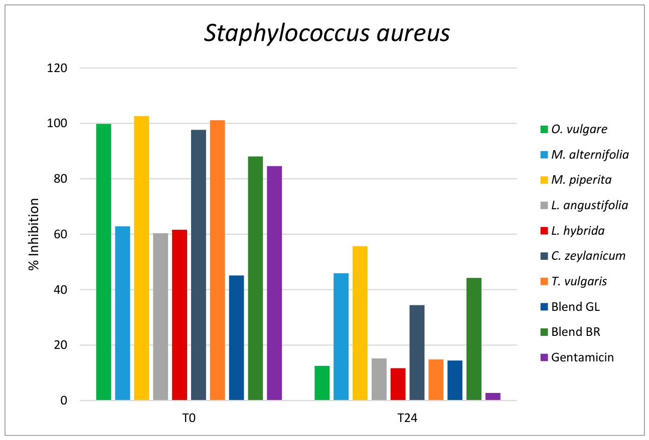
Figure 1. Percentage (%) of biofilm inhibition of seven essential oils and two blends at concentration of 0.39% v/v (CZ at 0.195% v/v) on Sta. aureus. Control with gentamicin at 1mg/mL. T0 = Treatment at 0 h (bacterial adhesion stage), T24 = Treatment after 24 h (pre-formed biofilm stage).
Preprints 230648 g001
Figure 2. Percentage (%) of biofilm inhibition of seven essential oils and two blends at concentration of 0.39% v/v (CZ at 0.195% v/v) on Sta. epidermidis. Control with gentamicin at 1mg/mL. T0 = Treatment at 0 h (bacterial adhesion stage), T24 = Treatment after 24 h (pre-formed biofilm stage).
Preprints 230648 g002
Table 1. MIC and MBC of essential oils and blends against Str. agalactiae. The MIC and MBC concentrations are given as percentage % v/v (μL/mL); in bold, the lower MIC and MBC values.
Streptococcus agalactiae
Essential oil MIC (% v/v) MBC (% v/v)
Origanum vulgare 0.390 >3.125
Melaleuca alternifolia 1.560 >3.125
Melaleuca leucadendrom 1.560 >3.125
Eucalyptus globosum >3.125 >3.125
Mentha piperita 0.780 >3.125
Lavandula angustifolia 0.780 >3.125
Lavandula hybrida 0.780 >3.125
Cinnamommum zeylanicum 0.050 0.098
Rosmarinum officinalis 1.560 >3.125
Citrus limon 3.125 >3.125
Thymus vulgaris 0.390 >3.125
Blend GL 0.780 >3.125
Blend BR 0.390 0.780
Table 2. MIC and MBC of essential oils and blends against Str. dysgalactiae. The MIC and MBC concentrations are given as percentage % v/v (μL/mL); in bold, the lower MIC and MBC values.
Streptococcus dysgalactiae
Essential oil MIC (% v/v) MBC (% v/v)
Origanum vulgare 0.098 0.195
Melaleuca alternifolia 0.780 1.560
Melaleuca leucadendrom 0.780 1.560
Eucalyptus globosum 1.560 3.125
Mentha piperita 0.195 0.780
Lavandula angustifolia 0.195 0.390
Lavandula hybrida 0.195 0.390
Cinnamommum zeylanicum 0.098 0.195
Rosmarinum officinalis 0.195 0.780
Citrus limon 3.125 >3.125
Thymus vulgaris 0.195 0.390
Blend GL 0.098 0.195
Blend BR 0.050 0.390
Table 3. MIC and MBC of essential oils and blends against Str. uberis. The MIC and MBC concentrations are given as percentage % v/v (μL/mL); in bold, the lower MIC and MBC values.
Streptococcus uberis
Essential oil MIC (% v/v) MBC (% v/v)
Origanum vulgare 0.195 0.390
Melaleuca alternifolia 0.780 1.560
Melaleuca leucadendrom 3.125 > 3.125
Eucalyptus globosum > 3.125 > 3.125
Mentha piperita 0.195 0.780
Lavandula angustifolia 0.195 0.195
Lavandula hybrida 1.560 3.125
Cinnamommum zeylanicum <0.001 <0.001
Rosmarinum officinalis 3.125 >3.125
Citrus limon >3125 >3.125
Thymus vulgaris 0.195 0.780
Blend GL 0.390 3.125
Blend BR 0.195 0.390
Table 4. MIC and MBC of essential oils and blends against Sta. aureus. The MIC and MBC concentrations are given as percentage % v/v (μL/mL); in bold, the lower MIC and MBC values.
Staphylococcus aureus
Essential oil MIC (% v/v) MBC (% v/v)
Origanum vulgare 0.390 > 3.125
Melaleuca alternifolia >3.125 > 3.125
Melaleuca leucadendrom >3.125 > 3.125
Eucalyptus globosum >3.125 > 3.125
Mentha piperita >3.125 > 3.125
Lavandula angustifolia >3.125 > 3.125
Lavandula hybrida >3.125 > 3.125
Cinnamommum zeylanicum 0.195 0.390
Rosmarinum officinalis >3.125 > 3.125
Citrus limon >3.125 > 3.125
Thymus vulgaris >3.125 > 3.125
Blend GL >3.125 > 3.125
Blend BR 3.125 > 3.125
Table 5. MIC and MBC of essential oils and blends against Sta. epidermidis. The MIC and MBC concentrations are given as percentage % v/v (μL/mL); in bold, the lower MIC and MBC values.
Staphylococcus epidermidis
Essential oil MIC (% v/v) MBC (% v/v)
Origanum vulgare >3.125 >3.125
Melaleuca alternifolia >3.125 >3.125
Melaleuca leucadendrom >3.125 >3.125
Eucalyptus globosum >3.125 >3.125
Mentha piperita >3.125 >3.125
Lavandula angustifolia >3.125 >3.125
Lavandula hybrida >3.125 >3.125
Cinnamommum zeylanicum 0.098 0.195
Rosmarinum officinalis >3.125 >3.125
Citrus limon >3.125 >3.125
Thymus vulgaris >3.125 >3.125
Blend GL >3.125 >3.125
Blend BR >3.125 >3.125
Table 6. Minimum Inhibitory Concentration (MIC) of essential oils and blends against all the tested strains. The MIC concentrations are given as percentage % v/v (μL/mL); in bold, the lower MIC values.
Essential oil Str. agalactiae Str. dysgalactiae Str. uberis Sta. aureus Sta. epidermidis
Origanum vulgare 0.390 0.098 0.195 0.390 >3.125
Melaleuca alternifolia 1.560 0.780 0.780 >3.125 >3.125
Melaleuca leucadendrom 1.560 0.780 3.125 >3.125 >3.125
Eucalyptus globosum >3.125 1.560 > 3.125 >3.125 >3.125
Mentha piperita 0.780 0.195 0.195 >3.125 >3.125
Lavandula angustifolia 0.780 0.195 0.195 >3.125 >3.125
Lavandula hybrida 0.780 0.195 1.560 >3.125 >3.125
Cinnamommum zeylanicum 0.050 0.098 <0.001 0.195 0.098
Rosmarinum officinalis 1.560 0.195 3.125 >3.125 >3.125
Citrus limon 3.125 3.125 >3125 >3.125 >3.125
Thymus vulgaris 0.390 0.195 0.195 >3.125 >3.125
Blend GL 0.780 0.098 0.390 >3.125 >3.125
Blend BR 0.390 0.050 0.195 3.125 >3.125
Disclaimer/Publisher’s Note: The statements, opinions and data contained in all publications are solely those of the individual author(s) and contributor(s) and not of MDPI and/or the editor(s). MDPI and/or the editor(s) disclaim responsibility for any injury to people or property resulting from any ideas, methods, instructions or products referred to in the content.
Copyright: This open access article is published under a Creative Commons CC BY 4.0 license, which permit the free download, distribution, and reuse, provided that the author and preprint are cited in any reuse.