

Submitted:
28 August 2026
Posted:
31 August 2026
You are already at the latest version
Abstract
Optical coherence tomography angiography (OCTA) has evolved from a novel method for non-invasive vascular visualization into an increasingly quantitative component of multimodal imaging in uveitis. Its distinctive value lies not in replacing fluorescein angiography (FA) or indocyanine green angiography (ICGA), but in providing depth-resolved visualization of the retinal and choroidal microvasculature without obscuration by dye leakage, while enabling quantitative assessment of vascular alterations. This review examines the current role of OCTA in uveitis, with particular emphasis on retinal vasculitis, macular microvascular injury, choriocapillaris involvement, inflammatory choroidal neovascularization, widefield imaging, and quantitative vascular biomarkers. Particular attention is given to the interpretation of OCTA abnormalities in the context of active inflammation, potentially reversible flow impairment, and irreversible vascular damage, as these processes may produce overlapping OCTA appearances yet have different clinical and prognostic implications. We also address acquisition, projection and segmentation artifacts; cross-device and algorithmic variability; the importance of multimodal interpretation; and emerging applications of automated image analysis and artificial intelligence. The most promising future role of OCTA may lie in longitudinal monitoring rather than stand-alone diagnosis, providing reproducible quantitative biomarkers of microvascular injury and treatment response. Realization of this potential will require prospective multicentre validation, standardized acquisition and reporting protocols, robust inter-device harmonization, and clinically meaningful thresholds for longitudinal change.
Keywords:
optical coherence tomography angiography
; OCTA
; uveitis
; retinal vasculitis
; choriocapillaris
; quantitative imaging
; biomarkers
; multimodal imaging
; artificial intelligence
1. Introduction
Modern uveitis imaging no longer serves merely to document findings visible at the slit lamp or through indirect ophthalmoscopy. Multimodal imaging has become an extension of the clinical examination, interrogating tissue architecture, vascular permeability, choroidal inflammatory involvement and microvascular perfusion. Structural optical coherence tomography (OCT) provides reproducible information on macular edema, subretinal fluid, outer-retinal disruption, retinal pigment epithelium (RPE) abnormalities and choroidal morphology. Fluorescein angiography (FA) remains the reference technique for assessing retinal vascular leakage and dynamic retinal vascular filling, whereas indocyanine green angiography (ICGA) remains particularly valuable for assessing choroidal stromal inflammatory involvement in selected posterior uveitic disorders. OCT angiography (OCTA) contributes a fundamentally different signal: depth-resolved maps of detectable blood flow generated without intravenous dye [1,2,3].
The first applications of OCTA in uveitis were necessarily descriptive. They demonstrated capillary rarefaction and non-detectable flow, foveal avascular zone (FAZ) abnormalities, deep capillary plexus (DCP) alterations, choriocapillaris flow deficits and inflammatory choroidal neovascularization (CNV) [1,4,5]. These observations established both the strengths and limitations of the technology. The absence of dye leakage allows capillary architecture to remain visible when FA is obscured by hyperfluorescence, but OCTA cannot directly demonstrate vascular permeability, one of the cardinal manifestations of active retinal vasculitis. OCTA and dye angiography therefore interrogate complementary biological features rather than competing to answer the same question [6,7,8].
The field has now moved beyond simple pattern recognition. Vessel density (VD), perfusion-related metrics, skeletonized vessel density, fractal dimension, FAZ metrics, measures of non-perfusion or flow-signal reduction, and choriocapillaris flow-deficit measurements can be quantified and followed longitudinally. The attraction of these measurements in uveitis is substantial because conventional activity assessment remains heterogeneous, partly subjective and strongly disease-dependent. The central question is therefore no longer whether OCTA can visualize inflammatory vascular alterations, but whether its measurements can provide reproducible information that helps differentiate potentially reversible vascular dysfunction from irreversible vascular damage, predicts clinically meaningful outcomes, and adds value to established multimodal imaging. Consensus-based reporting recommendations represent an important step toward that goal [9].
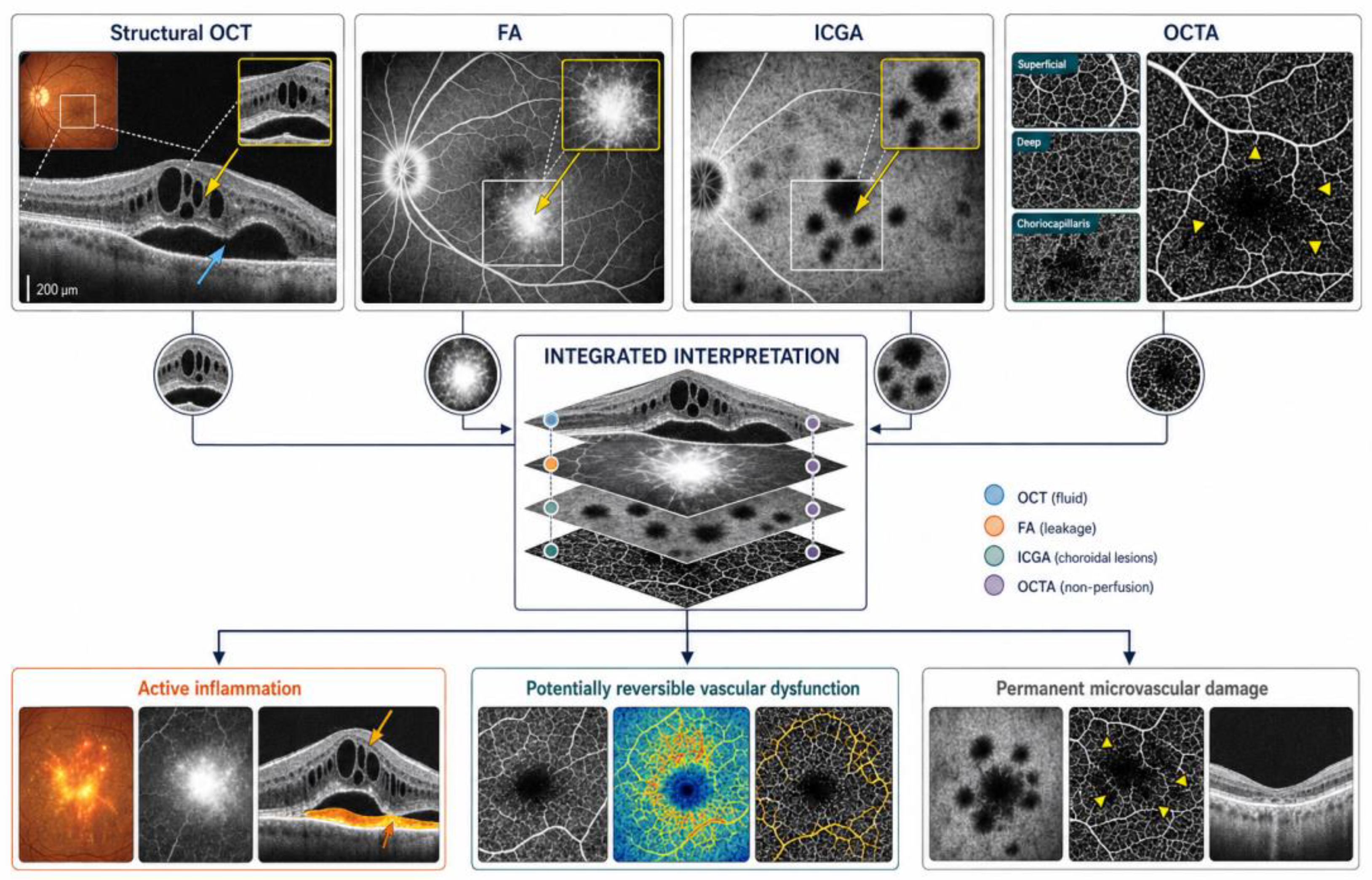
Figure 1.
From vascular imaging to candidate quantitative biomarkers. Conceptual evolution of OCTA in uveitis from qualitative visualization of retinal and choroidal vascular architecture to quantitative assessment of vascular density, network complexity, peripheral flow impairment and choriocapillaris flow deficits, and ultimately toward longitudinal risk assessment and individualized monitoring.
Figure 1.
From vascular imaging to candidate quantitative biomarkers. Conceptual evolution of OCTA in uveitis from qualitative visualization of retinal and choroidal vascular architecture to quantitative assessment of vascular density, network complexity, peripheral flow impairment and choriocapillaris flow deficits, and ultimately toward longitudinal risk assessment and individualized monitoring.
2. OCTA Principles, Quantification and Artifacts
OCTA detects temporal variation in the OCT signal between repeated B-scans acquired at the same retinal location. Commercial platforms use different motion-contrast algorithms, but the fundamental principle is similar: motion of erythrocytes generates changes in OCT signal that can be distinguished from relatively static tissue and reconstructed into a three-dimensional vascular dataset [2,3]. Segmentation of this volume allows separate visualization of the superficial vascular complex, intermediate and deep retinal capillary plexuses, outer retinal avascular slab and choriocapillaris. In inflammatory disease, this depth resolution is particularly valuable because vascular abnormalities may preferentially involve specific retinal or choroidal compartments.
The apparent simplicity of an en face angiogram conceals several acquisition-dependent variables. Small 3 × 3-mm scans provide dense sampling and excellent capillary detail but limited spatial context. Larger 6 × 6-mm and 12 × 12-mm scans increase coverage, generally at the cost of transverse sampling density when acquisition parameters are otherwise comparable. Swept-source systems, particularly those using longer wavelengths, improve penetration through the RPE and facilitate high-speed, larger-field acquisition, while montage techniques extend coverage further toward the retinal periphery. These advances are clinically important in uveitis because inflammatory vascular abnormalities may extend well beyond the posterior pole [10,11]. In accordance with published consensus terminology, OCTA images covering more than 70° may be described as widefield OCTA [9].
Quantitative terminology requires particular precision because definitions vary between devices and analytic pipelines. Vessel density may refer to the proportion of an analyzed area occupied by vascular signal or, when vessels are skeletonized, to vessel length per unit area. Perfusion density or vessel-area density generally reflects the proportion of the analyzed region occupied by perfused vasculature and is therefore influenced by vessel caliber. Skeletonized vessel-density measurements reduce the influence of vessel width and emphasize vascular length, while fractal dimension characterizes vascular branching complexity. FAZ area, perimeter and circularity describe aspects of perifoveal capillary geometry. Because terminology and implementation are not fully standardized across platforms, the exact mathematical definition of each metric should always be reported.
Choriocapillaris analysis requires a different approach because the normal choriocapillaris forms a dense vascular meshwork that is not fully resolved at the level of individual capillaries by current clinical OCTA systems. Quantification therefore commonly relies on flow-deficit number, mean or distribution of flow-deficit size, total flow-deficit area and percentage flow deficit rather than conventional retinal VD [12,13,14,15,16]. Table 1 summarizes the principal technical characteristics of OCTA relevant to inflammatory eye disease, while Table 2 outlines commonly used quantitative metrics.
Artifacts are particularly important in uveitis. Eye motion can create vessel duplication, displacement or discontinuity. Projection from superficial vessels may contaminate deeper slabs despite projection-removal algorithms. Macular edema, subretinal fluid, inflammatory deposits, RPE elevation, outer-retinal disruption and atrophy can distort automated segmentation. Hemorrhage, exudation, pigment and inflammatory material may attenuate signal and simulate reduced or absent flow, whereas RPE loss can increase transmission and alter deeper vascular contrast. OCTA also detects flow only within a velocity-dependent sensitivity range determined by the acquisition protocol and algorithm; flow below the detection threshold may therefore appear as a flow deficit. Consequently, a dark area within a choriocapillaris slab should not automatically be interpreted as anatomical non-perfusion.
For this reason, OCTA should be interpreted from the cross-sectional dataset outward. Structural B-scans and B-scans with flow overlay should be reviewed, segmentation boundaries verified, signal attenuation and shadowing assessed, and adjacent slabs inspected before an en face abnormality or numerical metric is accepted. This discipline is not merely technical. In uveitis, image validity is integral to biological interpretation. Consensus recommendations for OCTA reporting in uveitis appropriately emphasize device characteristics, scan protocol and field of view, segmentation, image-quality criteria, artifact and signal-attenuation assessment, and explicit definition of quantitative parameters [9].
3. Retinal Microvascular Injury in Uveitis
Retinal vasculitis represents one of the most compelling applications of OCTA. FA remains highly sensitive for demonstrating active vascular leakage and staining, but leakage may obscure the underlying capillary architecture. OCTA avoids this confounder and can delineate capillary rarefaction, areas of reduced or non-detectable flow, FAZ irregularity, perifoveal arcade disruption and vascular remodeling [4,5,17]. The clinically useful interpretation is therefore not that OCTA is intrinsically more sensitive than FA for retinal vasculitis, but that it reveals a different component of inflammatory vascular injury.
Behçet uveitis provides one of the best-studied examples. Multiple studies have demonstrated reduced macular VD, abnormalities involving both superficial and deeper retinal vascular plexuses, FAZ enlargement or distortion, and areas of capillary flow impairment [18,19,20,21,22,23]. Projection-resolved analysis has reinforced the observation that the intermediate and deep vascular plexuses may be particularly affected [20]. A systematic review and meta-analysis subsequently demonstrated lower superficial and deep VD and altered FAZ measurements in Behçet disease compared with controls [24]. Importantly, OCTA abnormalities may persist in clinically inactive eyes and have also been reported in some patients without clinically apparent ocular involvement [22,25,26]. Reduced VD therefore cannot be equated automatically with contemporaneous inflammatory activity.
Functional and angiographic correlations are beginning to strengthen the biological relevance of these measurements. OCTA abnormalities have been associated with altered retinal sensitivity on microperimetry, while quantitative OCTA parameters have also shown associations with FA-based measures of retinal inflammatory activity [27,28]. These findings are encouraging but should not be overinterpreted: an OCTA measurement may contain information about both current vascular dysfunction and cumulative damage from previous inflammatory episodes.
A useful conceptual framework is therefore the notion of residual functional microvascular integrity—or “capillary reserve”—after repeated inflammatory insults. This term is proposed here as a conceptual construct rather than an established OCTA biomarker. It helps distinguish two clinically different situations. In one eye, extensive FA leakage may coexist with relatively preserved capillary architecture, suggesting active permeability abnormalities before substantial capillary loss. In another, leakage may be minimal while OCTA demonstrates persistent extensive capillary rarefaction or non-detectable flow, suggesting clinically quiescent inflammation with substantial accumulated vascular damage. Longitudinal multimodal imaging is required to distinguish these states reliably.
Intermediate uveitis provides a complementary model. OCTA studies have demonstrated reduced retinal vascular density and complexity, including in eyes without macular edema, together with alterations in choriocapillaris perfusion [29,30]. Longitudinal evidence further suggests that baseline and longitudinal retinal VD measurements may carry prognostic information regarding subsequent clinical course [31]. If reproduced in larger prospective cohorts, such observations could represent an important transition from descriptive imaging toward risk stratification.
Birdshot chorioretinopathy (BSCR) is also well suited to longitudinal vascular assessment because central visual acuity may remain relatively preserved despite retinal and choroidal inflammatory abnormalities. OCTA studies have described abnormal capillary morphology, including telangiectatic changes, capillary loops and enlarged intercapillary spaces, as well as reduced retinal capillary density [32,33]. More recent multicenter evidence has supported alterations in superficial capillary density and FAZ parameters [34]. These findings support a potential role for OCTA as one component of multimodal longitudinal monitoring rather than as a stand-alone measure of inflammatory activity.
Macular edema complicates interpretation of retinal OCTA measurements. In uveitic cystoid macular edema, DCP density has been reported to be particularly reduced [4]. However, cystoid spaces displace retinal tissue, distort capillary geometry and may alter segmentation and projection, so an apparent DCP abnormality may reflect true vascular impairment, mechanical displacement, artifact, or a combination of these mechanisms. Serial reassessment after macular edema improves may therefore be more biologically informative than an isolated measurement obtained at peak swelling. Table 3 summarizes characteristic OCTA findings across selected inflammatory phenotypes.
4. Choriocapillaris and Choroidal Inflammation
The choriocapillaris is among the vascular compartments in which OCTA has most influenced mechanistic interpretation. ICGA remains fundamental for characterizing many choroidal inflammatory disorders because it depicts the distribution of hypofluorescent lesions and provides information related to choroidal stromal involvement over a broad field. OCTA asks a different question: does a region demonstrate reduced or non-detectable choriocapillaris flow signal, and does that abnormality recover or persist longitudinally?
Placoid inflammatory disorders were among the earliest examples. OCTA and en face OCT studies in acute posterior multifocal placoid pigment epitheliopathy (APMPPE) demonstrated focal inner-choroidal/choriocapillaris flow-signal reduction corresponding to lesions on multimodal imaging, with at least partial recovery of detectable flow during convalescence in some lesions [35,36]. These observations support a vascular or perfusion-related component to the lesion while simultaneously emphasizing the need to distinguish genuine hypoperfusion from signal attenuation produced by outer-retinal and RPE abnormalities. Automated approaches have subsequently enabled quantitative assessment of choriocapillaris lesion area in posterior uveitis [13].
Serpiginous choroiditis and tubercular serpiginous-like choroiditis provide another clinically relevant application. OCTA can demonstrate choriocapillaris flow deficits corresponding to involved lesions, and longitudinal imaging may document evolution at lesion borders without repeated intravenous dye administration [37,38,39]. Nevertheless, OCTA should be interpreted alongside fundus autofluorescence and structural OCT and, when the distribution or activity of choroidal stromal inflammation is the principal question, ICGA.
Vogt–Koyanagi–Harada (VKH) disease represents a different biological model because the primary inflammatory process involves the choroidal stroma, with secondary effects on the choriocapillaris and outer retina. OCTA studies have reported retinal microcirculatory abnormalities and reduced choriocapillaris flow-related measurements during active disease [40,41,42]. These measurements may complement enhanced-depth or swept-source OCT measures of choroidal structure and other multimodal markers, but should not be regarded as direct equivalents of stromal inflammatory activity.
Multiple evanescent white dot syndrome (MEWDS) remains an important cautionary example. Earlier pathogenetic hypotheses included primary choriocapillaris involvement, whereas OCTA studies have frequently demonstrated relatively preserved choriocapillaris flow beneath clinically active lesions, supporting predominant outer-retinal/photoreceptor involvement and illustrating how outer-retinal/RPE alterations may influence OCTA interpretation [1,43]. Quantitative studies have reported subtler retinal or choriocapillaris differences [44], but the central lesson remains: an apparent flow abnormality requires structural and multimodal validation before pathogenic significance is assigned.
Ocular sarcoidosis and tuberculosis-associated posterior uveitis further illustrate the heterogeneity of chorioretinal vascular involvement. OCTA studies have reported retinal and choroidal microvascular abnormalities, including changes involving the DCP and choriocapillaris [45,46]. Granulomatous lesions may, however, cause tissue displacement, vascular compression, shadowing and segmentation abnormalities, reinforcing the importance of structural co-registration. Across inflammatory choroidal disorders, the most clinically meaningful future biomarkers are likely to combine quantitative flow-deficit analysis with anatomical context and longitudinal behavior rather than relying on a single static threshold.
5. Inflammatory Choroidal Neovascularization
Inflammatory CNV is a vision-threatening complication of punctate inner choroidopathy, multifocal choroiditis, serpiginous choroiditis and other inflammatory chorioretinal disorders. Distinguishing inflammatory lesions from secondary neovascularization may be difficult because both can produce subretinal hyperreflective material, fluid and angiographic hyperfluorescence. OCTA offers a practical advantage in this setting: a vascular network can be visualized directly within an appropriately segmented outer-retinal/choriocapillaris-related slab without obscuration by dye leakage [1,6].
The presence of a vascular network, however, establishes neovascular tissue rather than the source of all accompanying exudation and does not determine whether the underlying inflammatory disease is controlled. Structural OCT remains essential for assessing intraretinal or subretinal fluid and lesion morphology, while FA provides information regarding leakage. The strongest interpretation therefore arises from integration of the OCTA vascular network with structural OCT, dye angiography when required, and the inflammatory clinical context.
Evidence supports the use of OCTA for detection and longitudinal monitoring of inflammatory CNV [47]. In punctate inner choroidopathy, OCTA has demonstrated high diagnostic performance compared with dye-based multimodal imaging [48]. Quantitative network descriptors—including lesion area, vessel length, junction or branching density and fractal characteristics—may eventually provide objective measures of lesion evolution or treatment response, but they are not currently validated as stand-alone therapeutic endpoints.
Standardized description of inflammatory CNV morphology may further improve longitudinal assessment and comparability between studies. Consensus-based OCTA recommendations in uveitis support describing neovascular networks as having a “loose” or “dense” vascular pattern according to the relative compactness and organization of the vascular network [9]. These descriptors may facilitate reproducible morphological reporting, although their relationship with exudative activity, treatment response and long-term prognosis requires further validation.
In clinical practice, inflammatory CNV requires two parallel assessments: whether the neovascular lesion is present and exudatively active, and whether the underlying inflammatory disease is active. OCTA contributes strongly to identification and morphological monitoring of the neovascular component but cannot answer either activity question reliably in isolation.
6. Widefield OCTA: Beyond the Macula
The original OCTA literature in uveitis was dominated by 3 × 3-mm and 6 × 6-mm macular scans. This represented an important limitation because many inflammatory vascular abnormalities extend into the retinal periphery. Widefield and ultrawide-field dye angiography have demonstrated that peripheral leakage and non-perfusion may provide clinically relevant information regarding disease extent and activity. Contemporary widefield OCTA extends depth-resolved perfusion assessment into the mid-periphery and, with montage techniques, substantially beyond it [10]. For terminology, current uveitis consensus recommendations define widefield OCTA as imaging covering more than 70° [9].
Widefield swept-source OCTA is particularly attractive in occlusive retinal vasculitis. In Behçet uveitis, widefield OCTA has demonstrated peripheral vascular abnormalities and areas of reduced or non-detectable flow that can be compared with FA findings [49]. More recent approaches have extended widefield swept-source OCTA assessment in retinal vasculitis and combined en face vascular information with cross-sectional OCT/OCTA assessment of perivascular structural abnormalities [50,51]. Conceptually, an OCTA-derived measure of flow-signal impairment and an FA-derived leakage measure interrogate different dimensions of inflammatory vascular disease: the former reflects detectable perfusion, whereas the latter reflects vascular permeability.
The technical expansion of field of view should not be interpreted as equivalence to ultrawide-field FA. Comparative studies in non-uveitic retinal vascular disease have demonstrated strong associations between widefield OCTA and UWF-FA measures of non-perfusion while also identifying systematic differences related to peripheral coverage, flow detection and lesion definition [11]. Extrapolation of these findings to uveitis should therefore be explicitly recognized as indirect rather than disease-specific evidence.
The likely clinical future is therefore complementary. Widefield OCTA may facilitate frequent non-invasive longitudinal assessment of peripheral vascular architecture and perfusion, while FA remains necessary when the principal question concerns vascular leakage, dynamic filling, far-peripheral disease or unresolved diagnostic uncertainty.
7. Integrating OCTA into Multimodal Imaging
The emergence of OCTA has substantially expanded the imaging armamentarium available for posterior uveitis. Nevertheless, its greatest clinical value is realized when it is integrated into a multimodal imaging strategy rather than interpreted in isolation. Each imaging modality interrogates overlapping but distinct components of inflammatory disease, and understanding their complementary roles is essential for diagnosis, activity assessment, therapeutic monitoring and prognostication [6,7,8,52,53].
Structural OCT revolutionized the assessment of inflammatory macular and chorioretinal disease by allowing non-invasive, high-resolution cross-sectional visualization of tissue morphology. Detection of cystoid macular edema, outer-retinal and ellipsoid-zone disruption, epiretinal membranes, vitreomacular interface abnormalities, serous retinal detachment, RPE abnormalities and choroidal structural change has become routine. Structural OCT, however, provides limited direct information regarding perfusion.
FA remains indispensable for evaluating retinal vascular leakage, optic-disc leakage or staining, macular leakage, vascular filling abnormalities, capillary non-perfusion and peripheral retinal vasculitis. Dynamic visualization of retinal vascular filling and permeability represents its principal advantage. Dye leakage, however, may obscure the underlying microvascular architecture. OCTA contributes precisely where FA is limited by depicting depth-resolved vascular morphology without leakage, thereby exposing capillary rarefaction, remodeling, collateralization and areas of reduced or non-detectable flow.
ICGA complements these modalities through assessment of the choroidal circulation and inflammatory involvement of the choroidal stroma. In disorders such as VKH, birdshot chorioretinopathy and selected inflammatory choroiditides, ICGA may identify abnormalities that are incompletely represented by retinal angiography or structural OCT. OCTA should therefore be regarded as complementary rather than competitive with conventional angiography.
Increasingly, the clinically relevant question is not which modality is “best,” but what biological information each contributes. Structural OCT principally depicts tissue architecture and exudative consequences; FA demonstrates retinal vascular filling and permeability; ICGA provides information on the choroidal circulation and stromal inflammatory involvement; and OCTA depicts depth-resolved vascular architecture and detectable flow. Together, these datasets provide a multidimensional representation of inflammatory disease that exceeds the capability of any individual technique. Table 4 summarizes these complementary signals.
This framework may also help distinguish inflammatory activity from accumulated vascular damage. In an eye with active retinal vasculitis, FA may demonstrate extensive leakage while OCTA shows relatively preserved capillary architecture. In another eye, FA leakage may have resolved while OCTA demonstrates persistent extensive capillary rarefaction. The first pattern is more consistent with active vascular permeability; the second suggests accumulated vascular injury in the absence of substantial current leakage. These patterns are illustrative rather than absolute, and clinical and longitudinal correlation remains essential. Similarly, in inflammatory choroidal disease, ICGA may delineate the territory of choroidal involvement, OCTA may determine whether detectable choriocapillaris flow is reduced, and structural OCT demonstrates associated outer-retinal and RPE consequences.
A practical imaging strategy should therefore be question-driven. During initial evaluation, broad characterization may require structural OCT, FA, ICGA, fundus autofluorescence and OCTA, selected according to the suspected phenotype rather than applied universally. During follow-up, imaging can be individualized according to disease phenotype, previous abnormalities and therapeutic objectives. OCTA is particularly useful when the clinical question concerns stability, deterioration or recovery of vascular architecture or detectable perfusion. Conversely, a normal OCTA study should never be interpreted as evidence that inflammatory activity is absent when FA, ICGA, structural imaging or clinical findings demonstrate otherwise. Table 5 provides a practical framework for interpreting longitudinal OCTA change.
Small clinical series have further illustrated how OCTA can add lesion-specific vascular information across non-infectious uveitic phenotypes when interpreted alongside conventional imaging, reinforcing its role as an adjunct rather than an isolated diagnostic test [54].
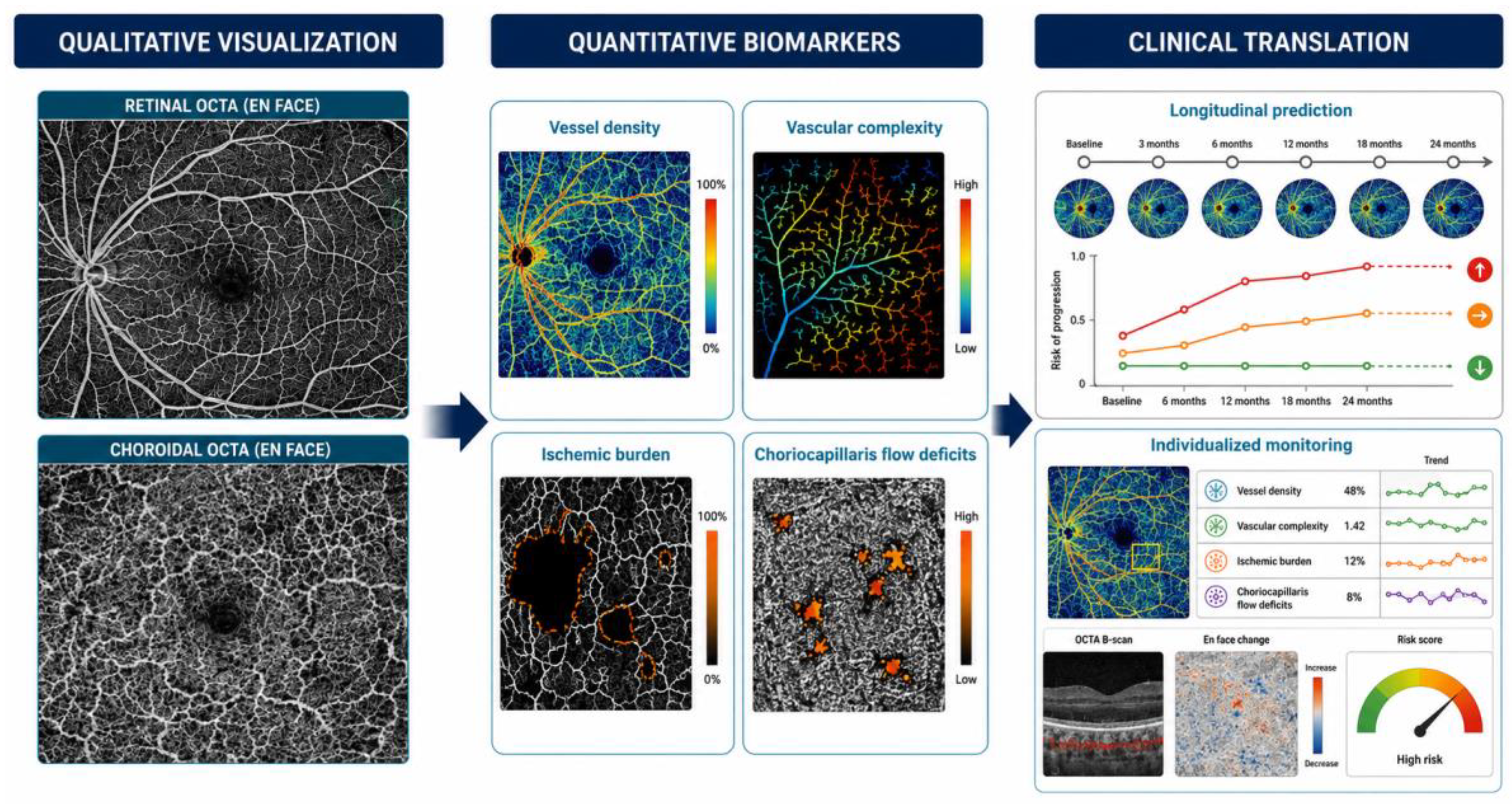
Figure 2.
Multimodal framework for separating inflammatory activity from vascular damage. Structural OCT depicts tissue architecture and exudative consequences; FA demonstrates retinal vascular filling and permeability; ICGA characterizes choroidal vascular and stromal inflammatory abnormalities; and OCTA depicts depth-resolved vascular architecture and detectable flow. Concordance and discordance among these signals, interpreted longitudinally, may help differentiate active inflammation, potentially reversible vascular dysfunction and accumulated microvascular damage.
Figure 2.
Multimodal framework for separating inflammatory activity from vascular damage. Structural OCT depicts tissue architecture and exudative consequences; FA demonstrates retinal vascular filling and permeability; ICGA characterizes choroidal vascular and stromal inflammatory abnormalities; and OCTA depicts depth-resolved vascular architecture and detectable flow. Concordance and discordance among these signals, interpreted longitudinally, may help differentiate active inflammation, potentially reversible vascular dysfunction and accumulated microvascular damage.
8. From Quantitative Measurements to Biomarkers
The proliferation of quantitative OCTA parameters has outpaced their clinical validation. A statistically significant difference between a uveitis cohort and healthy controls does not by itself establish a clinically useful biomarker, and even a validated biomarker does not automatically qualify as a surrogate or treatment endpoint. Clinical translation requires analytical validity and repeatability, biological interpretability, responsiveness to meaningful change, association with relevant clinical or functional outcomes, and demonstration of incremental clinical value.
For conceptual purposes, OCTA-derived abnormalities in uveitis may be considered in three overlapping categories. The first comprises activity-associated vascular dysfunction: potentially reversible reductions in detectable flow occurring in association with active inflammation. The second comprises damage-associated abnormalities: persistent capillary rarefaction, FAZ distortion and non-detectable flow after inflammatory activity has subsided. The third comprises candidate prognostic biomarkers: baseline or longitudinal features associated with subsequent clinical course, structural deterioration or functional loss. These categories are conceptual rather than validated classifications and may overlap within an individual measurement. Current evidence is substantially stronger for demonstrating vascular abnormalities and accumulated damage than for using OCTA alone to determine activity or predict outcome, although longitudinal studies in intermediate uveitis provide encouraging proof-of-concept data [31].
This framework explains why reductions in VD should not simply be labeled “active inflammation.” VD is influenced by signal strength, scan size, segmentation, image-processing algorithms, thresholding and the handling of larger vessels. FAZ measurements are intuitive but show substantial physiological and methodological variability. Fractal dimension and skeletonized metrics characterize network complexity or vascular length but remain incompletely standardized. Choriocapillaris flow-deficit analysis is biologically attractive but particularly sensitive to slab definition, signal attenuation, compensation strategies and thresholding methodology [12,13,14,15,16]. Cross-device numerical thresholds are therefore not currently justified.
The next generation of studies should focus less on introducing additional parameters and more on validating a limited set of robust measurements. Within-eye repeatability and reproducibility, minimal detectable change, longitudinal responsiveness, relationships with visual function and inflammatory activity, and incremental value beyond established clinical and angiographic measures should be prespecified. Systematic reviews of retinal vasculitis and quantitative choroidal imaging consistently identify acquisition and analytic heterogeneity as major barriers [14,17]. Consensus reporting recommendations provide an important framework for improving reproducibility, but prospective multicenter validation remains necessary [9].
In current clinical practice and research, quantitative OCTA is most defensible as a longitudinal, preferably same-device and same-protocol measurement interpreted alongside the source images and multimodal clinical context. Its immediate potential is not to establish universal cutoffs but to transform qualitative impressions—such as progressive capillary rarefaction or changing choriocapillaris flow deficits—into measurable longitudinal trajectories.
9. Artificial Intelligence, Radiomics and Precision Medicine
The evolution of OCTA has transformed the technology from predominantly qualitative vascular imaging toward a quantitative platform capable of generating candidate imaging biomarkers. A major future transition may arise not only from improvements in image acquisition but also from automated extraction of clinically meaningful information from increasingly complex imaging datasets.
Current quantitative OCTA interpretation commonly relies on predefined parameters such as VD, perfusion-related measurements, FAZ characteristics and choriocapillaris flow deficits. Although useful, these parameters inevitably reduce the complexity of vascular architecture to a limited number of numerical variables. Artificial intelligence (AI), machine-learning and radiomic approaches could potentially evaluate branching geometry, network topology, regional heterogeneity, vascular orientation, capillary connectivity, texture and spatial relationships simultaneously, including features that may not be reliably appreciated through conventional analysis.
Automated segmentation and image-quality control may represent among the most immediate practical applications. Uveitic macular edema, subretinal fluid, inflammatory deposits, RPE alterations and chorioretinal lesions frequently compromise default segmentation boundaries. Automated identification and correction—or at minimum automated flagging—of segmentation, motion, projection and shadowing artifacts could improve reproducibility and reduce grader dependency. This would be particularly relevant if quantitative OCTA is incorporated into multicenter observational studies or therapeutic trials.
A longer-term opportunity lies in disease phenotyping and prediction. Rather than relying exclusively on diagnostic labels, computational analysis might identify vascular signatures associated with particular mechanisms, activity states or accumulated damage. Integration of OCTA features with structural OCT, FA/ICGA findings, clinical phenotype, systemic disease and treatment history could eventually support models aimed at differentiating reversible vascular dysfunction from permanent remodeling, estimating future disease course, or identifying eyes at risk of progressive vascular or functional loss.
These possibilities remain largely investigational in uveitis. Uveitic disorders are heterogeneous and individually uncommon, making large, carefully phenotyped and externally validated datasets difficult to assemble. Algorithms trained on one OCTA platform may not generalize to another because segmentation, scan geometry, sampling density and signal processing differ. Dataset shift, annotation quality, external validation and explainability will therefore be critical considerations. A model with high internal performance but poor transportability or limited biological interpretability may have little clinical utility.
AI should therefore presently be viewed as a research and decision-support tool rather than a replacement for clinical expertise. Its near-term contribution may be greatest in automated image-quality control, segmentation and reproducible feature extraction. More ambitious applications involving individualized prediction require prospective validation before clinical implementation. Emerging approaches combining widefield en face OCTA with cross-sectional OCT/OCTA information in retinal vasculitis illustrate the increasingly rich vascular-tissue datasets that may ultimately support such analyses [50,51].
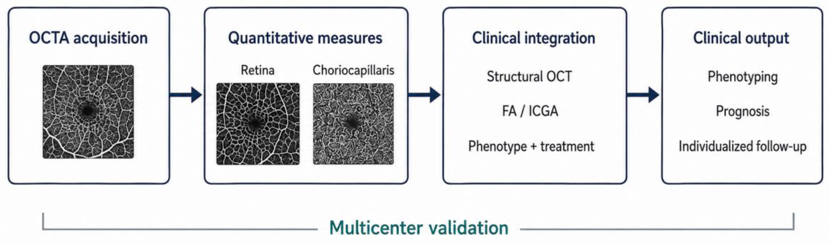
Figure 3.
Precision-imaging framework for uveitis. Standardized OCTA acquisition, segmentation and image-quality control generate retinal and choriocapillaris measurements that can be integrated with structural OCT, FA/ICGA, clinical phenotype and treatment history. Automated analysis and artificial intelligence may ultimately support quantitative phenotyping, prognostication and individualized longitudinal monitoring, contingent on prospective multicenter and cross-device validation.
Figure 3.
Precision-imaging framework for uveitis. Standardized OCTA acquisition, segmentation and image-quality control generate retinal and choriocapillaris measurements that can be integrated with structural OCT, FA/ICGA, clinical phenotype and treatment history. Automated analysis and artificial intelligence may ultimately support quantitative phenotyping, prognostication and individualized longitudinal monitoring, contingent on prospective multicenter and cross-device validation.
10. Perspective: Beyond Blood Flow
The introduction of OCTA represents an important advance in ocular inflammatory imaging. Within a relatively short period, OCTA has progressed from an experimental vascular imaging technique to an increasingly used component of multimodal assessment in posterior uveitis. Its most important potential contribution, however, is not simply the production of angiographic images, but the possibility of converting vascular architecture and detectable flow into reproducible quantitative measurements.
Historically, angiographic evaluation of uveitis has been closely linked to vascular leakage and choroidal inflammatory abnormalities. FA transformed assessment of retinal inflammatory disease by revealing blood-retinal barrier breakdown, retinal vasculitis, optic-disc leakage and capillary non-perfusion. ICGA substantially advanced the understanding and monitoring of selected choroidal inflammatory disorders by demonstrating abnormalities that may be occult on conventional fundus examination or FA. These modalities remain essential because they visualize biological phenomena that OCTA cannot directly demonstrate.
OCTA changes the perspective by focusing attention on depth-resolved vascular architecture and detectable flow. Layer-specific visualization of retinal vascular plexuses and the choriocapillaris has refined understanding of inflammatory vascular injury, while repeated non-invasive acquisition makes longitudinal quantification feasible. The central clinical challenge remains interpretation. Persistent capillary rarefaction or dropout may predominantly reflect accumulated vascular damage; FA leakage primarily reflects abnormal vascular permeability and is frequently associated with activity; and reduced OCTA flow signal may represent reversible hypoperfusion, flow below the detection threshold, signal attenuation, artifact or permanent vascular loss. Distinguishing these possibilities requires structural co-registration and longitudinal multimodal assessment.
The field should now move away from simply multiplying quantitative parameters. Vessel-density measurements, vascular complexity, FAZ characteristics, widefield flow-deficit burden and choriocapillaris flow-deficit measurements are promising candidate biomarkers, but none should currently be considered a universal treatment endpoint. Progress requires standardized acquisition and analysis, rigorous repeatability and reproducibility assessment, prospective demonstration of responsiveness, clinically meaningful thresholds for longitudinal change, and validation across devices, centers and uveitic phenotypes.
Widefield acquisition may progressively reduce the historical macular constraint of OCTA, although technical and peripheral coverage limitations remain. Automated analysis may make complex measurements more practical and reproducible. AI may eventually integrate OCTA with structural imaging and clinical variables to generate individualized risk estimates. Yet the decisive step remains clinical validation: demonstrating that an OCTA-derived measurement improves a clinical decision, predicts a meaningful outcome, or functions as a reliable endpoint in a therapeutic study.
In this context, the future value of OCTA should not be measured solely by faster acquisition or wider fields of view. Its greatest contribution will arise if vascular images can be converted into reproducible information that improves understanding, monitoring and ultimately management of inflammatory eye disease. The evolution of OCTA therefore extends beyond visualization of blood flow—from visualization to quantification, from quantification to biological interpretation, and ultimately toward precision monitoring in uveitis.
Author Contributions
Piergiorgio Neri and Shaikha Aljneibi equally contributed to the manuscript preparation and critical review.
Funding
This research received no external funding.
Institutional Review Board Statement
Not applicable.
Informed Consent Statement
Not applicable.
Data Availability Statement
No new data were created or analyzed in this study.
Conflicts of Interest
The authors declare no conflict of interest.
References
- Pichi, F.; Sarraf, D.; Arepalli, S.; et al. The application of optical coherence tomography angiography in uveitis and inflammatory eye diseases. Prog. Retin Eye Res. 2017, 59, 178–201. [Google Scholar] [CrossRef] [PubMed]
- Kashani, A.H.; Chen, C.L.; Gahm, J.K.; et al. Optical coherence tomography angiography: a comprehensive review of current methods and clinical applications. Prog. Retin Eye Res. 2017, 60, 66–100. [Google Scholar] [CrossRef] [PubMed]
- Spaide, R.F.; Fujimoto, J.G.; Waheed, N.K.; Sadda, S.R.; Staurenghi, G. Optical coherence tomography angiography. Prog. Retin Eye Res. 2018, 64, 1–55. [Google Scholar] [CrossRef] [PubMed]
- Kim, A.Y.; Rodger, D.C.; Shahidzadeh, A.; et al. Quantifying retinal microvascular changes in uveitis using spectral-domain optical coherence tomography angiography. Am. J. Ophthalmol. 2016, 171, 101–112. [Google Scholar] [CrossRef] [PubMed]
- Abucham-Neto, J.Z.; Torricelli, A.A.M.; Lui, A.C.F.; et al. Comparison between optical coherence tomography angiography and fluorescein angiography findings in retinal vasculitis. Int. J. Retin. Vitr. 2018, 4, 15. [Google Scholar] [CrossRef] [PubMed]
- Pichi, F.; Hay, S. Use of optical coherence tomography angiography in the uveitis clinic. Graefes Arch. Clin. Exp. Ophthalmol. 2023, 261, 23–36. [Google Scholar] [CrossRef] [PubMed]
- Invernizzi, A.; Cozzi, M.; Staurenghi, G. Optical coherence tomography and optical coherence tomography angiography in uveitis: a review. Clin. Exp. Ophthalmol. 2019, 47, 357–371. [Google Scholar] [CrossRef] [PubMed]
- Herbort CPJr Takeuchi, M.; Papasavvas, I.; et al. Optical coherence tomography angiography (OCT-A) in uveitis: a literature review and a reassessment of its real role. Diagnostics 2023, 13, 601. [Google Scholar] [CrossRef] [PubMed]
- Pichi, F.; Carreño, E.; Pavesio, C.; et al. Consensus-based recommendations for optical coherence tomography angiography reporting in uveitis. Br. J. Ophthalmol. 2023, 107, 959–965. [Google Scholar] [CrossRef] [PubMed]
- Nissen, A.H.K.; Vergmann, A.S. Clinical utilisation of wide-field optical coherence tomography and angiography: a narrative review. Ophthalmol. Ther. 2024, 13, 903–915. [Google Scholar] [CrossRef] [PubMed]
- Hafner, M.; Herold, T.R.; Deiters, V.; von Livonius, B.; Priglinger, S.G.; Gerhardt, M.J. Quantitative comparison of a novel wide-field OCT-angiography device with ultrawide-field fluorescein angiography in detecting retinal nonperfusion in vascular retinopathies. BMC Ophthalmol. 2025, 25, 642. [Google Scholar] [CrossRef] [PubMed]
- Chu, Z.; Weinstein, J.E.; Wang, R.K.; Pepple, K.L. Quantitative analysis of the choriocapillaris in uveitis using en face swept-source optical coherence tomography angiography. Am. J. Ophthalmol. 2020, 218, 17–27. [Google Scholar] [CrossRef] [PubMed]
- McKay, K.M.; Chu, Z.; Kim, J.B.; et al. Automated quantification of choriocapillaris lesion area in patients with posterior uveitis. Am. J. Ophthalmol. 2021, 231, 179–193. [Google Scholar] [CrossRef] [PubMed]
- Agarwal, A.; Singh, R.B.; Erckens, R.J.; Berendschot, T.T.J.M.; Webers, C.A.B. Quantitative analysis of the choroidal vascularity in eyes with uveitis using optical coherence tomography angiography: a systematic review. Ocul. Immunol. Inflamm. 2023, 31, 1792–1803. [Google Scholar] [CrossRef] [PubMed]
- Chu, Z.; Zhang, Q.; Gregori, G.; Rosenfeld, P.J.; Wang, R.K. Guidelines for imaging the choriocapillaris using OCT angiography. Am. J. Ophthalmol. 2021, 222, 92–101. [Google Scholar] [CrossRef] [PubMed]
- Khan, H.A.; Shahzad, M.A.; Iqbal, F.; et al. A novel method of quantifying the choriocapillaris in normal and post-inflammatory eyes. Ocul. Immunol. Inflamm. 2022, 30, 417–423. [Google Scholar] [CrossRef] [PubMed]
- Dhirachaikulpanich, D.; Chanthongdee, K.; Zheng, Y.; Beare, N.A.V. A systematic review of OCT and OCT angiography in retinal vasculitis. J. Ophthalmic Inflamm. Infect. 2023, 13, 1. [Google Scholar] [CrossRef] [PubMed]
- Khairallah, M.; Abroug, N.; Khochtali, S.; et al. Optical coherence tomography angiography in patients with Behçet uveitis. Retina 2017, 37, 1678–1691. [Google Scholar] [CrossRef] [PubMed]
- Koca, S.; Onan, D.; Kalaycı, D.; Allı, N. Comparison of optical coherence tomography angiography findings in patients with Behçet’s disease and healthy controls. Ocul. Immunol. Inflamm. 2020, 28, 806–813. [Google Scholar] [CrossRef] [PubMed]
- Pei, M.; Zhao, C.; Gao, F.; et al. Analysis of parafoveal microvascular abnormalities in Behçet’s uveitis using projection-resolved optical coherence tomographic angiography. Ocul. Immunol. Inflamm. 2021, 29, 524–529. [Google Scholar] [CrossRef] [PubMed]
- Smid, L.M.; Vermeer, K.A.; Missotten, T.O.A.R.; van Laar, J.A.M.; van Velthoven, M.E.J. Parafoveal microvascular alterations in ocular and non-ocular Behçet’s disease evaluated with optical coherence tomography angiography. Invest Ophthalmol. Vis. Sci. 2021, 62, 8. [Google Scholar] [CrossRef] [PubMed]
- Accorinti, M.; Gilardi, M.; De Geronimo, D.; et al. Optical coherence tomography angiography findings in active and inactive ocular Behçet disease. Ocul. Immunol. Inflamm. 2020, 28, 589–600. [Google Scholar] [CrossRef] [PubMed]
- Cheng, D.; Shen, M.; Zhuang, X.; et al. Inner retinal microvasculature damage correlates with outer retinal disruption during remission in Behçet’s posterior uveitis by optical coherence tomography angiography. Invest Ophthalmol. Vis. Sci. 2018, 59, 1295–1304. [Google Scholar] [CrossRef] [PubMed]
- Ji, K.B.; Hu, Z.; Zhang, Q.L.; Mei, H.F.; Xing, Y.Q. Retinal microvasculature features in patients with Behcet’s disease: a systematic review and meta-analysis. Sci. Rep. 2022, 12, 752. [Google Scholar] [CrossRef] [PubMed]
- Agarwal, A.; Bhatt, S.; Keshari, S.; et al. Retinal microvascular alterations in patients with quiescent posterior and panuveitis using optical coherence tomography angiography. Ocul. Immunol. Inflamm. 2022, 30, 1781–1787. [Google Scholar] [CrossRef] [PubMed]
- Karalezli, A.; Kaderli, S.T.; Sul, S.; Pektas, S.D. Preclinical ocular features in patients with Behçet’s disease detected by optical coherence tomography angiography. Eye (Lond) 2021, 35, 2719–2726. [Google Scholar] [CrossRef] [PubMed]
- Ferreira, B.F.A.; Higashi, A.H.; Prado, L.L.; et al. Optical coherence tomography angiography biomarkers and microperimetry features in Behçet’s uveitis. Retina 2023, 43, 1680–1690. [Google Scholar] [CrossRef] [PubMed]
- Ghanbarnia, M.J.; Ghochani, G.; Khalili Pour, E.; et al. Association of optical coherence tomography angiography biomarkers with fluorescein angiography retinal inflammation scores in Behcet’s retinal vasculitis. Ocul. Immunol. Inflamm. 2025, 33, 1534–1541. [Google Scholar] [CrossRef] [PubMed]
- Wintergerst, M.W.M.; Pfau, M.; Müller, P.L.; et al. Optical coherence tomography angiography in intermediate uveitis. Am. J. Ophthalmol. 2018, 194, 35–45. [Google Scholar] [CrossRef] [PubMed]
- Tian, M.; Tappeiner, C.; Zinkernagel, M.S.; Wolf, S.; Munk, M.R. Swept-source optical coherence tomography angiography reveals vascular changes in intermediate uveitis. Acta Ophthalmol. 2019, 97, e791. [Google Scholar] [CrossRef] [PubMed]
- Wintergerst, M.W.M.; Merten, N.R.; Berger, M.; et al. Vessel density on optical coherence tomography angiography is prognostic for future disease course in intermediate uveitis. Sci. Rep. 2024, 14, 2933. [Google Scholar] [CrossRef] [PubMed]
- de Carlo, T.E.; Bonini Filho, M.A.; Adhi, M.; DukerJS. Retinal and choroidal vasculature in birdshot chorioretinopathy analyzed using spectral domain optical coherence tomography angiography. Retina 2015, 35, 2392–2399. [Google Scholar] [CrossRef] [PubMed]
- Roberts, P.K.; Nesper, P.L.; Goldstein, D.A.; Fawzi, A.A. Retinal capillary density in patients with birdshot chorioretinopathy. Retina 2018, 38, 387–394. [Google Scholar] [CrossRef] [PubMed]
- Pichi, F.; Lembo, A.; Nucci, P.; Neri, P. Optical coherence tomography angiography in birdshot chorioretinopathy. Eur. J. Ophthalmol. 2024, 34, 781–788. [Google Scholar] [CrossRef] [PubMed]
- Burke, T.R.; Chu, C.J.; Salvatore, S.; et al. Application of OCT-angiography to characterise the evolution of chorioretinal lesions in acute posterior multifocal placoid pigment epitheliopathy. Eye (Lond) 2017, 31, 1399–1408. [Google Scholar] [CrossRef] [PubMed]
- Dolz-Marco, R.; Sarraf, D.; Giovinazzo, V.; Freund, K.B. Optical coherence tomography angiography shows inner choroidal ischemia in acute posterior multifocal placoid pigment epitheliopathy. Retin Cases Brief. Rep. 2017, 11 (Suppl 1)–S143. [Google Scholar] [CrossRef] [PubMed]
- Montorio, D.; Giuffrè, C.; Miserocchi, E.; et al. Swept-source optical coherence tomography angiography in serpiginous choroiditis. Br. J. Ophthalmol. 2018, 102, 991–995. [Google Scholar] [CrossRef] [PubMed]
- Shah, A.; Rao, V.G.; Verma, A.; Biswas, J. Evaluation of change in the vascular density of choriocapillaris on optical coherence tomography angiography in eyes with serpiginous choroiditis. Indian J. Ophthalmol. 2020, 68, 1901–1904. [Google Scholar] [CrossRef] [PubMed]
- Agarwal, A.; Aggarwal, K.; Mandadi, S.K.R.; et al. Longitudinal follow-up of tubercular serpiginous-like choroiditis using optical coherence tomography angiography. Retina 2021, 41, 793–803. [Google Scholar] [CrossRef] [PubMed]
- Liang, A.; Zhao, C.; Jia, S.; et al. Retinal microcirculation defects on OCTA correlate with active inflammation and vision in Vogt-Koyanagi-Harada disease. Ocul. Immunol. Inflamm. 2021, 29, 1417–1423. [Google Scholar] [CrossRef] [PubMed]
- Liang, A.; Jia, S.; Gao, F.; et al. Decrease of choriocapillary vascular density measured by optical coherence tomography angiography in Vogt-Koyanagi-Harada disease. Graefes Arch. Clin. Exp. Ophthalmol. 2021, 259, 3395–3404. [Google Scholar] [CrossRef] [PubMed]
- Aggarwal, K.; Agarwal, A.; Mahajan, S.; et al. The role of optical coherence tomography angiography in the diagnosis and management of acute Vogt-Koyanagi-Harada disease. Ocul. Immunol. Inflamm. 2018, 26, 142–153. [Google Scholar] [CrossRef] [PubMed]
- Pichi, F.; Sarraf, D.; Morara, M.; Mazumdar, S.; Neri, P.; Gupta, V. Pearls and pitfalls of optical coherence tomography angiography in the multimodal evaluation of uveitis. J. Ophthalmic Inflamm. Infect. 2017, 7, 20. [Google Scholar] [CrossRef] [PubMed]
- Tang, W.; Guo, J.; Liu, W.; Xu, G. Quantitative analysis of retinal and choriocapillary vascular density of multiple evanescent white dot syndrome by optical coherence tomography angiography. Graefes Arch. Clin. Exp. Ophthalmol. 2020, 258, 1697–1707. [Google Scholar] [CrossRef] [PubMed]
- Hassan, L.M.; Asaad, A.; ElSanabary, Z.; Youssef, M.M. Evaluation of the retinal and choroidal microvasculature changes in cases of sarcoid and tuberculosis-associated posterior uveitis using OCT angiography. Int. Ophthalmol. 2023, 43, 597–608. [Google Scholar] [CrossRef] [PubMed]
- Cerquaglia, A.; Iaccheri, B.; Fiore, T.; et al. New insights on ocular sarcoidosis: an optical coherence tomography angiography study. Ocul. Immunol. Inflamm. 2019, 27, 1057–1066. [Google Scholar] [CrossRef] [PubMed]
- Kongwattananon, W.; Grasic, D.; Lin, H.; et al. Role of optical coherence tomography angiography in detecting and monitoring inflammatory choroidal neovascularization. Retina 2022, 42, 1047–1056. [Google Scholar] [CrossRef] [PubMed]
- Gan, Y.; Zhang, X.; Su, Y.; Shen, M.; Peng, Y.; Wen, F. OCTA versus dye angiography for the diagnosis and evaluation of neovascularisation in punctate inner choroidopathy. Br. J. Ophthalmol. 2022, 106, 547–552. [Google Scholar] [CrossRef] [PubMed]
- Guo, S.; Liu, H.; Gao, Y.; et al. Analysis of vascular changes of fundus in Behçet uveitis by widefield swept source optical coherence tomography angiography and fundus fluorescein angiography. Retina 2023, 43, 841–850. [Google Scholar] [CrossRef] [PubMed]
- Harrigill, M.; Nguyen, M.; Noori, J. Diagnosis and monitoring of retinal vasculitis by widefield swept source OCT angiography. Diagnostics 2025, 15, 3129. [Google Scholar] [CrossRef] [PubMed]
- Mizuno, Y.; Srivastava, S.K.; Singh, S.; et al. Swept-source OCT and OCT angiography B-scan analysis of inflammatory retinal vascular changes: quantitative and qualitative assessment. Ophthalmol. Retin. 2026, 10, 552–561. [Google Scholar] [CrossRef] [PubMed]
- Dingerkus, V.L.S.; Munk, M.R.; Brinkmann, M.P.; et al. Optical coherence tomography angiography (OCTA) as a new diagnostic tool in uveitis. J. Ophthalmic Inflamm. Infect. 2019, 9, 10. [Google Scholar] [CrossRef] [PubMed]
- Tranos, P.; Karasavvidou, E.M.; Gkorou, O.; Pavesio, C. Optical coherence tomography angiography in uveitis. J. Ophthalmic Inflamm. Infect. 2019, 9, 21. [Google Scholar] [CrossRef] [PubMed]
- Melachuri, S.; Dansingani, K.K.; Wesalo, J.; et al. OCT angiography in noninfectious uveitis: a description of five cases and clinical applications. Diagnostics 2023, 13, 1296. [Google Scholar] [CrossRef] [PubMed]

Table 1.
Technical characteristics of OCTA relevant to uveitis.
| Characteristic | Clinical contribution | Principal limitation |
|---|---|---|
| Non-invasive, repeatable acquisition | Enables frequent longitudinal imaging without intravenous dye | Does not directly demonstrate vascular leakage or staining |
| Depth-resolved imaging | Separates superficial and deeper retinal vascular plexuses and choriocapillaris flow signals | Segmentation error is common when inflammatory anatomy is distorted |
| Leakage-free vascular maps | Reveals capillary architecture that may be obscured by hyperfluorescence on FA | Preserved vascular architecture does not exclude active vascular permeability |
| Quantitative analysis | Enables measurement of vascular density, FAZ geometry, network complexity and flow deficits | Measurements depend on device, acquisition protocol, segmentation and post-processing |
| Structural co-registration | Helps determine whether an apparent flow abnormality is anatomically plausible | Requires deliberate review of structural B-scans and flow overlays rather than en face interpretation alone |
| Widefield acquisition | Extends depth-resolved perfusion assessment beyond the macula | Peripheral coverage, sampling density and image quality remain device- and protocol-dependent |
Table 2.
Quantitative OCTA metrics with potential relevance in uveitis.
| Metric | What it describes | Potential clinical relevance | Major confounders |
|---|---|---|---|
| Vessel-area/perfusion density* | Proportion of an analyzed region occupied by detectable vascular flow signal | Overall vascular integrity and extent of flow-signal impairment | Signal strength, thresholding, scan size, segmentation, edema and large-vessel contribution |
| Skeletonized vessel density / vessel-length density* | Vascular length per unit area after reduction of vessels to their centerlines | Capillary rarefaction relatively independent of vessel caliber | Binarization, skeletonization, segmentation and image scale |
| Fractal dimension | Geometric complexity of the vascular branching pattern | Vascular network disorganization or remodeling | Algorithm, thresholding, image scale and region of interest |
| FAZ area, perimeter and circularity | Perifoveal capillary geometry | Macular microvascular injury and remodeling | Physiological variability, axial length/magnification, segmentation and image quality |
| Widefield flow-deficit/non-detectable-flow index* | Proportion of the analyzed retinal field showing predefined reduction or absence of detectable flow signal | Extent of peripheral vascular flow impairment | Field of view, flow-detection threshold, sampling density, montage artifacts and lesion-definition method |
| Choriocapillaris flow-deficit metrics | Number, size, distribution or proportion of areas with reduced/non-detectable choriocapillaris flow signal | Candidate measures of choriocapillaris perfusion abnormality and longitudinal change | Shadowing, signal attenuation, slow flow, slab definition, thresholding and RPE alterations |
| CNV network metrics | Area, vessel length, branching/junction characteristics and complexity of a neovascular network | Detection and morphological monitoring of inflammatory CNV | Segmentation, projection, thresholding, lesion definition and exudative structural changes |
*Terminology and mathematical definitions vary among devices and analysis pipelines and should be explicitly reported in individual studies.
Table 3.
Characteristic OCTA findings across selected inflammatory phenotypes.
| Phenotype | Predominant reported OCTA findings | Clinical interpretation |
|---|---|---|
| Behçet uveitis | Reduced superficial and deep retinal vascular density, deeper-plexus abnormalities, FAZ distortion and areas of reduced/non-detectable capillary flow | Useful for quantifying occlusive microvascular injury; inflammatory activity still requires multimodal correlation |
| Intermediate uveitis | Reduced retinal vascular density and complexity; choriocapillaris abnormalities reported | Candidate longitudinal and prognostic biomarkers; requires further prospective validation |
| Birdshot chorioretinopathy | Telangiectatic changes, capillary loops, enlarged intercapillary spaces and reduced retinal capillary density | Complements FA/ICGA and structural imaging in assessing retinal microvascular involvement and accumulated injury |
| APMPPE / placoid inflammatory disease | Focal choriocapillaris/inner-choroidal flow-signal reduction with partial recovery reported longitudinally | Supports a perfusion-related component but requires exclusion of signal attenuation and other artifacts |
| Serpiginous / tubercular serpiginous-like choroiditis | Choriocapillaris flow deficits corresponding to involved lesions | Useful for longitudinal lesion mapping alongside FAF, structural OCT and, where appropriate, ICGA |
| VKH disease | Retinal microcirculatory abnormalities and reduced choriocapillaris flow-related measurements reported | Adjunct to structural OCT and ICGA; not a stand-alone marker of choroidal stromal inflammatory activity |
| MEWDS | Frequently preserved choriocapillaris flow beneath outer-retinal lesions; subtler quantitative alterations reported | Illustrates the need for structural and multimodal validation of apparent choriocapillaris abnormalities |
| Inflammatory CNV | Vascular network within outer-retinal/choriocapillaris-related slabs; network morphology may be described using standardized “loose” or “dense” vascular patterns [9] | Strong application for CNV detection and longitudinal morphological monitoring; network presence or morphology alone does not establish exudative or inflammatory activity |
Table 4.
Complementary biological information provided by multimodal imaging.
| Modality | Primary signal | Particular strength in uveitis | Key limitation |
|---|---|---|---|
| Structural OCT | Tissue reflectivity, architecture and exudative/structural consequences | Macular edema, subretinal fluid, outer-retinal/RPE injury and choroidal structural changes | Limited direct information on vascular permeability or perfusion |
| FA | Retinal vascular filling and fluorescence dynamics | Retinal vascular leakage, disc leakage, macular leakage, vascular filling abnormalities and capillary non-perfusion | Invasive; leakage may obscure underlying capillary architecture |
| ICGA | Choroidal vascular fluorescence and tissue-dye interactions | Assessment of choroidal circulation and stromal inflammatory involvement, including occult choroidal lesions | Invasive; limited assessment of retinal capillary architecture |
| OCTA | Depth-resolved vascular architecture and detectable flow | Plexus-specific microvascular injury, flow deficits, choriocapillaris abnormalities and CNV visualization | Does not directly demonstrate leakage; susceptible to flow-threshold effects and artifacts |
| Fundus autofluorescence | Distribution of endogenous fluorophores, predominantly related to RPE/outer-retinal integrity | Mapping lesion distribution and evolution and identifying areas of RPE stress or loss | Indirect measure of inflammatory activity and vascular status |
Table 5.
Practical interpretation of longitudinal OCTA change.
| OCTA observation | Possible interpretation | Important alternatives/artifacts to exclude | Practical response |
|---|---|---|---|
| New focal flow deficit or non-detectable flow signal | New vascular flow impairment; potentially related to inflammatory vascular involvement | Shadowing/signal attenuation, segmentation error, displacement of vessels and flow below the detection threshold | Review structural B-scan and flow overlay, inspect adjacent slabs and correlate with multimodal imaging; repeat imaging if uncertain |
| Persistent reduced VD in a clinically quiet eye | Accumulated microvascular injury or persistent vascular remodeling | Device/protocol change, signal-strength difference, segmentation error or image-quality drift | Consider primarily as a candidate damage-associated measure rather than evidence of active inflammation |
| Progressive VD reduction on standardized serial scans | Possible progressive microvascular injury | Acquisition inconsistency, signal-strength change, segmentation and processing differences | Confirm reproducibility and correlate with examination, FA where indicated, structural imaging and treatment history |
| Reduction in choriocapillaris flow-deficit area | Increased detectable flow signal, potentially reflecting recovery of perfusion | Improved signal quality, reduced shadowing, altered segmentation or thresholding | Confirm concordant structural and multimodal evolution before attributing the change to treatment response |
| New outer-retinal vascular network | Development or newly detectable inflammatory CNV | Projection artifact, segmentation error and pre-existing but previously undetected network | Confirm on cross-sectional flow overlay and structural OCT; use FA and/or other multimodal imaging when lesion activity remains uncertain |
| Preserved OCTA architecture despite active FA leakage | Preserved detectable capillary architecture despite active vascular permeability | Limited field of view, inadequate scan quality or failure to sample the involved vessels | Do not use OCTA to exclude active retinal vasculitis |
Disclaimer/Publisher’s Note: The statements, opinions and data contained in all publications are solely those of the individual author(s) and contributor(s) and not of MDPI and/or the editor(s). MDPI and/or the editor(s) disclaim responsibility for any injury to people or property resulting from any ideas, methods, instructions or products referred to in the content. |
© 2026 by the authors. Licensee MDPI, Basel, Switzerland. This article is an open access article distributed under the terms and conditions of the Creative Commons Attribution (CC BY) license (http://creativecommons.org/licenses/by/4.0/).
Copyright: This open access article is published under a Creative Commons CC BY 4.0 license, which permit the free download, distribution, and reuse, provided that the author and preprint are cited in any reuse.