

Submitted:
28 August 2026
Posted:
31 August 2026
You are already at the latest version
Abstract
Background/Objectives: The stratum corneum restricts passive transport of hydrophilic macromolecules, viscous gels, and particulate suspensions. This exploratory study evaluated whether a roller-based platform combining plasma-mediated surface conditioning, electroporation, and iontophoresis could produce imaging changes consistent with needle-free cutaneous delivery of formulations with distinct physicochemical properties. Methods: Three adults with Fitzpatrick skin types III–IV received one model formulation each: autologous plasma rich in growth factors (PRGF), a dual-molecular-weight hyaluronic acid formulation (LongevHA), or calcium hydroxylapatite (CaHA). In the CaHA participant, adjacent dorsal-hand areas received undiluted, 1:1 diluted, and 1:2 diluted formulations. Each 2 cm treatment field received 0.5 mL. High-frequency micro-ultrasound was acquired at baseline, after PlasmaPeel conditioning, immediately after delivery, and, where available, 30 min later using a Vevo 3100 system with an MX400 38 MHz transducer. Results: PlasmaPeel was followed by changes in the superficial epidermal entry echo while continuity was preserved. PRGF produced increased superficial dermal heterogeneity immediately after treatment and a more diffuse low-echogenic pattern at 30 min. LongevHA produced a new, denser dermal/subdermal echotexture after treatment. CaHA appearance varied with dilution: undiluted product produced focal hyperechoic deposits, 1:1 dilution produced broader heterogeneous distribution, and 1:2 dilution produced the most diffuse pattern. No pain or immediate adverse events were recorded. Conclusions: Sequential ultrasound findings were compatible with formulation-dependent needle-free delivery, but they do not chemically identify the delivered material or independently prove transdermal passage. Controlled studies with quantitative imaging and orthogonal molecular validation are required.
Keywords:
transdermal delivery
; electroporation
; iontophoresis
; plasma
; PRGF
; hyaluronic acid
; calcium hydroxylapatite
; high-frequency ultrasound
1. Introduction
The stratum corneum is the principal barrier to cutaneous transport. Its corneocyte–lipid architecture restricts passive movement of hydrophilic, charged, high-molecular-weight, viscous, and particulate materials, motivating physical approaches that transiently modify barrier function.[1,2,3]
Electroporation uses short electrical pulses to create transient aqueous pathways, whereas iontophoresis applies electrical current to support electromigration and electro-osmosis. Because the mechanisms are distinct, sequential or combined application may broaden the range of molecules that can be transported through skin.[2,3,4,5,6] Plasma exposure has also been investigated as a pretreatment that can alter epidermal permeability, although the magnitude and reversibility of the effect depend on plasma source, dose, waveform, and exposure conditions.[7,8,9]
The model formulations were chosen to span a practical physicochemical range rather than to compare their clinical efficacy. PRGF is an autologous, leukocyte-poor and erythrocyte-free platelet preparation. [10] LongevHA (Caromed Italia, Italy) is a dual-molecular-weight hyaluronic acid formulation containing 80 mg HA per 2.5 mL together with mannitol, nicotinamide adenine dinucleotide, resveratrol, and peptides.[11,12] Radiesse is a particulate CaHA suspension whose rheology and ultrasound appearance change with dilution.[13,14]
The primary objective was to assess whether the Intraject platform could generate sequential high-frequency ultrasound findings consistent with needle-free cutaneous delivery across these formulation classes. The secondary objective was to describe how dilution influenced CaHA distribution. Clinical improvement, durability, and therapeutic efficacy were intentionally outside the study scope.
2. Materials and Methods
2.1. Study Design and Participants
This uncontrolled, exploratory in vivo proof-of-concept case series included three adults with Fitzpatrick skin types III–IV. One participant was assigned to each product category. PRGF and LongevHA were assessed in predefined facial skin treatment fields, whereas the CaHA comparison was performed in adjacent dorsal-hand fields. The CaHA sites received undiluted, 1:1 diluted, or 1:2 diluted product. The study included no replicate treatment fields, random allocation, sham controls, or repeated treatment sessions.
Eligibility required adult status, intact skin at the selected site, suitability for the assigned formulation and device, and no aesthetic or energy-based procedure in the target field during the preceding six months. Standard product- and device-specific contraindications were applied. All participants provided written informed consent for treatment and publication of de-identified observations and images.
2.2. Study Formulations

Table 1.
Model formulations and treatment allocation. NAD+, nicotinamide adenine dinucleotide; PRGF, plasma rich in growth factors; CaHA, calcium hydroxylapatite.
Table 1.
Model formulations and treatment allocation. NAD+, nicotinamide adenine dinucleotide; PRGF, plasma rich in growth factors; CaHA, calcium hydroxylapatite.
| Formulation | Treatment field | Preparation | Applied volume |
| PRGF–Endoret | Participant 1; facial skin | Closed-system, leukocyte-poor and erythrocyte-free PRGF; activated with 20 µL CaCl2 per 1 mL PRGF | 0.5 mL |
| LongevHA | Participant 2; facial skin | 80 mg dual-molecular-weight HA per 2.5 mL (40 mg high- and 40 mg low-molecular-weight HA), with mannitol, NAD+, resveratrol, and peptides | 0.5 mL |
| Radiesse CaHA | Participant 3; adjacent dorsal-hand fields | Undiluted; 1:1 (one volume CaHA plus one volume sterile saline); 1:2 (one volume CaHA plus two volumes sterile saline) | 0.5 mL per field |
2.3. Device and Treatment Protocol
A multifunctional Intraject system (Biotec Italia, Dueville, Italy) was used.[17] The roller applicator measured approximately 2 cm in length and diameter and contained 10 rings comprising 60 blunt 0.25 mm microelectrodes. The electrodes contacted the surface but were not intended to mechanically penetrate the epidermis. The same roller acted as the treatment interface during plasma conditioning and the subsequent electrical delivery phase (Figure 1).
Treatment comprised three stages. First, the roller was operated at 75 kHz and 500 V and moved over the 2 cm field in three multidirectional passes for approximately 30 s. Second, 0.5 mL of the assigned formulation was distributed over the field. Third, the same roller was used at device display settings of 20 W and 6.5 V in a monopolar configuration, with a negative return electrode applied to the participant. Multidirectional passes continued for 5–10 min until no visible product remained on the surface.
The device was described operationally as providing electroporation, iontophoretic electromigration/electrophoresis, and electro-osmosis. Current density, delivered charge, impedance, pulse waveform, and skin-surface temperature were not independently recorded; therefore, the displayed settings cannot be directly equated with conventional constant-current iontophoresis.
2.4. High-Frequency Micro-Ultrasound
Images were acquired with the FUJIFILM VisualSonics Vevo 3100 platform and MX400 38 MHz transducer.[16] Imaging was performed at baseline, immediately after PlasmaPeel conditioning and before formulation application, immediately after completion of the delivery protocol, and, where available, 30 min after treatment. Acquisition was confined to the predefined treatment fields.
2.5. Outcome Assessment
The primary exploratory outcome was a new or altered ultrasound pattern within the dermal or superficial subcutaneous compartment after visible surface clearance of the formulation, compared with baseline and post-PlasmaPeel images. Secondary outcomes were the qualitative distribution, homogeneity, and persistence of the post-treatment pattern and the comparative appearance of the three CaHA preparations.
A physician blinded to the formulation labels qualitatively reviewed the de-identified images. The analysis did not include predefined regions of interest, pixel-based echogenicity measurement, penetration-depth calculations, tracer assays, histology, chemical recovery, or inter-rater reliability. Hydration was interpreted cautiously because dermal water may increase low-echogenic pixels, while gain, probe pressure, coupling medium, edema, and acquisition angle can independently alter brightness.[19,20]
2.6. Safety and Statistical Analysis
Pain and visible immediate reactions were assessed during treatment and through the 30 min observation period. No longer-term safety assessment was performed. Because each condition was represented by one participant or one adjacent treatment field without replication, no inferential statistics were performed. Results are descriptive.
3. Results
3.1. Procedure Feasibility and Superficial Epidermal Appearance
All participants completed the assigned sequence. After the roller-mediated delivery phase, no residual formulation was visibly present on the treated surface. Relative to baseline, images acquired after PlasmaPeel demonstrated changes in the thickness, definition, or organization of the superficial parallel echoes while continuity of the epidermal entry echo was preserved. These findings were compatible with treatment-associated modification of the superficial interface but did not demonstrate stratum-corneum ablation.
3.2. LongevHA
At baseline, the superficial interface was relatively regular, and the underlying tissue showed a dispersed fibrillar echotexture. After PlasmaPeel, the entry echo appeared mildly reorganized. Following LongevHA delivery, the dermal and superficial subcutaneous compartments demonstrated a denser, more heterogeneous fibrillar pattern with altered interface definition (Figure 2). No discrete anechoic bolus was identified. The appearance was compatible with diffuse tissue distribution or altered hydration, but ultrasound could not directly identify HA or other molecular constituents.
3.3. PRGF
The PRGF baseline image showed a continuous superficial entry echo and a moderately heterogeneous fibrillar dermal pattern. PlasmaPeel preserved continuity but altered the arrangement of the superficial parallel echoes. Immediately after PRGF delivery, the superficial dermis appeared more heterogeneous, with increased low-echogenic spaces between reflective fibrillar structures. At 30 min, the low-echogenic pattern was more diffuse and less focally irregular (Figure 3). This temporal evolution was compatible with redistribution of water-rich material, but the images did not chemically identify PRGF.
3.4. Calcium Hydroxylapatite
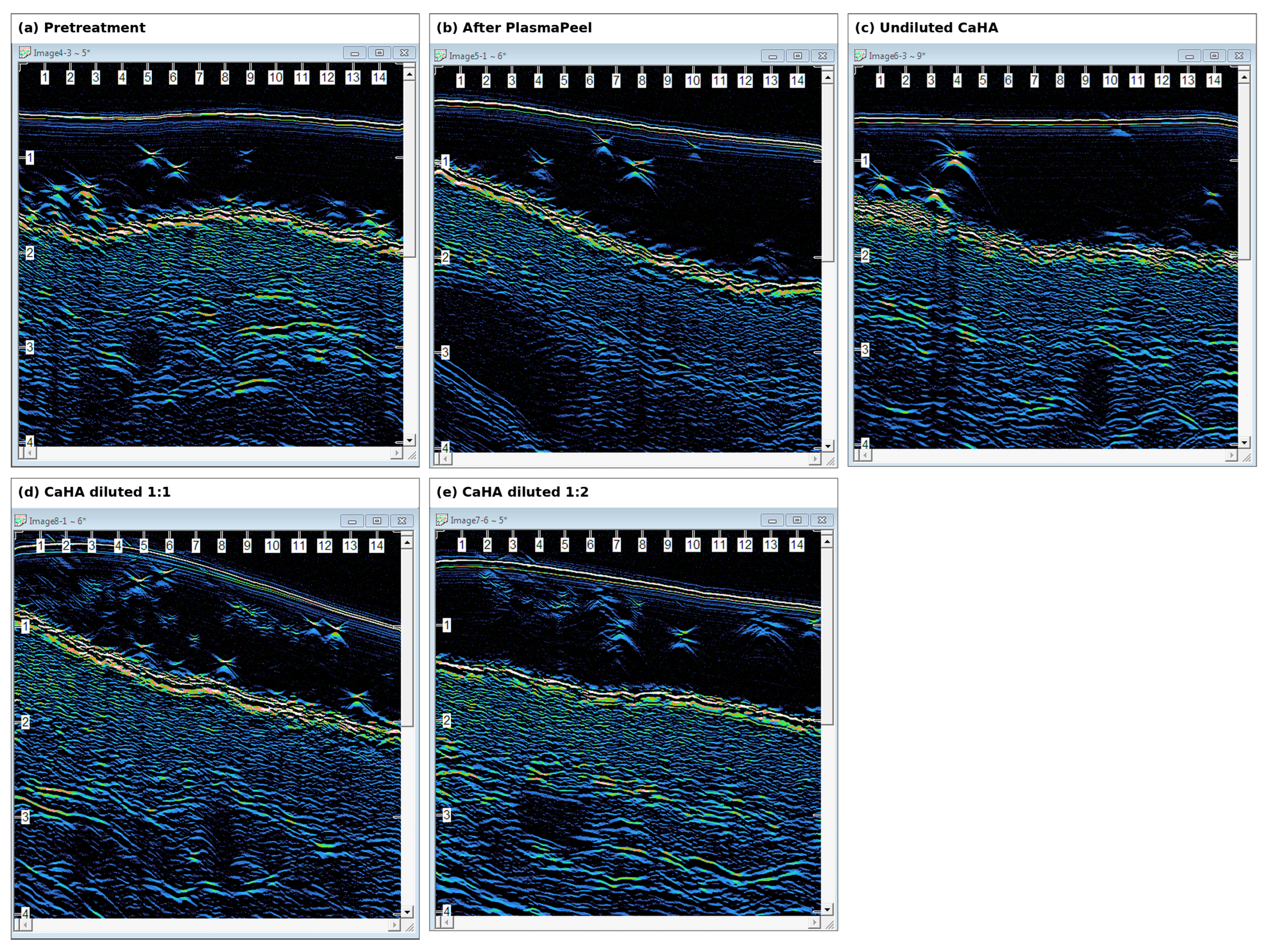
Pretreatment and post-PlasmaPeel images did not show a convincing coarse particulate CaHA pattern. After undiluted CaHA application, discrete intensely hyperechoic foci with localized posterior attenuation were observed, compatible with concentrated superficial deposits but not with broad diffuse distribution. The 1:1 dilution produced multiple hyperechoic foci distributed across a broader portion of the superficial subcutaneous plane, with less consistent shadowing. The 1:2 dilution produced the broadest and least sharply circumscribed change, with more diffuse integration into the surrounding echotexture (Figure 4).
The apparent gradient was therefore one of acoustic distribution rather than simple signal intensity: undiluted CaHA was focal and concentrated; the 1:1 preparation was intermediate; and the 1:2 preparation was the most dispersed. Because natural fibrous septa can also appear hyperechoic, only new patterns relative to pretreatment images were considered compatible with CaHA. The absence of matched imaging planes and quantitative analysis precluded estimates of delivered concentration or volume.
3.5. Comparative Findings and Immediate Safety
Table 2.
Qualitative ultrasound findings. Interpretations describe imaging compatibility and do not establish chemical identity or penetration depth.
Table 2.
Qualitative ultrasound findings. Interpretations describe imaging compatibility and do not establish chemical identity or penetration depth.
| Condition | Superficial interface | Dermal/subcutaneous pattern | Interpretation |
| PlasmaPeel | Continuity preserved; parallel echoes reorganized | No formulation-specific deposit | Treatment-associated surface change; not proof of stratum-corneum ablation |
| LongevHA | Continuous | Denser, heterogeneous fibrillar echotexture | Compatible with diffuse distribution or altered hydration |
| PRGF, immediate | Continuous | Greater heterogeneity and low-echogenic spaces | Compatible with acute distribution of water-rich material |
| PRGF, 30 min | Continuous | More diffuse low-echogenic pattern | Compatible with redistribution/persistent hydration |
| CaHA, undiluted | Continuous | Focal hyperechoic deposits with attenuation | Concentrated, limited distribution |
| CaHA, 1:1 | Continuous | Broader heterogeneous hyperechoic pattern | Intermediate distribution |
| CaHA, 1:2 | Continuous | Broad, diffuse, less circumscribed pattern | Greatest qualitative dispersion |
No participant reported pain during the procedure. No burn, bruising, clinically significant erythema, or other immediate adverse event was observed during the 30 min follow-up.
4. Discussion
4.1. Principal Findings
This exploratory study examined delivery feasibility rather than therapeutic efficacy. Across three model formulation classes, disappearance of visible surface material was followed by new ultrasound patterns in the dermal or superficial subcutaneous compartments. The findings best fit diffuse delivery for PRGF and LongevHA, while CaHA behavior depended on dilution. The results therefore support formulation properties as determinants of transport through a multimodal needle-free system.
The use of plasma peel demonstrated a temporary change in the stratum corneum as a preparatory step for the diffusion of the tested substances mediated by iontophoresis and electroporation. Naturally, even if the diagnostic test used shows the structural change, perhaps a study performed with a device that provides clearer results were needed. It remains evident, however, that after 30 minutes in all treated cases, the superficial dermal area returned to its original state without any residual damage..
4.2. Influence of Formulation Properties
PRGF is predominantly aqueous and produced an immediate increase in superficial low-echogenic spaces followed by a more diffuse pattern at 30 min. LongevHA is more viscous and hydrophilic and produced a denser, heterogeneous tissue pattern without a focal bolus. These differences are consistent with formulation-dependent acoustic behavior, although ultrasound cannot distinguish product from transient edema or other water-related changes.
The CaHA comparison was particularly informative. Undiluted product produced focal, intensely reflective deposits, whereas dilution broadened the distribution pattern and reduced the prominence of discrete deposits and shadowing. This is consistent with known changes in CaHA rheology and sonographic appearance with dilution [13,14]. The 1:2 preparation showed the broadest qualitative dispersion, supporting the hypothesis that reduced viscosity and particulate concentration expand the transportable range of the platform.
4.3. Mechanistic and Imaging Considerations
Electroporation and iontophoresis act through complementary mechanisms, and experimental work has reported increased transport when they are combined.[2,3,4,5,6] Plasma conditioning may provide an additional surface effect.[7,8,9] Nevertheless, the present study did not measure current density, waveform, impedance, temperature, transepidermal water loss, or electrical resistance. It therefore cannot determine each modality’s relative contribution or establish a mechanistic causal pathway.
Micro-ultrasound enabled repeated non-invasive imaging over a short interval but remains a surrogate. CaHA may generate hyperechoic bands or particulate deposits with posterior acoustic shadowing, whereas water-rich changes may increase low-echogenic pixels. [13,14,19] These patterns are not chemically specific. Confirming molecular identity and quantifying transport would require fluorescent or radiolabeled surrogates, Raman or confocal imaging, tissue recovery, or another validated chemical assay.
4.4. Future Studies
A controlled split-field or split-face study should incorporate untreated and sham areas, topical-only application, plasma alone, electroporation–iontophoresis without plasma, randomized field allocation, standardized dose, multiple participants per formulation, fixed imaging settings, co-registered acquisition planes, quantitative low-echogenic-pixel and thickness analysis, transepidermal water loss or impedance measurement, and an independent molecular or tracer-based validation method. Longer follow-up is necessary to evaluate persistence and delayed adverse events.
5. Conclusions
The Intraject multimodal platform produced sequential high-frequency ultrasound changes compatible with needle-free cutaneous delivery of a biologic fluid, a dual-molecular-weight HA formulation, and CaHA preparations with different dilutions. PRGF and LongevHA generated diffuse post-treatment tissue patterns, whereas CaHA distribution increased as the formulation was diluted. PlasmaPeel altered the superficial epidermal entry echo without disrupting continuity. Because ultrasound does not chemically identify the material and the study lacked controls and quantitative validation, the results constitute preliminary feasibility evidence rather than definitive proof of transdermal transport or clinical efficacy.
Author Contributions
Conceptualization, methodology, investigation, data curation, writing—original draft, writing—review and editing, and project administration, E.G. The author has read and agreed to the published version of the manuscript.
Funding
This research received no external funding.
Institutional Review Board Statement
The manuscript reports an exploratory interventional study involving human participants. Written informed consent was obtained.
Informed Consent Statement
Written informed consent for treatment and publication of de-identified study data and images was obtained from all participants.
Data Availability Statement
The de-identified ultrasound images supporting this report are available from the corresponding author upon reasonable request, subject to participant consent and applicable privacy restrictions.
Acknowledgments
The author acknowledges the independent physician who performed the blinded qualitative image review.
Conflicts of Interest
Caromed Italia, Biotec Italia, and Merz had no role in the study design; collection, analysis, or interpretation of data; preparation of the manuscript; or the decision to submit the results for publication. This study received no manufacturer funding.
References
- Prausnitz, M.R.; Langer, R. Transdermal drug delivery. Nat. Biotechnol. 2008, 26, 1261–1268. [Google Scholar] [CrossRef]
- Jadoul, A.; Bouwstra, J.; Préat, V. Effects of iontophoresis and electroporation on the stratum corneum: Review of the biophysical studies. Adv. Drug Deliv. Rev. 1999, 35, 89–105. [Google Scholar] [CrossRef]
- Kalia, Y.N.; Naik, A.; Garrison, J.; Guy, R.H. Iontophoretic drug delivery. Adv. Drug Deliv. Rev. 2004, 56, 619–658. [Google Scholar] [CrossRef]
- Wang, Y.; Thakur, R.; Fan, Q.; Michniak, B. Transdermal iontophoresis: Combination strategies to improve transdermal iontophoretic drug delivery. Eur. J. Pharm. Biopharm. 2005, 60, 179–191. [Google Scholar] [CrossRef]
- Préat, V.; Vanbever, R. Skin electroporation for transdermal and topical delivery. Adv. Drug Deliv. Rev. 1999, 35, 77–88. [Google Scholar] [CrossRef]
- Chang, S.L.; Hofmann, G.A.; Zhang, L.; Deftos, L.J.; Banga, A.K. The effect of electroporation on iontophoretic transdermal delivery of calcium-regulating hormones. J. Control. Release 2000, 66, 127–133. [Google Scholar] [CrossRef]
- Wen, X.; Xin, Y.; Hamblin, M.R.; Jiang, X. Applications of cold atmospheric plasma for transdermal drug delivery: A review. Drug Deliv. Transl. Res. 2021, 11, 741–747. [Google Scholar] [CrossRef]
- Kalghatgi, S.; Tsai, C.; Gray, R.; Pappas, D. Transdermal drug delivery using cold plasmas. In Plasma for Bio-Decontamination, Medicine and Food Security; Springer: Dordrecht, The Netherlands, 2012. [Google Scholar]
- Shimizu, K.; Hayashida, K.; Blajan, M. Novel method to improve transdermal drug delivery by atmospheric microplasma irradiation. Biointerphases 2015, 10, 029517. [Google Scholar] [CrossRef]
- Anitua, E.; Prado, R.; Sánchez, M.; Orive, G. Platelet-rich plasma: Preparation and formulation. Oper. Tech. Orthop. 2012, 22, 25–32. [Google Scholar] [CrossRef]
- Evoskin LLC. LongevHA Regenerative Hyaluronic Acid Powered by Cellular Longevity. Available online: https://evoskin.com/longevha/ (accessed on 25 August 2026).
- Italia, Caromed. LongevHA—Regenerative and Biostimulant Skin Booster. Available online: https://caromeditalia.com/products/regenerative/ (accessed on 25 August 2026).
- Wortsman, X.; Quezada, N.; Peñaloza, O.; Cavallieri, F.; Schelke, L.; Velthuis, P. Ultrasonographic patterns of calcium hydroxyapatite according to dilution and mix with hyaluronic acid. J. Ultrasound Med. 2023, 42, 2065–2072. [Google Scholar] [CrossRef]
- Wortsman, X. Identification and complications of cosmetic fillers: Sonography first. J. Ultrasound Med. 2015, 34, 1163–1172. [Google Scholar] [CrossRef]
- Young, S.R.; Bolton, P.A.; Downie, J. Use of high-frequency ultrasound in the assessment of injectable dermal fillers. Dermatol. Surg. 2008, 34, S188–S196. [Google Scholar]
- FUJIFILM VisualSonics. Vevo 3100 Preclinical Imaging System and MX400 Transducer. Available online: https://www.visualsonics.com/product/imaging-systems/vevo-3100 (accessed on 24 August 2026).
- Biotec Italia. Intraject Medical Platform. Available online: https://www.biotecitalia.com/medical/en/technologies/intraject/ (accessed on 25 August 2026).
- El Gammal, S.; El Gammal, C.; Kaspar, K.; Pieck, C.; Altmeyer, P.; Vogt, M.; Ermert, H. Sonography of the skin at 100 MHz enables in vivo visualization of stratum corneum and viable epidermis in palmar skin and psoriatic plaques. J. Investig. Dermatol. 1999, 113, 821–829. [Google Scholar] [CrossRef]
- Gniadecka, M.; Quistorff, B. Assessment of dermal water by high-frequency ultrasound: Comparative studies with nuclear magnetic resonance. Br. J. Dermatol. 1996. [Google Scholar] [CrossRef] [PubMed]
- Mlosek, R.K.; Malinowska, S.; Sikora, S.; Debowska, R.; Stepien, A.; Czekaj, K.; Dabrowska, A. The use of high-frequency ultrasound imaging in skin moisturization measurement. Skin. Res. Technol. 2013, 19, 169–175. [Google Scholar] [CrossRef] [PubMed]
Figure 1.
Intraject roller applicator used for roller-mediated plasma conditioning and the subsequent electrical delivery phase. The applicator contains multiple rings of blunt microelectrodes.
Figure 1.
Intraject roller applicator used for roller-mediated plasma conditioning and the subsequent electrical delivery phase. The applicator contains multiple rings of blunt microelectrodes.
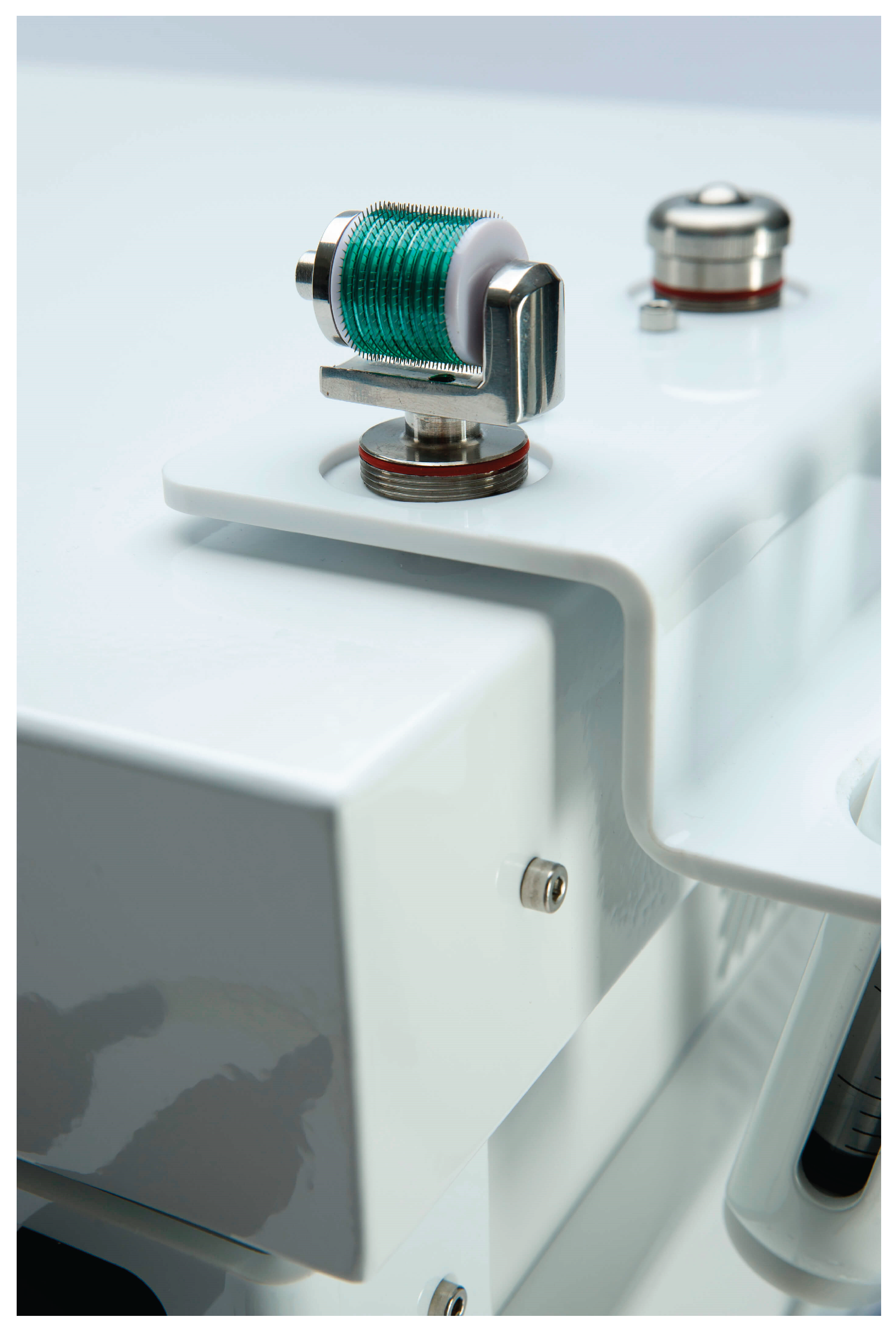
Figure 2.
Representative high-frequency micro-ultrasound sequence for LongevHA: (a) baseline; (b) after PlasmaPeel conditioning; and (c) after completion of roller-mediated product delivery. The post-treatment image shows an altered dermal/superficial subcutaneous echotexture without a circumscribed fluid collection. Images are qualitative and were not spatially co-registered.
Figure 2.
Representative high-frequency micro-ultrasound sequence for LongevHA: (a) baseline; (b) after PlasmaPeel conditioning; and (c) after completion of roller-mediated product delivery. The post-treatment image shows an altered dermal/superficial subcutaneous echotexture without a circumscribed fluid collection. Images are qualitative and were not spatially co-registered.
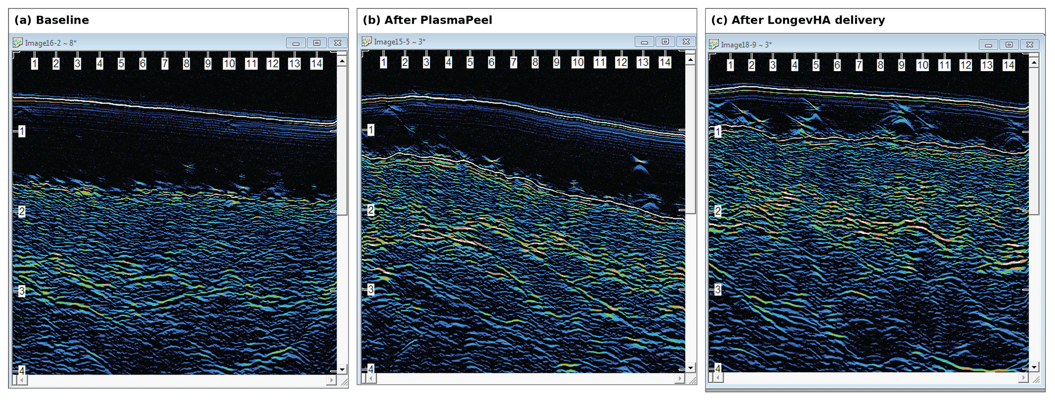
Figure 3.
Representative PRGF sequence: (a) baseline; (b) immediately after PlasmaPeel conditioning; (c) immediately after the complete PRGF delivery protocol; and (d) 30 min after treatment. The immediate image shows increased superficial heterogeneity; the 30 min image shows a more diffuse low-echogenic pattern. Differences in probe angle and tissue compression limit direct quantitative comparison.
Figure 3.
Representative PRGF sequence: (a) baseline; (b) immediately after PlasmaPeel conditioning; (c) immediately after the complete PRGF delivery protocol; and (d) 30 min after treatment. The immediate image shows increased superficial heterogeneity; the 30 min image shows a more diffuse low-echogenic pattern. Differences in probe angle and tissue compression limit direct quantitative comparison.
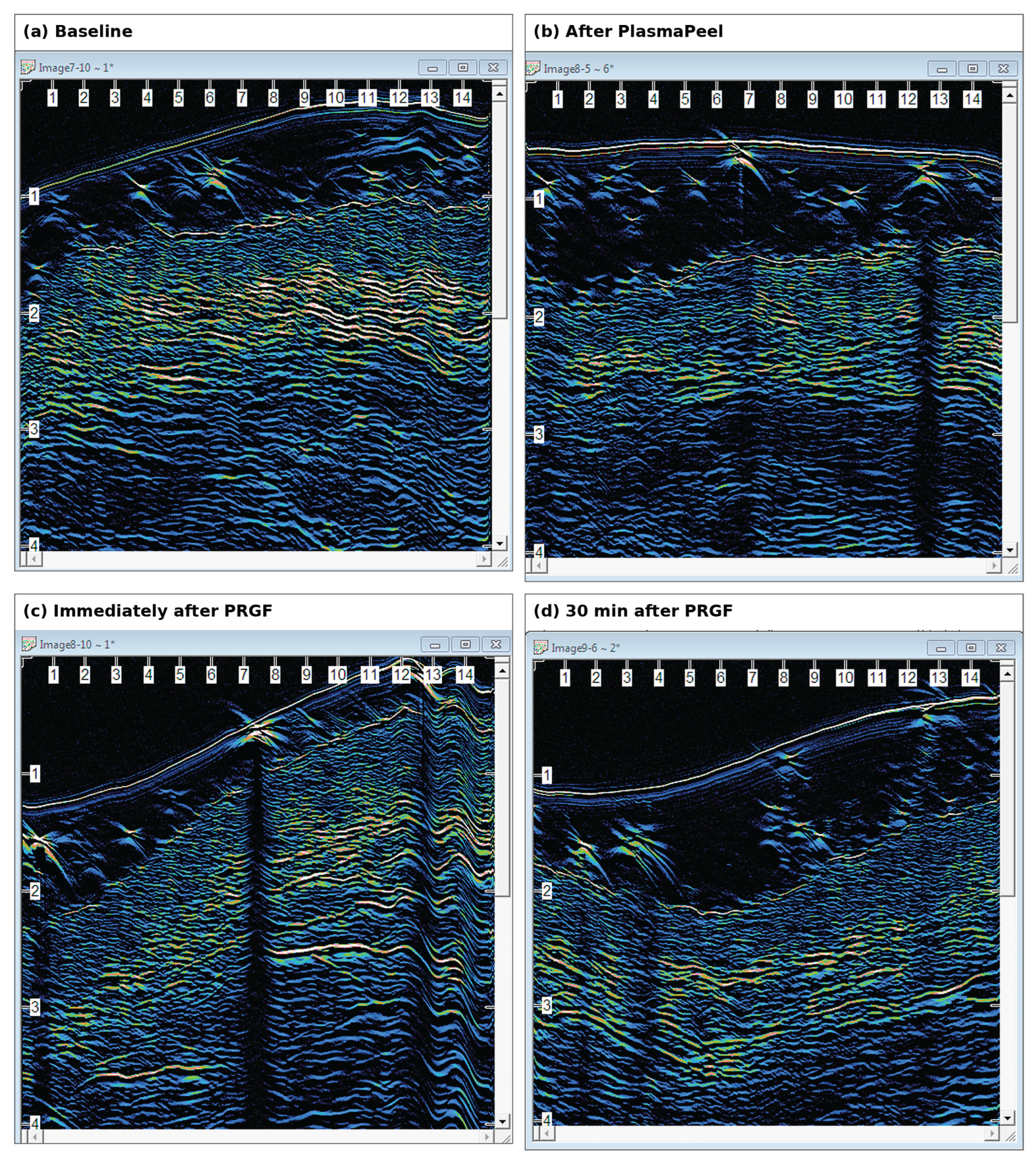
Figure 4.
Representative dorsal-hand CaHA images: (a) pretreatment; (b) after PlasmaPeel; (c) after undiluted CaHA; (d) after CaHA diluted 1:1 with sterile saline; and (e) after CaHA diluted 1:2. Undiluted CaHA shows focal hyperechoic deposits; increasing dilution is associated with a broader and less sharply defined pattern. The panels were acquired at different angles and are not spatially co-registered.
Figure 4.
Representative dorsal-hand CaHA images: (a) pretreatment; (b) after PlasmaPeel; (c) after undiluted CaHA; (d) after CaHA diluted 1:1 with sterile saline; and (e) after CaHA diluted 1:2. Undiluted CaHA shows focal hyperechoic deposits; increasing dilution is associated with a broader and less sharply defined pattern. The panels were acquired at different angles and are not spatially co-registered.
Disclaimer/Publisher’s Note: The statements, opinions and data contained in all publications are solely those of the individual author(s) and contributor(s) and not of MDPI and/or the editor(s). MDPI and/or the editor(s) disclaim responsibility for any injury to people or property resulting from any ideas, methods, instructions or products referred to in the content. |
© 2026 by the authors. Licensee MDPI, Basel, Switzerland. This article is an open access article distributed under the terms and conditions of the Creative Commons Attribution (CC BY) license (http://creativecommons.org/licenses/by/4.0/).
Copyright: This open access article is published under a Creative Commons CC BY 4.0 license, which permit the free download, distribution, and reuse, provided that the author and preprint are cited in any reuse.