
Submitted:
28 August 2026
Posted:
28 August 2026
You are already at the latest version
Abstract
Background/Objectives: Intraoral scanners (IOS) are increasingly integrated into dental curricula, yet evidence on how brief structured training affects novice students remains limited, particularly in resource-constrained settings. This pilot quasi-experimental study evaluated the effect of a structured IOS training program on the self-reported perceptions of undergraduate dental students with no prior IOS experience. Methods: Twenty first- and third-semester students at the School of Dentistry, Universidad de Especialidades Espíritu Santo (Ecuador), completed a 5-item Likert-type questionnaire (1–9 scale) before and after a training program comprising theoretical instruction and typodont-based practical sessions using a TRIOS 3 scanner (3Shape, Copenhagen, Denmark). Paired Student’s t-tests and Wilcoxon signed-rank tests were used to compare pre- and post-training scores, and effect sizes (Cohen’s d) were calculated for each dimension. Results: Significant improvements were observed in all five perception dimensions (all p < 0.001): familiarity (mean difference: +3.45; d = 1.74), comfort (+3.25; d = 1.91), confidence (+3.70; d = 2.06), perceived scan accuracy (+3.30; d = 1.61), and software competence (+3.55; d = 1.59). Conclusions: A brief, structured IOS training program produced large improvements in students’ self-reported perceptions. These findings support the early integration of IOS training into the dental curriculum. Future controlled studies should assess objective performance outcomes and long-term skill retention.
Keywords:
intraoral scanner
; dental education
; digital dentistry
; perception
; training
1. Introduction
In dentistry, the term “digital workflow” is widely used to describe the process of three-dimensional scanning of dental models or the patient’s oral cavity directly using intraoral scanners (IOS) [1,2]. The captured images were processed using dedicated software to accurately record the tooth morphology and surrounding tissues. The scanning accuracy is influenced by multiple factors, including moisture control, scanning distance, software algorithms, and hardware specifications [3]. Digital images are obtained with real-time visual feedback, allowing operators to identify and correct gaps or errors immediately, which is a practical advantage over conventional impression techniques that require complete remaking when defects are detected [4].
Digital and conventional impression techniques have been systematically compared in the literature [5,6,7,8]. Conventional impressions using elastomers or hydrocolloids are susceptible to dimensional variability arising from multi-step processing, manual handling and material distortion [9,10,11]. Although widely taught and practiced, these methods are technique-sensitive and depend heavily on clinician dexterity [12,13]. Intraoral scanners produce digital impressions directly, which removes several potential error sources linked to impression materials and plaster casting [14]. Research has reported comparable or superior marginal fit for restorations fabricated from digital impressions compared to conventional ones, especially for single-unit, tooth-supported restorations [5,8].
The integration of IOS into dental education has gained speed over the past decade; however, implementation remains uneven across institutions and regions [15]. Chatham et al. reported that 45% of UK dental schools had not incorporated digital impression technologies into their curricula, and that among the schools that had, only half ensured practical participation for all students [16]. More recent global survey data indicate that the adoption of digital tools varies by institutional ranking and country income level, with Latin American programmes lagging behind those in higher-income settings in IOS curriculum integration [17,18]. Competencies acquired during undergraduate training are critical for effective IOS use in clinical practice, which makes the design and evaluation of IOS training programmes a relevant priority for dental schools in the region [19].
Prior perceptions of technology play a meaningful role in how students engage with and adopt IOS [20,21]. Students who consider digital tools clinically relevant tend to engage with training more readily and report greater confidence once they have practiced under supervision. By contrast, students accustomed to conventional impression techniques may initially be reluctant to transition to digital workflows, and some may report lower self-efficacy and a longer adjustment period [20]. This variability in initial attitudes makes it important to measure student perceptions both before and after a structured training programme, not only as an outcome measure but also to identify students who may need additional support in the future.
Currently, evidence on the relative effectiveness of different IOS training formats, including lectures, live demonstrations, typodont-based practice, and simulations, on students’ perceived competence remains limited [22]. Several investigators have reported favorable student perceptions after structured IOS curricula, yet most of these studies were conducted in well-resourced institutions with a single scanner model, which limits the generalizability of the findings [20,21,23].
Given this context, the aim of the present pilot study was to evaluate the self-reported perceptions of first- and third-semester undergraduate dental students before and after a structured IOS training program covering five dimensions: familiarity, comfort, confidence, perceived scan accuracy, and software competence. The null hypothesis was that training would not significantly change students’ ratings across these dimensions.
2. Materials and Methods
2.1. Study Design and Participants
This quasi-experimental pilot study enrolled 20 undergraduate dental students at the School of Dentistry, Universidad de Especialidades Espíritu Santo, Samborondón, Ecuador. Students in the first and third semesters were selected because they were expected to have minimal or no prior IOS exposure, thus providing a more homogeneous baseline. Given the pilot nature of the study, a formal a priori sample size calculation was not performed; a target of 20 participants was chosen to obtain preliminary effect-size estimates for future confirmatory trials, consistent with recommendations for pilot feasibility studies [24]. The study protocol was reviewed and approved by the Human Research Ethics Committee of Universidad de Especialidades Espíritu Santo (CEISH UEES) under reference code C-UEES-26-81. All procedures were performed in accordance with the Declaration of Helsinki. Prior to the commencement of the study, written informed consent was obtained from all participants. Participant data were anonymized by replacing identifiers with numerical codes. The inclusion criteria required that the participants had no prior IOS experience, ensuring comparable baseline skill levels. Students with prior IOS experience or motor disabilities that could interfere with the scanning tasks were excluded.
2.2. Training Program
In the pre-training phase, each student performed a baseline scan of a dental typodont (Nissin Dental Products, Japan) mounted on a simulation mannequin head using a TRIOS 3 intraoral scanner (3Shape, Denmark)(Figure 1). The training program comprised two components: (a) a theoretical component consisting of slide presentations and video demonstrations covering the operating principles, clinical indications, limitations, and scanning workflow of the TRIOS 3 system; and (b) a hands-on practical component in which participants performed supervised scans of the typodont using the intraoral scanner and associated software (3Shape, Denmark), following step-by-step scanning procedures and quality-check checklists. Infection control protocols were applied throughout the study, including the use of gloves, caps, and gowns. A scan was considered acceptable if it showed complete arch coverage without significant overlapping artifacts or unfilled voids that the acquisition software could not automatically compensate for.
2.3. Outcome Measures
Perceptions were measured using a purpose-designed 5-item questionnaire (Table 1) administered electronically via Google Forms immediately before and after the training program. Each item was rated on a 9-point Likert-type scale (1 = lowest and 9 = highest). The five dimensions assessed were: (P1) familiarity with IOS use, (P2) comfort with IOS technology, (P3) confidence in performing scans, (P4) perceived accuracy of scans, and (P5) self-assessed competence in using the associated software.
2.4. Statistical Analysis
All statistical analyses were performed using Jamovi (version 2.3; The Jamovi Project, Australia). Categorical variables were summarized using frequencies and percentages. Continuous variables were described using the mean and standard deviation. Normality was assessed using the Shapiro-Wilk test. Paired Student’s t-tests were used to compare pre- and post-training scores. For variables with non-normal distributions (Shapiro-Wilk p < .05), Wilcoxon signed-rank tests were conducted as sensitivity analyses, and the results from both approaches were consistent. Effect sizes were calculated using Cohen’s d for paired samples (d ≥ 0.8 considered large). Statistical significance was set at α = 0.05.
Generative artificial intelligence was not used to generate data, design the study, or analyze or interpret the results. Details of its use during manuscript preparation are provided in the Acknowledgments section.
3. Results
All 20 enrolled students completed both the pre- and post-training questionnaire administrations, yielding a 100% response rate. The sample comprised 12 women (60%) and 8 men (40%), with a mean age of 19.05 years (SD = 2.09; median age = 19 years). Ten students were enrolled in the first semester and 10 in the third semester.
The Shapiro-Wilk test indicated non-normal distributions for four of the ten pre- and post-training variables (Table 2). For these variables, Wilcoxon signed-rank tests were conducted and yielded results consistent with those of the paired t-tests; therefore, parametric results are reported throughout the study.
All five perception dimensions showed statistically significant improvements from pre- to post-training (all p < .001; Table 2). The largest improvement was observed in confidence (P3: mean difference +3.70; t(19) = 6.87; d = 2.06), followed by software competence (P5: +3.55; d = 1.59), familiarity (P1: +3.45; d = 1.74), perceived accuracy (P4: +3.30; d = 1.61), and comfort (P2: +3.25; d = 1.91). Effect sizes were large for all five dimensions (Cohen’s d range: 1.59–2.06).
4. Discussion
Before training, the scores across all five dimensions were low. Familiarity averaged 1.90 on a 9-point scale, which was the lowest baseline value among the five dimensions and reflected the limited contact with IOS that early semester students typically have in settings where this technology is not yet part of the standard curriculum. After a single structured training session, the mean gains ranged from 3.25 to 3.70 points, with Cohen’s d values between 1.59 and 2.06 for all dimensions. A comparable pattern was reported by Schott et al., who enrolled 31 dental students with no prior digital impression experience in a brief teaching module [25]. In that study, 77% of the participants rated IOS handling as satisfactory, and 58% preferred the digital technique over alginate from the operator’s perspective. Although the study design was cross-sectional rather than pre/post, the findings point in the same direction: brief, structured exposure shifts students’ appraisal of IOS toward perceived competence.
The reasons why students prefer one impression technique over another are not clear. Lam et al. surveyed 97 final-year dental students across two cohorts and found that preference was driven primarily by perceived efficiency rather than by prior exposure [21]. Students who felt that scanning worked efficiently in their hands were significantly more likely to favor the digital technique (p = .000). This relationship is worth considering in light of the present study results. Of the five dimensions measured here, confidence showed the largest single gain (+3.70 points). A sense of personal confidence in performing a procedure is closely related to perceiving that procedure as efficient; therefore, the shift recorded here may have broader implications for how students approach IOS in subsequent coursework. However, whether this sense of confidence holds once students move from typodont practice to real clinical contexts requires a different study design to determine.
Studies that measure objective performance alongside student attitudes provide a useful context for interpreting perception data. In the crossover trial by Alfallaj et al. (n = 96 dental undergraduates), digital scanning averaged 293 s against 664 s for a conventional impression, and 76% of participants preferred the digital approach regardless of which technique they had practiced first [26]. Results from Bilir and Ayguzen, obtained in a preclinical typodont setting similar to the one used in the present study, showed a comparable time difference (272 vs. 639 s) and found that 85% of students considered the IOS method easier to handle [14]. Alfallaj et al. also observed that women completed conventional impressions faster, whereas men were quicker at digital scanning, which points to sex-related differences in psychomotor and technology familiarity patterns [26]. Because the current study measured perceptions only, without timing or objective quality data, the question of whether similar gender differences exist in the local student population cannot be addressed from these results and needs to be explored in subsequent work.
A note of caution regarding the relationship between perceptions and actual performance is warranted. Corne et al. randomly assigned 95 second-year dental students to three conditions: conventional silicone keys only, CEREC software-assisted self-assessment, and PrepCheck pedagogic software [27]. Despite having access to digital feedback tools in two of the three conditions, no significant differences in objective preparation quality emerged, and digital feedback alone did not improve how accurately students assessed their own work. The authors concluded that constructive critique requires human reflection and that instructor guidance remains essential for converting digital feedback into skill development. This finding is directly relevant to the interpretation of the perception gains recorded in this study. The questionnaire used in the current study captured what students believed about their abilities, which was not the same as what they could objectively demonstrate. Whether the two converge after additional supervised practice remains an open question, which would require objective assessments, such as digital model superimposition against a reference, to answer properly.
For dental programs in Latin America, where institutional resources shape curriculum design in ways that differ markedly from high-income contexts, the present results carry a practical message. A one-session IOS module that combined slide-based theory with typodont scanning under supervision generated large effect sizes without requiring multi-session or high-cost infrastructure. Introducing this type of module early in training, before students have completed several clinical semesters working with conventional impression materials, may reduce the resistance to technique change that has been reported in the literature [20]. At the same time, the absence of a control group in this study makes it impossible to attribute the observed improvements exclusively to the training. Maturation, the novelty effect of using new equipment, and the Hawthorne effect cannot be ruled out as contributing factors to this result. A randomized design comparing trained and untrained groups with a delayed follow-up measurement is needed to establish causality more firmly.
Several constraints of this study are relevant to the interpretation of the findings. The sample of 20 students from one institution was sufficient for a pilot estimate of effect sizes but did not support generalization to other programmes. Only one IOS model was used; therefore, it was not possible to determine whether the results reflect the properties of digital scanning in general or characteristics specific to the TRIOS 3 device. The study relied entirely on self-reported data, with no objective measures of scan quality, completion time, or number of retakes. The perception questionnaire was developed for this study and had not been formally validated beforehand, which is an acknowledged limitation of this study. In addition, no follow-up measurements were conducted; therefore, the durability of the observed perception gains is unknown.
5. Conclusions
This pilot study shows that a structured IOS training program can produce large improvements in the self-reported perceptions of undergraduate dental students with no prior experience with the technology. Gains were observed across all five dimensions assessed: familiarity, comfort, confidence, perceived scan accuracy, and software competence. The post-training scores converged across dimensions, suggesting that a single well-designed session can substantially close the gap between what novice students know and what IOS use in a pre-clinical setting demands. These findings support the incorporation of IOS training early in the dental curriculum. Future research should use randomized designs with larger samples, include objective performance measures alongside self-reported data, and track students over time to assess their skill retention.
Author Contributions
Conceptualization, P.L.B.S.; methodology, P.L.B.S.; formal analysis, P.L.B.S.; investigation, P.L.B.S., M.P.S.R. and C.P.S.B.; data curation, M.P.S.R. and C.P.S.B.; writing—original draft preparation, P.L.B.S., M.P.S.R. and C.P.S.B.; writing—review and editing, A.L.-F., M.A.B., E.B. and T.O.G.; visualization, P.L.B.S.; supervision, E.B. and T.O.G.; project administration, P.L.B.S. All authors have read and agreed to the published version of the manuscript.
Funding
This research was funded by the Research Center of Universidad de Especialidades Espíritu Santo (UEES), Samborondón, Ecuador.
Institutional Review Board Statement
The study was conducted in accordance with the Declaration of Helsinki and approved by the Human Research Ethics Committee of Universidad de Especialidades Espíritu Santo (CEISH UEES; Ecuadorian Ministry of Health registration DNIVS-CEISH-09-UEES-44) under reference code C-UEES-26-81.
Informed Consent Statement
Informed consent was obtained from all subjects involved in the study.
Data Availability Statement
The raw data supporting the conclusions of this article will be made available by the authors on request.
Acknowledgments
During the preparation of this manuscript, the authors used ChatGPT (GPT-4-turbo, OpenAI) and Claude (Anthropic) for the purposes of English-language editing, structural revision of the Discussion section, verification of reference metadata against PubMed records, and table formatting. The authors have reviewed and edited the output and take full responsibility for the content of this publication.
Conflicts of Interest
Co-author M.A.B. holds the position of Chair of the Human Research Ethics Committee of UEES (CEISH UEES). This is an administrative role that does not involve the scientific review of research protocols, and he had no involvement in the evaluation of the protocol reported here. The authors declare no conflicts of interest. The funder had no role in the design of the study; in the collection, analyses or interpretation of data; in the writing of the manuscript; or in the decision to publish the results.
Abbreviations
The following abbreviations are used in this manuscript:
| IOS | Intraoral scanner |
| SD | Standard deviation |
References
- Suese, K. Progress in Digital Dentistry: The Practical Use of Intraoral Scanners. Dent. Mater. J. 2020, 39, 52–56. [Google Scholar] [CrossRef]
- Róth, I.; Czigola, A.; Fehér, D.; Vitai, V.; Joós-Kovács, G.L.; Hermann, P.; Borbély, J.; Vecsei, B. Digital Intraoral Scanner Devices: A Validation Study Based on Common Evaluation Criteria. BMC Oral Health 2022, 22, 140. [Google Scholar] [CrossRef]
- Ahmed, K.E.; Wang, T.; Li, K.Y.; Luk, W.K.; Burrow, M.F. Performance and Perception of Dental Students Using Three Intraoral CAD/CAM Scanners for Full-Arch Scanning. J. Prosthodont. Res. 2019, 63, 167–172. [Google Scholar] [CrossRef]
- Ferrari, M.; Keeling, A.; Mandelli, F.; Lo Giudice, G.; Garcia-Godoy, F.; Joda, T. The Ability of Marginal Detection Using Different Intraoral Scanning Systems: A Pilot Randomized Controlled Trial. Am. J. Dent. 2018, 31, 272–276. [Google Scholar]
- Seelbach, P.; Brueckel, C.; Wöstmann, B. Accuracy of Digital and Conventional Impression Techniques and Workflow. Clin. Oral Investig. 2013, 17, 1759–1764. [Google Scholar] [CrossRef]
- Joda, T.; Lenherr, P.; Dedem, P.; Kovaltschuk, I.; Bragger, U.; Zitzmann, N.U. Time Efficiency, Difficulty, and Operator’s Preference Comparing Digital and Conventional Implant Impressions: A Randomized Controlled Trial. Clin. Oral Implant. Res. 2017, 28, 1318–1323. [Google Scholar] [CrossRef]
- Abdel-Azim, T.; Rogers, K.; Elathamna, E.; Zandinejad, A.; Metz, M.; Morton, D. Comparison of the Marginal Fit of Lithium Disilicate Crowns Fabricated with CAD/CAM Technology by Using Conventional Impressions and Two Intraoral Digital Scanners. J. Prosthet. Dent. 2015, 114, 554–559. [Google Scholar] [CrossRef]
- Ender, A.; Zimmermann, M.; Attin, T.; Mehl, A. In Vivo Precision of Conventional and Digital Methods for Obtaining Quadrant Dental Impressions. Clin. Oral Investig. 2016, 20, 1495–1504. [Google Scholar] [CrossRef]
- Zitzmann, N.U.; Kovaltschuk, I.; Lenherr, P.; Dedem, P.; Joda, T. Dental Students’ Perceptions of Digital and Conventional Impression Techniques: A Randomized Controlled Trial. J. Dent. Educ. 2017, 81, 1227–1232. [Google Scholar] [CrossRef]
- Ender, A.; Mehl, A. In-Vitro Evaluation of the Accuracy of Conventional and Digital Methods of Obtaining Full-Arch Dental Impressions. Quintessence Int. 2015, 46, 9–17. [Google Scholar] [CrossRef]
- Chochlidakis, K.M.; Papaspyridakos, P.; Geminiani, A.; Chen, C.-J.; Feng, I.J.; Ercoli, C. Digital versus Conventional Impressions for Fixed Prosthodontics: A Systematic Review and Meta-Analysis. J. Prosthet. Dent. 2016, 116, 184–190.e12. [Google Scholar] [CrossRef]
- Wöstmann, B.; Rehmann, P.; Trost, D.; Balkenhol, M. Effect of Different Retraction and Impression Techniques on the Marginal Fit of Crowns. J. Dent. 2008, 36, 508–512. [Google Scholar] [CrossRef]
- Davidowitz, G.; Kotick, P.G. The Use of CAD/CAM in Dentistry. Dent. Clin. North Am. 2011, 55, 559–570, ix. [Google Scholar] [CrossRef]
- Bilir, H.; Ayguzen, C. Comparison of Digital and Conventional Impression Methods by Preclinical Students: Efficiency and Future Expectations. J. Int. Soc. Prev. Community Dent. 2020, 10, 402–409. [Google Scholar] [CrossRef]
- Brownstein, S.A.; Murad, A.; Hunt, R.J. Implementation of New Technologies in U.S. Dental School Curricula. J. Dent. Educ. 2015, 79, 259–264. [Google Scholar] [CrossRef]
- Chatham, C.; Spencer, M.H.; Wood, D.J.; Johnson, A. The Introduction of Digital Dental Technology into BDS Curricula. Br. Dent. J. 2014, 217, 639–642. [Google Scholar] [CrossRef]
- Quinn, B.; Chau, R.C.W.; Felszeghy, S.; Liukkonen, M.; Didilescu, A.C.; Pasqualini, D.; Valenzuela, D.P.; Rice, D.P.; Spagnuolo, G.; Camilleri, J.; et al. Immersive Technologies in Dental Education: Global Adoption Patterns From a 2025 Survey. J. Dent. Educ. 2026. [Google Scholar] [CrossRef]
- Olegário, I.C.; Coffey, N.; Chaurasia, A.; Leung, A. International Perspectives on Digital and Generative AI Adoption and Governance in Undergraduate Dental Education: A Cross-Sectional Survey. Dent. J. 2026, 14, 128. [Google Scholar] [CrossRef]
- Al Hamad, K.Q. Learning Curve of Intraoral Scanning by Prosthodontic Residents. J. Prosthet. Dent. 2020, 123, 277–283. [Google Scholar] [CrossRef]
- Marti, A.M.; Harris, B.T.; Metz, M.J.; Morton, D.; Scarfe, W.C.; Metz, C.J.; Lin, W.-S. Comparison of Digital Scanning and Polyvinyl Siloxane Impression Techniques by Dental Students: Instructional Efficiency and Attitudes towards Technology. Eur. J. Dent. Educ. Off. J. Assoc. Dent. Educ. Eur. 2017, 21, 200–205. [Google Scholar] [CrossRef]
- Lam, W.Y.-H.; Mak, K.C.-K.; Maghami, E.; Molinero-Mourelle, P. Dental Students’ Preference and Perception on Intraoral Scanning and Impression Making. BMC Med. Educ. 2021, 21, 501. [Google Scholar] [CrossRef]
- Cheah, C.; Lim, C.; Ma, S. The Dentist Will Scan You Now: The next Generation of Digital-Savvy Graduates. Eur. J. Dent. Educ. Off. J. Assoc. Dent. Educ. Eur. 2021, 25, 232–237. [Google Scholar] [CrossRef]
- Gavounelis, N.A.; Gogola, C.-M.C.; Halazonetis, D.J. The Effect of Scanning Strategy on Intraoral Scanner’s Accuracy. Dent. J. 2022, 10, 123. [Google Scholar] [CrossRef]
- Whitehead, A.L.; Julious, S.A.; Cooper, C.L.; Campbell, M.J. Estimating the Sample Size for a Pilot Randomised Trial to Minimise the Overall Trial Sample Size for the External Pilot and Main Trial for a Continuous Outcome Variable. Stat. Methods Med. Res. 2016, 25, 1057–1073. [Google Scholar] [CrossRef]
- Schott, T.C.; Arsalan, R.; Weimer, K. Students’ Perspectives on the Use of Digital versus Conventional Dental Impression Techniques in Orthodontics. BMC Med. Educ. 2019, 19, 81. [Google Scholar] [CrossRef]
- Alfallaj, H.A.; Alsaloum, M.A.; Altuwaijri, S.H.; Aldibasi, O.S.; Alkadi, L.T. Procedure Time and Students’ Perception Comparing Full Arch Digital Scans with Conventional Impressions: A Cross-Over Randomized Experimental Trial. Int. J. Dent. 2022, 2022, 6320251. [Google Scholar] [CrossRef]
- Corne, P.; Joseph, D.; Hirtz, P.; Durand, J.-C.; Fages, M.; Vincent, M.; Balthazard, R. Study of the Use in Autonomy of Intraoral Scanning Systems to Optimise Dental Students’ Performances in Fixed Prosthodontics Practical Learning in Second Year. Eur. J. Dent. Educ. Off. J. Assoc. Dent. Educ. Eur. 2023, 27, 888–897. [Google Scholar] [CrossRef]
Figure 1.
Baseline intraoral scanning on a mannequin-mounted typodont using the TRIOS 3 intraoral scanner (3Shape) during the pre-training phase.
Figure 1.
Baseline intraoral scanning on a mannequin-mounted typodont using the TRIOS 3 intraoral scanner (3Shape) during the pre-training phase.
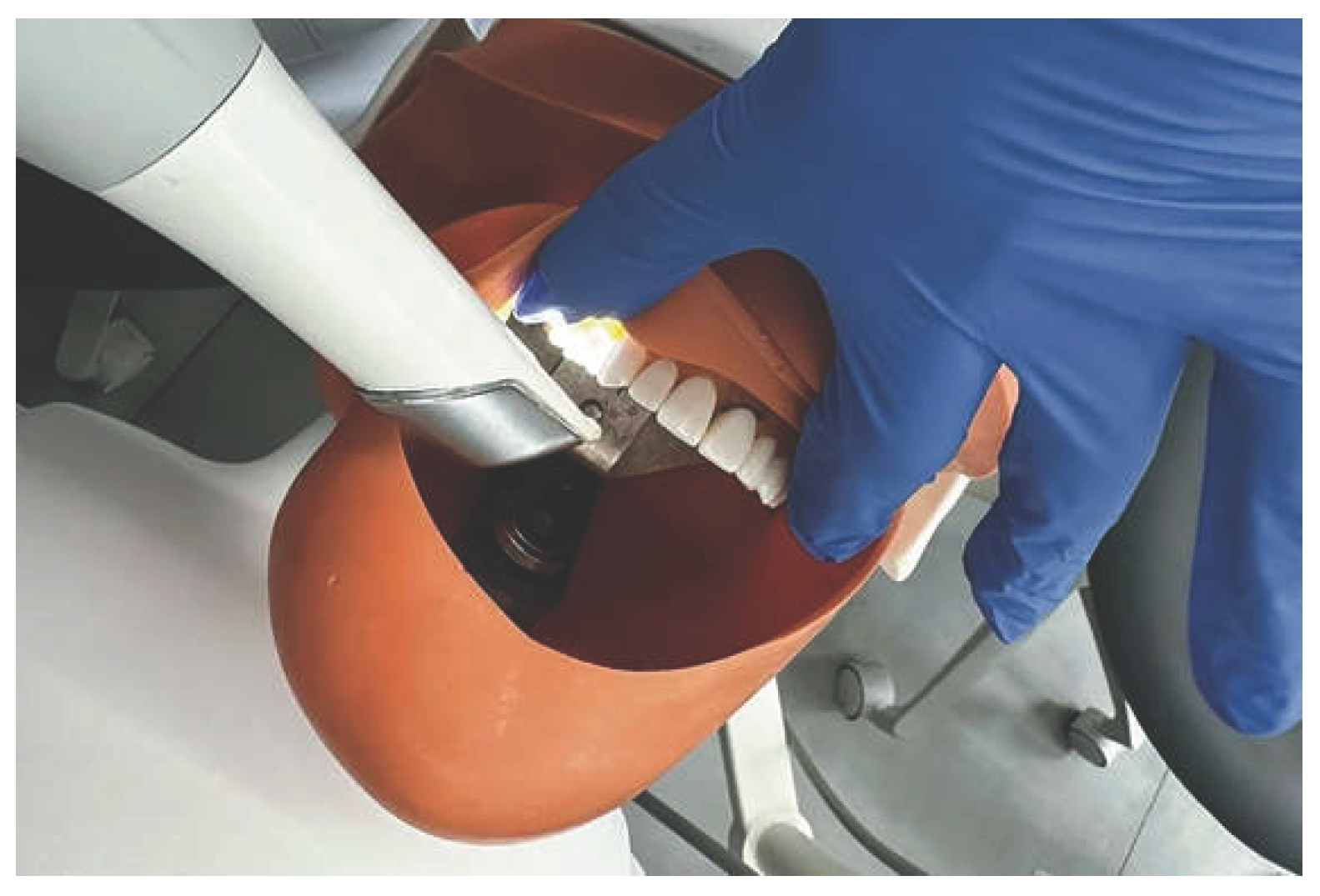

Table 1.
Perception questionnaire items administered before and after training.
| Item | Pre-Training | Post-Training |
|---|---|---|
| P1. How familiar are you with the use of intraoral scanners? | 1–9 | 1–9 |
| P2. How comfortable are you with intraoral scanning technology? | 1–9 | 1–9 |
| P3. How confident are you in your ability to use an intraoral scanner effectively? | 1–9 | 1–9 |
| P4. How would you rate the accuracy of your scans? | 1–9 | 1–9 |
| P5. How competent do you feel using the software associated with the intraoral scanner? | 1–9 | 1–9 |
Items were rated on a 9-point Likert-type scale (1 = lowest; 9 = highest). Both administrations used the same response format.
Table 2.
Pre- and post-training perception scores for each dimension (n = 20).
| Perception Dimension | Pre-Training Mean (SD) | Post-Training Mean (SD) | Mean Difference | p | Cohen’s d |
|---|---|---|---|---|---|
| Familiarity (P1) | 1.90 (1.52) 1 | 5.35 (2.32) | +3.45 | <0.001 | 1.74 |
| Comfort (P2) | 4.25 (2.05) | 7.50 (1.32) 1 | +3.25 | <0.001 | 1.91 |
| Confidence (P3) | 3.30 (2.08) | 7.00 (1.38) | +3.70 | <0.001 | 2.06 |
| Perceived accuracy (P4) | 3.80 (2.35) | 7.10 (1.59) | +3.30 | <0.001 | 1.61 |
| Software competence (P5) | 3.85 (2.58) 1 | 7.40 (1.35) 1 | +3.55 | <0.001 | 1.59 |
Scale: 1 (lowest) to 9 (highest). Mean difference = post-training minus pre-training score. All comparisons used paired Student’s t-tests (df = 19). 1 Shapiro–Wilk p < 0.05 (non-normal distribution); Wilcoxon signed-rank tests yielded consistent results. SD = standard deviation.
Disclaimer/Publisher’s Note: The statements, opinions and data contained in all publications are solely those of the individual author(s) and contributor(s) and not of MDPI and/or the editor(s). MDPI and/or the editor(s) disclaim responsibility for any injury to people or property resulting from any ideas, methods, instructions or products referred to in the content. |
© 2026 by the authors. Licensee MDPI, Basel, Switzerland. This article is an open access article distributed under the terms and conditions of the Creative Commons Attribution (CC BY) license (http://creativecommons.org/licenses/by/4.0/).
Copyright: This open access article is published under a Creative Commons CC BY 4.0 license, which permit the free download, distribution, and reuse, provided that the author and preprint are cited in any reuse.