

Submitted:
18 August 2026
Posted:
28 August 2026
You are already at the latest version
Abstract
COPD involves progressive airflow limitation driven by chronic inflammation, largely triggered by cigarette smoke. Disease progression is linked to heightened Th17/IL-17 responses, with STAT3 playing a key regulatory role. Targeting STAT3 such as with the selective inhibitor Cryptotanshinone may offer a promising strategy to reduce COPD associated inflammation. This study aimed to evaluate the effects of Cryptotanshinone administration on the Th17 immune response in a murine model of chronic cigarette smoke exposure. C57BL/6 mice were exposed to cigarette smoke for six months, while controls breathed filtered air. From the fourth month, mice received intramuscular Cryptotanshinone treatment. Lung function, airspace enlargement, and STAT3/Th17 responses were assessed through respiratory mechanics, histology, immunohistochemistry, and ELISA. Cigarette-smoke reduced tissue elastance and increased alveolar enlargement. Cryptotanshinone attenuated structural damage but did not improve lung function. Smoke exposure elevated STAT3 and IL-17 positive cells and IL-17 levels, all of which were significantly reduced by treatment, indicating suppression of the STAT3/IL-17 inflammatory axis. Pharmacological inhibition of STAT3 with Cryptotanshinone mitigated structural lung damage and reduced Th17-driven inflammation in a chronic cigarette smoke exposure model. These findings reinforce the importance of the STAT3/Th17 axis in COPD pathogenesis and progression.
Keywords:
cigarette-smoke
; COPD
; Cryptotanshinone
; IL-17
; STAT3
; Th17
Introduction
Chronic Obstructive Pulmonary Disease (COPD) is a heterogeneous lung disease characterized by persistent and progressive airflow limitation and respiratory symptoms such as cough, dyspnea, sputum production, and exacerbation episodes (1). Currently, COPD is the third leading cause of death worldwide (2). The progression of the disease is associated with a chronic inflammatory process mediated by an imbalance between pro- and anti-inflammatory cells, cytokines, and inflammatory mediators (3).
T helper type 17 (Th17) cells are key components of the adaptive immune system. They constitute a subpopulation of CD4⁺ T lymphocytes involved in pro-inflammatory mechanisms (4). Their differentiation depends on chemokines present in the microenvironment and is initiated through phosphorylation of the Signal Transducer and Activator of Transcription 3 (STAT3) protein, mediated by interleukin-6 (IL-6) activation. Once phosphorylated, STAT3 translocates to the nucleus and induces the expression of the transcription factor retinoic acid–related orphan receptor gamma t (RORγt). Together, STAT3 and RORγt act synergistically to regulate Th17 cell differentiation and function. Th17 cells, in turn, promote the release of pro-inflammatory cytokines such as IL-17A, IL-17F, IL-22, and IL-23R (7,8). In patients with chronic obstructive pulmonary disease (COPD), increased levels of Th17 cells and IL-17 have been observed, contributing to amplified inflammation, tissue damage, and deterioration of pulmonary function (9,10).
Cryptotanshinone (CTS) is a quinoid diterpene compound isolated from Salvia miltiorrhiza, known for its potent and rapid inhibitory effect on STAT3 phosphorylation (11). This compound exhibits various biological activities, including anti-inflammatory, antioxidant, cytotoxic, and anti-fibrotic effects in diverse pathological contexts (12,13). Previous studies have demonstrated that CTS effectively protects the lungs from pulmonary fibrosis by inhibiting STAT3 signaling (14). However, the mechanisms underlying the action of CTS in chronic obstructive pulmonary disease (COPD) remain poorly understood. Further investigations are warranted, especially considering the increased presence of Th17 cells during the progression of COPD.
Previous clinical and experimental studies have shown that, in COPD, the response mediated by Th17 cell activity plays an important role in inflammatory progression. Lourenço et al. demonstrated in both local and systemic samples that patients with COPD exhibit increased gene expression of STAT3 and RORγt, with a concomitant increase in IL-6 (10). Sales et al. reported an increase in IL-17 positive cells in the airways of COPD patients compared to healthy individuals and non-obstructed smokers (15). Progressive analyses in murine models by Ito et al. 2019 and Silva et al. 2020, showed that mice exposed to cigarette smoke for 1, 3, and 6 months exhibited an increase in IL-17 positive cells after 6 months and IL-6 positive cells after 3 months of exposure (7,16). These findings suggest that COPD progression is associated with a concomitant increase in the pro-inflammatory response, primarily mediated by Th17 cell activity.
Considering the importance of the Th17 response in COPD development and progression, and the pivotal role of STAT3 in the differentiation of these cells, we aimed to evaluate the effects of administering a specific STAT3 inhibitor in a cigarette smoke–induced model of COPD.
Materials and Methods
Experimental Groups
The present study was approved by the Ethics Committee on Human and Animal Research of the Faculty of Medicine, University of São Paulo (Animal Use Ethics Committee – CEUA; protocol number 1678/2021). Male C57BL/6 mice (6–8 weeks old), weighing 26–28 g at the beginning of the experiment, were obtained from the Central Animal Facility of the Faculty of Medicine, University of São Paulo (FMUSP) and used
in this study. All animals received humane care in accordance with the National Institutes of Health Guide for the Care and Use of Laboratory Animals (NIH Publication No. 85-23, revised 1996). This study is reported in accordance with the ARRIVE guidelines for reporting animal research.
Animals were fully anesthetized with thiopental prior to euthanasia, and exsanguination was performed only after the complete loss of reflexes was confirmed. Humane endpoints were established before the beginning of the study, and animal welfare was monitored throughout the experimental period. Criteria for euthanasia included signs of distress such as marked weight loss, reduced mobility, respiratory distress, or lack of response to stimuli. Animals meeting these criteria were humanely euthanized to minimize suffering.The animals were divided into four different experimental groups:
Control Vehicle Group (C-VEHICLE): animals not exposed to cigarette smoke that received vehicle saline administration (N= 12).
Control Crypto Group (C-CRYPTO): animals not exposed to cigarette smoke that received Cryptotanshinone administration (N= 12).
Cigarette smoker Group (CS-VEHICLE): animals exposed to cigarette smoke for 6 months (N= 12).
Cigarette-smoker + Crypto (CS- CRYPTO): animals were exposed to cigarette smoke for 6 months and received Cryptotanshinone administration (N= 12).
The experiment was carried out in two independent phases to meet the specific methodological requirements. The first phase aimed at the collection and processing of samples for respiratory function and histological analysis, while the second was focused on obtaining biological material for biomolecular analyses. Thus, the total number of animals for both phases was described.
Induction of COPD
The animals were exposed to cigarette smoke as previously described (16) for 30 minutes per exposure, twice daily, 5 days per week, for 6 months. The exposure was conducted in an inhalation chamber, a 28-liter plastic box (approximately 40x27 cm at the base, with a height of 26 cm) with two air inlets: synthetic air and cigarette smoke. A small fan for air homogenization is at the top of this box. The airflow inside the compartment is controlled by a flow meter connected to a compressed air torpedo and will be maintained at 2 L/min. The second air inlet receives a mixture of synthetic air and cigarette smoke, drawn in by a Venturi system connected to the lit cigarette. The laminar flow of synthetic air passes through a region of smaller diameter, accelerating the flow and reducing pressure at that point the Venturi effect which facilitates the aspiration of cigarette smoke. The decrease in pressure at the point of diameter reduction in the tube depends on the airflow, which is kept constant. This system creates a concentration of carbon monoxide ranging from 250 to 350 ppm (ToxiPro, Biosystems, USA).
STAT3 Inhibitor Administration
In this study, we used the Cryptotanshinone, a diterpene quinone compound isolated from the root of the Asian medicinal plant Salvia miltiorrhiza Bunge, traditionally used in Chinese medicine for the treatment of allergic disorders. Previous studies have demonstrated that Cryptotanshinone possesses multiple biological properties, including anti-inflammatory, antioxidant, antiproliferative, anticancer, and antifibrotic effects in various disease models.
Cryptotanshinone (Abcam, Cambridge, UK; cat. no. ab120666) functions as a specific inhibitor of the STAT3 signaling pathway. In the experimental protocol, Cryptotanshinone administration started in the fourth month and was carried out twice weekly, one hour before cigarette smoke exposure. The compound was diluted in dimethyl sulfoxide (DMSO) and saline and administered via intramuscular injection at a dose of 20 mg/kg (18). This dosage was selected based on previous research demonstrating its efficacy in reducing inflammatory cell infiltration and tissue remodeling in a murine model of chronic allergic pulmonary inflammation induced by cigarette smoke exposure. Over the course of the experiment, each animal received a total of 24 doses.
Respiratory Mechanics
The animals were anesthetized with Thiopental (50 mg/kg, intraperitoneally), tracheostomized, and placed on a rodent mechanical ventilator (flexiVent, SCIREQ, Montreal, Canada) for respiratory mechanics assessment. They were ventilated with a tidal volume of 10 mL/kg and a respiratory rate of 120 cycles per minute. Using the previously described constant-phase model, tissue elastance (Htis) parameters were calculated (17).
Lung Preparation
Lung preparation and morphometric analysis were performed as previously described (16). After the respiratory mechanic assessment, mice were euthanized by exsanguination under deep thiopental anesthesia through the abdominal aorta, and their lungs were removed and fixed at a constant pressure of 20 cmH2O using 10% buffered formalin infused through the trachea for 24 hours. The lungs were embedded in paraffin and cut into 5-µm sections for histological and morphometric evaluation.
The tissue samples were stained with hematoxylin and eosin (H.E) for conventional morphometry to perform the mean linear intercept (Lm) measurements. Lm was obtained by counting the number of times that the lines of the reticulum, containing 50 lines and 100 points, intercepted the alveolar walls. We performed the Lm analysis in distal areas of parenchyma (peripheral airspaces) and used the following equation: Lm = Ltotal/NI where Ltotal is the sum of all grid segments, calculated by measuring each segment with a ruler (Carl Zeiss Microscopy GmbH, Jena, Germany) attached to the microscope, and NI is the average number of times that the lines intersected the alveolar walls. All Lm values were expressed in micrometers (μm) (19).
Positive Cells by Immunohistochemistry for STAT 3 and IL-17 Evaluation
Immunohistochemical analysis was performed according to previously published protocols in experimental models of COPD (16). Lung sections (5 µm thick) were deparaffinized and hydrated. Antigen retrieval was performed, and the sections were washed in phosphate-buffered saline (PBS) and blocked with 3% hydrogen peroxide at room temperature. Then, the sections were incubated with a rabbit anti Stat3 (Santa Cruz, SC482) at a dilution of 1:300 and anti-IL-17 antibody (Santa Cruz, SC374218) at a dilution of 1:200. The primary antibody was diluted in bovine serum albumin (BSA) overnight (16–18 hours) in a humidified chamber at 4–8 °C. Subsequently, the sections were washed in PBS and incubated with a secondary antibody (Vector ABCElite, horseradish peroxidase [HRP]; anti-mouse) at 37 °C in a humidified chamber. Three additional 5-minute washes in PBS were performed, and the samples were stained with 3,3-diaminobenzidine (DAB) (code K3468, Dako Citomation, Fort Collins, CO, USA) for 5 min. Subsequently, the tissues were washed with tap water and counterstained with Harris hematoxylin. Cell density was assessed by the number of cells divided by the respective peribronchovascular area (104 cells/µm2) in 15 fields/slide. Analysis was performed using an optical microscope equipped with an integrating eyepiece containing a known area (104 μm² at ×1000 magnification), consisting of 50 lines and 100 points (20).
Cytokine Analysis
The lungs were removed, stored in labeled tubes containing crushed ice, and then individually homogenized. Cytokine levels were detected using ELISA (OptEIA, BD PharMingen, Oxford, UK) on microplates (Costar, Cambridge, MA, USA) sensitized with specific monoclonal antibodies for each cytokine. After the samples were washed, specific antibodies were added to the different cytokines conjugated to biotin. After the solution contained streptavidin-peroxidase, substrate, and chromogen enzyme conjugate were added. The reaction was read at 450 nm using an M2 spectrophotometer (Molecules Devices, San Jose, CA, USA). Sample concentrations were calculated from the standard curves obtained with the recombinant cytokines, and the results were expressed in units of pg/ml. R&D Systems ELISA kits determined interleukin (IL-6 and IL-17) (21).
Generative Artificial Intelligence Assistance
Generative Artificial Intelligence Assistance was used solely to assist in the visual preparation and graphical design of schematic figures. The AI-generated outputs were reviewed and edited by the authors. Generative AI was not used to generate, modify, or analyze experimental data, and all scientific content and interpretations presented in the figures were defined and verified by the authors.
Statistical Analysis
Statistical analysis was performed using the SigmaStat program (version 11.0; Systat Software, San Jose, CA, USA). For respiratory mechanics and Lm parameters, we compared four experimental groups, using Two-way analysis of variance, followed by Holm Sidak for multiple comparisons. For histological parameters and the expression of IL-17 and STAT3 analysis, we compared only three groups, using the one-way ANOVA test followed by the multiple-comparison test (Holm-Sidak Test or Tukey Test, depending on the normality of variables).
Figure 1.
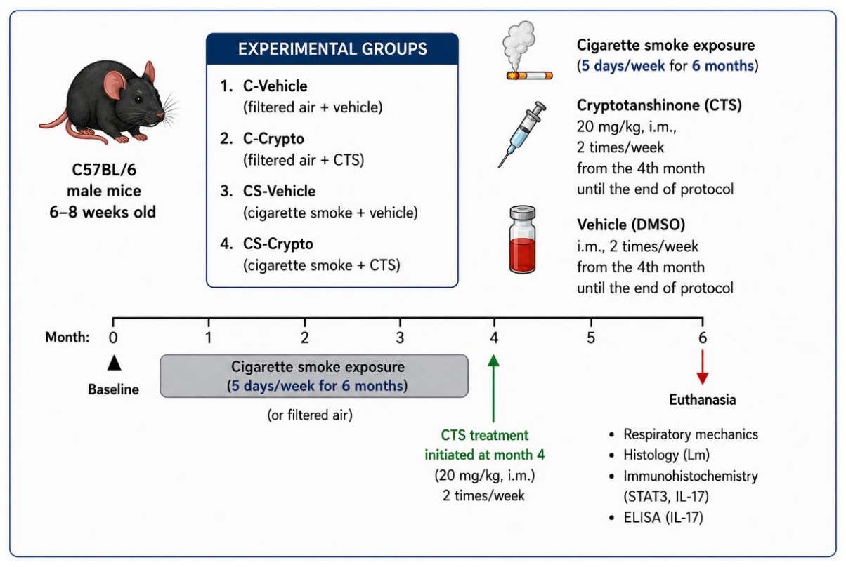
Experimental design. Male C57BL/6 mice were exposed to cigarette smoke or filtered air for six months. Cryptotanshinone (20 mg/Kg, intramuscular) or vehicle was administered twice weekly from the fourth month until the end of the protocol. Respiratory mechanics, morphometric analysis, immunohistochemistry, and ELISA were performed at the end of the experimental period.
Figure 1.
Experimental design. Male C57BL/6 mice were exposed to cigarette smoke or filtered air for six months. Cryptotanshinone (20 mg/Kg, intramuscular) or vehicle was administered twice weekly from the fourth month until the end of the protocol. Respiratory mechanics, morphometric analysis, immunohistochemistry, and ELISA were performed at the end of the experimental period.
Results
A significant reduction in tissue elastance (Htis) was observed in cigarette-smoke exposed groups compared with the controls. Cryptotanshinone treatment did not significantly restore tissue elastance. (Figure 2)
A significant increase in alveolar enlargement was observed in the CS-Vehicle and CS-Crypto groups compared to both Control groups, as evidenced by the mean linear intercept analysis. Moreover, there was a significant decrease in Lm values in the CS-Crypto group compared with CS-Vehicle group (Figure 3).
Discussion
In this study, we demonstrated that Cryptotanshinone administration inhibits the inflammatory response mediated by the STAT3/Th17 axis, thereby attenuating the structural alterations observed in a cigarette smoke–induced model of COPD.
Cigarette-smoke (CS) exposure leads to a reduction of tissue elastance, indicating impaired elastic recoil of the lung tissue, concomitant with an increase in the mean linear intercept, consistent with alveolar enlargement. In accordance with these findings, a pronounced accumulation of STAT3⁺ cells and IL-17⁺ lymphocytes was observed in the peribronchovascular areas of lung tissue samples, accompanied by elevated expression levels of IL-17 and IL-6 in lung homogenates.
Cryptotanshinone administration was initiated in the fourth month, when the alveolar enlargement and tissue elastance decrease have already been established in this animal model (16), and it was maintained until the end of the protocol. We observed that inhibitor administration attenuated alveolar enlargement, concomitant with a decrease in IL-17+ and STAT3+ lymphocytes in the peribronchovascular areas. Moreover, the IL-17 expression was also decreased in the lung homogenate in animals that received the inhibitor administration.
Interestingly, no significant changes were observed in tissue elastance, indicating that the structural improvements were not sufficient to restore lung mechanical function. It is important to emphasize that in most studies conducted in experimental models of COPD, the structural alterations assessed by morphometric methods precede the functional changes, and they only begin to affect functional parameters as they become more pronounced. This is due to the small size of these rodents, which creates technical challenges for obtaining accurate respiratory mechanics measurements. We are only able to detect functional differences when there is substantial structural destruction or recovery (21,23,24).
These findings contrast with the results reported by Riani et al., who demonstrated that IL-17 neutralization in the same CS-induced COPD model attenuated both structural damage and functional impairment, accompanied by a reduction in Th17-associated cytokines in lung tissue. It is worth noting that IL-17 blockade not only suppressed the STAT3/Th17 signaling axis but also reduced the inflammatory response driven by innate immune cells—particularly neutrophils, which are a major source of IL-17.
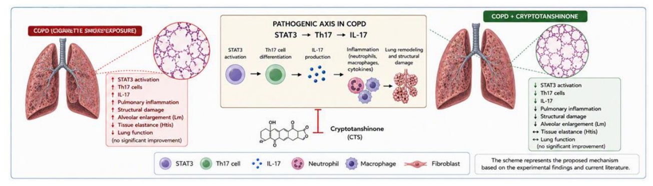
Interleukin-17 (IL-17) has been shown to induce IL-6 production in airway epithelial cells, fibroblasts, and other lung-resident structural cells, thereby amplifying the local inflammatory microenvironment (25). In parallel, IL-6 is enrolled in the differentiation of naïve CD4⁺ T cells into Th17 cells through STAT3 activation and RORγt induction, establishing a self-reinforcing inflammatory loop in which IL-6 enhances Th17 development and IL-17 further stimulates IL-6 production (26). This reciprocal relationship helps explain why IL-17 neutralization reduces IL-6 levels and results in concurrent improvements in both lung structure and function, as reported in previous studies. It is possible that IL-17 inhibition helped re-establish the balance of extracellular matrix components, including the proportion between type I and type III collagen fiber deposition in the alveolar walls. Since IL-17 acts as a chemotactic stimulus for neutrophils (27,28), key effector cells involved in alveolar wall destruction and tissue remodeling, its blockade may have reduced neutrophil-driven matrix degradation, thereby favoring the preservation of structural integrity and contributing to the partial recovery of elastic recoil lost during COPD progression. In contrast, the present intervention, which targeted only a portion of this pathway, led to partial improvement in structural parameters that was not pronounced enough to restore the functional lung elastic recoil. Based on the present findings, we propose the mechanistic model illustrated in Figure 6.
Conclusions
In the present study, Cryptotanshinone administration effectively inhibited the STAT3/Th17 signaling axis, as demonstrated by the reduced density of STAT3+ and IL-17⁺ cells and the reduction in IL-17 levels. This inhibition attenuated the structural alterations induced by CS exposure, reinforcing the importance of STAT3-mediated immune responses in the development and progression of COPD.
Author Contributions
Conceptualization, F.J.L. and F.D.Q.S.L.; Methodology, F.J.L., A.F.S., C.U.S., A.R.M. and M.R.G.G.; Validation, F.J.L.; Formal Analysis, F.J.L., M.R.G.G., F.M.A., L.H.A.V., T.Y.T., A.T.C. and V.C.R.S.; Investigation, F.J.L., A.F.S., A.R.M., C.U.S. and L.H.A.V.; Resources, F.J.L., A.F.S. and F.D.Q.S.L.; Data Curation, L.P.M.C., L.N.C., C.N.S. and V.B.S.; Writing – Original Draft Preparation, F.J.L., A.F.S., C.U.S., A.R.M. and F.D.Q.S.L.; Writing – Review & Editing, F.J.L., A.F.S., C.U.S., A.R.M., L.H.A.V. and F.D.Q.S.L.; Visualization, F.J.L.; Supervision, F.M.A. and F.D.Q.S.L.; Funding Acquisition, I.F.L.C.T. and F.D.Q.S.L. All authors have read and agreed to the published version of the manuscript.
Funding
Fundação de Amparo à Pesquisa do Estado de São Paulo - FAPESP (2023/06001-0).
Institutional Review Board Statement
The animal study protocol was approved by the Animal Use Ethics Committee of the Faculty of Medicine, University of São Paulo (CEUA; protocol No. 1678/2021; approved on 1 July 2021).
Informed Consent Statement
Not applicable.
Data Availability Statement
The data presented in this study are available from the corresponding author upon reasonable request.
Acknowledgments
The: authors thank all those who, directly or indirectly, contributed to the completion of this work. We particularly acknowledge the members of the research group and the co-authors for their scientific support, constructive discussions, and collaboration throughout all stages of the study.We are especially grateful to Prof. Dr. Fernanda Degobbi TQS Lopes for sharing her expertise, for her rigorous scientific guidance, and for her continuous support throughout all phases of the project, which was fundamental to the academic and professional development of the author. We also thank Prof. Dr. Iolanda de Fátima L. C. Tibério, head of the laboratory, for the institutional and scientific support provided during the development of this study.Finally, the authors acknowledge the São Paulo Research Foundation (FAPESP) for the financial support that made this research possible. During the preparation of this manuscript, the authors used ChatGPT (OpenAI) for assistance with the visual preparation and graphical design of schematic figures. The authors reviewed and edited the output and take full responsibility for the content of this publication.
Conflicts of Interest
The authors declare no conflicts of interest.
Abbreviations
The following abbreviations are used in this manuscript:
| ABCElite | Avidin-Biotin Complex Elite Immunohistochemistry System (Vector Laboratories) |
| ANOVA | Analysis of Variance |
| BD | Becton Dickinson |
| BSA | Bovine Serum Albumin |
| CEUA | Animal Use Ethics Committee |
| CO | Carbon Monoxide |
| COPD | Chronic Obstructive Pulmonary Disease |
| CS | Cigarette Smoke |
| CTS | Cryptotanshinone |
| DAB | 3,3'-Diaminobenzidine |
| DMSO | Dimethyl Sulfoxide |
| ELISA | Enzyme-Linked Immunosorbent Assay |
| H.E | Hematoxylin and Eosin |
| Htis | Tissue Elastance |
| HRP | Horseradish Peroxidase |
| IL | Interleukin |
| IL-6 | Interleukin-6 |
| IL-17 | Interleukin-17 |
| IL-22 | Interleukin-22 |
| IL-23R | Interleukin-23 Receptor |
| Lm | Mean Linear Intercept |
| NIH | National Institutes of Health |
| PBS | Phosphate-Buffered Saline |
| RORγt | Retinoic Acid–Related Orphan Receptor Gamma t |
| SCIREQ | Scientific Respiratory Equipment (manufacturer of the flexiVent system) |
| SE | Standard Error |
| STAT | Signal Transducer and Activator of Transcription |
| STAT3 | Signal Transducer and Activator of Transcription 3 |
| Th17 | T Helper 17 Cell |
| Treg | Regulatory T Cell |
References
- Global Initiative for Chronic Obstructive Lung Disease (GOLD). Global Strategy for the Diagnosis, Management, and Prevention of COPD: 2025 Report; Global Initiative for Chronic Obstructive Lung Disease: 2025.
- Kahnert, K.; Jörres, R.A.; Behr, J.; Welte, T. The diagnosis and treatment of COPD and its comorbidities. Dtsch. Arztebl. Int. 2023, 120, 434–444.
- Brightling, C.; Greening, N. Airway inflammation in COPD: Progress to precision medicine. Eur. Respir. J. 2019, 54. [CrossRef]
- Lourenço, J.D.; Ito, J.T.; Martins, M.A.; Tibério, I.F.L.C.; Lopes, F.D.T.Q.S. Th17/Treg imbalance in chronic obstructive pulmonary disease: Clinical and experimental evidence. Front. Immunol. 2021, 12, 804919. [CrossRef]
- Lopes, F.D.T.Q.S.; Tibério, I.F.L.C.; Leme, A.; Fairclough, L. Editorial: The importance of Th17/Treg imbalance in asthma and COPD development and progression. Front. Immunol. 2022, 13, 1025215. [CrossRef]
- Thomas, R.; Qiao, S.; Yang, X. Th17/Treg imbalance: Implications in lung inflammatory diseases. Int. J. Mol. Sci. 2023, 24, 4865. [CrossRef]
- Silva, L.E.F.; Lourenço, J.D.; Silva, K.R.; Santana, F.P.R.; Kohler, J.B.; Moreira, A.R.; et al. Th17/Treg imbalance in COPD development: Suppressors of cytokine signaling and STAT proteins. Sci. Rep. 2020, 10, 15287. [CrossRef]
- Damasceno, L.E.A.; Prado, D.S.; Veras, F.P.; Fonseca, M.M.; Toller-Kawahisa, J.E.; Rosa, M.H.; et al. PKM2 promotes Th17 cell differentiation and autoimmune inflammation by fine-tuning STAT3 activation. J. Exp. Med. 2020, 217, e20190613. [CrossRef]
- Barnes, P.J. Inflammatory mechanisms in patients with chronic obstructive pulmonary disease. J. Allergy Clin. Immunol. 2016, 138, 16–27. [CrossRef]
- Lourenço, J.D.; Teodoro, W.R.; Barbeiro, D.F.; Velosa, A.P.P.; Silva, L.E.F.; Kohler, J.B.; et al. Th17/Treg-related intracellular signaling in patients with chronic obstructive pulmonary disease: Comparison between local and systemic responses. Cells 2021, 10, 1569. [CrossRef]
- Wang, Y.; Zhou, C.; Gao, H.; Li, C.; Li, D.; Liu, P.; Huang, M.; Shen, X.; Liu, L. Therapeutic effect of Cryptotanshinone on experimental rheumatoid arthritis through downregulating p300-mediated STAT3 acetylation. Biochem. Pharmacol. 2017, 138, 119–129. [CrossRef]
- Chen, L.; Wang, H.J.; Xie, W.; Yao, Y.; Zhang, Y.S.; Wang, H. Cryptotanshinone inhibits lung tumorigenesis and induces apoptosis in cancer cells in vitro and in vivo. Mol. Med. Rep. 2014, 9, 2447–2452. [CrossRef]
- Ye, Z.; Wang, P.; Feng, G.; Wang, Q.; Liu, C.; Lu, J.; Chen, J.; Liu, P. Cryptotanshinone attenuates LPS-induced acute lung injury by regulating metabolic reprogramming of macrophage. Front. Med. 2022, 9, 1075465. [CrossRef]
- Zhang, Y.; Lu, W.; Zhang, X.; Lu, J.; Xu, S.; Chen, S.; Zhong, Z.; Zhou, T.; Wang, Q.; Chen, J.; Liu, P. Cryptotanshinone protects against pulmonary fibrosis through inhibiting Smad and STAT3 signaling pathways. Pharmacol. Res. 2019, 147, 104307. [CrossRef]
- Sales, D.S.; Ito, J.T.; Zanchetta, I.A.; Annoni, R.; Aun, M.V.; Ferraz, L.F.S.; Cervilha, D.A.B.; Negri, E.; Mauad, T.; Martins, M.A.; Lopes, F.D.T.Q.S. Regulatory T-cell distribution within lung compartments in COPD. COPD 2017, 14, 533–542. [CrossRef]
- Ito, J.T.; Cervilha, D.A.B.; Lourenço, J.D.; Gonçalves, N.G.; Volpini, R.A.; Caldini, E.G.; et al. Th17/Treg imbalance in COPD progression: A temporal analysis using a CS-induced model. PLoS ONE 2019, 14, e0209351. [CrossRef]
- Biselli, P.J.C.; Lopes, F.D.T.Q.S.; Righetti, R.F.; Moriya, H.T.; Tibério, I.F.L.C.; Martins, M.A. Lung mechanics over the century: From bench to bedside and back to bench. Front. Physiol. 2022, 13, 817263. [CrossRef]
- Wang, C.; Zheng, M.; Choi, Y.; Jiang, J.; Li, L.; Li, J.; Xu, C.; Xian, Z.; Li, Y.; Piao, H.; Li, L.; Yan, G. Cryptotanshinone attenuates airway remodeling by inhibiting crosstalk between tumor necrosis factor-like weak inducer of apoptosis and transforming growth factor beta 1 signaling pathways in asthma. Front. Pharmacol. 2019, 10, 1338. [CrossRef]
- Cervilha, D.A.B.; Ito, J.T.; Lourenço, J.D.; Olivo, C.R.; Saraiva-Romanholo, B.M.; Volpini, R.A.; Oliveira-Junior, M.C.; Mauad, T.; Martins, M.A.; Tibério, I.F.L.C.; Vieira, R.P.; Lopes, F.D.T.Q.S. The Th17/Treg cytokine imbalance in chronic obstructive pulmonary disease exacerbation in an animal model of cigarette smoke exposure and lipopolysaccharide challenge association. Sci. Rep. 2019, 9, 1921. [CrossRef]
- de Genaro, I.S.; de Almeida, F.M.; dos Santos Lopes, F.D.T.Q.; Kunzler, D.D.C.H.; Tripode, B.G.B.; Kurdejak, A.; et al. Low-dose chlorine exposure impairs lung function, inflammation and oxidative stress in mice. Life Sci. 2021, 267, 118912. [CrossRef]
- Simao, J.J.; Bispo, A.F.S.; Plata, V.T.G.; Abel, A.B.M.; Saran, R.J.; Barcella, J.F.; et al. The activation of the NF-κB pathway in human adipose-derived stem cells alters the deposition of epigenetic marks on H3K27 and is modulated by fish oil. Life 2024, 14, 1653. [CrossRef]
- Anciães, A.M.; Olivo, C.R.; Prado, C.M.; Kagohara, K.H.; Pinto, T.S.; Moriya, H.T.; et al. Respiratory mechanics do not always mirror pulmonary histological changes in emphysema. Clinics 2011, 66, 1797–1803. [CrossRef]
- Bates, J.H.T. CORP: Measurement of lung function in small animals. J. Appl. Physiol. 2017, 123, 1039–1046. [CrossRef]
- Irvin, C.G.; Bates, J.H.T. Measuring the lung function in the mouse: The challenge of size. Respir. Res. 2003, 4, 4. [CrossRef]
- Laan, M.; Lötvall, J.; Chung, K.F.; Lindén, A. IL-17-induced cytokine release in human bronchial epithelial cells in vitro: Role of MAP kinases. Br. J. Pharmacol. 2001, 133, 200–206. [CrossRef]
- Qin, H.; Wang, L.; Feng, T.; Elson, C.O.; Niyongere, S.A.; Lee, S.J.; et al. TGF-β promotes Th17 cell development through inhibition of SOCS3. J. Immunol. 2009, 183, 97–105. [CrossRef]
- Griffin, G.K.; Newton, G.; Tarrio, M.L.; Bu, D.X.; Maganto-Garcia, E.; Azcutia, V.; et al. IL-17 and TNF-α sustain neutrophil recruitment during inflammation through synergistic effects on endothelial activation. J. Immunol. 2012, 188, 6287–6299. [CrossRef]
- Fan, X.; Shu, P.; Wang, Y.; Ji, N.; Zhang, D. Interactions between neutrophils and T-helper 17 cells. Front. Immunol. 2023, 14, 1279837. [CrossRef]
Figure 2.
Respiratory mechanics. Htis parameter. C-Vehicle (n= 5), C-Crypto (n= 5), CS (n= 5), CS- Crypto (n= 4). Compared to the control group. Data were analyzed using Two-way ANOVA and are presented as mean ± standard error (SE). *p = 0,03.
Figure 2.
Respiratory mechanics. Htis parameter. C-Vehicle (n= 5), C-Crypto (n= 5), CS (n= 5), CS- Crypto (n= 4). Compared to the control group. Data were analyzed using Two-way ANOVA and are presented as mean ± standard error (SE). *p = 0,03.
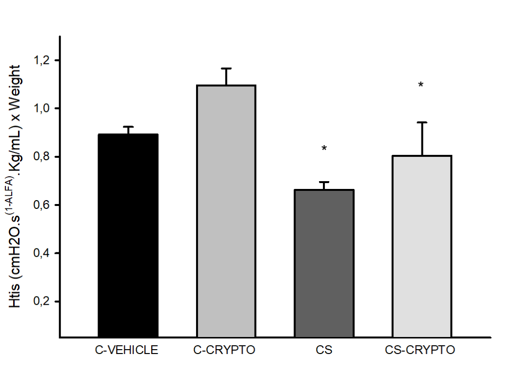
Figure 3.
Mean Linear Intercept. C-Vehicle (n= 6), C-Crypto (n= 5), CS-Vehicle (n= 5), and CS- Crypto (n = 6). The Lm values, measured in the distal pulmonary parenchyma. A significant difference was observed when compared to the C-Vehicle and C-Crypto groups (#p = 0.045); between the CS-Vehicle and C-Vehicle groups, p < 0.001; and between the CS-Crypto and C-Crypto groups, p = 0.021. Data were analyzed using Two-way ANOVA and are presented as mean ± standard error (SE).
Figure 3.
Mean Linear Intercept. C-Vehicle (n= 6), C-Crypto (n= 5), CS-Vehicle (n= 5), and CS- Crypto (n = 6). The Lm values, measured in the distal pulmonary parenchyma. A significant difference was observed when compared to the C-Vehicle and C-Crypto groups (#p = 0.045); between the CS-Vehicle and C-Vehicle groups, p < 0.001; and between the CS-Crypto and C-Crypto groups, p = 0.021. Data were analyzed using Two-way ANOVA and are presented as mean ± standard error (SE).
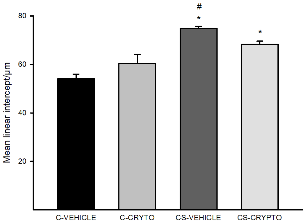
Figure 4.
Immunohistochemical analysis. (A) STAT3 immunostaining in lung tissue from C-Vehicle (n = 4), CS (n = 6), and CS-Crypto (n = 6) groups. (B) IL-17 immunostaining in lung tissue from C-Vehicle (n = 6), CS (n = 6), and CS-Crypto (n = 5) groups. Data are presented as mean ± standard error (SE). For STAT3, *p < 0.001 compared with the CS-Crypto and C-Vehicle groups (ANOVA followed by Holm–Sidak post hoc test). For IL-17, *p< 0.001 for the CS group compared with the other groups (ANOVA followed by Tukey’s post hoc test). Representative images are shown at 200× magnification.
Figure 4.
Immunohistochemical analysis. (A) STAT3 immunostaining in lung tissue from C-Vehicle (n = 4), CS (n = 6), and CS-Crypto (n = 6) groups. (B) IL-17 immunostaining in lung tissue from C-Vehicle (n = 6), CS (n = 6), and CS-Crypto (n = 5) groups. Data are presented as mean ± standard error (SE). For STAT3, *p < 0.001 compared with the CS-Crypto and C-Vehicle groups (ANOVA followed by Holm–Sidak post hoc test). For IL-17, *p< 0.001 for the CS group compared with the other groups (ANOVA followed by Tukey’s post hoc test). Representative images are shown at 200× magnification.
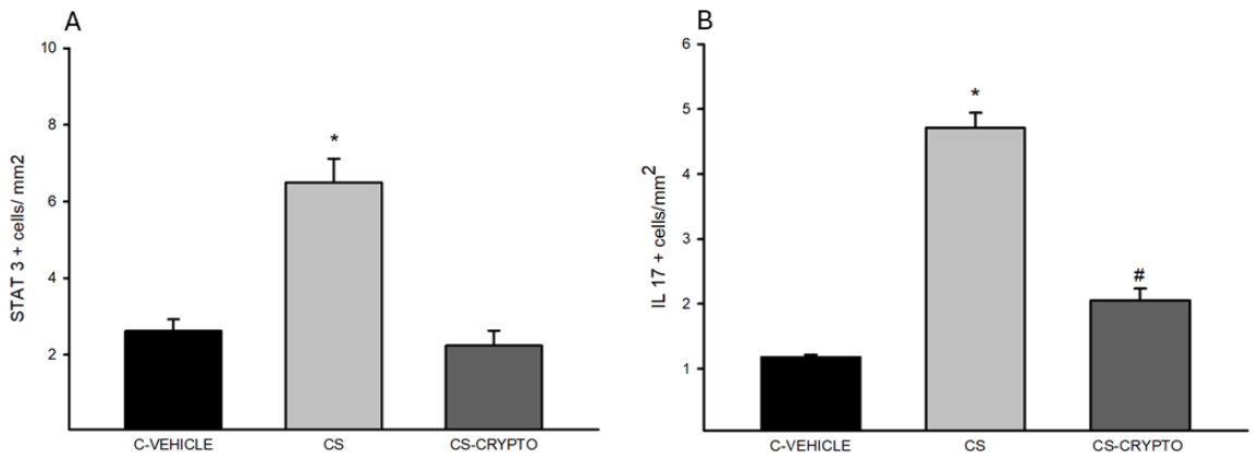
Figure 5.
Cytokine protein expression in lung tissue. (A) IL-6 protein levels in C-Vehicle (n = 5), CS (n = 4), and CS-Crypto (n = 5) groups. A significant increase was observed in the CS and CS-Crypto groups compared with the C-Vehicle group (*p < 0.05; one-way ANOVA followed by Tukey’s post hoc test). (B) IL-17 protein levels in C-Vehicle (n = 10), CS (n = 9), and CS-Crypto (n = 9) groups. A marked increase was observed in the CS group compared with the C-Vehicle and CS-Crypto groups (*p < 0.001; Kruskal–Wallis test followed by Dunn’s post hoc test). Data are expressed as mean ± standard error (SE).
Figure 5.
Cytokine protein expression in lung tissue. (A) IL-6 protein levels in C-Vehicle (n = 5), CS (n = 4), and CS-Crypto (n = 5) groups. A significant increase was observed in the CS and CS-Crypto groups compared with the C-Vehicle group (*p < 0.05; one-way ANOVA followed by Tukey’s post hoc test). (B) IL-17 protein levels in C-Vehicle (n = 10), CS (n = 9), and CS-Crypto (n = 9) groups. A marked increase was observed in the CS group compared with the C-Vehicle and CS-Crypto groups (*p < 0.001; Kruskal–Wallis test followed by Dunn’s post hoc test). Data are expressed as mean ± standard error (SE).
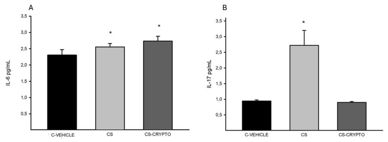
Figure 6.
Proposed mechanism by which Cryptotanshinone attenuates cigarette smoke-induced pulmonary inflammation and structural remodeling. Cigarette smoke exposure increases STAT3 expression, Th17-associated responses, and IL-17 production, contributing to pulmonary inflammation and alveolar enlargement. Cryptotanshinone reduces STAT3/Th17- associated responses and IL-17 production, and attenuates alveolar enlargement, although lung mechanical function remains impaired. .
Figure 6.
Proposed mechanism by which Cryptotanshinone attenuates cigarette smoke-induced pulmonary inflammation and structural remodeling. Cigarette smoke exposure increases STAT3 expression, Th17-associated responses, and IL-17 production, contributing to pulmonary inflammation and alveolar enlargement. Cryptotanshinone reduces STAT3/Th17- associated responses and IL-17 production, and attenuates alveolar enlargement, although lung mechanical function remains impaired. .
Disclaimer/Publisher’s Note: The statements, opinions and data contained in all publications are solely those of the individual author(s) and contributor(s) and not of MDPI and/or the editor(s). MDPI and/or the editor(s) disclaim responsibility for any injury to people or property resulting from any ideas, methods, instructions or products referred to in the content. |
© 2026 by the authors. Licensee MDPI, Basel, Switzerland. This article is an open access article distributed under the terms and conditions of the Creative Commons Attribution (CC BY) license.
Copyright: This open access article is published under a Creative Commons CC BY 4.0 license, which permit the free download, distribution, and reuse, provided that the author and preprint are cited in any reuse.