
Submitted:
27 August 2026
Posted:
27 August 2026
You are already at the latest version
Abstract
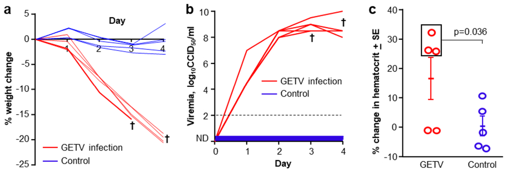
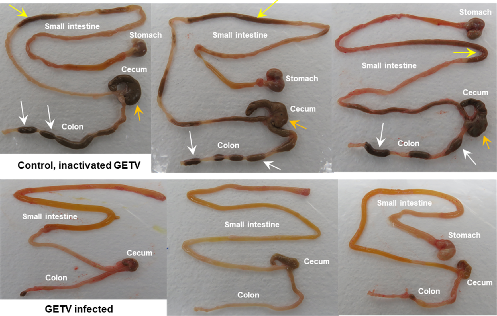
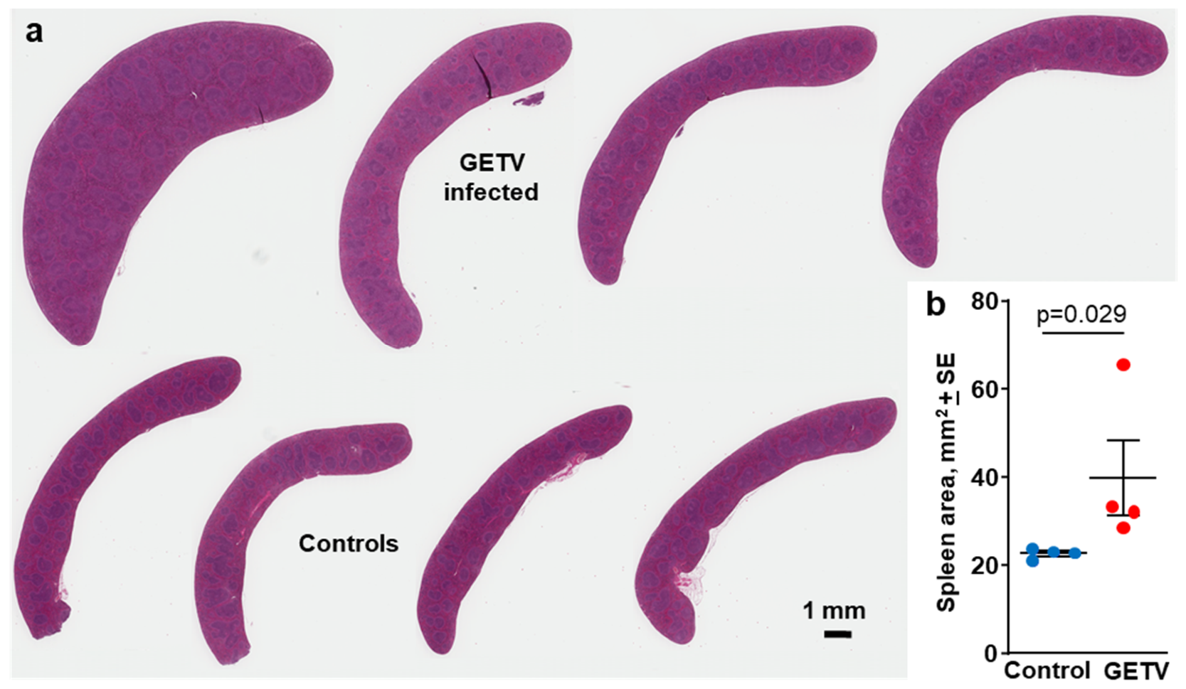
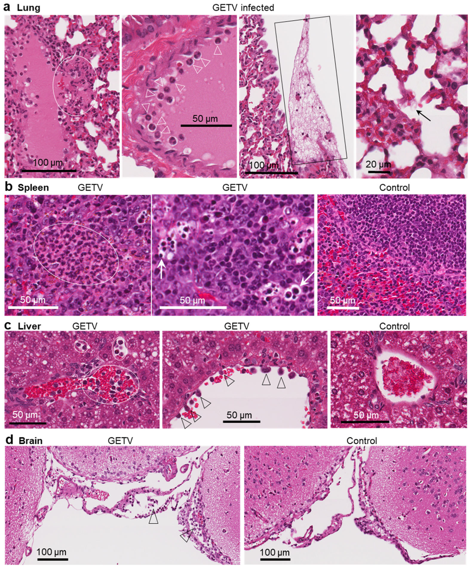
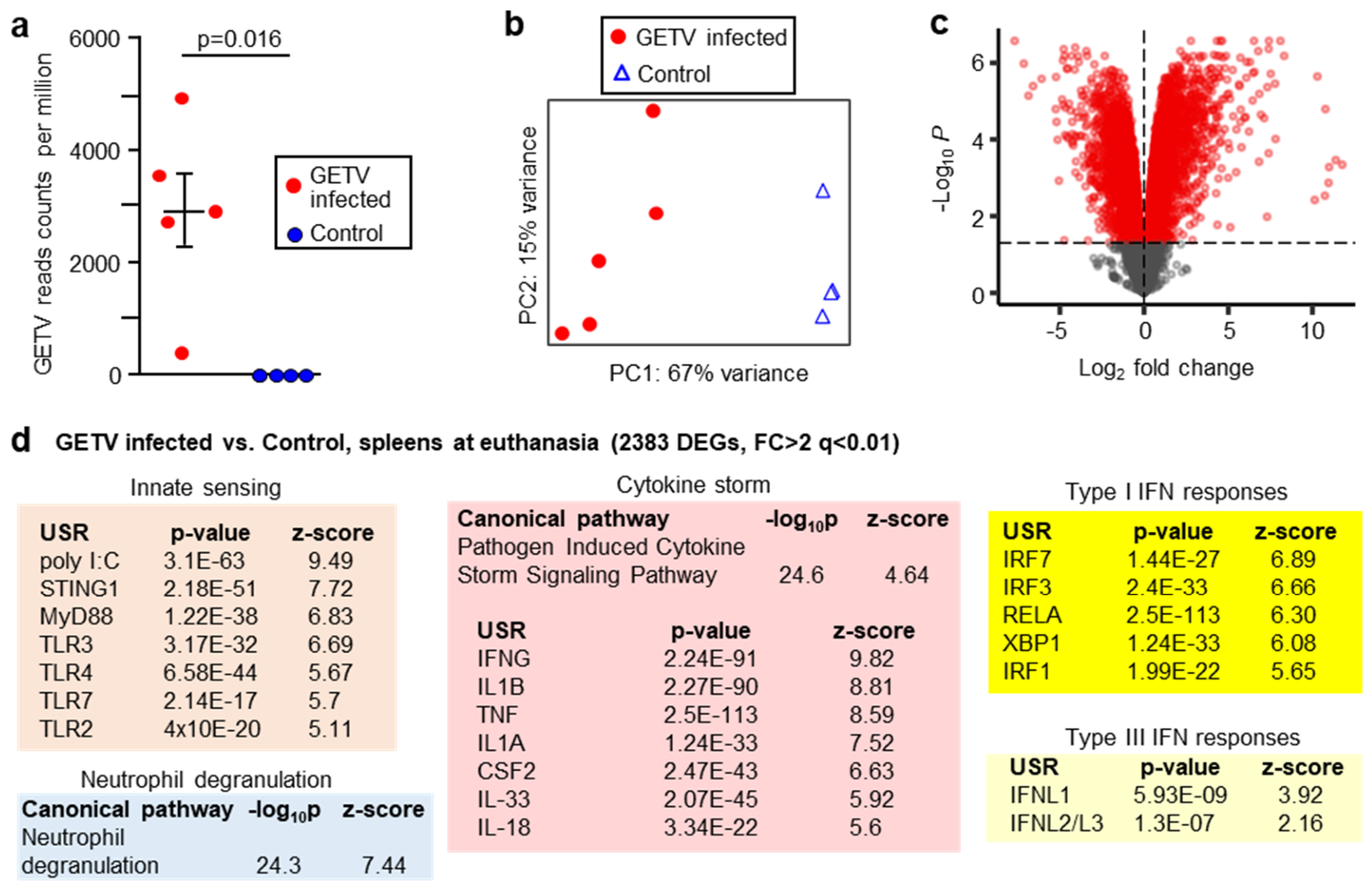
Getah virus (GETV) is a mosquito borne Old World alphavirus that has recently increased its geographic range, particularly in China. GETV causes rheumatic disease in horses, but in recent years its primary impact has been lethal infections in newborn piglets. Herein we characterize a lethal Ifnar1-/- mouse model of GETV, which recapitulated many of the features seen in piglets. These include lethality, high viremia, diarrhoea, and splenomegaly, with histopathological findings aligning with those seen in piglets and indicating severe, acute, multi-organ inflammatory immunopathology. RNA-Seq of spleens from GETV infected Ifnar1-/- mice and subsequent bioinformatic analyses illustrated a cytokine storm response that included robust type I interferon signatures and a pattern of cytokine/chemokine signatures indicative of viral sepsis/septic shock. Ifnar1-/- mice thus offer a framework for understanding, and a mouse model for, GETV disease in piglets. The model also indicates that lethal GETV infection in piglets ay involve viral sepsis/septic shock.
Keywords:
1. Introduction
2. Materials and Methods
2.1. Ethics and Regulatory Statement
2.2. The GETV Isolate, Culture and Titration
2.3. Mouse Infection and Monitoring
2.4. Histology
2.5. RNA-Seq and Bioinformatics
2.6. Statistics
3. Results
3.1. GETV Virus Infection of Ifnar1-/- Mice, Weight Loss, Viremia and Haematocrit
3.2. GETV Virus Infection in Ifnar1-/- Mice Was Associated with Overt Diarrhoea
3.3. Splenomegaly in GETV Infected Ifnar1-/- Mice
3.4. Histopathological Findings in GETV Infected Mice
3.5. RNA-Seq and Bioinformatic Analyses of Spleens from GETV Infected vs. Control Ifnar1-/- Mice
3.6. mRNA Expression Signatures in Spleens of GETV Infected Ifnar1-/- Mice Indicate Viral Sepsis and/or Viral Septic Shock
4. Discussion
5. Conclusions
Supplementary Materials
Author Contributions
Funding
Institutional Review Board Statement
Acknowledgments
Conflicts of Interest
Abbreviations
| GETV | getah virus |
| Ifn/IFN | interferon |
| Ifnar1 | interferon alpha receptor subunit 1 (gene) |
| CHIKV | chikungunya virus |
| COVID-19 | Coronavirus disease of 2019 |
| RNA-Seq | ribonucleic acid sequencing |
| USR | upstream regulator |
| mRNA | messenger ribonucleic acid |
| DEGs | differentially expressed genes |
| CCID50 | 50% cell culture infectivity dose |
| TLR | Toll like receptor |
| CPE | cytopathic effect |
| H&E | hematoxylin and eosin |
References
- Li, B.; Wang, H.; Liang, G. Getah Virus (Alphavirus): An Emerging, Spreading Zoonotic Virus. Pathogens 2022, 11, 945. [Google Scholar] [CrossRef] [PubMed]
- Shen, J.; Liu, S.; Liu, S.; Shen, S.; Lei, M.; Xu, Q.; Li, W.; He, Q.; Xu, X.; Cai, X. Genomic surveillance and evolution of Getah virus. Virus Evol. 2025, 11, veaf007. [Google Scholar] [CrossRef] [PubMed]
- Nguyen, W.; Nakayama, E.; Yan, K.; Tang, B.; Le, T.T.; Liu, L.; Cooper, T.H.; Hayball, J.D.; Faddy, H.M.; Warrilow, D.; et al. Arthritogenic Alphavirus Vaccines: Serogrouping Versus Cross-Protection in Mouse Models. Vaccines 2020, 8, 209. [Google Scholar] [CrossRef] [PubMed]
- Suhrbier, A.; Jaffar-Bandjee, M.-C.; Gasque, P. Arthritogenic alphaviruses—An overview. Nat. Rev. Rheumatol. 2012, 8, 420–429. [Google Scholar] [CrossRef] [PubMed]
- Suhrbier, A. Rheumatic manifestations of chikungunya: Emerging concepts and interventions. Nat. Rev. Rheumatol. 2019, 15, 597–611. [Google Scholar] [CrossRef] [PubMed]
- Yu, L.; Chen, L. The epidemiological and evolutionary dynamics of Getah virus in China. Vet. Vaccine 2026, 5, 100172. [Google Scholar] [CrossRef]
- Chu, P.-P.; Chen, S.-N.; Zhou, X.; Wei, Z.-Z.; Zhai, S.-L. Getah Virus: A New Contaminant in Veterinary Vaccines. Vet. Sci. 2025, 12, 82. [Google Scholar] [CrossRef] [PubMed]
- Irekeola, A.A.; Shueb, R.H. Global seroprevalence and distribution of Getah virus in domestic and wild animals: A systematic review and meta-analysis. Vet. World 2025, 18, 3464–3475. [Google Scholar] [CrossRef] [PubMed]
- Ochi, A.; Bannai, H.; Aonuma, H.; Kanuka, H.; Uchida-Fujii, E.; Kinoshita, Y.; Ohta, M.; Kambayashi, Y.; Tsujimura, K.; Ueno, T.; et al. Surveillance of Getah virus in mosquitoes and racehorses from 2016 to 2019 at a training center in Ibaraki Prefecture, Japan, a site of several previous Getah virus outbreaks. Arch. Virol. 2023, 168, 35. [Google Scholar] [CrossRef] [PubMed]
- Zhong, D.; Zheng, J.; Ma, Z.; Wang, Y.; Wei, J. Rapid Detection of Getah Virus Antibodies in Horses Using a Recombinant E2 Protein-Based Immunochromatographic Strip. Animals 2024, 14, 2309. [Google Scholar] [CrossRef] [PubMed]
- Lan, J.; Fang, M.; Duan, L.; Liu, Z.; Wang, G.; Wu, Q.; Fan, K.; Huang, D.; Ye, Y.; Wan, G. Novel porcine getah virus from diarrheal piglets in Jiangxi province, China: Prevalence, genome sequence, and pathogenicity. Animals 2024, 14, 2980. [Google Scholar] [CrossRef] [PubMed]
- Qiu, Z.; Zhang, D.; Fan, Q.; Hu, Z.; Xie, H.; Li, N.; Xiao, P. Getah Virus in Review: An Emerging Zoonotic Arbovirus. Transbound. Emerg. Dis. 2026, 2026, 9245726. [Google Scholar] [CrossRef] [PubMed]
- Kanamitsu, M.; Taniguchi, K.; Urasawa, S.; Ogata, T.; Wada, Y.; Wada, Y.; Saroso, J.S. Geographic distribution of arbovirus antibodies in indigenous human populations in the Indo-Australian archipelago. Am. J. Trop. Med. Hyg. 1979, 28, 351–363. [Google Scholar] [CrossRef] [PubMed]
- Marchette, N.J.; Rudnick, A.; Garcia, R. Alphaviruses in Peninsular Malaysia: II. Serological evidence of human infection. Southeast Asian J. Trop. Med. Public Health 1980, 11, 14–23. [Google Scholar] [PubMed]
- Li, X.; Qiu, F.; Yang, H.; Rao, Y.; Calisher, C.H. Isolation of Getah virus from mosquitoes collected on Hainan island, China, and results of a serosurvey. Southeast Asian J. Trop. Med. Public Health 1992, 23, 730–734. [Google Scholar] [PubMed]
- Lvov, S.D.; Gromashevsky, V.L.; Andreev, V.P.; Skvortsova, T.M.; Kondrashina, N.G.; Morozova, T.N.; Avershin, A.D.; Aristova, V.A.; Dmitriev, G.A.; Kandaurov, Y.K.; et al. Natural foci of arboviruses in far northern latitudes of Eurasia. Arch. Virol. 1990, 267–275. [Google Scholar] [CrossRef]
- Farmer, J.F.; Suhrbier, A. Interpreting paired serology for Ross River virus and Barmah Forest virus diseases. Aust. J. Gen. Pract. 2019, 48, 645–649. [Google Scholar] [CrossRef] [PubMed]
- Rawle, D.J.; Nguyen, W.; Dumenil, T.; Parry, R.; Warrilow, D.; Tang, B.; Le, T.T.; Slonchak, A.; Khromykh, A.A.; Lutzky, V.P.; et al. Sequencing of Historical Isolates, K-mer Mining and High Serological Cross-Reactivity with Ross River Virus Argue against the Presence of Getah Virus in Australia. Pathogens 2020, 9, 848. [Google Scholar] [CrossRef] [PubMed]
- Fukunaga, Y.; Kumanomido, T.; Kamada, M. Getah virus as an equine pathogen. Vet. Clin. N. Am. Equine Pract. 2000, 16, 605–617. [Google Scholar] [CrossRef] [PubMed]
- Barton, A.J.; Bielefeldt-Ohmann, H. Clinical Presentation, Progression, and Management of Five Cases of Ross River Virus Infection in Performance Horses Located in Southeast Queensland: A Longitudinal Case Series. J. Equine Vet. Sci. 2017, 51, 34–40. [Google Scholar] [CrossRef]
- Nemoto, M.; Bannai, H.; Tsujimura, K.; Kobayashi, M.; Kikuchi, T.; Yamanaka, T.; Kondo, T. Getah Virus Infection among Racehorses, Japan, 2014. Emerg. Infect. Dis. 2015, 21, 883–885. [Google Scholar] [CrossRef] [PubMed]
- Miao, Q.; Nguyen, W.; Zhu, J.; Liu, G.; van Oers, M.M.; Tang, B.; Yan, K.; Larcher, T.; Suhrbier, A.; Pijlman, G.P. A getah virus-like-particle vaccine provides complete protection from viremia and arthritis in wild-type mice. Vaccine 2024, 42, 126136. [Google Scholar] [CrossRef] [PubMed]
- Rawle, D.J.; Hugo, L.E.; Cox, A.L.; Devine, G.J.; Suhrbier, A. Generating prophylactic immunity against arboviruses in vertebrates and invertebrates. Nat. Rev. Immunol. 2024, 24, 621–636. [Google Scholar] [CrossRef] [PubMed]
- Pan, J.; Zhang, H.; Chen, X.; Zeng, M.; Han, H.; Guo, Y.; Li, J.; Luo, S.; Yan, G.; Chen, S. Evolutionary characterization and pathogenicity of Getah virus from pigs in Guangdong Province of China. Arch. Virol. 2023, 168, 258. [Google Scholar] [CrossRef] [PubMed]
- Yuan, X.; Yu, B.; Ma, X.; Xu, L.; Su, F.; Sun, H.; Shao, K.; Xiong, T.; Li, J.; Ye, S. Isolation and Molecular Characterization of Emerging Getah Virus Genotype III Variant Strains from Swine in Southeastern China (2024–2025). Int. J. Mol. Sci. 2026, 27, 6016. [Google Scholar] [CrossRef] [PubMed]
- Sun, Y.; Yang, H.; Zhang, Y.; Liu, R.; Li, L.; Shi, M.; Liu, T.; Du, J.; Guo, Z.; Zhao, L. Outbreak and epidemic of Getah virus infection in swine by virulence-enhanced GIII variant in Henan, central China in 2024. Virulence 2025, 16, 2530661. [Google Scholar] [CrossRef] [PubMed]
- Yang, Z.; Yan, W.; Wang, X.; Zhang, M.; Peng, Z.; Zhang, M.; Ghonaim, A.H.; Yu, X.; Vy, T.T.; Ku, X.; et al. Isolation and Characterization of a Highly Virulent Getah Virus Causing Acute Lethal Infection in Neonatal Piglets. Transbound. Emerg. Dis. 2026, 2026, 2271732. [Google Scholar] [CrossRef] [PubMed]
- Jia, H.; Kang, H.; Chu, P.; Wang, T.; Guo, Y.; Chen, J.; Li, J.; Zhou, X.; Ran, D.-L.; Du, L.-Y.; et al. Re-Emergence and Characterization of a Highly Pathogenic Getah Virus on a Pig Farm in Guangdong Province, China. Microorganisms 2026, 14, 846. [Google Scholar] [CrossRef] [PubMed]
- Chu, P.P.; Guo, H.; Zhou, X.; Chen, S.N.; Sun, X.; Tian, S.; Zou, Y.G.; Li, C.L.; Zhang, R.; Zhai, S.L. Emergence of a novel GIII Getah virus variant in pigs in Guangdong, China, 2023. Microbiol. Spectr. 2024, 12, e0048324. [Google Scholar] [CrossRef] [PubMed]
- Wu, Y.; Gao, X.; Kuang, Z.; Lin, L.; Zhang, H.; Yin, L.; Hong, J.; Ren, B.; Li, Q.; Wang, L. Isolation and pathogenicity of a highly virulent group III porcine Getah virus in China. Front. Cell Infect. Microbiol. 2024, 14, 1494654. [Google Scholar] [CrossRef] [PubMed]
- Kumanomido, T.; Wada, R.; Kanemaru, T.; Kamada, M.; Akiyama, Y.; Matumoto, M. Transplacental infection in mice inoculated with Getah virus. Vet. Microbiol. 1988, 16, 129–136. [Google Scholar] [CrossRef] [PubMed]
- Taylor, A.; Herrero, L.J.; Rudd, P.A.; Mahalingam, S. Mouse models of alphavirus-induced inflammatory disease. J. General. Virol. 2015, 96, 221–238. [Google Scholar] [CrossRef] [PubMed]
- Wang, A.; Zhou, F.; Liu, C.; Gao, D.; Qi, R.; Yin, Y.; Liu, S.; Gao, Y.; Fu, L.; Xia, Y.; et al. Structure of infective Getah virus at 2.8 A resolution determined by cryo-electron microscopy. Cell Discov. 2022, 8, 12. [Google Scholar] [CrossRef] [PubMed]
- Danis, B.; George, T.C.; Goriely, S.; Dutta, B.; Renneson, J.; Gatto, L.; Fitzgerald-Bocarsly, P.; Marchant, A.; Goldman, M.; Willems, F.; et al. Interferon regulatory factor 7-mediated responses are defective in cord blood plasmacytoid dendritic cells. Eur. J. Immunol. 2008, 38, 507–517. [Google Scholar] [CrossRef] [PubMed]
- Lau-Kilby, A.W.; Turfkruyer, M.; Kehl, M.; Yang, L.; Buchholz, U.J.; Hickey, K.; Malloy, A.M.W. Type I IFN ineffectively activates neonatal dendritic cells limiting respiratory antiviral T-cell responses. Mucosal Immunol. 2020, 13, 371–380. [Google Scholar] [CrossRef] [PubMed]
- De Wit, D.; Olislagers, V.; Goriely, S.; Vermeulen, F.; Wagner, H.; Goldman, M.; Willems, F. Blood plasmacytoid dendritic cell responses to CpG oligodeoxynucleotides are impaired in human newborns. Blood 2004, 103, 1030–1032. [Google Scholar] [CrossRef] [PubMed]
- Cormier, S.A.; Shrestha, B.; Saravia, J.; Lee, G.I.; Shen, L.; DeVincenzo, J.P.; Kim, Y.-i.; You, D. Limited type I interferons and plasmacytoid dendritic cells during neonatal respiratory syncytial virus infection permit immunopathogenesis upon reinfection. J. Virol. 2014, 88, 9350–9360. [Google Scholar] [CrossRef] [PubMed]
- Carpentier, K.S.; Morrison, T.E. Innate immune control of alphavirus infection. Curr. Opin. Virol. 2018, 28, 53–60. [Google Scholar] [CrossRef] [PubMed]
- Ryman, K.D.; Klimstra, W.B.; Nguyen, K.B.; Biron, C.A.; Johnston, R.E. Alpha/Beta Interferon Protects Adult Mice from Fatal Sindbis Virus Infection and Is an Important Determinant of Cell and Tissue Tropism. J. Virol. 2000, 74, 3366–3378. [Google Scholar] [CrossRef] [PubMed]
- Rudd, P.A.; Wilson, J.; Gardner, J.; Larcher, T.; Babarit, C.; Le, T.T.; Anraku, I.; Kumagai, Y.; Loo, Y.M.; Gale, M., Jr.; et al. Interferon response factors 3 and 7 protect against Chikungunya virus hemorrhagic fever and shock. J. Virol. 2012, 86, 9888–9898. [Google Scholar] [CrossRef] [PubMed]
- Prow, N.A.; Tang, B.; Gardner, J.; Le, T.T.; Taylor, A.; Poo, Y.S.; Nakayama, E.; Hirata, T.D.C.; Nakaya, H.I.; Slonchak, A.; et al. Lower temperatures reduce type I interferon activity and promote alphaviral arthritis. PLoS Pathog. 2017, 13, e1006788. [Google Scholar] [CrossRef] [PubMed]
- Marín-Lopez, A.; Calvo-Pinilla, E.; Moreno, S.; Utrilla-Trigo, S.; Nogales, A.; Brun, A.; Fikrig, E.; Ortego, J. Modeling Arboviral Infection in Mice Lacking the Interferon Alpha/Beta Receptor. Viruses 2019, 11, 35. [Google Scholar] [CrossRef] [PubMed]
- da Silva, J.S.d.F.; Ferreira Junior, C.V.; Dos Santos, L.M.S.; Andrade-Neto, V.F.d.; Guedes, P.M.d.M.; Yaochite, J.N.U.; França, R.F.D.O.; Brito, R.M.d.M.; Nascimento, M.S.L. Innate immune sensing of alphavirus chikungunya: Balancing antiviral defense and pathogenesis. Front. Immunol. 2026, 17, 1810208. [Google Scholar] [CrossRef] [PubMed]
- Chen, J.; Wu, H.; Lin, A.; Miu, X.; Li, Y.; Zhu, Q.; Lu, Y.; Zhang, X.; Zhang, Z.; Li, K. Attenuation Profile and Molecular Characterization of a High-Passaged Attenuated Getah Virus Strain. Transbound. Emerg. Dis. 2025, 2025, 2787909. [Google Scholar] [CrossRef] [PubMed]
- Jiang, Z.; Merits, A.; Qin, Y.; Xing, G.; Zhang, L.; Chen, J.; Wang, N.; Varjak, M.; Zhai, X.; Li, D.; et al. Attenuated Getah virus confers protection against multiple arthritogenic alphaviruses. PLoS Pathog. 2024, 20, e1012700. [Google Scholar] [CrossRef] [PubMed]
- Liu, B.; Wang, Y.; Shao, L.; Chen, Y.; Xu, Z.; Zhu, L. Antiviral activity of Scutellaria baicalensis Georgi Extract against Getah virus in vivo and in vitro. Front. Vet. Sci. 2025, 12, 1551501. [Google Scholar] [CrossRef] [PubMed]
- Ren, T.; Zhou, L.; Min, X.; Sui, M.; Zhi, X.; Mo, Y.; Huang, J.; Zhang, K.; Liu, W.; Wang, H.; et al. Development of a recombinant reporter Getah virus for antiviral drug screening assays. Vet. Microbiol. 2023, 281, 109742. [Google Scholar] [CrossRef] [PubMed]
- Wei, B.; Xing, L.; Sun, S.; Zhang, P.; Dong, Y.; Yang, H. Establishment of reverse transcription recombinase-aided amplification with lateral flow dipstick for the rapid visual detection of Getah virus. Front. Cell. Infect. Microbiol. 2025, 15, 1631048. [Google Scholar] [CrossRef] [PubMed]
- Liu, Z.; Yang, F.; Fang, M.; Wu, Q.; Fan, K.; Huang, D.; Ye, Y.; Wan, G.; Song, D. Rapid and Sensitive One-Tube Detection of Getah Virus Using RT-LAMP Combined with Pyrococcus furiosus Argonaute. Vet. Sci. 2025, 12, 93. [Google Scholar] [CrossRef] [PubMed]
- Ivashkiv, L.B.; Donlin, L.T. Regulation of type I interferon responses. Nat. Rev. Immunol. 2014, 14, 36–49. [Google Scholar] [CrossRef] [PubMed]
- Wilson, J.A.; Prow, N.A.; Schroder, W.A.; Ellis, J.J.; Cumming, H.E.; Gearing, L.J.; Poo, Y.S.; Taylor, A.; Hertzog, P.J.; Di Giallonardo, F.; et al. RNA-Seq analysis of chikungunya virus infection and identification of granzyme A as a major promoter of arthritic inflammation. PLoS Pathog. 2017, 13, e1006155. [Google Scholar] [CrossRef] [PubMed]
- Hick, T.A.H.; Geertsema, C.; Nguyen, W.; Bishop, C.R.; van Oosten, L.; Abbo, S.R.; Dumenil, T.; van Kuppeveld, F.J.M.; Langereis, M.A.; Rawle, D.J.; et al. Safety concern of recombination between self-amplifying mRNA vaccines and viruses is mitigated in vivo. Mol. Ther. 2024, 32, 2519–2534. [Google Scholar] [CrossRef] [PubMed]
- Zhao, B.; Du, B.; Wang, J.; Zhang, Z.; Yang, D.; He, X.; Wang, J. Establishment of a mouse model for studying emerging Getah virus pathogenesis and vaccine efficacy. J. Integr. Agric. 2026, in press. [Google Scholar] [CrossRef]
- Couderc, T.; Chrétien, F.; Schilte, C.; Disson, O.; Brigitte, M.; Guivel-Benhassine, F.; Touret, Y.; Barau, G.; Cayet, N.; Schuffenecker, I. A mouse model for Chikungunya: Young age and inefficient type-I interferon signaling are risk factors for severe disease. PLoS Pathog. 2008, 4, e29. [Google Scholar] [CrossRef] [PubMed]
- Kurosu, T.; Okuzaki, D.; Sakai, Y.; Kadi, M.A.; Phanthanawiboon, S.; Ami, Y.; Shimojima, M.; Yoshikawa, T.; Fukushi, S.; Nagata, N. Dengue virus infection induces selective expansion of Vγ4 and Vγ6TCR γδ T cells in the small intestine and a cytokine storm driving vascular leakage in mice. PLoS Neglected Trop. Dis. 2023, 17, e0011743. [Google Scholar] [CrossRef] [PubMed]
- Liu, J.; Jing, W.; Fang, Y.; He, X.; Chen, G.; Jia, H.; Wang, J.; Jing, Z. The Infection of the Japanese Encephalitis Virus SA14-14-2 Strain Induces Lethal Peripheral Inflammatory Responses in IFNAR Deficiency Mice. Front. Microbiol. 2022, 12, 823825. [Google Scholar] [CrossRef] [PubMed]
- Rawle, D.J.; Le, T.T.; Dumenil, T.; Bishop, C.; Yan, K.; Nakayama, E.; Bird, P.I.; Suhrbier, A. Widespread discrepancy in Nnt genotypes and genetic backgrounds complicates granzyme A and other knockout mouse studies. eLife 2022, 11, e70207. [Google Scholar] [CrossRef] [PubMed]
- Nakayama, E.; Tang, B.; Stewart, R.; Cox, A.L.; Yan, K.; Bishop, C.R.; Dumenil, T.; Nguyen, W.; Slonchak, A.; Sng, J.; et al. Evolution of Zika virus in Rag1-deficient mice selects for unique envelope glycosylation motif mutants that show enhanced replication fitness. Virus Evol. 2025, 11, veaf021. [Google Scholar] [CrossRef] [PubMed]
- Dumenil, T.; Le, T.T.; Rawle, D.J.; Yan, K.; Tang, B.; Nguyen, W.; Bishop, C.; Suhrbier, A. Warmer ambient air temperatures reduce nasal turbinate and brain infection, but increase lung inflammation in the K18-hACE2 mouse model of COVID-19. Sci. Total Environ. 2023, 859, 160163. [Google Scholar] [CrossRef] [PubMed]
- Poo, Y.S.; Rudd, P.A.; Gardner, J.; Wilson, J.A.; Larcher, T.; Colle, M.A.; Le, T.T.; Nakaya, H.I.; Warrilow, D.; Allcock, R.; et al. Multiple immune factors are involved in controlling acute and chronic chikungunya virus infection. PLoS Negl. Trop. Dis. 2014, 8, e3354. [Google Scholar] [CrossRef] [PubMed]
- Bankhead, P.; Loughrey, M.B.; Fernández, J.A.; Dombrowski, Y.; McArt, D.G.; Dunne, P.D.; McQuaid, S.; Gray, R.T.; Murray, L.J.; Coleman, H.G. QuPath: Open source software for digital pathology image analysis. Sci. Rep. 2017, 7, 16878. [Google Scholar] [CrossRef] [PubMed]
- Bishop, C.R.; Yan, K.; Nguyen, W.; Rawle, D.J.; Tang, B.; Larcher, T.; Suhrbier, A. Microplastics dysregulate innate immunity in the SARS-CoV-2 infected lung. Front. Immunol. 2024, 15, 1382655. [Google Scholar] [CrossRef] [PubMed]
- Carolin, A.; Yan, K.; Bishop, C.R.; Tang, B.; Nguyen, W.; Rawle, D.J.; Suhrbier, A. Tracking inflammation resolution signatures in lungs after SARS-CoV-2 omicron BA.1 infection of K18-hACE2 mice. PLoS ONE 2024, 19, e0302344. [Google Scholar] [CrossRef] [PubMed]
- Carolin, A.; Frazer, D.; Yan, K.; Bishop, C.R.; Tang, B.; Nguyen, W.; Helman, S.L.; Horvat, J.; Larcher, T.; Rawle, D.J.; et al. The effects of iron deficient and high iron diets on SARS-CoV-2 lung infection and disease. Front. Microbiol. 2024, 15, 1441495. [Google Scholar] [CrossRef] [PubMed]
- IPA Training: Maximizing the Biological Interpretation of Gene, Transcript & Protein Expression Data with IPA. Available online: https://chhe.research.ncsu.edu/wordpress/wp-content/uploads/2015/10/IPA-Data-Analysis-training-slides-2016_04.pdf.
- Pinto, A.K.; Brien, J.D.; Lam, C.Y.; Johnson, S.; Chiang, C.; Hiscott, J.; Sarathy, V.V.; Barrett, A.D.; Shresta, S.; Diamond, M.S. Defining New Therapeutics Using a More Immunocompetent Mouse Model of Antibody-Enhanced Dengue Virus Infection. mBio 2015, 6, e01316-01315. [Google Scholar] [CrossRef] [PubMed]
- Nurhalimah, C. The Comprehensive Systematic Review of Platelet Count and Hematocrit as Predictors of Plasma Leakage in Dengue Hemorrhagic Fever. Int. J. Med. Sci. Health Res. 2026, 35, 97–133. [Google Scholar] [CrossRef]
- Mahapatra, S.; Heffner, A.; Atarthi-Dugan, J. Septic Shock. In StatPearls [Internet]; StatPearls Publishing: Treasure Island, FL, USA, 2023. Available online: https://www.ncbi.nlm.nih.gov/books/NBK430939/.
- Nwe, K.M.; Ngwe Tun, M.M.; Myat, T.W.; Sheng Ng, C.F.; Htun, M.M.; Lin, H.; Hom, N.S.; Soe, A.M.; Elong Ngono, A.; Hamano, S.; et al. Acute-phase Serum Cytokine Levels and Correlation with Clinical Outcomes in Children and Adults with Primary and Secondary Dengue Virus Infection in Myanmar between 2017 and 2019. Pathogens 2022, 11, 558. [Google Scholar] [CrossRef] [PubMed]
- Shresta, S.; Sharar, K.L.; Prigozhin, D.M.; Beatty, P.R.; Harris, E. Murine model for dengue virus-induced lethal disease with increased vascular permeability. J. Virol. 2006, 80, 10208–10217. [Google Scholar] [CrossRef] [PubMed]
- Green, S.; Vaughn, D.W.; Kalayanarooj, S.; Nimmannitya, S.; Suntayakorn, S.; Nisalak, A.; Lew, R.; Innis, B.L.; Kurane, I.; Rothman, A.L.; et al. Early Immune Activation in Acute Dengue Illness Is Related to Development of Plasma Leakage and Disease Severity. J. Infect. Dis. 1999, 179, 755–762. [Google Scholar] [CrossRef] [PubMed]
- Yu, H.; Yang, Q.; Qian, Y.; Luo, S.; Kong, T.; Yang, G.; Chen, W.; Xie, F.; Chen, J.; Xiong, X. A positive correlation between serum lactate dehydrogenase level and in-hospital mortality in ICU sepsis patients: Evidence from two large databases. Eur. J. Med. Res. 2024, 29, 525. [Google Scholar] [CrossRef] [PubMed]
- Azar, S.R.; Campos, R.K.; Bergren, N.A.; Camargos, V.N.; Rossi, S.L. Epidemic Alphaviruses: Ecology, Emergence and Outbreaks. Microorganisms 2020, 8, 1167. [Google Scholar] [CrossRef] [PubMed]
- Barr, K.L.; Vaidhyanathan, V. Chikungunya in Infants and Children: Is Pathogenesis Increasing? Viruses 2019, 11, 294. [Google Scholar] [CrossRef] [PubMed]
- Waechter, R.; Ingraham, E.; Evans, R.; Cudjoe, N.; Krystosik, A.; Isaac, R.; Watts, A.; Noel, T.; Landon, B.; Fernandes, M.; et al. Pre and postnatal exposure to Chikungunya virus does not affect child neurodevelopmental outcomes at two years of age. PLoS Negl. Trop. Dis. 2020, 14, e0008546. [Google Scholar] [CrossRef] [PubMed]
- Ferraz, A.C.; Almeida, L.T.; da Silva Caetano, C.C.; da Silva Menegatto, M.B.; Lima, R.L.S.; de Senna, J.P.N.; de Oliveira Cardoso, J.M.; Perucci, L.O.; Talvani, A.; de Lima, W.G. Hepatoprotective, antioxidant, anti-inflammatory, and antiviral activities of silymarin against mayaro virus infection. Antivir. Res. 2021, 194, 105168. [Google Scholar] [CrossRef] [PubMed]
- Arshad, K.; Sheikh, S.; Naqvi, S.U.-u.-B.; Sarwar, I.; Javaid, S.; Asghar, M.; Butt, M.A. Frequency of splenomegaly in dengue fever in children. J. Ayub Med. Coll. Abbottabad 2015, 27, 356–359. [Google Scholar] [PubMed]
- Gardner, J.; Anraku, I.; Le, T.T.; Larcher, T.; Major, L.; Roques, P.; Schroder, W.A.; Higgs, S.; Suhrbier, A. Chikungunya virus arthritis in adult wild-type mice. J. Virol. 2010, 84, 8021–8032. [Google Scholar] [CrossRef] [PubMed]
- Bishop, C.R.; Caten, F.T.; Nakaya, H.I.; Suhrbier, A. Chikungunya patient transcriptional signatures faithfully recapitulated in a C57BL/6J mouse model. Front. Immunol. 2022, 13, 1092370. [Google Scholar] [CrossRef] [PubMed]
- Bishop, C.R.; Dumenil, T.; Rawle, D.J.; Le, T.T.; Yan, K.; Tang, B.; Hartel, G.; Suhrbier, A. Mouse models of COVID-19 recapitulate inflammatory pathways rather than gene expression. PLoS Pathog. 2022, 18, e1010867. [Google Scholar] [CrossRef] [PubMed]
- Baxter, V.K.; Heise, M.T. Immunopathogenesis of alphaviruses. Adv. Virus Res. 2020, 107, 315–382. [Google Scholar] [CrossRef] [PubMed]
- Schuermans, S.; Kestens, C.; Marques, P.E. Systemic mechanisms of necrotic cell debris clearance. Cell Death Dis. 2024, 15, 557. [Google Scholar] [CrossRef] [PubMed]
- Hazlewood, J.E.; Dumenil, T.; Le, T.T.; Slonchak, A.; Kazakoff, S.H.; Patch, A.-M.; Gray, L.-A.; Howley, P.M.; Liu, L.; Hayball, J.D.; et al. Injection site vaccinology of a recombinant vaccinia-based vector reveals diverse innate immune signatures. PLoS Pathog. 2021, 17, e1009215. [Google Scholar] [CrossRef] [PubMed]
- Xie, F.; Zhu, Q. The regulation of cGAS-STING signaling by RNA virus-derived components. Virol. J. 2024, 21, 101. [Google Scholar] [CrossRef] [PubMed]
- Beisel, C.; Ziegler, S.; Martrus Zapater, G.; Chapel, A.; Griesbeck, M.; Hildebrandt, H.; Lohse, A.W.; Altfeld, M. TLR7-mediated activation of XBP1 correlates with the IFNα production in humans. Cytokine 2017, 94, 55–58. [Google Scholar] [CrossRef] [PubMed]
- Nguyen, B.H.; Stanley, E.; Baxter, V.K.; Griffin, D.E. Interferon-β Modulates Early Viral Replication Kinetics and Innate Responses to Non-Fatal Alphavirus Encephalomyelitis. Pathogens 2026, 15, 441. [Google Scholar] [CrossRef] [PubMed]
- Feyzi, K.; Mousavi, F.S.; Abbasi, S. Pathogen-Specific Evasion of Innate Immunity by Alphaviruses (CHIKV, VEEV, and SINV). Rev. Med. Virol. 2026, 36, e70174. [Google Scholar] [CrossRef] [PubMed]
- Gürtler, L.; Schramm, W.; Seitz, R. Viral sepsis–pathophysiology and disease manifestation. Infection 2025, 53, 775–784. [Google Scholar] [CrossRef] [PubMed]
- Sharma, A.; Kontodimas, K.; Bosmann, M. The MAVS immune recognition pathway in viral infection and sepsis. Antioxid. Redox Signal. 2021, 35, 1376–1392. [Google Scholar] [CrossRef] [PubMed]
- Marques, A.; Torre, C.; Pinto, R.; Sepodes, B.; Rocha, J. Treatment advances in sepsis and septic shock: Modulating pro-and anti-inflammatory mechanisms. J. Clin. Med. 2023, 12, 2892. [Google Scholar] [CrossRef] [PubMed]
- Vella, R.; Panci, D.; Carini, F.; Malta, G.; Vieni, S.; David, S.; Albano, G.D.; Puntarello, M.; Zerbo, S.; Argo, A. Cytokines in sepsis: A critical review of the literature on systemic inflammation and multiple organ dysfunction. Front. Immunol. 2025, 16, 1682306. [Google Scholar] [CrossRef] [PubMed]
- Schulte, W.; Bernhagen, J.; Bucala, R. Cytokines in sepsis: Potent immunoregulators and potential therapeutic targets—An updated view. Mediat. Inflamm. 2013, 2013, 165974. [Google Scholar] [CrossRef] [PubMed]
- Zanotti, S.; Kumar, A.; Kumar, A. Cytokine modulation in sepsis and septic shock. Expert Opin. Investig. Drugs 2002, 11, 1061–1075. [Google Scholar] [CrossRef] [PubMed]
- Chang, D.; Dela Cruz, C.; Sharma, L. Beneficial and Detrimental Effects of Cytokines during Influenza and COVID-19. Viruses 2024, 16, 308. [Google Scholar] [CrossRef] [PubMed]
- Thomas-Rüddel, D.; Giamarellos-Bourboulis, E.; Neumann, C.; Briegel, J.; Roquilly, A.; Annane, D.; Wetzker, R.; Bauer, M. IFNγ in human sepsis: A scoping review. Ann. Intensive Care 2025, 15, 112. [Google Scholar] [CrossRef] [PubMed]
- Martín-Fernández, M.; Tamayo-Velasco, Á.; Aller, R.; Gonzalo-Benito, H.; Martínez-Paz, P.; Tamayo, E. Endothelial Dysfunction and Neutrophil Degranulation as Central Events in Sepsis Physiopathology. Int. J. Mol. Sci. 2021, 22, 6272. [Google Scholar] [CrossRef] [PubMed]
- Rolle, A.; Schepers, K.; Cassadou, S.; Curlier, E.; Madeux, B.; Hermann-Storck, C.; Fabre, I.; Lamaury, I.; Tressieres, B.; Thiery, G. Severe sepsis and septic shock associated with chikungunya virus infection, Guadeloupe, 2014. Emerg. Infect. Dis. 2016, 22, 891. [Google Scholar] [CrossRef] [PubMed]
- Karki, R.; Sharma, B.R.; Tuladhar, S. Synergism of TNF-α and IFN-γ triggers inflammatory cell death, tissue damage, and mortality in SARS-CoV-2 infection and cytokine shock syndromes. Cell 2021, 184, 149–168.e117. [Google Scholar] [CrossRef] [PubMed]
- Takahama, M.; Patil, A.; Richey, G. A pairwise cytokine code explains the organism-wide response to sepsis. Nat. Immunol. 2024, 25, 226–239. [Google Scholar] [CrossRef] [PubMed]
- Giamarellos-Bourboulis, E.J.; Antonelli, M.; Bloos, F.; Kotsamidi, I.; Psarrakis, C.; Dakou, K.; Thomas-Rüddel, D.; Montini, L.; Briegel, J.; Damoraki, G. Interferon-gamma driven elevation of CXCL9: A new sepsis endotype independently associated with mortality. eBioMedicine 2024, 109, 105414. [Google Scholar] [CrossRef] [PubMed]
- de Farias, E.C.F.; Carvalho, P.B.; do Nascimento, L.M.P.P.; Pinheiro, A.H.O.; Alves, M.C.B.; Machado-Ferraro, K.M.M.; Aires, L.F.Q.; Dias, L.G.; Machado, M.M.M.; Serrão, M.J.D.; et al. IL-6, IFN-γ, IL-17A elevations and glutathione depletion predict severe MODS and mortality in critically ill children with COVID-19 and bacterial sepsis: A prospective cohort study from the Brazilian Amazon. Cytokine 2025, 196, 157043. [Google Scholar] [CrossRef] [PubMed]
- Kawa, K.; Tsutsui, H.; Uchiyama, R.; Kato, J.; Matsui, K.; Iwakura, Y.; Matsumoto, T.; Nakanishi, K. IFN-γ is a master regulator of endotoxin shock syndrome in mice primed with heat-killed Propionibacterium acnes. Int. Immunol. 2010, 22, 157–166. [Google Scholar] [CrossRef] [PubMed]
- Chakravarti, A.; Kumaria, R. Circulating levels of tumour necrosis factor-alpha & interferon-gamma in patients with dengue & dengue haemorrhagic fever during an outbreak. Indian J. Med. Res. 2006, 123, 25–30. [Google Scholar] [PubMed]
- Tilahun, A.Y.; Holz, M.; Wu, T.-T.; David, C.S.; Rajagopalan, G. Interferon Gamma-Dependent Intestinal Pathology Contributes to the Lethality in Bacterial Superantigen-Induced Toxic Shock Syndrome. PLoS ONE 2011, 6, e16764. [Google Scholar] [CrossRef] [PubMed]
- Li, H.; Liu, L.; Zhang, D.; Xu, J.; Dai, H.; Tang, N.; Su, X.; Cao, B. SARS-CoV-2 and viral sepsis: Observations and hypotheses. Lancet 2020, 395, 1517–1520. [Google Scholar] [CrossRef] [PubMed]
- Tuyen, T.T.; Viet, N.T.; Hang, N.T.; Giang, N.T.; Anh, D.D.; Anh, D.T.; Hung, H.V.; Quyet, D.; Toan, N.L.; Cam, T.D.; et al. Proinflammatory Cytokines Are Modulated in Vietnamese Patients with Dengue Fever. Viral Immunol. 2020, 33, 514–520. [Google Scholar] [CrossRef] [PubMed]
- Pan, P.; Zhang, Q.; Liu, W. Dengue virus infection activates interleukin-1β to induce tissue injury and vascular leakage. Front. Microbiol. 2019, 10, 2637. [Google Scholar] [CrossRef] [PubMed]
- Cannon, J.G.; Tompkins, R.G.; Gelfand, J.A.; Michie, H.R.; Stanford, G.G.; van der Meer, J.W.M.; Endres, S.; Lonnemann, G.; Corsetti, J.; Chernow, B.; et al. Circulating Interleukin-1 and Tumor Necrosis Factor in Septic Shock and Experimental Endotoxin Fever. J. Infect. Dis. 1990, 161, 79–84. [Google Scholar] [CrossRef] [PubMed]
- Pachot, A.; Monneret, G.; Voirin, N.; Leissner, P.; Venet, F.; Bohé, J.; Payen, D.; Bienvenu, J.; Mougin, B.; Lepape, A. Longitudinal study of cytokine and immune transcription factor mRNA expression in septic shock. Clin. Immunol. 2005, 114, 61–69. [Google Scholar] [CrossRef] [PubMed]
- Hottz, E.D.; Lopes, J.F.; Freitas, C.; Valls-de-Souza, R.; Oliveira, M.F.; Bozza, M.T.; Da Poian, A.T.; Weyrich, A.S.; Zimmerman, G.A.; Bozza, F.A.; et al. Platelets mediate increased endothelium permeability in dengue through NLRP3-inflammasome activation. Blood 2013, 122, 3405–3414. [Google Scholar] [CrossRef] [PubMed]
- Li, Z.; Hu, J.; Bao, C.; Gao, C.; Zhang, N.; Cardona, C.J.; Xing, Z. Activation of the NLRP3 inflammasome and elevation of interleukin-1β secretion in infection by sever fever with thrombocytopenia syndrome virus. Sci. Rep. 2022, 12, 2573. [Google Scholar] [CrossRef] [PubMed]
- Griffiths, M.J.; Ooi, M.H.; Wong, S.C.; Mohan, A.; Podin, Y.; Perera, D.; Chieng, C.H.; Tio, P.H.; Cardosa, M.J.; Solomon, T. In Enterovirus 71 Encephalitis With Cardio-Respiratory Compromise, Elevated Interleukin 1β, Interleukin 1 Receptor Antagonist, and Granulocyte Colony-Stimulating Factor Levels Are Markers of Poor Prognosis. J. Infect. Dis. 2012, 206, 881–892. [Google Scholar] [CrossRef] [PubMed]
- Jia, F.; Wang, G.; Xu, J.; Long, J.; Deng, F.; Jiang, W. Role of tumor necrosis factor-α in the mortality of hospitalized patients with severe and critical COVID-19 pneumonia. Aging 2021, 13, 23895. [Google Scholar] [CrossRef] [PubMed]
- Marks, J.D.; Marks, C.B.; Ljuce, J.M.; Montgomery, A.B.; Turner, J.; Metz, C.A.; Murray, J.F. Plasma tumor necrosis factor in patients with septic shock: Mortality rate, incidence of adult respiratory distress syndrome, and effects of methylprednisolone administration. Am. Rev. Respir. Dis. 1990, 141, 94–97. [Google Scholar] [CrossRef] [PubMed]
- O’Dwyer, M.J.; Mankan, A.K.; Stordeur, P.; O’Connell, B.; Duggan, E.; White, M.; Kelleher, D.P.; McManus, R.; Ryan, T. The occurrence of severe sepsis and septic shock are related to distinct patterns of cytokine gene expression. Shock 2006, 26, 5–6. [Google Scholar] [CrossRef]
- Endo, S.; Inada, K.; Inoue, Y.; Kuwata, Y.; Suzuki, M.; Yamashita, H.; Hoshi, S.; Yoshida, M. Two types of septic shock classified by the plasma levels of cytokines and endotoxin. Circ. Shock 1992, 38, 264–274. [Google Scholar] [PubMed]
- Tamayo-Velasco, A.; Martinez-Paz, P.; Penarrubia-Ponce, M.J.; De la Fuente, I.; Perez-Gonzalez, S.; Fernandez, I.; Duenas, C.; Gomez-Sanchez, E.; Lorenzo-Lopez, M.; Gomez-Pesquera, E. HGF, IL-1α, and IL-27 Are Robust Biomarkers in Early Severity Stratification of COVID-19 Patients. J. Clin. Med. 2021, 10, 2017. [Google Scholar] [CrossRef] [PubMed]
- Benjamin, J.T.; Moore, D.J.; Bennett, C.; van der Meer, R.; Royce, A.; Loveland, R.; Wynn, J.L. Cutting Edge: IL-1α and Not IL-1β Drives IL-1R1–Dependent Neonatal Murine Sepsis Lethality. J. Immunol. 2018, 201, 2873–2878. [Google Scholar] [CrossRef] [PubMed]
- Mehta, P.; Porter, J.C.; Manson, J.J.; Isaacs, J.D.; Openshaw, P.J.M.; McInnes, I.B.; Summers, C.; Chambers, R.C. Therapeutic blockade of granulocyte macrophage colony-stimulating factor in COVID-19-associated hyperinflammation: Challenges and opportunities. Lancet Respir. Med. 2020, 8, 822–830. [Google Scholar] [CrossRef] [PubMed]
- Huang, H.; Wang, S.; Jiang, T.; Fan, R.; Zhang, Z.; Mu, J.; Li, K.; Wang, Y.; Jin, L.; Lin, F. High levels of circulating GM-CSF+ CD4+ T cells are predictive of poor outcomes in sepsis patients: A prospective cohort study. Cell. Mol. Immunol. 2019, 16, 602–610. [Google Scholar] [CrossRef] [PubMed]
- Thwaites, R.S.; Sanchez Sevilla Uruchurtu, A.; Siggins, M.K.; Liew, F.; Russell, C.D.; Moore, S.C.; Fairfield, C.; Carter, E.; Abrams, S.; Short, C.-E.; et al. Inflammatory profiles across the spectrum of disease reveal a distinct role for GM-CSF in severe COVID-19. Sci. Immunol. 2021, 6, eabg9873. [Google Scholar] [CrossRef] [PubMed]
- Lee, S.J.; Kim, J.; Han, M.; Lee, J.A.; Lee, Y.; Kim, J.H.; Ahn, J.Y.; Jeong, S.J.; Ku, N.S.; Yeom, J.-S. Distinct plasma cytokine and chemokine profiles in severe COVID-19 and septic shock. PLoS ONE 2026, 21, e0347126. [Google Scholar] [CrossRef] [PubMed]
- Torre, D.; Tambini, R.; Manfredi, M.; Mangani, V.; Livi, P.; Maldifassi, V.; Chiaranda, M.; Campi, P.; Speranza, F. Circulating levels of granulocyte macrophage colony-stimulating factor in patients with the systemic inflammatory response syndrome. J. Infect. 2003, 47, 296–299. [Google Scholar] [CrossRef] [PubMed]
- Basu, S.; Dunn, A.R.; Marino, M.W.; Savoia, H.; Hodgson, G.; Lieschke, G.J.; Cebon, J. Increased tolerance to endotoxin by granulocyte-macrophage colony-stimulating factor-deficient mice. J. Immunol. 1997, 159, 1412–1417. [Google Scholar] [CrossRef]
- Gowri Sankar, S.; Alwin Prem Anand, A. Cytokine IP-10 and GM-CSF are prognostic biomarkers for severity in secondary dengue infection. Hum. Immunol. 2021, 82, 438–445. [Google Scholar] [CrossRef] [PubMed]
- Linch, S.N.; Kelly, A.M.; Danielson, E.T.; Pero, R.; Lee, J.J.; Gold, J.A. Interleukin 5 is protective during sepsis in an eosinophil-independent manner. Am. J. Respir. Crit. Care Med. 2012, 186, 246–254. [Google Scholar] [CrossRef] [PubMed]
- Taha, S.I.; El Sehsah, E.M.; Fouad, S.H.; Ezzelregal, H.G.; Moussa, A.H.; Abdalgeleel, S.A.; Abdelmaksoud, M.F. Interleukin-5, Interleukin-6 and Eosinophils in COVID-19 Egyptian Patients: Potential Clues for Prognosis and Immunotherapy. Int. J. Med. Arts 2022, 4, 1993–2000. [Google Scholar] [CrossRef]
- Karhu, J.; Ala-Kokko, T.I.; Vuorinen, T.; Ohtonen, P.; Julkunen, I.; Syrjälä, H.T. Interleukin-5, interleukin-6, interferon induced protein-10, procalcitonin and C-reactive protein among mechanically ventilated severe community-acquired viral and bacterial pneumonia patients. Cytokine 2019, 113, 272–276. [Google Scholar] [CrossRef] [PubMed]
- Gong, Y.; Yan, X.; Sun, X.; Chen, T.; Liu, Y.; Cao, J. Oncostatin M is a prognostic biomarker and inflammatory mediator for sepsis. J. Infect. Dis. 2020, 221, 1989–1998. [Google Scholar] [CrossRef] [PubMed]
- Ricaño-Ponce, I.; Riza, A.-L.; de Nooijer, A.H.; Pirvu, A.; Dorobantu, S.; Dragos, A.; Streata, I.; Roskanovic, M.; Grondman, I.; Dumitrescu, F.; et al. Characterization of sepsis inflammatory endotypes using circulatory proteins in patients with severe infection: A prospective cohort study. BMC Infect. Dis. 2022, 22, 778. [Google Scholar] [CrossRef] [PubMed]
- López-Cortés, A.; Guerrero, S.; Ortiz-Prado, E.; Yumiceba, V.; Vera-Guapi, A.; León Cáceres, Á.; Simbaña-Rivera, K.; Gómez-Jaramillo, A.M.; Echeverría-Garcés, G.; García-Cárdenas, J.M. Pulmonary inflammatory response in lethal COVID-19 reveals potential therapeutic targets and drugs in phases III/IV clinical trials. Front. Pharmacol. 2022, 13, 833174. [Google Scholar] [CrossRef] [PubMed]
- Van Nynatten, L.R.; Tweddell, D.; Daley, M.; Cepinskas, G.; Basmaji, J.; Slessarev, M.; Fraser, D.D. Pathway-level profiling of the sepsis proteome reveals immune and transcriptional dysregulation. Mol. Med. 2026, 32, 74. [Google Scholar] [CrossRef] [PubMed]
- Froeschle, G.M.; Bedke, T.; Boettcher, M.; Huber, S.; Singer, D.; Ebenebe, C.U. T cell cytokines in the diagnostic of early-onset sepsis. Pediatr. Res. 2021, 90, 191–196. [Google Scholar] [CrossRef] [PubMed]
- Vivanco-Cid, H.; Maldonado-Renteria, M.; Sanchez-Vargas, L.; Izaguirre-Hernandez, I.; Hernandez-Flores, K.; Remes-Ruiz, R. Dynamics of interleukin-21 production during the clinical course of primary and secondary dengue virus infections. Immunol. Lett. 2014, 161, 89–95. [Google Scholar] [CrossRef] [PubMed]
- Öztürk, N.A.A.; Ursavaş, A.; Dilektaşlı, A.G.; Demirdöğen, E.; Coşkun, F.; Ediger, D.; Uzaslan, E.; Yöyen-Ermiş, D.; Karaca, M.; Terzi, O. IL-21: A Potential Biomarker For Diagnosis and Predicting Prognosis in COVID-19 Patients. Turk. J. Med. Sci. 2021, 51, 2274–2284. [Google Scholar] [CrossRef] [PubMed]
- Xiong, L.; Tang, H.; Xie, Q.; Fang, H.; Jing, D.; Chen, L. Immune Signatures Distinguish Pure and Mixed Sepsis in Critical COVID-19: A Retrospective Cohort Study. J. Inflamm. Res. 2025, 18, 11139–11153. [Google Scholar] [CrossRef] [PubMed]
- Puc, I.; Ho, T.-C.; Yen, K.-L.; Vats, A.; Tsai, J.-J.; Chen, P.-L.; Chien, Y.-W.; Lo, Y.-C.; Perng, G.C. Cytokine signature of dengue patients at different severity of the disease. Int. J. Mol. Sci. 2021, 22, 2879. [Google Scholar] [CrossRef] [PubMed]
- Van Ta, T.; Tran, H.T.; Ha, Q.N.T.; Nguyen, X.T.; Tran, V.K.; Simmons, C. The role of cytokines in the prognosis of children with dengue shock syndrome in Vietnam. Int. J. Res. Pharm. Sci. 2019, 10, 2519–2524. [Google Scholar] [CrossRef]
- Cekmez, F.; Fidanci, M.K.; Ayar, G.; Saldir, M.; Karaoglu, A.; Gündüz, R.C.; Tunc, T.; Kalkan, G. Diagnostic value of Upar, IL-33, and ST2 levels in childhood sepsis. Clin. Lab. 2016, 62, 751–755. [Google Scholar] [CrossRef] [PubMed]
- Halil, H.; Tayman, C.; Buyuktiryaki, M.; Okur, N.; Cakır, U.; Serkant, U. Serum interleukin-33 as a biomarker in predicting neonatal sepsis in premature infants. Comb. Chem. High Throughput Screen. 2018, 21, 510–515. [Google Scholar] [CrossRef] [PubMed]
- Zhang, Y.; Zhang, C.; Zhuang, R.; Ma, Y.; Zhang, Y.; Yi, J.; Yang, A.; Jin, B. IL-33/ST2 correlates with severity of haemorrhagic fever with renal syndrome and regulates the inflammatory response in Hantaan virus-infected endothelial cells. PLoS Neglected Trop. Dis. 2015, 9, e0003514. [Google Scholar] [CrossRef] [PubMed]
- Wang, H.; Hosakote, Y.M.; Boor, P.J.; Yang, J.; Zhang, Y.; Yu, X.; Gonzales, C.; Levine, C.B.; McLellan, S.; Cloutier, N. The alarmin IL-33 exacerbates pulmonary inflammation and immune dysfunction in SARS-CoV-2 infection. Iscience 2024, 27, 110117. [Google Scholar] [CrossRef] [PubMed]
- Marques, R.E.; Besnard, A.G.; Maillet, I.; Fagundes, C.T.; Souza, D.G.; Ryffel, B.; Teixeira, M.M.; Liew, F.Y.; Guabiraba, R. Interleukin-33 contributes to disease severity in Dengue virus infection in mice. Immunology 2018, 155, 477–490. [Google Scholar] [CrossRef] [PubMed]
- Chang, D.; Jia, J.; Zang, B. Changes in plasma interleukin-33 concentration in sepsis and its correlation with seriousness of sepsis. Zhonghua Wei Zhong Bing Ji Jiu Yi Xue 2015, 27, 138–142. [Google Scholar] [PubMed]
- Mehta, P.; McAuley, D.F.; Brown, M.; Sanchez, E.; Tattersall, R.S.; Manson, J.J. COVID-19: Consider cytokine storm syndromes and immunosuppression. Lancet 2020, 395, 1033–1034. [Google Scholar] [CrossRef] [PubMed]
- Waage, A.; Brandtzaeg, P.; Halstensen, A.; Kierulf, P.; Espevik, T. The complex pattern of cytokines in serum from patients with meningococcal septic shock. Association between interleukin 6, interleukin 1, and fatal outcome. J. Exp. Med. 1989, 169, 333–338. [Google Scholar] [CrossRef] [PubMed]
- Calandra, T.; Gerain, J.; Heumann, D.; Baumgartner, J.-D.; Glauser, M.P.; Group, S.-D.J.I.S. High circulating levels of interleukin-6 in patients with septic shock: Evolution during sepsis, prognostic value, and interplay with other cytokines. Am. J. Med. 1991, 91, 23–29. [Google Scholar] [CrossRef] [PubMed]
- Yulianto, S.; Runtunuwu, A.; Pateda, V.; Mandei, J.; Lolombulan, J. Correlation between interleukin-6 and septic shock in children. Paediatr. Indones. 2012, 52, 352–355. [Google Scholar] [CrossRef]
- Huang, L.; Zhao, X.; Qi, Y.; Li, H.; Ye, G.; Liu, Y.; Zhang, Y.; Gou, J. Sepsis-associated severe interleukin-6 storm in critical coronavirus disease 2019. Cell. Mol. Immunol. 2020, 17, 1092–1094. [Google Scholar] [CrossRef] [PubMed]
- Suharti, C.; van Gorp, E.; Dolmans, W.; Setiati, T.E.; Hack, C.E.; Djokomoeljanto, R.; van der Meer, J.W. Cytokine patterns during dengue shock syndrome. Eur. Cytokine Netw. 2003, 14, 172–177. [Google Scholar] [PubMed]
- Juffrie, M.; Meer, G.v.; Hack, C.; Haasnoot, K.; Veerman, A.; Thijs, L. Inflammatory mediators in dengue virus infection in children: Interleukin-6 and its relation to C-reactive protein and secretory phospholipase A2. Am. J. Trop. Med. Hyg. 2001, 65, 70–75. [Google Scholar] [CrossRef] [PubMed]
- Grobmyer, S.R.; Lin, E.; Lowry, S.F.; Rivadeneira, D.E.; Potter, S.; Barie, P.S.; Nathan, C.F. Elevation of IL-18 in human sepsis. J. Clin. Immunol. 2000, 20, 212–215. [Google Scholar] [CrossRef] [PubMed]
- Oberholzer, A.; Steckholzer, U.; Kurimoto, M.; Trentz, O.; Ertel, W. Interleukin-18 plasma levels are increased in patients with sepsis compared to severely injured patients. Shock 2001, 16, 411–414. [Google Scholar] [CrossRef] [PubMed]
- Valero, N.; Mosquera, J.; Torres, M.; Duran, A.; Velastegui, M.; Reyes, J.; Fernandez, M.; Fernandez, G.; Veliz, T. Increased serum ferritin and interleukin-18 levels in children with dengue. Braz. J. Microbiol. 2019, 50, 649–656. [Google Scholar] [CrossRef] [PubMed]
- Nasser, S.M.; Rana, A.; Doffinger, R.; Kafizas, A.; Khan, T.A.; Nasser, S. Elevated free interleukin-18 associated with severity and mortality in prospective cohort study of 206 hospitalised COVID-19 patients. Intensive Care Med. Exp. 2023, 11, 9. [Google Scholar] [CrossRef] [PubMed]
- Nakamura, T.; Kawagoe, Y.; Suzuki, T.; Shoji, H.; Ueda, Y.; Kobayashi, N.; Koide, H. Changes in Plasma Interleukin-18 by Direct Hemoperfusion with Polymyxin B-Immobilized Fiber in Patients with Septic Shock. Blood Purif. 2005, 23, 417–420. [Google Scholar] [CrossRef] [PubMed]
- Eidt, M.V.; Nunes, F.B.; Pedrazza, L.; Caeran, G.; Pellegrin, G.; Melo, D.A.S.; Possuelo, L.; Jost, R.T.; Dias, H.B.; Donadio, M.V.F.; et al. Biochemical and inflammatory aspects in patients with severe sepsis and septic shock: The predictive role of IL-18 in mortality. Clin. Chim. Acta 2016, 453, 100–106. [Google Scholar] [CrossRef] [PubMed]
- Mustafa, A.S.; Elbishbishi, E.A.; Agarwal, R.; Chaturvedi, U.C. Elevated levels of interleukin-13 and IL-18 in patients with dengue hemorrhagic fever. FEMS Immunol. Med. Microbiol. 2001, 30, 229–233. [Google Scholar] [CrossRef] [PubMed]
- Nanda, J.D.; Jung, C.J.; Satria, R.D.; Jhan, M.K.; Shen, T.J.; Tseng, P.C.; Wang, Y.T.; Ho, T.S.; Lin, C.F. Serum IL-18 is a potential biomarker for predicting severe dengue disease progression. J. Immunol. Res. 2021, 2021, 7652569. [Google Scholar] [CrossRef] [PubMed]
- Nguyen, D.N.; Huyghens, L.; Schiettecatte, J.; Smitz, J.; Vincent, J.-L. High prolactin levels are associated with more delirium in septic patients. J. Crit. Care 2016, 33, 56–61. [Google Scholar] [CrossRef] [PubMed]
- Dennhardt, R.; Gramm, H.J.; Meinhold, K.; Voigt, K. Patterns of endocrine secretion during sepsis. Prog. Clin. Biol. Res. 1989, 308, 751–756. [Google Scholar] [CrossRef]
- Asadikaram, M.; Bahrampour, S.; Rahimi Naiini, M.; Jafarzadeh, A.; Rosen, C.; Asadikaram, G. Prolactin: A Key Immunoregulator in Viral Infections and Autoimmune Diseases. Int. J. Endocrinol. 2025, 2025, 2312675. [Google Scholar] [CrossRef] [PubMed]
- Vardas, K.; Apostolou, K.; Psarra, K. Prolactin, cortisol and heat shock proteins in early sepsis: Preliminary data. Crit. Care 2014, 18, P234. [Google Scholar] [CrossRef]
- Borkowski, J.; Siemiatkowski, A.; Jedynak, M.; Czaban, S.L.; Wołczyński, S. Serum levels of luteinizing hormone, testosterone and prolactin in patients with septic shock. Prz. Lek. 2003, 60, 706–709. [Google Scholar]
- Ramachandra, R.N.; Sehon, A.H.; Berczi, I. Neuro-hormonal host defence in endotoxin shock. Brain Behav. Immun. 1992, 6, 157–169. [Google Scholar] [CrossRef] [PubMed]
- Yang, X.; Cheng, X.; Tang, Y.; Qiu, X.; Wang, Z.; Fu, G.; Wu, J.; Kang, H.; Wang, J.; Wang, H. The role of type 1 interferons in coagulation induced by gram-negative bacteria. Blood J. Am. Soc. Hematol. 2020, 135, 1087–1100. [Google Scholar] [CrossRef] [PubMed]
- Farkas, L. The endothelium is in shock and IFN-α/STAT1 signaling is to blame. Am. J. Respir. Cell Mol. Biol. 2021, 65, 128–129. [Google Scholar] [CrossRef] [PubMed]
- Karaghiosoff, M.; Steinborn, R.; Kovarik, P.; Kriegshäuser, G.; Baccarini, M.; Donabauer, B.; Reichart, U.; Kolbe, T.; Bogdan, C.; Leanderson, T.; et al. Central role for type I interferons and Tyk2 in lipopolysaccharide-induced endotoxin shock. Nat. Immunol. 2003, 4, 471–477. [Google Scholar] [CrossRef] [PubMed]
- Hu, M.; Zheng, Q.; Jia, X.; Li, Y.; Dong, Y.; Wang, C.; Lin, Q.; Zhang, F.; Zhao, R.; Xu, H. Neuroprotection effect of interleukin (IL)-17 secreted by reactive astrocytes is emerged from a high-level IL-17-containing environment during acute neuroinflammation. Clin. Exp. Immunol. 2014, 175, 268–284. [Google Scholar] [CrossRef] [PubMed]
- Retnoningrum, D.; Mulyono, B.; Intansari, U.S.; Jaludamascena, A. Interleukin-17 as predictor mortality of septic patients: A systematic review and meta-analysis. Wiadomości Lek. 2024, 77, 1134–1140. [Google Scholar] [CrossRef] [PubMed]
- Jain, A.; Pandey, N.; Garg, R.; Kumar, R. IL-17 level in patients with Dengue virus infection & its association with severity of illness. J. Clin. Immunol. 2013, 33, 613–618. [Google Scholar] [CrossRef] [PubMed]
- Bédard-Matteau, J.; Soulé, A.; Liu, K.Y.; Fourcade, L.; Fraser, D.D.; Emad, A.; Rousseau, S. Circulating IL-17F, but not IL-17A, is elevated in severe COVID-19 and leads to an ERK1/2 and p38 MAPK-dependent increase in ICAM-1 cell surface expression and neutrophil adhesion on endothelial cells. Front. Immunol. 2024, 15, 1452788. [Google Scholar] [CrossRef] [PubMed]
- Maravitsa, P.; Adamopoulou, M.; Pistiki, A.; Netea, M.G.; Louis, K.; Giamarellos-Bourboulis, E.J. Systemic over-release of interleukin-17 in acute kidney injury after septic shock: Clinical and experimental evidence. Immunol. Lett. 2016, 178, 68–76. [Google Scholar] [CrossRef] [PubMed]
- Haque, A.; Kajiwara, C.; Matsumoto, T. IL-17A/IL-17F double KO mice are resistant to lipopolysaccharide induced endotoxic shock. Eur. Exp. Biol. 2017, 7, 14. [Google Scholar] [CrossRef]
- Lauw, F.N.; Simpson, A.J.; Prins, J.M.; Smith, M.D.; Kurimoto, M.; Van Deventer, S.J.; Speelman, P.; Chaowagul, W.; White, N.J.; Van Der Poll, T. Elevated plasma concentrations of interferon (ifn)-γ and the Ifn-γ—Inducing cytokines interleukin (il)-18, Il-12, and Il-15 in severe melioidosis. J. Infect. Dis. 1999, 180, 1878–1885. [Google Scholar] [CrossRef] [PubMed]
- Zhao, J.; Zhang, Y.; Wang, J.; Wei, B.; Liu, Y. Potential role and prognostic value of interleukin-15 for mortality among elderly patients with sepsis. J. Inflamm. Res. 2023, 16, 4481–4488. [Google Scholar] [CrossRef] [PubMed]
- Ueda, T.; Takeyama, Y.; Yasuda, T.; Shinzeki, M.; Sawa, H.; Nakajima, T.; Takase, K.; Matsumoto, I.; Fujita, T.; Ajiki, T.; et al. Serum interleukin-15 level is a useful predictor of the complications and mortality in severe acute pancreatitis. Surgery 2007, 142, 319–326. [Google Scholar] [CrossRef] [PubMed]
- Singh, A.K.; Kasarpalkar, N.; Bhowmick, S.; Paradkar, G.; Talreja, M.; Shah, K.; Tiwari, A.; Palav, H.; Kaginkar, S.; Kulkarni, R.; et al. Opposing roles for sMAdCAM and IL-15 in COVID-19 associated cellular immune pathology. J. Leukoc. Biol. 2022, 111, 1287–1295. [Google Scholar] [CrossRef] [PubMed]
- Patro, A.; Mohanty, S.; Prusty, B.K.; Singh, D.K.; Gaikwad, S.; Saswat, T.; Chattopadhyay, S.; Das, B.K.; Tripathy, R.; Ravindran, B. Cytokine Signature Associated with Disease Severity in Dengue. Viruses (1999–4915) 2019, 11, 34. [Google Scholar] [CrossRef] [PubMed]
- Bambini, K.S.; Fuentefria, A.M.; Reginatto, P. Monitoring and Detection of Cytokine Storm: A Review. Bratisl. Med. J. 2026, 127, 1327–1351. [Google Scholar] [CrossRef]
- Guo, Y.; Luan, L.; Patil, N.K.; Wang, J.; Bohannon, J.K.; Rabacal, W.; Fensterheim, B.A.; Hernandez, A. IL-15 Enables Septic Shock by Maintaining NK Cell Integrity and Function. J. Immunol. 2017, 198, 1320–1333. [Google Scholar] [CrossRef] [PubMed]
- Jiravejchakul, N.; Chan-In, W.; Thuncharoen, W.; Sungnak, W.; Charoensawan, V.; Vacharathit, V.; Matangkasombut, P. Cytokine and chemokine kinetics in natural human dengue infection as predictors of disease outcome. Sci. Rep. 2025, 15, 15612. [Google Scholar] [CrossRef] [PubMed]
- Pittet, J.-F.; Morel, D.R.; Hemsen, A.; Gunning, K.; Lacroix, J.-S.; Suter, P.; Lundberg, J. Elevated plasma endothelin-1 concentrations are associated with the severity of illness in patients with sepsis. Ann. Surg. 1991, 213, 261. [Google Scholar] [CrossRef] [PubMed]
- Ebihara, I.; Nakamura, T.; Shimada, N.; Shoji, H.; Koide, H. Effect of hemoperfusion with polymyxin B-immobilized fiber on plasma endothelin-1 and endothelin-1 mRNA in monocytes from patients with sepsis. Am. J. Kidney Dis. 1998, 32, 953–961. [Google Scholar] [CrossRef] [PubMed]
- Abraham, G.R.; Kuc, R.E.; Althage, M.; Greasley, P.J.; Ambery, P.; Maguire, J.J.; Wilkinson, I.B.; Hoole, S.P.; Cheriyan, J.; Davenport, A.P. Endothelin-1 is increased in the plasma of patients hospitalised with Covid-19. J. Mol. Cell. Cardiol. 2022, 167, 92–96. [Google Scholar] [CrossRef] [PubMed]
- Beishuizen, A.; Götz, J.; Kip, L.; Haanen, C.; Vermes, I. Elevated plasma levels of endothelin are associated with the severity of sepsis and presence of shock in contrast to the levels of atrial natriuretic peptide. Mediat. Inflamm. 1992, 1, 419–423. [Google Scholar] [CrossRef] [PubMed]
- Brauner, J.; Rohde, L.; Clausell, N. Circulating endothelin-1 and tumor necrosis factor-α: Early predictors of mortality in patients with septic shock. Intensive Care Med. 2000, 26, 305–313. [Google Scholar] [CrossRef] [PubMed]
- Lundberg, O.H.M.; Bergenzaun, L.; Rydén, J.; Rosenqvist, M.; Melander, O.; Chew, M.S. Adrenomedullin and endothelin-1 are associated with myocardial injury and death in septic shock patients. Crit. Care 2016, 20, 178. [Google Scholar] [CrossRef] [PubMed]
- Xia, A.; Li, X.; Zhao, C.; Meng, X.; Kari, G.; Wang, Y. For better or worse: Type I interferon responses in bacterial infection. Pathogens 2025, 14, 229. [Google Scholar] [CrossRef] [PubMed]
- Mertowska, P.; Smolak, K.; Mertowski, S.; Grywalska, E. Immunomodulatory role of interferons in viral and bacterial infections. Int. J. Mol. Sci. 2023, 24, 10115. [Google Scholar] [CrossRef] [PubMed]
- Hwaiz, R.; Rahman, M.; Syk, I.; Zhang, E.; Thorlacius, H. Rac1-dependent secretion of platelet-derived CCL5 regulates neutrophil recruitment via activation of alveolar macrophages in septic lung injury. J. Leukoc. Biol. 2015, 97, 975–984. [Google Scholar] [CrossRef] [PubMed]
- Lee, K.; Kim, H.; Park, H.S.; Kim, K.-J.; Song, H.; Shin, H.-I.; Kim, H.-S.; Seo, D.; Kook, H.; Ko, J.-H. Targeting of the osteoclastogenic RANKL–RANK axis prevents osteoporotic bone loss and soft tissue calcification in Coxsackievirus B3–infected mice. J. Immunol. 2013, 190, 1623–1630. [Google Scholar] [CrossRef] [PubMed]
- Bergquist, M.; Andersson, S.; Rylander, C.; Lindholm, C.; Gjertsson, I. Increased RANKL/OPG ratio and sclerostin in patients with septic shock. Clin. Microbiol. Infect. Dis. 2017, 2, 1–3. [Google Scholar] [CrossRef]
- Guo, Y.; Ren, M.; Ge, L.; Sun, C.; Li, R.; Ma, C.; Sui, S. Increased serum concentrations of TNF-like weak inducer of apoptosis predict higher 28-day mortality in patients with sepsis. Emerg. Med. Int. 2019, 2019, 7238705. [Google Scholar] [CrossRef] [PubMed]
- Nagai, M.; Hirayama, K.; Ebihara, I.; Higuchi, T.; Imaizumi, M.; Maruyama, H.; Miyamoto, Y.; Kakita, T.; Ogawa, Y.; Fujita, S. Serum TNF-related and weak inducer of apoptosis levels in septic shock patients. Ther. Apher. Dial. 2011, 15, 342–348. [Google Scholar] [CrossRef] [PubMed]
- Maier, S.; Emmanuilidis, K.; Entleutner, M.; Zantl, N.; Werner, M.; Pfeffer, K.; Heidecke, C.-D. Massive chemokine transcription in acute renal failure due to polymicrobial sepsis. Shock 2000, 14, 187–192. [Google Scholar] [CrossRef] [PubMed]
- Demoule, A.; Divangahi, M.; Yahiaoui, L.; Danialou, G.; Gvozdic, D.; Petrof, B.J. Chemokine Receptor and Ligand Upregulation in the Diaphragm during Endotoxemia and Pseudomonas Lung Infection. Mediat. Inflamm. 2009, 2009, 860565. [Google Scholar] [CrossRef] [PubMed]
- Salimi, V.; Ramezani, A.; Mirzaei, H.; Tahamtan, A.; Faghihloo, E.; Rezaei, F.; Naseri, M.; Bont, L.; Mokhtari-Azad, T.; Tavakoli-Yaraki, M. Evaluation of the expression level of 12/15 lipoxygenase and the related inflammatory factors (CCL5, CCL3) in respiratory syncytial virus infection in mice model. Microb. Pathog. 2017, 109, 209–213. [Google Scholar] [CrossRef] [PubMed]
- Tekkanat, K.K.; Maassab, H.; Miller, A.; Berlin, A.A.; Kunkel, S.L.; Lukacs, N.W. RANTES (CCL5) production during primary respiratory syncytial virus infection exacerbates airway disease. Eur. J. Immunol. 2002, 32, 3276–3284. [Google Scholar] [CrossRef] [PubMed]
- Zdrodowska, A.; Swierzbińska, R.; Kondrusik, M.; Pancewicz, S.A.; Zajkowska, J.M.; Grygorczuk, S.; Hermanowska-Szpakowicz, T. Cilliary neurotrophic factor in tick-borne encephalitis. Pol. Merkur. Lek. Organ Pol. Tow. Lek. 2007, 23, 100–102. [Google Scholar]
- Kępa, L. Evaluation of the concentration of cerebrospinal fluid ciliary neurotrophic factor (CNTF) in patients with purulent, bacterial meningitis--own observations. Prz. Epidemiol. 2012, 66, 425–430. [Google Scholar]
- Zuo, Y.-H.; Song, Z.-j.; Luo, Z.; Ju, M.-j.; Zhang, J. Plasma level of M-CSF was independently related to 30-day survival in patients with suspected sepsis, and correlated to pathogen load: A prospective cohort study. Inflamm. Cell Signal. 2022, 9, e1186. [Google Scholar] [CrossRef]
- Oren, H.; Duman, N.; MD, H.A.l.; Ozkan, H.; MD, G.l.I.r. Association between serum macrophage colony-stimulating factor levels and monocyte and thrombocyte counts in healthy, hypoxic, and septic term neonates. Pediatrics 2001, 108, 329–332. [Google Scholar] [CrossRef] [PubMed]
- Francois, B.; Trimoreau, F.; Vignon, P.; Fixe, P.; Praloran, V.; Gastinne, H. Thrombocytopenia in the sepsis syndrome: Role of hemophagocytosis and macrophage colony-stimulating factor. Am. J. Med. 1997, 103, 114–120. [Google Scholar] [PubMed]
- Wolszczak-Biedrzycka, B.; Dorf, J.; Wojewódzka-Żelezniakowicz, M.; Żendzian-Piotrowska, M.; Dymicka-Piekarska, V.; Matowicka-Karna, J.; Maciejczyk, M. Changes in chemokine and growth factor levels may be useful biomarkers for monitoring disease severity in COVID-19 patients; a pilot study. Front. Immunol. 2024, 14, 1320362. [Google Scholar] [CrossRef] [PubMed]
- De Jong, H.K.; Van Der Poll, T.; Wiersinga, W.J. The systemic pro-inflammatory response in sepsis. J. Innate Immun. 2010, 2, 422–430. [Google Scholar] [CrossRef] [PubMed]
- Dolmatova, E.V.; Wang, K.; Mandavilli, R.; Griendling, K.K. The effects of sepsis on endothelium and clinical implications. Cardiovasc. Res. 2021, 117, 60–73. [Google Scholar] [CrossRef] [PubMed]
- Opdenakker, G. New insights in the regulation of leukocytosis and the role played by leukocytes in septic shock. Verh.-K. Acad. Voor Geneeskd. Van. Belg. 2001, 63, 531–538. [Google Scholar]
- Hotchkiss, R.S.; Swanson, P.E.; Freeman, B.D.; Tinsley, K.W.; Cobb, J.P.; Matuschak, G.M.; Buchman, T.G.; Karl, I.E. Apoptotic cell death in patients with sepsis, shock, and multiple organ dysfunction. Crit. Care Med. 1999, 27, 1230–1251. [Google Scholar] [CrossRef] [PubMed]
- Gunia, S.; Albrecht, K.; May, M.; Stosiek, P. The white pulp in the setting of the septic spleen caused by different bacteria: A comparative morphometric study. Apmis 2005, 113, 675–682. [Google Scholar] [CrossRef] [PubMed]
- Li, J.; Gao, X.; Liu, X.; Wu, T.; Song, H.; Gao, W.; Jia, H.; Li, Y.; Zhang, Z. The host transcriptome change involved in the inhibitory effect of exogenous interferon-γ on Getah virus replication. Front. Microbiol. 2023, 14, 1214281. [Google Scholar] [CrossRef] [PubMed]
- Kawamura, H.; Yago, K.; Narita, M.; Imada, T.; Nishimori, T.; Haritani, M. A fatal case in newborn piglets with Getah virus infection: Pathogenicity of the isolate. Nihon Juigaku Zasshi 1987, 49, 1003–1007. [Google Scholar] [CrossRef] [PubMed]
- Zhao, J.; Dellicour, S.; Yan, Z.; Veit, M.; Gill, M.S.; He, W.-T.; Zhai, X.; Ji, X.; Suchard, M.A.; Lemey, P. Early genomic surveillance and phylogeographic analysis of Getah virus, a reemerging arbovirus, in livestock in China. J. Virol. 2023, 97, e01091-01022. [Google Scholar] [CrossRef] [PubMed]
- Chen, H.; Ding, K.; Tang, C.; Xu, J.; Zhang, F.; Yan, Y.; Li, B.; Zhou, Y.; Yang, Y.; Yang, H. Chikungunya virus drives gut microbiota shifts and IFN-Mediated intestinal repair: Insights into microbiota-immune interplay. Gut Microbes 2025, 17, 2512900. [Google Scholar] [CrossRef] [PubMed]
- Keely, S.J.; Barrett, K.E. Intestinal secretory mechanisms and diarrhea. Am. J. Physiol.-Gastrointest. Liver Physiol. 2022, 322, G405–G420. [Google Scholar] [CrossRef] [PubMed]
- Kreydiyyeh, S.I.; Al-Sadi, R. Interleukin-1 β Increases urine flow rate and inhibits protein expression of na+/K+-ATPase in the rat jejunum and kidney. J. Interferon Cytokine Res. 2002, 22, 1041–1048. [Google Scholar] [CrossRef] [PubMed]
- Schmitz, H.; Fromm, M.; Bode, H.; Scholz, P.; Riecken, E.; Schulzke, J. Tumor necrosis factor-alpha induces Cl-and K+ secretion in human distal colon driven by prostaglandin E2. Am. J. Physiol.-Gastrointest. Liver Physiol. 1996, 271, G669–G674. [Google Scholar] [CrossRef] [PubMed]
- Rocha, F.; Musch, M.W.; Lishanskiy, L.; Bookstein, C.; Sugi, K.; Xie, Y.; Chang, E.B. IFN-γ downregulates expression of Na+/H+ exchangers NHE2 and NHE3 in rat intestine and human Caco-2/bbe cells. Am. J. Physiol.-Cell Physiol. 2001, 280, C1224–C1232. [Google Scholar] [CrossRef] [PubMed]
- Dejager, L.; Vandevyver, S.; Ballegeer, M.; Van Wonterghem, E.; An, L.-L.; Riggs, J.; Kolbeck, R.; Libert, C. Pharmacological inhibition of type I interferon signaling protects mice against lethal sepsis. J. Infect. Dis. 2014, 209, 960–970. [Google Scholar] [CrossRef] [PubMed]
- Nansen, A.; Randrup Thomsen, A. Viral Infection Causes Rapid Sensitization to Lipopolysaccharide: Central Role of IFN-αβ. J. Immunol. 2001, 166, 982–988. [Google Scholar] [CrossRef] [PubMed]
- Howe, M.; Bauer, J.; Schulze, A.; Kropp, S.; Locksley, R.M.; Alferink, J.; Weighardt, H.; Scheu, S. Production of IFNβ by Conventional Dendritic Cells after Stimulation with Viral Compounds and IFNβ-Independent IFNAR1-Signaling Pathways are Associated with Aggravation of Polymicrobial Sepsis. Int. J. Mol. Sci. 2019, 20, 4410. [Google Scholar] [CrossRef] [PubMed]
- Pinto, A.K.; Ramos, H.J.; Wu, X.; Aggarwal, S.; Shrestha, B.; Gorman, M.; Kim, K.Y.; Suthar, M.S.; Atkinson, J.P.; Gale, M., Jr. Deficient IFN signaling by myeloid cells leads to MAVS-dependent virus-induced sepsis. PLoS Pathog. 2014, 10, e1004086. [Google Scholar] [CrossRef] [PubMed]
- Zhao, M.; Ji, L.; Zhao, Y.; Yang, S.; Yang, H.; He, B.; Wang, S.; Zhang, W. Getah virus non-structural protein 2 inhibits type I interferon production by preventing K63-linked polyubiquitination of IKKε and causing widespread cellular shutoff. Microbiol. Spectr. 2026, 14, e03419-03425. [Google Scholar] [CrossRef] [PubMed]
- Taylor, M.M.; Roberts, R.W.; Rayner, J.O. CHIKV-Infected Human Dermal Fibroblasts Mount an IFNβ Transcriptional Response Independent of TBK1/IKKε Signaling That Fails to Prevent Lethal Infection. Viruses 2026, 18, 503. [Google Scholar] [CrossRef] [PubMed]
- Mostafavi, H.; Abeyratne, E.; Zaid, A.; Taylor, A. Arthritogenic Alphavirus-Induced Immunopathology and Targeting Host Inflammation as A Therapeutic Strategy for Alphaviral Disease. Viruses 2019, 11, 290. [Google Scholar] [CrossRef] [PubMed]
- Hean Tang, P.C.; Hann Ng, W.; Liu, X. Host immune responses to arthritogenic alphavirus infection, with emphasis on type I IFN responses. Zoonoses 2022, 2, 972. [Google Scholar] [CrossRef]
- Nguyen, B.H.; Troisi, E.; Griffin, D.E. Interferon Regulatory Factor 7-mediated induction of interferon-α is required for survival from alphavirus encephalomyelitis. J. Immunol. 2023, 210, 75.35. [Google Scholar] [CrossRef]
- Hata, N.; Sato, M.; Takaoka, A.; Asagiri, M.; Tanaka, N.; Taniguchi, T. Constitutive IFN-α/β signal for efficient IFN-α/β gene induction by virus. Biochem. Biophys. Res. Commun. 2001, 285, 518–525. [Google Scholar] [CrossRef] [PubMed]
- Barchet, W.; Cella, M.; Odermatt, B.; Asselin-Paturel, C.; Colonna, M.; Kalinke, U. Virus-induced interferon α production by a dendritic cell subset in the absence of feedback signaling in vivo. J. Exp. Med. 2002, 195, 507–516. [Google Scholar] [CrossRef] [PubMed]
- Kochs, G.; Anzaghe, M.; Kronhart, S.; Wagner, V.; Gogesch, P.; Scheu, S.; Lienenklaus, S.; Waibler, Z. In vivo conditions enable IFNAR-independent type I interferon production by peritoneal CD11b+ cells upon Thogoto virus infection. J. Virol. 2016, 90, 9330–9337. [Google Scholar] [CrossRef] [PubMed]
- Anzaghe, M.; Kronhart, S.; Niles, M.A.; Höcker, L.; Dominguez, M.; Kochs, G.; Waibler, Z. Type I interferon receptor-independent interferon-α induction upon infection with a variety of negative-strand RNA viruses. J. General. Virol. 2021, 102, 001616. [Google Scholar] [CrossRef] [PubMed]
- Asano, A. Host Cell-Intrinsic Antiviral Defense Induced by Type I Interferons. Korean J. Agric. Sci. 2008, 35, 177–182. [Google Scholar]
- Voigt, E.A.; Swick, A.; Yin, J. Rapid induction and persistence of paracrine-induced cellular antiviral states arrest viral infection spread in A549 cells. Virology 2016, 496, 59–66. [Google Scholar] [CrossRef] [PubMed]
- van den Pol, A.N.; Ding, S.; Robek, M.D. Long-distance interferon signaling within the brain blocks virus spread. J. Virol. 2014, 88, 3695–3704. [Google Scholar] [CrossRef] [PubMed]
- Efa, B.B.; Antwi-Osei Jnr, J. Can a mosquito-borne virus become a cancer therapy? rethinking Getah virus through the lens of oncolytic virotherapy. Front. Cell. Infect. Microbiol. 2026, 16, 1768174. [Google Scholar] [CrossRef] [PubMed]
- Wollmann, G.; Robek, M.D.; van den Pol, A.N. Variable deficiencies in the interferon response enhance susceptibility to vesicular stomatitis virus oncolytic actions in glioblastoma cells but not in normal human glial cells. J. Virol. 2007, 81, 1479–1491. [Google Scholar] [CrossRef] [PubMed]
- Jain, J.; Narayanan, V.; Kumar, A.; Shrinet, J.; Srivastava, P.; Chaturvedi, S.; Sunil, S. Establishment and comparison of pathogenicity and related neurotropism in two age groups of immune competent mice, C57BL/6J using an Indian isolate of chikungunya virus (CHIKV). Viruses 2019, 11, 578. [Google Scholar] [CrossRef] [PubMed]
- Conabady, E.; Vernochet, C.; Gulmez, A.; Marti, F.; Faure, P.; Parnaudeau, S.; Tronche, F. Social hierarchy and behavioral individuality in colonies of isogenic female mice. bioRxiv 2025. [Google Scholar] [CrossRef]
- Horii, Y.; Nagasawa, T.; Sakakibara, H.; Takahashi, A.; Tanave, A.; Matsumoto, Y.; Nagayama, H.; Yoshimi, K.; Yasuda, M.T.; Shimoi, K. Hierarchy in the home cage affects behaviour and gene expression in group-housed C57BL/6 male mice. Sci. Rep. 2017, 7, 6991. [Google Scholar] [CrossRef] [PubMed]
- Williamson, C.M.; Lee, W.; DeCasien, A.R.; Lanham, A.; Romeo, R.D.; Curley, J.P. Social hierarchy position in female mice is associated with plasma corticosterone levels and hypothalamic gene expression. Sci. Rep. 2019, 9, 7324. [Google Scholar] [CrossRef] [PubMed]
- Quatrini, L.; Wieduwild, E.; Escaliere, B.; Filtjens, J.; Chasson, L.; Laprie, C.; Vivier, E.; Ugolini, S. Endogenous glucocorticoids control host resistance to viral infection through the tissue-specific regulation of PD-1 expression on NK cells. Nat. Immunol. 2018, 19, 954–962. [Google Scholar] [CrossRef] [PubMed]
- Stark, D.L.; Cauceglia, J.W.; Sitzman, V.N.; Repetto, M.C.; Tadje, J.M.; Potts, W.K. Friend virus severity is associated with male mouse social status and environmental temperature. Anim. Behav. 2022, 187, 221–231. [Google Scholar] [CrossRef] [PubMed]
- Vialard, F.; Olivier, M. Thermoneutrality and Immunity: How Does Cold Stress Affect Disease? Front. Immunol. 2020, 11, 588387. [Google Scholar] [CrossRef] [PubMed]
- Prow, N.A.; Tang, B.; Gardner, J.; Le, T.T.; Taylor, A.; Poo, Y.S.; Nakayama, E.; Hirata, T.D.; Nakaya, H.I.; Slonchak, A. Lower temperatures reduce type I interferon activity and promote alphaviral arthritis. PLoS Pathog. 2017, 13, e1006788. [Google Scholar] [CrossRef] [PubMed]
- Maher, R.L.; Barbash, S.M.; Lynch, D.V.; Swoap, S.J. Group housing and nest building only slightly ameliorate the cold stress of typical housing in female C57BL/6J mice. Am. J. Physiol.-Regul. Integr. Comp. Physiol. 2015, 308, R1070–R1079. [Google Scholar] [CrossRef] [PubMed]
- Landen, J.G.; Vandendoren, M.; Killmer, S.; Bedford, N.L.; Nelson, A.C. Huddling substates in mice facilitate dynamic changes in body temperature and are modulated by Shank3b and Trpm8 mutation. Commun. Biol. 2024, 7, 1186. [Google Scholar] [CrossRef] [PubMed]
- Zepeda, J.A.; Bautista, A.; Rangassamy, M.; Monclus, R.; Bocquet, C.; Martínez-Gómez, M.; Gouat, P.; Feron, C.; Hudson, R.; Rödel, H.G. Individual differences in early body mass affect thermogenic performance and sibling interactions in litter huddles of the house mouse. Dev. Psychobiol. 2018, 60, 825–835. [Google Scholar] [CrossRef] [PubMed]
- Mata, F.; Araújo, J.; Kowalczky, A.; Cerqueira, J. The effect of litter hierarchy and teat allocation on suckling piglets’ growth. Arch. Anim. Breed. 2025, 68, 263–269. [Google Scholar] [CrossRef] [PubMed]
- Lessard, M.; Blais, M.; Beaudoin, F.; Deschene, K.; Verso, L.L.; Bissonnette, N.; Lauzon, K.; Guay, F. Piglet weight gain during the first two weeks of lactation influences the immune system development. Vet. Immunol. Immunopathol. 2018, 206, 25–34. [Google Scholar] [CrossRef] [PubMed]
- Graham, V.A.; Easterbrook, L.; Rayner, E.; Findlay-Wilson, S.; Flett, L.; Kennedy, E.; Fotheringham, S.; Kempster, S.; Almond, N.; Dowall, S. Comparison of chikungunya virus-induced disease progression and pathogenesis in type-i interferon receptor-deficient mice (A129) and two wild-type (129Sv/Ev and C57BL/6) mouse strains. Viruses 2024, 16, 1534. [Google Scholar] [CrossRef] [PubMed]

| Regulator | z-Score |
Sepsis Association |
Sepsis References |
Septic Shock Association |
Septic Shock References |
|---|---|---|---|---|---|
| IFNG | 9.82 | Both | [98,99,100] | Both | [98,101,102,103,104] |
| IL1B | 8.81 | Both | [99,105,106]. Dengue virus infected Ifnar1-/- mice [107] | Both | [98,108,109,110,111,112] |
| TNF | 8.59 | Both | [98,99] | Both | [98,113,114,115,116] |
| IL1A | 7.52 | Both | [117,118]. Signature similar to IL-1B | - | Similar to IL-1B |
|
CSF2 (GM-CSF) |
6.63 | Both | [119,120,121,122,123] | Both | [124,125] |
| IL5 | 6.47 | Both | [126,127]. Severe infections [128] | — | — |
| OSM | 6.37 | Both | [129,130,131] | Bacterial | [132] |
| IL21 | 6.29 | Viral | [133,134]. Severe COVID-19 [135] | — | — |
| IL2 | 5.97 | Both | [136,137] | Both | [116,138] |
| IL33 | 5.92 | Both | [139,140,141,142,143] | Bacterial | [144] |
| IL6 | 5.89 | Both | [99,145] | Both | [146,147,148,149,150,151] |
| IL18 | 5.60 | Both | [152,153,154,155] | Both | [156,157,158,159] |
| PRL Prolactin | 5.53 | Both | [160,161,162] | Bacterial | [163,164,165] |
| IFNA2/A4 | 5.44/4.62 | Both | [166] (Not IFNα subtype specific) | Both | [167,168] (Not IFNα subtype specific) |
| IL17A | 5.11 | Both | [169,170,171,172] | Bacterial | [173,174]. Severe dengue [171] |
| IL15 | 4.94 | Bacterial | [175,176,177]. Virus infection [178,179,180] | Bacterial | [122,181]. Dengue haemorrhagic fever [182] |
|
EDN1 Endothelin-1 |
4.91 | Both | [183,184]. Severe viral [185] | Bacterial | [186,187,188] |
| IFNB1 | 4.88 | — | Viral and bacterial infections [189,190] | — | — |
| TNFSF11 (RANKL) | 4.83 | Bacterial | [191]. Virus infection [192] | Bacterial | Higher sRANKL/OPG ratio [193] |
| TNFSF12 (TWEAK) | 4.60 | Both | [194,195] | Bacterial | [195] |
| CCL5 (RANTES) | 4.57 | Bacterial | [191,196,197]. Virus infections [198,199] |
— | — |
| CNTF Ciliary neurotrophic factor | 4.56 | — | Viral and bacterial infections [200,201] | — | — |
| CSF1 (M-CSF) | 4.20 | Bacterial | [202,203,204]. Virus infections [205] | — | — |
Disclaimer/Publisher’s Note: The statements, opinions and data contained in all publications are solely those of the individual author(s) and contributor(s) and not of MDPI and/or the editor(s). MDPI and/or the editor(s) disclaim responsibility for any injury to people or property resulting from any ideas, methods, instructions or products referred to in the content. |
© 2026 by the authors. Licensee MDPI, Basel, Switzerland. This article is an open access article distributed under the terms and conditions of the Creative Commons Attribution (CC BY) license (http://creativecommons.org/licenses/by/4.0/).