
Submitted:
18 August 2026
Posted:
27 August 2026
You are already at the latest version
Abstract
Pes planus in adults and elderly is commonly caused by advanced tibialis posterior insufficiency with 2ndry osteoarthritis; however, the deformity can also be caused by arthritis of tarsometaarsal joints or an old injury of the mentioned joints. Concomitant valgus deformities of the ankle joint are found in approximately 3% patients with symptomatic flat foot deformities. Conservative treatment is mostly successful only in the short term or in low-demand patients. The operative treatment of flat foot deformities follows the standard algorithm for flat foot treatment. The ankle joint can be treated while retaining mobility or by arthrodesis depending on the degree and rigidity of the deformity, degenerative changes, patient factors, and expectations. Achieving an orthograde hindfoot and midfoot is obligatory for successful treatment as well as in ankle reconstructive or arthrodesis procedures. On clinical examination, the medial arch of the foot has collapsed, and the calcaneus is in a valgus alignment, the forefoot abducts at the tarsometataral articulations.Treatment is nonoperative with orthotics and ankle braces in initial stages, custom moulded in-shoe orthosis. The surgical treatments are indicated for symptomatic pesplanovalgus when there is pressure and pain symptoms failing to respond to pain killers and splints. The following surgical procedures has been proposed; first TMT joint arthrodesis, calcaneal osteotomy, ± lateral column lengthening, isolated subtalar arthrodesis, hindfoot arthrodesis, triple arthrodesis, and TAL + deltoid ligament reconstruction, forefoot correction osteotomy. The choice of the procedure selected for surgical correction depends on where the peak of the deformity; the peak of the deformity must be identified and an appropriate procedure is chosen accordingly chosen. Triple arthrodesis is the commonest procedure used for treating degenerative pesplanovalgus deformity. The outcome of surgery is dependent on the multitude of procedures. The success of triple arthrodesis depends on the patient's overall health, the extent of deformity, and the surgeon's expertise. While the procedure effectively relieves pain and corrects deformity, it may result in some loss of motion in the foot and possible complications. However, most patients experience significant improvement in foot function and pain relief, allowing them to resume normal daily activities with better stability. The aim of the current review article is to highlight that the appropriate procedure must correct the peak of the deformity.
Keywords:
adult rigid flatfoot
; arthrodesis
; osteotomy
; medial column
adnan faraj@rocketmail.com; Tel.: 00447438229521
Introduction
The medial column of the foot is slightly longer in normal well aligned foot, the medical column length may become longer in flat foot. There are debates though that the lateral column is short (not truly short) because of the forefoot abduction which is associated with the pesplanovalgus deformity [1,2,3,4,5,6,7,8,9,10].
Biomechanical tests have shown that as flat foot the posture of the foot progresses into hind foot valgus and forefoot abduction through attenuation of the medial structures of the foot; the medial column begins to change shape. The first ray elevates and the joints of the medial column may begin to collapse. Medial column fusion has also been described to correct adult flat foot [10,11].
The management of patients with flat foot starts with history including past medical history. The clinical examination includes lower limb alignment, to identify the site joint deformity which has led to pesplanovalgus deformity, hip adduction, knee valgus deformity, deformity at the tibia, and the ankle.
The range of movement of the hind and mid-foot joints, site of pain and any neurovascular deficit is checked. The site of valgus deformity is divided to deformities at or above the ankle joint or deformities below the ankle joint. Deformities at the subtalar, talonavicular, mid-tarsal, or the tarsometatarsal (TMT) joints. The most suitable procedure is contemplated to correct the deformity at the site it is arising from.
Standing weight bearing radiographs of the foot is crucial in identifying the site oof valgus deviation of the joint which has collapsed and led into the valgus deformity, whether it is in the tarsometatarsal, Naivculocuniform, talonavicular joints. And the talonavicular uncovering.
Radiographic assessment includes, magnetic resonance imaging of the foot, Ultrasound scan pf the soft tissues in addition to plain radiography may deem necessary to order. CT or three-dimensional CT reconstruction, Spect CT will guide was also used to determine the joint arthritis and de fusion of the arthrodesis. The current paper evaluates the outcome of a series of patients with rigid flat foot who underwent medial column fusion mixed with other procedures.
Surgical treatment algorithm:
The peak of the deformity: Progressive collapsing foot deformity (PCFD) is a complex 3D deformity with varying degrees of hindfoot valgus, forefoot abduction, and midfoot supination. Although a medial displacement calcaneal osteotomy can correct heel valgus, it has far less ability to correct forefoot abduction and midfoot pronation. More severe forefoot abduction, most frequently measured preoperatively by assessing talonavicular coverage on an anteroposterior (AP) weightbearing conventional radiographic view of the foot, can be more effectively corrected with a lateral column lengthening procedure than by other osteotomies in the foot. Care must be taken intraoperatively to not overcorrect the deformity by restricting passive eversion of the subtalar joint or causing adduction at the talonavicular joint on simulated AP weightbearing fluoroscopic imaging. Overcorrection can lead to lateral column overload with persistent lateral midfoot pain. The typical amount of lengthening of the lateral column is between 5 and 10 mm.
The following procedures are described in treating pesplanovalgus deformity ([10,11,12,13,14,15,16,17,18,19,20]:
1. Deformity at the level of the knee leading to valgus hindfoot and 2ndry flat foot. Supracondylar femoral osteotomy is a surgical option to manage the deformity.
2. Valgus deformity at or above the level of ankle joint, leading to pesplanovalgus deformity: Supramalleolar osteotomy can improve the deformity both in the ankle and the foot if the ankle joint is not arthritic
3. If the valgus deformity is in the ankle joint and the joint is arthritic, ankle fusion is the sensible option.
4. Triple arthrodesis is a procedure of permanently fuses three key hindfoot joints the subtalar, talonavicular, and calcaneocuboid joints creating a stable, pain-free foundation for walking. The surgical procedure involves fusing the talocalcaneal, subtalar, and calcaneocuboid joints to create a rigid structure, eliminating the motion, and providing stability to the foot. During the surgery: longitudinal incisions are made on the foot to expose the affected joints. The damaged cartilage is removed from the joint surfaces. The bones are fixed with screws, plates, or other fixation devices to hold them in place during the healing process. If need be, bone grafts or bone substitutes are inserted to promote fusion between the bones.
The success of triple arthrodesis depends on the patient's overall health, the extent of deformity, and the surgeon's expertise. While the procedure effectively relieves pain and corrects deformity, it may result in some loss of motion in the foot. However, most patients experience significant improvement in foot function and pain relief, allowing them to resume normal daily activities with better stability.
5. A severe deformity at the level of the hindfoot with valgus heel, the preferable surgical option is triple arthrodesis, with or without calcaneal osteotomy
6. A significantly longer Medial column with associated arthritis in the subtalar joint, the preferable surgical option is medial column to shorten the medial column.
7. Valgus deviation at the level of the subtalar joint with an associated osteoarthritis would benefit from subtalar joint fusion. Having the hind foot in neutral alignment following surgery, will reduce the pronation of the midfoot and forefoot abduction.
8. When medial column shortening is not enough to correct the valgus foot, it is indicated to do medial column shortening and lengthening of lateral column using Evan’s procedure if the calcaneocuboid joint is not arthritis, if it is distraction arthrodesis using a wedge or tricortical bone graft will help.
9.If the peak of the deformity is at the naviculo-cunifiorm and talonavicular joints, then a shot medial cuneiform fusion including the talus, navicular and medial cuneiform, this will shorten the medial column as well. Many companies make medial column plates.
10. When the main valgus deformity is at the level of the talonavicular joint with the talus getting uncovered the procedure of choice is talonavicular fusion, (Figure 1)
9. If the peak of the deformity is at the tarsometatarsal joint, often following an old Lisfranc injury, the procedure of choice is tarsometatarsal fusion, (Figure 2).
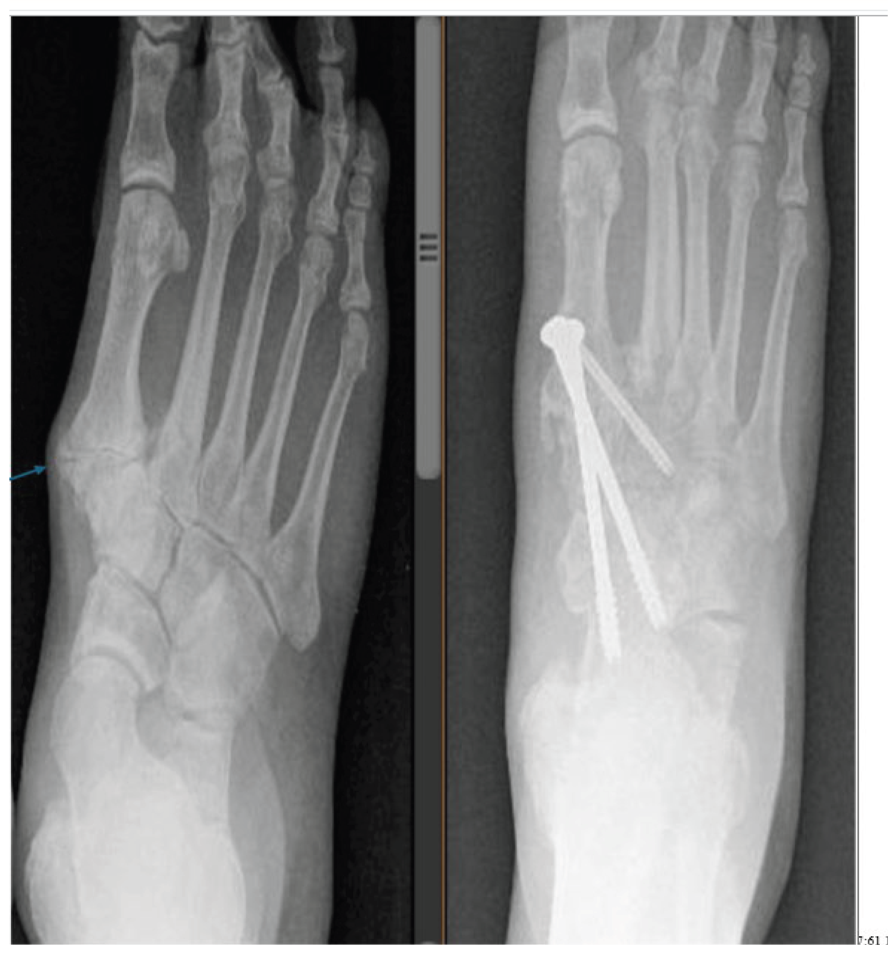
11. When the valgus deformity on weight bearing radiographs spans across the talonavicular, and tarsometatarsal joint, a long medial column fusion from talus to the first metatarsal is an option (Figure 3).
12. In relatively younger patients when the deformity is only partially fixed with no established osteoarthritis of the talonavicular jointly lateral column lengthenin may suffice or when the amount of talonavicular joint uncover age is above 40%. The amount of lengthening needed in the lateral column should be judged intraoperatively by the amount of correction of the uncoverage and by adequate residual passive eversion range of motion of the subtalar joint. When titrating the amount of correction of abduction deformity intraoperatively, the presence of adduction at the talonavicular joint on simulated weightbearing fluoroscopic assessment imaging is an important sign of hypercorrection and higher risk for lateral column.overload. The typical range for performing a lateral column lengthening is between 5 and 10 mm to achieve an adequate amount of talonavicular coverage.[19]
13. Medial column fusion of the foot is a surgical procedure aimed at correcting deformities such as flatfoot by stabilizing the medial arch of the foot. This procedure involves fusing the bones of the medial column, typically the talus, navicular, and medial cuneiform, to create a solid joint. It is particularly useful for treating conditions like arthritis, fractures that have not healed, and other foot deformities. The surgery is performed under general anaesthesia, and the goal is to reduce pain, improve and restore stability to the foot.
The technique medial column fusion:
The procedure is performed under anaesthetics and tourniquet through a medial incision, extending from the talar region to the first metatarsal base. If subtalar arthrodesis is added, it is performed through the same incision, protecting the neurovascular structures.
Preliminary decision as to which joints to fuse is made analysing clinical and radiological deformities.
Guided image is used to ensure the level identified and the placement of the fixation implant, plate and screws.
The fusion procedure is undertaken by removing the articular surface, compressing the joint and adding bone graft, if need be, prior to the fixation.
It is vital that, the talonavicular joint is reduced in supination prior to the internal fixation. Plates and screws are used for fixation. Talonavicular plate or medial column fusion plates are chosen according to the extent of arthritis, deformity, and planned fusion. The subtalar joint is fused in neutral, or 5-degree valgus heel position using two auto fix Stryker headless screws or cannulated 6.5 screws. The C-arm is used preoperatively to check if the Meary angle, the calcaneus tilt angle has improved.
After surgery, a below knee plaster of Paris is applied to be kept for 2 weeks to be changed to a light below knee cast after two weeks and after wound check-up,
The patient is allowed mobilizing without weight bearing for 6-12 weeks or until bone bridging across the fusion is seen.
After 6 weeks of surgery a below knee boot is applied, if appropriate gradual weight bearing in walking boot is then allowed. The boot is discarded after 6 weeks. Deep vein prophylaxis using subcutaneous Fragmin 5000 for 6 weeks.
Standing foot and ankle check X-rays are taken at 6 weeks, three and six months after surgery to evaluate the fusion and implant complications.
At the final follow-up, pain relief and patient satisfaction in addition to Manchester Oxford foot score is recorded.
Results are assessed on subjective measure and objective measure, union rate across the joints with attempted fusion.
The results are considered good, if the pain relief and the deformity correction is significant and the Manchester Oxford score has improved (mean 45) and there are radiological signs of angle improvement and union with minimal complication not affecting the outcome.
Fair results are considered for patients who have developed mild recurrence of the pesplanovalgus deformity not necessitating revision surgery. Two patients had mild recurrence of valgus deformity proximal to cuneiform metatarsal fusion.
Poor results are considered for patients who continue to have pain, and the deformity is not corrected, and further surgery deem to be necessary.
Fusion rate seen on CT scan or plan radiographs has shown that single-Tarsometatarsal (TMT) joint surgeries had a significantly greater proportion of fusions occurring by 12 weeks than the multiple-TMT-joint surgeries, 74.9% vs 67.0%, respectively, P = .0002. Many non-union were asymptomatic; the combined asymptomatic non-union and union rate was 95.4%. The revision rate for all non-union was 25.5%. Staple fixation was associated with higher non-union rates in the second (46.15%) and third (37.5%) TMT joints, whereas screw fixation showed the highest non-union at the first TMT joint (54.6%). The lowest non-union rates were observed with combined interfragmentary screw fixation and plating across all joints. Diabetic patients and current smokers experienced higher complication and non-union rates. Although the radiographic non-union rate (18.2%) exceeded prior reports, symptomatic non-union was rare (4.6%). Multiple MT-joint surgeries required more time to confirm radiographic union than single joint surgeries. Among the modifiable factors, implant and graft choice were associated with differences in union rates.[17]
Reported Complications:
Fusion surgery for degenerative pesplanovalgus is daunting and time consuming with a significant complication rate. Infection, venous thromboembolism, wound problems, neurovascular deficit and others. Persistent of leg swelling can take as long as 12-24 months.
Non-union, recurrence of deformity and implant related problems; remain to be of considerable concern resulting in patient dissatisfaction. Screw breakages however can be found accidentally on radiographs of the foot and may be asymptomatic requiring no further surgery. It is vital that the patient is informed of the need of further revision surgery if need be.
Discussion
Post-degenerative valgus foot deformity is a challenging condition which often requires surgical intervention if conservative management fails, however there is no one procedure of choice and all the options are fraught with relatively high complication rate. Triple arthrodesis used to be considered the procedure of choice however, the deformity caused by this condition is multifaceted and triple arthrodesis may not be sufficient. Several complications are reported, non-union of fusion surgery, under or over corrected deformity and secondary arthritis of the remaining joints of the mid foot and the ankle joint, infection of surgical site and deeper, and the failure of internal fixation. Wallinget al performed triple-joint arthrodesis on twenty-five patients with flatfoot [l l]. One patient (4%) had talonavicular joint non-union after operation; two patients (8%) had calcaneal varus deformity due to the inadequate correction of the flatfoot; one patient (4%) had no significant improvement in pain after operation, and the arch of foot failed to return to normal height. Mann et al performed 3joint fusion on eighteen cases of adult flatfoot. At the last follow-up, the AOFAS score increased significantly, but three patients developed bone non-union [12-2016].
Medial column fusion with the inclusion of the peak of valgus deformity does seem to improve the deformity; however, in a biomechanical study it is found that medial column fusion in isolation as compared to a combined medial and lateral column fusion, may increase plantar and calcaneocuboid joint pressure [16].
The armamentarium for the medial column fusion is varied, and a bolt has been tried for Charcot foot with valgus deformity. The medial column bolt provides satisfactory correction of the deformity but failed to provide adequate fixation for fusion in CN deformities in the foot. In its present form, we cannot recommend the routine use of this bolt.
Progressive collapsing foot deformity (PCFD; commonly referred to as flatfoot deformity) is a complex condition classically characterized by hindfoot valgus, midfoot abduction, and forefoot varus. Medial column arthrodesis can be used to reliably correct severe, arthritic, and unstable PCFD involving the medial column. Although both naviculocuneiform arthrodesis and talonavicular arthrodesis have their own indications, patient selection and careful radiographic and clinical assessment are crucial for any medial column arthrodesis.
The medial column bolt provided satisfactory correction of the deformity but failed to provide adequate fixation for fusion in Charcot deformities in the foot. In its present form, we cannot recommend the routine use of this bolt [18].
The results of this small cohort series suggest that lag screw with plantar plate NC arthrodesis yielded improved short-term radiographic and clinical outcomes in PCFD patients with medial arch collapse through the Naviculocunform joint. [19]
The results of double arthrodesis (talonavicular and subtalar joints) through a medial approach were better. Brilhault et al, evaluated fourteen patients treated using double arthrodesis. The average follow-up time was less than 21.5 months [13]. All deformities were well corrected. None of them had bone non-union. Jackson followed up eight patients with 100% of the fusion rate and whose average fusion time was 5.25 months. After joint fusion and osteotomy, twenty-six patients were followed up for the last time [14]. Calcaneal valgus deformity and flat arch deformity were corrected. In the current series, the calcaneocuboid fusion was not performed in most of the patients without any complications, the duration of the operation was shorter and only one incision was used. In a cadaveric study, using medial and lateral column fusion, it was found that the combination of fusion significantly increased lateral and calcaneocuboid pressures with loading compared with isolated medial column fusion [15]. Some believe that non-inclusion of the calcaneocuboid joint does not have a negative impact in the correction of rigid pesplanovalgus.
Lateral column lengthening (Evan's procedure is performed to aid in the correction and equalizations of the medial and lateral column. The longer the medial column, the more joints need to be fused to shorten the column. The medial column consists of forefoot, mid and rear foot joints, the talonavicular joint (rear foot), and naviculocuneiform and the first tarsometatarsal joints (forefoot). The loss of medial column support can be viewed as having either a direct or indirect causal effect on pronation during the gait cycle.
Bearing in ming that biomechanical studies suggest that Medial and lateral column fusion significantly increased lateral and calcaneocuboid pressures with loading compared with isolated medial column fusion and the intact state. No difference was observed between isolated medial column fusion and the intact state [21]. There is some evidence that bone grafting can aid in the success of the fusion of [22].
In conclusion; Identifying the peak of valgus foot deformity, and any mechanical lower limb malalignment which may be leading to pesplanovalgus deformity and planning corrective surgery, would be beneficial. Deformity at the knee or the ankle may lead to valgus foot deformity and must be addressed accordingly.
Declaration of interest
none.
References
- Kohls-Gatzoulis, J.; Woods, B.; Angel, J.C.; et al. The prevalence of symptomatic posterior tibial tendon dysfunction in women over the age of forty in England. Foot Ankle Surg. 2009, 15, 75–81. [Google Scholar] [CrossRef]
- Orr, J.D.; Nunley, J.A. Il Isolated Spring ligament failure as a cause of adult-acquired flatfoot deformity. Foot Ankle Int. 2013, 34, 818–23. [Google Scholar] [CrossRef]
- Abousayed, M.M.; Alley, M.C.; Shakked, R.; et al. Adult-acquired flatfoot deformity: etiology, diagnosis, and management. Jbjs Rev. 2017, 5, e7. [Google Scholar]
- Bluman, E.; Myerson, M. Stage IV posterior tibial tendon rupture. Foot AnkleC1in 2007, 12, 341–363. [Google Scholar] [CrossRef]
- Lee, M.S.; Vanore, J.V.; Thomas, J.L.; et al. Diagnosis and treatment of adult flatfoot. J. Foot Ankle Surg. 2005, 44, 78–113. [Google Scholar] [CrossRef]
- Tang, K.L.; Zhou, J.B.; Yang, H.F.; et al. Triple arthrodesis with osteotomy for the treatment of stage Il B and stage Ill adult-acquired flatfoot deformity. Zhonghua Yi Xue Za Zhi 2010, 90, 2313–6. [Google Scholar]
- Yang, Zongyu; Liu, Fei; Cui, Liang, MD; Liu, Heda, MD; Zuo, Junshui, MD; Liu, Lin, MD; Li, Sentian. Adult rigid flatfoot. Triple arthrodesis and osteotomy. In Medicine (Baltimore); McCormick, Jeremy J, Ed.; Feb 2020; Volume 99, 7, p. el 8826. [Google Scholar]
- Loretta B Chou, L.; Halligan, Benjamin W. Treatment of severe, painful pes planovalgus deformity with hindfoot arthrodesis and wedge-shaped tricortical allograft. Foot Ankle 28(5), 56. [CrossRef]
- Kang 1, Steve; Charlton, Timothy P; Thordarson, David B. Lateral column length in adult flatfoot deformity. Foot Ankle Int. Epub. 2013, 34(3), 392–7. [Google Scholar] [CrossRef]
- Faraj, Adnan. Surgical Treatment of Adult Acquired Pesplanovalgus Deformity Report of 15 Patients. J. Orthop. Bone Disord. 2023, 7(4), 1–6. [Google Scholar] [CrossRef]
- Johnson, Jeffrey E. Medial column procedures in the correction of adult acquired flatfoot deformity. Foot Ankle Clin. 2012, 17(2), 28. [Google Scholar] [CrossRef]
- Walling, A.K.; Fortin, P.T. Triple arthrodesis. Clin. Orthop. 1999, 91–9. [Google Scholar] [CrossRef]
- Mann, R.A.; Graves, S.C.; Graves, K.O. Triple arthrodegis in older adults. Results after long term follow up. J. Bone Jt. Surg. Am. 1993, 5, 355–62. [Google Scholar] [CrossRef]
- Brilhault, J. Single medial approach to modified double arthrodesis in rigid flatfoot with lateral deficient skin. Foot Ankle Int. 2009, 30, 21–6. [Google Scholar] [CrossRef]
- Jackson, W.F.; Tryfonidis, M.; Cooke, P.H.; et al. Arthrodesis of the hindfoot for valgus deformity: an entirely medial approach. Bone Jt. Surg. (Br) 2007, 89, 925–7. [Google Scholar]
- Nadaud, Joshua P; Parks, Brent G; Schon, Lew C. Plantar and calcaneocuboid joint pressure after isolated medial column fusion versus medial and lateral column fusion: a biomechanical study. Foot Ankle Int. 2011, 32(11), 1069–74. [Google Scholar] [CrossRef]
- Derner, Brian; Wolfe, Josh; Scott, Ryan. Union Outcomes in Medial and Central Column Tarsometatarsal Fusions: Single vs Multiple Joints. FAI 2026, Volume 47(Issue 5), 12–18. [Google Scholar]
- Butt, D. A.; Hester, T.; Bilal, A.; Edmonds, M.; Kavarthapu, V. The medial column Synthes Midfoot Fusion Bolt is associated with unacceptable rates of failure in corrective fusion for Charcot deformity: Results from a consecutive case series. Bone Jt. J. 2015, 97-B(6), 80913. [Google Scholar]
- Wininger, Austin E; Klavas, Derek M; Gardner, Stephanie S; Ahuero, Jason S; Harris, Joshua D; Varner, Kevin E. Plantar Plating for Medial Naviculocuneiform Arthrodesis in Progressive Collapsing Foot Deformity. Foot Ankle Orthop. 2022, 7(1), 24730114221088517. [Google Scholar] [CrossRef]
- Thordarson, D.B.; Schon, L.C.; de Cesar Netto, C.; Deland, J.T.; Ellis, S.J.; Johnson, J.E.; Myerson, M.S.; Sangeorzan, B.J.; Hintermann, B. Consensus for the Indication of Lateral Column Lengthening in the Treatment of Progressive Collapsing Foot Deformity. Foot Ankle Int. 2020, 41(10), 1286–1288. [Google Scholar] [CrossRef]
- Nadaud, Joshua P; Parks, Brent G; Schon, Lew C. Plantar and calcaneocuboid joint pressure after isolated medial column fusion versus medial and lateral column fusion: a biomechanical study. Foot Ankle Int. 2011, 32(11), 1069–74. [Google Scholar] [CrossRef]
- Dariusch Arbab , Bertil Bouillon , Sebastian Schilde , Natalia Gutteck , Philipp Lichte , Eugen Ulrich [Talonavicular arthrodesis] Operative Orthopädie und Traumatologie 38(2), Seiten/Artikel-Nr.:91-103 [Article in German].
Figure 1.
pesplanovalgus deformity at the Talonavicular area managed with fusion of the joint.
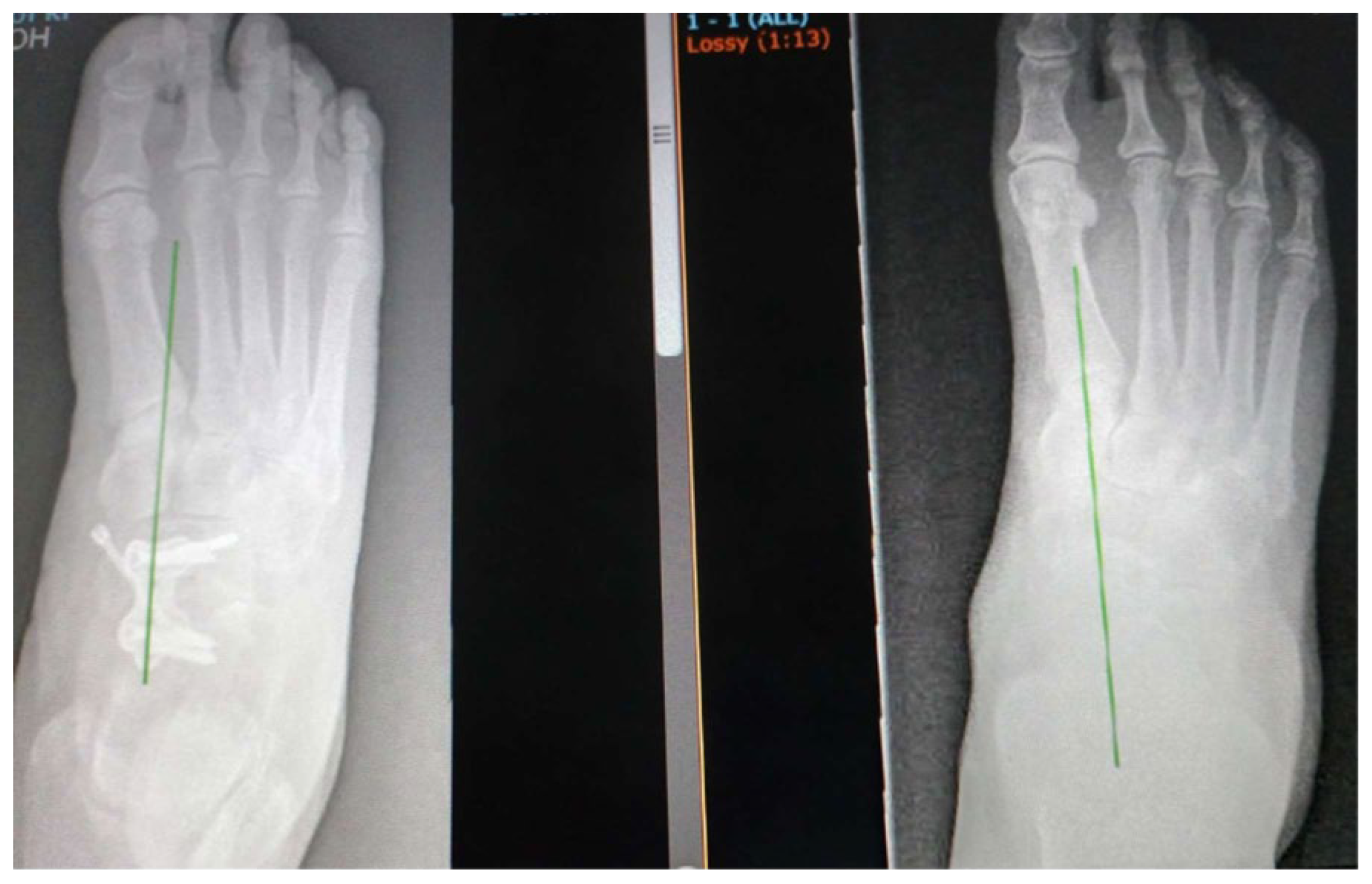
Figure 2.
tarsometatarsal fusion to correct degenerative pesplanovalgus.
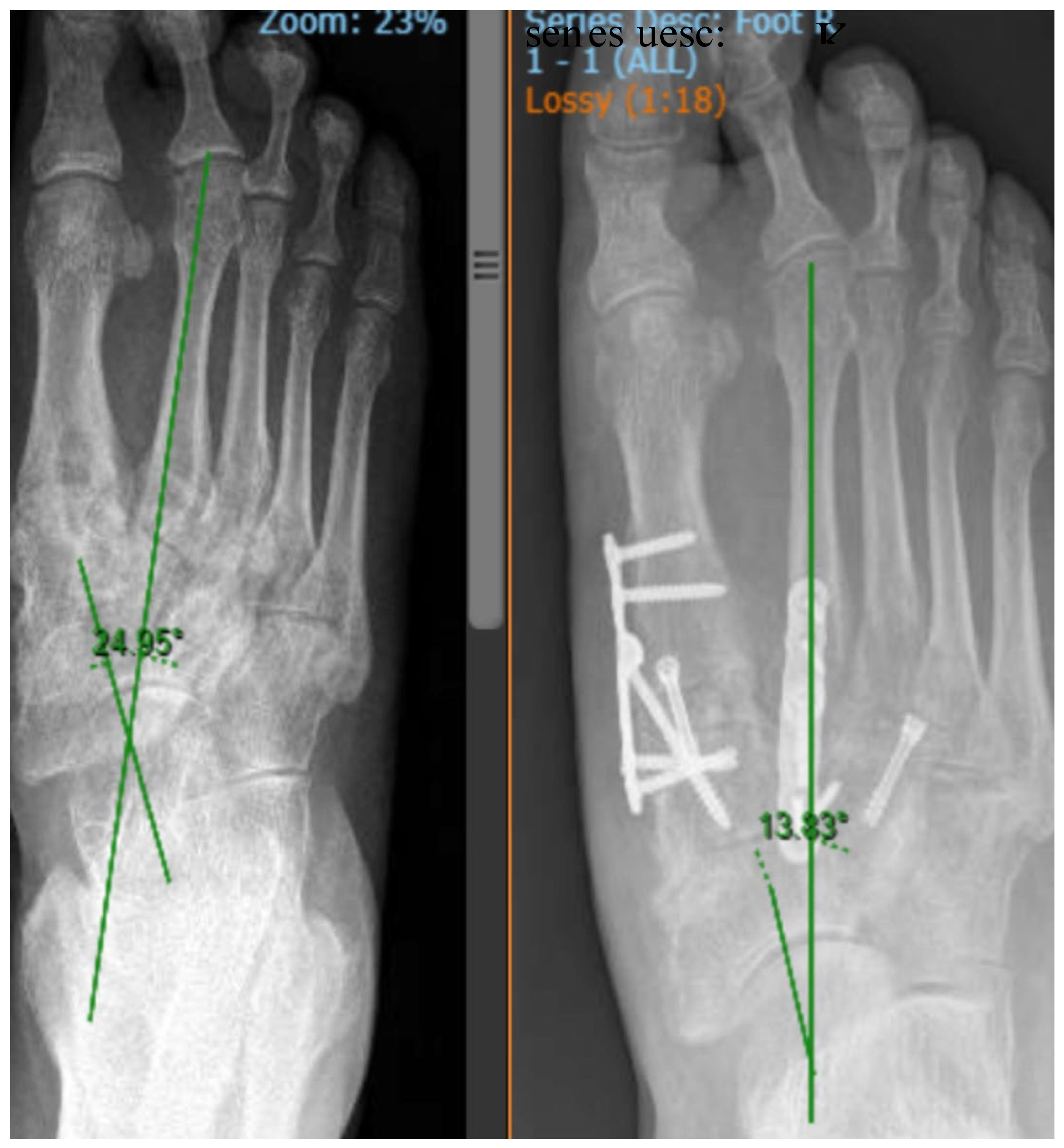
Figure 3.
medial coulmn fusion involving the fist metatarsal, navicula. Talar fusion. The peak of the deformity is at the first tarsometatarsal joint, which has to be involved in the fusion.
Figure 3.
medial coulmn fusion involving the fist metatarsal, navicula. Talar fusion. The peak of the deformity is at the first tarsometatarsal joint, which has to be involved in the fusion.
Disclaimer/Publisher’s Note: The statements, opinions and data contained in all publications are solely those of the individual author(s) and contributor(s) and not of MDPI and/or the editor(s). MDPI and/or the editor(s) disclaim responsibility for any injury to people or property resulting from any ideas, methods, instructions or products referred to in the content. |
© 2026 by the authors. Licensee MDPI, Basel, Switzerland. This article is an open access article distributed under the terms and conditions of the Creative Commons Attribution (CC BY) license (http://creativecommons.org/licenses/by/4.0/).
Copyright: This open access article is published under a Creative Commons CC BY 4.0 license, which permit the free download, distribution, and reuse, provided that the author and preprint are cited in any reuse.