
Preprint
Review

This version is not peer-reviewed.

Orthodontic Screw–Assisted Intermaxillary Fixation for Mandibular Fracture Fragments: A Narrative Review and Technical Rationale for a Minimally Invasive Intraoral Method

Submitted:

26 August 2026

Posted:

27 August 2026

You are already at the latest version

Abstract

Background: Mandibular fractures are among the most common injuries encountered in oral and maxillofacial trauma and frequently require restoration of occlusion, stabilization of bone fragments, and maintenance of intermaxillary relationships during healing. Conventional Erich arch bars remain a widely used method of maxillomandibular fixation (MMF), but their application is associated with prolonged operative time, gingival and mucosal trauma, plaque accumulation, needle-stick risk to the operator, and reduced patient comfort. Bone-supported intermaxillary fixation (IMF) screws have been introduced as an alternative, yet screw-related complications — including root injury, mucosal overgrowth, loosening, and mechanical failure — continue to limit their universal adoption. Objective: This narrative review synthesizes the clinical, morphological, biomechanical, technical, and practical rationale for a proposed method of mandibular fracture stabilization using self-tapping titanium orthodontic screws, with heads placed intraorally in the alveolar process of the maxilla and mandible, connected by elastic intermaxillary traction. Methods: A narrative literature review was undertaken across five thematic domains: mandibular fracture management, Erich arch bars, intermaxillary fixation screws, orthodontic miniscrews, and the biomechanics of elastic maxillomandibular fixation. The proposed technique was then appraised against these existing fixation concepts and their documented clinical limitations. Results: The reviewed evidence indicates that screw-based intermaxillary fixation can shorten operative time, facilitate oral hygiene, and avoid extensive circumdental wiring relative to conventional arch bars. A closely related precedent already exists in the literature in the form of a published technical report combining IMF screws with orthodontic elastic chains; the present proposal extends this concept by using orthodontic titanium screws placed within explicitly defined interradicular safe zones — approximately 8 mm from the gingival margin and at least one tooth away from the fracture edge — connected by elastic rings applied between opposing screw heads to control the maxillomandibular relationship. Morphologically, placement within interradicular alveolar bone requires careful evaluation of root anatomy, cortical thickness, keratinized mucosa, and fracture configuration. Biomechanically, elastic intermaxillary traction distributes force between the maxilla and mandible while permitting controlled stabilization of the repositioned mandibular fragments. Conclusion: Orthodontic screw-assisted intermaxillary fixation, understood as a refinement of an approach already shown to be feasible in limited clinical use, represents a promising, minimally invasive intraoral strategy for selected mandibular fractures, with potential advantages that include reduced intraoral metal bulk, improved hygiene access, simplified elastic control, and controlled stabilization of mandibular fragments. Prospective clinical trials, cone-beam computed tomography (CBCT)–based anatomical safety studies, and dedicated biomechanical analyses are required before the method can be recommended for broad clinical use.
Keywords: 
;  ;  ;  ;  ;  ;  ;  ;  

1. Introduction

Mandibular fractures constitute a major component of oral and maxillofacial trauma because the mandible is a mobile, prominent, load-bearing facial bone that participates in mastication, speech, occlusion, airway support, and lower facial contour. Contemporary retrospective series continue to identify the mandible as one of the most frequently fractured facial bones, most commonly caused by assaults, falls, and road traffic accidents, with the parasymphyseal, angle, and condylar regions most often involved [19]. Treatment aims to restore anatomical continuity, re-establish dental occlusion, stabilize bone fragments, preserve neurosensory function, and permit bone healing with minimal morbidity. In dentate patients, occlusion remains one of the most reliable intraoperative and postoperative references for the accuracy of reduction.
Maxillomandibular fixation (MMF), also termed intermaxillary fixation (IMF), has historically occupied a central role in mandibular fracture management. It may serve as a closed treatment modality in selected fractures, as an intraoperative aid during open reduction and internal fixation (ORIF), or as a postoperative stabilization method. Traditional Erich arch bars secure the dental arches effectively but require circumdental wiring, occupy considerable intraoral space, increase plaque retention, and may cause soft-tissue trauma. These limitations have driven the development of bone-supported alternatives, including IMF screws and hybrid arch-bar systems.
Orthodontic miniscrews and self-tapping titanium screws have become widely used in orthodontics and maxillofacial surgery as temporary anchorage devices. Their small dimensions, ease of placement, potential for immediate loading, and intraoral accessibility make them attractive for controlled elastic traction. Translating this principle to mandibular fracture stabilization, however, requires careful attention to anatomical safety, biomechanical force vectors, mucosal tolerance, screw stability, and the intended duration of fixation.
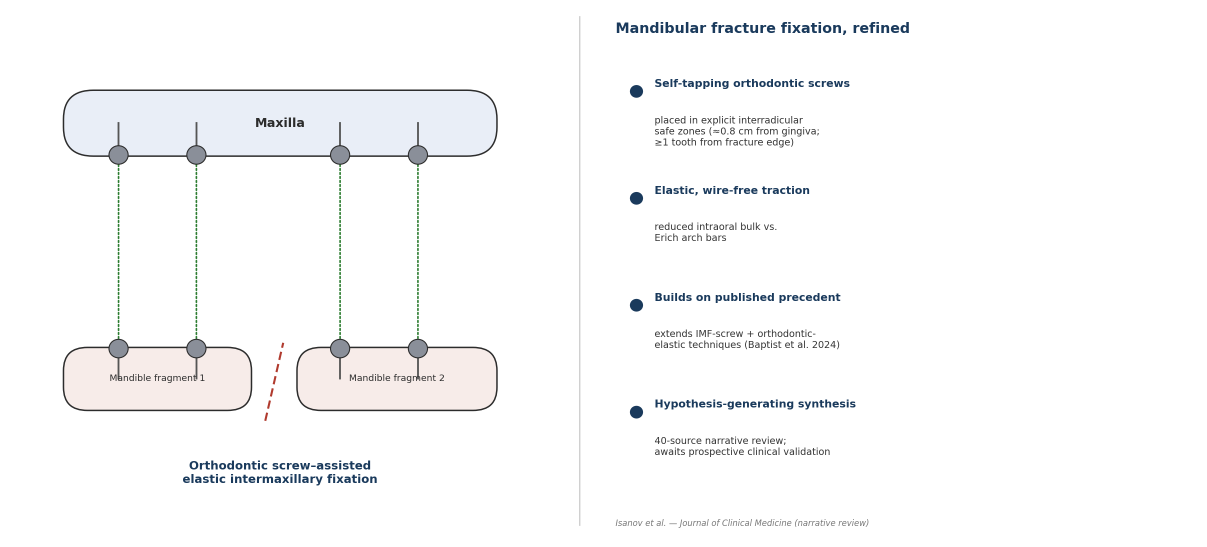
This review discusses a proposed method of mandibular fracture fixation based on the placement of self-tapping titanium orthodontic screws, with heads positioned in the alveolar process of the lower and upper jaws, followed by elastic intermaxillary fixation. The method is intended to reduce the disadvantages of bulky metallic arch-bar constructions while preserving a clinically useful maxillomandibular stabilization effect.
As a narrative review with technical rationale, this article does not report original clinical, cadaveric, or biomechanical data of its own; the proposed method is presented as a synthesis-derived, hypothesis-generating concept, positioned within the existing evidence base and intended to guide future prospective validation rather than to document a tested clinical outcome.

2. Materials and Methods

This article was designed as a narrative review rather than a systematic review. The literature was considered across five thematic domains: mandibular fracture stabilization, conventional Erich arch bars, intermaxillary fixation screws, orthodontic miniscrews, and the biomechanical principles of elastic maxillomandibular fixation. Priority was given to systematic reviews, randomized clinical trials, comparative studies, clinical audits, and technical references in oral and maxillofacial surgery.
The conceptual analysis was organized along the following dimensions: clinical indication, anatomical and morphological safety, biomechanical stability, technical reproducibility, patient hygiene, potential complications, and future validation requirements. The proposed method was then compared with its closest existing analogues, particularly Erich arch bars and bone-supported IMF screws.
Because no validated checklist exists for reporting narrative reviews in oral and maxillofacial surgery, the structure and reporting transparency of this article were guided by the Scale for the Assessment of Narrative Review Articles (SANRA), which addresses the explanation of topic importance and review aims, the description of the literature search, and the referencing and evidence presentation used throughout the manuscript [13].
The literature search was conducted iteratively rather than as a single pre-registered protocol, consistent with the narrative (non-systematic) design of this review. Searches targeted PubMed-indexed and Scopus-indexed publications using combinations of the terms “mandibular fracture,” “intermaxillary fixation,” “maxillomandibular fixation,” “Erich arch bar,” “IMF screw,” “orthodontic miniscrew,” “interradicular,” “CBCT safe zone,” and “orthodontic elastics,” supplemented by manual screening of the reference lists of the identified systematic reviews and meta-analyses. Because this was not a systematic review, no formal database-count, deduplication, or PRISMA flow diagram is reported. To help the reader weight the cited evidence appropriately despite this, a design-based risk-of-bias appraisal of the primary comparative studies is presented in Table 4; a corresponding limitation on the scope of this appraisal is stated in Section 14.

3. Clinical Background of Mandibular Fracture Fixation

The mandible is subject to complex functional loading generated by the muscles of mastication. Depending on fracture site and displacement pattern, these forces may cause rotation, shortening, fragment overlap, lingual displacement, or open bite. In tooth-bearing segments, restoration of dental occlusion is a practical and clinically important guide to anatomical reduction. Any fixation method applied to mandibular fractures must therefore immobilize bone fragments while also maintaining the correct spatial relationship between the maxillary and mandibular dental arches.
Conventional treatment options include closed reduction with MMF, ORIF with plates and screws, or a combination of both. Closed treatment may be appropriate for minimally displaced or favorable fractures, for patients with contraindications to open surgery, or in cases where occlusion can be restored and maintained without direct exposure of the fracture. ORIF is preferred when direct fragment stabilization is required, particularly in displaced, comminuted, unstable, or unfavorable fractures. Randomized comparisons of open versus closed treatment for condylar and subcondylar fractures generally favor ORIF for objective functional outcomes, while closed treatment remains an acceptable option with a more conservative risk profile [28,38], underscoring that the choice between closed and open strategies — and, by extension, the role of any intermaxillary fixation method — remains fracture- and patient-specific rather than universal.
Intermaxillary fixation remains clinically relevant even in open surgery, since it helps establish occlusion before definitive plate fixation. The method used to achieve this fixation, however, matters considerably: a bulky, plaque-retentive appliance may compromise oral hygiene, whereas an insufficiently stable system may permit malocclusion or relapse. The search for a less traumatic yet mechanically reliable intraoral fixation method therefore remains clinically important.

4. Conventional Erich Arch Bars: Strengths and Limitations

Erich arch bars provide broad dental-arch engagement and have long been considered a standard method for MMF. Their advantages include operator familiarity, relatively low cost, applicability across a wide range of occlusal situations, and the capacity for either rigid wire fixation or elastic guidance, with multiple points of attachment distributed along the dental arch.
Despite these advantages, several limitations are well recognized. Placement is time-consuming because the bar must be adapted to the dental arch and secured with circumdental wires around multiple teeth. Repeated passage and tightening of these wires can cause glove perforation and needle-stick injury to the operator. Wire ends and bar hooks irritate the oral mucosa and gingiva, and the bar itself creates a retentive surface for plaque and food debris, making oral hygiene difficult over several weeks of fixation. In susceptible patients, this can progress to periodontal inflammation, gingival recession, enamel scratching, tooth mobility, and mucosal ulceration.
The occupational risk to the surgical team is not theoretical. In a one-year survey of residents performing intermaxillary fixation, 40 needlestick injuries were recorded across 172 procedures — an incidence of 23% — most involving the maxillary left quadrant [20]. Early gloving studies similarly found that 108 of 120 outer gloves were perforated during Erich arch bar placement, with triple gloving substantially outperforming double gloving in preventing inner-glove breach [16]. A subsequent randomized controlled trial comparing hybrid, bone-supported arch bars with conventional Erich arch bars confirmed significantly fewer glove perforations and shorter placement time with the bone-supported alternative [17], reinforcing that the wire-and-bar interface itself, rather than operator technique alone, is the principal driver of this risk.
The hygiene burden deserves particular emphasis. Patients with mandibular fractures often present with pain, trismus, swelling, dietary restriction, and a reduced ability to perform effective brushing. The addition of circumdental wires and arch bars further increases the difficulty of cleaning, and in clinical practice this combination frequently results in gingivitis, halitosis, plaque accumulation, and patient discomfort.

5. Intermaxillary Fixation Screws as the Closest Analogue

The closest technical analogue to the proposed method is the use of intermaxillary fixation screws. These bone-supported devices are placed through the oral mucosa into the alveolar bone of the maxilla and mandible; wires or elastics are then applied between opposing screw heads to maintain occlusion or immobilize the jaws.
AO Surgery Reference describes skeletally based intermaxillary fixation using screws and plates as an alternative technique for maxillomandibular immobilization. It emphasizes that screw placement patterns vary according to fracture location and dentition, and that screws must be positioned within the alveolus while avoiding tooth roots, the infraorbital nerve, and the inferior alveolar nerve [1]. This is a crucial anatomical caveat, because the principal safety challenge of screw-based fixation is the limited interradicular bone available for placement.
Clinical studies support several advantages of IMF screws over Erich arch bars. A randomized clinical trial comparing Erich arch bars with IMF screws during mandibular fracture reduction reported no significant difference in occlusal stability between the two groups, while the arch-bar group required longer application and removal times and showed poorer oral hygiene parameters [3]. A separate randomized study of 20 adult patients with uncomplicated mandibular fractures, using six to eight MMF screws, found adequate fracture healing and acceptable occlusion at 5–6 weeks, although overall screw failure occurred in 25.5% of screws, with a markedly higher failure rate in closed MMF than in ORIF-assisted use [4].
These findings indicate that screw-based fixation is not uniformly superior across all clinical scenarios. Rather, it is a technique with a defined set of benefits and a defined set of risks: it may reduce operative time and improve hygiene, but it demands accurate screw placement, adequate bone support, appropriate case selection, and structured monitoring for loosening or mucosal overgrowth.
This balanced picture is echoed by a further comparative study and literature review of IMF screws versus Erich arch bars, which likewise concluded that screws reduce chair-side time and needle-stick exposure but require careful patient selection and radiographic planning to avoid root injury [21]. Additional randomized and prospective comparisons report consistent directional findings: shorter application time with screws, generally comparable or better plaque scores, and low but non-zero rates of root contact and screw fracture [35,36]. A recent systematic review and meta-analysis spanning multiple MMF techniques similarly positions bone-supported screws as a time-efficient option whose safety depends heavily on operator technique and case selection [37].
A closely related precedent for the present proposal already exists in the published literature. A 2024 technical report described a novel IMF technique in which orthodontic elastic (“E”) chains were looped around conventional IMF screws to achieve maxillomandibular fixation without wires, reporting adequate stabilization without the need for specialized wire-passing instruments [29]. A related report has separately described using a patient’s existing orthodontic brackets, rather than dedicated IMF screws, as anchorage for maxillomandibular fixation when a fracture occurs during active orthodontic treatment [33]. Together, these reports confirm that combining bone- or tooth-anchored fixation points with orthodontic elastic components is clinically feasible and has already been attempted, at least at the level of individual case reports and small case series. The present review differs from these reports chiefly in scope and specificity: it proposes self-tapping orthodontic screws (rather than dedicated IMF screws or pre-existing orthodontic brackets) as the anchorage element, defines explicit interradicular placement criteria relative to the gingival margin and fracture edge, and positions the concept within a systematic evidence synthesis rather than a single technical case description. This distinction is stated here explicitly so that the proposed method is not misread as an entirely novel category of intervention, but rather as a more detailed, anatomically constrained refinement of an approach that has already been shown to be feasible in limited clinical use.
Table 1. Positioning of the proposed method relative to conventional fixation and the closest published precedent.
Feature Erich arch bars Standard IMF screws Baptist et al., 2024 [29] Proposed method
Anchorage element Teeth (circumdental wire) Bone (generic bicortical screw) Bone (generic IMF screw) Bone (self-tapping orthodontic screw)
Force-transmission element Wire ligature Wire or elastic Orthodontic elastic chain Elastic ring
Explicit interradicular placement criteria Not applicable General (avoid roots) Not specified in the source report Explicit (≈0.8 cm from gingival margin; ≥1 tooth from fracture edge)
Reported evidence level Multiple RCTs / meta-analyses Multiple RCTs / meta-analyses Single technical report / small series None (proposed; hypothesis-generating)
Needle-stick / glove-perforation risk High [16,17,20] Low Low (anticipated) Low (anticipated)
Specialized instrumentation required No No No No
This comparison is intended to make the incremental, rather than categorical, nature of the proposed refinement explicit, in line with the honesty requirements of a narrative review (see Section 2 and Section 14).

6. Limitations of the Closest Analogue

Although IMF screws represent a logical alternative to arch bars, the published literature identifies several recurring complications, including iatrogenic root injury, screw loosening, screw fracture, mucosal coverage of the screw head, soft-tissue irritation, peri-implant infection, and reduced reliability during prolonged immobilization. A dedicated complications series specifically addressing IMF screws catalogued exactly this pattern of root damage, screw loosening, and mucosal problems as the principal drawbacks limiting routine use [22], and an early technical report on IMF screw complications similarly described root and mucosal problems as the main limiting factors shortly after the devices entered clinical use [32]. The risk of root injury is particularly relevant in the anterior and premolar regions, where interradicular spaces are often narrow: a prospective radiographic study of 232 transalveolar screws placed adjacent to 440 teeth found major root contact (more than 50% of the screw hole impinging on the root) in 11.2% of screws and minor contact in a further 15.9%, with a subset of teeth subsequently testing non-vital [23]. A dedicated systematic review of root damage associated with intermaxillary screws corroborates this order of magnitude across multiple studies and identifies screw angulation and inadequate preoperative imaging as the principal modifiable risk factors [30]. These figures illustrate that root injury is not a rare, anecdotal complication but a measurable and clinically relevant risk that any interradicular screw-placement algorithm must explicitly design around.
Early descriptions of IMF screw application already emphasized that the technique, while fast, is technique-sensitive and benefits from a dedicated, purpose-built screw design rather than adaptation of generic osteosynthesis hardware [31]. This observation is directly relevant to the present proposal, which substitutes orthodontic screws — designed from the outset for elastic-traction anchorage — for either generic IMF screws or arch bars.
A further limitation is that standard IMF screws are typically designed primarily for wire fixation rather than elastic, orthodontic-type traction. Their head geometry is not always optimized for comfortable elastic retention, and wire-based fixation can complicate emergency release. Moreover, when screws are placed without adequate consideration of fracture biomechanics, the resulting elastic vectors may fail to provide balanced stabilization.
The proposed orthodontic screw-assisted approach builds on the premise that self-tapping titanium orthodontic screws with accessible heads can function as controlled intraoral anchorage points for elastic rings. By explicitly defining the placement distance from the gingival margin and the fracture edge, and by positioning screws in antagonist zones and on the contralateral side, the technique aims to create a symmetrical and clinically manageable elastic fixation system.

7. Comparative Summary of Reviewed Authors and Evidence Synthesis

Because this article is designed as a narrative review with technical rationale rather than an original clinical study, direct statistical correlations cannot be calculated from patient-level data. The reviewed literature is first summarized comparatively across authors, and an author-based evidence correlation matrix is then constructed to show how existing authors link fixation technique, technical variables, oral hygiene, occlusal stability, screw-related complications, and patient-centered outcomes.
In the correlation tables, the term “correlation” denotes an evidence-based relationship reported or implied by previously published clinical studies, randomized trials, systematic reviews, and technical surgical references — it is not a statistical correlation coefficient derived from original data. The direction of association is classified as positive, negative, neutral, mixed, or not reported. This structure allows the proposed orthodontic screw-assisted intermaxillary fixation method to be positioned within the existing evidence base and used to generate testable hypotheses for future clinical studies.
Table 2. Comparative summary of the principal authors and sources reviewed.
Author(s) / source Year Study design Sample / units analyzed Comparison or focus Key finding Level of evidence*
AO Surgery Reference [1] Technical / expert reference Not applicable Bone-supported IMF screws vs wire-based MMF Defines screw placement principles and anatomical structures to avoid. Level 5
AO closed-treatment protocol [2] Technical / expert reference Not applicable Elastic vs wire MMF duration Recommends 4-week heavy elastics/wires, then lighter training elastics; ≤6 weeks total. Level 5
Fernandes et al. [3] 2023 Randomized clinical trial Patients undergoing ORIF (n as reported) Erich arch bars vs IMF screws Comparable occlusal stability; arch bars slower to apply/remove and more biofilm. Level 2b
West et al. [4] 2014 Prospective randomized study 20 patients, 106 screws MMF screws in open vs closed treatment 25.5% overall screw failure; failure linked to low torque, closed treatment, posterior site. Level 2b
Falci et al. [5] 2015 Systematic review Multiple pooled studies Erich arch bars as the reference MMF method Evaluates whether arch bars remain the best available MMF method. Level 1a
Fernandes et al. [6] 2021 Meta-analysis Multiple pooled studies Erich arch bars vs intermaxillary screws Synthesizes comparative outcomes in fractures involving dental occlusion. Level 1a
Jain and Rai [7] 2021 Systematic review and meta-analysis 8 included studies Bone-supported arch bars vs Erich arch bars Screws faster to apply; hygiene difference not statistically significant. Level 1a
Nandini et al. [8] 2011 Comparative clinical study Clinical cohort (as reported) Self-tapping screws vs Erich arch bars Compares efficacy, complications, and indications of both methods. Level 3b
Purmal et al. [11] 2013 CBCT volumetric mapping study 193 patients Safe vs danger zones for IMF screw placement Defines mesiodistal/buccolingual safe zones for 1.0×7 mm screws. Level 4
Tepedino et al. [12] 2020 Systematic review and meta-analysis Multiple pooled CBCT studies Interradicular sites for orthodontic miniscrews Posterior mandible offers more interradicular bone than the maxilla. Level 1a
Fabbroni et al. [23] 2004 Prospective radiographic study 232 screws / 440 teeth Root contact during transalveolar screw placement Major root contact in 11.2% and minor contact in 15.9% of screws. Level 4
Bali et al. [20] 2011 Prospective survey 172 IMF procedures Needlestick injury incidence during IMF 23% procedure-level incidence of needlestick injury. Level 4
Papageorgiou et al. [14] 2012 Meta-analysis 4987 miniscrews / 2281 patients Orthodontic miniscrew failure and risk factors Overall failure rate 13.5%; failure linked to jaw of insertion. Level 1a
Baptist et al. [29] 2024 Technical report / case series Not applicable IMF screws + orthodontic E-chains Novel wireless technique combining screws with elastic chains; closest published precedent. Level 4
Rai et al. [36] 2011 Randomized clinical study Clinical cohort (as reported) MMF screws vs Erich arch bars, plaque index Plaque index lower with screws (1.88 vs 2.69); root damage 5.81% of screws. Level 2b
Kalluri et al. [37] 2024 Systematic review and meta-analysis Multiple pooled studies Outcomes across MMF techniques Positions bone-supported screws as time-efficient; safety operator-dependent. Level 1a
* Simplified Oxford Centre for Evidence-Based Medicine (OCEBM)-style classification, assigned by the authors for orientation only: 1a = systematic review/meta-analysis of comparative studies; 2b = individual randomized trial; 3b = individual comparative/cohort study; 4 = cross-sectional anatomical/imaging study; 5 = expert opinion or technical reference. Not a formal risk-of-bias assessment (see Section 14, Limitations).
Table 3. Author-based correlation between fixation method and clinical-technical outcomes.
Author / source Study type / evidence level Main fixation method analyzed Reported relationship between technique and outcomes Author-based correlation relevant to the proposed method
AO Surgery Reference Technical surgical reference Bone-supported maxillomandibular fixation with IMF screws Screw placement must avoid tooth roots, the infraorbital nerve, and the inferior alveolar nerve; screws should be placed symmetrically in opposing jaws; long-term immobilization is not recommended because of mucosal injury risk. Safe interradicular screw positioning is negatively associated with anatomical injury risk; symmetrical screw distribution is positively associated with controlled intermaxillary stabilization.
Fernandes et al., 2023 [3] Randomized clinical trial Erich arch bars vs IMF screws during mandibular fracture reduction No significant difference in occlusal stability between arch bars and IMF screws; application and removal times were longer with arch bars; the arch-bar group showed higher biofilm and poorer hygiene status. Screw-based fixation is negatively associated with application/removal time and plaque-retentive burden, with no clear negative association with occlusal stability in selected fractures.
West et al., 2014 [4] Prospective randomized study, 20 adult patients, 106 screws MMF screws in open and closed treatment of uncomplicated mandibular fractures All patients achieved adequate fracture healing and acceptable occlusion at 5–6 weeks; overall screw failure was 25.5%, with 40% failure in the closed-MMF group versus 6% in the ORIF group; lower insertion torque, closed treatment, and posterior placement significantly affected failure. Screw stability is positively associated with adequate insertion torque and negatively affected by posterior placement, closed-treatment use, and reduced patient compliance.
Nandini et al., 2011 [8] Comparative clinical study Self-tapping IMF screws vs Erich arch bars The study compared efficacy, advantages, disadvantages, indications, and complications of Erich arch bars and self-tapping IMF screws in mandibular fracture management. Self-tapping screw fixation is clinically relevant as an alternative to conventional arch bars, but requires careful assessment of complications and indications.
Jain and Rai, 2021 [7] Systematic review and meta-analysis, 8 included studies IMF screws vs Erich arch bars IMF screws required significantly less time to achieve MMF, but no statistically significant difference in oral hygiene status was found between groups; complications such as mucosal coverage, root injury, screw loosening, and screw fracture limited the certainty of recommending IMF screws as a universal alternative. Evidence supports faster application with screw-based fixation, but the correlation between screw use and improved hygiene remains mixed; screw-related complications must be treated as limiting variables.
AO closed-treatment protocol [2] Technical surgical reference MMF with elastics or wires Heavy elastics or wires are commonly used for approximately 4 weeks, followed by lighter training elastics; MMF is usually maintained for no more than 6 weeks. Duration and intensity of intermaxillary fixation are positively associated with stabilization needs, but prolonged fixation may increase hygiene and soft-tissue risk.
Purmal et al., 2013 [11] CBCT volumetric mapping study, 193 patients 3D safe/danger zone mapping for IMF screw placement A 1.0 mm × 7 mm screw could be placed safely distal to the maxillary canines and <8 mm between the molars; in the mandible the safest sites were distal to the first premolar (>5 mm) and distal to the second premolar (>2 mm); right–left differences were significant (p<0.05). Provides direct anatomical validation for interradicular, image-guided screw positioning and supports the proposed gingival-offset distance as anatomically conservative.
Note: “Author-based correlation” reflects a qualitative synthesis of the cited authors’ own reported associations, not a statistic computed by the present authors.
Table 4. Design-based risk-of-bias considerations for the primary comparative studies cited.
Study Design Randomization reported Sample size Main design-related limitation Design-based risk level*
Fernandes et al., 2023 [3] RCT Yes Not stated in abstract Single-center; blinding of outcome assessors not specified Moderate
West et al., 2014 [4] Prospective randomized Yes 20 patients / 106 screws Small sample; mixed open/closed subgroups reduce precision Moderate
Rai et al., 2011 [36] RCT Yes Not stated in abstract Single-center; outcome assessment method not detailed in abstract Moderate
Nandini et al., 2011 [8] Comparative clinical study Not stated 20 patients Small sample; randomization method not reported Moderate–High
Singh et al., 2010 [28] RCT Yes 40 patients Single-center; condylar/subcondylar subgroup only Moderate
Purmal et al., 2013 [11] Cross-sectional (CBCT) Not applicable 193 patients Retrospective imaging sample; selection criteria not detailed in abstract Moderate
Jain and Rai, 2021 [7] Systematic review + meta-analysis Not applicable 8 pooled studies Pooled studies of heterogeneous design; formal GRADE not reported in abstract Low–Moderate
Bins et al., 2015 [34] Systematic review Not applicable 22 pooled studies (17 case series, 4 RCTs, 1 cohort) Authors themselves rated included RCTs as low methodological quality Moderate–High
* This is a design-based appraisal derived from the information reported in each study’s published abstract, not a formal item-by-item Cochrane RoB 2 or ROBINS-I assessment with full-text dual review; see Section 14 for this limitation. Risk levels are indicative rather than definitive and are intended to help the reader weight the evidence synthesized in Table 4 and Table 5.
Table 5. Direction of author-based correlations for key variables.
Variable / exposure Outcome Expected direction Supporting authors / sources Interpretation for the proposed method
Interradicular screw placement outside root projection Root injury risk Negative AO Surgery Reference; West et al. Correct placement outside root projection should reduce the risk of root trauma.
Symmetrical placement of screws in opposing jaws Occlusal control Positive AO Surgery Reference Bilateral, antagonist-based screw positioning may improve distribution of fixation vectors.
Use of screw-based MMF instead of Erich arch bars Application time Negative Fernandes et al.; Jain and Rai Screw-based fixation is generally associated with shorter application time than arch bars.
Use of screw-based MMF instead of Erich arch bars Removal time Negative Fernandes et al. Screw removal is expected to be faster than arch-bar removal.
Use of arch bars Dental biofilm / plaque retention Positive Fernandes et al. Bulky arch bars and circumdental wires may increase plaque accumulation and hygiene difficulty.
Use of IMF screws Oral hygiene improvement Mixed Fernandes et al.; Jain and Rai Some studies report better hygiene with screws, whereas meta-analytic evidence shows insufficient consistent support.
Lower insertional torque Screw failure Positive West et al. Reduced primary stability increases the probability of screw loosening or failure.
Posterior jaw screw placement Screw failure Positive West et al. Posterior placement may carry higher failure risk, plausibly due to force vectors, bone anatomy, and functional loading.
Closed MMF treatment using screws Screw failure / noncompliance Positive West et al. Screw-only closed treatment may be more vulnerable to failure than screw use as an ORIF adjunct.
Prolonged immobilization with IMF screws Mucosal damage Positive AO Surgery Reference Long-term screw-based immobilization may increase mucosal irritation or coverage of screw heads.
Elastic intermaxillary fixation Controlled occlusal stabilization Positive AO closed-treatment protocol Elastics can maintain occlusion and allow controlled stabilization when applied with appropriate vectors.
Excessive elastic traction Fragment rotation / open-bite risk Positive AO Surgery Reference Over-tightening or unbalanced force vectors may cause fragment rotation or posterior open bite.
Direct root contact during insertion Root injury / screw failure Positive Fabbroni et al.; Papageorgiou et al. Root contact is directly associated with higher failure and non-vitality rates; supports strict interradicular targeting.
Use of circumdental wiring (Erich bars) vs screws Needlestick injury risk Negative (for screws) Bali et al.; Pieper et al.; King and Christensen Wire-based fixation carries a substantially higher documented needlestick/glove-perforation risk than screw-based anchorage.
Direction is classified relative to the exposure listed in column 1, as reported or reasonably inferable from the cited source(s).
Table 6. Author-based evidence map for the proposed orthodontic screw-assisted fixation.
Domain Evidence from authors Existing limitation How the proposed method responds
Clinical domain Occlusal stability with IMF screws may be comparable to Erich arch bars in selected mandibular fractures. Existing evidence does not support unrestricted use of IMF screws for all fracture types. The proposed method is limited to selected fractures with adequate dentition, stable occlusion, sufficient interradicular bone, and good patient compliance.
Morphological domain AO emphasizes avoidance of tooth roots and neurovascular structures during IMF screw placement. Root injury and mucosal coverage remain important complications. The method specifies placement in the interradicular area, outside root projection, at approximately 0.8 cm from the gingival margin and at least one tooth away from the fracture edge.
Biomechanical domain Screw failure is associated with low insertional torque, closed-treatment use, and posterior jaw placement. Screw-based systems may fail under excessive loading or weak anchorage. The method uses multiple screws distributed across both jaws and both sides of the fracture to distribute elastic traction forces.
Technical domain IMF screws reduce application time compared with Erich arch bars. Standard IMF screws are not always optimized for elastic traction and often require wires. Orthodontic screws with accessible heads are used as intraoral anchorage points designed specifically for elastic rings.
Hygiene domain Some studies show less biofilm and easier hygiene with screw-based fixation, but meta-analytic evidence is mixed. Screw heads can still irritate mucosa or become covered if fixation is prolonged. Reduced intraoral bulk compared with arch bars may facilitate oral hygiene, but follow-up and hygiene instruction remain mandatory.
Patient-centered domain West et al. reported tolerability of MMF screws but also noted compliance problems in closed MMF treatment. Patient noncompliance may compromise closed screw-based fixation. The proposed method includes patient education, elastic-replacement instructions, hygiene monitoring, and emergency-release guidance.
Domains synthesize evidence from Section 3, Section 4, Section 5, Section 6, Section 7, Section 8, Section 9, Section 10, Section 11, Section 12 and Section 13; “How the proposed method responds” reflects the authors’ design rationale, not measured outcomes.

Interpretation of Author-Based Correlations

The correlation matrix indicates that the scientific justification for orthodontic screw-assisted intermaxillary fixation is strongest in three areas: reduction of technical complexity, reduction of intraoral bulk, and improved access for hygiene. Fernandes et al. reported comparable occlusal stability between Erich arch bars and IMF screws, while also showing that arch bars required longer application and removal times and were associated with greater biofilm accumulation. This supports the hypothesis that screw-based fixation may reduce procedure-related burden without necessarily compromising occlusal stability in selected fracture cases.
West et al. provide important cautionary evidence. Although all patients in their cohort achieved adequate fracture healing and acceptable occlusion at 5–6 weeks, screw failure occurred in 25.5% of screws overall. The authors identified lower insertion torque, closed-MMF use, and posterior jaw placement as significant factors associated with failure. In the proposed method, screw placement should therefore not be regarded as a purely mechanical step; it must be planned according to alveolar bone thickness, interradicular anatomy, force-vector direction, and the anticipated duration of fixation.
The systematic review and meta-analysis by Jain and Rai demonstrate that IMF screws significantly reduce the time required to achieve MMF, but that the evidence remains insufficient to recommend IMF screws as a universal replacement for Erich arch bars. The same review highlights complications including mucosal coverage, root injury, screw loosening, and screw fracture — findings directly relevant to the proposed technique, in that they support a balanced interpretation: orthodontic screw-assisted fixation may be advantageous, but only under controlled anatomical, technical, and clinical conditions.
AO Surgery Reference supplies the anatomical and technical framework for safe screw-supported maxillomandibular fixation, emphasizing that screw placement is constrained by the position of tooth roots and adjacent neurovascular structures, and that screws should be positioned symmetrically in opposing jaws. These principles directly inform the proposed method, in which orthodontic screws are placed within interradicular safe zones and connected by elastic rings to achieve controlled intermaxillary stabilization.

8. Morphological Rationale

The morphological basis of the method lies in the anatomy of the dentate alveolar process. In adults, interradicular alveolar bone provides potential sites for temporary screw placement, although this space varies with tooth region, root divergence, periodontal condition, patient age, and any prior orthodontic tooth movement. Safe insertion requires avoidance of the periodontal ligament, root surface, mental foramen, inferior alveolar canal, and mobile fracture margins.
Direct CBCT-based mapping data support this rationale. In a volumetric analysis of 193 patients, Purmal et al. defined safe and danger zones for IMF screw placement in the maxilla and mandible at 2, 5, 8, and 11 mm from the alveolar crest, reporting that a screw of 1.0 mm diameter and 7 mm length could be placed safely distal to the maxillary canines and less than 8 mm mesiodistally between the molars, while in the mandible the safest sites were located distal to the first premolar (more than 5 mm of interradicular width) and distal to the second premolar (more than 2 mm) [11]. These figures are consistent with — and provide independent anatomical support for — the proposed 0.8 cm gingival offset and the recommendation to avoid placement immediately adjacent to the fracture margin. A systematic review and meta-analysis of interradicular sites for orthodontic miniscrew insertion similarly concluded that the posterior mandible generally offers more interradicular bone than the maxilla, that predrilling is usually advisable in the mandible, and that available space and cortical thickness vary considerably by tooth region and vertical level, reinforcing the need for individualized, image-based site selection rather than a fixed anatomical template [12].
The proposed placement formula specifies the interradicular region, outside the projection of the tooth roots. A distance of approximately 0.8 cm from the gingival margin is intended to keep the screw within alveolar bone while maintaining adequate separation from the free gingival margin and limiting gingival compression by the screw head. Placement at least one tooth away from the fracture edge is designed to avoid the inherently unstable bone immediately adjacent to the fracture line and to reduce the risk of inserting the screw into a mobile fragment margin.
The mucosal environment must also be considered, since intraoral screws penetrate the mucosa and remain exposed throughout the fixation period. Excessive compression of mucosa by the screw head may cause ulceration, pain, or mucosal overgrowth. The screw head should therefore remain accessible rather than buried under mucosa, and should permit elastic attachment without soft-tissue strangulation.
Beyond IMF-specific data, the broader orthodontic miniscrew literature provides a useful reference range for expected failure rates of small interradicular titanium screws under functional load. Pooled meta-analytic estimates report an overall miniscrew failure rate of approximately 13.5% [14,15], with root contact during insertion associated with a substantially increased risk of failure (risk ratio up to 8.7 in one anatomical-site analysis). While these figures come from orthodontic anchorage screws rather than IMF-specific devices, they are directionally consistent with the root-contact and screw-loosening rates reported for transalveolar IMF screws [22,23] and support treating accurate interradicular placement as the single most consequential technical variable in the proposed method.

9. Biomechanical Rationale

Intermaxillary fixation functions by connecting the maxillary and mandibular arches so that mandibular movement is restricted and occlusal relationships are preserved. In the proposed method, the screws act as anchorage units, while the elastic rings act as force-transmitting elements. Unlike rigid wire ligatures, elastic rings provide continuous traction while allowing a degree of functional adaptability, depending on the number, diameter, and force of the elastics used. This anchorage principle is well established outside the trauma setting: comprehensive reviews of orthodontic miniscrew anchorage describe small titanium screws as capable of providing rigid, temporary skeletal anchorage against sustained orthodontic load, which is the same underlying mechanical requirement as elastic intermaxillary traction [25].
The biomechanical effect depends on screw distribution. Screws placed on both fragments of the mandible, in corresponding antagonist areas of the maxilla, and on the contralateral side create multiple vertical and diagonal force vectors that help maintain occlusion and reduce rotational displacement of the mandibular fragments (Figure 1). When the elastics are arranged symmetrically, the system distributes forces across several anchorage points rather than concentrating stress on a single tooth or wire loop.
The proposed minimum distance of one tooth from the fracture margin carries mechanical, not merely anatomical, significance. Screws placed too close to the fracture line risk loosening because the local bone is inherently unstable, or because micromotion at the fracture site concentrates stress around the screw. Screws placed at a more stable distance are expected to provide better anchorage and reduce the risk of fragment displacement during healing.

10. Technical Description of the Proposed Method

The proposed method begins with clinical and radiological evaluation of the fracture. Panoramic radiography, computed tomography, or cone-beam computed tomography may be used to determine fracture location, displacement, tooth-root anatomy, and safe interradicular zones. After local infiltration or regional anesthesia and antiseptic preparation, the mandibular fragments are repositioned into anatomical occlusion.
  • A guiding hole is created with a bur in the interradicular region of the mandibular alveolar process, outside the projection of the tooth roots.
  • A self-tapping titanium orthodontic screw with an accessible head is inserted using a screwdriver, positioned approximately 0.8 cm from the gingival margin and at least one tooth away from the edge of the fracture fragment.
  • Corresponding screws are placed in the second mandibular fracture fragment, in the antagonist maxillary dentition, and in the maxilla and mandible on the side contralateral to the fracture.
  • Elastic rings are applied between opposing screw heads to achieve intermaxillary fixation, arranged vertically, obliquely, or in a crossed configuration according to the fracture pattern and the desired vector of stabilization.
  • Fixation is maintained until clinical and radiographic signs of fragment stabilization are achieved, with elastics inspected and replaced at scheduled follow-up visits.
Preferred technical parameters may include titanium-alloy screws with a diameter of approximately 1.5–2.0 mm and a length of 6–10 mm, selected according to alveolar bone thickness. The guiding bur diameter may be approximately 1.0–1.2 mm. Elastic rings may have a diameter of approximately 3.2–4.8 mm; based on the general orthodontic elastics literature, light configurations (approximately 2–3 oz) are typically used for finer occlusal control, while medium-force elastics (approximately 3.5–4.5 oz) are more commonly selected for stronger sagittal or vertical correction [18]. The specific force level for this application has not been established and should be determined by future biomechanical testing rather than extrapolated directly from orthodontic tooth-movement protocols.

11. Clinical Advantages of the Proposed Method

  • Avoids circumdental wiring, reducing manipulation around multiple teeth and the amount of bulky metal retained in the mouth.
  • May simplify oral hygiene, since screw heads occupy substantially less surface area than full-arch bars.
  • Allows elastic rings to be replaced and adjusted more readily than wire ligatures.
  • May reduce application and removal time relative to traditional arch bars.
  • Remains fully intraoral and bone-supported, avoiding external incisions.
A further potential advantage is improved patient comfort: reduced intraoral bulk may facilitate speech, oral rinsing, and access for cleaning, all of which support the patient compliance that strongly influences the success of closed or semi-closed fixation methods. Elastic fixation may also offer safer emergency release than rigid wire fixation, provided the patient and caregiver are properly instructed.

12. Possible Complications and Risk Control

The method must be evaluated critically, since screw-based systems carry well-documented risks. Root injury is the most consequential anatomical complication and can occur during drilling or screw insertion if the interradicular space is misjudged. This risk can be reduced through preoperative imaging, careful selection of insertion points, use of small-diameter guiding burs, and immediate cessation of insertion if unusual resistance is encountered.
Screw loosening is a further concern and may occur with poor bone quality, insufficient insertion torque, placement in mobile bone, or excessive elastic loading. Peri-screw inflammation may develop if hygiene is inadequate or if the screw head compresses the mucosa, and mucosal overgrowth may occur when screw heads are positioned too close to soft tissue or when immobilization is prolonged.
The method should accordingly be reserved for selected patients with adequate dentition, sufficient interradicular bone, simple or moderately displaced fracture patterns, and the capacity to maintain oral hygiene. It may be contraindicated in severely comminuted or unstable segmented fractures, in children with developing tooth buds, in patients with multiple mobile teeth or severe periodontal disease, and in patients unable to comply with hygiene and follow-up instructions. The pediatric exclusion is consistent with the broader mandibular trauma literature, which generally advises against true jaw immobilization in growing patients because of interference with condylar remodeling and the availability of better-tolerated functional alternatives such as guiding elastics and functional appliances [24,27]. Edentulous or severely atrophic mandibles fall outside the scope of the method for a related reason: systematic reviews of edentulous mandibular fracture management consistently favor rigid plate fixation over any form of maxillomandibular fixation, since dental anchorage points are absent or unreliable in these patients [26].

13. Comparative Assessment

Compared with Erich arch bars, the proposed method is less bulky, avoids circumdental wiring, and may offer easier access for oral hygiene. Compared with standard IMF screws, it emphasizes orthodontic screw design, defined interradicular positioning, elastic rather than wire-based fixation, and a placement algorithm that incorporates both mandibular fragments, the antagonist maxillary zones, and the contralateral side. Compared with ORIF, it is not a substitute for plate osteosynthesis in unstable or displaced fractures that require rigid internal fixation; rather, it may be applied in selected closed-treatment cases or as an adjunct to reduction and occlusal stabilization.
This distinction matters for scientific and clinical accuracy: no screw-elastic system should be presented as universally replacing ORIF. The method is best understood as a minimally invasive intraoral intermaxillary fixation strategy that may improve comfort and hygiene in properly selected dentate patients.

14. Limitations of This Review

This article has the inherent limitations of a narrative review. The literature search was conducted by a single reviewer without independent duplicate screening, was not registered as a pre-specified protocol, and did not follow a formal database-count or PRISMA flow diagram; publication bias in the underlying literature cannot be excluded. The design-based risk-of-bias appraisal presented in Table 4 is likewise a single-reviewer, abstract-level assessment rather than a full-text, dual-reviewer application of a validated instrument such as Cochrane RoB 2 or ROBINS-I, and should be read as an orientation aid rather than a definitive quality judgment. The proposed method itself has not been evaluated in any original clinical series, cadaveric study, or biomechanical bench test; the rationale presented here is therefore theoretical and derived by analogy from the IMF-screw and orthodontic-miniscrew literature rather than from direct evidence for the technique described. All numerical placement parameters (screw dimensions, distances, elastic diameters, and forces) are proposed technical values intended to guide future study design and clinical piloting, not values validated by outcome data. Readers should interpret the clinical, morphological, and biomechanical claims accordingly, as hypotheses to be tested rather than established findings.

15. Research Gaps and Future Directions

Further research is required before this method can be recommended broadly. Cadaveric or CBCT-based studies should define safe interradicular zones for screw placement across different mandibular and maxillary regions. Biomechanical testing should compare elastic force distribution between orthodontic screw systems, standard IMF screws, and arch bars. Prospective clinical studies should assess occlusal stability, fracture healing, screw failure, mucosal complications, oral hygiene indices, patient-reported comfort, operative time, and cost-effectiveness.
A future randomized controlled trial could compare three parallel groups: Erich arch bars, standard IMF screws, and orthodontic screw-assisted elastic fixation. Primary outcomes might include occlusal stability and fracture healing; secondary outcomes might include application time, removal time, plaque index, gingival index, pain, quality of life, root injury on CBCT, screw loosening, and emergency-release safety.
Digital planning workflows represent a further avenue worth exploring. On-site three-dimensional printing and virtual surgical planning have already been shown to shorten operative time and improve reduction accuracy in other mandibular fracture contexts, including edentulous and atrophic mandibles [39,40]. An analogous CBCT-to-3D-printed-guide workflow could, in principle, be adapted to plan patient-specific interradicular screw trajectories for the proposed method, reducing reliance on freehand anatomical judgment during placement.

16. Conclusions

Orthodontic screw-assisted intermaxillary fixation is a scientifically plausible and clinically relevant concept for selected mandibular fractures. The method is based on the intraoral placement of self-tapping titanium orthodontic screws within safe interradicular alveolar zones of the upper and lower jaws, followed by elastic intermaxillary fixation. Its rationale rests on reduced intraoral bulk, improved hygiene access, simplified elastic control, and controlled stabilization of repositioned mandibular fragments.
The technique is not without risk. Root injury, screw loosening, mucosal irritation, and inappropriate case selection remain important concerns, and none of the clinical or biomechanical claims presented here has yet been validated for this specific technique. Successful clinical translation will require anatomical planning, radiological assessment, precise screw positioning, controlled elastic loading, patient instruction, and scheduled follow-up. The proposed method warrants dedicated biomechanical and clinical evaluation as a candidate minimally invasive alternative or adjunct to existing intermaxillary fixation methods.

Author Contributions

[To be confirmed by the author team using CRediT roles — initials for reference: S.B. (Shukhrat Boymurodov), K.I. (Komiljon Iminov), S.Y. (Shokhrukh Yusupov), B.N. (Bakhtyor Narmurotov), N.I. (Nuriddin Isanov), L.K. (Lola Khasanova), M.Y. (Madina Yunuskhodjayeva), K.M. (Khayrulla Mirzakarimov). Suggested draft: “Conceptualization, N.I. and S.B.; methodology, N.I. and K.I.; investigation, N.I., K.I. and B.N.; resources, S.Y., M.Y. and K.M.; writing—original draft preparation, N.I.; writing—review and editing, S.B., S.Y., M.Y., L.K., B.N. and K.M.; supervision, S.B. and L.K. All authors have read and agreed to the published version of the manuscript.” Please amend to reflect each author’s actual contribution before submission.].

Funding

This research received no external funding.

Institutional Review Board Statement

Not applicable. This article is a narrative literature review and technical rationale; it does not report original patient data, and no human or animal experimentation was performed by the authors.

Data Availability Statement

No new data were created or analyzed in this review. Data sharing is not applicable to this article.

Acknowledgments

[Optional — to be completed if any non-author contributions, such as institutional or technical support, should be acknowledged.].

Conflicts of Interest

The authors declare no conflict of interest. [Amend if a conflict exists.].

References

  1. AO Foundation Surgery Reference. Maxillomandibular fixation (MMF): bone-supported devices. In AO Surgery Reference.
  2. AO Foundation Surgery Reference. Closed treatment with maxillomandibular fixation for simple mandibular body fractures. In AO Surgery Reference.
  3. Fernandes, I.A.; Al-Moraissi, E.A.; Galvão, E.L.; Falci, S.G.M. Erich arch bars vs intermaxillary fixation screws for mandibular fracture reduction during ORIF: a randomized clinical trial. Clin. Oral Investig. 2023, 27(10), 6063–6071. [Google Scholar] [CrossRef] [PubMed]
  4. West, G.H.; Griggs, J.A.; Chandran, R.; Precheur, H.V.; Buchanan, W.; Caloss, R. Treatment outcomes with the use of maxillomandibular fixation screws in the management of mandible fractures. J. Oral Maxillofac. Surg. 2014, 72(1), 112–120. [Google Scholar] [CrossRef] [PubMed]
  5. Falci, S.G.; Douglas-de-Oliveira, D.W.; Stella, P.E.; Santos, C.R. Is the Erich arch bar the best intermaxillary fixation method in maxillofacial fractures? A systematic review. Med. Oral Patol. Oral Y Cirugía Bucal 2015, 20. [Google Scholar] [CrossRef] [PubMed]
  6. Fernandes, I.A.; Lopes, A.B.S.; Fonseca, P.G.; da Silva, T.A.; Rodrigues, A.B.; Galvão, E.L.; et al. Comparison between Erich arch bars and intermaxillary screws in maxillofacial fractures involving the dental occlusion: a meta-analysis. Int. J. Oral Maxillofac. Surg. 2021, 50, 83–95. [Google Scholar] [CrossRef] [PubMed]
  7. Jain, A.; Taneja, S.; Rai, A. What is a better modality of maxillomandibular fixation: bone-supported arch bars or Erich arch bars? A systematic review and meta-analysis. Br. J. Oral Maxillofac. Surg. 2021, 59, 858–866. [Google Scholar] [CrossRef] [PubMed]
  8. Nandini, G.D.; Balakrishna, R.; Rao, J. Self-tapping screws versus Erich arch bar for intermaxillary fixation: a comparative clinical study in the treatment of mandibular fractures. J. Maxillofac. Oral Surg. 2011, 10, 127–131. [Google Scholar] [CrossRef] [PubMed]
  9. An audit of mandibular fractures treated by intermaxillary fixation using intraoral cortical bone screws. PubMed. [CrossRef] [PubMed]
  10. Is healing outcome of 2 weeks intermaxillary fixation different from that of 4 to 6 weeks intermaxillary fixation in the treatment of mandibular fractures? PubMed. [CrossRef] [PubMed]
  11. Purmal, K.; Alam, M.K.; Pohchi, A.; Abdul Razak, N.H. 3D mapping of safe and danger zones in the maxilla and mandible for the placement of intermaxillary fixation screws. PLoS ONE 2013, 8(12), e84202. [Google Scholar] [CrossRef] [PubMed]
  12. Tepedino, M.; Cattaneo, P.M.; Niu, X.; Cornelis, M.A. Interradicular sites and cortical bone thickness for miniscrew insertion: a systematic review with meta-analysis. Am. J. Orthod. Dentofac. Orthop. 2020, 158(6), 783–798.e20. [Google Scholar] [CrossRef] [PubMed]
  13. Baethge, C.; Goldbeck-Wood, S.; Mertens, S. SANRA—a scale for the quality assessment of narrative review articles. Res. Integr. Peer Rev. 2019, 4, 5. [Google Scholar] [CrossRef] [PubMed]
  14. Papageorgiou, S.N.; Zogakis, I.P.; Papadopoulos, M.A. Failure rates and associated risk factors of orthodontic miniscrew implants: a meta-analysis. Am. J. Orthod. Dentofac. Orthop. 2012, 142(5), 577–595.e7. [Google Scholar] [CrossRef] [PubMed]
  15. Alharbi, F.; Almuzian, M.; Bearn, D. Miniscrews failure rate in orthodontics: systematic review and meta-analysis. Eur. J. Orthod. 2018, 40(5), 519–530. [Google Scholar] [CrossRef] [PubMed]
  16. Pieper, S.P.; Schimmele, S.R.; Johnson, J.A.; Harper, J.L. A prospective study of the efficacy of various gloving techniques in the application of Erich arch bars. J. Oral Maxillofac. Surg. 1995, 53(10), 1174–1176. [Google Scholar] [CrossRef] [PubMed]
  17. King, B.J.; Christensen, B.J. Hybrid arch bars reduce placement time and glove perforations compared with Erich arch bars during the application of intermaxillary fixation: a randomized controlled trial. J. Oral Maxillofac. Surg. 2019, 77(6), 1228.e1–1228.e8. [Google Scholar] [CrossRef] [PubMed]
  18. Gamal Hassan, A.; Elshial, M.; Hassan, B.; Ismail Ahmed, M.; Nabil, H.; Sabry, E. Orthodontic elastics: a narrative review of biomechanics, biological responses, and evidence-based clinical guidelines for everyday practice. Cureus 2026, 18(1), e100972. [Google Scholar] [CrossRef] [PubMed]
  19. Gualtieri, M.; Pisapia, F.; Fadda, M.T.; Priore, P.; Valentini, V. Mandibular fractures epidemiology and treatment plans in the center of Italy: a retrospective study. J. Craniofac Surg. 2021, 32(4), e346–e349. [Google Scholar] [CrossRef] [PubMed]
  20. Bali, R.; Sharma, P.; Garg, A. Incidence and patterns of needlestick injuries during intermaxillary fixation. Br. J. Oral Maxillofac. Surg. 2011, 49(3), 221–224. [Google Scholar] [CrossRef] [PubMed]
  21. Qureshi, A.A.; Reddy, U.K.; Warad, N.M.; Badal, S.; Jamadar, A.A.; Qurishi, N. Intermaxillary fixation screws versus Erich arch bars in mandibular fractures: a comparative study and review of literature. Ann. Maxillofac. Surg. 2016, 6(1), 25–30. [Google Scholar] [CrossRef] [PubMed]
  22. Hashemi, H.M.; Parhiz, A. Complications using intermaxillary fixation screws. J. Oral Maxillofac. Surg. 2011, 69(5), 1411–1414. [Google Scholar] [CrossRef] [PubMed]
  23. Fabbroni, G.; Aabed, S.; Mizen, K.; Starr, D.G. Transalveolar screws and the incidence of dental damage: a prospective study. Int. J. Oral Maxillofac. Surg. 2004, 33(5), 442–446. [Google Scholar] [CrossRef] [PubMed]
  24. Bottini, G.B.; Roccia, F.; Sobrero, F. Management of pediatric mandibular condyle fractures: a literature review. J. Clin. Med. 2024, 13(22), 6921. [Google Scholar] [CrossRef] [PubMed]
  25. Papadopoulos, M.A.; Tarawneh, F. The use of miniscrew implants for temporary skeletal anchorage in orthodontics: a comprehensive review. Oral Surg. Oral Med. Oral Pathol. Oral Radiol. Endod. 2007, 103(5), e6–e15. [Google Scholar] [CrossRef] [PubMed]
  26. Kumar, M.; Gupta, A.; Mittal, A.; Kaur, H.; Verma, A.; Kasrija, R.; Gupta, S. Management of edentulous and atrophic mandibular fractures: a systematic review of treatment modalities and outcomes. Cureus 2025, 17(6), e85164. [Google Scholar] [CrossRef] [PubMed]
  27. Stähli, C.; Eliades, T.; Papageorgiou, S.N. Functional appliance treatment for mandibular fractures: a systematic review with meta-analyses. J. Oral Rehabil. 2021, 48(8), 945–954. [Google Scholar] [CrossRef] [PubMed]
  28. Singh, V.; Bhagol, A.; Goel, M.; Kumar, I.; Verma, A. Outcomes of open versus closed treatment of mandibular subcondylar fractures: a prospective randomized study. J. Oral Maxillofac. Surg. 2010, 68(6), 1304–1309. [Google Scholar] [CrossRef] [PubMed]
  29. Alves, M., Jr.; Baratieri, C.; Araújo, M.T.S.; Souza, M.M.G.; Maia, L.C. Root damage associated with intermaxillary screws: a systematic review. Int. J. Oral Maxillofac. Surg. 2012, 41(11), 1445–1450. [Google Scholar] [CrossRef] [PubMed]
  30. Coletti, D.P.; Salama, A.; Caccamese, J.F., Jr. Application of intermaxillary fixation screws in maxillofacial trauma. J. Oral Maxillofac. Surg. 2007, 65(9), 1746–1750. [Google Scholar] [CrossRef] [PubMed]
  31. Coburn, D.G.; Kennedy, D.W.; Hodder, S.C. Complications with intermaxillary fixation screws in the management of fractured mandibles. Br. J. Oral Maxillofac. Surg. 2002, 40(3), 241–243. [Google Scholar] [CrossRef] [PubMed]
  32. Baptist, J.; Parachuri, V.N.; Shetty, P.; Madhu, S.K. IMF screws with orthodontic E chains as a novel technique for intermaxillary fixation. Oral Surg. 2024, 17(4), 315–320. [Google Scholar] [CrossRef]
  33. Bang, K.; Shenoi, R.; Waghchoure, A.V.; Situt, N. Maxillomandibular fixation using orthodontic brackets as a treatment option for mandibular fractures. Cureus 2025, 17(4), e83085. [Google Scholar] [CrossRef] [PubMed]
  34. Bins, A.; Oomens, M.A.E.; Boffano, P.; Forouzanfar, T. Is there enough evidence to regularly apply bone screws for intermaxillary fixation in mandibular fractures? J. Oral Maxillofac. Surg. 2015, 73(10), 1963–1969. [Google Scholar] [CrossRef] [PubMed]
  35. Sahoo, N.K.; Mohan, R. IMF screw: an ideal intermaxillary fixation device during open reduction of mandibular fracture. J. Maxillofac. Oral Surg. 2010, 9(2), 170–172. [Google Scholar] [CrossRef] [PubMed]
  36. Rai, A.; Datarkar, A.; Borle, R.M. Are maxillomandibular fixation screws a better option than Erich arch bars in achieving maxillomandibular fixation? A randomized clinical study. J. Oral Maxillofac. Surg. 2011, 69(12), 3015–3018. [Google Scholar] [CrossRef] [PubMed]
  37. Kalluri, M.H.; Edalatpour, A.; Thadikonda, K.M.; Blum, J.D.; Garland, C.B.; Cho, D.Y. Patient outcomes and complications following various maxillomandibular fixation techniques: a systematic review and meta-analysis. J. Plast. Reconstr. Aesthet. Surg. 2024, 92, 151–176. [Google Scholar] [CrossRef] [PubMed]
  38. Jazayeri, H.E.; Lopez, J.; Khavanin, N.; Xun, H.; Lee, U.K.; Best, D.L.; Reategui, A.; Urata, M.M.; Dorafshar, A.H. Comparative benefits of open versus closed reduction of condylar fractures: a systematic review and meta-analysis. Plast. Reconstr. Surg. 2023, 151(4), 664e–672e. [Google Scholar] [CrossRef] [PubMed]
  39. Gugliotta, Y.; Roccia, F.; Novaresio, A.; Cassano, P.; Sobrero, F.; Ramieri, G.; Zavattero, E. Virtual reduction and 3D printing in the management of edentulous atrophic mandibular body fractures: case series and literature review. Int. J. Comput Assist Radiol. Surg. 2025. [Google Scholar] [CrossRef] [PubMed]
  40. King, B.J.; Park, E.P.; Christensen, B.J.; Danrad, R. On-site 3-dimensional printing and preoperative adaptation decrease operative time for mandibular fracture repair. J. Oral Maxillofac. Surg. 2018, 76(9), 1950.e1–1950.e8. [Google Scholar] [CrossRef] [PubMed]
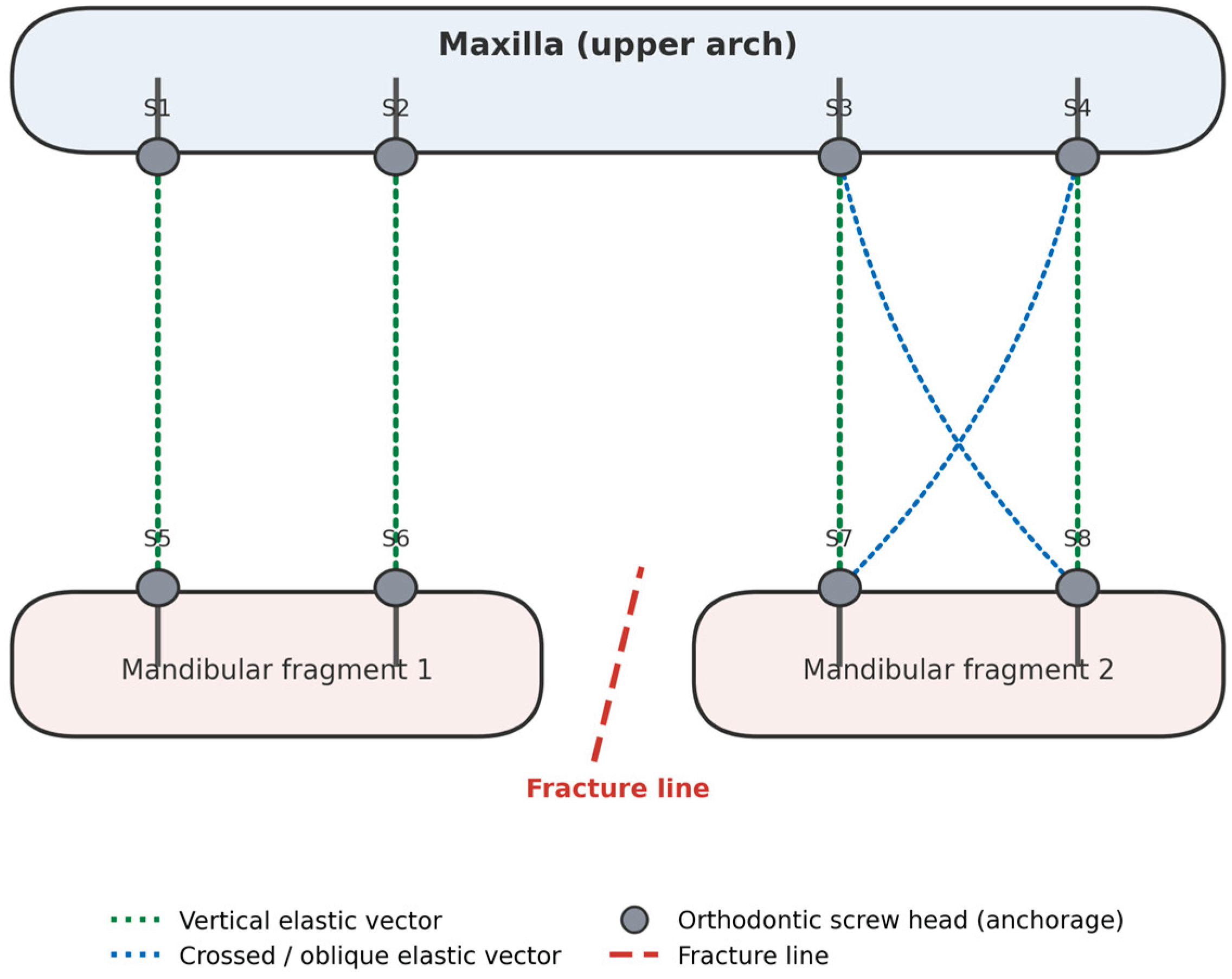
Figure 1. Schematic distribution of orthodontic screws (S1–S8) and elastic traction vectors in the proposed method. Vertical elastics (green) connect antagonist screw pairs across the maxilla and mandible; crossed/oblique elastics (blue) provide an additional rotational-control vector near the fracture line. The diagram is a conceptual illustration of the proposed placement algorithm and does not represent measured patient data.
Preprints 230316 g001
Disclaimer/Publisher’s Note: The statements, opinions and data contained in all publications are solely those of the individual author(s) and contributor(s) and not of MDPI and/or the editor(s). MDPI and/or the editor(s) disclaim responsibility for any injury to people or property resulting from any ideas, methods, instructions or products referred to in the content.
Copyright: This open access article is published under a Creative Commons CC BY 4.0 license, which permit the free download, distribution, and reuse, provided that the author and preprint are cited in any reuse.