

Submitted:
22 August 2026
Posted:
27 August 2026
You are already at the latest version
Abstract
Ecthyma gangrenosum (EG) is one of the manifestations of skin infections affecting immunocompromised patients. Pseudomonas aeruginosa is an important etiologic factor. The cutaneous localization of EG lesions usually is the consequence of bacteraemia. Neutropenia is the main risk factor for EG in patients with haematological malignancies. In this paper we present four cases of EG in children with acute leukaemia, who underwent chemotherapy and were treated with broad spectrum antibiotics, immunoglobulins and G-CSF. Granulocyte mass was given in three cases and one patient underwent hyperbaric treatment. All of those methods were insufficient and the patients did not improve until wide surgical excision of necrotic changes was done. The outcomes of severe EG were poor. Only in one case the patient recovered from infection and the leukaemia. One patient died of P. aeruginosa sepsis and septic shock. Two were healed of infection, but later died of leukaemia relapses. We conclude that EG leads to prolonged delays in haematological treatment and EG management should also include early surgical intervention to remove the necrotic lesions and to incise the regions of inflammatory changes.
Keywords:
ecthyma gangrenosum
; Pseudomonas aeruginosa
; sepsis
; acute leukaemia
1. Introduction
In recent years, drug-resistant Gram-negative infections are increasingly observed among hospitalized patients. The most common pathogens causing such infections include Pseudomonas aeruginosa, Acinetobacter baumannii, Enterobacter faecium and Klebsiella pneumonia [1,2]. Aggressive chemotherapy followed by prolonged and severe neutropenia (<100 cells/mm for >7 days) play an important part in the susceptibility of children with acute lymphoblastic leukaemia (ALL) or acute myeloblastic leukaemia (AML) to infections [3]. Additionally, central venous catheters and invasive procedures (lumbar punctures, suprapubic catheters) during the intensive phase of chemotherapy serve as routes of infection. Gram (+) pathogens cause the majority of infections (57%) in this patient group. However, it is the less frequent Gram (-) infections that cause higher mortality rates (18%) [4,5]. The poor outcomes among the oncological patients (of whom 15% will eventually suffer from P. aeruginosa bacteraemia) [6] are related to these pathogens’ high genetic variability, rapid resistance to several groups of antibiotics (e.g. -lactamases or carbapenems) and the increased frequency of multidrug resistance [7].
Pseudomonas aeruginosa rods are G(-) organisms found in moist environments with ambient temperatures ranging from 40 C to 430 C and pH ranging from normal to basic. Due to its resistance to antiseptics and low-dose antibiotics, ability to form biofilms and to move on the surface of medical equipment, P. aeruginosa is frequently found among the hospital flora. [6,8,9].
People with fully active immune systems are generally resistant to P. aeruginosa. Whereas the immunodeficient patients can present with localized P. aeruginosa infections of the urinary tract, external or middle ear, paranasal sinuses, eyes or the lungs (particularly the mechanically ventilated patients or those with cystic fibrosis) [6,10,11,12]. Sometimes the bones or joints are involved as the result of bacteraemia or the spread from local infection [13,14,15].
Neutropenia seems to be the most important factor in host susceptibility and in such neutropenic patients, P. aeruginosa infections usually present as bacteraemia with rapidly-progressing septic shock [16,17]. The characteristic feature of P. aeruginosa sepsis among these patients is ecthyma gangrenosum (EG): necrotic skin lesions resulting from the vascular invasion of bacteria in the skin and soft tissues. EG is known to develop in oncological patients, prematures, diabetics, as well as patients who are on steroid therapy, malnourished or with extensive burn wounds [6]. The most frequent EG risk factors are carriers of P. aeruginosa in the gastrointestinal tract and an infection of a central vessel catheter.
Although EG foci can localize in any part of the skin, the majority of these lesions are found in the anogenital region (57%), limbs (30%), buttocks and axillae [6]. Despite the use of targeted antibiotics, the treatment of EG is difficult due to increasing drug resistance and poor drug penetration into the necrotic areas. For the above reasons, early excision of the necrotic changes and wide incision of the inflamed tissue is recommended and was demonstrated to significantly improve treatment outcomes [17].
Fournier gangrene is a specific presentation of EG in males that spreads along the fascia of the perineal and genital regions [18]. In the advanced stage of the inflammation, mixed pathogens were revealed in microbiological tests in most of the patients and P. aeruginosa was indicated as one of etiological factors [19]. To complete the treatment with antibiotics, oncological patients with neutropenia and EG are treated with immunoglobulins, G-CSF and granulocyte transfusions in order to boost the immune response. During treatment it is also important to maintain caloric balance and prevent catabolism by providing appropriate enteral and parenteral nutrition [20]. Published data indicate that EG treatment is not successful in 15% of adults with neutropenia [6].
2. Case Series
In this paper we present a case series of four paediatric oncology patients with EG.
2.1. Case 1
A 17-year-old male was diagnosed with AML type M (FAB classification) and treated according to the Interim AML-BFM 1998 protocol. A hematologic remission was achieved after the first two cycles of chemotherapy. Four months after the diagnosis and after the third cycle of chemotherapy (HAE), the patient presented with agranulocytosis, dysuria, inflammation of the external urethral meatus and rapidly increasing inflammatory markers (CRP up to 228 mg/L, PCT up to 22 mg/dL). Despite the empiric antimicrobial therapy (piperacillin/tazobactam, netilmicin and ciprofloxacin), the patient developed signs of septic shock in <24 hours and needed intensive care with vasopressors, continuous intravenous fluids and immunomodulatory treatment (G-CSF, immunoglobulins). Urine and stool cultures were positive for P. aeruginosa and the stool was also positive for Enterococcus faecium, VRE (+), β-haemolytic Streptococcus and Candida albicans. The antibiotic therapy was revised according to the culture and sensitivity results (colistin, teicoplanin, rifampicin) and an amphotericin together with caspofungin was added. 24 hours later, the septic shock was successfully managed, however, the inflammatory changes of the skin and soft tissues of the patient’s groin and penis (see Figure 1) evolved into growing necrotic foci, diagnosed as Fournier gangrene. Metronidazole and acyclovir were added for anaerobic and antiviral coverage. Antiseptics (chlorhexidine and octenidine dihydrochloride) were used locally on the skin lesions and a suprapubic urinary catheter was inserted. The surgical resection of the necrotic tissues was postponed due to the persistently severe state of the patient and prolonged agranulocytosis. Six days later, the patient once again presented signs of shock. Intensive therapy did not yield improvement. No matching familial donor of granulocytes was found for this patient. Signs of infection worsened, inflammatory foci were confirmed in the lungs and the patient died due to multiorgan failure.
2.2. Case 2
A 9-year-old female with a relapse of ALL without central nervous system (CNS) involvement was treated according to the ALL-REZ BFM 2002 protocol for relapses. Two days after the second cycle (F2), the patient complained of pain in the left periauricular area. The laryngological examination revealed a lesion in the left external ear canal with slight bleeding. The patient was neutropenic and intravenous cefoperazone was administered, followed by additional meropenem and teicoplanin. On the following day, an increased erythema with central necrosis and oedema of the left periauricular area were revealed and EG was recognized. Blood tests indicated increased inflammatory markers (CRP = 70 mg/L) and blood cultures were positive for P. aeruginosa. In the next several hours, the patient developed signs of septic shock with hepatic and renal insufficiency, lost consciousness and was transferred to the intensive care unit. Computer tomography (CT) of the head revealed bleeding into the CNS as the result of DIC and possible involvement of CNS by inflammatory process. Despite stabilizing the blood pressure and an improved hepatic and renal function, the patient was in a coma and needed continued intubation with mechanical ventilation. It was decided to proceed with wide debridement and excision of the necrotic changes (Figure 2). The same antimicrobial treatment was continued with the addition of antifungal agents (amphotericin and caspofungin) and immunomodulating treatment (G-CSF, immunoglobulins, granulocyte transfusions). Over the next week, the patient gradually returned to consciousness and was extubated. Four weeks later, the skin lesions were healed and the patient resumed chemotherapy with doses reduced to 80%. Due to the fungal changes in the chest CT during the sepsis, the patient received antifungal treatment until the completion of chemotherapy. Although the patient suffered from severe side effects of myelosuppression until the end of therapy, no new bacterial infections were noted. The treatment was completed with an allogenic hematopoietic stem cell transplant (HSCT) from an unrelated donor. The patient has undergone extensive rehabilitation that allowed her to regain speech and motor function. As of four years post-transplantation, the patient remains in remission and in overall good condition.
2.3. Case 3
An 8-year-old female was diagnosed with a relapse of high-risk ALL and was treated according to the ALL-REZ BFM 2002 protocol. On the third day of the F1 cycle, agranulocytosis appeared along with pain and erythema of the groin. As the inflammatory markers rapidly increased, cefoperazone and amphotericin were administered. Blood, urine and groin swab cultures were positive for P. aeruginosa. Despite the early antibiotic treatment, the patient developed signs of septic shock in <24 hours, accompanied by focal necrosis of the inflamed tissues. The patient was stabilized after intensive therapy with vasopressors and intravenous fluids but remained in severe condition despite the immunomodulatory treatment (G-CSF, immunoglobulins and granulocytes). Over the next four days, the skin changes spread to the suprapubic region and the patient was referred for hyperbaric therapy. Three days later, the patient again developed signs of septic shock, was stabilised and referred for surgical resection of the necrotic changes. Wide incisions of the inflammatory tissues were performed and due to the location of the skin changes (Figure 3) the patient required an ileostomy. The patient gradually improved and the infection of her soft tissues was resolved within two weeks. The incised tissues were sutured and the ileostomy was removed three months after the surgery. Six weeks post-op, the patient resumed chemotherapy with reduced doses of cytostatics. Chemotherapy was complicated by severe toxicity (myelosuppression, mucositis and gastrotoxicity) and subsequently shortened. The treatment was completed with an allogenic HSCT. Eight months after the transplantation, the patient was diagnosed with a second relapse of ALL with death in the next two months as the result.
2.4. Case 4
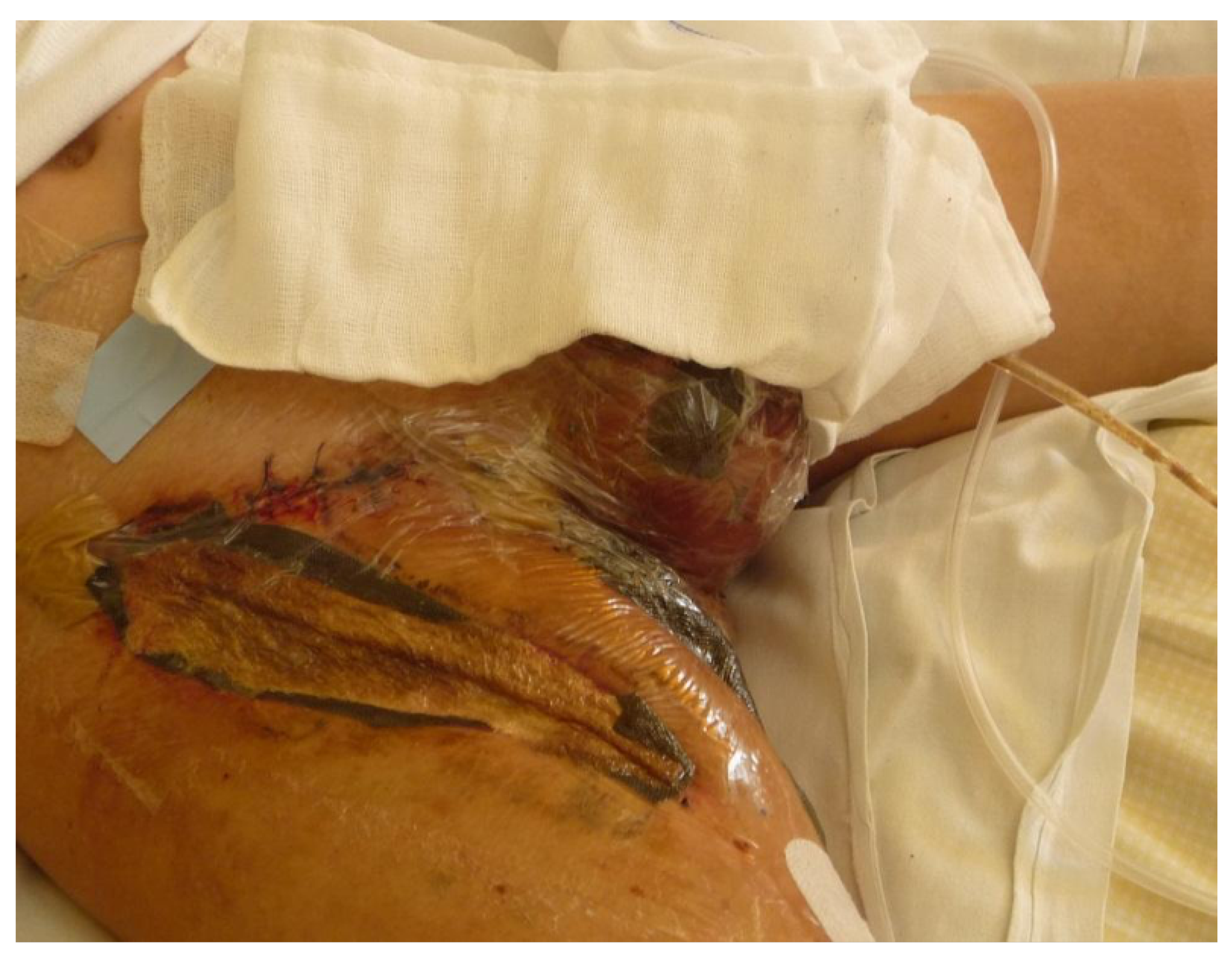
A 12-year-old boy was diagnosed with bilinear ALL and was treated according to the ALLIC 2002 protocol. From the start, the patient poorly tolerated the chemotherapy and presented with severe adverse effects (prolonged agranulocytosis, fungal lung infection and gastrotoxicity). After the HR3 cycle, the patient presented with erythema of the external urethral meatus and oedema of the glans. He was immediately treated with broad spectrum antibiotics (cefepime, teicoplanin, amikacin and metronidazole) and posaconazole (for the ongoing pulmonary fungal infection). Cultures of the blood and skin changes were positive for P. aeruginosa and the treatment was augmented with G-CSF, immunoglobulins and granulocyte transfusion. Despite the intensive treatment over the next three days, the inflammation progressed to focal necrosis and Fournier gangrene was diagnosed. Moreover, the inflammation spread to the right inguinal and the thigh regions of the skin. Five days after the onset of infection, the boy presented with septic shock. After stabilizing the patient, a meatotomy was performed, along with an excision of the necrotic changes in the groin. The inflamed parts of the skin were incised and a vacuum drainage was applied. The modification in antibiotics according to susceptibility of pathogens was performed using ciprofloxacin, tygacillin, and imipenem, followed by ceftazidime, rifampicin, and chloramphenicol. Additionally antifungal drugs (caspofungin and amphotericin) and antiviral medication (acyclovir) were used over the time of infection. In the next 10 weeks, the patient required several additional incisions of the inflamed tissues as well as widening of the initial incisions (Figure 4). Agranulocytosis persisted for a total of two months. Once the patient’s bone marrow gradually regenerated itself, his skin changes also healed. The patient resumed chemotherapy with reduced doses but it was stopped due to severe toxicity (myelosuppression, gastrotoxicity and mucositis) and an allogenic HSCT was performed. The transplantation was complicated by severe fungal lung infection with septic shock and the patient died due to multiorgan failure.
3. Discussion and Clinical Implications
Our observations of leukaemia patients with EG-type P. aeruginosa infections of the skin confirm the severity, resistance to treatment and poor outcome of such cases. Therapy with broad spectrum antibiotics is frequently insufficient due to their poor penetration into the necrotic and inflamed tissues. Although initially the lesions were produced by P. aeruginosa, the tissue cultures were frequently positive for several pathogens, including anaerobes, fungi and other microorganisms [20,21]. We didn’t observe any results using conservative treatment with broad spectrum antibiotics in any of our patients. We agree with the previously published reports that early surgical excision of the necrotic tissues offers a chance to manage the infection and allows to shorten the delay in chemotherapy, thus improving the prognosis of the underlying leukaemia [17].
Some publications confirmed the importance of colony stimulating factor G-CSF as supportive therapy of EG [22]. All of our patients received immunomodulatory treatment (G-CSF, immunoglobulins and granulocyte infusions) together with broad spectrum antibiotics, however this was not sufficient to achieve recovery. Just as in the case of antibiotics, it seems that the ischemic and necrotic tissues also inhibit the penetration of the humoral and cellular immune response factors. In consequence, the elimination of pathogens “in situ” is impossible.
The prolonged delays in oncological therapy led to the relapses in ½ of our patients. It is noteworthy that from the beginning of chemotherapy all the patients presented serious and multiple complications and they needed modified and reduced doses. This was probably the main reason for their poor outcomes and it suggests an alteration in the metabolism of cytostatic drugs.
As it is observed, the P. aeruginosa infections tend to develop during periods of agranulocytosis. In ¾ of our cases the use of granulocyte infusion allowed us to achieve improvement of the systemic inflammatory reaction. Although the local inflammation persisted, the septic reactions were stopped and provided time to perform the surgical procedures. Thus, we suggest that it is clinically important to administer the granulocyte infusions at the onset of an infection. Good results of using granulocyte infusions in EG cases have been published previously [23]. We suggest that in case of neutropenic fever, patients should be immediately treated empirically with antimicrobials aimed at P. aeruginosa. It has been postulated that a delay of >2 days in target antimicrobial therapy is associated with a doubled mortality rate in ALL or AML patients [24]. The immunocompromised patients require strict monitoring of vital signs and skin changes. It is recommended in patients who are colonized with P. aeruginosa to start with antibiotics immediately once the neutropenic fever appears [24].
Patients who are carriers of P. aeruginosa are at the greatest risk of developing such infections, thus we agree that leukaemia patients should be discharged home in between cycles of chemotherapy to minimize the exposure to the nosocomial infections [25]. It is also important to follow proper antisepsis protocols at the haematology-oncology wards. A detailed epidemiologic analysis is also recommended on the wards for early implementation of procedures aimed at P. aeruginosa eradication [26].
Author Contributions
Conceptualization, M.N.; methodology, M.N., P.C., N.I.-J.; software, M.C.; validation, M.N., P.C., N.I.-J. ; formal analysis, M.N., N.I.-J., P.C., J.S. M.C.; investigation, M.N., N.I.-J., P.C., J.S. M.C.; resources, M.N., N.I.-J., P.C., J.S. M.C.; data curation, M.N., N.I.-J., P.C., J.S. M.C.; writing—original draft preparation, M.N., J.S.; writing—review and editing, M.C.; visualization, M.N. M.C.; supervision, N.I.-J.; project administration, M.N., M.C.; All authors have read and agreed to the published version of the manuscript.
Funding
This research received no external funding.
Institutional Review Board Statement
Not applicable.
Informed Consent Statement
Not applicable.
Data Availability Statement
Basic data is provided within the manuscript. Other data is available on request. In order to obtain this data, contact the corresponding Author.
Acknowledgments
Not applicable. No generative AI was used while working on this manuscript.
Conflicts of Interest
The authors declare no conflicts of interest.
Abbreviations
The following abbreviations are used in this manuscript:
| ALL | Acute Lymphocytic Leukaemia |
| ALL-REZ BFM | Acute Lymphoblastic Leukaemia Relapse Berlin-Frankfurt-Münster |
| ALLIC 2002 | Acute Lymphoblastic Leukaemia - Intercontinental Berlin-Frankfurt-Münster 2002 |
| AML | Acute Myelocytic Leukaemia |
| AML-BFM 1998 | Acute Myeloid Leukaemia Berlin-Frankfurt-Münster 1998 |
| CNS | Central Nervous System |
| CT | Computer Tomography |
| DIC | Disseminated Intravasal Coagulation |
| EG | Ecthyma Gangrenosum |
| FAB | French-American-British Classification |
| G-CSF | Granulocyte-Colony Stimulating Factor |
| HAE | Homoharringtonine, Ara-C [cytarabine], Etoposide |
| HSCT | Haematopoietic Stem Cell Transplantation |
| VRE | Vancomycin-Resistant Enterococci |
References
- Gaynes, R.; Edwards, J.R. Overview of nosocomial infections caused by Gram-negative bacilli. Clin. Infect. Dis. 2005, 41, 848–854. [Google Scholar] [CrossRef]
- Kunz, A.N.; Brook, I. Emerging resistant Gram-negative aerobic bacilli in hospital-acquired infections. Chemotherapy 2010, 56, 492–500. [Google Scholar] [CrossRef]
- Cao, B.; Wang, H.; Sun, H.; Zhu, Y.; Chen, M. Risk factors and clinical outcomes of nosocomial multi-drug resistant Pseudomonas aeruginosa infections. J. Hosp. Infect. 2004, 57, 112–118. [Google Scholar] [CrossRef]
- Bodey, G.P. Pseudomonas aeruginosa infections in cancer patients: have they gone away? Curr. Opin. Infect. Dis. 2001, 14, 403–407. [Google Scholar] [CrossRef]
- Freifeld, A.G.; Bow, E.J.; Sepkowitz, K.A.; Boeckh, M.J.; Ito, J.I.; Mullen, C.A.; Raad, I.I.; Rolston, K.V.; Young, J.A.H.; Wingard, J.R. Clinical practice guideline for the use of antimicrobial agents in neutropenic patients with cancer: 2010 update by the Infectious Diseases Society of America. Clin. Infect. Dis. 2011, 52, e56–e93. [Google Scholar] [CrossRef]
- Pier, G.B.; Ramphal, R. Pseudomonas aeruginosa. In Principles and Practice of Infectious Diseases; Mandell, G.L., Douglas, R.G., Bennett, J.E., Eds.; Churchill Livingstone Elsevier: Philadelphia, 2010; pp. 2835–2858. [Google Scholar]
- Aubron, C.; Poirel, L.; Fortineau, N.; Nicolas, P.; Collet, L.; Nordmann, P. Nosocomial spread of Pseudomonas aeruginosa isolates expressing the metallo-beta-lactamase VIM-2 in a hematology unit of a French hospital. Microb. Drug Resist. 2005, 11, 254–259. [Google Scholar] [CrossRef]
- Silvestri, L.; van Saene, H.K.F.; Gullo, A.; de la Cal, M.A. Guidelines for prevention, diagnosis, and treatment of ventilator-associated pneumonia (VAP) in the trauma patient. J. Trauma;Comment 2007, 62, 1062–1064. [Google Scholar] [CrossRef]
- Schwaber, M.J.; Carmeli, Y. Mortality and delay in effective therapy associated with extended-spectrum beta-lactamase production in Enterobacteriaceae bacteraemia: a systematic review and meta-analysis. J. Antimicrob. Chemother. 2007, 60, 913–920. [Google Scholar] [CrossRef]
- Chastre, J.; Fagon, J.Y. Ventilator-associated pneumonia Review. Am. J. Respir. Crit. Care Med. 2002, 165, 867–903. [Google Scholar] [CrossRef] [PubMed]
- Goolamali, S.I.; Fogo, A.; Killian, L.; Shaikh, H.; Brathwaite, N.; Ford-Adams, M.; Macfarlane, S. Ecthyma gangrenosum: an important feature of pseudomonal sepsis in a previously well child. Clin. Exp. Dermatol. 2009, 34, e180–e182. [Google Scholar] [CrossRef]
- Soria, A.; Francès, C.; Guihot, A.; Varnous, S.; Bricaire, F.; Caumes, E. Etiology of ecthyma gangrenosum: four cases. Ann. De Dermatol. Et. De Vénéréologie 2010, 137, 472–476. [Google Scholar]
- Lang, A.G.; Peterson, H.A. Osteomyelitis following puncture wounds of the foot in children. J. Trauma 1976, 16, 993–999. [Google Scholar] [CrossRef]
- Ross, J.J.; Hu, L.T. Septic arthritis of the pubic symphysis: review of 100 cases. Medicine 2003, 82, 340–345. [Google Scholar] [CrossRef]
- Rybtke, M.T.; Jensen, P.O.; Høiby, N.; Givskov, M.; Tolker-Nielsen, T.; Bjarnsholt, T. The implication of Pseudomonas aeruginosa biofilms in infections. Inflamm. Allergy-Drug Targets 2011, 10, 141–157. [Google Scholar] [CrossRef]
- Cryz, S.J.J.; Fürer, E.; Germanier, R. Simple model for the study of Pseudomonas aeruginosa infections in leukopenic mice. Infect. Immun. 1983, 39, 1067–1071. [Google Scholar] [CrossRef]
- Khalil, B.A.; Baillie, C.T.; Kenny, S.E.; Lamont, G.L.; Turnock, R.R.; Pizer, B.L.; van Saene, H.K.F.; Losty, P.D. Surgical strategies in the management of ecthyma gangrenosum in paediatric oncology patients. Pediatr. Surg. Int. 2008, 24, 793–797. [Google Scholar] [CrossRef]
- Jarvis, W.R. Controlling healthcare-associated infections: the role of infection control and antimicrobial use practices. Semin. Pediatr. Infect. Dis. 2004, 15, 30–40. [Google Scholar] [CrossRef]
- Czymek, R.; Hildebrand, P.; Kleemann, M.; Roblick, U.; Hoffmann, M.; Jungbluth, T.; Bürk, C.; Bruch, H.P.; Kujath, P. New insights into the epidemiology and etiology of Fournier’s gangrene: a review of 33 patients. Infection 2009, 37, 306–312. [Google Scholar] [CrossRef]
- Kang, C.I.; Song, J.H.; Chung, D.R.; Peck, K.R.; Ko, K.S.; Yeom, J.S.; Ki, H.K.; Son, J.S.; Lee, S.S.; Kim, Y.S.; et al. Risk factors and pathogenic significance of severe sepsis and septic shock in 2286 patients with gram-negative bacteremia. In Journal of Infection; Korean Network for Study of Infectious Diseases (KONSID), 2011; Volume 62, pp. 26–33. [Google Scholar]
- Szmyd, K.; Wrobel, G.; Dobaczewski, G.; Gołębiewski, W.; Chybicka, A. Pseudomonas aeruginosa sepsis, ecthyma gangrenosum in immunocompromised children. Acta Haematol. Pol. 2004, 35, 377–382. [Google Scholar]
- Becherel, P.A.; Chosidow, O.; Berger, E.; Martin, S.; Khayat, D.; Herson, S.; Francès, C. Granulocyte-macrophage colony-stimulating factor in the management of severe ecthyma gangrenosum related to myelodysplastic syndrome. Arch. Dermatol. 1995, 131, 892–894. [Google Scholar] [CrossRef]
- Béjaoui, M. Successful treatment of Fusarium solani ecthyma gangrenosum in a patient affected by leukocyte adhesion deficiency type 1 with granulocytes transfusions. BMC Dermatol. 2010, 10, 10–15. [Google Scholar] [CrossRef]
- Lodise, T.P.J.; Patel, N.; Kwa, A.; Graves, J.; Furuno, J.P.; Graffunder, E.; Lomaestro, B.; McGregor, J.C. Predictors of 30-day mortality among patients with Pseudomonas aeruginosa bloodstream infections: impact of delayed appropriate antibiotic selection. Antimicrob. Agents Chemother. 2007, 51, 3510–3515. [Google Scholar] [CrossRef]
- Jenkins, C.E.; Swiatoniowski, A.; Issekutz, A.C.; Lin, T.J. Pseudomonas aeruginosa exotoxin A induces human mast cell apoptosis by a caspase-8 and -3-dependent mechanism. J. Biol. Chem. 2004, 279, 37201–37207. [Google Scholar] [CrossRef]
- Reich, H.L.; Williams Fadeyi, D.; Naik, N.S.; Honig, P.J.; Yan, A.C. Nonpseudomonal ecthyma gangrenosum. J. Am. Acad. Dermatol. 2004, 50, 114–117. [Google Scholar] [CrossRef]
Figure 1.
Fournier’s gangrene in a 17 old boy - initial clinical presentation.
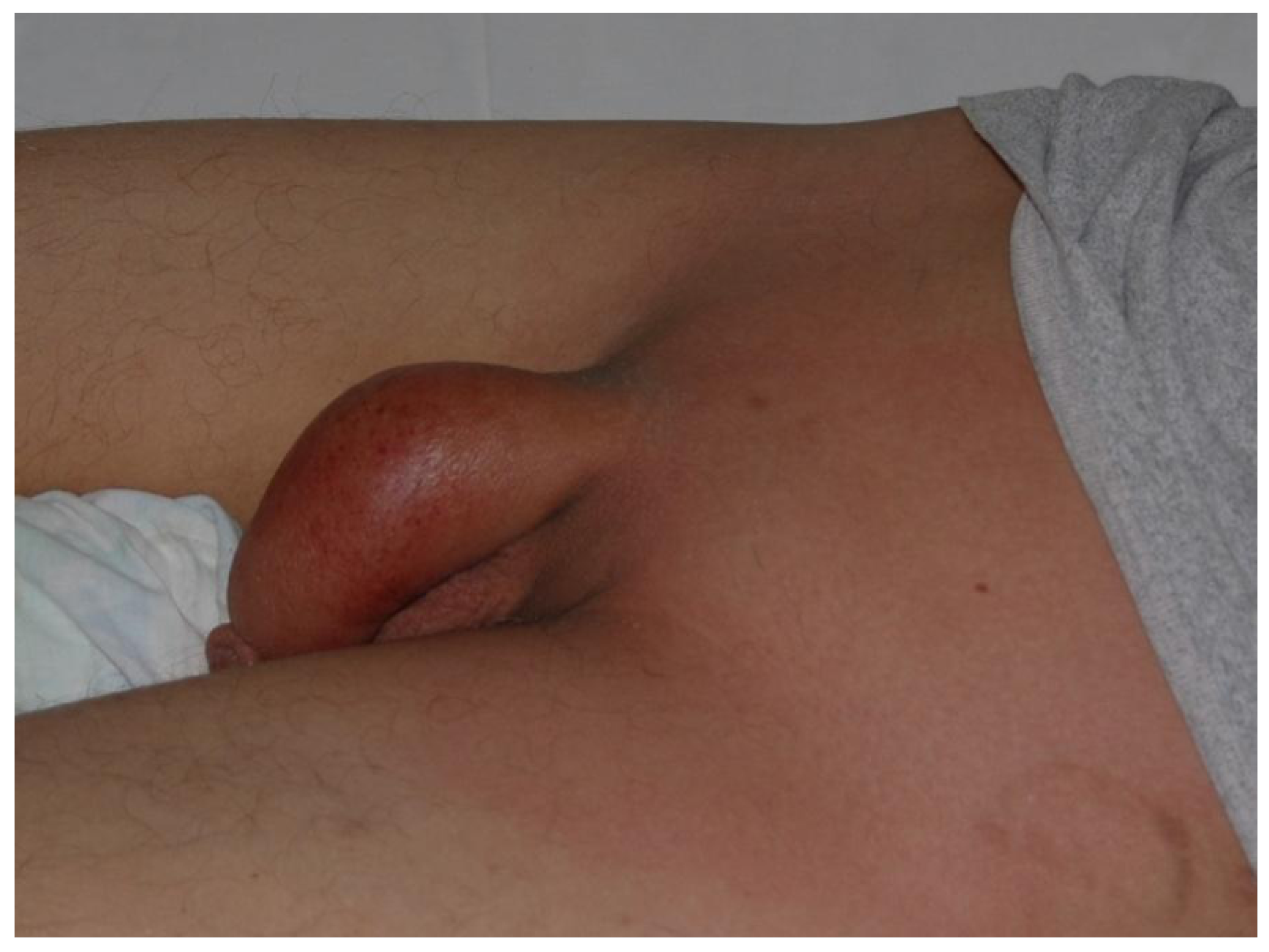
Figure 2.
Ecthyma gangrenosum in periauricular region in a 12-year-old girl – wide surgical incisions of the inflammatory skin.
Figure 2.
Ecthyma gangrenosum in periauricular region in a 12-year-old girl – wide surgical incisions of the inflammatory skin.
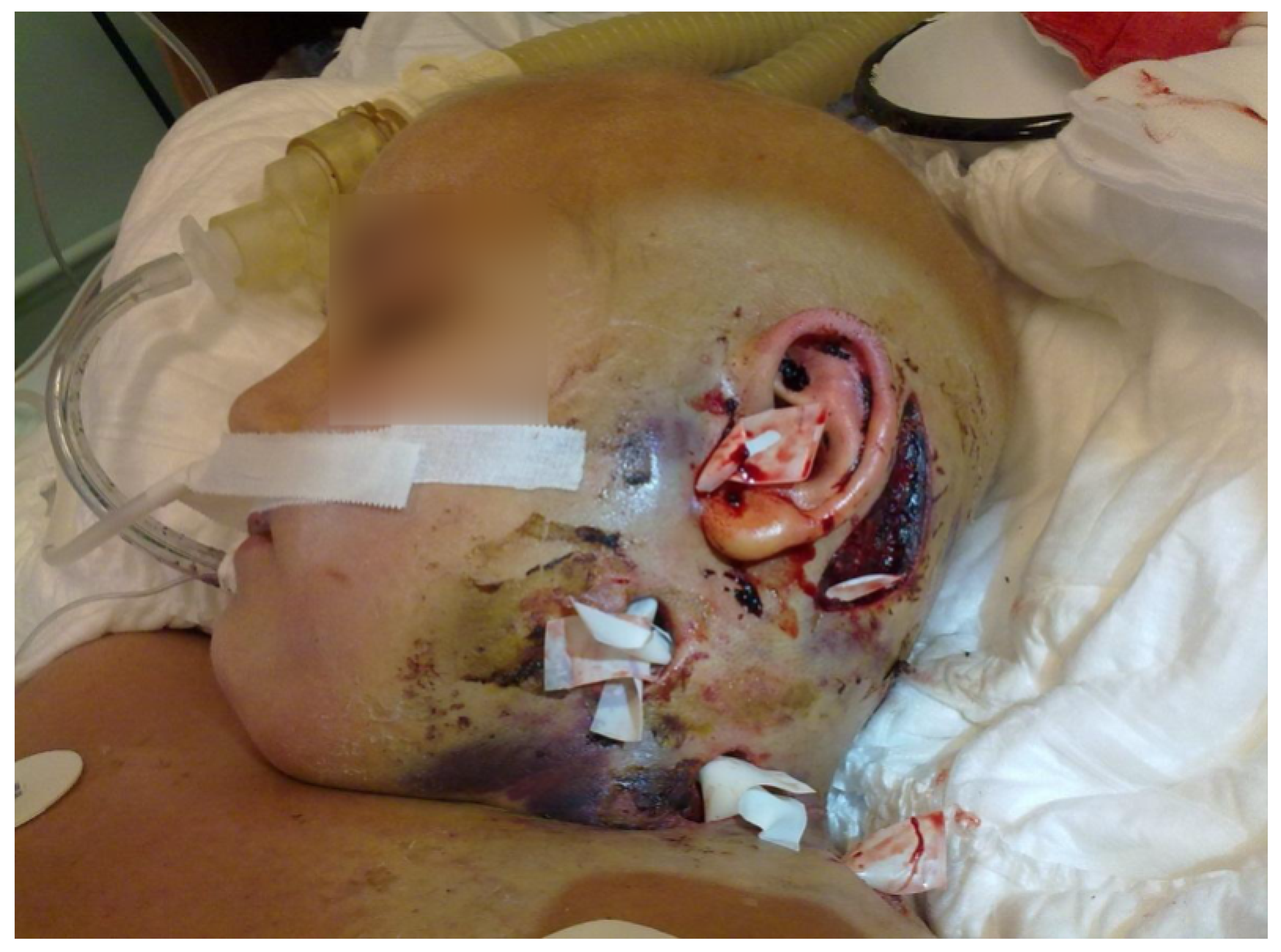
Figure 3.
Ecthyma gangrenosum in a 9-year-old girl after surgical excision of necrotic lesions and wide incision of the region of inflammation.
Figure 3.
Ecthyma gangrenosum in a 9-year-old girl after surgical excision of necrotic lesions and wide incision of the region of inflammation.
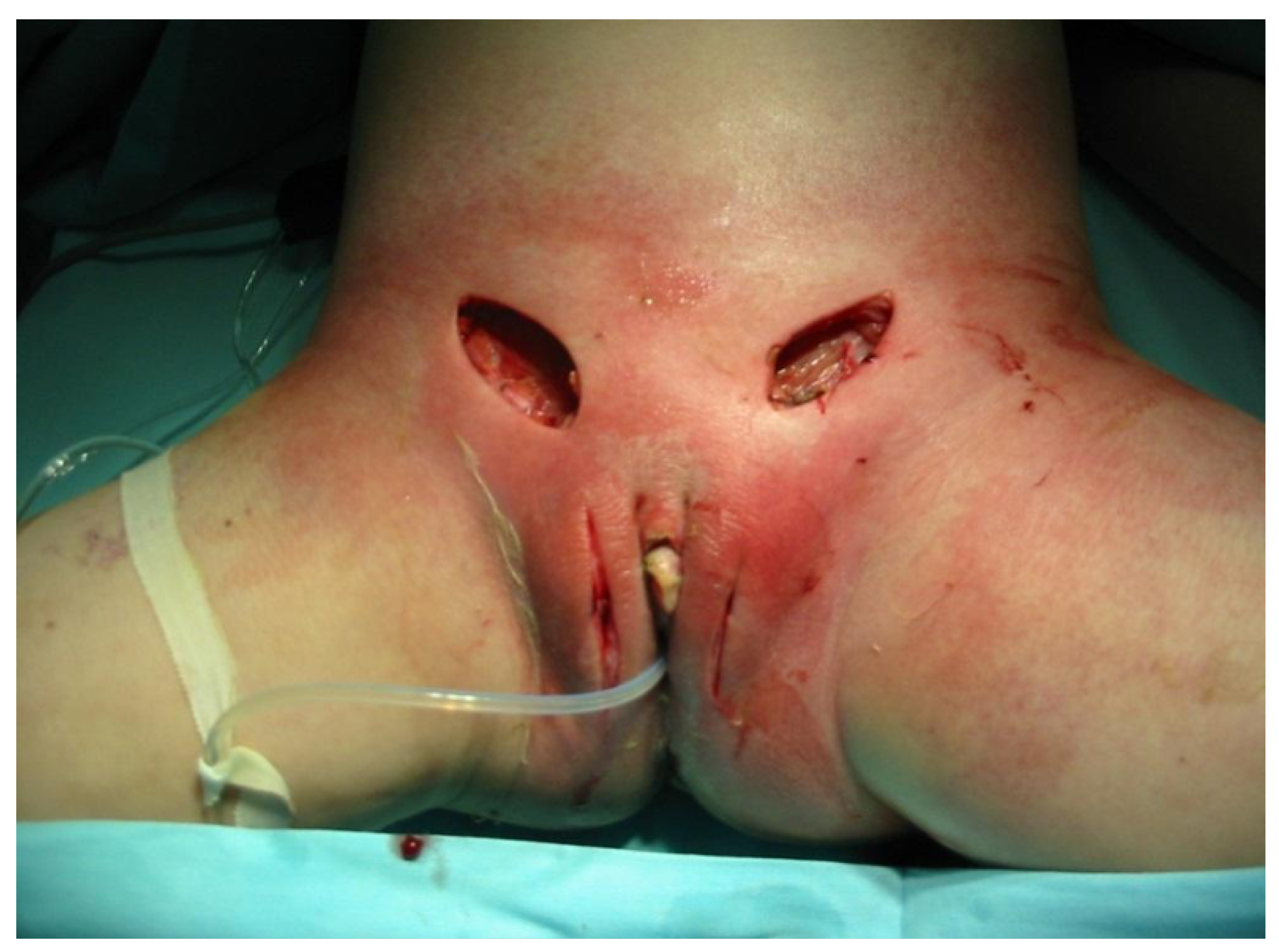
Figure 4.
Fournier’s gangrene in an 8-year-old boy treated with wide incision and local drainage.
Disclaimer/Publisher’s Note: The statements, opinions and data contained in all publications are solely those of the individual author(s) and contributor(s) and not of MDPI and/or the editor(s). MDPI and/or the editor(s) disclaim responsibility for any injury to people or property resulting from any ideas, methods, instructions or products referred to in the content. |
© 2026 by the authors. Licensee MDPI, Basel, Switzerland. This article is an open access article distributed under the terms and conditions of the Creative Commons Attribution (CC BY) license (http://creativecommons.org/licenses/by/4.0/).
Copyright: This open access article is published under a Creative Commons CC BY 4.0 license, which permit the free download, distribution, and reuse, provided that the author and preprint are cited in any reuse.