
Submitted:
25 August 2026
Posted:
27 August 2026
You are already at the latest version
Abstract
Osseointegration is strongly influenced by the physicochemical properties of implant surfaces, including topography, roughness, and biofunctionalization. This study investigated the effects of laser surface texturing and hydroxyapatite (HA) biofunctionalization on the viability, morphology, and inflammatory profile of human osteoblasts. Ti-6Al-4V discs were mechanically polished or laser-textured using an Nd laser to generate checkerboard patterns with spatial periodicities of 0.25 or 0.8 mm. Textured surfaces were subsequently coated with nanohydroxyapatite and sintered using either conventional furnace or CO2 laser sintering. Immortalized human osteoblasts (hFOB 1.19) were cultured on the seven experimental surfaces for up to 14 days. Cell viability was assessed by a resazurin-based assay, while cell adhesion and morphology were evaluated by scanning electron microscopy. IL-1β, IL-8, and IL-10 secretion was quantified by ELISA. Smooth titanium exhibited higher osteoblast viability than textured surfaces, whereas the 0.25 mm pattern generally performed better than the 0.8 mm pattern. HA biofunctionalization consistently improved viability within both texture patterns, irrespective of the sintering method, while the sintering technique itself had no significant effect. The 0.8 mm pattern showed lower IL-1β secretion, whereas IL-10 remained largely unchanged. Within the limitations of this in vitro study, the 0.25 mm texture combined with HA biofunctionalization showed a more favorable cellular response than the 0.8 mm pattern. Further studies should characterize baseline surface roughness and investigate corrosion and microbiological responses.
Keywords:
osseointegration
; titanium implants
; laser surface-texturing
; hydroxyapatite
; biofunctionalization
; osteoblasts
1. Introduction
Osseointegration is a key factor for the success of oral rehabilitation with dental implants [1]. Described by Brånemark as the direct structural and functional connection between living bone and the surface of an implant—without the interposition of soft tissue and capable of withstanding mechanical load [2,3] - it is essential to understand the underlying cellular dynamics to optimize this process.
Two fundamental biological phenomena are involved in osseointegration: osteoconduction and osteoinduction. Osteoinduction refers to the biological process by which pluripotent cells are stimulated and induced to differentiate into osteogenic lineages. Osteoconduction, on the other hand, is defined as the growth of bone along a surface [4,5].
Therefore, when selecting an implant, the goal is to choose a surface that enhances both osteoconduction and osteoinduction, making the osseointegration process more predictable [4,6]. Ideally, the implant surface should promote cellular adhesion and osteoblastic differentiation, while simultaneously avoiding features that encourage bacterial colonization. Parameters such as topography, roughness, and wettability are thus evaluated [7,8,9].
Implant topography encompasses its geometric design, grooves, ridges, and striations, which directly influence surface roughness [1]. The macro-topography (100 μm to millimeters) should be selected based on the bone quality and the mechanical load the implant is expected to bear. Studies have shown that macro-topography significantly influences cell adhesion and proliferation by modifying the available surface area for cellular interaction [10,11].
Micro-topography (100 nm to 100 μm) and nano-topography (1 to 100 nm) are achieved through surface modification techniques aimed at eliciting a faster and more effective cellular response, enhancing interactions with extracellular matrix components [8,12]. These topographical features regulate the formation of anchoring junctions between cells and the implant surface [13]. Despite extensive research, there is no consensus regarding the ideal surface topography [14,15,16,17,18].
Regarding surface roughness, based on the classification proposed by Wennerberg et al. [19] the average roughness (Ra) can be categorized as smooth (<0.5 μm), minimally rough (0.5 to 1 μm), moderately rough (1 to 2 μm), or rough (2 to 3 μm). Several studies indicate that rougher surfaces lead to improved force distribution around the implant, increased osteoblastic adhesion and proliferation, and enhanced adsorption of extracellular matrix proteins and growth factors, thus stimulating signaling pathways related to osteoblast differentiation [1,12,20,21,22]. Osteoblasts tend to adhere more effectively to surfaces with moderate roughness (Ra ~1–2 μm), which facilitates osseointegration [10,21,23,24,25].
Wettability is another crucial parameter, strongly influenced by surface topography, roughness, and chemical composition [26]. It plays a central role in cell adhesion and proliferation at the bone–implant interface, as well as in bacterial adhesion [27,28]. Surfaces with higher surface energy—and therefore greater wettability—tend to exhibit increased adsorption of key proteins such as albumin and fibronectin, which are essential for cellular adhesion [29,30,31].
To enhance osseointegration, implant surfaces were modified to increase bone contact area through enhanced surface roughness, promoting osteogenic cell adhesion and extracellular matrix proliferation, without increasing bacterial adhesion [7,32]. Laser techniques were employed to texture the surface by ablating material via direct absorption of laser energy [33,34]. In addition, biofunctionalization of the implant surface was achieved by incorporating bioactive materials—specifically hydroxyapatite—due to its chemical similarity to native bone. This incorporation enhances the biological response by promoting cellular adhesion through increased adsorption of fibronectin and vitronectin and stimulating osteoconduction by recruiting osteoblastic cells [35,36,37].
Being composed of calcium and phosphate salts, hydroxyapatite also facilitates mineral nucleation, thereby promoting bone formation [38]. In vitro studies evaluating the cytotoxicity of ceramic bioactive coatings such as hydroxyapatite have shown increased osteoblast viability [36,37].
Therefore, the aim of this study was to understand how the texturing of titanium implant surfaces using laser techniques and subsequent biofunctionalization influences the viability, morphology and cellular response of a line of immortalized osteoblast cell line.
2. Materials and Methods
2.1. Sample Processing
Based on a titanium alloy bar (Ti-6Al-4V), with the chemical characteristics described in Table 1, discs with 8 mm in diameter and approximately 2.5 mm in height will be obtained, which will be subsequently polished with sandpaper to remove cutting risks or any other mark that could interfere with the results. The production of samples will take place in the CMEMS laboratories.
2.2. Texture Pattern
After producing the samples, the surface texturing of the titanium samples was carried out with a laser. An Nd:YVO4 laser (OEM Plus, SISMA, Vincenza, Italy) will be used with λ= 1064 nm and a pulse of 10 ns and with an initial focus at 11.1 cm under normal atmospheric pressure and with constant air renewal using a fan and a jet of compressed air, promoting the removal of debris from the surface of the sample. Laser specifications are detailed in Table 2.
The texturing pattern of the samples is a checkerboard formed by grooves and ridges. This pattern will be drawn using software and later transferred to the laser.
Two designs will be created, with the abovementioned pattern, but which differ in the length of the crest and groove: one of the samples the length of the crest and groove is 0.8 mm while in the other design it is 0.25 mm; This way, the studied design will not present differences in the distance between the ridges, but rather in the surface area of the non-textured material. After texturing, all samples will be disinfected again in an isopropyl alcohol solution for 1 minute in ultrasound.
2.3. Bioactive Functionalization
The addition of hydroxyapatite to the samples were done through a suspension of droxyapatite (nanoXIM Hap400, Fluidinova; Maia, Portugal) powder and water. This solution was deposited on the previously textured samples using a pipette to create a homogeneous film on the disc. The chemical formula and other parameters are described in Table 3, according to the information provided by the manufacturer.
After placing the hydroxyapatite coating on the samples, they will be sintered using two different methods: laser or conventional. Laser sintering will be carried out using a Carbon Dioxide Laser (BD-50C, Bende China) with a maximum power of 50 W and a λ= 1064 nm (complete specifications are detailed in Table 4). Conventional sintering is where samples are sintered in a furnace at a temperature of 950 to 1175ºC for 1 hour.
Afterall, were produced seven sample types: Flat titanium (TiL), texturized titanium in a 0.25 pattern (TiT025); texturized titanium in a 0.25 pattern plus bioactive in a convention sintering (TiT025CS), texturized titanium in a 0.25 pattern plus bioactive in a laser sintering (TiT025LS); texturized titanium in a 0.8 pattern (TiT08); texturized titanium in a 0.8 pattern plus bioactive in a convention sintering (TiT08CS), texturized titanium in a 0.8 pattern plus bioactive in a laser sintering (TiT08LS).
2.4. Surface Roughness Analysis
Surface roughness was evaluated for all groups using a contact profilometer (SurfTest SJ-201, Mitutoyo, Tokyo, Japan) equipped with a diamond stylus with a tip radius of 2 μm. Ten measurements were obtained from randomly selected regions on each specimen, using a scanning speed of 0.5 mm/s and a sampling length of 0.8 mm. All measurements were performed perpendicular to the surface texture to ensure reliable topographical characterization.
The roughness parameter assessed was the arithmetic mean roughness (Ra). Quantitative profilometric analysis was restricted to the uncoated metallic substrates (TiL, TiT025, and TiT08). Due to the inherent limitations of contact profilometry—specifically the application of mechanical load via a sharp stylus—measurements on the thin hydroxyapatite coatings were avoided, as they could induce microcracking, surface scratching, or delamination of the brittle ceramic layer.
To preserve the integrity of the biofunctionalized surfaces for subsequent biological evaluation, coated samples were excluded from contact profilometry. Instead, their surface morphology and structural integrity were qualitatively assessed by scanning electron microscopy (SEM).
2.5. Surface Wettability
Surface wettability of all experimental groups was determined by static contact angle measurements using two probe liquids: deionized water (H₂O) and diiodomethane (CH₂I₂). These liquids were selected to obtain polar and dispersive components for subsequent surface free energy calculations.
Measurements were conducted at room temperature using the sessile drop method with an optical goniometer (OCA 15 Plus, Dataphysics, Filderstadt, Germany). A 5 μL droplet of ultrapure deionized water (18.2 MΩ·cm) was dispensed using a micrometric syringe at a rate of 2.5 μL/s. After deposition, the droplet was allowed to stabilize on the surface for 15 s prior to image acquisition and analysis.
For each sample, five independent measurements were performed, and the mean value was calculated. Before testing, all specimens were ultrasonically cleaned in isopropyl alcohol for 1 min to eliminate potential surface contaminants.
2.6. Cell Culture
Human Fetal Osteoblast – hFOB 1.19 were purchased from ATCC® (CRL-11372 ™; American Culture Collection, Manassas, VA, USA). Cells were cultured in an atmosphere of 5% CO2, 100% humidity and a restrictive temperature of 37ºC in a culture medium compose of a mixture (1:1 v/v) of Ham’s F12 Medium (Sigma-Aldrich 51651C, Hampshire, UK) and Dulbecco’s Modified Eagle’s Medium – DMEM (Biowhittaker, Lonza, Walksville, USA) supplemented with 0.3 mg/ml G418 (Roche, Indiana, USA) and 10% Bovine Fetal serum (Biowest, Nuaillé, France).
When cells reached 80% confluence were detached using trypsin-EDTA (Lonza TM, Basel, Switzerland), centrifuged approximately 100 x g for 5 minutes and re-suspended in culture media. Cells were seeded on the discs and distributed in 48 well plate. Sample discs were distributed in 48’well culture plates (Corning, Coning NY, USA) under a sterile condition at a density of 1.0 x10⁴ cells/well and cultured at 37ºC for all biological assays. All experiments were conducted using a fifth passage.
2.7. Cell Viability and Proliferation Assay
Eight groups were considered (seven descripts before and positive control). This assay was evaluated in three independent experiments with duplicates of 5 samples per group (N=15). Cell viability and proliferation were evaluated using a resazurin-based viability assay – CellTiter-Blue® reagent (Promega®, EUA) according to the manufacturer protocol. The conversion rate was measured as fluorescence intensity in arbitrary fluorescence units (AU) after 1, 3, 7 and 14 days of culture. Fluorescence intensity was detected at excitation/emission wavelengths of 560/590 nm using a Luminescence spectrometer -Victor Nivo Multimode Plate Reader (PerkinElmer®, EUA).
2.8. Cell Morphology
Osteoblast were cultured on discs for 1 day. Culture cells were washed with phosphate buffered saline (PBS – VWR®, Radnor, PA, USA) and then fixed with 2,5% glutaraldehyde solution (VWR®, Radnor, PA, USA) for 1 hour. For dehydration process were used a serial dilution of ethanol. Samples were metallized using a gold target in a JEOL JFC 1200 (Jeol Ltd. Tokyo, Japan) sputtering chamber. Samples were observed under JEOL JSM5200-LV (Jeol Ltd. Tokyo, Japan) and secondary images were carried out an acceleration voltage of 15kV and 25kV with a different magnification (100, 500 and 1000x). Two calibrated researchers performed the image analysis considering cell morphology and adhesion to the materials and cells spreading.
2.9. Interleukin-1β, Interleukin -8 and Interleukin -10 Quantification Assay
The quantification of IL-1β, IL -8 and IL-10 present on supernatant were analyzed after 1 and 3 days of cell culture, using Human IL-1β or IL 8 or IL-10 kit (DuoSet ELISA (R&D Systems Inc, Minneapolis, Minnesota, EUA) according to the manufacturer’s instructions. After the incubation period, the samples were reading Victor Nivo Multimode Plate Reader (PerkinElmer ®, EUA) at λ= 540 nm e de λ= 450 nm.
The results were obtained in absorbance units (nm) and subsequently converted into μg/ml using the linear regression function of the absorbance values recorded by the calibration line. One this experiment with duplicates of 3 samples per group were performed.
2.10. Statistical Analysis
Statistical analyses were performed using IBM® SPSS® 29.0 statistics software for Mac (SPSS, Chicago, USA). The Kolmogorov-Smirnov test was used to test data for normality. Comparisons between groups were carried out using a factorial analysis of variance ANOVA or Kruskal-Wallis test as appropriated, and significant differences between groups were identified with Turkey’s post-hoc test. The significance level was set as p<0,05. All data are presented as mean ± standard deviation (SD).
3. Results
3.1. Surface Roughness and Wettability
Surface roughness values for the TiL, TiT025, and TiT08 groups are presented in Table 5, which also summarizes the wettability measurements for all experimental groups.
3.2. Cell Viability
Concerning the cell viability of hFOB1.19 cells, the following observations were made:
- Comparison of cell viability between smooth titanium samples and those with two distinct surface texture patterns (Figure 1).
Over the 14-day experimental period, hFOB 1.19 cell viability was consistently higher on smooth titanium surfaces, with statistically significant differences observed at all time points (p<0.05), except on day 3. When comparing the two textured surface patterns (0.25 mm and 0.8 mm), a reduction in cell viability was noted in the 0.8 mm samples, with statistically significant differences emerging from day 7 onward. (p<0.05).
- 2.
In the 0.25 mm texture group, titanium samples coated with HA, either by conventional sintering or laser sintering, exhibited higher cell viability compared to uncoated samples. This difference reached statistical significance on day 14 (p<0.05). Regarding the sintering technique within the 0.25 mm group, cell viability was markedly higher in conventionally sintered HA samples at day 1. However, over the course of the study, a progressive increase in viability was observed in the laser-sintered HA group, although no statistically significant differences were detected between the two methods (p>0.05). For the 0.8 mm texture group, HA-coated samples, regardless of the sintering method, also demonstrated enhanced cell viability compared to uncoated titanium. These differences were statistically significant from day 3 onward (p<0.05). With respect to the sintering technique in the 0.8 mm group, laser-sintered HA samples showed higher viability values; however, the differences were not statistically significant (p>0.05).
No significant differences in osteoblast viability were observed between laser and conventional sintering methods at any time point (p<0.05), suggesting that the sintering method did not significantly influence cell viability.
3.3. Cell Morphology
SEM images obtain on samples after 1 day of osteoblast culture are present with their respective magnifications (Figure 3). Images show adherent cells in all samples after 1 day. However, HA coated samples appear to have a lower number of cells than other groups. Osteoblast present on all samples displayed typical morphology and adequate attachment to the material surface.
3.4. Interleukin 1β, Interleukin 8 and Interleukin 10
IL-1β, IL-8 IL-10 secretion by osteoblasts were measured in culture media at 1 and 3 days and its presented-on Figure 4, as a mean cytokine concentration in pg/ml and considering the ratio with cell viability, presented on Figure 5.
In general, IL-1β expression in the 0.8 mm surface pattern was markedly lower than that observed in the 0.25 mm pattern, regardless of HA incorporation or the sintering method used. At day 1, IL-1β expression in the TiT025 group was higher compared to the TIL group; however, this difference was not statistically significant (p>0.05). At day 3, IL-1β expression was significantly higher in the TIL group, with the difference reaching statistical significance (p<0.05). Within the 0.25 mm surface pattern, IL-1β expression was markedly reduced in the group with hydroxyapatite incorporated via laser sintering (TiT025LS) (p<0.05). In contrast, the group subjected to conventional sintering (TiT025CS) exhibited IL-1β levels comparable to those of the uncoated textured titanium (TiT025) at 1 day (p>0.05). However, by day 3, a significant increase in IL-1β expression was detected in the TiT025CS group, with the difference reaching statistical significance (p<0.05). For the 0.8 mm surface pattern, the uncoated textured titanium consistently demonstrated higher IL-1β expression across all time points, with statistically significant differences observed. (p<0.05). Notably, at day 3, a significant decrease in IL-1β expression was observed in the conventionally sintered HA group (TiT08CS), relative to the uncoated textured titanium (p<0.05).
Regarding IL-10 expression by osteoblasts, levels remained relatively constant across all groups. No statistically significant differences were observed, either between the textured titanium surfaces or following hydroxyapatite incorporation, regardless of the sintering method employed (p>0.05).
Considering IL-8 expression Ti vs 0.25 (TiT025) and 0.8 (TiT08) pattern there was no statistical differences across 3 days studied (p>0.05). Within 0.25mm pattern, at day, IL-8 expression was significant lower in TiT025LS than TiT025CS and TiT025 (p<0.05) however at day 3, the behavior was similar but there were no statistical differences (p>0.05). Considering 0.08 mm pattern, at day 1 there was a significant lower expression in TiT08CS group compared to TiT08 and TiT08LS (p<0.05), but at day 3, there was no statistical different within groups (p>0.05).
When normalized to cell viability, the secretion results for IL-1β and IL-8 showed comparable trends. However, for IL-10 expression at day 1 there was no statistical differences in all groups studied (p>0.05). At day 3 was observed a decrease in IL-10 secretion, special in LS groups (TiT025LS and TiT08LS) comparing TiT025, TiT025 CS and TiT08, TiT08CS, respectively (p<0.05).
4. Discussion
The present study aimed to investigate novel coating surfaces for titanium implants, for that we developed a Titanium implant surface with a laser pattern, incorporating two different surfaces pattern values of 0.25 mm and 0.8 mm, followed by the deposition and sintering of bioactive materials either using laser or convectional sintering methods. We compared the different osteoblast response, regarding cell viability, morphology and interaction with the material as well as the inflammatory secretory profile.
Considering the literature the addiction of bioactive such HA showing a profile that increases a positive response to osteointegration [35,40,41]. As osteoblasts are the primary cells involved in bone regeneration and remodeling, enhancing their differentiation is a critical objective in the design of implant surface [35,42,43]. In line with this, numerous studies have investigated not only the physical and chemical properties of implant surfaces, but also advanced surface modification strategies aimed at improving cellular adhesion while actively promoting osteoblastic differentiation [23,28,30,36,44].
These modifications are intended to generate bioactive surfaces capable of accelerating and enhancing the predictability of osseointegration, ultimately contributing to improved clinical outcomes [45]. Osteoblast viability in this study appear to contradict what is commonly reported in the literature. An increase in cell viability would typically be expected on textured surfaces, due to the higher surface roughness compared to smooth titanium [20,46]. Moreover, this effect would be anticipated to be further enhanced by the addition of hydroxyapatite [10,21,23]. However, such a trend was not observed in the present study. It is important to note that surface roughness measurements were not performed for the smooth titanium samples. As a result, the degree of polishing remains unknown. This limitation may be critical for interpreting the unexpectedly high cell viability observed in this group, as even minimal surface roughness could have favored cell adhesion and proliferation [24,25,47].
Laser patterning using Nd:YAG technology enables the creation of directed and programmable surface textures on implants, offering high reproducibility, as the patterns are generated via software and can be consistently replicated across multiple samples. Additionally, the clean action of the laser reduces the risk of sample contamination [34,48,49,50]. Regarding the two surface patterns studied (0.25 mm and 0.8 mm), lower cell viability was consistently observed over time in the 0.8 mm pattern. This finding aligns with previous reports in the literature, as the 0.8 mm pattern produces a rougher surface texture compared to the 0.25 mm pattern, which may adversely affect cell proliferation. When analyzing the influence of the sintering method, no significant impact on cell viability was observed. This result is consistent with the findings reported by Mesquita et al., indicating that the sintering technique itself does not markedly alter osteoblastic viability [33,34,48,49,50,51].
It is crucial to understand the role of immune cells in the process of osseointegration of these modified implant surfaces. A balanced interaction between M1 macrophages (which exert a pro-inflammatory response) and M2 macrophages (which promote tissue healing) is essential [52,53,54]. If this balance is disrupted, it may lead to an exacerbated immune response, potentially resulting in a foreign body reaction and, consequently, inhibition of the osseointegration process.
In this study, the behavior of IL-1β, IL-8 and IL-10 in the supernatant of the cell culture was analyzed after 1 and 3 days. As expected from previous reports in the literature [55], IL-1β expression was higher after one day compared to IL-10, reflecting the initiation of the inflammatory response typical of the early phase of wound healing [52,53,56]. However, the expression levels of both interleukins were lower on the rougher surfaces (TIT08) compared to the smooth surface (TIL), which contrasts with reports in the literature [43,57]. After 3 days, a decrease in IL-1β expression was observed but an increase in IL-10 expression weren’t observed as were expected [55]. It is important to emphasize that the observed differences in interleukin production do not necessarily indicate the absence of an immune response. Rather, the nature and magnitude of this response are influenced by factors such as the specific cell type involved and the extent of cellular adhesion to the implant surface material [55].
The biofunctionalization of implant surfaces aims to promote and accelerate wound healing and osseointegration, thereby enhancing the predictability of clinical outcomes. Additionally, it seeks to confer antimicrobial properties to the surface, minimizing the risk of peri-implant disease by preventing bacterial colonization and the onset of peri-implantitis.
5. Conclusions
Those results may suggest better results on 0.25 mm pattern than 0.8mm. Based on the results obtained, the improvement in cell viability was consistent across the hydroxyapatite-coated samples. However, when considering the sintering method, there were no difference in cellular response. Further studies are required to validate these findings. In particular, the influence of sample corrosion and surface roughness should be investigated. Additionally, it would be relevant to assess the microbiologic response.
Author Contributions
Conceptualization, A.B.L. and J.F.M; methodology, B.F.F. and J.F.M; software, A.B.L.; validation, A.B.L., N.S. and O.C; formal analysis, A.B.L.; investigation, A.B.L. and N.S.; resources, N.S. and O.C.; data curation, A.B.L, N.S.; writing—original draft preparation, A.B.L; writing—review and editing, B.F.F.; visualization, B.F.F; supervision, B.F.F. and J.F.M; project administration, A.M. and J.F.M; funding acquisition, A.M, O.C. and J.F.M. All authors have read and agreed to the published version of the manuscript.
Funding
This research was funded by Fundação para a Ciência e Tecnologia (UID/04559/2025; 2021.07095.BD, PEX_2024.18086) and partially funded by the Faculty of Dental Medicine of the University of Lisboa Scholarship for Open Access Publications.
Data Availability Statement
The original contributions presented in this study are included in the article. Further inquiries can be directed to the corresponding author.
Acknowledgments
We acknowledge the scanning electron microscopy services provided by technician Telmo Nunes at the Faculty of Sciences, Universidade de Lisboa and the Center for MicroElectroMechanics Systems (CMEMS) at the Minho University which provided the samples.
Conflicts of Interest
The authors declare no conflicts of interest.
Abbreviations
The following abbreviations are used in this manuscript:
| HA | Hydroxyapatite |
| SD | Standard deviation |
References
- Pellegrini, G.; et al. Novel surfaces and osseointegration in implant dentistry . J. Investig. Clin. Dent. 2018, 9(4), e12349. [Google Scholar] [CrossRef] [PubMed]
- Branemark, P.I. Vital microscopy of bone marrow in rabbit . Scand. J. Clin. Lab Invest 1959, 1–82. [Google Scholar] [PubMed]
- Branemark, R.; et al. Osseointegration in skeletal reconstruction and rehabilitation: a review . J. Rehabil. Res. Dev. 2001, 38(2), 175–81. [Google Scholar] [PubMed]
- van Oirschot, B.; et al. Surface Engineering for Dental Implantology: Favoring Tissue Responses Along the Implant . Tissue Eng. Part A 2022, 28(11-12), 555–572. [Google Scholar] [CrossRef] [PubMed]
- Gnilitskyi, I.; et al. Enhanced osteointegration and osteogenesis of osteoblast cells by laser-induced surface modification of Ti implants . Nanomedicine 2024, 62, 102785. [Google Scholar] [CrossRef] [PubMed]
- Walter, N.; et al. Evolution of implants and advancements for osseointegration: A narrative review . Injury 2022, 53 Suppl 3, S69–S73. [Google Scholar] [CrossRef] [PubMed]
- Corvino, E.; et al. Influence of Modified Titanium Abutment Surface on Peri-implant Soft Tissue Behavior: A Systematic Review of In Vitro Studies . Int. J. Oral Maxillofac. Implant. 2020, 35(3), 503–519. [Google Scholar] [CrossRef] [PubMed]
- Albrektsson, T.; et al. Osseointegrated titanium implants. Requirements for ensuring a long-lasting, direct bone-to-implant anchorage in man . Acta Orthop. Scand. 1981, 52(2), 155–70. [Google Scholar] [CrossRef] [PubMed]
- Saini, M.; et al. Implant biomaterials: A comprehensive review . World J. Clin. Cases 2015, 3(1), 52–7. [Google Scholar] [CrossRef] [PubMed]
- Sykaras, N.; et al. Implant materials, designs, and surface topographies: their effect on osseointegration. A literature review . Int. J. Oral Maxillofac. Implant. 2000, 15(5), 675–90. [Google Scholar]
- Smeets, R.; et al. Impact of Dental Implant Surface Modifications on Osseointegration . BioMed Res. Int. 2016, 6285620. [Google Scholar] [CrossRef] [PubMed]
- Albrektsson, T.; Wennerberg, A. On osseointegration in relation to implant surfaces . Clin. Implant Dent. Relat. Res. 2019, 21 Suppl 1, 4–7. [Google Scholar] [CrossRef] [PubMed]
- Luo, J.; et al. The influence of nanotopography on cell behaviour through interactions with the extracellular matrix - A review . Bioact. Mater. 2022, 15, 145–159. [Google Scholar] [CrossRef] [PubMed]
- Cooper, L.F.; et al. Formation of mineralizing osteoblast cultures on machined, titanium oxide grit-blasted, and plasma-sprayed titanium surfaces . Int. J. Oral Maxillofac. Implant. 1999, 14(1), 37–47. [Google Scholar]
- Tsukimura, N.; et al. The effect of superficial chemistry of titanium on osteoblastic function . J. BioMed Mater. Res. A 2008, 84(1), 108–16. [Google Scholar] [CrossRef] [PubMed]
- Capellato, P.; Camargo, S.E.A.; Sachs, D. Biological Response to Nanosurface Modification on Metallic Biomaterials . Curr. Osteoporos. Rep. 2020, 18(6), 790–795. [Google Scholar] [CrossRef] [PubMed]
- Da Cruz, M.B.; et al. Laser surface treatment on Yttria-stabilized zirconia dental implants: Influence on cell behavior . J. Biomed. Mater. Res. Part B Appl. Biomater. 2022, 110(1), 249–258. [Google Scholar] [CrossRef] [PubMed]
- Cruz, M.B.; et al. Biomimetic Implant Surfaces and Their Role in Biological Integration-A Concise Review . Biomimetics 2022, 7(2). [Google Scholar] [CrossRef] [PubMed]
- Wennerberg, A.; Albrektsson, T. Effects of titanium surface topography on bone integration: a systematic review . Clin. Oral Implant. Res. 2009, 20 Suppl 4, 172–84. [Google Scholar] [CrossRef] [PubMed]
- de Souza, V.Z.; et al. Viability and collagen secretion by fibroblasts on titanium surfaces with different acid-etching protocols . Int. J. Implant Dent. 2019, 5(1), 41. [Google Scholar] [CrossRef] [PubMed]
- Wang, G.; et al. Bioactivity of micropatterned TiO2 nanotubes fabricated by micro-milling and anodic oxidation . Mater. Sci. Eng. C 2019, 95, 114–121. [Google Scholar] [CrossRef] [PubMed]
- Schwarz, F.; et al. Peri-implantitis . J. Clin. Periodontol. 2018, 45 Suppl 20, S246–S266. [Google Scholar] [CrossRef] [PubMed]
- Lukaszewska-Kuska, M.; et al. Osteoblastic cell behaviour on modified titanium surfaces . Micron 2018, 105, 55–63. [Google Scholar] [CrossRef] [PubMed]
- Gittens, R.A.; et al. The roles of titanium surface micro/nanotopography and wettability on the differential response of human osteoblast lineage cells . Acta Biomater. 2013, 9(4), 6268–6277. [Google Scholar] [CrossRef] [PubMed]
- Rønold, H.J.; Ellingsen, J.E. Effect of micro-roughness produced by TiO2 blasting—tensile testing of bone attachment by using coin-shaped implants . Biomaterials 2002, 23(21), 4211–4219. [Google Scholar] [CrossRef] [PubMed]
- Luke Yeo, I.S. Dental Implants: Enhancing Biological Response Through Surface Modifications . Dent. Clin. North Am. 2022, 66(4), 627–642. [Google Scholar] [PubMed]
- Rupp, F.; et al. Surface characteristics of dental implants: A review . Dent. Mater. 2018, 34(1), 40–57. [Google Scholar] [CrossRef] [PubMed]
- Nakazawa, M.; et al. Activation of Osteoblastic Function on Titanium Surface with Titanium-Doped Hydroxyapatite Nanoparticle Coating: An In Vitro Study . Int. J. Oral Maxillofac. Implant. 2017, 32(4), 779–791. [Google Scholar] [CrossRef] [PubMed]
- Michiardi, A.; et al. The influence of surface energy on competitive protein adsorption on oxidized NiTi surfaces . Biomaterials 2007, 28(4), 586–94. [Google Scholar] [CrossRef] [PubMed]
- Harimoto, K.; et al. Osteoblast compatibility of materials depends on serum protein absorbability in osteogenesis . Dent. Mater. J. 2012, 31(4), 674–80. [Google Scholar] [CrossRef] [PubMed]
- Majhy, B.; Priyadarshini, P.; Sen, A.K. Effect of surface energy and roughness on cell adhesion and growth - facile surface modification for enhanced cell culture . RSC Adv. 2021, 11(25), 15467–15476. [Google Scholar] [CrossRef] [PubMed]
- Blazquez-Hinarejos, M.; et al. Influence of surface modified dental implant abutments on connective tissue attachment: A systematic review . Arch. Oral Biol. 2017, 80, 185–192. [Google Scholar] [CrossRef] [PubMed]
- Faria, D.; et al. Ti6Al4V laser surface preparation and functionalization using hydroxyapatite for biomedical applications . J. BioMed Mater. Res. B Appl. Biomater. 2018, 106(4), 1534–1545. [Google Scholar] [CrossRef] [PubMed]
- Faria, D.; et al. Novel laser textured surface designs for improved zirconia implants performance . Mater. Sci. Eng. C Mater. Biol. Appl. 2020, 108, 110390. [Google Scholar] [CrossRef] [PubMed]
- Zastulka, A.; et al. Recent Trends in Hydroxyapatite Supplementation for Osteoregenerative Purposes . Materials 2023, 16(3). [Google Scholar] [CrossRef] [PubMed]
- Knabe, C.; et al. The effect of different titanium and hydroxyapatite-coated dental implant surfaces on phenotypic expression of human bone-derived cells . J. BioMed Mater. Res. A 2004, 71(1), 98–107. [Google Scholar] [CrossRef] [PubMed]
- Penarrieta-Juanito, G.M.; et al. Bioactivity of novel functionally structured titanium-ceramic composites in contact with human osteoblasts . J. BioMed Mater. Res. A 2018, 106(7), 1923–1931. [Google Scholar] [CrossRef] [PubMed]
- Huang, S.; Wei, H.; Li, D. Additive manufacturing technologies in the oral implant clinic: A review of current applications and progress . Front Bioeng. Biotechnol. 2023, 11, 1100155. [Google Scholar] [CrossRef] [PubMed]
- Barbosa, G.G.M.R. Surface Functionalization with Antibacterial and Bioactive Compounds Using Hybrid Techniques (Subtractive and Addictive) via Laser for the Improvement of Knee Prostheses Properties; Universidade do Minho: Braga, Portugal, 2022. [Google Scholar]
- Bosco, R.; et al. Surface Engineering for Bone Implants: A Trend from Passive to Active Surfaces . Coatings 2012, 95–119. [Google Scholar] [CrossRef]
- de Jonge, L.T.; et al. Organic-inorganic surface modifications for titanium implant surfaces . Pharm. Res. 2008, 25(10), 2357–69. [Google Scholar] [CrossRef] [PubMed]
- Dulski, M.; et al. Key Properties of a Bioactive Ag-SiO(2)/TiO(2) Coating on NiTi Shape Memory Alloy as Necessary at the Development of a New Class of Biomedical Materials . Int. J. Mol. Sci. 2021, 22(2). [Google Scholar] [CrossRef] [PubMed]
- Thalji, G.; Cooper, L.F. Molecular assessment of osseointegration in vitro: a review of current literature . Int. J. Oral Maxillofac. Implant. 2014, 29(2), e171–99. [Google Scholar] [CrossRef] [PubMed]
- Panda, S.; Biswas, C.K.; Paul, S. A comprehensive review on the preparation and application of calcium hydroxyapatite: A special focus on atomic doping methods for bone tissue engineering . Ceram. Int. 2021, 47(20), 28122–28144. [Google Scholar] [CrossRef]
- Esposito, M.; Ardebili, Y.; Worthington, H.V. Interventions for replacing missing teeth: different types of dental implants . Cochrane Database Syst. Rev. 2014, 7, CD003815. [Google Scholar] [CrossRef] [PubMed]
- Albrektsson, T.; Johansson, C. Osteoinduction, osteoconduction and osseointegration . Eur. Spine J. 2001, 10 Suppl 2(Suppl 2), S96–101. [Google Scholar] [CrossRef] [PubMed]
- Hotchkiss, K.M.; et al. Titanium surface characteristics, including topography and wettability, alter macrophage activation . Acta Biomater. 2016, 31, 425–434. [Google Scholar] [CrossRef] [PubMed]
- Fernandes, B.F.; et al. Laser Nd:YAG patterning enhance human osteoblast behavior on zirconia implants . Lasers Med. Sci. 2020, 35(9), 2039–2048. [Google Scholar] [CrossRef] [PubMed]
- da Cruz, M.B.; et al. Laser surface treatment on Yttria-stabilized zirconia dental implants: Influence on cell behavior . J. BioMed Mater. Res. B Appl. Biomater. 2022, 110(1), 249–258. [Google Scholar] [CrossRef] [PubMed]
- Cruz, M.B.D.; et al. Gingival fibroblasts behavior on bioactive zirconia and titanium dental implant surfaces produced by a functionally graded technique . J. Appl. Oral Sci. 2020, 28, e20200100. [Google Scholar] [CrossRef] [PubMed]
- Mesquita-Guimaraes, J.; et al. Cell adhesion evaluation of laser-sintered HAp and 45S5 bioactive glass coatings on micro-textured zirconia surfaces using MC3T3-E1 osteoblast-like cells . Mater. Sci. Eng. C Mater. Biol. Appl. 2020, 109, 110492. [Google Scholar] [CrossRef] [PubMed]
- Murray, P.J.; et al. Macrophage activation and polarization: nomenclature and experimental guidelines . Immunity 2014, 41(1), 14–20. [Google Scholar] [CrossRef] [PubMed]
- Martinez, F.O.; Gordon, S. The M1 and M2 paradigm of macrophage activation: time for reassessment . F1000Prime Rep. 2014, 13. [Google Scholar] [CrossRef] [PubMed]
- Mosser, D.M.; Edwards, J.P. Exploring the full spectrum of macrophage activation . Nat. Rev. Immunol. 2008, 8(12), 958–69. [Google Scholar] [CrossRef] [PubMed]
- Rydén, L.; et al. Early inflammatory response in soft tissues induced by thin calcium phosphates . J. BioMed Mater. Res. A 2013, 101(9), 2712–7. [Google Scholar] [CrossRef] [PubMed]
- Vallés, G.; et al. Modulation of the cross-talk between macrophages and osteoblasts by titanium-based particles . Biomaterials 2008, 29(15), 2326–35. [Google Scholar] [CrossRef] [PubMed]
- Fu, J.; et al. Novel Ti-base superelastic alloys with large recovery strain and excellent biocompatibility . Acta Biomater. 2015, 17, 56–67. [Google Scholar] [CrossRef] [PubMed]
Figure 1.
Bar graphs showing osteoblasts viability on titanium surfaces. (A) Cell viability on laser-textured titanium samples with two pattern sizes (0.25 and 0.8mm) compared with the smooth titanium surfaces (TiL); (B) cell viability on 0.25 mm patterns modified with conventionally sintered (TiT025CS) or laser-sintered (TiT025LS) hydroxyapatite (HA); (C) cell viability on the 0.8 mm patterns modified with conventionally sintered (TiT08CS) or laser-sintered (TiT08LS) HA coatings. Cell viability is expressed in arbitrary units (AU). Data are presented as mean ± standard deviation (SD). Each bar represents the mean of three assays; n=15 per group. Statistical significance: * p<0.05, **p<0.01, ***p<0.005, ****p<0.001.
Figure 1.
Bar graphs showing osteoblasts viability on titanium surfaces. (A) Cell viability on laser-textured titanium samples with two pattern sizes (0.25 and 0.8mm) compared with the smooth titanium surfaces (TiL); (B) cell viability on 0.25 mm patterns modified with conventionally sintered (TiT025CS) or laser-sintered (TiT025LS) hydroxyapatite (HA); (C) cell viability on the 0.8 mm patterns modified with conventionally sintered (TiT08CS) or laser-sintered (TiT08LS) HA coatings. Cell viability is expressed in arbitrary units (AU). Data are presented as mean ± standard deviation (SD). Each bar represents the mean of three assays; n=15 per group. Statistical significance: * p<0.05, **p<0.01, ***p<0.005, ****p<0.001.
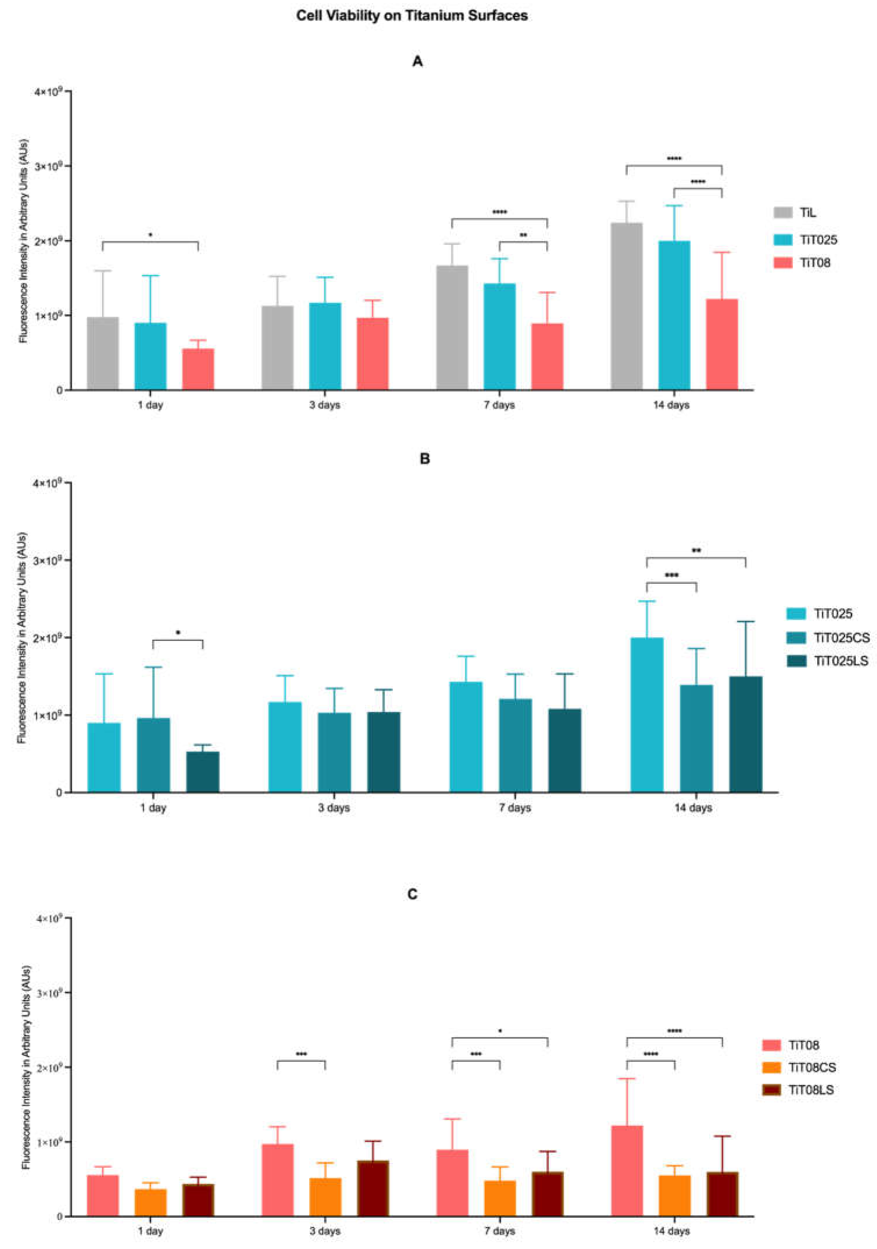
Figure 2.
Bar graph showing hFOB 1.19 cell viability on titanium surfaces coated with HA using conventional or laser sintering over 14 days. Data represent the combined viability values of the 0.25 mm and 0.8 mm patterned groups processed via either conventional sintering (CS) or laser sintering (LS). Cell viability is expressed in arbitrary units (AU). Each bar represents the mean of three assays ± standard deviation (SD); n = 15 per group.
Figure 2.
Bar graph showing hFOB 1.19 cell viability on titanium surfaces coated with HA using conventional or laser sintering over 14 days. Data represent the combined viability values of the 0.25 mm and 0.8 mm patterned groups processed via either conventional sintering (CS) or laser sintering (LS). Cell viability is expressed in arbitrary units (AU). Each bar represents the mean of three assays ± standard deviation (SD); n = 15 per group.
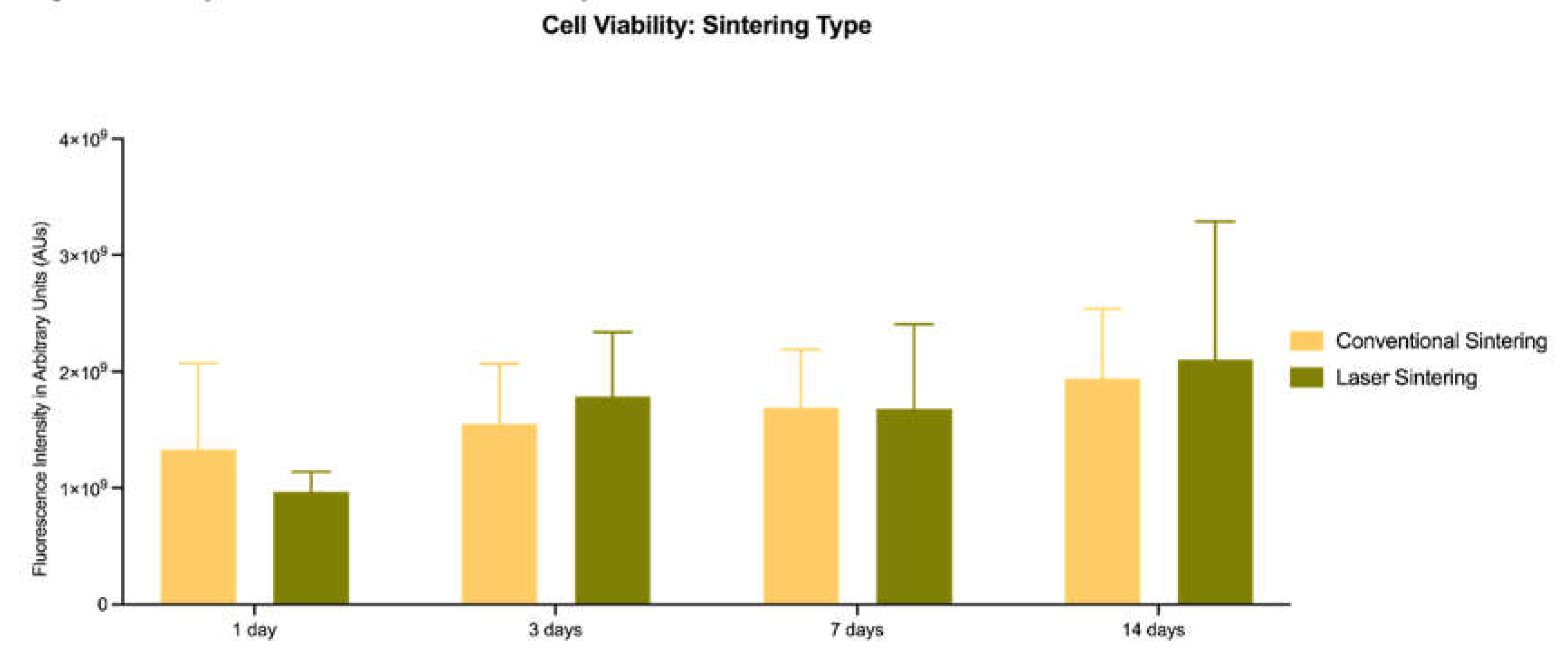
Figure 3.
SEM images of laser-textured and hydroxyapatite (HA)-coated titanium surfaces at magnifications of 100 x (A), 500 x (E, F) or 1000x (B-D, G). Osteoblasts (hFOB1.19) cultured for 24h on (A) smooth titanium surface (TiL), (B) 0.25 mm Nd:YAG-textured pattern, (C) 0.25 mm Nd:YAG-textured pattern with HA added by conventional sintering, (D) 0.25 mm Nd:YAG-textured pattern with HA added by CO₂ laser sintering, (E) 0.8 mm Nd:YAG-textured pattern, (F) 0.8 mm Nd:YAG-textured pattern with HA added by conventional sintering, and (G) 0.8 mm Nd:YAG-textured pattern with HA added by CO₂ laser sintering.
Figure 3.
SEM images of laser-textured and hydroxyapatite (HA)-coated titanium surfaces at magnifications of 100 x (A), 500 x (E, F) or 1000x (B-D, G). Osteoblasts (hFOB1.19) cultured for 24h on (A) smooth titanium surface (TiL), (B) 0.25 mm Nd:YAG-textured pattern, (C) 0.25 mm Nd:YAG-textured pattern with HA added by conventional sintering, (D) 0.25 mm Nd:YAG-textured pattern with HA added by CO₂ laser sintering, (E) 0.8 mm Nd:YAG-textured pattern, (F) 0.8 mm Nd:YAG-textured pattern with HA added by conventional sintering, and (G) 0.8 mm Nd:YAG-textured pattern with HA added by CO₂ laser sintering.
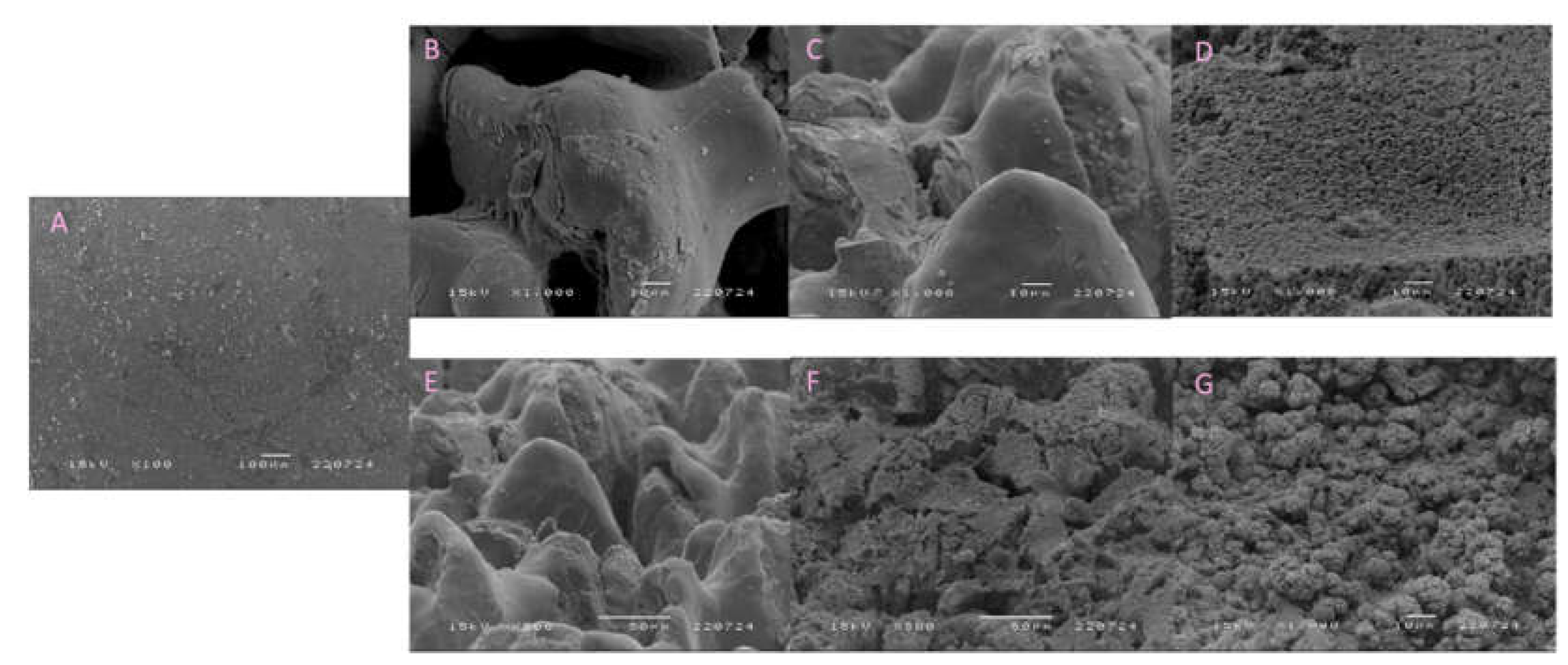
Figure 4.
Bar graphs showing the secretion profile of IL-1β, IL-10 and IL-8 from human osteoblasts (hFOB 1.19) after 1 and 3 days of culture. (A) Cytokine secretion on laser-textured titanium samples (TiT025 and TiT08) compared to the smooth titanium control (TiL); (B) cytokine secretion on the 0.25 mm patterns modified with conventionally sintered (TiT025CS) or laser-sintered (TiT025LS) HA coatings; (C) cytokine secretion on the 0.8 mm patterns modified with conventionally sintered (TiT08CS) or laser-sintered (TiT08LS) HA coatings. Cytokine concentrations are expressed in pg/mL. Data are presented as mean ± standard deviation (SD) from independent experimental replicates; n=3 for each group. Statistical significance: **p<0.01, ***p<0.005, ****p<0.001.
Figure 4.
Bar graphs showing the secretion profile of IL-1β, IL-10 and IL-8 from human osteoblasts (hFOB 1.19) after 1 and 3 days of culture. (A) Cytokine secretion on laser-textured titanium samples (TiT025 and TiT08) compared to the smooth titanium control (TiL); (B) cytokine secretion on the 0.25 mm patterns modified with conventionally sintered (TiT025CS) or laser-sintered (TiT025LS) HA coatings; (C) cytokine secretion on the 0.8 mm patterns modified with conventionally sintered (TiT08CS) or laser-sintered (TiT08LS) HA coatings. Cytokine concentrations are expressed in pg/mL. Data are presented as mean ± standard deviation (SD) from independent experimental replicates; n=3 for each group. Statistical significance: **p<0.01, ***p<0.005, ****p<0.001.
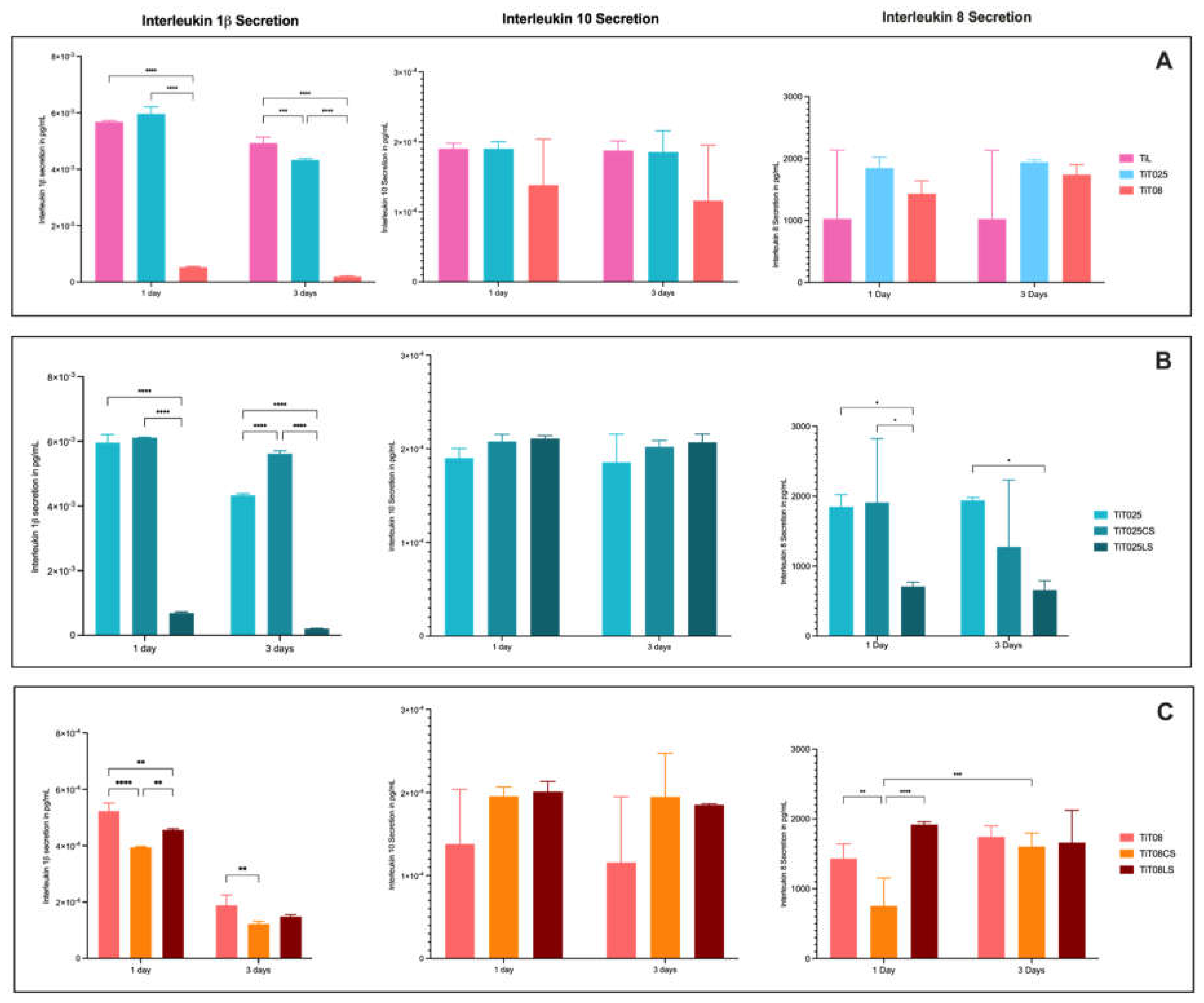
Figure 5.
Bar graphs showing the normalized ratio of cytokine secretion to human osteoblasts (hFOB 1.19) cell viability after 1 and 3 days of culture. Panels display the ratio of IL-1β, IL-10 and IL-8 pro-inflammatory to viability, respectively. (A) Cytokine/viability ratios on laser-textured titanium samples (TiT025 and TiT08) compared to the smooth titanium control (TiL); (B) cytokine/viability ratios on the 0.25 mm patterns modified with conventionally sintered (TiT025CS) or laser-sintered (TiT025LS) HA coatings; (C) cytokine/viability ratios on the 0.8 mm patterns modified with conventionally sintered (TiT08CS) or laser-sintered (TiT08LS) HA coatings. Ratios are expressed in picograms per milliliter per arbitrary unit (pg/mL/AU). Data are presented as mean ± standard deviation (SD) from independent experimental replicates; n=3 for each group. Statistical significance: *p<0.05, **p<0.01, ***p<0.005, ****p<0.001.
Figure 5.
Bar graphs showing the normalized ratio of cytokine secretion to human osteoblasts (hFOB 1.19) cell viability after 1 and 3 days of culture. Panels display the ratio of IL-1β, IL-10 and IL-8 pro-inflammatory to viability, respectively. (A) Cytokine/viability ratios on laser-textured titanium samples (TiT025 and TiT08) compared to the smooth titanium control (TiL); (B) cytokine/viability ratios on the 0.25 mm patterns modified with conventionally sintered (TiT025CS) or laser-sintered (TiT025LS) HA coatings; (C) cytokine/viability ratios on the 0.8 mm patterns modified with conventionally sintered (TiT08CS) or laser-sintered (TiT08LS) HA coatings. Ratios are expressed in picograms per milliliter per arbitrary unit (pg/mL/AU). Data are presented as mean ± standard deviation (SD) from independent experimental replicates; n=3 for each group. Statistical significance: *p<0.05, **p<0.01, ***p<0.005, ****p<0.001.
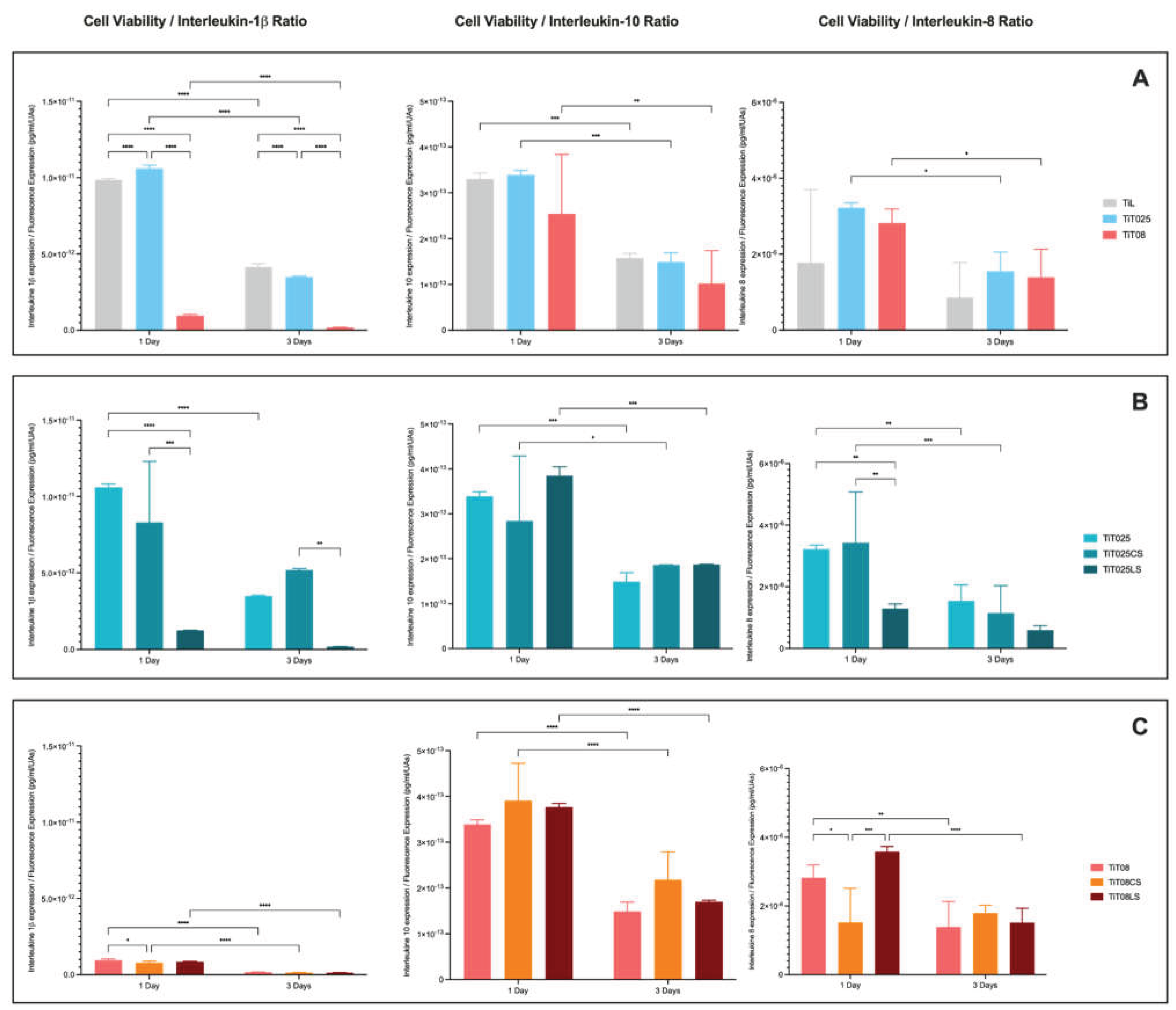

Table 1.
Chemical composition of titanium samples [39].
Table 1.
Chemical composition of titanium samples [39].
| Chemical Composition | Ti | O | Al | V |
| Wt (%) | 78,2 | 12,7 | 5,6 | 3,6 |
Table 2.
Nd:YVO4 laser specifications.
| Laser Specifications | Values |
| Maximum Output Power (W) | 30 |
| Wavelength (nm) | 1064 |
| Laser Technology | Nd1 |
| Repetition Rate Range (kHz) | 20 |
| Pulse Width (ns) | 10 |
| Spot Size (mm) | 0.01 |
| Cooling System | Forced Air-Cooling |
1 Nd stands for Neodymium, a rare-earth element embedded into the host crystal matrix that acts as the active gain medium responsible for producing the laser emission.
Table 3.
Chemical formula and physicochemical specifications of the hydroxyapatite powder nanoXIM Hap400 (as provided by the manufacturer—Fluidinova, Maia, Portugal).
Table 3.
Chemical formula and physicochemical specifications of the hydroxyapatite powder nanoXIM Hap400 (as provided by the manufacturer—Fluidinova, Maia, Portugal).
| Parameter | Specification/Value |
| Chemical Formula | (Ca10(PO4)6(OH)2) |
| Phase Purity | 100% |
| Ca/P Atomic Ratio | 1.67–1.68 |
| Particle Size (d50) | 10.0 ± 2.0 μm |
| Specific Gravity | 0.60 ± 0.10 g/cm3 |
| Specific Surface Area | ≥100 m2/g |
Table 4.
CO2 laser specifications.
| Laser Specifications | Values |
| Maximum Output Power (W) | 50 |
| Wavelength (nm) | 10.640 |
| Laser Technology | CO2 |
| Spot Size (mm) | 0.02 |
| Cooling System | None |
Table 5.
Comparative analysis of roughness profiles and wetting properties across samples.
| Groups | Ra (µm) | CA (°) |
| TiL | 0.2 | 82.48 ± 2.56 |
| TiT025 | 1.9 | 22.22 ± 4.83 |
| TiT025CS | - | 6.98 ± 7.14 |
| TiT025LS | - | 9.14 ± 6.3 |
| TiT08 | 2.7 | 11.25 ±11.31 |
| TiT08CS | - | 0 ± 0 |
| TiT08LS | - | 0 ± 0 |
Disclaimer/Publisher’s Note: The statements, opinions and data contained in all publications are solely those of the individual author(s) and contributor(s) and not of MDPI and/or the editor(s). MDPI and/or the editor(s) disclaim responsibility for any injury to people or property resulting from any ideas, methods, instructions or products referred to in the content. |
© 2026 by the authors. Licensee MDPI, Basel, Switzerland. This article is an open access article distributed under the terms and conditions of the Creative Commons Attribution (CC BY) license.
Copyright: This open access article is published under a Creative Commons CC BY 4.0 license, which permit the free download, distribution, and reuse, provided that the author and preprint are cited in any reuse.