
Submitted:
25 August 2026
Posted:
27 August 2026
You are already at the latest version
Abstract
Paederus dermatitis is an acute irritant contact dermatitis caused by inadvertent contact with rove beetles of the genus Paederus (Coleoptera: Staphylinidae). The condition may be difficult to recognize in individuals and healthcare professionals from non‑endemic regions because the characteristic skin lesions can mimic other inflammatory, infectious, or vesiculobullous dermatoses. The causative toxin, pederin, is associated predominantly with female beetles and is produced by endosymbiotic bacteria of the genus Pseudomonas. We present a series of cases of Paederus dermatitis observed in Russian physicians working in Nicaragua. Several hours after presumed contact with Paederus beetles, the patients developed burning and mild pruritus followed by erythematous macules and streak‑like lesions. Over the following days, grouped vesicles and non‑tense bullae developed, with subsequent erosions, crusting, and post‑inflammatory hyperpigmentation and hypopigmentation. The diagnoses were established clinically on the basis of the characteristic morphology, exposure history, and epidemiological setting. Conservative treatment resulted in gradual resolution of the inflammatory lesions within approximately 2–3 weeks, with residual post‑inflammatory hyperpigmentation. This case series highlights the diagnostic value of recognizing the characteristic linear and bizarrely configured lesions of Paederus dermatitis, particularly in healthcare professionals working in endemic regions.
Keywords:
paederus dermatitis
; pederin
; rove beetle
; irritant contact dermatitis
; nicaragua
; vesiculobullous dermatitis
Figure 1.
Bullous lesion in Paederus dermatitis. Several Russian physicians working in Nicaragua developed acute cutaneous lesions after inadvertent crushing of rove beetles (Paederus sp.) against their skin. The initial symptoms included burning and mild pruritus, followed by erythematous macules and streak-like lesions. In one patient, a non-tense bulla with a flaccid whitish roof is seen on a mildly erythematous background, representing an early vesiculobullous response to pederin exposure. Pederin is a potent vesicant synthesized by endosymbiotic Pseudomonas bacteria and stored in the hemolymph of female beetles; males carry only negligible amounts of the toxin [1,2]. Such bullous lesions are typical of the acute phase of this irritant contact dermatitis [3,4,5].
Figure 1.
Bullous lesion in Paederus dermatitis. Several Russian physicians working in Nicaragua developed acute cutaneous lesions after inadvertent crushing of rove beetles (Paederus sp.) against their skin. The initial symptoms included burning and mild pruritus, followed by erythematous macules and streak-like lesions. In one patient, a non-tense bulla with a flaccid whitish roof is seen on a mildly erythematous background, representing an early vesiculobullous response to pederin exposure. Pederin is a potent vesicant synthesized by endosymbiotic Pseudomonas bacteria and stored in the hemolymph of female beetles; males carry only negligible amounts of the toxin [1,2]. Such bullous lesions are typical of the acute phase of this irritant contact dermatitis [3,4,5].
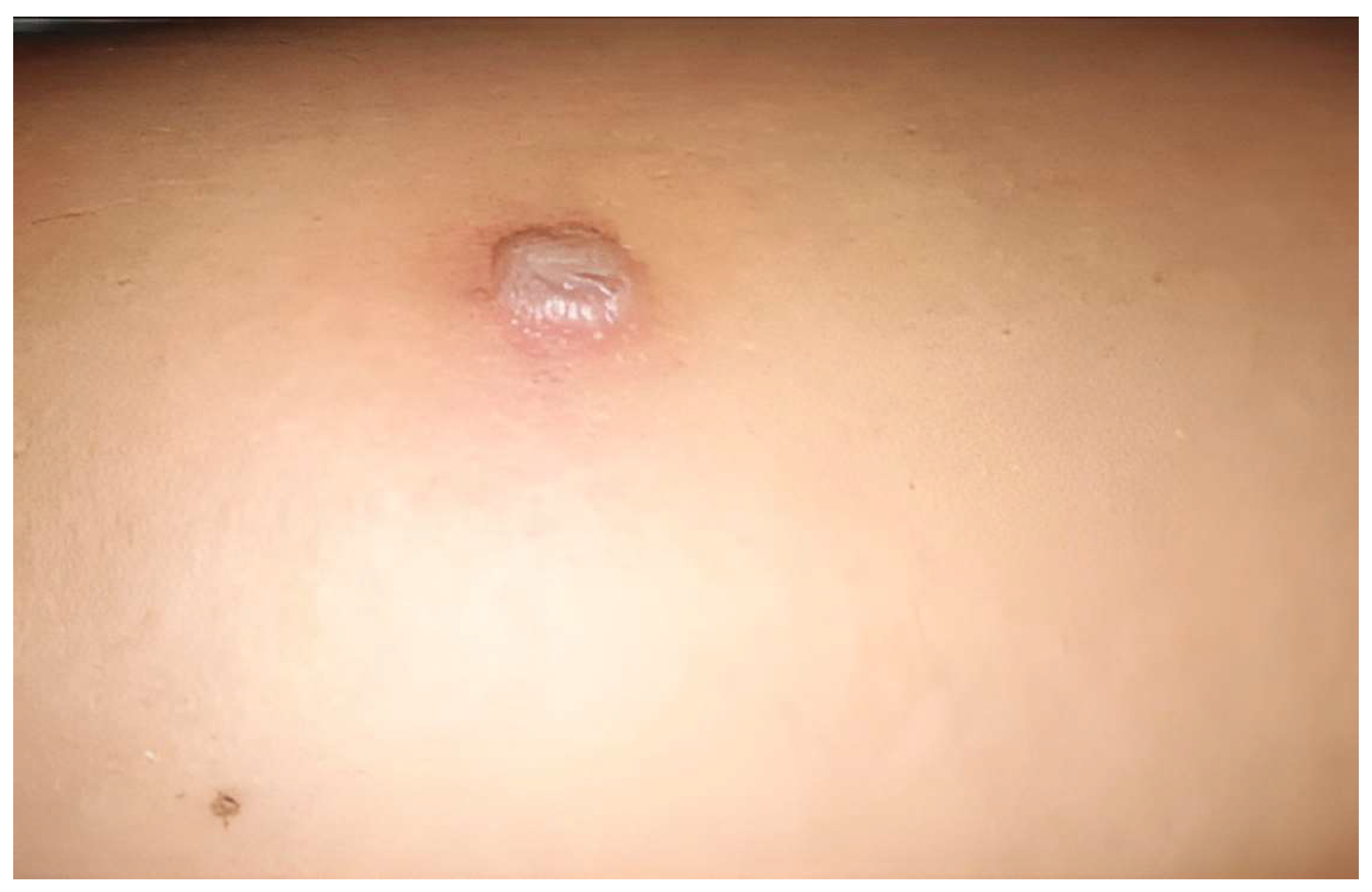
Figure 2.
Figure 2. Linear vesiculobullous lesions. Over the following days, grouped vesicles and bullae appeared in the affected individuals, many arranged in a linear or irregular pattern – a hallmark of mechanical toxin transfer across the skin surface [3,4]. This image from another patient shows multiple linearly arranged vesicles, a non-tense bulla with clear contents, and a collapsed bulla on an erythematous background. Irregular areas of hypopigmentation are also visible, indicating early pigmentary changes. The linear configuration is highly characteristic and helps distinguish Paederus dermatitis from other vesiculobullous disorders [4,5].
Figure 2.
Figure 2. Linear vesiculobullous lesions. Over the following days, grouped vesicles and bullae appeared in the affected individuals, many arranged in a linear or irregular pattern – a hallmark of mechanical toxin transfer across the skin surface [3,4]. This image from another patient shows multiple linearly arranged vesicles, a non-tense bulla with clear contents, and a collapsed bulla on an erythematous background. Irregular areas of hypopigmentation are also visible, indicating early pigmentary changes. The linear configuration is highly characteristic and helps distinguish Paederus dermatitis from other vesiculobullous disorders [4,5].
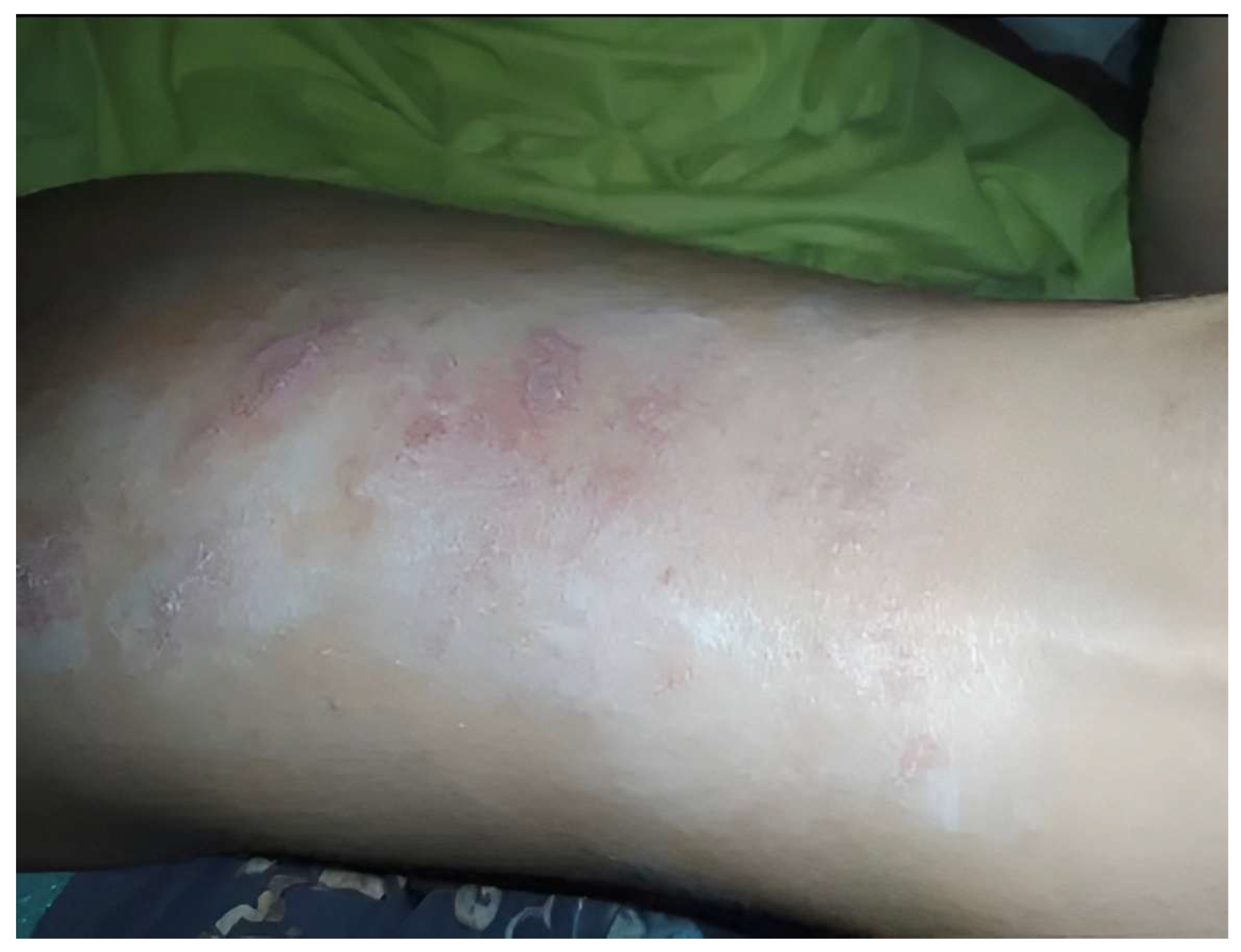
Figure 3.
Post-inflammatory hyperpigmentation following resolution. As the acute inflammation subsided in several patients, brownish, irregularly shaped foci of post-inflammatory hyperpigmentation with poorly defined borders developed, as seen here on the anterior surface of the lower leg. Thin central crusts correspond to the roofs of ruptured bullae. Multiple smaller pigmented spots are also present, reflecting the healing phase of the dermatitis. Residual hyperpigmentation is a frequent sequela and may persist for weeks to months [4,5].
Figure 3.
Post-inflammatory hyperpigmentation following resolution. As the acute inflammation subsided in several patients, brownish, irregularly shaped foci of post-inflammatory hyperpigmentation with poorly defined borders developed, as seen here on the anterior surface of the lower leg. Thin central crusts correspond to the roofs of ruptured bullae. Multiple smaller pigmented spots are also present, reflecting the healing phase of the dermatitis. Residual hyperpigmentation is a frequent sequela and may persist for weeks to months [4,5].
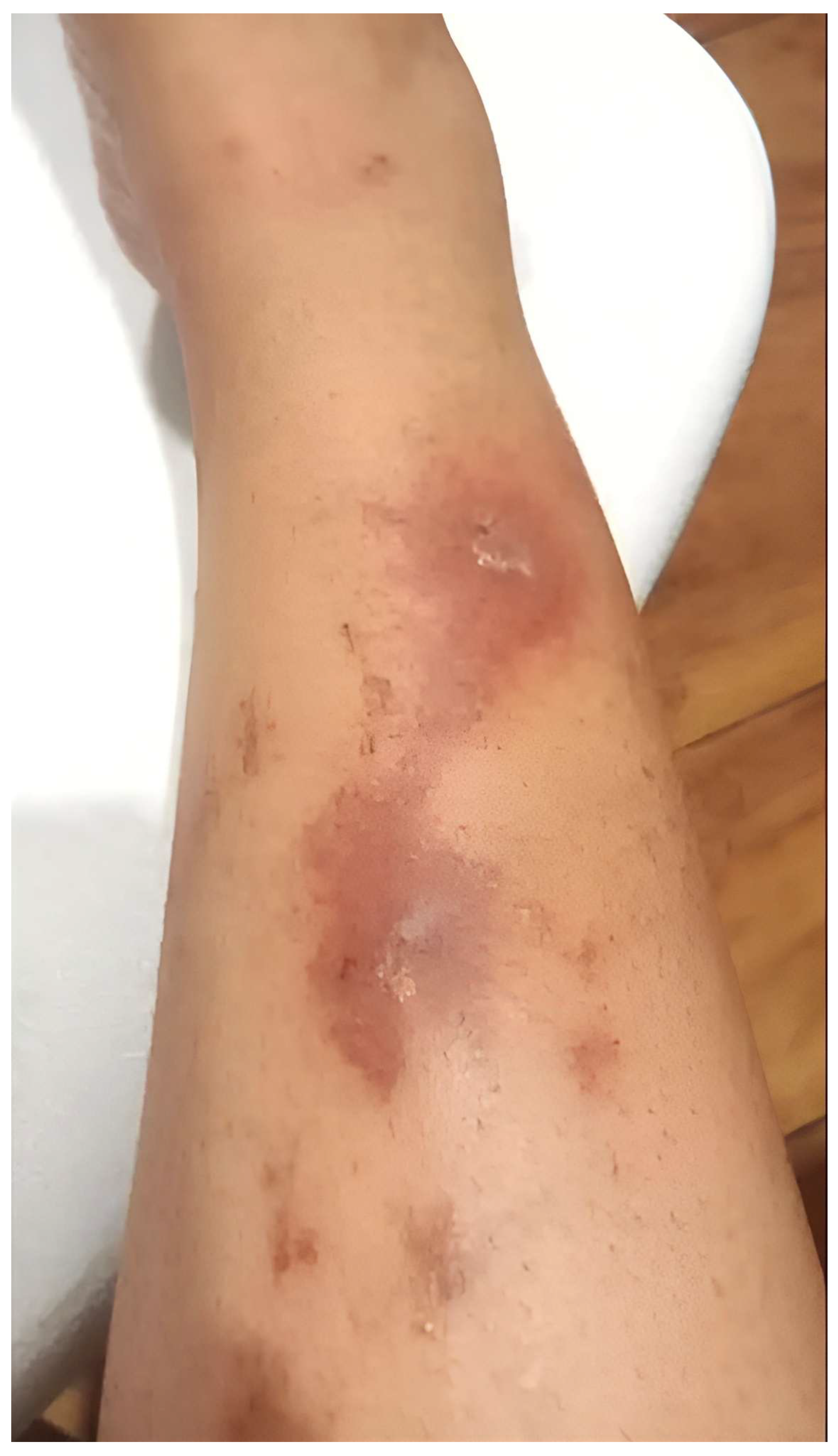
Figure 4.
Acute phase: polymorphic lesions representing early stages. During the acute phase, the cutaneous manifestations were not uniform; fresh vesiculobullous lesions coexisted with resolving ones. In this patient, an irregularly shaped erythematous focus with rounded and linear margins contains pale grouped vesicles centrally and light-brown discoloration toward the periphery, illustrating the initial evolution from active inflammation to early pigmentary alteration. The simultaneous presence of lesions at various stages is a common feature of Paederus dermatitis [3].
Figure 4.
Acute phase: polymorphic lesions representing early stages. During the acute phase, the cutaneous manifestations were not uniform; fresh vesiculobullous lesions coexisted with resolving ones. In this patient, an irregularly shaped erythematous focus with rounded and linear margins contains pale grouped vesicles centrally and light-brown discoloration toward the periphery, illustrating the initial evolution from active inflammation to early pigmentary alteration. The simultaneous presence of lesions at various stages is a common feature of Paederus dermatitis [3].
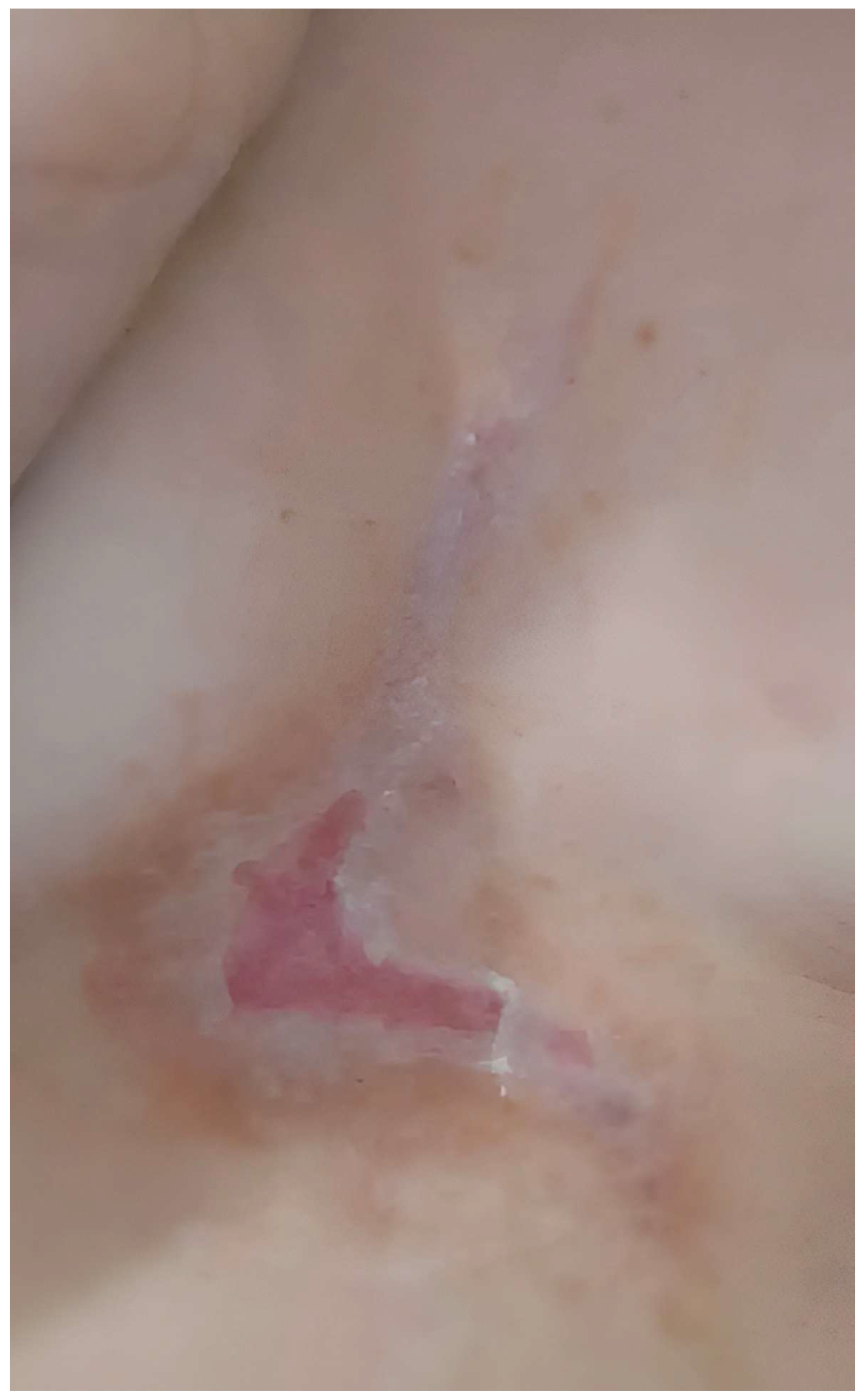
Figure 5.
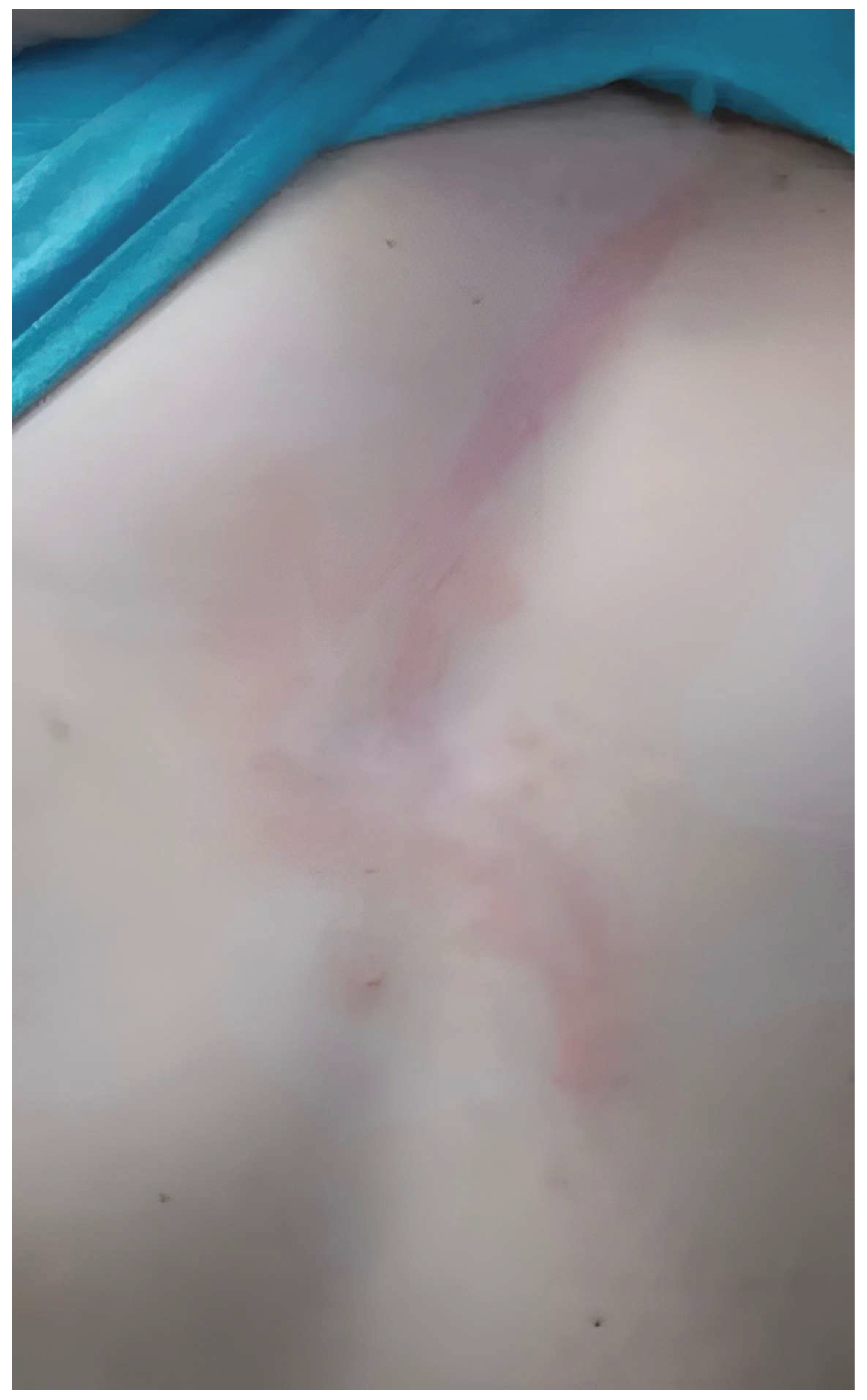
Progressive phase: characteristic linear inflammatory lesion. As the inflammatory process progressed, the lesions became more defined and assumed their typical bizarre configurations. This image demonstrates a linear erythematous macule with areas of relatively spared skin within the streak and a single papule, observed in another physician. Such geographical configurations are highly suggestive of Paederus dermatitis and help distinguish it from other vesiculobullous disorders, including herpes simplex, phytophotodermatitis, and allergic contact dermatitis [6,3]. The diagnoses were made clinically based on morphology, exposure history, and the tropical setting; no laboratory tests were required. The toxin's mechanism and the beetle's biology have been detailed elsewhere [1,2].
Figure 5.
Progressive phase: characteristic linear inflammatory lesion. As the inflammatory process progressed, the lesions became more defined and assumed their typical bizarre configurations. This image demonstrates a linear erythematous macule with areas of relatively spared skin within the streak and a single papule, observed in another physician. Such geographical configurations are highly suggestive of Paederus dermatitis and help distinguish it from other vesiculobullous disorders, including herpes simplex, phytophotodermatitis, and allergic contact dermatitis [6,3]. The diagnoses were made clinically based on morphology, exposure history, and the tropical setting; no laboratory tests were required. The toxin's mechanism and the beetle's biology have been detailed elsewhere [1,2].
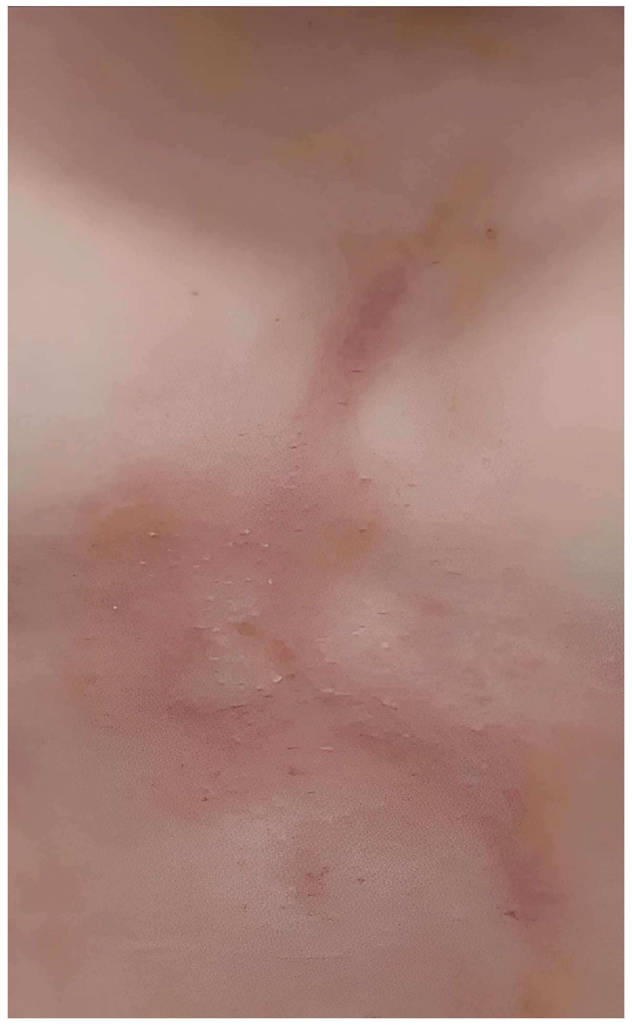
Figure 6.
Resolution phase: resolving linear lesion with residual hyperpigmentation. During the convalescent stage, the acute inflammation gradually subsided. In this patient, a bright-red linear inflammatory macule with areas of resolution and maceration is seen superimposed on an irregular focus of brown post-inflammatory hyperpigmentation. The affected physicians were treated conservatively with local skin care, antiseptics, and topical corticosteroids. The inflammatory lesions resolved within 2–3 weeks, leaving only pigmentary sequelae. No systemic complications or recurrence occurred. Thus, Figure 4, Figure 5 and Figure 6 illustrate the temporal evolution of Paederus dermatitis in different patients: from the acute polymorphic phase (Figure 4) through the progressive stage with characteristic linear lesions (Figure 5) to the resolution phase with post-inflammatory hyperpigmentation (Figure 6). This case series underscores that recognition of linear and bizarrely shaped lesions is crucial for prompt diagnosis, especially for healthcare workers from non-endemic regions practicing in tropical areas, as misdiagnosis may lead to unnecessary investigations and inappropriate therapy. The epidemiological context is consistent with a previous report of Paederus dermatitis among medical volunteers in the same region of Nicaragua [7].
Figure 6.
Resolution phase: resolving linear lesion with residual hyperpigmentation. During the convalescent stage, the acute inflammation gradually subsided. In this patient, a bright-red linear inflammatory macule with areas of resolution and maceration is seen superimposed on an irregular focus of brown post-inflammatory hyperpigmentation. The affected physicians were treated conservatively with local skin care, antiseptics, and topical corticosteroids. The inflammatory lesions resolved within 2–3 weeks, leaving only pigmentary sequelae. No systemic complications or recurrence occurred. Thus, Figure 4, Figure 5 and Figure 6 illustrate the temporal evolution of Paederus dermatitis in different patients: from the acute polymorphic phase (Figure 4) through the progressive stage with characteristic linear lesions (Figure 5) to the resolution phase with post-inflammatory hyperpigmentation (Figure 6). This case series underscores that recognition of linear and bizarrely shaped lesions is crucial for prompt diagnosis, especially for healthcare workers from non-endemic regions practicing in tropical areas, as misdiagnosis may lead to unnecessary investigations and inappropriate therapy. The epidemiological context is consistent with a previous report of Paederus dermatitis among medical volunteers in the same region of Nicaragua [7].
Author Contributions
For research articles with several authors, a short paragraph specifying their individual contributions must be provided. Conceptualization, Y.V.Z.; writing—original draft preparation, Y.V.Z. and E.A.S.; writing—review and editing, Y.V.Z., V.A.G., and N.P.M.; supervision, Y.V.Z.; project administration, Y.V.Z. All authors have read and agreed to the published version of the manuscript.
Funding
This research received no external funding.
Institutional Review Board Statement
The study was conducted in accordance with the Declaration of Helsinki. Ethical review and approval were waived for this retrospective case series by the Ethics Committee of I.M. Sechenov First Moscow State Medical University (Sechenov University) (waiver code 2025-123-W, dated 15 April 2025) because it involves only anonymized clinical photographs and does not constitute human subject research.
Informed Consent Statement
Verbal informed consent was obtained from the participants. Verbal consent was obtained rather than written because the study was retrospective, all data were anonymized, and no additional procedures beyond routine clinical care were performed.
Data Availability Statement
Data is contained within the article.
Acknowledgments
The authors sincerely thank Dr. Kirill Mokhniuk and the members of the international humanitarian medical mission for providing photographic materials for the clinical description. The mission has been actively engaged in building a hospital and reconstructing the infrastructure for patient care in La Salvia, Nicaragua, since the end of 2018.
Conflicts of Interest
The authors declare no conflicts of interest.
References
- Marchiori, C.H.; de Oliveira Santana, M.V.; de Paula Malheiros, K. Characteristics of venomous Coleopterans and their venom with therapeutic power (Insecta: Coleoptera). J. Med. Sci. 2024, 4, 105–115. [Google Scholar] [CrossRef]
- Kellner, R.L.L.; Dettner, K. Allocation of pederin during lifetime of Paederus rove beetles (Coleoptera: Staphylinidae): evidence for polymorphism of hemolymph toxin. J. Chem. Ecol. 1995, 21, 1719–1733. [Google Scholar] [CrossRef] [PubMed]
- Inbamani, A.; Sundaram, G.; Ramalingam, R. Morphological and clinical patterns of Paederus dermatitis. Cureus 2024, 16, e58813. [Google Scholar] [CrossRef] [PubMed]
- Zargari, O.; Kimyai-Asadi, A.; Fathalikhani, F.; Panahi, M. Paederus dermatitis in northern Iran: a report of 156 cases. Int. J. Dermatol. 2003, 42, 608–612. [Google Scholar] [CrossRef] [PubMed]
- Sendur, N.; Savk, E.; Karaman, G. Paederus dermatitis: a report of 46 cases in Aydin, Turkey. Dermatology 1999, 199, 353–355. [Google Scholar] [CrossRef] [PubMed]
- Nasir, S.; Akram, W.; Khan, R.R.; Arshad, M.; Nasir, I. Paederus beetles: the agent of human dermatitis. J. Venom. Anim. Toxins Incl. Trop. Dis. 2015, 21, 5. [Google Scholar] [CrossRef] [PubMed]
- Mokhniuk, K.; Zhernov, Y.V. Case report of a Paederus dermatitis in international humanitarian medical mission in Nicaragua. Allergy 2020. [Google Scholar] [CrossRef]
Disclaimer/Publisher’s Note: The statements, opinions and data contained in all publications are solely those of the individual author(s) and contributor(s) and not of MDPI and/or the editor(s). MDPI and/or the editor(s) disclaim responsibility for any injury to people or property resulting from any ideas, methods, instructions or products referred to in the content. |
© 2026 by the authors. Licensee MDPI, Basel, Switzerland. This article is an open access article distributed under the terms and conditions of the Creative Commons Attribution (CC BY) license (http://creativecommons.org/licenses/by/4.0/).
Copyright: This open access article is published under a Creative Commons CC BY 4.0 license, which permit the free download, distribution, and reuse, provided that the author and preprint are cited in any reuse.