

Submitted:
25 August 2026
Posted:
27 August 2026
You are already at the latest version
Preprints on COVID-19 and SARS-CoV-2
Abstract
The management of COVID-19 continues to evolve as clinical experience and scientific research refine approaches to diagnosis, isolation protocols, and treatment. Diagnosing COVID-19 relies either on molecular tests to detect specific viral genetic material or antigen tests to detect viral proteins. Molecular tests generally have higher analytical sensitivity and accuracy than antigen tests, particularly early in infection. The SARS-CoV-2 RNA genetic material may remain detectable for prolonged periods after infection. Immunoassay antigen tests detect specific viral proteins but are less sensitive than molecular tests and can miss early infection. Laboratory diagnosis of acute COVID-19 commonly relies on molecular or antigen testing of respiratory specimens, including nasopharyngeal or other appropriate specimens. Both tests, molecular and antigenic, may yield a false negative result due to the insufficient viral load in the nasopharynx, or a false positive result caused by sample contamination or cross-reactivity. Given that sputum often contains a higher viral load and demonstrates significantly higher detection rates, we propose sputum testing as a highly effective alternative to nasopharyngeal swabs for COVID-19 diagnosis. Pretreatment of sputum with surfactants and proteolytic enzymes effectively breaks down the sample to release viral antigens. This liquefaction method improves assay performance and helps to enhance the sensitivity of the diagnostic test. Furthermore, the virus remains detectable several months after initial infection using either spontaneous or induced sputum samples. This simple method has the potential to be significantly cost-effective, offering a faster and more affordable alternative to molecular testing. This overview explores how affinity capture-migration and affinity capture-separation techniques utilize sputum and other biological fluid samples to successfully monitor the long-term effect of COVID-19, which can lead to long-lasting multi-organ issues, and the impact of quantifying pro-inflammatory cytokines and aberrant glycoproteins in the lingering inflammation of post-COVID infection.
Keywords:
affinity capture-separation
; aberrant glycopeptides
; cytokine storm
; immunoaffinity capillary electrophoresis
; infectious diseases
; Long COVID
; liquefied sputum
; low abundance biomarkers
; non-invasive diagnostics
; proteoforms
; preventive medicine
; sputum biomarkers
Introduction
Severe acute respiratory syndrome coronavirus 2 (SARS-CoV-2), the novel coronavirus that causes coronavirus disease 2019 (COVID-19) continues to be a concern for public health since it began to spread globally. According to global COVID-19 tracker, as of August 2, 2026, there have been 779,844,137 cumulative cases of COVID-19, 6,849 weekly new cases, and 7,121,990 cumulative deaths worldwide [1]. COVID-19 patients with mild-to-moderate illness are highly unlikely to be infectious beyond 10 days of symptoms. However, evidence from a limited number of studies indicates that patients with severe-to-critical illness or who are immunocompromised, may shed infectious virus for extended periods [2].
Some viruses appear to be dormant in the body for a certain period. Many others cause acute infections and are completely cleared by the immune system. For example, the Andes orthohantavirus (ANDV), endemic in Chile and Argentina, has an incubation period between seven to 39 days, and the transmission to humans occurs primarily via the inhalation of virus-containing aerosols from rodent excretions such as urine, feces, and saliva [3]. As an exception among hantaviruses, person-to-person transmission has been demonstrated for ANDV, including possible sexual transmission. Peculiarly, the viral RNA remained detectable in semen samples for about 6 years. There is evidence that at least 27 different viruses, across a broad range of virus families or orders, can be found in human semen. Some viruses cause chronic or latent infections; others cause acute infections [3].
SARS-CoV-2 transmission occurs predominantly through respiratory aerosol particles and droplets. However, the virus has also been detected in blood, feces, and semen, indicating that the infection may at times be systemic [4,5]. Although SARS-CoV-2 RNA has been detected in semen, sexual transmission has not been established as a meaningful route of viral components. Among patients with moderate to severe COVID-19, studies have detected SARS-CoV-2 RNA in about 40% to 85% of fecal samples, for as much as 7 months [6,7].
While SARS-CoV-2 infections primarily target the respiratory system, the virus and the body’s resulting inflammatory response can damage tissues beyond the lung, such as heart, endothelium of blood vessels, kidneys, liver, muscle, endocrine system, immune system, gastrointestinal tract, and brain. Injury to the organs may become apparent long after the acute infection has subsided. Different organs may be affected at different times [8,9,10,11,12]. Findings of a multicohort study indicate that SARS-CoV-2 antigens can be detected in the blood of a substantial proportion of individuals up to 14 months after infection. While approximately one in five asymptomatic individuals was antigen-positive, roughly half of all individuals reporting ongoing cardiopulmonary, musculoskeletal, and neurological symptoms were antigen positive [13].
Long COVID, also known as post-acute sequelae of COVID-19 (PASC), is an infection-associated chronic condition that occurs after SARS-CoV-2 infection and can lead to sometimes severe disability. It is a significant health concern affecting millions of people globally [14]. A recent report estimated that the cumulative global incidence of Long COVID was approximately 400 million people, with an estimated annual global economic impact of US$1 trillion [15,16]. Six years into the COVID-19 pandemic, scientific understanding of Long COVID has advanced significantly, and evidence implicates several pathomechanisms, including aberrant immunity, persistent pathogens, endothelial dysfunction, and mitochondrial dysfunction [16].
It remains to be demonstrated that SARS-CoV-2 might infect organ systems by gaining access to the bloodstream. Although highly speculative now, SARS-CoV-2 might cause a persistent chronic infection in certain individuals. In these patients, certain sites might act as a potential “viral reservoir”, in which the virus can persist for prolonged periods accompanied by recurrent viral shedding. This phenomenon is not new for RNA viruses, as this has been described in Ebola virus survivors. SARS-CoV-2 persistence and recurrent shedding has been demonstrated in the respiratory tract, but also sites of attenuated immunity such as the testis, eye and brain might potentially preserve the virus for longer times [9].
Is sputum considered to be a long-term reservoir of the COVID-19 virus? Because sputum is a dense, sticky hydrogel, it has long been recognized as a temporary primary vehicle or reservoir for respiratory viruses (it is not an anatomical refuge). This complex molecular network traps and shelters viruses, allowing them to evade clearance. Consequently, airway mucus may serve as a transient matrix in which SARS-CoV-2 RNA, viral proteins and, in some circumstances, infectious virus can persist [17,18,19,20]. Additionally, it has been demonstrated that coronaviruses can persist on inanimate surfaces like metal, glass, or plastic from hours to up to 9 days [21]. Notably, even if testing for the COVID-19 virus in nose and throat yields negative results, the patients can still shed the virus in their sputum and feces [7,19,22,23]. These cases highlight the necessity of a comprehensive approach to COVID-19 diagnosis. For instance, Balla et al. [24] and Wiseman et al. [25] documented a patient whose RT-PCR result was initially false negative but later tested positive. False negative results can have significant implications on public health due to high reproductive factor of COVID-19. Consequently, some medical centers prohibited the discharge of patients solely based on negative RT-PCR molecular tests on specimens obtained from nasopharyngeal and oropharyngeal swabs [26]. Patients were only cleared for release once their symptoms resolved, their molecular tests were negative, and serial chest CT imaging showed no lingering inflammation. CT scans of the chest have been shown to have a high sensitivity and specificity for identifying lung lesions in SARS-CoV infection [27].
Furthermore, other studies suggest that the virus has developed sophisticated ways to “cloak” or hide itself from the immune system. The virus uses the classical mechanism of camouflaging and exploiting the host machinery for its own protein synthesis. It goes inside the host cell, and its messenger RNA looks the same as the host messenger RNA. In SARS coronaviruses, the non-structural protein 16 (nsp16), in conjunction with nsp10, methylates the 5′-end of virally encoded mRNA to mimic cellular mRNAs, thus protecting the virus from host innate immune restriction [28]. Comparing data from more than 1000 specimens collected from 205 patients with COVID-19, it was found that bronchoalveolar lavage fluid specimens showed the highest positive rates (93%), followed by sputum (72%), nasal swabs (63%), fibrobronchoscope brush biopsy (46%), pharyngeal swabs (32%), feces (29%), and blood (15%) [23].
Sputum is considered an excellent and highly reliable specimen source for diagnosing COVID-19, particularly for patients experiencing a productive cough or deep respiratory symptoms [19,29]. The method is simple, non-invasive, and inexpensive, and the sample is easy to collect without requiring highly trained personnel. Additionally, it naturally harbors diverse pathogens, allowing modern molecular and antigenic techniques to accurately detect the specific virus and bacteria causing various respiratory and systemic diseases [30]. Multiple studies demonstrate that the lower respiratory tract, origin of sputum, yields a very high viral load, often resulting in higher SARS-CoV-2 detection rates than standard nasopharyngeal or oropharyngeal swabs, especially later in the course of the infection [29]. Lin et al. [31] demonstrated that among patients with concurrent positive nasopharyngeal or throat swab, expectorated sputum specimens exhibited both a superior positivity rate and the highest viral titers of infectious SARS-CoV-2. Another report indicates that SARS-CoV-2 RNA is more readily detected in induced sputum than in throat swabs of convalescent COVID-19 patients [32].
This overview describes current information on the types of laboratory tests used to detect SARS-CoV-2 infection, highlighting some technical modifications to identify viral and bacterial markers for accurate diagnosis, guiding treatment for at-risk patients.
Current Tests for the Diagnosis of COVID-19
At present, there are three main types of COVID-19 tests available to detect current infection or past exposure. Rapid antigen tests (RATs) and molecular tests (nucleic acid amplification tests-NAATs/reverse transcription polymerase chain reaction-RT-PCR) are used to detect current infection, whereas antibody tests (serology) are used to detect past infection [33]. A simplified visual comparison of rapid tests and molecular test is shown in Figure 1. The remainder of this text focuses exclusively on these tests, excluding antibody testing. Generally, antibodies against SARS-CoV-2 (IgG, IgA, IgM) can be detected in blood samples within 1 to 3 weeks after the initial infection or vaccination. They can be detected using indirect immunofluorescence assays, enzyme-linked immunosorbent assays, and chemiluminescence microparticle immunoassay [34]. Other tests have been reported for the detection of SARS-CoV-2, including loop-mediated isothermal amplification (LAMP), clustered regularly interspaced short palindromic repeats (CRISPR), colloidal gold immunochromatography assay (GICA), and electrochemical sensors. However, these different tests vary greatly in terms of their detection efficiency, analytical specificity, accuracy, analytical sensitivity, cost and throughput [35]. Besides, most of the current detection methods are conducted in central hospitals and clinical laboratories, which is a great challenge for remote and underdeveloped areas. It is noteworthy to mention that some tests for COVID-19 should be performed carefully, and the results should be judged comprehensively, considering the clinical symptoms and patient background. Analysis of diagnostic performance and factors causing nonspecific reactions, have been evaluated in SARS-CoV-2 rapid antigen detection tests [36].
In a rapid antigen test, the solubilized sample migrates via capillary action across the test strip to the conjugate pad, where it interacts with detection antibodies. If the target viral protein is present, it binds to these antibodies immobilized on the ‘T’ line, producing a visible colored signal. Residual labeled antibodies proceed to the ‘C’ line, where they bind to the antibody immobilized on the ‘C’ line where a visible colored signal is formed as well, confirming the test’s analytical validity regardless of whether the target is found [18,19,38,39]. While these rapid tests are useful, Nucleic Acid Amplification Tests (NAATs) remain the gold standard of COVID-19 diagnosis due to their superior sensitivity in early stage and asymptomatic infections [40].
Regarding the nucleic acid amplification test, quantitative fluorescence-based transcription-polymerase chain reaction (RT-qPCR) is widely acknowledged as the gold standard for SARS-CoV-2 detection. The first step of sample preparation for RT-qPCR is the extraction of nucleic acids, specifically RNA for SARS-CoV-2, which can be time-consuming, labor-intensive, and prone to contamination. This is followed by the reverse transcription of the viral RNA into complementary DNA (cDNA). The third step is the qPCR amplification of the cDNA using specific primers. A real-time thermocycler monitors and records this fluorescence, allowing for the determination of the cycle threshold (Ct). Ct represents the number of cycles in which the fluorescence significantly exceeds the background and enables the measurement of the exponential accumulation of amplicons. By comparing the Ct values of the controls and samples, the relative gene expression can be estimated. Details of the procedure have been reported elsewhere [40].
Diagnostic methods for viral diseases have evolved rapidly due to the pandemic. New genetic techniques, such as isothermal amplification, CRISPR-based methods, next-generation sequencing, and artificial intelligence have opened challenging opportunities that can help with the correct interpretation of results and epidemiological surveillance [35,41,42].
Sputum is a Promising Alternative Specimen as a Reliable Source for Diagnosing COVID-19
The lung and tracheobronchial tree are usually sterile below the larynx in healthy individuals. An infectious agent reaches this site via a breach in the host defense bypassing the multi-tiered defense system [43]. Physical and cellular barriers of the respiratory tract mucosal surface utilize a variety of strategies to obstruct microbe entry [44]. Nonetheless, organisms enter the distal airway by inhalation, aspiration or by hematogenous seeding. The pathogen multiplies in or on the epithelium, causing inflammation, increased mucus secretion, and impaired mucociliary function; other lung functions may also be affected [45]. If these microorganisms can overcome or subvert the innate host defenses, such as the mucociliary system, bronchoconstriction, cough reflex, or acquired immunity (mucosal immunoglobulin A), then the infectious process can begin.
A sputum sample reveals various microorganisms, including bacteria, viruses and occasionally fungi. In sputum cultures, the most frequently isolated pathogens are typically bacteria, such as S. pneumoniae, H. influenzae, S. aureus, and Klebsiella. More than 200 different types of viruses are responsible for human upper respiratory tract infections. The most common are rhinoviruses (30–50% of all colds), followed by coronaviruses (10–15% of all colds). Influenza viruses cause 5–15% of colds [45]. However, the clinical significance of mixed viral-bacterial infections remains unclear [30]. Furthermore, respiratory viruses have been detected in sputum samples from patients with chronic obstructive pulmonary disease (COPD), asthma, and cystic fibrosis [46].
Sputum was traditionally considered a suboptimal specimen due to its viscosity. However, with the improvement of antigen and molecular methods, sputum has been introduced for the diagnosis of respiratory viral infections [18,19,47]. Several studies have supported the use of sputum testing as a valuable method for SARS-CoV-2 antigen and molecular testing and highlight the importance of early testing after symptoms onset to increase the rates of COVID-19 diagnosis [19,48]. Figure 2 illustrates an improved procedure for liquefying viscous sputum to isolate viral proteins for COVID-19 diagnosis. Other types of procedures applied to release bacterial, viral, and diverse specimen constituents from the complex sputum meshwork have been described elsewhere [18,19].
When working with complex matrices containing low concentrations of a target substance, it is necessary to maximize analyte recovery. Extraction, cleaning and enrichment processes are critical for achieving the necessary high rate of analytical sensitivity and detection limit to have a valid assay [49]. Because sputum forms a dense and viscous matrix, extracting biomarkers of interest without degrading the molecules demands meticulous sample preparation methods. We propose that incorporating at least a surfactant and a controlled proteolysis step overcomes this technical challenge, enabling the isolation of essential bacterial and viral peptides for diagnostic purposes [18,19]. It is important to keep in mind that analysis of sputum and other biofluids could contribute to this type of research when specimen collection and sample preparation are matched with the intended measurand.
Methods to Identify Simultaneously Multiple Components in Sputum for Diagnosing COVID-19 and Long COVID
Having already discussed the antigenic and molecular methods for acute COVID-19 diagnosis in the “Current Tests for the Diagnosis of COVID-19” section, we now evaluate the current methods available for the determination of the post-acute sequelae of SARS-CoV-2 infection (PASC), commonly known as Long COVID or long-haul COVID. Unfortunately, few diagnostic tools are available for identifying the Long COVID infection-associated chronic condition.
Long COVID has emerged as a new concern, as it may have affected more than half of the COVID-19 survivors which involves multi-organ systems [50], with a wide range of persistent symptoms that can remain long after acute SARS-CoV-2 episode [51]. Large-scale clinical and mechanistic studies have not yet confirmed the theoretical drivers of Long COVID, which include residual viral reservoirs, autoimmunity triggered by antibody cross-reactivity of SARS-CoV-2 antibodies with host proteins, and dysregulation of the renin-angiotensin system. As a result, identifying, diagnosing, and treating the syndrome remains a clinical hurdle, particularly because definitive early-detection biomarkers are still missing [52,53,54].
The persistence of the COVID-19 virus in biological fluids relies on specimen viral load. Sputum generally exhibits higher viral concentrations when compared to nasopharyngeal and oropharyngeal swabs [32]. Meanwhile, pharyngeal swabs still serve in many places as the standard metric to confirm patient eligibility for hospital discharge and the continued need for isolation. However, Chen et al. [22] has demonstrated that 22 patients continue to test positive for SARS–CoV-2, using the RT-qPCR method, when testing sputum or fecal samples, even after pharyngeal swabs became negative. Figure 3 demonstrates that SARS-CoV-2 is still detected in expectorated sputum specimens several weeks after collection [18,19]. The intensity of the band colors is semi-quantitative, rather than a true quantitative measure of the viral load.
Several reports have indicated that viral proteins can be detected not only in sputum samples for at least 3 months after the acute phase of the SARS-CoV-2 infection [19,55] but shedding of SARS-CoV-2 in feces and urine has also been reported [56] and detected in water and wastewater [57]. Nevertheless, there is no evidence that any-one has ever contracted COVID-19 from sewage exposure or drinking water. Among patients with moderate to severe COVID-19, studies detected SARS-CoV-2 RNA in about 40% to 85% of fecal samples. Approximately 4% of patients remained positive for at least 7 months [6,7].
Multiple proteins, used as a panel of biomarkers, significantly improves diagnostic accuracy, sensitivity, and specificity compared to a single biomarker. Their clinical utility spans disease prediction, diagnosis, patient stratification, therapeutic monitoring, and prognostic evaluation across virtually all disease domains [58]. Furthermore, multiplexed analysis offers significant advantages regarding time, reagent cost, sample requirements and the amount of data that they can generate [59]. Since Long COVID is a highly heterogeneous disease, serving as an umbrella term for a wide spectrum of multi-system syndromes that vary in their clinical presentation and underlying biological causes [60], biomarkers with high reproducibility and accurate prediction performance can contribute to understanding the underlying pathogenesis of this complex disease and further facilitate disease diagnosis and therapy.
An extensive number of studies have demonstrated that increased levels of certain circulating inflammatory immunomodulators (e.g., cytokines, chemokines, etc.) are associated with all chronic diseases and can be used as biomarkers in several pathological conditions, including chronic respiratory diseases [61,62]. Consequently, analyzing pro-inflammatory cytokines can serve as additional diagnostic biomarkers for Long COVID, given that ongoing systemic inflammation triggered by viral remnants is hypothesized to be a primary pathophysiological driver of the disease [63,64]. Bustos et al. [65] have reported that the IL-6 family of cytokines is increasingly recognized as a potential contributor to the immune alterations observed in COVID-19 and its long-term sequelae. Recent advances, including the development of multispecific fusion proteins capable of simultaneously blocking viral entry and selectively modulating IL-6–related inflammatory pathways, highlight the expanding therapeutic potential of targeting this cytokine axis [65]. Therefore, existing immunomodulatory drugs warrant further investigation as potential treatments [66]. Recently, it has been demonstrated that upadacitinib, a Janus kinase (JAK) inhibitor, yields favorable responses in Long COVID patients with predominantly neuropsychiatric symptoms [67].
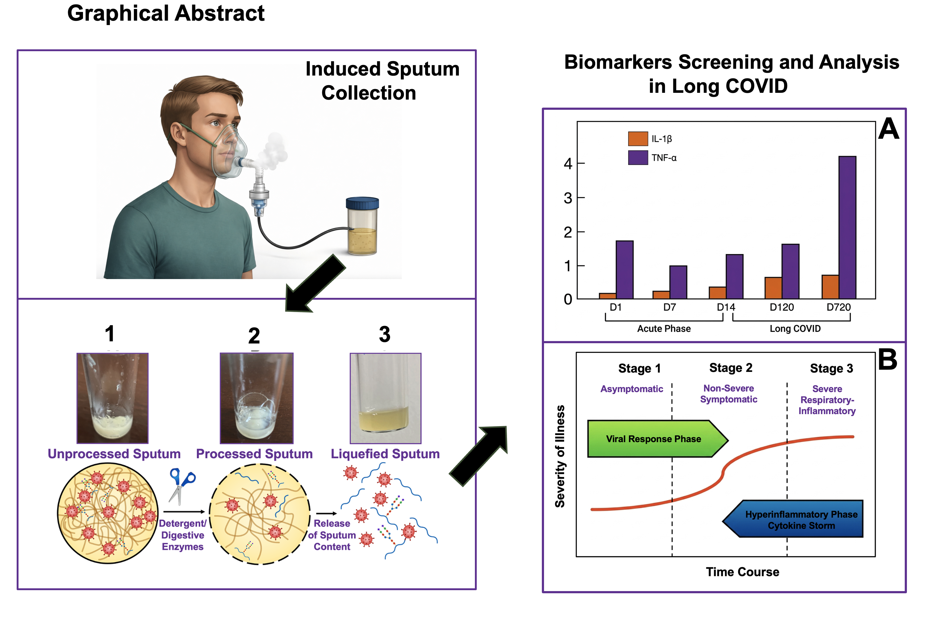
The systemic levels of cytokines caused by COVID-19 may be lower than those observed in sepsis, but the local response is more intense. As indicated in Figure 4-A, proinflammatory cytokines are elevated in the serum of a study involving 80 patients with COVID-19 and Long COVID [61]. In patients with long COVID-associated respiratory symptoms the pro-inflammatory profile was significantly elevated two years after acute severe COVID-19. It appears that systemic cytokine profiles can diverge significantly from localized tissue responses. Thus, bronchoalveolar lavage fluid (BALF) yields the highest diagnostic positivity for SARS-CoV-2, followed by sputum from the lower respiratory tract [68]. Additionally, the multifaceted presentation of Long COVID – which exhibits marked sex-based disparities, particularly affecting females – varies with symptom duration and preexisting conditions. These disparities underscore the critical need for individualized therapeutic frameworks and enhanced strategies to preserve daily function [69].
Intriguingly, not all individuals who are exposed to the SARS-CoV-2 virus acquire the infection. However, once infected, the disease progresses through three main stages (Figure 4-B). Stage 1 is an asymptomatic-paucisymptomatic incubation period where the virus may or may not be clinically detectable. Stage 2 is a period of non-severe symptomatic illness with detectable virus that may resolve or progress. In infected individuals, approximately 80% will end in stage 2. Finally, stage 3 is characterized by severe respiratory illness with progressive pneumonitis that may or may not lead to respiratory failure, which in its final stages, causes diffuse alveolar damage. A syndrome characterized by hypercytokinemic inflammation referred to as “cytokine storm” can occur in patients with advanced COVID-19 disease [66]. In the case of Figure 4-B, it represents a model of acute COVID-19 cytokine storm, rather than Long COVID. Although hyperinflammatory responses – such as cytokine storms – may precipitate Long COVID, the syndrome frequently manifests in patients who initially experienced only mild acute infections [70].
Given the variability of Long COVID in its clinical presentation and array of underlying mechanisms, the difficulty of capturing key components such as post-exertional malaise (PEM), and a rapidly evolving research base, there is unlikely to be one outcome that can capture the scope of Long COVID’s progression or resolution [71]. At the present time, long COVID has no approved treatment or validated biomarkers. As understanding of the pathophysiology of Long COVID evolves, biomarkers may clarify disease mechanisms and phenotypes, guide phenotype specific treatments, and refine endpoints for targeted therapies.
New-Generation Protein Assays for Diagnosing COVID-19 and Long COVID
Several studies demonstrate that Long COVID can be effectively predicted and diagnosed using biological indicators associated with viral persistence, immune system dysregulation, hormonal imbalances, and chronic systemic inflammation [64]. A comprehensive meta-analysis of 28 studies identified 113 biomarkers linked to Long COVID. Among these, 69.9% of the biomarkers were significantly elevated and 25.7% were decreased, while 4.4% required additional research to determine their clinical relevance in Long COVID patients [72].
Protein biomarkers derived from easily accessible biofluids, such as urine, saliva, and sputum could be utilized as contributing factors for the early prediction of Long COVID. Unlike older, single-analyte tests, new-generation platforms are uncovering structural changes (such as protein misfolding) and multi-protein signatures that outperform single-gene or single-protein assays for complex conditions like Long COVID. The ultimate diagnostic challenge is to detect the onset of the disease as early as possible [73]. In terms of patient risk, earlier diagnoses are correlated with better patient outcomes, assuming that efficacious therapeutic modalities can be employed.
The foremost diagnostic imperative is the presymptomatic detection of disease, which directly correlates with superior clinical outcomes followed by timely, targeted interventions. Next-generation proteomics require structural, highly sensitive, and multiplexed technologies capable of quantifying ultra-low-abundance proteins. Overcoming the limits of traditional ELISAs, these tools shift the diagnostic paradigm from isolated biomarkers to complex protein networks, facilitating early intervention and personalized medicine [74,75]. Exemplary technologies include Single Molecule Array (Simoa), Singulex Single Molecule Counting (SMC), Proximity Extension Assay (PEA), Aptamer-Based Array, Single-Molecule Sequencing, among others [53,76,77,78,79].
Integrating a multi-protein biomarker panel analysis can significantly improve diagnostic accuracy. Unfortunately, some of these technologies may still yield false positive and false negative results. Experiments do not always proceed smoothly, and issues such as low detection rates, poor reproducibility, or biased results may lead to unusable data or distorted conclusions. In the case of the proximity extension two-antibody assay (Olink) or aptamer assay (SomaScan), cross-reactivity represents a significant hurdle, where capture and detection antibodies (Olink) or aptamer (SomaScan) may bind to non-target antigens, as well as the effect of hemolysis and repeated freeze-thaw cycles, leading to affected results [80]. Comparative analyses indicate that the Olink platform demonstrates superior target specificity and uncovers a greater density of phenotypic associations; conversely, the SomaScan platform exhibits superior analytical precision and broader proteomic coverage [81].
Furthermore, the rapid development and commercialization of therapeutic proteins have increased the need to develop orthogonal analytical techniques to monitor and control the product quality heterogeneity of these proteins. Many of these proteins are heterogeneous glycoproteins and are subject to molecular changes during the production. Regulatory agencies consider charge heterogeneity to be a critical product quality attribute due to the potential impact of acidic and basic isoforms on pharmacokinetics, biological activity, and stability during long-term storage [82].
Identifying specific proteoforms, distinct molecular forms of a protein, could unlock the next wave of breakthrough in drug discovery and disease diagnostics [83]. However, for proteoforms to truly propel drug development and biomarker discovery forward, we need the tools to measure them routinely. The potential applications of proteoforms in medicine are immense, and the characterization of proteoform heterogeneity is essential in modern biomedical research [84]. One promising emerging tool to reveal heterogeneous proteoform profiles is capillary electrophoresis (CE) and immunoaffinity capillary electrophoresis (IACE). Capillary electrophoresis is an umbrella term encompassing various capillary-based electrophoretic techniques used in medical and life science applications [85,86,87,88]. IACE is a powerful two-dimensional analytical technique that combines the target-specific analyte capture, using the principle of immunoassays (i.e., ELISA), with the high-speed, high-resolution separation of capillary electrophoresis (CE) [19,89,90,91,92,93,94,95,96,97,98,99,100,101,102,103,104,105,106,107,108]. It has been used to rapidly isolate a target biomarker while removing the rest of the simple or complex matrix, concentrate, and quantify a wide range of biomarker concentrations, including proteins and drugs in a variety of fluids [109]. As a clean-up and pre-analysis concentration step, IACE significantly improves the signal-to-noise ratio for trace analytes [19]. Multiple affinity capture ligands can be immobilized in a single area, known as the analyte concentrator-microreactor (ACM) device, to isolate and quantify several analytes in a single run [19,102]. Furthermore, it can distinguish between closely related variants, post-translational modifications, and isoforms of intact proteins.
Characterization of several proteoforms by one of the several modes of capillary electrophoresis or by IACE have been reported. A representative separation of four heterogeneous proteoforms is shown in Figure 5. Alpha-1-acid glycoprotein or orosomucoid (A) is a heavily glycosylated protein with a carbohydrate content of about 45% of its total molecular mass. Immunoglobulin G is composed of about 2% to 3% sugars by weight (B). Heparan N-sulfatase is a glycoprotein that contains approximately 4% to 8% carbohydrates by weight (C). Erythropoietin is also heavily glycosylated where carbohydrates account for 40% of its total molecular weight (D).
The market for several of these therapeutic glycoproteins is one of the most lucrative sectors in the pharmaceutical industry. The global market of monoclonal antibodies (mAb) is valued in the hundreds of billions of dollars. Similarly, erythropoietin is also a multi-billion-dollar market. The U.S. Food and Drug Administration (FDA) has several regulatory requirements to ensure the safety, efficacy, and quality of these protein products. The safety consequences of immunogenicity vary widely and are often unpredictable in patients administered therapeutic protein products. Cytokine release syndrome is a symptom complex caused by the rapid release of proinflammatory cytokines from target immune cells. Although cytokine release syndrome is not related to immunogenicity, the clinical presentation of cytokine release syndrome overlaps with anaphylaxis and other immunologically related adverse reactions [113]. In the case of cytokines, cytokines and chemokines are present in vivo as mixtures of proteoforms with different amino- or carboxyterminal or carrying heterogeneous glycan chains and possibly also being subject to citrullination, pyroglutamination and other post-translational modifications (PTMs) [114].
It has been reported that widely used immunological detection and quantification systems for cytokines, such as ELISAs and multiplex assays, based on the use of either polyclonal or monoclonal antibodies, are often fraught with misinterpretations, because the results are affected by the possible occurrence of PTMs of the analytes. Furthermore, different proteoforms often bind differently to their cell-surface receptors, which changes the resulting immune or inflammatory response [114].
Therefore, to solve some of these problems, capillary electrophoresis (CE) is becoming an exceptionally powerful and highly efficient method for characterizing intact proteoforms. When CE is coupled to an immunoassay, it can carry out the analysis of several cytokines or major inflammatory mediators and neuropeptides simultaneously. Figure 6-B depicts the separation of 12 cytokines/chemokines/neuropeptides using IACE, in which each analyte was captured by a specific antibody against the corresponding cytokine and then separated by high-resolution CE [102]. Conventional CE has a low sample load capacity limited to a few nanoliters of sample introduction into the capillary, which can constrain limits for low abundance proteoforms in highly complex mixtures. Conversely, IACE can use microliters or milliliters of sample introduction into the capillary [19,97,98]. The purpose is to apply as much of the target analyte to be bound to the analyte concentrator-microreactor (ACM) device (Figure 6-A) to hopefully occupy all its binding sites filled with affinity ligands. The excess matrix is removed, and the bound analytes are released by a special elution solution to finally separate the target analyte(s) purified and concentrated. To increase accuracy, it is recommended to use an affinity ligand interacting with a known substance, used as an internal standard, and bound to the same ACM device together with one or more affinity ligands to capture the intended target analytes. Figure 6-C demonstrates that after 45 uses of the ACM device, it is possible to obtain very reproducible results with the adjustments of peak areas and migration times of the target analyte(s) and the known internal standard used, making the IACE technology a platform that offers excellent cost-efficiency during high-throughput utilization, yielding highly reproducible data across sequential analytical runs. Consistency of results, high-resolution power, and speed of analysis make affinity-capture immunoaffinity capillary electrophoresis a powerful tool for quality control of therapeutic proteins, vaccines, and all other heterogeneous proteins. As indicated in Figure 6-A, affinity capture ligands can be several molecules, including antibodies, antibody fragments, aptamers, lectins, and many others with binding capacities, used individually or in combination.
Analysis of Glycopeptides of SARS-CoV-2 Glycoproteins as Potential Diagnostic Biomarkers
There are several proteins that are constituents of the SARS-CoV-2. The Envelope (E) protein and the Nucleocapsid (N) protein are structural proteins, but they are not glycosylated. Conversely, the Membrane (M) glycoprotein and the Spike (S) glycoprotein are glycosylated. The sugar molecules of the S protein act as a protective shield to help the virus evade the human immune system, while the protein portion binds directly to human cells to cause infection [116,117,118].
Glycosylation is known to play an important role in the efficacy and antigenicity of therapeutic proteins. The study of glycosylation on viral glycoproteins has been an important aspect of vaccine development for more than three decades [119]. Studies on the mapping of glycopeptides of the Spike protein has allowed researchers to understand how the Spike protein binds to human host cells. Identification, mapping and relative quantitation of SARS-CoV-2 Spike glycopeptides by analytical methods have been reported [119,120]. The unusually heavy and complex glycosylation observed in Spike protein is believed to play an important role in the pathogenicity of SARS-CoV-2 by mimicking host cell glycans and allowing the virus to evade the normal immune response [120].
Figure 7 shows the glycopeptide analysis of SARS-CoV-2 of a glycoprotein expressed and purified on 293-S cells. The cells were lysed, and the purified glycoprotein was subjected to proteolysis. Analysis was performed by high-resolution LC/MS experiments, and the data analysis was obtained by GlycoPep DB and Byonic glycan databases. For details of the experiment see Go et al., 2021 [119]. Under these experimental conditions, it was possible to identify 828 different glycopeptides on the SARS-CoV-2 S protein. A few glycopeptides are shown in Figure 7A. Furthermore, a summary of the results from two different analysis approaches for the SARS-CoV-2 S data set was performed. As indicated in Figure 7B, more false positives than true positives were present: 460 correct assignments were verified, while 528 were rejected as false positives. However, when comparing the overall true positives and false positives for the assignment with scores > 100, the true positives [237] outnumbered the false positives [130]. More than 550 assignable glycopeptides were missed by the Byonic software when compared to the Expert Analysis.
Go et al. [119] noted alarming inaccuracies in the software used, noting that it yielded more incorrect than correct annotations despite a claimed false discovery rate of 1%. Even when applying a stringent filtering threshold of scores exceeding 100 for glycopeptides, the program still generated erroneous assignments. Ultimately, these unassigned or incorrectly assigned glycopeptides may lead to misinterpretations regarding how glycans influence the function of key proteins. Analyzing the glycosylation profiles of viral glycoproteins provides critical insights that directly inform immunogen design and optimization. Consequently, comprehensive characterization of the SARS-CoV-2 spike S glycoprotein’s glycan landscape is paramount for advancing the development and manufacturing of efficacious vaccine candidates. These results provide strong cautionary evidence that the field of automated glycopeptide analysis is still a field in flux, and better tools are needed to support warp speed science. Glycoprotein analysis is still a challenge for many scientists. Reliable glycopeptide identification also depends on the analytical software and glycan database used for spectral interpretation. In a head-to-head comparison of five contemporary glycoproteomic search programs using matched glycan databases, substantial software-dependent differences were observed in glycoprotein and glycosite assignments. Byonic reported the largest number of software-specific glycoproteins and glycosites and the lowest proportional agreement of identified glycosites with UniProt (~57%). Lack of UniProt annotation alone, however, cannot distinguish a novel glycosite from an incorrect assignment. Importantly, manual examination of selected Byonic-specific results revealed incorrect assignments. Hogan et al. [121] therefore recommended using more than one search program to establish confidence through consensus. Differences in glycan-database input formats among software packages further complicate standardization and reproducibility of glycoproteomic analysis [121]. Converting the glycomic results into a database for each available software is difficult. A universal glycan database format would be critical to establishing better consistency between software and would eliminate a significant amount of work involved in reformatting databases [121]. Today, these analytical hurdles are successfully addressed by specialized glycan analysis facilities equipped with highly sensitive, advanced nano-LC-MS-MS systems, which are useful for the identification, mapping, and relative quantitation of glycopeptides from SARS-CoV-2 Spike protein [120].
Glycosylation of Spike protein in COVID-19 patients is likely to be variable and may change over the course of infection [120]. Furthermore, excessive glycosylation could block antibody binding when using in vitro antibody tests and yield false negative results. Therefore, continuous development of analytical strategies enables advancements in deciphering the roles of post-translational modifications, which influence organism maturation, physiological processing, and immune response [122]. Numerous labeling strategies have enabled facile, accurate investigations of disease-relevant glycoproteins and are well suited to uncover future biomarkers and discern symptomatic protein profiles [122].
Because quantitative glycomics and glycoproteomics will remain an area of significant active focus for years to come, we would like to discuss the complementary use of capillary electrophoresis and IACE in the development of quantitative glycomics and glycoproteomics. Since the early publications on the analysis of glycoproteins by CE [123], numerous publications have advanced this area of research [124,125,126,127].
It has been reported that aberrant glycosylation, the abnormal attachment of sugar molecules to proteins, is recognized as a hallmark in various diseases. Glycans greatly enrich the biological information of glycoproteins and thus influence cellular interaction and behavior. Sialylation, fucosylation, and complex branch structures have been revealed to be among the most commonly observed structural patterns in malignancies. Recent findings have also highlighted the relationship between glycosylation and immunity, and pinpointed the novel strategies based on deciphering cancer “glyco-code” [128]. Aberrant cellular glycosylation is one of the earliest events that occur during malignant transformation, having a significant impact in shaping an immunosuppressive tumor microenvironment [129].
Similarly, an aberrant glycosylation in COVID patients has been investigated. It has been reported that aberrant glycosylation on SARS-CoV-2 spike IgG is a prothrombotic stimulus for platelets. Immune complexes of SARS-CoV-2 spike protein and anti-spike IgG increase the formation of platelet-mediated thrombosis on von Willebrand factor in vitro, but only when the glycosylation of the Fc domain of IgG is altered in a fashion identified in patients with severe COVID-19 [130]. Also, it has been demonstrated that the IgG glycome in severe COVID-19 patients is statistically significantly altered in a way that it indicates decreased immunosuppressive action of circulating immunoglobulins. The magnitude of observed changes in IgG glycosylation is associated with the severity of the disease [131]. Quantification of plasma N-glycans can portray the presence of SARS-CoV-2 infection and in turn, implies the disease course of patients [132]. Serum N-glycomic profiling has been reported to provide potential signatures for surveillance of COVID-19 [133]. Protein glycosylation patterns represent a promising resource of biomarkers for post-acute sequelae of COVID-19 (PASC). Notably, high-throughput OxoScan mass spectrometry has demonstrated that acute, severe SARS-CoV-2 infection induces persistent, differential glycosylation across critical plasma glycoproteins, including immunoglobulin A (IgA), haptoglobin, and transferrin [134].
Other compelling research has been carried out in Long COVID. Swank et al. [2024] have determined the proportion of individuals with detectable circulating viral antigens in blood after SARS-CoV-2 infection and the association of antigen detection with Long COVID symptoms. The primary outcome measure was to quantify SARS-CoV-2 antigens, including S1 subunit of spike protein, full-length spike protein, and nucleocapsid in participant samples, using single molecule array (Simoa) assays. In a period of 4 and 7 months postinfection, the full-length spike protein was the most predominantly detected. The authors concluded that SARS-CoV-2 antigens can be detected in the blood of a substantial proportion of individuals up to 14 months after infection. Furthermore, while it was not possible to confirm that the presence of viral antigen is linked with a persistence of viral reservoir, these findings highlight the need to further investigate the source of the persistent antigens.
Another interesting observation is that extracellular nanosized vesicles (EVs) isolated from serum have been found to harbor viral RNA and proteins and may serve as potential biomarkers in Long COVID [135]. Extracellular vesicles and glycans exhibit extreme heterogeneity in terms of structure, composition, and function. Extracellular vesicles are currently considered a new mode of communications between cells, and glycans as a principle of coding and storage for biological information [136]. Plasma from Long COVID patients contained elevated levels of both large and small extracellular vesicles. However, concentrations of mannose-positive large EVs (100-500 nm) of Long COVID patients were significantly increased in comparison to recovered controls [137].
Studies on how glycoproteins on the surface of viruses may aid the spread of protein aggregates associated with neurodegenerative diseases have been reported. Soluble and aggregated proteins associated with a diverse number of neurodegenerative diseases have been found secreted by neurons and other cells as free proteins or in association with extracellular vesicles [138]. These lines of investigations are very interesting but need to be followed up before we can understand whether extensive glycosylation of glycoproteins in viruses, particularly in SARS-CoV-2, play a significant role in the relationship of Long COVID in the prolonged dysfunction of many organs, including heart, lung, brain, kidneys, liver and pancreas. Symptoms are often caused by persistent inflammation, microclots, or ongoing viral presence. Some symptoms persist for months or even years after COVID illness first appears.
Aberrant glycosylation is a critical frontier in disease diagnostics, because structural changes of sugars attached to proteins act as early, highly specific indicators of pathological changes across many diseases. Consequently, we proposed to determine glycopeptides and sugars bound to glycoproteins by immunoaffinity capillary electrophoresis, as a complementary technique to the currently existing set of technologies.
Aberrant Glycopeptides as Potential Diagnostic Biomarkers
Aberrant glycopeptides are transforming diagnostic medicine. Although not related directly to glycopeptides in sputum, information on aberrant glycopeptides obtained in other fluids underscores the importance of these biomarkers in the accuracy of diagnosing a disease. An example of glycoprotein and glycopeptide profiling is provided in Figure 8 for prostate specific antigen (PSA). The PSA test for prostate cancer screening is still a controversial test [139]. There is no single threshold that distinguishes between normal and abnormal PSA result. This is in part because there is no specific PSA level that means that someone has prostate cancer. However, the higher someone’s PSA level, the likelier it is that prostate cancer is present [140].
Immunoaffinity capillary electrophoresis and high-performance PSA glycomic assay can help detect the presence and extent of prostate disease in its early stages, even before abnormalities can be detected with other methods, including digital rectal examination, multiparametric magnetic resonance, and/or prostate biopsy. The PSA glycomics assay reveals multiple glycoforms of urinary PSA, including sialic acid linkage isomers as well as the level of (core-)fucosylation [141]. Similarly, IACE has a significant potential to analyze glycopeptides derived from several biological fluids and tissues to accurately identify diseases before they can progress to an advanced stage [142].
Currently, the FDA has approved more than ten different glycoproteins as biomarkers for different types of cancer. In addition to serving as biomarkers for disease prognosis and diagnosis, glycoproteins are also extremely important drug and vaccine candidates [143]. Experiments have demonstrated that enzymatic deglycosylation led to widespread conformational changes, both in regions with glycosylation sites and those without [144].
Research has shown that in severe COVID-19 cases, the antibodies (IgG) produced by the body against the Spike protein can feature abnormal (aberrant) glycosylation. These atypical sugar structures on antibodies can cause pathological platelet activation and drive persistent blood-clotting issues seen in post-acute sequelae of COVID-19 [130]. Conversely, while the N protein is not a glycoprotein, persistent N protein antigens (viral fragments) have been detected in the blood and tissue reservoirs (such as the gut) of patients suffering from Long COVID. The ongoing presence of these viral components is strongly hypothesized to fuel chronic, low-grade inflammation [145].
Conclusions
Since the COVID-19 pandemic in late 2019, there have been ongoing efforts to study the mechanism of action of the novel coronavirus and to develop potent anti-SARS-CoV-2 drugs. Despite significant advances in understanding coronavirus mechanisms and therapeutics, the validation of both SARS-CoV-2 targets and treatments remains ongoing [146]. Additionally, a significant percentage of patients experience a prolonged recovery period where COVID-19 symptoms persist, clinically identified as Long COVID or post-acute sequelae of SARS-CoV-2 (PASC). A multi-system array of symptoms, such as dyspnea, myalgia, and fatigue, have been reported for months or even years after recovery from the initial infection [13,147]. Long COVID can affect anyone who has previously had acute COVID-19. Some experts have defined long-COVID as a long-lasting chronic condition triggered by the virus that causes COVID-19. It is critical to distinguish between casual and correlative relationships regarding viral persistence in the pathogenesis of post-acute sequelae of SARS-CoV-2 (PASC). Emerging evidence suggests that persistent viral reservoirs may drive PASC pathophysiology, supported by the prolonged detection of circulating SARS-CoV-2 S1 subunit spike proteins in a subset of patients, months after initial infection. Persistent SARS-CoV-2 infections may act as viral reservoirs that could seed future outbreaks [148]. Although sputum is a rich source of microbial biomarkers, it does not constitute a true anatomical reservoir for SARS-CoV-2. Rather, it functions as a temporary biological vehicle transporting viral particles and infected cells shed from primary tissue reservoirs in the lower respiratory tract [9]. Nonetheless, the SARS-CoV-2 viral load within sputum directly correlates with the risk of COVID-19 progression [149]. This association provides a strong rationale for early antiviral intervention to mitigate disease severity and reduce mortality rates. Notably, virus particles often hide in “immunoprivileged sites” around the human body, often known as sanctuary sites or viral reservoirs, that our immune system does not monitor or protect as closely as the rest of our bodies [150]. The causal link between acute RNA virus persistence and post-viral sequalae should raise awareness of the long-term risks of what are usually short-course viral infections. If these acute infections are severe, there is a significant chance that a chronic condition will manifest years later. Preventing severe acute infections in susceptible individuals through vaccination or isolation may diminish the risk [151].
Addressing Long COVID is highly challenging due to its nature as a multisystemic syndrome with over 200 fluctuating and overlapping symptoms. Because it is driven by complex, interconnected biological processes, unmasking its root causes requires a multi-layered diagnostic approach. This involves tracking traditional inflammatory markers alongside structural protein anomalies, such as aberrant glycosylation, and other key molecular indicators. One area of research in the understanding of the presence of SARS-CoV-2, or certain constituents of it, in different tissues is the role of the glycoproteins of the S1 protein. Coronaviruses heavily modify their viral proteins with carbohydrates. Specifically, the viral spike S protein, which contains approximately 66 to 87 N-linked glycosylation sites per trimer. N-glycoproteomic data indicates that treating the virus with Cepharanthine – a plant-derived alkaloid – modulates this S protein glycosylation. By altering these glycan structures, Cepharanthine disrupts viral assembly and impairs the virus’s capacity to recognize and bind to host receptors, thereby demonstrating potent antiviral activity [146].
Coronavirus-induced glycoprotein modifications are vital for host cell invasion and subsequent cellular dysfunction. These aberrant N-glycans shield the virus from the immune system and stabilize the spike protein in an open conformation for receptor binding. This evidence paves the way for exploring N-glycosylation as a target for COVID-19. Some of the inhibitors tested (i.e., miglustat, miglitol, acarbose, and celgosivir) are drugs in advanced clinical trials or FDA-approved, used to treat unrelated human diseases, and could be considered for repurposing if found effective in trials, alone or in combination. Further new treatments may consist of blocking essential spike N-glycans using antibodies or lectins to reduce the spread of the infection [152]. In addition, these investigators provide experimental evidence that glycosylation inhibitors are universally effective against major SARS-CoV-2 variants worldwide, including Alpha (B.1.1.7), Beta (B.1.351), Gamma (P.1), and Delta (B.1.617.2).
Interestingly, other investigators have demonstrated that since significant alterations in fucosylation patterns of immunoglobulin G are observed among patients with severe COVID-19, defucosylation may be a crucial strategy to prevent the progression from mild to severe COVID-19 [153]. Other researchers [154] have reported that both hyperglycemia and elevated glycemic variability are each associated with increased risk of severe COVID-19 outcomes. Furthermore, bulk IgG glycosylation predicts COVID-19 severity and vaccine antibody response [155]. Studies in other areas with the hope of understanding the mysteries of COVID-19, such as metabolomics, have indicated metabolic dysregulation associated with COVID-19 opening a path for predicting potential biomarkers based on pathway analysis [156,157]. Additionally, the IL-6 family of cytokines is increasingly recognized as a potential contributor to the immune alterations observed in COVID-19 and its long-term sequelae [65].
To conclude, evaluating proinflammatory cytokines and abnormal protein glycosylation patterns of glycoproteins related to COVID-19 provides a promising foundation for comprehending, diagnosing, and managing Long COVID. Analyzing these important biological components in bodily fluids – such as sputum, saliva, blood, urine and cerebrospinal fluid – can be valuable in the study of biomarkers of Long COVID, as they provide objective biological evidence that clarify persistent symptoms, map complex pathobiology, and guides personalized therapies [158]. Dysregulation of cytokine responses can substantially increase the risk of disease and consequently their activity requires tight control. The formation of cytokine homodimers, heterodimers and multimers has evolved as a versatile mechanism to regulate cytokine biology, in which multimerization can enable or attenuate their activity, diversify signaling outcomes and drive signaling bias [159]. Additionally, characterization of biological and chemical pathways – frequently categorized as biointeractions – is critical for elucidating underlying disease mechanisms, advancing targeted therapeutics, and engineering novel analytical assays for clinical applications [160]. For research purposes, we suggest pairing these analyses with the two-dimensional immunoaffinity capillary electrophoresis (IACE) (affinity capture-CE separation), a fast, high resolution (resolves subtle structural variations of analytes), high analytical sensitivity for low-abundance biomarkers, a potential cost-effective methodology (ACM can be re-used at least 45 times with high reproducibility), reducing false results (due to its two-dimensional format), and generating information with significant accuracy (employing a known internal standard, used under the same experimental conditions as the target analyte with its appropriate immobilized affinity ligand in the same ACM device).
While this review demonstrates the practicality and usefulness of affinity capture migration/separation technologies, certain advantages and limitations should be acknowledged. The workflow is designed to be simple and rapid, offering operational efficiency and reduced resource requirements relative to molecular diagnostics, yet it necessitates comprehensive validation through large-scale comparative analytical trials, clinical outcome evaluations, and rigorous meta-analyses to establish definitive performance and economic parameters.
Ongoing technical optimizations are currently underway to enhance assay performance, specifically regarding analyte recovery, ACM device binding capacity and re-usability, quantitative data fidelity, and analytical sensitivity. These critical parameters govern overall diagnostic reliability and are inherently modulated by the chemical-biological nature of the target analyte, affinity ligand quality, stability of the immobilization process, matrix complexity, and device architecture. Ultimately, elucidating proinflammatory cytokines and protein glycosylation profiles may offer deeper insights into Long COVID pathogenesis and facilitate the identification of candidate biomarkers.
The integration of sputum and alternative biological matrix analyses into post-acute sequelae of SARS-CoV-2 infection (PASC) investigation necessitates rigorous harmonization between biospecimen acquisition protocols and targeted measurand physico-chemistry. Immunoaffinity capillary electrophoresis (IACE) provides a multifunctional operational framework facilitating selective target analyte(s) capture, matrix cleanup, target analyte(s) enrichment, and high-resolution electrophoresis separation. Nevertheless, translation of IACE into a clinically viable diagnostic modality for Long COVID remains contingent upon comprehensive analytical validation and empirical clinical qualifications for disease-specific biomarkers.
Consequently, while emerging investigations have successfully delineated diverse biological correlates and prospective classifiers of post-acute sequelae of SARS-CoV-2 infection – encompassing viral persistence, immunological dysregulation, endocrinological alterations, systemic inflammation, and aberrant glycosylation – these empirical discoveries remain restricted to a candidate-biomarker paradigm. Rigorous independent analytical and clinical validation is a requisite prior to clinical translation for routine diagnostic deployment. Furthermore, despite incremental advancements in elucidating core pathology to mitigate the burden experienced by nearly one in five adults who have had COVID-19, they continue to suffer from symptoms of Long COVID after acute infection, and a definitive therapeutic resolution remains unrealized.
Author Contributions
Conceptualization and writing, N.A.G.; review and editing, N.A.G., C.M.B., S.N.K. All authors have read and agreed to the final version of the manuscript.
Funding
This research received no external funding.
Institutional Review Board Statement
Not applicable.
Informed Consent Statement
Not applicable.
Data Availability Statement
The data used in this article were sourced from the materials mentioned in the References section.
Acknowledgments
In Memoriam: N.A.G. would like to dedicate this paper to the memory of a trusted colleague, innovator, educator, and dear friend Julio Humberto Dölz. As the founding father of the School of Pharmacy at the Universidad Austral de Chile, Valdivia, Chile, he leaves behind a remarkable balance of groundbreaking research and profound, lifelong dedication to student mentorship.
Conflicts of Interest
N.A.G is the inventor of patents related to the IACE technology.
Abbreviations
ACM, analyte concentrator-microreactor; (ANDV), Andes orthohantavirus; COVID-19, coronavirus disease 2019; IACE, immunoaffinity capillary electrophoresis; NAAT, nucleic acid amplification test; PASC, post-acute sequelae of COVID-19 or Long COVID; RAT, rapid antigen test; RT-PCR, reverse transcription polymerase chain reaction; SARS-CoV-2, severe acute respiratory syndrome coronavirus 2.
References
- KFF Global COVID-19 Tracker. 2 August 2026. Available online: https://www.kff.org/covid-19/global-covid-19-tracker/.
- Walsh, K.A.; Spillane, S.; Comber, L.; Cardwell, K.; Harrington, P.; Connell, J.; Teljeur, C.; Broderick, N.; de Gascun, C.F.; Smith, S.M.; Ryan, M.; O’Neill, M. The duration of individuals infected with SARS-CoV-2. J. Infect. 2020, 81(6), 847–856. [Google Scholar] [CrossRef] [PubMed]
- Züst, R.; Ackermann-Gäumann, R.; Liechti, N.; Siegrist, D.; Ryler, S.; Portmann, J.; Lenz, N.; Beuret, C.; Koller, R.; Staehelin, C.; Kuenzli, A.B.; Marschall, J.; Rothenberger, S.; Engler, O. Presence and persistence of Andes virus RNA in human semen. Viruses 2023, 15(11), 2266. [Google Scholar] [CrossRef] [PubMed]
- Sharun, K.; Tiwari, R.; Dhama, K. SARS-CoV-2 in semen. Potential for sexual transmission in COVID-19. Int. J. Surg. 2020, 84, 156–158. [Google Scholar] [CrossRef] [PubMed]
- Kutti-Sridharan, G.; Vegunta, R.; Vegunta, R.; Mohan, B.P.; Rokkam, V.R.P. SARS-CoV-2 in different body fluids, risks of transmission, and preventing COVID-19: A comprehensive evidence-based review. Int. J. Prev. Med. 2020, 11, 97. [Google Scholar] [CrossRef] [PubMed]
- Wong, M.C.S.; Huang, J.; Lai, C.; Chan, F.K.L.; Chan, K.S. Detection of SARS-CoV-2 RNA in fecal specimens of patients with confirmed COVID-19: A meta-analysis. J. Infect. 2020, 81(2), e31–e38. [Google Scholar] [CrossRef] [PubMed]
- Rubin, R. SARS-CoV-2 RNA can persist in stool months after respiratory tract clears virus. JAMA 2022, 327(22), 2175–2176. [Google Scholar] [CrossRef] [PubMed]
- Jain, U. Effect of COVID-19 on Organs. Cureus 2020, 12(8), e9540. [Google Scholar] [CrossRef] [PubMed]
- Trypsteen, W.; Van Cleemput, J.V.; van Snippenberg, W.; Gerlo, S.; Vanderkerckhove, L. On the whereabouts of SARS-CoV-2 in the human body: A systematic review. PLoS Pathog. 2020, 16(10), e1009037. [Google Scholar] [CrossRef] [PubMed]
- Shah, M.D.; Sumeh, A.S.; Sheraz, M.; Kavitha, M.S.; Maran, B.A.V.; Rodrigues, K.F. A mini-review on the impact of COVID 19 on vital organs. Biomed. Pharmacother. 2021, 143, 112158. [Google Scholar] [CrossRef] [PubMed]
- Balcioglu, S.S.K.; Dindar, G.Z.; Guclu, O.; Karabulut, N.; Ozturk, N. A rare presentation after COVID-19: Manic episode with psychotic features. Psych. Res. Case Rep. 2023, 2, 100115. [Google Scholar] [CrossRef] [PubMed]
- Ewing, A.G.; Salamon, S.; Pretorius, E.; Joffre, D.; Fox, G.; Bilodeau, S.; Bar-Yam, Y. Review of organ damage from COVID and long COVID: a disease with spectrum of pathology. Med. Rev. 2025, 5(1), 66–75. [Google Scholar] [CrossRef] [PubMed]
- Swank, Z.; Borberg, E.; Chen, Y.; Senussi, Y.; Claise, S.; Manickas-Hill, Z.; Yu, X.G.; Li, J.Z.; Alter, G.; Henrich, T.J.; Kelly, J.D.; Hoh, R.; Goldberg, S.A.; Deeks, S.G.; Martin, J.N.; Peluso, M.J.; Talla, A.; Li, X.; Skene, P.; Bumol, T.F.; Torgerson, T.R.; Czartoski, J.L.; McElrath, M.J.; Karlosn, E.W.; Walt, D.R.; RECOVER consortium. Measurements of circulating viral antigens post-SARS-CoV-2 infection in a multicohort study. Clin. Microbiol. Infect. 2024, 30(12), 1599–1605. [Google Scholar] [CrossRef] [PubMed]
- Baptista, S.N.; Atkins, T.; Chakraborty, S.; Bakhit, M.; Glasziou, P.; Byambasarusen, O. Candidate treatments for long COVID: a narrative review of expert and patient-driven priorities. Front. Med. 2026, 13, 17346600. [Google Scholar] [CrossRef] [PubMed]
- Al-Aly, Z.; Davis, H.; McCorkell, L.; Soares, L.; Wulf-Hanson, S.; Iwasaki, A.; Topol, E.J. Long COVID science, research and policy. Nat. Med. 2024, 30, 2148–2164. [Google Scholar] [CrossRef] [PubMed]
- Soares, L.; Romatowski, C.H.; Assaf, G.; de Canson, C. The case for routine patient review in long COVID research. Lancet Infect. Dis. 2026a, 26(5), 446–447. [Google Scholar] [CrossRef] [PubMed]
- Zhao, H.; Tu, H.; Yu, X.; Su, J.; Zhang, X.; Xu, K.; Shi, Y.; Qiu, Y.; Sheng, J. Delayed clearance of viral RNA in sputum for severity COVID-19 patients with initial high viral load. Infect. Drug Resist. 2022, 15, 1971–1979. [Google Scholar] [CrossRef] [PubMed]
- Guzman, N.A.; Guzman, D.E. A COVID-19 infection with positive sputum and negative nasopharyngeal/oropharyngeal rapid antigen-based testing: A case report and pilot study. Med. Clin. Case Rep. J. 2023a, 1(2), 77–84. [Google Scholar] [CrossRef]
- Guzman, N.A.; Guzman, A. Human sputum proteomics: Advancing non-invasive diagnosis of respiratory diseases with enhanced biomarker analysis methods. Int. J. Transl. Med. 2024, 4(2), 309–333. [Google Scholar] [CrossRef]
- Guzman, N.A. Method and system for exposing hidden or masked antigenic sites of viral specimens present in biosamples using a home-based COVID-19 rapid lateral flow immunoassay test. U.S. Patent No. US12,416,633 B2, 16 September 2025. [Google Scholar]
- Eccles, R. Respiratory mucus and persistence of virus on surfaces. J. Hosp. Infect. 2020, 105(2), 350. [Google Scholar] [CrossRef] [PubMed]
- Chen, C.; Gao, G.; Xu, Y.; Pu, L.; Wang, Q.; Wang, L.; Wang, W.; Song, Y.; Chen, M.; Wang, L.; Yu, F.; Yang, S.; Tang, Y.; Zhao, L.; Wang, H.; Wang, Y.; Zeng, H.; Zhang, F. SARS-CoV-2-positive sputum and feces after conversion of pharyngeal samples in patients with COVID-19. Ann. Inter. Med. 2020. [Google Scholar] [CrossRef] [PubMed]
- Wang, W.; Xu, Y.; Gao, R.; Lu, R.; Han, K.; Wu, G.; Tan, W. Detection of SARS-CoV-2 in different types of clinical specimens. JAMA 2020, 323(18). [Google Scholar] [CrossRef] [PubMed]
- Balla, M.; Merugu, G.P.; Pokald, M.; Gayam, V.; Adapa, S.; Naramala, S.; Konala, V.M. A comprehensive approach is vital for diagnosing COVID-19: A case of false negative. J. Clin. Med. Res. 2020, 12(5), 315–319. [Google Scholar] [CrossRef] [PubMed]
- Wiseman, J.; D’Amico, T.A.; Zawadzka, S.; Anyimadu, H. False negative SARS-CoV-2 PCR- A case report and literature review. Respir. Med. Case Rep. 2020, 31, 101140. [Google Scholar] [CrossRef] [PubMed]
- Qu, Y.-M.; Kang, E.-M.; Cong, H.-Y. Positive results of SARS-CoV-2 in sputum from a cured patient with COVID-19. Travel Med. Infect. Dis. 2020, 34, 101619. [Google Scholar] [CrossRef] [PubMed]
- Shi, L.; Chen, J.; He, J. How to prevent rehospitalization in patients with COVID-19. Arch. Med. Case Rep. 2021, 3(1), 17–28. [Google Scholar] [CrossRef]
- Viswanathan, T.; Arya, S.; Chan, S.-H.; Qi, S.; Dai, N.; Misra, A.; Park, J.-G.; Oladunni, F.; Kovalskyy, D.; Hromas, R.A.; Matinez-Sobrido, L.; Gupta, Y.K. Structural basis of RNA cap modifications by SARS-CoV-2. Nat. Commun. 2020, 11, 3718. [Google Scholar] [CrossRef] [PubMed]
- Ali, D.Y.; Hussein, R.A.; Elshafie, S.M.; Mohamed, R.A.; El Reheem, F.A. Comparable detection of nasopharyngeal swabs and induced sputum specimens for viral nucleic acid detection of suspected coronavirus (SARS-CoV-2) patients in Fayoum governorate, Egypt. Beni Suef Univ. J. Basic Appl. Sci. 2023, 12(1), 43. [Google Scholar] [CrossRef] [PubMed]
- Honkinen, M.; Lahti, E.; Österback, R.; Ruuskanen, O.; Waris, M. Viruses and bacteria in sputum samples of children with community-acquired pneumonia. Clin. Microbiol. Infect. 2012, 18(3), 300–307. [Google Scholar] [CrossRef] [PubMed]
- Lin, Y.-C.; Malott, R.J.; Ward, L.; Kiplagat, L.; Pabbaraju, K.; Gill, K.; Berenger, B.M.; Hu, J.; Fonseca, K.; Noyce, R.S.; Louie, K.; Evans, D.H.; Conly, J.M. Detection and quantification of infectious severe acute respiratory coronavirus-2 in diverse clinical and environmental samples. Sci. Rep. 2022, 12, 5418. [Google Scholar] [CrossRef] [PubMed]
- Han, H.; Luo, Q.; Mo, F.; Long, L.; Zheng, W. SARS-CoV-2 RNA more readily detected in induced sputum than in throat swabs of convalescent COVID-19 patients. Lancet Infect. Dis. 2020, 20(6), 655–656. [Google Scholar] [CrossRef] [PubMed]
- Peeling, R.W.; Heyman, D.L.; Teo, Y.-Y.; Garcia, P.J. Diagnostic for COVID-19: moving from pandemic response to control. Lancet 2021, 399(10326), 757–768. [Google Scholar] [CrossRef] [PubMed]
- Meumann, E.M.; Robson, J.M.B. Testing for COVID-19: a 2023 update. Aust. Prescr. 2023, 46(1), 13–17. [Google Scholar] [CrossRef] [PubMed]
- Cheng, L.; Lan, L.; Ramalingam, M.; He, J.; Yang, Y.; Gao, M.; Shi, Z. A review of current effective COVID-19 testing methods and quality control. Arch. Microbiol. 2023, 205(6), 239. [Google Scholar] [CrossRef] [PubMed]
- Narumi, N.; Kondo, T.; Sato, Y.; Katayama, Y.; Nirasawa, S.; Saeki, M.; Yakuwa, Y.; Fujiya, Y.; Kuronuma, K.; Takahashi, S. Analysis of diagnostic performance and factors causing nonspecific reactions in SARS-CoV-2 rapid antigen detection tests. J. Infect. Chemother. 2023, 29, 157–162. [Google Scholar] [CrossRef] [PubMed]
- Kinyua, D.M.; Memeu, D.M.; Mwenda, C.N.M.; Ventura, B.D.; Velotta, R. Advancements and applications of lateral flow assays (LFAs): A comprehensive review. Sensors 2025, 25(17), 5414. [Google Scholar] [CrossRef] [PubMed]
- Ren, Q.; Wang, Y.; Ma, H.; Xie, J.; Jin, J.; Tian, R.; Yu, H.; Gao, X. Recent advances in lateral flow immunoassay for raid diagnosis of viral diseases. Transbound. Emerg. Dis. 2026, 5701806. [Google Scholar] [CrossRef] [PubMed]
- Koczula, K.M.; Gallotta, A. Lateral flow assays. Essays Biochem. 2016, 60(1), 111–120. [Google Scholar] [CrossRef] [PubMed]
- Dorta-Gorrín, A.; Navas-Méndez, J.; Gozalo-Margüello, M.; Miralles, L.; García-Hevia, L. Detection of SARS-CoV-2 based on nucleic acid amplification tests (NAATs) and its integration into nanomedicine and microfluidic devices as point-of-care testing (POCT). Int. J. Mo. Sci. 2023, 24(12), 10233. [Google Scholar] [CrossRef] [PubMed]
- John, G.; Sahajpal, N.S.; Mondal, A.K.; Ananth, S.; Williams, C.; Chaubey, A.; Rojiani, A.M.; Kolhe, R. Next-generation sequencing (NGS) in COVID-19: A tool for SARS-CoV-2 diagnosis, monitoring new strains and phylodynamic modeling in molecular epidemiology. Curr. Issues Mol. Biol. 2021, 43, 845–867. [Google Scholar] [CrossRef] [PubMed]
- Ding, X.; Shang, B.; Xie, C.; Xin, J.; Yu, F. Artificial intelligence in the COVID-19 pandemic: balancing benefits and ethical challenges in China’s response. Humanit. Soc. Sci. Commun. 2025, 12. [Google Scholar] [CrossRef]
- Cukic, V.; Hadzic, A. The most common detected bacteria in sputum of patients with community acquired pneumonia (CAP) treated in hospital. Med. Arch. 2016, 70(5), 354–358. [Google Scholar] [CrossRef] [PubMed]
- LeMessurier, K.S.; Tiwary, M.; Morin, N.P.; Samarasinghe, A.E. Respiratory barrier as a safeguard and regulator of defense against influenza A virus and Streptococcus pneumoniae. Front Immnunol 2020, 11, 3. [Google Scholar] [CrossRef] [PubMed]
- McChlery, S.; Ramage, G.; Bagg, J. Respiratory tract infections and pneumonia. Periodontol. 2000, 49(1), 151–165. [Google Scholar] [CrossRef] [PubMed]
- Branche, A.R.; Walsh, E.E.; Formica, M.A.; Falsey, A.R. Detection of respiratory viruses in sputum from adults by use of automated multiplex PCR. J. Clin. Microbiol. 2014, 52(10), 3590–3596. [Google Scholar] [CrossRef] [PubMed]
- Kim, K.J.; Yun, S.G.; Cho, Y.; Nam, M.-H.; Lee, C.K. Usefulness of combining sputum and nasopharyngeal samples for viral detection by reverse transcriptase PCR in adults hospitalized with acute respiratory illness. Microbiol. Spectr. 2022, 10(6), 1–6. [Google Scholar] [CrossRef] [PubMed]
- Mohammadi, A.; Esmaeilzadeh, E.; Li, Y.; Bosch, R.J.; Li, J.Z. SARS-CoV-2 detection in different respiratory sites: A systematic review and meta-analysis. eBioMedicine 2020, 59, 102903. [Google Scholar] [CrossRef] [PubMed]
- Williams, M.L.; Olomukoro, A.B.; Emmons, R.V.; Godage, N.H.; Gionfriddo, E. Matrix effects demystified: Strategies for resolving challenges in analytical separations of complex samples. J. Sep. Sci. 2023, 46(23), 2300571. [Google Scholar] [CrossRef] [PubMed]
- Liang, X.; Wang, Y.; Guo, T. Proteomics approaches to long COVID: status and outlooks. Life Medi 2023, 2, 1–3. [Google Scholar] [CrossRef] [PubMed]
- Merhavy, Z.I.; Junor, T.; Gonzalez, A.; De Filippis, S.M.; Oveisitork, S.; Rivera, E.; Ndukwu, I.; Bhatara, K. Long COVID: A comprehensive overview of the signs and symptoms across multiple organ systems. Korean J. Fam. Med. 2024, 45(6), 305–316. [Google Scholar] [CrossRef] [PubMed]
- Gu, X.; Wang, S.; Zhang, W.; Li, C.; Guo, L.; Wang, Z.; Li, H.; Zhang, H.; Liang, W.; Li, H.; Liu, Y.; Wang, Y.; Huang, L.; Dong, T.; Zhang, D.; Wong, C.C.; Cao, B. Probing long COVID through a proteomic lens: a comprehensive two-year longitudinal cohort study of hospitalized survivors. eBioMedicine 2023, 98, 104851. [Google Scholar] [CrossRef] [PubMed]
- Iosef, C.; Knauer, M.J.; Nicholson, M.; Van Nynatten, L.R.; Cepinskas, G.; Draghici, S.; Han, V.K.M.; Fraser, D.D. Plasma proteome of Long-COVID patients indicates HIF-mediated vasculo-proliferative disease with impact on brain and heart function. J. Transl. Med. 2023, 21, 377. [Google Scholar] [CrossRef] [PubMed]
- Cojocaru, E.; Cojocaru, C.; Vlad, C.-E.; Eva, L. Role of the renin-angiotensin system in long COVID’s cardiovascular injuries. Biomedicines 2023, 11(7). [Google Scholar] [CrossRef] [PubMed]
- Manckoundia, P.; Franon, E. Is persistent thick copious mucus a long-term symptom of COVID-19? Eur. J. Case Rep. Intern. Med. 2020, 7(12), 002145. [Google Scholar] [CrossRef] [PubMed]
- Jones, D.L.; Baluja, M.Q.; Graham, D.W.; Corbishley, A.; McDonald, J.E.; Malham, S.K.; Hillary, L.S.; Connor, T.R.; Gaze, W.H.; Moura, I.B.; Wilcox, M.H.; Farkas, K. Shedding of SARS-CoV-2 in feces and urine and its potential role in person-to-person transmission and the environment-based spread of COVID-19. Sci. Total Environ. 2020, 749, 141364. [Google Scholar] [CrossRef] [PubMed]
- Tran, H.N.; Le, G.T.; Nguyen, D.T.; Juang, R.-S.; Rinklebe, J.; Bhatnagar, A.; Lima, E.C.; Iqbal, H.M.N.; Sarmah, A.J.; Chao, H.-P. SARS-CoV-2 coronavirus in water and wastewater: a critical review about presence and concern. Environ. Res. 2020, 193, 110265. [Google Scholar] [CrossRef]
- Shanmugam, Y.; Ravikumar, L. Multi-omics biomarkers as emerging determinants in precision disease diagnosis and therapeutic monitoring. Adv. Biom. Sci. Technol. 2026, 8, 464–478. [Google Scholar] [CrossRef]
- Fu, Q.; Schoenhoff, F.S.; Savage, W.J.; Zhang, P.; Van Eyk, J.E. Multiplex assays for biomarker research and clinical application: Translational science coming of age. Proteom. Clin. Appl. 2010, 4(3), 271–284. [Google Scholar] [CrossRef] [PubMed]
- Goxhaj, L.; McCorkell, L.; van Rhijn-Brouwer, F.; Soares, L.; Vogel, J.M.; de Canson, C. Negative results in long COVID clinical trials: choosing outcome measures for heterogeneous disease. Lancet Infect. Dis. 2026, 26, 13–15. [Google Scholar] [CrossRef] [PubMed]
- Goulart, G.L.; Da Silva Maciel, A.B.; Rezende, A.G.R.A.; Valente, J.V.J.; Da Silva, B.M.S.B.M.; Sampaio, V.S.V.; Guimaraes, A.G.A.; Areas, G.P.T.A.G.P.; De Almeida, F.F.A.V.F.F.; Val, E. Pro-inflammatory cytokines are elevated two-years after acute severe COVID-19: a pre-vaccine, two-years, five-point longitudinal assessment of individuals with long COVID. Eur. J. Prev. Cardiol. 2024, 31 Suppl. 1, i426–i427. zwae175.289. [Google Scholar] [CrossRef]
- Chavda, V.P.; Bezbaruah, R.; Ahmed, N.; Alom, S.; Bhattacharjee, B.; Nall, L.V.; Rynjah, D.; Gadanec, L.K.; Apostolopoulos, V. Proinflammatory cytokines in chronic respiratory diseases and their management. Cells 2025, 14, 400. [Google Scholar] [CrossRef] [PubMed]
- Chen, K.; Wang, Z.; Li, J.; Xu, Y.; Gu, S.; Li, H.; Li, J.; Zhang, Y.; Mao, N. Chronic inflammation in Long COVID relationship to autoimmune diseases. Autoimmun. Rev. 2025, 24(10), 103882. [Google Scholar] [CrossRef] [PubMed]
- Aid, M.; Boero-Teyssier, V.; McMahan, K.; Dang, R.; Doyle, M.; Belabbaci, N.; Borducchi, E.; Collier, A-i.; Mullington, J.; Barouch, D.H. Long COVID involves activation of proinflammatory and immune exhaustion pathways. Nat. Immunol. 2025, 27, 61–71. [Google Scholar] [CrossRef] [PubMed]
- Bustos, M.; Rodríguez-Hernández, M.A. IL-6 family cytokines behind acute COVID-19 and long COVID syndrome: Mechanisms, biomarkers, and interventions strategies. BBA-Mol. Cell Res. 2026, 1873, 120198. [Google Scholar] [CrossRef]
- Calabrese, L.H. Cytokine storm and the prospects for immunotherapy with COVID-19. Clevel. Clin. J. Med. 2020, 87(7), 389–393. [Google Scholar] [CrossRef] [PubMed]
- Jyonouchi, H.; Kornitzer, J.; Geng, L. Favorable responses to upadacitinib, a JAK1 inhibitor, in long COVID patients with predominantly neuropsychiatric symptoms: case report in 2 autistic patients and one typically developing patient. MBC Neurol. 2025, 25, 342. [Google Scholar] [CrossRef] [PubMed]
- Wang, C.; Kang, K.; Lan, X.; Fei, D.; Wang, Q.; Li, X.; Chong, Y.; Gao, Y.; Wang, H.; Li, X.; Zhao, M.; Yu, K. Cytokine levels in sputum, not serum, may be more helpful for indicating the damage in the lung and the prognosis of severe COVID-19 – A case series. J. Infect. 2021, 83, e-6-e4. [Google Scholar] [CrossRef] [PubMed]
- Felix, J.; Gonçalves, J.; Cabedo, C.; Brito, J.; Gamas, M.; Neves, M.I.; Soares, H. Long-term sex differences in symptoms and immune profile in long COVID. Biol. Sex. Differ. 2026, 17(1), 32. [Google Scholar] [CrossRef] [PubMed]
- Low, R.N.; Low, R.J.; Akrami, A. A review of cytokine-based pathophysiology of Long COVID symptoms. Front. Med. 2023, 10, 1011936. [Google Scholar] [CrossRef] [PubMed]
- Soares, L.; Davis, H.; Spier, E.; Walker, T.; Davenport, T.; Putrino, D.; Peluso, M.; Vogel, J.M. Recommended long COVID outcome measures and their implications for clinical trial design, with a focus on post-exertional malaise. eBioMedicine 2026b, 123, 106083. [Google Scholar] [CrossRef] [PubMed]
- Lai, Y.-J.; Liu, S.-H.; Manachevakul, S.; Lee, T.-A.; Kuo, C.-T.; Bello, D. Biomarkers in long COVID-19: A systematic review. Front. Med. 2023, 10, 1085988. [Google Scholar] [CrossRef] [PubMed]
- Chen, Y.-J.; Jian, W.-H.; Liang, Z.-Y.; Guan, W.-J.; Liang, W.-H.; Chen, R.-C.; Tang, C.-L.; Wang, T.; Liang, H.-R.; Li, Y.-M.; Liu, X.-Q.; Sang, L.; Cheng, L.-L.; Ye, F.; Li, S.-Y.; Zhang, N.-F.; Zhang, Z.; Fang, Y.; He, J.-X.; Zhong, N.-S.; Zheng, J.-P. Earlier diagnosis improves COVID-19 prognosis: a nationwide retrospective cohort analysis. Ann. Transl. Med. 2021, 9(11), 941. [Google Scholar] [CrossRef] [PubMed]
- Todd, J.; Agee, S.J. Next generation immunoassay systems and the revival of protein biomarkers. Drug Discovery World, 7 October 2008.
- Babaei, M.; Kashanian, S.; Lee, H.-T.; Harding, F. Proteomics techniques in protein biomarker discovery. Quant. Biol. 2024, 12(1), 53–69. [Google Scholar] [CrossRef] [PubMed]
- Pai, S.S.; Ellington, A.D. Using RNA aptamers and the proximity ligation assay for the detection of cell surface antigens. Methods Mol. Biol. 2009, 504, 385–398. [Google Scholar] [CrossRef] [PubMed]
- Hwang, J.; Banerjee, M.; Venable, A.S.; Walden, Z.; Jolly, J.; Zimmerman, C.; Adkisson, E.; Xiao, Q. Quantitation of low abundant soluble biomarkers using high sensitive Single Molecule Counting technology. Methods 2019, 158, 69–76. [Google Scholar] [CrossRef] [PubMed]
- deMelo, B.P.; da Silva, J.A.M.; Rodrigues, M.A.; Palmeira, J.dF.; Saldanha-Araujo, F.; Argañaraz, G.A.; Argañaraz, E.R. SARS-CoV-2 spike protein and long COVID—Part1: Impact of spike protein in pathophysiological mechanisms of long COVID syndrome. Viruses 2025, 17(5), 617. [Google Scholar] [CrossRef] [PubMed]
- Shraim, R.; Diorio, C.; Canna, S.W.; McDonald-Dunlop, E.; Bassiri, H.; Martinez, Z.; Mälarstig, A.; Abbaspour, A.; Teachey, D.T.; Lindell, R.B.; Behrens, E.M. A method for comparing proteins measured in serum and plasma Olink proximity extension assay. Mol. Cell. Proteom. 2025, 24(7), 101000. [Google Scholar] [CrossRef] [PubMed]
- Candia, J.; Fantoni, G.; Moaddel, R.; Delgado-Peraza, F.; Shehadeh, N.; Tanaka, T.; Ferrucci, L. Effects of in vitro hemolysis and repeated freeze-thaw cycles in protein abundance quantification using the SomaScan and Olink assays. J. Proteome Res. 2025, 24(5), 2517–2528. [Google Scholar] [CrossRef] [PubMed]
- Katz, D.H.; Robbins, J.M.; Deng, S.; Tahir, U.A.; Bick, A.G.; Pampana, A.; Yu, Z.; Ngo, D.; Benson, M.D.; Chen, Z.-Z.; Cruz, D.E.; Shen, D.; Gao, Y.; Bouchard, C.; Sarzynski, M.A.; Correa, A.; Natarajan, P.; Wilson, J.G.; Gerszten, R.E. Proteomic profiling platforms head to head: Leveraging genetics and clinical traits to compare aptamer- and antibody-based methods. Sci. Adv. 2022, 8, eabm5164. [Google Scholar] [CrossRef] [PubMed]
- Salas-Solano, O.; Kennel, B.; Park, S.S.; Roby, K.; Sosic, Z.; Boumajny, B.; Free, S.; Reed-Bogan, A.; Michels, D.; McElroy, W.; Bonasia, P.; Hong, M.; He, X.; Ruesch, M.; Moffatt, F.; Kiessig, S.; Nunnally, B. Robustness of iCIEF methodology for the analysis of monoclonal antibodies: an interlaboratory study. J. Sep. Sci. 2012, 35(22), 3124–3129. [Google Scholar] [CrossRef] [PubMed]
- Korchak, J.A.; Yi, S.S.; Kelleher, N.L.; Sahni, N.; Scheynkman, G.M. Proteoform medicine: characterizing and targeting protein isoforms in human disease. Nat. Rev. Genet. 2026, 27, 271–291. [Google Scholar] [CrossRef] [PubMed]
- Forgrave, L.M.; Wang, M.; Yang, D.; DeMarco, M.L. Proteoforms and their expanding role in laboratory medicine. Pract. Lab. Med. 2021, 28, e00260. [Google Scholar] [CrossRef] [PubMed]
- Voeten, R.L.C.; Ventouri, I.K.; Haselberg, R.; Somsen, G.W. Capillary electrophoresis: Trends and recent advances. Anal. Chem. 2018, 90(3), 1464–1481. [Google Scholar] [CrossRef] [PubMed]
- Li, Y.; Miao, S.; Tan, J.; Zhang, Q.; Chen, D.D.Y. Capillary electrophoresis: A three-year literature review. Anal. Chem. 2024, 96, 7799–7816. [Google Scholar] [CrossRef] [PubMed]
- Guttman, A.; Auer, F. Capillary gradient gel electrophoresis. Gels 2026, 12(1), 29. [Google Scholar] [CrossRef] [PubMed]
- Stepánová, S.; Kasicka, V. Recent developments and applications of capillary and microchip electrophoresis in proteomics and peptidomics (2023-2025). J. Sep. Sci. 2026, 49(3), e70395. [Google Scholar] [CrossRef] [PubMed]
- Guzman, N.A.; Trebilcock, M.A.; Advis, J.P. The use of a concentration step to collect urinary components separated by capillary electrophoresis and further characterization of collected analytes by mass spectrometry. J. Liq. Chromatogr. 1991, 14(5), 997–1015. [Google Scholar] [CrossRef]
- Guzman, N.A.; Stubbs, R.J. The use of selective adsorbents in capillary electrophoresis-mass spectrometry for analyte preconcentration and microreactions: a powerful three-dimensional tool for multiple chemical and biological applications. Electrophoresis 2001, 22(17), 3602–3628. [Google Scholar] [CrossRef]
- Guzman, N.A.; Phillips, T.M. Immunoaffinity CE for proteomics studies. Anal. Chem. 2005, 77(3), 60A–67A. [Google Scholar] [CrossRef] [PubMed]
- Guzman, N.A.; Stubbs, R.J.; Phillips, T.M. Determination of inflammatory biomarkers by immunoaffinity capillary electrophoresis. Drug Disc. Today Technol. 2006, 3(1), 29–37. [Google Scholar] [CrossRef] [PubMed]
- Guzman, N.A.; Blanc, T.; Phillips, T.M. Immunoaffinity capillary electrophoresis as a powerful strategy for the quantification of low-abundance biomarkers, drugs, and metabolites in biological matrices. Electrophoresis 2008, 29(16), 3259–3278. [Google Scholar] [CrossRef]
- Guzman, N.A.; Phillips, T.M. Immunoaffinity capillary electrophoresis: A new versatile tool for determining protein biomarkers in inflammatory processes. Electrophoresis 2011, 32, 1565–1578. [Google Scholar] [CrossRef] [PubMed]
- Guzman, N.A.; Guzman, D.E. An emerging micro-scale immune-analytical diagnostic tool to see the unseen. Holding promise for precision medicine and P4 medicine. J. Chromatogr. B. 2016, 1021, 14–29. [Google Scholar] [CrossRef] [PubMed]
- Guzman, N.A.; Guzman, D.E. From a central laboratory to the bedside: A point-of-care instrument for monitoring wellness and disease using two-dimensional immunoaffinity capillary electrophoresis technology. Arch. Med. Res. 2018, 1(001). [Google Scholar] [CrossRef]
- Guzman, N.A.; Guzman, D.E. A two-dimensional affinity capture and separation mini-platform for the isolation, enrichment, and quantification of biomarkers and its potential use for liquid biopsy. Biomedicines 2020, 8(8), 255. [Google Scholar] [CrossRef] [PubMed]
- Guzman, N.A.; Guzman, D.E. Immunoaffinity capillary electrophoresis in the era of proteoforms, liquid biopsy and preventive medicine: A potential impact in the diagnosis and monitoring of disease progression. Biomolecules 2021, 11(10), 1443. [Google Scholar] [CrossRef] [PubMed]
- Guzman, N.A.; Guzman, D.E.; Blanc, T. Advancements in portable instruments based on affinity-capture-migration and affinity-capture-separation for use in clinical testing and life science applications. J. Chromatogr. A 2023b, 1704, 464109. [Google Scholar] [CrossRef] [PubMed]
- Phillips, T.M.; Dickens, B.F. Analysis of recombinant cytokines in human body fluids by immunoaffinity capillary electrophoresis. Electrophoresis 1998, 19(16-17), 2991–2996. [Google Scholar] [CrossRef] [PubMed]
- Phillips, T.M.; Smith, P. Analysis of intracellular regulatory proteins by immunoaffinity capillary electrophoresis coupled with laser-induced fluorescence detection. Biomed. Chromatogr. 2003, 17(2-3), 182–187. [Google Scholar] [CrossRef] [PubMed]
- Phillips, T.M.; Wellner, E.F. Analysis of inflammatory biomarkers from tissue biopsies by chip-based immunoaffinity CE. Electrophoresis 2007, 28(17), 3041–3048. [Google Scholar] [CrossRef] [PubMed]
- Phillips, T.M.; Wellner, E. Detection of cerebral spinal fluid-associated chemokines in birth traumatized premature babies by chip-based immunoaffinity CE. Electrophoresis 2013, 34(11), 1530–1538. [Google Scholar] [CrossRef] [PubMed]
- Phillips, T.M.; Wellner, E.F. Analysis of inflammatory mediators in newborn dried blood spot samples by chip-based immunoaffinity capillary electrophoresis. Methods Mol. Biol. 2019, 1972, 185–198. [Google Scholar] [CrossRef] [PubMed]
- Wellner, E.F.; Kalish, H. A chip-based immunoaffinity capillary electrophoresis assay for assessing hormones in human biological fluids. Electrophoresis 2008, 29(16), 3477–3483. [Google Scholar] [CrossRef] [PubMed]
- Yang, W.; Yu, M.; Sun, X.; Woolley, A.T. Microdevices integrating affinity columns and capillary electrophoresis for multi-biomarker analysis in human serum. Lab Chip 2010, 10(19), 2527–2533. [Google Scholar] [CrossRef] [PubMed]
- Mendonca, M.; Kalish, H. Cytokine analysis by immunoaffinity capillary electrophoresis. Methods Mol. Biol. 2013, 919, 259–271. [Google Scholar] [CrossRef] [PubMed]
- Holladay, J.D.; Berkheimer, Z.A.; Haggard, M.K.; Nielsen, J.B.; Nordin, G.P.; Woolley, A.T. 3D printed microfluidic devices for integrated immunoaffinity extraction, solid-phase extraction, and fluorescent labeling of preterm birth biomarkers. Precis. Chem. 2025, 3, 261–271. [Google Scholar] [CrossRef] [PubMed]
- Han, M.; Wang, Y.; Cook, K.; Bala, N.; Soto, M.; Rock, D.A.; Pearson, J.T.; Rock, B.M. Universal automated immunoaffinity purification-CE-MS platform for accelerating next generation biologic design. Anal. Chem. 2021, 93(13), 5562–5569. [Google Scholar] [CrossRef] [PubMed]
- Bristow, A.; Charton, E. Assessment of the suitability of a capillary zone electrophoresis method for determining isoform distribution of erythropoietin. PharmaEuropa 1999, 11(2), 290–300. [Google Scholar]
- Roseman, D.S.; Weinberger, R. Quantitative capillary zone electrophoresis method for the precise determination of charge differences arising from the manufacture of heparan-N-sulfatase. J. Pharm. Biomed. Anal. 2013, 85, 67–73. [Google Scholar] [CrossRef]
- Morales-Cid, G.; Diez-Masa, J.C.; de Frutos, M. On-line immunoaffinity capillary electrophoresis based on magnetic beads for the determination of alpha-1 acid glycoprotein isoforms profile to facilitate its use as biomarker. Anal. Chim. Acta. 2013, 773, 89–96. [Google Scholar] [CrossRef] [PubMed]
- U.S. Department of Health and Human Services; FDA; CDER; CBER. Guidance for Industry-Immunogenicity assessment for therapeutic protein products; August 2014.
- Van Damme, J.; Opdenakker, G.; Van Damme, S.; Struyf, S. Antibodies as tools in cytokine discovery and usage for diagnosis and therapy of inflammatory diseases. Eur. Cytokine Netw. 2023, 34(1), 1–9. [Google Scholar] [CrossRef] [PubMed]
- Wang, A.; Liu, J.; Yang, J.; Yang, L. Aptamer affinity-based microextraction in-line coupled to capillary electrophoresis mass spectrometry using porous layer/nanoparticle-modified open tubular column. Anal. Chim. Acta 2023, 1239, 340750. [Google Scholar] [CrossRef] [PubMed]
- Ujike, M.; Taguchi, F. Incorporation of spike and membrane glycoproteins into coronavirus virions. Viruses 2015, 7(4), 1700–1725. [Google Scholar] [CrossRef] [PubMed]
- Banerjee, S.; Wang, X.; Du, S.; Zhu, C.; Jia, Y.; Wang, Y.; Cai, Q. Comprehensive role of SARS-CoV-2 spike glycoproteins in regulating host signaling pathway. J. Med. Virol. 2022, 94(9), 4071–4087. [Google Scholar] [CrossRef] [PubMed]
- Jackson, C.B.; Farzan, M.; Chen, B.; Choe, H. Mechanism of SARS-Co-2 entry into cells. Nat. Rev. Mol. Cell Biol. 2022, 23, 3–20. [Google Scholar] [CrossRef] [PubMed]
- Go, E.P.; Zhang, S.; Ding, H.; Kappes, J.C.; Sodroski, J.; Desaire, J. The opportunity cost of automated glycopeptide analysis: case study profiling the SARS-CoV-2 S glycoprotein. Anal. Bioanal. Chem. 2021, 413, 7215–7227. [Google Scholar] [CrossRef] [PubMed]
- Chalk, R.; Greenland, W.E.P.; Moreira, T.; Coker, J.; Mukhopadhyay, S.M.M.; Williams, E.; Manning, C.; Bohstedt, T.; McCrorie, R.; Fernandez-Cid, A.; Burgess-Brown, N.A. Identification, mapping and relative quantitation of SARS-CoV-2 spike glycopeptides by mass-retention time finger printing. Commun. Biol. 2021, 4, 934. [Google Scholar] [CrossRef] [PubMed]
- Hogan, R.A.; Pepi, L.E.; Riley, N.M.; Chalkley, R.J. Comparative analysis of glycoproteomic software using a tailored glycan database. Anal. Bioanal. Chem. 2025, 417(10), 1985–2001. [Google Scholar] [CrossRef] [PubMed]
- Delafield, D.G.; Li, L. Recent advances in analytical approaches for glycan and glycopeptide quantification. Mol. Cell. Proteom. 2021, 20, 100054. [Google Scholar] [CrossRef] [PubMed]
- Kakehi, K.; Honda, S. Analysis of glycoproteins, glycopeptides and glycoprotein-derived oligosaccharides by high-performance capillary electrophoresis. J. Chromatogr. A 1996, 720(1-2), 377–393. [Google Scholar] [CrossRef] [PubMed]
- Szabo, Z.; Guttman, A.; Rejtar, T.; Karger, B.L. Improved sample preparation method for glycan analysis of glycoproteins by CE-LIF and CE-MS. Electrophoresis 2010, 31(8), 1389–1395. [Google Scholar] [CrossRef] [PubMed]
- Alley, Jr. W.R.; Novotny, M.V. Structural glycomic analyses at high sensitivity: a decade of progress. Annu. Rev. Anal. Chem. 2013, 6, 237–265. [Google Scholar] [CrossRef] [PubMed]
- Váradi, C.; Lew, C.; Guttman, A. Rapid magnetic bead based sample preparation for automated and high throughput N-glycan analysis of therapeutic antibodies. Anal. Chem. 2014, 86(12), 5682–5687. [Google Scholar] [CrossRef] [PubMed]
- Pont, L.; Kuzyk, V.; Benavente, F.; Sanz-Nebot, V.; Mayboroba, O.A.; Wuhrer, M.; Lageveen-Kammeijer, G.S.M. Site-specific-N-linked glycosylation analysis of human carcinoembryonic antigen by sheathless capillary electrophoresis-tandem mass spectrometry. J. Proteome Res. 2021, 20(3), 1666–1675. [Google Scholar] [CrossRef] [PubMed]
- Wang, M.; Zhu, J.; Lubman, D.M.; Gao, C. Aberrant glycosylation and cancer biomarker discovery: a promising and thorny journey. Clin. Chem. Lab. Med. 2019, 57(4), 407–416. [Google Scholar] [CrossRef] [PubMed]
- Pinho, S.S.; Macauley, M.S.; Läubli, H. Tumor glyco-immunology, glyco-immune checkpoints and immunotherapy. J. Immunother. Cancer 2025, 13960, e012391. [Google Scholar] [CrossRef] [PubMed]
- Bye, A.P.; Hoepel, W.; Mitchell, J.L.; Jégouic, S.; Loureiro, S.; Tanya, S.; Vidarsson, G.; Nout, J.; Wuhrer, M.; de Taeye, S.; van Gils, M.; Kriek, N.; Cooper, N.; Jones, I.; den Dunnen, J.; Gibbins, J.M. Aberrant glycosylation of anti-SARS-CoV-2 spike IgG is a prothrombotic stimulus for platelets. Blood 2021, 138(16), 1481–1489. [Google Scholar] [CrossRef] [PubMed]
- Petrovic, T.; Vuckovic, F.; Trbojevic-Akmacic, I.; Ollivere, B.J.; Marjonovic, D.; Bego, T.; Prnjavorac, B.; Derek, L.; Markotic, A.; Luksic, I.; Jurin, I.; Valdes, A.M.; Hadzibegovic, I.; Lauc, G. IgG N-glycome changes during the course of severe COVID-19: An observational study. eBiomed 2022, 81, 104101. [Google Scholar] [CrossRef] [PubMed]
- Elgood-Hunt, G.; Cheeseman, J.; Gardner, R.A.; Sénard, T.; Urbanowicz, P.A.; Garcia-Leon, A.G.; McCarthy, C.; Monopoli, M.P.; Fernandes, D.L.; Thompson, C.P.; Mayboroba, O.A.; Mallon, P.W.G.; Spenser, D.I.R. Total plasma N-glycomic patterns of COVID-19 disease. Gycoconjugate. J. 2026, 43, 9. [Google Scholar] [CrossRef] [PubMed]
- Xie, Y.; Butler, M. Serum N-glycomic profiling may provide potential signatures for surveillance of COVID-19. Glycobiology 2022, 32(10), 871–885. [Google Scholar] [CrossRef] [PubMed]
- White, M.E.H.; Sinn, L.R.; Jones, D.M.; de Folter, J.; Aulakh, S.K.; Wang, Z.; Flynn, H.R.; Krüger, L.; Tober-Lau, P.; Demichev, V.; Kurth, F.; Mülleder, M.; Blanchard, V.; Messner, C.B.; Ralser, M. Oxonium ion scanning mass spectrometry for large-scale plasma glycoproteomics. Nat. Biomed. Eng. 2023, 8, 233–247. [Google Scholar] [CrossRef] [PubMed]
- Abbasi, A.; Sharma, R.; Hansen, N.; Pirrotte, P.; Stringer, W.W. Possible long COVID biomarker: identification of SARS-CoV-2 related protein(s) in serum extracellular vesicles. Infection 2025, 53, 2909–2912. [Google Scholar] [CrossRef] [PubMed]
- Jankovic, T.; Jankovic, M. Extracellular vesicles and glycans: new avenue for biomarker research. Biochem. Med. 2024, 34(2), 020503. [Google Scholar] [CrossRef] [PubMed]
- Pesqueira-Sanchez, M.A.; de Necochea-Campion, R.; Dalhuisen, T.; Fehrman, E.A.; Chhabra, P.S.; Kelly, J.D.; Martin, J.N.; Deeks, S.G.; Henrich, T.J.; Peluso, M.J.; La Rosa, S.P. Increased mannosylation of extracellular vesicles in Long COVID plasma provides a potential therapeutic target for Galanthus nivalis agglutin (GNA) affinity resin. bioRxiv 2025, 11.21.689519. [Google Scholar] [CrossRef] [PubMed]
- Liu, S.; Hossinger, A.; Heumüller, S.-E.; Hornberger, A.; Buravlova, O.; Konstantoulea, K.; Müller, S.A.; Paulsen, L.; Rousseau, F.; Schymkowitz, J.; Lichtenthaler, S.F.; Neumann, M.; Denner, P.; Vorberg, I.M. Highly efficient intercellular spreading of protein misfolding mediated by viral-receptor interactions. Nat. Commun. 2021, 12, 5739. [Google Scholar] [CrossRef] [PubMed]
- Franco, J.V.; Hwang, E.C.; Jung, J.H.; Vaimberg, O.; Ilic, D.; Cleves, A.; Dahm, P. Prostate-specific antigen (PSA) test for prostate cancer screening. Cochrane Database Syst. Rev. 2026, 5(5), CD004720. [Google Scholar] [CrossRef] [PubMed]
- National cancer Institute, January 31, 2025, U.S.A. Available online: https://www.cancer.gov/types/prostate/psa-fact-sheet.
- Kammeijer, G.S.M.; Nouta, J.; de la Rosette, J.J.M.C.H.; de Reijke, T.M.; Wuhrer, M. An in-depth glycosylation assay for urinary prostate-specific antigen. Anal. Chem. 2018, 90, 4414–4421. [Google Scholar] [CrossRef] [PubMed]
- Barrabés, S.; Farina-Gomez, N.; Llop, E.; Puerta, A.; Diez-Masa, J.C.; Perry, A.; de Llorens, R.; de Frutos, M.; Peracula, R. Comparative analysis of prostate-specific antigen by two-dimensional gel electrophoresis and capillary electrophoresis. Electrophoresis 2017, 38, 408–416. [Google Scholar] [CrossRef]
- Cao, L.; Qu, Y.; Zhang, Z.; Wang, Z.; Prykova, I.; Wu, S. Intact glycopeptide characterization using mass spectrometry. Exp. Rev. Proteom. 2016, 13(5), 513–522. [Google Scholar] [CrossRef] [PubMed]
- Osman, S.H.; Haynes, C.A.; Woolfitt, A.R.; Keppel, T.R.; Barr, J.R.; Wang, D. Role of glycosylation in SARS-CoV-2 spike glycoprotein conformational dynamics, structure, and function. ACS Infect. Dis. 2026, 12(5), 1649–1661. [Google Scholar] [CrossRef] [PubMed]
- Prakash, S.; Karan, S.; Lekbach, Y.; Tifrea, D.F.; Figueroa, C.J.; Ulmer, J.B.; Young, J.F.; Glenn, G.; Gil, D.; Jones, T.M.; Redfield, R.R.; BenMohamed, L. Insights into persistent SARS-CoV-2 reservoirs in chronic long COVID. Viruses 2025, 17(10), 1310. [Google Scholar] [CrossRef] [PubMed]
- An, W.; Tian, F.; Li, J.; Chen, J.; Tong, Y. N-glycoproteomic profiling revealing novel coronavirus therapeutic target potentially involved Cepharanthine’s intervention. Med. Nov. Technol. Devices 2022, 16, 100156. [Google Scholar] [CrossRef] [PubMed]
- Haque, A.; Pant, A.B. Long Covid: Untangling the complex syndrome and the search for therapeutics. Viruses 2022, 15(1), 42. [Google Scholar] [CrossRef] [PubMed]
- Ghafari, M.; Hall, M.; Golubchik, T.; Ayoubkhani, D.; House, T.; Macintyre-Cockett, G.; Fryer, H.R.; Thomson, L.; Nurtay, A.; Kemp, S.A.; Ferretti, L.; Buck, D.; Green, A.; Trebes, A.; Piazza, P.; Lonie, L.J.; Studley, R.; Rourke, E.; Smith, D.L.; Bashton, M.; Nelson, A.; Crown, M.; McCann, C.; Young, G.R.; Nunes dos Santos, R.A.; Richards, Z.; Tariq, M.A.; Cahuantzi, R.; et al. Prevalence of persistent SARS-CoV-2 in a large community surveillance study. Nature 2024, 626, 1094–1101. [Google Scholar] [CrossRef] [PubMed]
- Yu, X.; Sun, S.; Shi, Y.; Wang, H.; Zhao, R.; Sheng, J. SARS-CoV-2 viral load in sputum correlates with risk of COVID-19 progression. Crit. Care 2020, 24, 170. [Google Scholar] [CrossRef] [PubMed]
- Extance, A. Where do viruses hide in the human body? BMJ 2023, 382, 1156. [Google Scholar] [CrossRef] [PubMed]
- Vidal, D.; Castro, I.deA.; López, C.B. Implications of RNA virus persistence for post-acute sequelae and chronic inflammatory syndromes. Nat. Immunol. 2026, 27, 1553–1562. [Google Scholar] [CrossRef] [PubMed]
- Casas-Sanchez, A.; Romero-Ramirez, A.; Hargreaves, E.; Ellis, C.C.; Grajeda, B.I.; Estevao, I.L.; Patterson, E.I.; Hughes, G.L.; Almeida, I.C.; Zech, T.; Acosta-Serrano, A. Inhibition of protein N-glycosylation blocks SARS-CoV-2 infection. mBio 2022, 13(1), e03718-21. [Google Scholar] [CrossRef] [PubMed]
- Zhao, C.; Wang, J.; Liu, Y.; Shang, B.; Lin, D.; Xiao, Y.; Ren, H.; Li, Y.; Rui, W.; Zou, X.; Pan, H.; Liu, L. IgG fucosylation: An emerging key player in the treatment of severe COVID-19. Engineering 2026, 57, 72–86. [Google Scholar] [CrossRef]
- Pah, A.-M.; Gavrilescu, D.-M.; Mateescu, D.-M.; Cotet, I.-G.; Craciun, M.-L.; Florescu, E.; Crisan, S.; Avram, A. Association of chronic hyperglycemia and glycemic variability with mortality in COVID-19: Meta-analysis of cohort studies. Medicina 2026, 62(2), 310. [Google Scholar] [CrossRef] [PubMed]
- Ash, M.K.; Bhimalli, P.P.; Cho, B.-K.; Mattamana, B.B.; Gambut, S.; Tarhoni, I.; Fhied, C.L.; Reyes, A.F.; Welninski, S.J.; Arivalagan, J.; Negrão, F.; Goel, R.; Beck, T.L.; Hope, T.J.; Sha, B.E.; Goo, Y.A.; Al-Harthi, L.; Mamede, J.I.; Borgia, J.A.; Kelleher, N.L.; Schneider, J.R. Bulk IgG glycosylation predicts COVID-19 severity and vaccine antibody response. Cell Rep. 2022, 41(11), 111799. [Google Scholar] [CrossRef] [PubMed]
- Camelo, A.L.M.; Obando, H.R.Z.; Rocha, I.; Dias, A.C.; Mesquita, Ad.S.; Simionato, A.V.C. COVID-19 and comorbidities: What has been unveiled by Metabolomics? Metabolites 2024, 14(4), 195. [Google Scholar] [CrossRef] [PubMed]
- Amiri-Dashatan, N.; Koushki, M.; Parsamanesh, N.; Ahmadi, N.; Chiti, H.; Razzaghi, Z.; Robati, R.M. COVID-19: A systematic review of metabolomics data and predicting potential biomarkers based on pathway analysis. Tanaffos 2025, 24(1), 9–29. [Google Scholar] [PubMed]
- Bevc, C. RECOVER pathobiology studies look inside the body to learn more about Long COVID. RECOVER (Researching COVID to Enhance Recovery) June 4. 2025. Available online: https://recovercovid.org/news/recover-pathobiology-studies-look-inside-body-learn-more-about-long-covid.
- Rudloff, I.; Christie, M.; Deen, N.S.; Ellisdon, A.M.; Nold-Petry, C.A.; Nold, M.F. Cytokine multimerization: when more is more and sometimes less. Nat. Rev. Immunol. 2026, 26, 591–612. [Google Scholar] [CrossRef] [PubMed]
- Hage, D.S.; Pinky, N.S.; Sajeeb, B.K.; Rahman, Md.M.; Olupathage, H.; Alim, S.; Kyei, I.; Zingler, Z.; Heenkenda, S. Microscale affinity chromatography for biointeraction analysis: Strategies, principles and applications. J. Sep. Sci. 2026, 49, e70361. [Google Scholar] [CrossRef] [PubMed]
Figure 1.
Diagrammatic representation of the rapid antigen test (A) and the nucleic acid amplification test (B). The rapid antigen test (A) detects specific proteins on the COVID-19 virus in about 30 minutes. In this case, a nasopharyngeal specimen from a patient is obtained using a swab. The collected sample is transferred to a tube containing a surfactant solution, and the dissolved specimen is applied to the sample pad of a lateral flow immunoassay platform strip as described elsewhere [18,19,37,38]. Author-generated conceptual schematics, as informed by Guzman et al. [18]; Guzman et al. [19]; Kinyua et al. [37]; and Ren et al. [38]. Reproduced under Creative Commons Attribution License.
Figure 1.
Diagrammatic representation of the rapid antigen test (A) and the nucleic acid amplification test (B). The rapid antigen test (A) detects specific proteins on the COVID-19 virus in about 30 minutes. In this case, a nasopharyngeal specimen from a patient is obtained using a swab. The collected sample is transferred to a tube containing a surfactant solution, and the dissolved specimen is applied to the sample pad of a lateral flow immunoassay platform strip as described elsewhere [18,19,37,38]. Author-generated conceptual schematics, as informed by Guzman et al. [18]; Guzman et al. [19]; Kinyua et al. [37]; and Ren et al. [38]. Reproduced under Creative Commons Attribution License.
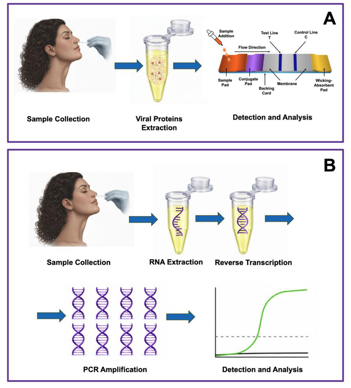
Figure 2.
Diagrammatic representation of the sputum sample preparation (disruption-extraction-digestion) workflow using the nonionic surfactant extraction process and the enzymatic digestion using subtilisin A (Alcalase) and Triton X-100. Panel A shows that sputum is a thick, rubbery, sticky, viscous, and gel-like meshwork. This respiratory mucus traps numerous cells, cell debris, microorganisms, and chemical-biochemical entities. Upon introducing the surfactant and protease to the sample, significant disruption to the matrix and proteolysis of viral proteins occurs (panel B). This process is governed by time of incubation, proteolytic enzyme quantity, temperature, and pH of the solution. The incubation temperature for most of the experiments was maintained at 25 degrees Celsius. This process resulted in a solution containing primarily soluble material and some precipitate of insoluble components (panel C). After decantation or centrifugation, the supernatant was tested for the presence of SARS-CoV-2 virus, or virus components, on a LFIA platform or strip. Author-generated conceptual schematic, as informed by Guzman et al. [18], Guzman et al. [19]. Reproduced under Creative Commons Attribution License.
Figure 2.
Diagrammatic representation of the sputum sample preparation (disruption-extraction-digestion) workflow using the nonionic surfactant extraction process and the enzymatic digestion using subtilisin A (Alcalase) and Triton X-100. Panel A shows that sputum is a thick, rubbery, sticky, viscous, and gel-like meshwork. This respiratory mucus traps numerous cells, cell debris, microorganisms, and chemical-biochemical entities. Upon introducing the surfactant and protease to the sample, significant disruption to the matrix and proteolysis of viral proteins occurs (panel B). This process is governed by time of incubation, proteolytic enzyme quantity, temperature, and pH of the solution. The incubation temperature for most of the experiments was maintained at 25 degrees Celsius. This process resulted in a solution containing primarily soluble material and some precipitate of insoluble components (panel C). After decantation or centrifugation, the supernatant was tested for the presence of SARS-CoV-2 virus, or virus components, on a LFIA platform or strip. Author-generated conceptual schematic, as informed by Guzman et al. [18], Guzman et al. [19]. Reproduced under Creative Commons Attribution License.
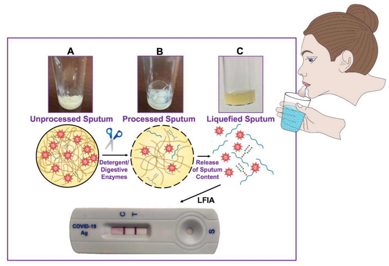
Figure 3.
Illustration of a time-series evaluation of SARS-CoV-2 in sputum samples collected over a period of 15 weeks, using a modified disruption-extraction sample preparation protocol. The color intensity of the test (T) line diminishes as the persistence of the coughing in the patient lessens and the amount of sputum collected is reduced as well. The sputum specimen was obtained from a patient that had COVID-19-like symptoms for approximately 4-5 days but was asymptomatic for the rest of the 15-week period of testing, except for some minor coughing. Author-generated conceptual schematic, as informed by Guzman et. al. [18]; Guzman et al. [19]. Reproduced under Creative Commons Attribution License.
Figure 3.
Illustration of a time-series evaluation of SARS-CoV-2 in sputum samples collected over a period of 15 weeks, using a modified disruption-extraction sample preparation protocol. The color intensity of the test (T) line diminishes as the persistence of the coughing in the patient lessens and the amount of sputum collected is reduced as well. The sputum specimen was obtained from a patient that had COVID-19-like symptoms for approximately 4-5 days but was asymptomatic for the rest of the 15-week period of testing, except for some minor coughing. Author-generated conceptual schematic, as informed by Guzman et. al. [18]; Guzman et al. [19]. Reproduced under Creative Commons Attribution License.
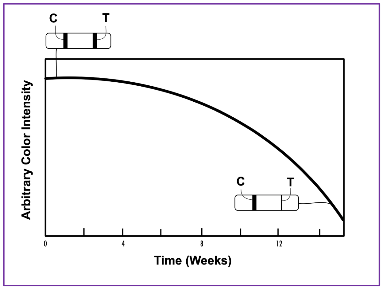
Figure 4.
Illustration of the time-dependent experiment for pro-inflammatory cytokines IL-1 and TNF- in serum samples of a study involving 80 patients (Panel A) at Day 1, Day 7, and Day 14 (acute phase); Day 120 (convalescence phase); and Day 720/24 months (post-acute phase). Measurement of circulating cytokines in the participants’ serum samples was performed using the Flow Cytometry Cytometric Bead Array technique. The concentrations of IL-1 and TNF- are expressed in pg/mL. Panel B, Course of SARS-CoV-2 infection. Panel A is our author-generated visualization based on the Goulart et al. data [61]; Panel B is reproduced with permission from Calabrese [66]. Copyright © 2026 Cleveland Clinic Foundation. All rights reserved.
Figure 4.
Illustration of the time-dependent experiment for pro-inflammatory cytokines IL-1 and TNF- in serum samples of a study involving 80 patients (Panel A) at Day 1, Day 7, and Day 14 (acute phase); Day 120 (convalescence phase); and Day 720/24 months (post-acute phase). Measurement of circulating cytokines in the participants’ serum samples was performed using the Flow Cytometry Cytometric Bead Array technique. The concentrations of IL-1 and TNF- are expressed in pg/mL. Panel B, Course of SARS-CoV-2 infection. Panel A is our author-generated visualization based on the Goulart et al. data [61]; Panel B is reproduced with permission from Calabrese [66]. Copyright © 2026 Cleveland Clinic Foundation. All rights reserved.
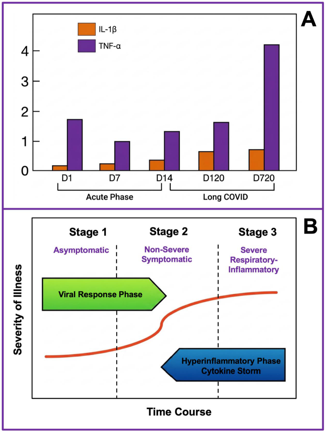
Figure 5.
Representative separation of four different heterogeneous proteins and their respective molecular forms. Panel A represents an electropherogram profile of the various molecular forms of immunoreactive alpha-1-acid glycoprotein (AGP) or orosomucoid separated by capillary zone electrophoresis (CZE). Orosomucoid is an important protein in inflammation and pharmacokinetics, acting as a major transport protein in the blood. Panel B represents an electropherogram of 7 molecular forms of a recombinant monoclonal antibody (rMAb) using imaged capillary isoelectric focusing (iCIEF) and two pI markers. Panel C represents an electropherogram of the various glycoforms of heparan-N-sulfatase separated by CZE. Heparan-N-sulfatase is a critical lysosomal enzyme responsible for the breakdown of heparan sulfate, a complex sugar molecule essential for cell signaling and tissue development. Panel D represents the separation by CZE of the various glycoforms of erythropoietin, a vital hormone, produced primarily by the kidneys, that regulates red blood cell production. Author-generated conceptual schematics, as informed by Bristow et al. [110]; Salas-Solano et al. [82]; Roseman et al. [111]; Morales-Cid et al. [112]; and Guzman et al. [98]. Reproduced under Creative Commons Attribution License.
Figure 5.
Representative separation of four different heterogeneous proteins and their respective molecular forms. Panel A represents an electropherogram profile of the various molecular forms of immunoreactive alpha-1-acid glycoprotein (AGP) or orosomucoid separated by capillary zone electrophoresis (CZE). Orosomucoid is an important protein in inflammation and pharmacokinetics, acting as a major transport protein in the blood. Panel B represents an electropherogram of 7 molecular forms of a recombinant monoclonal antibody (rMAb) using imaged capillary isoelectric focusing (iCIEF) and two pI markers. Panel C represents an electropherogram of the various glycoforms of heparan-N-sulfatase separated by CZE. Heparan-N-sulfatase is a critical lysosomal enzyme responsible for the breakdown of heparan sulfate, a complex sugar molecule essential for cell signaling and tissue development. Panel D represents the separation by CZE of the various glycoforms of erythropoietin, a vital hormone, produced primarily by the kidneys, that regulates red blood cell production. Author-generated conceptual schematics, as informed by Bristow et al. [110]; Salas-Solano et al. [82]; Roseman et al. [111]; Morales-Cid et al. [112]; and Guzman et al. [98]. Reproduced under Creative Commons Attribution License.
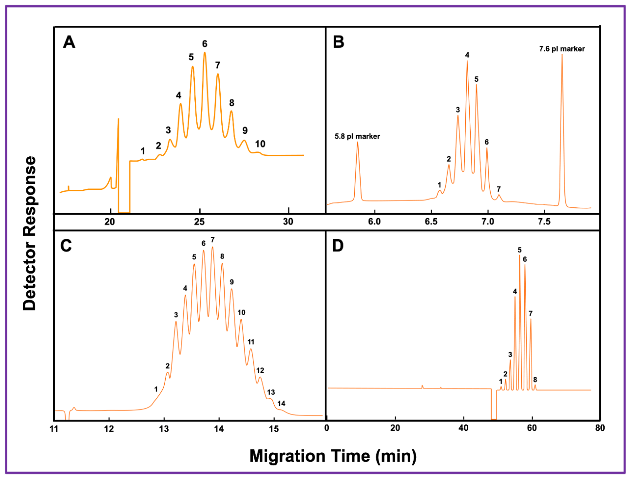
Figure 6.
Diagrammatic representation of an analyte concentrator microreactor (ACM) device on a unidirectional mode, employed for the capture and separation of target biomarkers. (panel A). Electropherogram of a multi-biomarker analysis based on affinity-capture–separation analytical technology performed with microchip electrophoresis (panel B). The peaks in the electropherogram resolved in the following order: 1, TGF-β (transforming growth factor beta); 2, IL-6 (interleukin-6); 3, IL-1β (interleukin-1 beta); 4, IFNγ (interferon gamma); 5, MIP-1α (macrophage inflammatory protein 1 alpha); 6, MCP-1 (macrophage chemoattractant protein 1); 7, TNF-α (tumor necrosis factor-alpha); 8, CGRP (calcitonin gene-related peptide); 9, NY (neuropeptide Y); 10, IL-8 (interleukin-8); 11, VIP (vasoactive intestinal peptide); 12, SP (substance P). A small amount of free dye was always present. All antibodies directed against the targeted biomarkers were co-immobilized to disposable glass fiber disks and inserted within the extraction port of the microchip. The captured analytes were labeled with a 635 nm light-emitting laser dye and electroeluted into the separation channel as described in ref. 102. On panel C, single-stranded aptamers were used as affinity-capture ligands for the selective recognition of two low molecular-weight compounds (1, aflatoxin B1 and 2, ochratoxin A). Two distinct aptamers specific for these two analytes were co-immobilized via covalent bonds on the surface of the inlet end of the capillary. The separation, detection, and quantification were performed by capillary coupled to a mass spectrometer. Author-generated conceptual schematics, as informed by Phillips et al. [102]; Wang et al. [115]; Guzman et al. [95]; and Guzman et al. [19]. Reproduced under Creative Commons Attribution License.
Figure 6.
Diagrammatic representation of an analyte concentrator microreactor (ACM) device on a unidirectional mode, employed for the capture and separation of target biomarkers. (panel A). Electropherogram of a multi-biomarker analysis based on affinity-capture–separation analytical technology performed with microchip electrophoresis (panel B). The peaks in the electropherogram resolved in the following order: 1, TGF-β (transforming growth factor beta); 2, IL-6 (interleukin-6); 3, IL-1β (interleukin-1 beta); 4, IFNγ (interferon gamma); 5, MIP-1α (macrophage inflammatory protein 1 alpha); 6, MCP-1 (macrophage chemoattractant protein 1); 7, TNF-α (tumor necrosis factor-alpha); 8, CGRP (calcitonin gene-related peptide); 9, NY (neuropeptide Y); 10, IL-8 (interleukin-8); 11, VIP (vasoactive intestinal peptide); 12, SP (substance P). A small amount of free dye was always present. All antibodies directed against the targeted biomarkers were co-immobilized to disposable glass fiber disks and inserted within the extraction port of the microchip. The captured analytes were labeled with a 635 nm light-emitting laser dye and electroeluted into the separation channel as described in ref. 102. On panel C, single-stranded aptamers were used as affinity-capture ligands for the selective recognition of two low molecular-weight compounds (1, aflatoxin B1 and 2, ochratoxin A). Two distinct aptamers specific for these two analytes were co-immobilized via covalent bonds on the surface of the inlet end of the capillary. The separation, detection, and quantification were performed by capillary coupled to a mass spectrometer. Author-generated conceptual schematics, as informed by Phillips et al. [102]; Wang et al. [115]; Guzman et al. [95]; and Guzman et al. [19]. Reproduced under Creative Commons Attribution License.
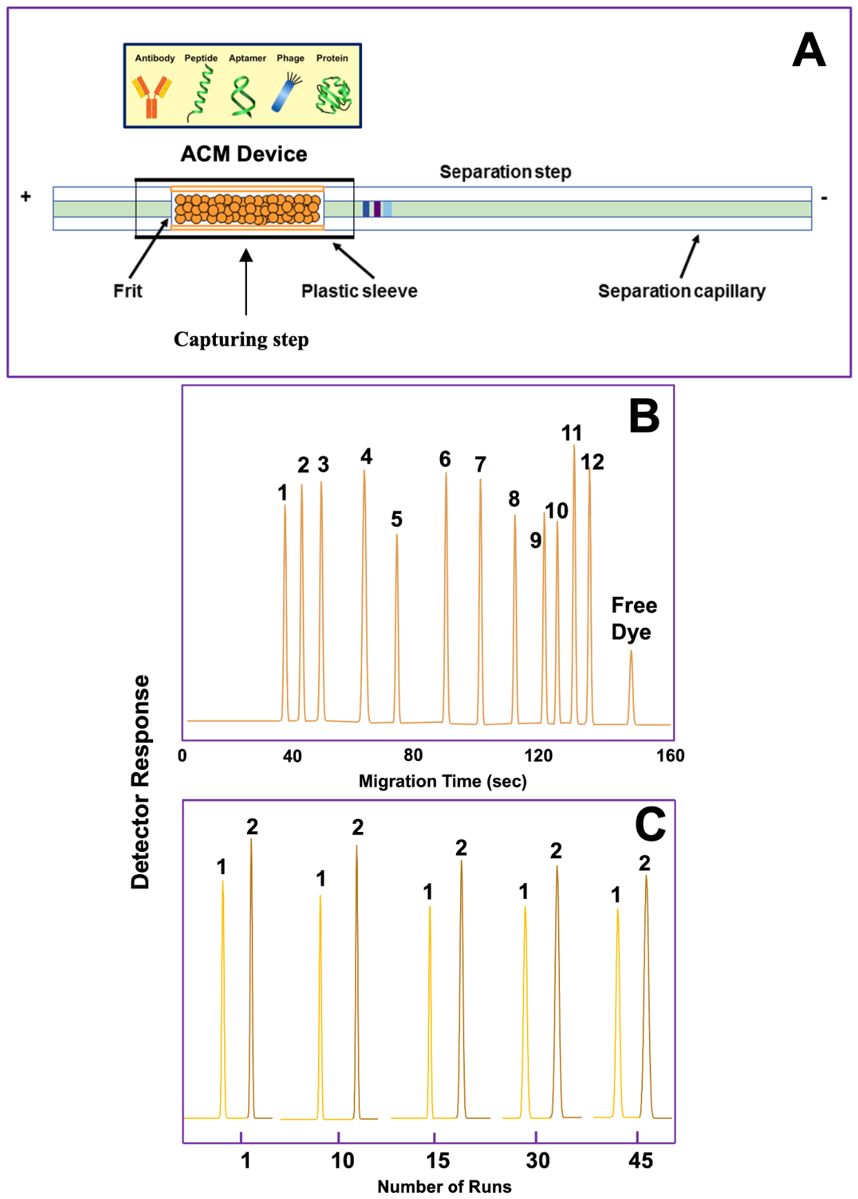
Figure 7.
Illustration of LC-MS data on the SARS-CoV-2 S glycopeptides (at N-linked glycosylation sites). Panel A illustrates representative SARS-CoV-2 glycopeptides identified in the analysis. Panel B shows the number of glycopeptides as summary of results of the two different approaches for the SARS-CoV-2 S data set. Tally of the number of correctly identified SARS-CoV-2 S glycopeptides (orange bars) and false positives (blue bars), based on the expert-based assignment criteria. Author-generated conceptual schematics, as informed by Go et al. [119].
Figure 7.
Illustration of LC-MS data on the SARS-CoV-2 S glycopeptides (at N-linked glycosylation sites). Panel A illustrates representative SARS-CoV-2 glycopeptides identified in the analysis. Panel B shows the number of glycopeptides as summary of results of the two different approaches for the SARS-CoV-2 S data set. Tally of the number of correctly identified SARS-CoV-2 S glycopeptides (orange bars) and false positives (blue bars), based on the expert-based assignment criteria. Author-generated conceptual schematics, as informed by Go et al. [119].
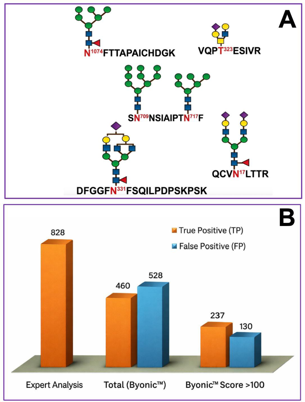
Figure 8.
Illustration of the analysis of prostate-specific antigen using three different methods. Panel A depicts the analysis of PSA using the ELISA method. Panel B depicts the analysis of PSA isoforms by capillary zone electrophoresis method. Panel C, D, and E depict the analysis of PSA by the glycomic method. Author-generated conceptual schematics, as informed by Barrabés et al. [142]; Kammeijer et al. [141]; and Guzman et al. [99]. Reproduced under Creative Commons Attribution License.
Figure 8.
Illustration of the analysis of prostate-specific antigen using three different methods. Panel A depicts the analysis of PSA using the ELISA method. Panel B depicts the analysis of PSA isoforms by capillary zone electrophoresis method. Panel C, D, and E depict the analysis of PSA by the glycomic method. Author-generated conceptual schematics, as informed by Barrabés et al. [142]; Kammeijer et al. [141]; and Guzman et al. [99]. Reproduced under Creative Commons Attribution License.
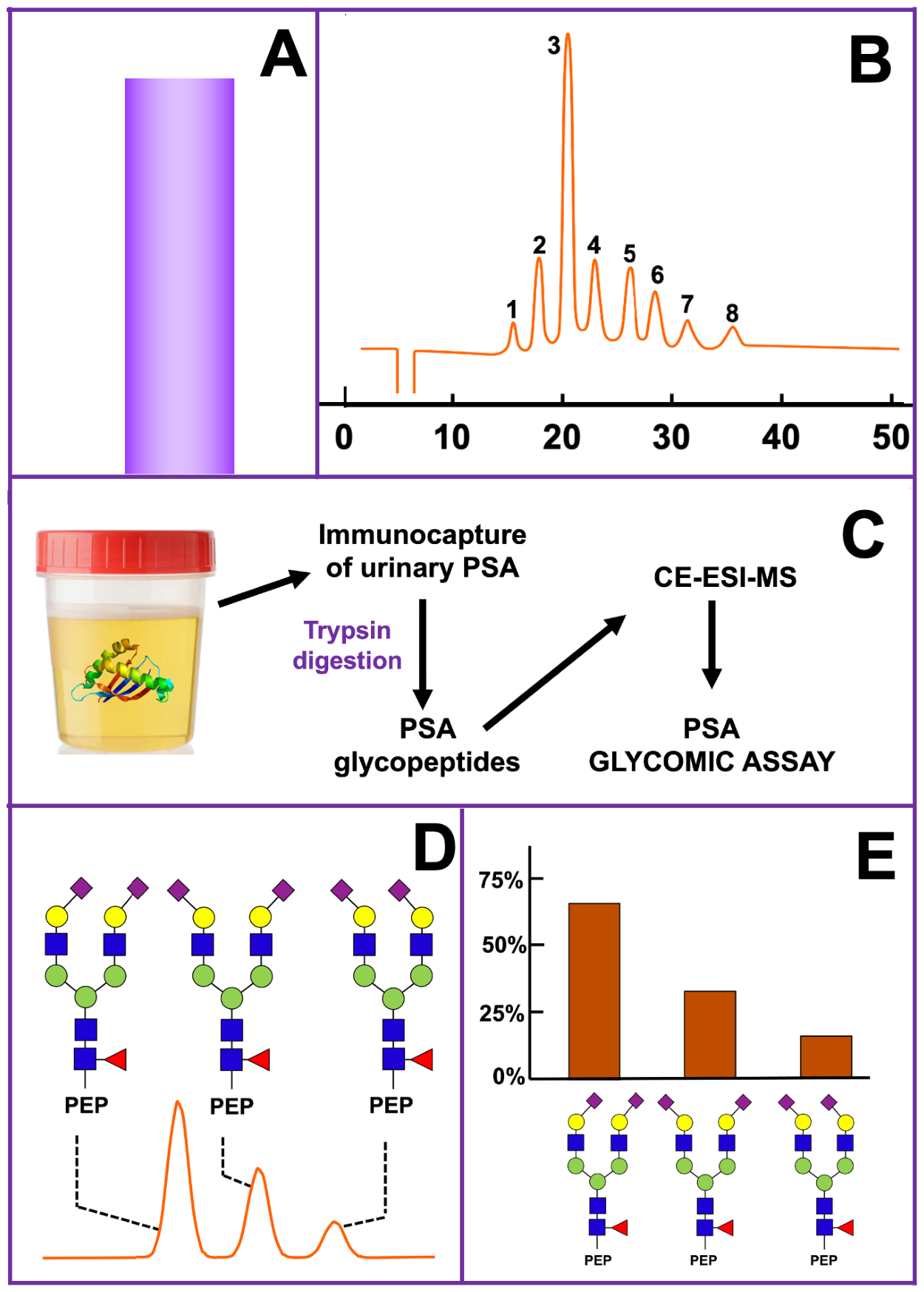
Disclaimer/Publisher’s Note: The statements, opinions and data contained in all publications are solely those of the individual author(s) and contributor(s) and not of MDPI and/or the editor(s). MDPI and/or the editor(s) disclaim responsibility for any injury to people or property resulting from any ideas, methods, instructions or products referred to in the content. |
© 2026 by the authors. Licensee MDPI, Basel, Switzerland. This article is an open access article distributed under the terms and conditions of the Creative Commons Attribution (CC BY) license (http://creativecommons.org/licenses/by/4.0/).
Copyright: This open access article is published under a Creative Commons CC BY 4.0 license, which permit the free download, distribution, and reuse, provided that the author and preprint are cited in any reuse.