

Submitted:
26 August 2026
Posted:
26 August 2026
You are already at the latest version
Abstract
Preeclampsia (PE), a multi-system disorder, is not confined to the puerperium and may represent a sentinel event leading to increased maternal risk for future cardiovascular disease (CVD). Early-onset PE appears to carry a distinct, and possibly more pronounced, vascular signature than late-onset PE. Accumulating data consistently link previous early-onset PE, to an elevated lifetime risk of CVD. Arterial stiffness (AS), most commonly quantified by carotid-femoral pulse wave velocity (cf-PWV) and augmentation index (AIx, or AIx corrected to a heart rate of 75 beats per minute, AIx-75), is an established, independent predictor of CVD in the general population and has therefore attracted interest as a candidate marker for postpartum CVD risk stratification. However, whether AS remains elevated once the puerperium has resolved, has been a persistently unsettled question. Existing literature is inconclusive regarding the association between postpartum AS measurements and previous early-onset PE. This comprehensive review synthesizes the available literature on oscillometric AS measurements before and after delivery in pregnancies complicated by early-onset PE and attempts to quantify the differences between ante- and postpartum oscillometric AS measurements. Although not a systematic review, this quantitative review was conducted according to the PRISMA (Preferred Reporting Items for Systematic Reviews and Meta-Analyses) and MOOSE (Meta-Analysis of Observational Studies in Epidemiology) guidelines. The study protocol was registered at PROSPERO (Prospective Register of Systematic Reviews System) (ID CRD42020167749) a priori. Only longitudinal studies reporting on both pregnancies complicated by early-onset PE (case group) and normotensive pregnancies (control group) were included. cf-PWV, Alx and Alx-75 were assessed both ante- and postpartum. Quantitative synthesis was performed for indices presented among at least 2 studies. Data were presented as standardized mean differences (SMDs) and 95% confidence intervals (95%CIs). Overall, from 14 citations of relevance, 3 studies with a total of 35 cases and 155 controls fulfilled the inclusion/exclusion criteria. In pregnancies complicated by early-onset PE assessed both ante- and postpartum with either cf-PWV (SMD: -0.61, 95%CI: -1.3 to 0.07) and/or Alx (SMD: -0.32, 95%CI: -2.83 to 2.2) and/or Alx-75 (SMD: -0.61, 95%CI: -1.27 to 0.05), there was no statistically significant difference in AS measurements. In conclusion, current literature suggests that AS measurements (cf-PWV, AIx and AIx-75) are not significantly altered in pregnancies complicated by early-onset PE. Alx and Alx-75 should be further evaluated. Substantial heterogeneity in study design, small sample sizes, and inconsistent postpartum follow-up windows continue to limit firm conclusions, underscoring the need for larger, standardized, and adequately powered longitudinal studies before AS measurements can be recommended for routine postpartum CVD risk stratification.
Keywords:
cardiovascular disease
; postpartum period
; pregnancy complications
; early-onset pre-eclampsia
; pulse wave analysis
; arterial stiffness
1. Introduction
Preeclampsia (PE) is no longer viewed simply as a self-limiting complication of the second half of pregnancy. It is now recognized as a sentinel event for future maternal cardiovascular disease (CVD).[1,2,3] Women with a history of PE[1,2,4,5] are at increased risk of subsequent CVD later in life compared to unaffected women.[6,7,8] CVD and cardiovascular mortality seem to be strongly associated with the severity of PE.[9] Early-onset PE occurs before 34 weeks of gestation and accounts for 10% of PE cases.[10] It is characterized by increased severity and is associated with increased maternal and perinatal morbidity and mortality.[11,12] The latter raises the probability that early-onset PE also leaves a more pronounced vascular imprint that persists after delivery.
Arterial stiffness (AS) has emerged as an attractive candidate biomarker for future maternal CVD risk stratification because it is a validated, independent predictor of CVD in the general population.[13,14,15,16,17,18] Among a variety of measurements, AS is mainly quantified through augmentation index (Alx) and carotid–femoral pulse wave velocity (cf-PWV). AIx is a composite measure of systemic AS derived from the ascending aortic pressure waveform.[19] Because AIx is sensitive to heart rate, many studies additionally report AIx corrected to a standard heart rate of 75 beats per minute (AIx-75).[20] AIx is a direct marker of wave reflection but an indirect marker of AS, whereas cf-PWV is a direct marker of AS.[21,22,23,24] cf-PWV is the “gold standard” of AS measurements.[25] The relative ease and non-invasiveness of oscillometric devices as well as the ever increasing burden of CVD-related morbidity and mortality, have fueled a renewed interest in oscillometric AS measurements for postpartum CVD risk stratification in pregnancies complicated by early-onset PE.[26,27,28,29]
Hausvater et al. reported that cf-PWV and AIx were increased in pregnancies complicated by PE compared with normotensive pregnancies.[13] Moreover, Osman et al. concluded in a meta-analysis (assessing different hemodynamic parameters during pregnancy but before the onset of PE) that, in comparison to normotensive pregnancies, pregnancies complicated by PE had increased: 1) Alx-75 (Alx corrected for a heart rate of 75 beats per minute) in the 1st trimester, 2) cf-PWV in the 2nd trimester, and 3) cf-PWV and Alx in the 3rd trimester.[30] These findings suggest that vascular dysfunction in PE is detectable well before delivery. What has proved far less consistent is whether this dysfunction persists after delivery.
Current literature is inconsistent regarding the association between postpartum AS measurements and previous early-onset PE. Studies reporting on AS measurements between 7 weeks and 2 years postpartum in pregnancies complicated by PE, compared with previous normotensive pregnancies, demonstrated significant heterogeneity.[31] Grand’ Maison et al. concluded in a meta-analysis (assessing vascular dysfunction at least 3 months postpartum) that cIMT (carotid Intima-Media Thickness), cf-PWV and Alx were increased.[32] Hamad et al. found that flow-mediated dilation (FMD) was decreased in women 1 year postpartum.[33] By contrast, Lampinen et al. and Ronnback et al. reported non-significant differences in AS measurements between 5 and 9 years postpartum.[34,35] Yinon et al. added a further layer of complexity by demonstrating that AS measurements differed between women with a history of early- and late-onset PE when assessed between six and twenty-four months postpartum, suggesting that timing of PE onset may itself be a source of inconsistency.[36]
Conclusively, no consensus has been reached regarding the difference between ante- and postpartum AS measurements in pregnancies complicated by early-onset PE. This comprehensive review synthesizes all available English-language literature, to summarize what is currently known about ante- to postpartum changes in oscillometric AS measurements (cf-PWV, AIx, and AIx-75) and attempts to quantify the differences between ante- and postpartum oscillometric AS measurements (cf-PWV, AIx and AIx-75) in pregnancies complicated by early-onset PE.
2. Pathophysiological Rationale and Measurements of Arterial Stiffness
AS reflects the mechanical properties of the vessel wall and rises with age, hypertension, and a wide range of cardiometabolic risk factors. cf-PWV is measured by recording the pressure wave at two distinct arterial sites (typically the carotid and femoral arteries) and calculating the velocity at which the wave travels between them; a faster wave indicates a stiffer, less compliant arterial tree.[20,22,24,25,37] Because cf-PWV is a direct mechanical measurement of the aorta and large elastic arteries, it is comparatively unaffected by peripheral vascular tone and is widely regarded as the reference standard for AS assessment.[20,22,24,25,37]
AIx is derived differently, it quantifies the augmentation of the central aortic pressure waveform produced by the reflection of the forward pressure wave from the periphery back toward the heart. Because the timing of wave reflection is influenced by heart rate, peripheral vascular resistance, and stroke volume in addition to arterial stiffness itself, AIx is best regarded as an indirect marker of AS and a more direct marker of wave reflection; correcting it to a standard heart rate of 75 beats per minute (AIx-75) removes one major source of variability and is the preferred metric in most contemporary studies.[20] Oscillometric brachial cuff devices and applanation tonometry systems using a micromanometer are the two methodologies most commonly used to obtain these measurements non-invasively.[37]
A separate but important pathophysiological point is that AIx and cf-PWV appear to be differentially sensitive to age. McEniery et al. inidicated that AIx tends to be a more sensitive marker of AS and CVD risk in younger individuals (<50 years of age), whereas cf-PWV becomes better discrimination measurement in older individuals (≥50 years of age).[38] Given that most women assessed after early-onset PE are relatively young, this distinction is directly relevant to interpreting postpartum measurements: normal findings for cf-PWV do not necessarily mean that CVD risk is absent, and conversely, elevated AIx may carry more prognostic value than the same finding would in an older population.[38]
3. Arterial Stiffness During Pregnancies Complicated by Preeclampsia
While the spotlight often shines on the postpartum period, it is worth summarizing what is known about AS during pregnancy, because much of the postpartum literature is implicitly asking whether an antepartum abnormality preexists or evolves and whether it resolves or persists. Khalil et al. reported that AS measurements differ between early-onset and late-onset PE, with severe, earlier-onset PE being associated with greater AS.[39,40] Moreover, Alx-75 was the only AS measurement demonstrating statistically significant difference in the first trimester between pregnancies that later developed PE and those that remained normotensive.[41] General patterns across gestation show AIx falling in the first and second trimesters before rising again in the third trimester and peaking postpartum. However, because AIx is a direct measure of wave reflection, it is also influenced by peripheral vascular resistance, heart rate, and stroke volume, which are independent of AS itself (thus AIx-75 is generally preferred over unadjusted AIx). cf-PWV, by contrast, is primarily affected by age and blood pressure, so any postpartum elevation in cf-PWV among women with a history of PE could, at least in part, reflect persistently higher blood pressure rather than a structural change in the arterial wall.[41,42,43,44,45]
4. Postpartum Arterial Stiffness After Early-Onset Preeclampsia
The most direct evidence on early-onset PE specifically comes from a small number of longitudinal studies that assessed the same women both during pregnancy and again after delivery, alongside normotensive controls. Carty et al. prospectively followed pregnancies from before the clinical onset of PE until six to nine months postpartum, and found that AIx and AIx-75 were both elevated in the postpartum period among those who developed early-onset PE, whereas differences on some antepartum measures did not reach statistical significance until closer to delivery.[46] Franz et al. assessed cf-PWV and AIx in a case-control cohort three to six months after early-onset PE and likewise reported evidence of a higher CVD risk profile in the affected group.[42] Robb et al., assessing women as early as seven weeks postpartum found a persistently elevated cf-PWV and AIx-75 among women with a recent history of PE, although their cohort included both early- and late-onset PE cases.[47]
Taken individually, these three studies point toward persistently elevated wave-reflection indices (AIx, AIx-75) after early-onset PE, with inconsistent data for cf-PWV, particularly given the modest sample sizes involved (35 early-onset PE cases and 155 controls in total across the three studies) and the wide confidence intervals that accompany such small numbers. When these data are grouped together, the overall picture across cf-PWV, AIx, and AIx-75 falls short of statistical significance, and the very small number of eligible studies (a consequence of the strict requirement for both ante- and postpartum measurement within the same cohort) limits how far any conclusion can be generalized. What the three studies agree on is a numerical trend toward higher postpartum values across all three indices in the early-onset PE group, even where formal statistical significance is not reached. This trend is consistent with, rather than contradictory to, the broader hypertensive disorders of pregnancy (HDP) literature described below.
It is worth emphasizing that these are the only three published longitudinal studies identified that report on both ante- and postpartum oscillometric AS data specifically restricted to, or separable for, early-onset PE with a normotensive comparison group. The latter underscores just how sparse the evidence base is for this question, despite early-onset PE being the clinically most severe phenotype.
5. Broader Evidence Across the Spectrum of Hypertensive Disorders of Pregnancy
Because studies restricted to early-onset PE with paired ante- and postpartum measurement are scarce, much of what is known about postpartum AS comes from a wider literature examining HDP as a whole (encompassing gestational hypertension, chronic hypertension, and PE of any onset) typically compared against normotensive controls at a single postpartum timepoint rather than longitudinally against the same woman’s antepartum values. This literature has expanded considerably over the past five years and offers a more complete, if still imperfect, picture.
Mbongozi et al., drawing on twelve studies and 856 women, found that both AIx and cf-PWV were significantly elevated overall in women with a history of HDP compared with normotensive controls (pooled mean difference for AIx of 11.63, 95% CI 1.72–21.54; pooled mean difference for cf-PWV of 0.53 m/s, 95% CI 0.27–0.78).[31] Notably, when the data were stratified by time since delivery, AIx showed no significant elevation within the first postpartum year but became significantly elevated beyond one year (mean difference 9.11, 95% CI 4.20–14.02), whereas cf-PWV was elevated both within and beyond the first postpartum year.[31] When restricted to early-onset PE, AIx again failed to reach significance, but cf-PWV was significantly elevated (mean difference 1.86, 95% CI 0.25–3.47); for late-onset PE, neither index differed significantly from controls.[31] This pattern, a stronger, earlier cf-PWV signal in early-onset disease, and an AIx signal that emerges only with longer follow-up, offers one plausible explanation for why the smaller, early-focused literature produced inconsistent results: several of those studies assessed women within the first postpartum year, precisely within the window in which AIx appears least likely to distinguish HDP from normotensive pregnancy.
Several other studies published within the last five years reinforce and add nuance to this picture. The EVA (Early Vascular Aging) study by Werlang et al. compared women with a history of PE against normotensive controls between six months and six years postpartum and found significantly elevated AIx and cf-PWV, consistent with accelerated vascular aging.[8] Moe et al., assessing functional and structural vascular biomarkers exactly one year after a hypertensive pregnancy, reported that AIx was not significantly different at that specific timepoint, mirroring the “within one year” null finding highlighted in the Mbongozi et al. meta-analysis.[29,31] Kim etal. using the cardio-ankle vascular index to follow women longitudinally for a full year after delivery, similarly found that any early postpartum elevation in arterial stiffness among women with PE tended to track closely with persistence of hypertension and metabolic risk factors rather than emerging as an isolated, fixed vascular abnormality.[48]
Namugowa et al. found increased AS in women with a history of PE approximately fifteen weeks postpartum.[49] Kaihara et al. compared macrovascular and microvascular function in pregnant women with chronic hypertension or PE and found that large-artery stiffness (cf-PWV and AIx-75) was increased in both hypertensive groups relative to normotensive pregnancy, even though microvascular function measured by peripheral arterial tonometry did not differ (i.e. suggesting that the vascular signature of HDP is most reliably captured at the level of the large, elastic arteries rather than the microcirculation).[50] Anthoulakis et al. have also reported that pulse wave velocity and AIx-75 are elevated in preeclamptic compared with normotensive pregnant women using newer oscillometric devices, adding further, methodologically independent confirmation of the core finding.[51] Nakimuli et al. followed a large prospective cohort of women one year after a pregnancy complicated by PE and found not only a markedly higher prevalence of persistent hypertension (36.4% versus 4.5% in controls) but also a significantly higher postpartum aortic pulse wave velocity, underscoring that the vascular consequences of PE are not confined to higher-resource settings and may be at least as pronounced, if not more so, in under-resourced populations with limited access to postpartum cardiovascular surveillance.[52]
Two recent studies illustrate that the vascular changes associated with PE extend beyond the two “classical” oscillometric indices and may be detectable through complementary vascular ultrasound techniques. Barr et al. used carotid ultrasound to demonstrate reduced carotid far-wall circumferential strain (indicating increased local carotid stiffness) in women with a history of PE assessed between six months and five years postpartum, with the greatest impairment seen in those with a history of severe PE.[53] Davis et al. reported higher carotid backward pressure wave amplitude and load-dependent stiffness in young women with a history of PE compared with normotensive controls, although in that cohort these haemodynamic differences did not appear to mediate the modestly reduced cognitive processing speed also observed in the PE group.[54] Together, these studies suggest that the vascular remodeling associated with PE is detectable across multiple complementary methodologies, not solely through cf-PWV and AIx, which strengthens confidence that the phenomenon is real rather than an artefact of any single measurement technique.[53,54]
However, not every study points in the same direction. As already noted, cf-PWV in cohort-design studies pooled by Mbongozi et al. did not reach significance despite a clear signal in cross-sectional designs, a discrepancy the authors partly attribute to differing degrees of confounder control between the two study designs.[31] Palmiero et al., reviewing long-term CVD risk after PE, likewise report that while the epidemiological association between PE and later CVD risk is now well established, the specific mechanistic pathway (in particular, how much of that risk is mediated through persistently elevated AS as opposed to shared underlying risk factors, incident chronic hypertension, or metabolic syndrome) remains incompletely resolved.[55] This caution is echoed in a broader American Heart Association scientific statement on adverse pregnancy outcomes and CVD risk, which frames PE as one of several pregnancy complications that should prompt earlier, more structured CVD risk assessment, without yet specifying a validated postpartum vascular biomarker for routine use.[56]
6. Materials and Methods
A quantitative review was performed in compliance with the recommendations of the Preferred Reporting Items for Systematic Reviews and Meta-analyses (PRISMA) statement[57] and the Meta-Analysis of Observational Studies in Epidemiology (MOOSE) guidelines.[58] The research protocol (ID CRD42020167749) was registered at the International Prospective Register of Systematic Reviews (PROSPERO) a priori.
6.1. Study Inclusion Criteria
6.1.1. Types of Studies
To gather any available information, this quantitative review included longitudinal observational studies with both ante- and postpartum arms as well as control groups.
6.1.2. Types of Participants
PE was defined as new-onset hypertension (systolic blood pressure ≥ 140mmHg or diastolic blood pressure ≥ 90mmHg) and proteinuria (≥300mg/24hr or protein-to-creatinine ratio >30 mg/mmol or ≥2+ in dipstick testing) after 20 weeks of gestation.[59] This has been revised by the International Society for the Study of Hypertension in Pregnancy (ISSHP) and the American College of Obstetricians and Gynecologists (ACOG) to include cases without proteinuria but with evidence of renal, hepatic or hematological dysfunction.[60,61] In our study, the exposure of interest was early-onset PE (appearing before 34 weeks of gestation) in singleton pregnancies.
6.1.3. Types of Diagnostic Interventions
No studies other than those reporting on imaging modalities of vascular dysfunction antepartum and postpartum were included. Only non-invasive, automated oscillometric AS measurements were evaluated. The included studies used normotensive pregnancies as the control group.
6.1.4. Types of Outcome Measures
The outcomes of interest were: 1) cf-PWV as a direct marker of AS, and 2) Alx and Alx-75 as indirect markers of AS and direct markers of wave reflection.
6.1.5. General Admission Requirements
Conclusively, studies were included when the following criteria were met: 1) longitudinal studies reporting on pregnancies complicated by early-onset PE (case group) and normotensive pregnancies (control group); 2) ante- and postpartum data available; 3) outcomes reported as cf-PWV and/or Alx and/or Alx-75; and 4) data or subsets of data not published more than once.
6.2. Study Exclusion Criteria
We have excluded studies assessing imaging modalities other than cf-PWV and/or Alx and/or Alx-75, studies that evaluated arterial stiffness and wave reflection pre-pregnancy and/or during pregnancy and the postpartum period but did not address early-onset PE. Cross-sectional studies, review articles, letters, comments, non-clinical studies, technical papers, expert opinions, single case reports and case series reporting five cases or fewer on either arm or articles without original data were excluded.
6.3. Identification of Studies
A PRISMA- and MOOSE-compliant quantitative review was conducted to identify studies reporting on postpartum AS and wave reflection after early-onset PE. Prominent online medical databases including MEDLINE via PubMed, EMBASE, Scopus, Cochrane Database of Systematic Reviews (CDSR), Cochrane Central Register of Controlled Trials (CENTRAL), and ClinicalTrials.org registry were searched from 1st January 2000 until 31st December 2025, due to potentially markedly altered practices and techniques over an extensively elongated time frame. Reference lists from identified articles and relevant systematic reviews were screened manually to locate further eligible studies. The search algorithm included: combinations of medical subject heading (MeSH) terms, keywords, and word variants for ‘vascular stiffness’, ‘arterial stiffness’, ‘preeclampsia’, ‘cardiovascular disease’, ‘postpartum period’, ‘pregnancy complications’, ‘early-onset pre-eclampsia’, and ‘pulse wave analysis’. We also searched Clinicaltrials.gov for (arterial stiffness/preeclampsia, cardiovascular disease/preeclampsia). Cochrane’s Central Register of Controlled Trials was searched with 2 different strategies (preeclampsia and arterial stiffness) and (preeclampsia and cardiovascular disease). An extended search through Google Scholar was carried out using the terms, (arterial stiffness, preeclampsia, cardiovascular disease) to render feasible a manual search. After reading the abstracts, two reviewers (C.A. and E.I.) independently assessed all potentially eligible studies. Filters were applied for English-language studies and articles involving human female population. Grey literature, including conference proceedings, dissertations, and unpublished studies, was excluded.
6.4. Study Selection
Duplicates were removed and two independent reviewers (C.A. and E.I.) initially screened the title and abstract of the retrieved studies. If the inclusion/exclusion criteria were met, full-text articles were retrieved and independently evaluated by both reviewers to determine final eligibility. Discrepancies during the selection process between the two reviewers were resolved through discussion and consensus involving a third senior reviewer (T.T.).
6.5. Data Extraction
For each eligible study, two independent reviewers (C.A. and E.I.) extracted appropriate data using a predefined form designed specifically for this quantitative review. Standardized preformulated tables were used to ensure completeness and allow subsequent data analysis and outcome synthesis. The reviewers were not blind to journal names, article authors or affiliations as this was deemed unnecessary. If required, corresponding authors were contacted via institutional email.
The following data were recorded for each article: (1) first author and year of publication, (2) type of study, (3) sample size (number of cases versus controls both antepartum and postpartum), (4) imaging modality (cf-PWV and/or Alx and/or Alx-75) and technique, (5) mean maternal age, (6) mean body mass index (BMI), (7) ethnicity, (8) parity, (9) pre-existing hypertension rate, (10) PE history, (11) mean systolic blood pressure (SBP), (12) mean diastolic blood pressure (DBP), (13) timing of antepartum assessment (before or after early-onset PE diagnosis), (14) mean gestational age (GA) at PE diagnosis, (15) mean GA at delivery, (16) caesarian section (CS) rate, and (17) birthweight. If overlapping samples were detected, the study with the largest sample size was selected for data extraction.
6.6. Study Design Characteristics
The following study design characteristics were recorded for each included study: (1) patient selection (consecutive or non-consecutive), (2) eligibility criteria, (3) methods of data collection (prospective, retrospective, or unknown in cases of doubt), (4) baseline characteristics of the study population regarding the most important prognostic indicators (sufficient or insufficient; sufficient if most of the following characteristics were described: mean maternal age, BMI, ethnicity, gravidity, parity, pre-existing hypertension, and PE history), (5) reporting of diagnostic intervention (sufficient or insufficient; the diagnostic intervention should be described with sufficient detail to allow for replication, validation, and generalization of the study), (6) co-interventions (anti-hypertensive medication) and confounders (maternal age, chronic hypertension, and diabetes mellitus), (7) follow-up (timing of postpartum assessment), and (8) drop-out rate.
6.7. Quality Assessment
Two independent reviewers (C.A. and E.I.) assessed the quality of each included study using NOS (Newcastle-Ottawa Scale),[62] NIH Quality Assessment Tool for Observational Cohort and Cross-Sectional Studies,[63] and NIH Quality Assessment Toll for Case-Control Studies.[63] After reaching consensus through discussion between reviewers, it was collectively decided that included studies should meet at least 50% of the points for each scale. Discrepancies were discussed and, if consensus could not be reached between the two reviewers, a third senior reviewer (T.T.) was consulted.
6.8. Statistical Analysis
In singleton pregnancies complicated by early-onset PE, cf-PWV and/or Alx and/or Alx-75 were measured. Normally distributed continuous variables were reported as mean and SD (standard deviation) for descriptive data or as mean and 95%CI (95% confidence interval) for outcome measures. Binary variables were reported as absolute number and percentages. For data analysis, x2 and Fisher’s exact test were used appropriately. P<0.05 was statistically significant. Quantitative synthesis was performed for indices presented among at least 2 studies. Standardized mean differences (SMDs) with 95% confidence intervals (CIs) for the outcomes of interest were calculated using the random effects model.[64] Heterogeneity was quantified with I2 index.[65]
The following statistical software packages were used for data analysis and outcome synthesis:
- Comprehensive Meta-Analysis (CMA), Version 3.3.070, 2014 (Biostat Inc., 14 North Dean Street, Englewood, NJ 07631, USA)
- Medcalc for Windows, version 12.7 (Medcalc Software, Mariakerke, Belgium)
- Review Manager (RevMan), Version 5.3, 2014 (Copenhagen: The Nordic Cochrane Centre, The Cochrane Collaboration)
7. Results
7.1. Search Results and Study Selection
In total, 86 potentially available records were identified. Following duplicate removal and exclusion due to predefined criteria (n=42), 44 studies underwent title and abstract screening, of which 27 were excluded. A total of 17 studies underwent full-text review, of which 3 studies were ultimately included (Figure 1). Exclusion reasons were: (1) no separate data on early-onset PE (n=6); (2) no data on cf-PWV and/or Alx and/or Alx-75 (n=8)(Table 1)(Figure 2) (Table A1 and Table A2).
7.2. Study Range and Characteristics
From the 3 studies included, [42,46,47] only one article assessed Alx and Alx-75 before early-onset PE development.[46]
cf-PWV was evaluated in 2 studies. [42,47] Two different methods were used: 1) the TensioClinic TL1Arteriograph (Tensiomed Ltd, Budapest, Hungary) oscillometric brachial pressure cuff device and TENSIOCLINIC software,[21,42] and 2) applanation tonometry using a micromanometer (Millar Instruments) and the SphygmoCor system (Atcor Medical, Sydney, Australia).[42,47]
Alx was evaluated in 2 studies.[42,46] Two different methods were used: 1) the TensioClinic TL1Arteriograph (Tensiomed Ltd, Budapest, Hungary) oscillometric brachial pressure cuff device and TENSIOCLINIC software,[21,42] and 2) applanation tonometry using a micromanometer (Millar Instruments) and the SphygmoCor system (Atcor Medical, Sydney, Australia).[42,46]
7.3. Study Quality and Potential Sources of Bias
Study quality was assessed in 3 articles, and all 3 studies were included in the quantitative review. (Figure 3) (Table A3). Only one prospective cohort study was identified (Table A4).[46]
This quantitative review identified several limitations in study design characteristics among the included studies: for instance, patient selection was non-consecutive or unknown in all 3 studies (100%), the outcome assessors were not blinded to the exposure status of the participants in all 3 studies (100%), mode of conception was not specified in all 3 studies (100%), reporting of study population was insufficient in 2 studies (66.7%)(Table A5),[42,47] no exclusion criteria were applied for cases with early-onset PE in 1 study(33.3%),[42] data were derived from bar chart in 1 study (33.3%),[47] antihypertensive medication was not recorded either antepartum or postpartum in all 3 studies (100%), missing data rates and handling were not reported in all 3 studies (100%), postpartum PE pregnancies were not divided into early-onset and late-onset groups in 1 study (33.3%),[47] postpartum matching on maternal age was inadequate in 1 study (33%),[46] however there was sufficient description of the study conditions in all 3 studies (100%).
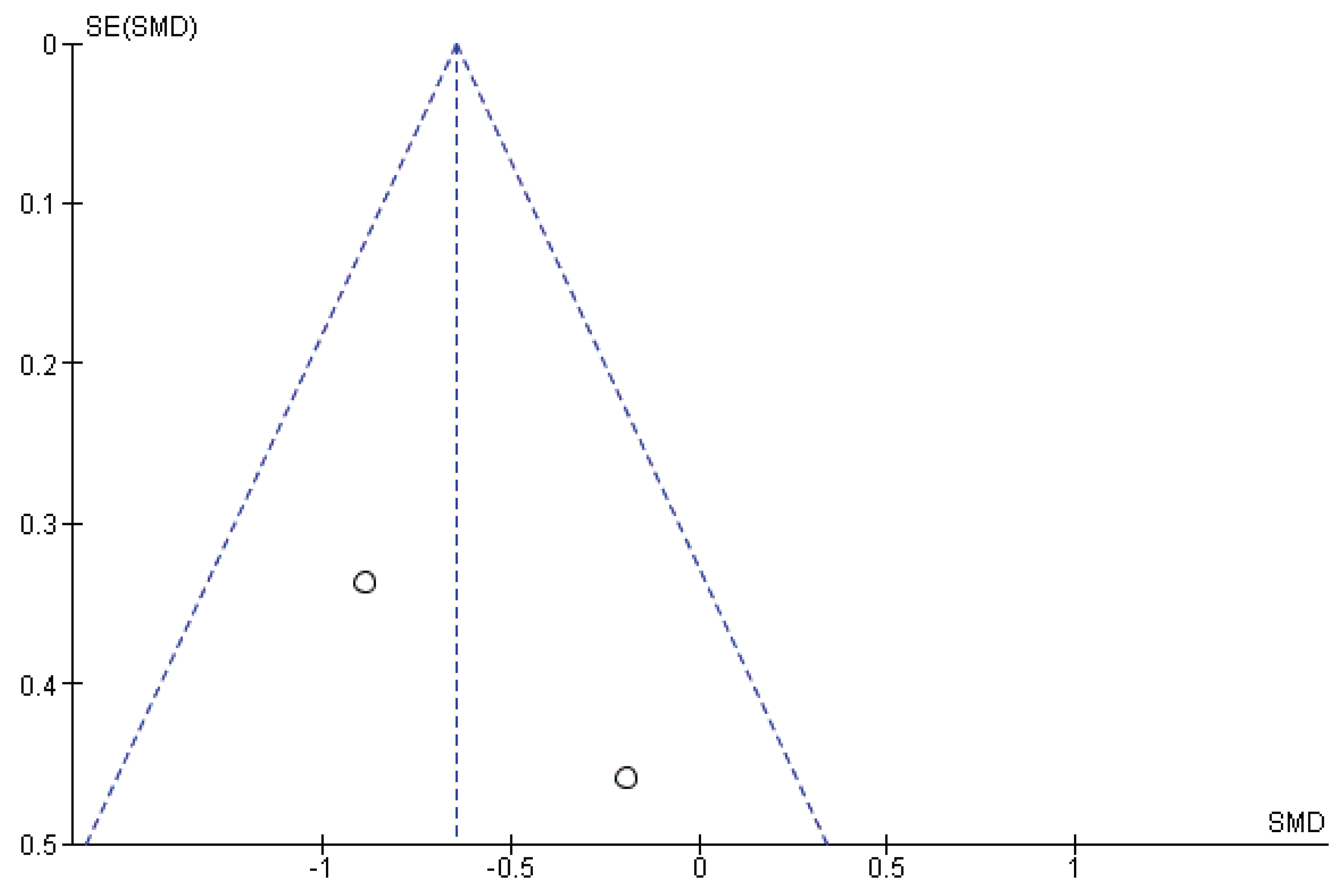
The potential for reporting biases was carefully assessed, given the constrictions of only a few articles analyzed. The visual inspection of the derived funnel plots, both of standard error by Logit event rate and of precision (1/Std Err) by Logit event rate, appear asymmetrical, and thus indicative of reporting bias (Review Manager (RevMan), Version 5.3, Copenhagen: The Nordic Cochrane Centre, The Cochrane Collaboration, 2014) (Figure A1, Figure A2 and Figure A3). Moreover, the exclusion of grey literature may introduce bias, especially given the limited evidence base.
The classical measure of heterogeneity is Cochran’s Q, which is included because it forms part of the DerSimonian-Laird random effects pooling method. Increased heterogeneity due to different included studies’ designs and outcomes, necessitates using the random effect model for the subsequent quantitative synthesis (Table A6).[71]
Quantity I2 describes the percentage of total variation across studies that is due to true heterogeneity rather than chance, thus quantifying the effect of heterogeneity and providing a measure of the degree of inconsistency in the studies’ results. High I2 values in this quantitative review indicate that most of the variability across studies is due to heterogeneity rather than chance (Table A6). Statistical analyses were performed using Review Manager (RevMan), Version 5.3, 2014 (Copenhagen: The. Nordic Cochrane Centre, The Cochrane Collaboration) and Medcalc for Windows, Version 12.7.7, 2013 (Medcalc Software, Ostend, Belgium).
7.4. Combined Data
In early-onset PE pregnancies assessed antepartum with cf-PWV (n=18), the mean and SD were 7.3 and 0.6, respectively. In early-onset PE pregnancies assessed postpartum with cf-PWV (n=23), the mean and SD were 8.2 and 1, respectively. In this population there was an insignificant change in cf-PWV (SMD: -0.61, 95%CI: -1.3 to 0.07) (Figure 4).
In early-onset PE pregnancies assessed antepartum with Alx (n=28), the mean and SD were 6.6 and 14.4, respectively. In early-onset PE pregnancies assessed postpartum with Alx (n=31), the mean and SD were 3.8 and 14.8, respectively. In this population there was an insignificant change in Alx (SMD: -0.32, 95%CI: -2.83 to 2.2) (Figure. 5).
In early-onset PE pregnancies assessed antepartum with Alx-75 (n=24), the mean and SD were 11.3 and 7.9, respectively. In early-onset PE pregnancies assessed postpartum with Alx-75 (n=38), the mean and SD were 15.9 and 4.9, respectively. In this population there was an insignificant change in Alx-75 (SMD: -0.61, 95%CI: -1.27 to 0.05) (Figure 6).
8. Discussion
The main finding of this study is that, given the low statistical power and thus level of evidence, AS measurements (cf-PWV, AIx and AIx-75) are not significantly altered in pregnancies complicated by early-onset PE.
The greatest advantage of this quantitative review is that, contrary to the existing literature, only studies with both interventional and expectant arm were included. This design increases the strength of the subsequent meta-analysis by directly comparing outcomes within the same studies, reducing confounding variables, and enhancing reliability. Therefore, the possibility of selective reporting bias is reduced but not entirely ruled out. The combined number of the included patients allows for quantitative synthesis and analysis of the pooled results.
However, this quantitative review has several disadvantages, mainly due to increased heterogeneity among included studies. Common limitations were: (1) it is unclear whether AS was already increased before the index pregnancy, thus contributing to the development of PE; (2) different time intervals since the index pregnancy were used in cf-PWV, AIx and AIx-75 measurements. Postpartum assessment times vary widely (from weeks to years), hampering biological and statistical comparisons; (3) the time of the day and study conditions (e.g. abstaining from caffeine) were not standardized or not reported. Key confounders (i.e. blood pressure, BMI, and medication use) are not controlled, making AS comparisons unreliable; and (4) no subgroup analysis based on maternal age was performed. Moreover, the duration of follow-up period since index pregnancy was reported in different and inconsistent manners, thus precluded a preplanned subgroup analysis to address the question whether a longer or shorter follow-up period affected heterogeneity of pooled results.
The aforementioned accurately reflect the weaknesses within this research design and methodology: (1) only 3 studies with small number of participants were included, (2) all 3 studies were potentially suffering from biases, (3) studies published after 2014 may have used a different definition of PE [1,2], and (4) non-English publications were missed. In particular, the potential for publication and reporting biases, especially given the reliance on retrospective studies, should also be identified as a serious limitation hampering certainty assessment. The included studies exhibit substantial heterogeneity in measurement techniques, timing, and populations, thus rendering data pooling questionable.
AS after a pregnancy complicated by PE has been a controversial issue.[72] Early-onset PE is often more severe than late-onset PE and studies have homogenously indicated that more severe PE is associated with greater AS. Khalil et al.[39] reported in a longitudinal study that AS measurements are different amongst preterm and term PE pregnancies.[39,73] In the first trimester, Alx-75 was the only measurement that was significantly different between pregnancies subsequently complicated by PE compared with normotensive pregnancies.[74] In general, AIx is reduced in the 1st and 2nd trimester but then increased in the 3rd trimester, reaching its peak in the postpartum period. Alx as a direct measure of wave reflection is affected by peripheral vascular resistance, heart rate, stroke volume and AS. Therefore, to limit the impact of heart rate, Alx-75 may be a more useful measurement. cf-PWV is affected by age and blood pressure (BP), thus increased cf-PWV may be a direct result of increased BP in PE pregnancies.[42,50,51,52,53] Grand’ Maison et al. suggest that younger women, who have less traditional CVD factors, demonstrate increased cf-PWV, cIMT and AIx closer to the index pregnancy.[32] Furthermore, McEniery et al. advocate that AIx may be a more sensitive marker of AS and CVD risk in younger individuals (age<50 years) but cf-PWV is likely to be a better measure in older individuals (age>50 years).[76]
Read as a whole, the literature converges on several points even as it diverges on others. First, there is reasonably consistent evidence that cf-PWV is elevated after early-onset PE specifically.[31,42,47] Second, AIx and AIx-75 appear to be more sensitive to the duration of postpartum follow-up (several studies assessing women within the first postpartum year found no significant elevation, while studies with follow-up extending beyond one year, and in some cases out to five or ten years, more consistently found AIx to be elevated).[8,29,31] Third, late-onset PE and milder forms of HDP (gestational hypertension without proteinuria or evidence of organ dysfunction) appear to carry a less pronounced vascular signature than early-onset PE, consistent with a dose-response relationship between disease severity and residual vascular abnormality.[31,32] A plausible unifying explanation is that the immediate postpartum period represents a transitional physiological state in which haemodynamic parameters are still normalizing from the profound cardiovascular adaptations of pregnancy, and that AS measurements obtained very early after delivery may partly reflect this normalization process rather than a stable, underlying vascular phenotype. [31,32] Studies that measure AS closer to a year or more after delivery, once haemodynamics have more fully stabilized, may therefore be better positioned to detect a persistent, pathological elevation in AS that is more likely to be clinically meaningful for long-term CVD risk.[31,32]
In the present study, 3 AS measurements were assessed: cf-PWV, Alx and Alx-75. Regarding ante- vs postpartum cf-PWV in PE pregnancy, a significant difference was not confirmed by our results either with fixed or random effects model (fixed effect model SMD: -0.642, 95% CI: -1.30 to 0.02, P=0.057 vs random effects model SMD: -0.646, 95% CI: -1.37 to 0.07, P=0.079). Regarding ante- vs postpartum Alx and Alx-75 in PE pregnancy, fixed and random effects model disagree about the significance (fixed effects model Alx SMD: -0.612, 95%CI: -1.18 to -0.04, P=0.036 vs random effects model Alx SMD: -0.249, 95%CI: -2.72 to 2.23, P=0.844 and fixed effects model Alx-75 SMD: -0.667, 95%CI: -1.19 to -0.13, P=0.015 vs random effects model Alx-75 SMD: -0.622, 95%CI: -1.29 to 0.05, P=0.068). However, the number of included studies (n=3) and total sample size (35 early-onset PE cases) are statistically insufficient to support robust conclusions. In view of the modest numbers and the wide 95%CI, these results should be interpreted with caution.
The clinical case for incorporating AS measurements into routine postpartum care after PE or other HDP is suggestive rather than definitive. On one hand, the consistency with which cf-PWV in particular is found to be elevated after early-onset PE and more severe HDP lends biological plausibility to the idea that postpartum AS assessment could help identify women at elevated future CVD risk earlier than would otherwise be recognized through blood pressure monitoring alone. On the other hand, no study to date has established that AS measurements change clinical outcomes when incorporated into postpartum surveillance, nor have validated, age- and time-since-delivery-specific reference ranges been established for postpartum populations.
Several priorities emerge from this synthesis. Longitudinal cohorts that track the same women from before or during the index pregnancy through multiple postpartum timepoints (rather than relying on cross-sectional comparisons at a single, often arbitrarily chosen, postpartum interval) would clarify whether AS genuinely evolves over time after HDP, and if so, over what trajectory. Larger, adequately powered studies restricted specifically to early-onset PE, ideally multi-centre and multi-ethnic, are needed given how sparse the directly comparable evidence remains for this clinically important subgroup. Standardization of measurement technique, timing relative to caffeine and food intake, and reporting of antihypertensive medication use at the time of testing would reduce a substantial and avoidable source of heterogeneity. Studies should also, where feasible, capture pre-pregnancy or early-pregnancy baseline AS to help disentangle whether elevated postpartum AS represents a persistent consequence of PE or reflects a pre-existing vascular predisposition that also contributed to the development of PE in the first place. Finally, because AIx and cf-PWV appear to behave somewhat differently across HDP subtype and follow-up duration, future work would benefit from pre-specified subgroup analyses by onset timing (early versus late), severity, and time since delivery, along with outcome-linked follow-up.
Our findings suggest that, especially in younger women, when the top priority is prevention of CVD through lifestyle modifications, Alx and Alx-75 seem to be a reasonable choice, although not yet validated, in distinguishing women at risk of CVD after early-onset PE. However, taking into consideration that young women who had early-onset PE are unlikely to develop CVD especially only less than a year postpartum, further longitudinal studies are required to accurately determine the optimal postpartum period for data collection. The ability to predict an at-risk of CVD population at the earliest opportunity is important. In the absence of high-quality RCTs, quantitative reviews of lower-level data provide the best available evidence. Nevertheless, robust conclusions mandate the inclusion of appropriately designed and adequately powered studies. Future research should aim for standardized methodology and reporting to enable both subgroup analysis as well as certainty assessment in evidence and recommendations. Until larger, prospective, outcome-linked studies are available, AS measurements are perhaps best regarded as a promising research tool for stratifying CVD risk, rather than a validated clinical test ready for routine postpartum use.[5,56]
In conclusion, PE leaves a vascular signature that appears to extend well beyond the puerperium. cf-PWV is the more consistently elevated AS index after early-onset PE, while Alx appears to become a more discriminating marker with longer postpartum follow-up, particularly beyond the first year after delivery. AS may remain elevated for years after a hypertensive pregnancy, even as the magnitude, consistency, and clinical significance of that elevation continue to vary across studies. Substantial heterogeneity in measurement technique, timing, and study design, together with a persistently modest evidence base, mean that AS measurements are not yet ready for routine postpartum clinical use.
Qualitative synthesis suggests that cf-PWV, AIx and AIx-75 are not significantly altered, given the low statistical power and thus level of evidence, in pregnancies complicated by early-onset PE and are therefore not appropriate for preliminary CVD risk stratification after early-onset PE. Although hampered by low numbers, indications exist that Alx and Alx-75 could have clinical significance and should be further evaluated.
Author Contributions
Conceptualization, C.A.; methodology, C.A. and E.I.; software, C.A.; validation, T.T. and G.G.; formal analysis, C.A.; investigation, C.A., E.I. and T.T.; resources, C.A., T.T. and G.G.; data curation, C.A. and E.I.; writing—original draft preparation, C.A.; writing—review and editing, T.T. and G.G.; visualization, C.A. and E.I.; supervision, T.T. and G.G.; project administration, C.A. and E.I.; funding acquisition, C.A., T.T. and G.G. All authors have read and agreed to the published version of the manuscript.
Funding
This research received no external funding.
Institutional Review Board Statement
Not applicable.
Informed Consent Statement
Not applicable.
Data Availability Statement
The original contributions presented in this study are included in the article/supplementary material. Further inquiries can be directed to the corresponding author.
Acknowledgments
None.
Conflicts of Interest
The authors declare no conflicts of interest.
Abbreviations
The following abbreviations are used in this manuscript:
| AS | Arterial Stiffness |
| PE | PreEclampsia |
| cf-PWV | pulse wave velocity |
| Alx | augmentation index |
| Alx-75 | Alx corrected for a heart rate of 75 beats per minute |
| HDP | Hypertensive Disoredrs of Preganancy |
| EVA | Early Vascular Aging |
| SMS | Standardized Mean Difference |
| CI | Confidence Intervals |
| PROSPERO | Prospective Register of Systematic Reviews System |
| CVD | cardiovascular disease |
| cIMT | carotid Intima-Media Thickness |
| FMD | flow-mediated dilation |
| MOOSE | Meta-Analysis of Observational Studies in Epidemiology |
| ISSHP | International Society for the Study of Hypertension in Pregnancy (ISSHP |
| ACOG | American College of Obstetricians and Gynecologists (ACOG) |
| PRISMA | Preferred Reporting Items for Systematic Reviews and Meta-analyses |
| MEDLINE | Medical Literature Analysis and Retrieval System Online |
| EMBASE | Excerpta Medica dataBASE |
| CDSR | Cochrane Database of Systematic Reviews (CDSR) |
| DARE | Database of Abstracts of Reviews of Effectiveness |
| CENTRAL | Cochrane Central Register of Controlled Trials |
| GA | Gestational Age |
| BMI | Body Mass Index |
| SBP | Systolic Blood Pressure |
| DBP | Diastolic Blood Pressure |
| CS | Caesarian Section |
| NOS | Newcastle-Ottawa Scale |
| NIH | National Institute of Health |
Appendix A

Table A1.
Excluded from abstract studies list and reason(s) for exclusion.
| Author(s) | Year | DOI | Reason for exclusion |
|---|---|---|---|
|
2018 | 10.1016/j.preghy.2018.04.017 |
Excluded from abstract No data during pregnancy |
|
2016 | 10.1016/j.jash.2016.01.012 |
Excluded from abstract No data during pregnancy |
|
2016 | 10.1016/j.preghy.2016.07.001 |
Excluded from abstract No data during pregnancy |
|
2017 | 10.1111/aogs.13173 |
Excluded from abstract No data during pregnancy |
|
2016 | 10.1016/j.preghy.2016.04.001 |
Excluded from abstract No data during pregnancy |
|
2019 | 10.1016/j.preghy.2019.09.001 |
Excluded from abstract No data during pregnancy |
|
2014 | 10.17772/gp/1880 |
Excluded from abstract No data during pregnancy |
|
2005 | 10.1097/00004872-200501000-00025 |
Excluded from abstract No data during pregnancy |
|
2013 | 10.1016/j.preghy.2013.01.006 |
Excluded from abstract No postpartum data |
|
2019 | 10.1016/j.jacc.2019.09.052 |
Excluded from abstract No data during pregnancy |
|
2015 | 10.1016/j.ajog.2015.05.045 |
Excluded from abstract No data during pregnancy |
|
2020 | 10.1186/s12884-020-03374-0. | Excluded from abstract Only data on cardio-ankle vascular index |
|
2012 | 10.1016/j.preghy.2012.04.143 |
Excluded from abstract No data during pregnancy |
|
2006 | 10.1097/01.hjh.0000217859.27864.19 |
Excluded from abstract No data during pregnancy |
|
2013 | 10.3109/14767058.2012.733774 |
Excluded from abstract No data during pregnancy |
|
2015 | 10.1016/j.ajog.2015.01.005 |
Excluded from abstract No data during pregnancy |
|
2019 | 10.1080/10641963.2018.1441858 |
Excluded from abstract No data during pregnancy |
|
2018 | 10.1002/uog.18825 |
Excluded from abstract No data during pregnancy |
|
2017 | 10.1002/uog.17394 |
Excluded from abstract No data during pregnancy |
|
2016 | 10.1002/uog.14838 |
Excluded from abstract No data during pregnancy |
|
2016 | 10.1002/uog.14838 |
Excluded from abstract No data during pregnancy |
|
2013 | 10.1038/hr.2013.81 |
Excluded from abstract No data during pregnancy |
|
2014 | 10.1016/j.repc.2013.11.006 |
Excluded from abstract No data during pregnancy |
|
2019 | 10.1097/HJH.0000000000001888 |
Excluded from abstract No data during pregnancy |
|
2020 | 10.1152/ajpheart.00578.2019 |
Excluded from abstract No data during pregnancy |
|
2016 | 10.1152/ajpheart.00010.2016 |
Excluded from abstract No data during pregnancy |
|
2019 | 10.1159/000500358 |
Excluded from abstract No data during pregnancy |
|
2010 | 10.1161/CIRCULATIONAHA.110.948455 |
Excluded from abstract No data during pregnancy |
Abbreviations: PE, preeclampsia.
Table A2.
Excluded studies list and reason(s) for exclusion.
| Author(s) | Year | DOI | Reason for exclusion |
|---|---|---|---|
|
2017 | 10.1016/j.ejogrb.2017.07.030 | No data on cfPWV and/or Alx and/or Alx-75 |
|
2015 | 10.1016/j.preghy.2015.09.003. | No data on cfPWV and/or Alx and/or Alx-75 |
|
2013 | 10.1093/ajh/hps067 | No separate data on early-onset PE |
|
2013 | 10.1016/j.preghy.2013.01.006 | No separate data on early-onset PE |
|
2017 | 10.1016/j.preghy.2017.08.001 | No separate data on early-onset PE |
|
2014 | 10.1111/cpf.12063 | No separate data on early-onset PE |
|
2013 | 10.1016/j.ejogrb.2013.04.010 | No data on cfPWV and/or Alx and/or Alx-75 |
|
2019 | 10.1016/j.preghy.2019.05.021 | No data on cfPWV and/or Alx and/or Alx-75 |
|
2013 | 10.1038/hr.2013.24 | No data on cfPWV and/or Alx and/or Alx-75 |
|
2019 | 10.1016/j.preghy.2019.02.001 | No separate data on early-onset PE |
|
2012 | 10.1161/HYPERTENSIONAHA.112.194159 | No data on cfPWV and/or Alx and/or Alx-75 |
|
2005 | 10.1081/PRG-200059871 | No separate data on early-onset PE |
|
2014 | 10.1186/1471-2393-14-113 | No data on cfPWV and/or Alx and/or Alx-75 |
|
2013 | 10.1002/uog.12409 | No separate data on early-onset PE |
|
2014 | 10.3109/10641955.2013.821481 | No data on cfPWV and/or Alx and/or Alx-75 |
Abbreviations: Alx, augmentation index; Alx-75, Alx corrected for a heart rate of 75 beats per minute; cf-PWV, carotid-femoral pulse wave velocity; PE, preeclampsia;.
Table A3.
Quality assessment of the included studies.
| Author(s) | Year | NOS for Cohort Studies | NOS for Case-Control Studies | NIH Quality Assessment Tool for Observational Cohort and Cross-Sectional Studies | NIH Quality Assessment Tool for Case-Control Studies |
|---|---|---|---|---|---|
| Carty et al. | 2013 | 7/9 | - | 7/14, Faira (Y7N4O3) | - |
| Franz et al. | 2013 | - | 6/9 | - | 6/12, Faira (Y6N4O2) |
| Robb et al. | 2009 | - | 6/9 | - | 6/12, Faira (Y6N4O2) |
Abbreviations: NIH Quality Assessment Tools, National Institutes of Health Assessment Tools; NOS, Newcastle Ottawa Quality Assessment Scale; Y, yes; N, no; O, other (i.e. cannot determine, not applicable, not reported)aPoor (4≥Y≥0), Fair (9≥Y≥5), Good (14≥Y≥10).
Table A4.
Design characteristics of the included studies.
| Author(s) | Year | Study Design | Measurement (s) | Timing of Antepartum Case Assessment (PE pregnancy) | Timing of Postpartum Case Assessment (PE pregnancy) |
|---|---|---|---|---|---|
| Carty et al. | 2013 | longitudinal observational/prospective cohort | Alx, Alx-75 | before PE development | 6-9 months |
| Franz et al. | 2013 | longitudinal observational/prospective case-control | cf-PWV, Alx | after PE development | 3-6 months |
| Robb eta al. | 2009 | longitudinal observational/prospective case-control | cf-PWV, Alx-75 | after PE development | 7 weeks |
Abbreviations: Alx, augmentation index; Alx-75, Alx corrected for a heart rate of 75 beats per minute; cf-PWV, carotid-femoral pulse wave velocity; PE, preeclampsia.
Table A5.
Baseline characteristics of study population.
| Author(s) |
Year | Number (Case vs Control) | MA (years) | BMI (kg/m2) | Smoking, n (%) | Ethnicity, (% non-Caucasian) | SBP (mmHg) | DBP (mmHg) | Nulliparous (%) | Parity (median)a | Pre-existing hypertension | PE history (%) | GA at diagnosis (weeks) | GA at delivery (weeks) | Caesarian section, n | Birthweight, g |
|---|---|---|---|---|---|---|---|---|---|---|---|---|---|---|---|---|
| Antepartum (Case group vs Controls) | ||||||||||||||||
| Carty et al. | 2013 | 17 vs 123 | 30.5±5.6 vs 32.7±5.3 (P=0.11) | 30.7±6.3 vs 27.6±5.4 (P=0.03) | 1/17 (5.8%) vs 8/123 (6.5%) (P=1.0) | 1/17 (5.8%) vs 7/123 (5.6%) (P=1.0) | 125±11 vs 120±12.7 (P=0.089)(at booking) | 79±7 vs 73±9 (P=0.005) (at booking) | 16/17 (94%) vs 67/123 (54%) (P=0.001) | - | - | 6/17 (35%) vs 19/123 (15%) (P=0.08) | - | 38.9±2 vs 40±1.7 (P=0.016) | 4/17 (24%) vs 25/123 (20%) (P=0.75) | 3166±499 vs 3611±523 (P=0.001) |
| Franz et al. | 2013 | 11 vs 10 | 32±5 vs 32 ± 4 (P=1) | 29.3 ±6 vs 24.2±1.8 (P=0.0184) | - | - | 170±18 vs 106±9 (P<0.001)(at diagnosis) | 97±13 vs 64±7 (P<0.001)(at diagnosis) | - | 0 (0–1) vs 0 (0–1) (P=n.s.) | 6/11 (55%) vs 0/10 (0%) (P=0.012) | - | 28±3 vs 28±2 (P=1) | 30±4 vs 40±1 (P<0.001) | - | 1326±555 vs 3538±268 (P<0.001) |
| Robb et al. | 2009 | 7 vs 22 | 30±4 vs 30±1 (P=1) | 25±2 vs 27±1 (P=0.0014) | 140±3 vs 113±1 (P<0.0001)(at diagnosis) | 87±3 vs 70±1 (P<0.0001)(at diagnosis) | 30±1.4 vs 41±0.3 (P<0.0001) | 1371±222 vs 3497±112 (P<0.0001) | ||||||||
| Total | 35 vs 155 | 30.8±4.9 vs 31.6±3.4 (P=0.2515) | 28.3±4.8 vs 26.3±2.7 (P=0.0010) | 145±11 vs 113±8 (P<0.0001) | 88±8 vs 69±6 (P<0.0001) | 33±2.5 vs 40.3±1 (P<0.0001) | 1954±425 vs 3549±301 (P<0.0001) | |||||||||
| Postpartum (Case group vs Controls) | ||||||||||||||||
| Carty et al. | 2013 | 22 vs 69 | 31.5±4.7 vs 35.6±4.9 (P<0.001) | 28.6±8 vs 27.5±5.5 (P=0.346) | 1/23 (4.3%) vs 6/69 (8.7%) (P=0.67) | 1/23 (4.3%) vs 4/69 (5.8%) (P=1.0) | 126±13 vs 124±13 (P=0.385) | 83±10 vs 78±11 (P=0.074) | - | - | - | 6/23 (23%) vs 9/69 (13%) (P=0.19) | - | 38.5±1.6 vs 40.2±1.2 (P<0.001) | 8/23 (35%) vs 11/69 (16%) (P=0.07) | 3080±478 vs 3710±493 (P<0.001) |
| Franz et al. | 2013 | 8 vs 8 | 35±7 vs 34±4 (P=0.7309) | 28.8±4.9 vs 23.2±4 (P=0.028) | - | - | 135±19 vs 114±12 (P=0.038) | 82±13 66±9 (P=0.005) | - | - | - | - | - | - | - | - |
| Robb et al. | 2009 | 7 vs 22 | 30±4 vs 30±1 (P=1) | 25±2 vs 27±1 (P=0.0014) | 123±3 vs 113±1 (P=0.002) | 77±3 vs 69±1 (P=0.004) | 30±1.4 vs 41±0.3 (P< 0.0001) | 1371±222 vs 3497±112 (P< 0.0001) | ||||||||
| Total | 37 vs 99 | 32.2±5.2 vs 33.2±3.3 (P=0.1858) | 27.5±5 vs 25.9±3.5 (P=0.0378) | 128±12 vs 117±9 (P<0.0001) | 81±9 vs 71±7 (P<0.0001) | 34.3±1.5 vs 40.6±0.8 (P<0.0001) | 2226±350 vs 3604±303 (P<0.0001) | |||||||||
Note - Values represent means ± standard deviation (SD) or n (%) Abbreviations: BMI, body mass index; DBP, diastolic blood pressure; SBP, systolic blood pressure; n.s., not significant aValues represent medians and range.
Table A6.
Meta-analysis statistical results for cf-PWV, Alx and Alx-75 (based on Comprehensive Meta-Analysis software).
Table A6.
Meta-analysis statistical results for cf-PWV, Alx and Alx-75 (based on Comprehensive Meta-Analysis software).
| Meta-analysis (Std diff in means) |
Std diff in means (random effects) | 95% CI | Q | DF | P | I2 (%) |
|---|---|---|---|---|---|---|
| cf-PWV | -0.646 | -1.365-0.074 | 1.187 | 1 | 0.276 | 15.778 |
| Alx | -0.249 | -2.724-2.226 | 17.050 | 1 | 0.000 | 94.135 |
| Alx-75 | -0.622 | -1.290-0.047 | 1.503 | 1 | 0.220 | 33.462 |
Abbreviations: Alx, augmentation index; Alx-75, Alx corrected for a heart rate of 75 beats per minute; cf-PWV, carotid-femoral pulse wave analysis; CI, confidence interval; DF, degrees of freedom; I, inconsistency; P, possibility; Q, Cochran’s Q; Std diff in means, standardized difference in means.
Figure A1.
Funnel plot of standard error by standardized mean difference for studies reporting on antepartum and postpartum cf-PWV indicating publication bias. Abbreviations: cf-PWV, carotid-femoral pulse wave analysis; SE, standard error; SMD, standardized mean difference.
Figure A1.
Funnel plot of standard error by standardized mean difference for studies reporting on antepartum and postpartum cf-PWV indicating publication bias. Abbreviations: cf-PWV, carotid-femoral pulse wave analysis; SE, standard error; SMD, standardized mean difference.
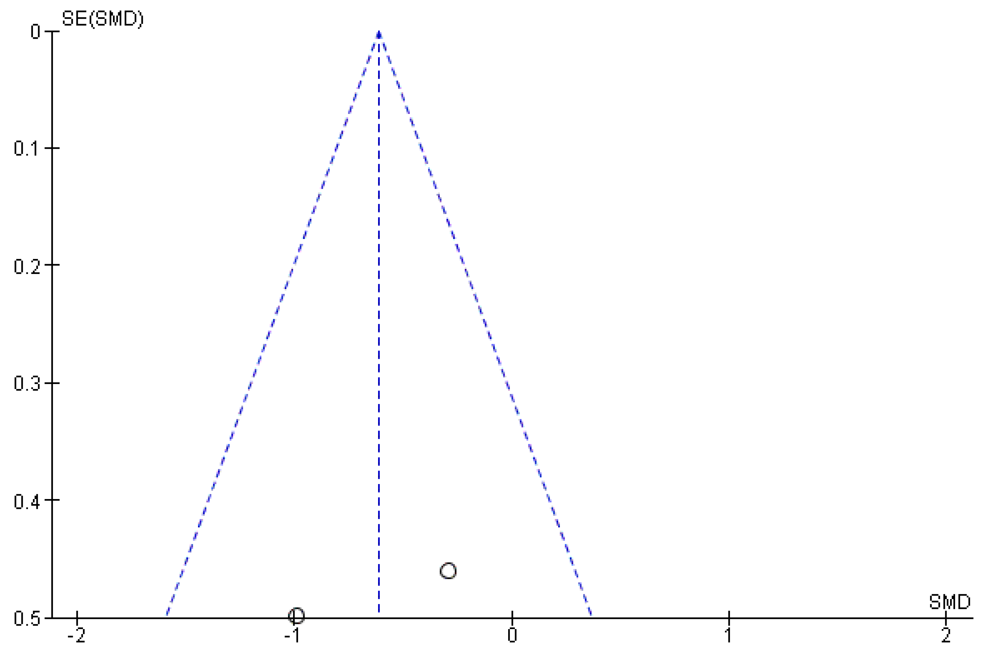
Figure A2.
Funnel plot of standard error by standardized mean difference for studies reporting on antepartum and postpartum Alx indicating publication bias. Abbreviations: Alx, augmentation index; SE, standard error; SMD, standardized mean difference.
Figure A2.
Funnel plot of standard error by standardized mean difference for studies reporting on antepartum and postpartum Alx indicating publication bias. Abbreviations: Alx, augmentation index; SE, standard error; SMD, standardized mean difference.
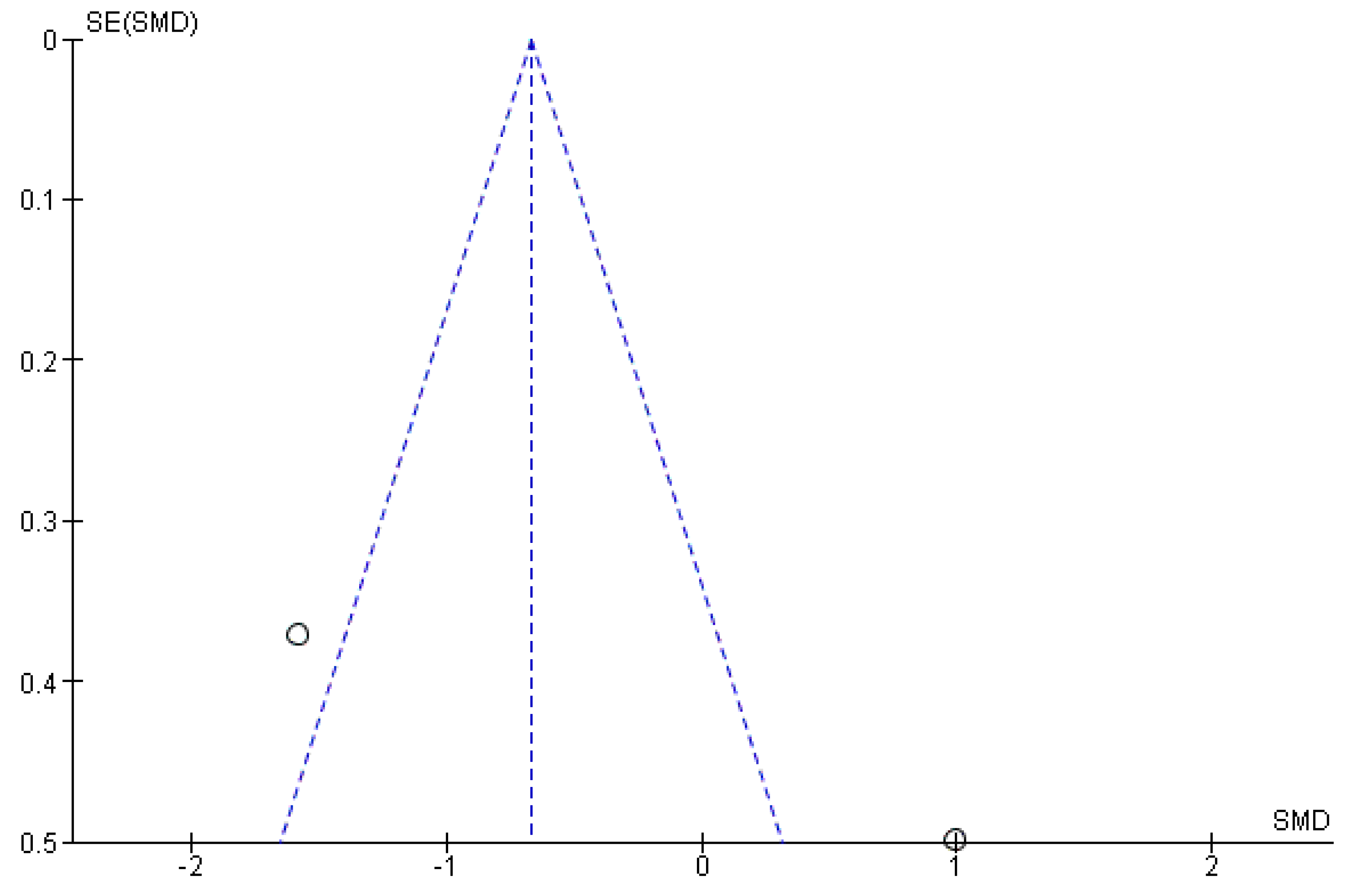
Figure A3.
Funnel plot of standard error by standardized mean difference for studies reporting on antepartum and postpartum Alx-75 indicating publication bias. Abbreviations: Alx, augmentation index; Alx-75, Alx corrected for a heart rate of 75 beats per minute; SE, standard error; SMD, standardized mean difference.
Figure A3.
Funnel plot of standard error by standardized mean difference for studies reporting on antepartum and postpartum Alx-75 indicating publication bias. Abbreviations: Alx, augmentation index; Alx-75, Alx corrected for a heart rate of 75 beats per minute; SE, standard error; SMD, standardized mean difference.
References
- Magee, L.A.; Pels, A.; Helewa, M.; Rey, E.; von Dadelszen, P. SOGC Hypertension Guideline Committee Diagnosis, Evaluation, and Management of the Hypertensive Disorders of Pregnancy: Executive Summary. J. Obstet. Gynaecol. Can. 2014, 36, 575–576. [Google Scholar] [CrossRef]
- American College of Obstetricians and Gynecologists. Task Force on Hypertension in Pregnancy Hypertension in Pregnancy. Report of the American College of Obstetricians and Gynecologists’ Task Force on Hypertension in Pregnancy. Obstet. Gynecol. 2013, 122, 1122–1131. [Google Scholar] [CrossRef]
- Pauli, J.M.; Repke, J.T. Pitfalls With the New American College of Obstetricians and Gynecologists Task Force on Hypertension in Pregnancy. Clin. Obstet. Gynecol. 2017, 60, 141–152. [Google Scholar] [CrossRef]
- Stanhewicz, A.E. Residual Vascular Dysfunction in Women with a History of Preeclampsia. Am. J. Physiol. Regul. Integr. Comp. Physiol. 2018, 315, R1062–R1071. [Google Scholar] [CrossRef]
- Palmiero, P.; Caretto, P.; Ciccone, M.M.; Maiello, M. On Behalf Of The I C I S C U Italian Chapter Of International Society Cardiovascular Ultrasound, null Long-Term Cardiovascular Risk and Maternal History of Pre-Eclampsia. J. Clin. Med. 2025, 14, 3121. [Google Scholar] [CrossRef]
- Bellamy, L.; Casas, J.-P.; Hingorani, A.D.; Williams, D.J. Pre-Eclampsia and Risk of Cardiovascular Disease and Cancer in Later Life: Systematic Review and Meta-Analysis. BMJ 2007, 335, 974. [Google Scholar] [CrossRef]
- Brown, M.C.; Best, K.E.; Pearce, M.S.; Waugh, J.; Robson, S.C.; Bell, R. Cardiovascular Disease Risk in Women with Pre-Eclampsia: Systematic Review and Meta-Analysis. Eur. J. Epidemiol. 2013, 28, 1–19. [Google Scholar] [CrossRef]
- Werlang, A.; Paquin, A.; Coutinho, T. The EVA Study: Early Vascular Aging in Women With History of Preeclampsia. J. Am. Heart Assoc. 2023, 12, e028116. [Google Scholar] [CrossRef]
- Ray, J.G.; Vermeulen, M.J.; Schull, M.J.; Redelmeier, D.A. Cardiovascular Health after Maternal Placental Syndromes (CHAMPS): Population-Based Retrospective Cohort Study. Lancet 2005, 366, 1797–1803. [Google Scholar] [CrossRef]
- Lisonkova, S.; Joseph, K.S. Incidence of Preeclampsia: Risk Factors and Outcomes Associated with Early- versus Late-Onset Disease. Am. J. Obstet. Gynecol. 2013, 209, 544.e1–544.e12. [Google Scholar] [CrossRef]
- Lain, K.Y.; Roberts, J.M. Contemporary Concepts of the Pathogenesis and Management of Preeclampsia. JAMA 2002, 287, 3183–3186. [Google Scholar] [CrossRef]
- van Esch, J.J.A.; van Heijst, A.F.; de Haan, A.F.J.; van der Heijden, O.W.H. Early-Onset Preeclampsia Is Associated with Perinatal Mortality and Severe Neonatal Morbidity. J. Matern. Fetal. Neonatal. Med. 2017, 30, 2789–2794. [Google Scholar] [CrossRef]
- Hausvater, A.; Giannone, T.; Sandoval, Y.-H.G.; Doonan, R.J.; Antonopoulos, C.N.; Matsoukis, I.L.; Petridou, E.T.; Daskalopoulou, S.S. The Association between Preeclampsia and Arterial Stiffness. J. Hypertens. 2012, 30, 17–33. [Google Scholar] [CrossRef]
- Laurent, S.; Boutouyrie, P.; Asmar, R.; Gautier, I.; Laloux, B.; Guize, L.; Ducimetiere, P.; Benetos, A. Aortic Stiffness Is an Independent Predictor of All-Cause and Cardiovascular Mortality in Hypertensive Patients. Hypertension 2001, 37, 1236–1241. [Google Scholar] [CrossRef]
- Boutouyrie, P.; Tropeano, A.I.; Asmar, R.; Gautier, I.; Benetos, A.; Lacolley, P.; Laurent, S. Aortic Stiffness Is an Independent Predictor of Primary Coronary Events in Hypertensive Patients: A Longitudinal Study. Hypertension 2002, 39, 10–15. [Google Scholar] [CrossRef]
- Willum-Hansen, T.; Staessen, J.A.; Torp-Pedersen, C.; Rasmussen, S.; Thijs, L.; Ibsen, H.; Jeppesen, J. Prognostic Value of Aortic Pulse Wave Velocity as Index of Arterial Stiffness in the General Population. Circulation 2006, 113, 664–670. [Google Scholar] [CrossRef]
- Mattace-Raso, F.U.S.; van der Cammen, T.J.M.; Hofman, A.; van Popele, N.M.; Bos, M.L.; Schalekamp, M.A.D.H.; Asmar, R.; Reneman, R.S.; Hoeks, A.P.G.; Breteler, M.M.B.; et al. Arterial Stiffness and Risk of Coronary Heart Disease and Stroke: The Rotterdam Study. Circulation 2006, 113, 657–663. [Google Scholar] [CrossRef]
- Waddell, T.K.; Dart, A.M.; Medley, T.L.; Cameron, J.D.; Kingwell, B.A. Carotid Pressure Is a Better Predictor of Coronary Artery Disease Severity than Brachial Pressure. Hypertension 2001, 38, 927–931. [Google Scholar] [CrossRef]
- Wilkinson, I.B.; MacCallum, H.; Flint, L.; Cockcroft, J.R.; Newby, D.E.; Webb, D.J. The Influence of Heart Rate on Augmentation Index and Central Arterial Pressure in Humans. J. Physiol. 2000, 525, 263–270. [Google Scholar] [CrossRef]
- Wilkinson, I.B.; MacCallum, H.; Flint, L.; Cockcroft, J.R.; Newby, D.E.; Webb, D.J. The Influence of Heart Rate on Augmentation Index and Central Arterial Pressure in Humans. J. Physiol. 2000, 525 Pt 1, 263–270. [Google Scholar] [CrossRef]
- Baulmann, J.; Schillings, U.; Rickert, S.; Uen, S.; Düsing, R.; Illyes, M.; Cziraki, A.; Nickering, G.; Mengden, T. A New Oscillometric Method for Assessment of Arterial Stiffness: Comparison with Tonometric and Piezo-Electronic Methods. J. Hypertens. 2008, 26, 523–528. [Google Scholar] [CrossRef]
- Williams, B. Pulse Wave Analysis and Hypertension: Evangelism versus Scepticism. J. Hypertens. 2004, 22, 447–449. [Google Scholar] [CrossRef]
- O’Rourke, M.F.; Nichols, W.W.; Safar, M.E. Pulse Waveform Analysis and Arterial Stiffness: Realism Can Replace Evangelism and Scepticism. J. Hypertens. author reply 1634. 2004, 22, 1633–1634. [Google Scholar] [CrossRef]
- Williams, B.; Lacy, P.S.; Thom, S.M.; Cruickshank, K.; Stanton, A.; Collier, D.; Hughes, A.D.; Thurston, H.; O’Rourke, M.; CAFE Investigators; et al. Differential Impact of Blood Pressure-Lowering Drugs on Central Aortic Pressure and Clinical Outcomes: Principal Results of the Conduit Artery Function Evaluation (CAFE) Study. Circulation 2006, 113, 1213–1225. [Google Scholar] [CrossRef]
- DeLoach, S.S.; Townsend, R.R. Vascular Stiffness: Its Measurement and Significance for Epidemiologic and Outcome Studies. CJASN 2008, 3, 184–192. [Google Scholar] [CrossRef]
- Papaioannou, T.G.; Argyris, A.; Protogerou, A.D.; Vrachatis, D.; Nasothimiou, E.G.; Sfikakis, P.P.; Stergiou, G.S.; Stefanadis, C.I. Non-Invasive 24hour Ambulatory Monitoring of Aortic Wave Reflection and Arterial Stiffness by a Novel Oscillometric Device: The First Feasibility and Reproducibility Study. Int. J. Cardiol. 2013, 169, 57–61. [Google Scholar] [CrossRef]
- Lortz, J.; Halfmann, L.; Burghardt, A.; Steinmetz, M.; Radecke, T.; Jánosi, R.A.; Rassaf, T.; Rammos, C. Rapid and Automated Risk Stratification by Determination of the Aortic Stiffness in Healthy Subjects and Subjects with Cardiovascular Disease. PLoS ONE 2019, 14. [Google Scholar] [CrossRef]
- Usselman, C.W.; Adler, T.E.; Coovadia, Y.; Leone, C.; Paidas, M.J.; Stachenfeld, N.S. A Recent History of Preeclampsia Is Associated with Elevated Central Pulse Wave Velocity and Muscle Sympathetic Outflow. Am. J. Physiol. Heart Circ. Physiol. 2020, 318, H581–H589. [Google Scholar] [CrossRef]
- Moe, K.; Sugulle, M.; Dechend, R.; Angel, K.; Staff, A.C. Functional and Structural Vascular Biomarkers in Women 1 Year after a Hypertensive Disorder of Pregnancy. Pregnancy Hypertens. 2020, 21, 23–29. [Google Scholar] [CrossRef]
- Osman, M.W.; Nath, M.; Breslin, E.; Khalil, A.; Webb, D.R.; Robinson, T.G.; Mousa, H.A. Association between Arterial Stiffness and Wave Reflection with Subsequent Development of Placental-Mediated Diseases during Pregnancy: Findings of a Systematic Review and Meta-Analysis. J. Hypertens. 2018, 36, 1005–1014. [Google Scholar] [CrossRef]
- Mbongozi, X.B.; Galloway, S.D.R.; Hunter, A.M.; Milambo, J.P.M.; Businge, C.B. Arterial Stiffness after 6 weeks Postdelivery in Women with a History of Hypertensive Disorders of Pregnancy: A Systematic Review and Meta-Analysis. Front Med. 2025, 12, 1665100. [Google Scholar] [CrossRef]
- Grand’Maison, S.; Pilote, L.; Okano, M.; Landry, T.; Dayan, N. Markers of Vascular Dysfunction After Hypertensive Disorders of Pregnancy: A Systematic Review and Meta-Analysis. Hypertension 2016, 68, 1447–1458. [Google Scholar] [CrossRef]
- Hamad, R.R.; Eriksson, M.J.; Silveira, A.; Hamsten, A.; Bremme, K. Decreased Flow-Mediated Dilation Is Present 1 Year after a Pre-Eclamptic Pregnancy. J. Hypertens. 2007, 25, 2301–2307. [Google Scholar] [CrossRef]
- Lampinen, K.H.; Rönnback, M.; Kaaja, R.J.; Groop, P.-H. Impaired Vascular Dilatation in Women with a History of Pre-Eclampsia. J. Hypertens. 2006, 24, 751–756. [Google Scholar] [CrossRef]
- Rönnback, M.; Lampinen, K.; Groop, P.-H.; Kaaja, R. Pulse Wave Reflection in Currently and Previously Preeclamptic Women. Hypertens. Pregnancy 2005, 24, 171–180. [Google Scholar] [CrossRef]
- Yinon, Y.; Kingdom, J.C.P.; Odutayo, A.; Moineddin, R.; Drewlo, S.; Lai, V.; Cherney, D.Z.I.; Hladunewich, M.A. Vascular Dysfunction in Women with a History of Preeclampsia and Intrauterine Growth Restriction: Insights into Future Vascular Risk. Circulation 2010, 122, 1846–1853. [Google Scholar] [CrossRef]
- Baulmann, J.; Schillings, U.; Rickert, S.; Uen, S.; Düsing, R.; Illyes, M.; Cziraki, A.; Nickering, G.; Mengden, T. A New Oscillometric Method for Assessment of Arterial Stiffness: Comparison with Tonometric and Piezo-Electronic Methods. J. Hypertens. 2008, 26, 523–528. [Google Scholar] [CrossRef]
- McEniery, C.M.; Yasmin, null; Hall, I.R.; Qasem, A.; Wilkinson, I.B.; Cockcroft, J.R. ACCT Investigators Normal Vascular Aging: Differential Effects on Wave Reflection and Aortic Pulse Wave Velocity: The Anglo-Cardiff Collaborative Trial (ACCT). J. Am. Coll. Cardiol. 2005, 46, 1753–1760. [Google Scholar] [CrossRef]
- Khalil, A.; Jauniaux, E.; Harrington, K. Antihypertensive Therapy and Central Hemodynamics in Women with Hypertensive Disorders in Pregnancy. Obstet. Gynecol. 2009, 113, 646–654. [Google Scholar] [CrossRef]
- Avni, B.; Frenkel, G.; Shahar, L.; Golik, A.; Sherman, D.; Dishy, V. Aortic Stiffness in Normal and Hypertensive Pregnancy. Blood Press 2010, 19, 11–15. [Google Scholar] [CrossRef]
- Khalil, A.; Garcia-Mandujano, R.; Maiz, N.; Elkhouli, M.; Nicolaides, K.H. Longitudinal Changes in Maternal Hemodynamics in a Population at Risk for Pre-Eclampsia. Ultrasound Obstet. Gynecol. 2014, 44, 197–204. [Google Scholar] [CrossRef]
- Franz, M.B.; Burgmann, M.; Neubauer, A.; Zeisler, H.; Sanani, R.; Gottsauner-Wolf, M.; Schiessl, B.; Andreas, M. Augmentation Index and Pulse Wave Velocity in Normotensive and Pre-Eclamptic Pregnancies. Acta Obstet. Gynecol. Scand. 2013, 92, 960–966. [Google Scholar] [CrossRef]
- Khalil, A.; Akolekar, R.; Syngelaki, A.; Elkhouli, M.; Nicolaides, K.H. Maternal Hemodynamics at 11-13 Weeks’ Gestation and Risk of Pre-Eclampsia. Ultrasound Obstet. Gynecol. 2012, 40, 28–34. [Google Scholar] [CrossRef]
- Khalil, A.; Cowans, N.J.; Spencer, K.; Goichman, S.; Meiri, H.; Harrington, K. First-Trimester Markers for the Prediction of Pre-Eclampsia in Women with a-Priori High Risk. Ultrasound Obstet. Gynecol. 2010, 35, 671–679. [Google Scholar] [CrossRef]
- Estensen, M.-E.; Remme, E.W.; Grindheim, G.; Smiseth, O.A.; Segers, P.; Henriksen, T.; Aakhus, S. Increased Arterial Stiffness in Pre-Eclamptic Pregnancy at Term and Early and Late Postpartum: A Combined Echocardiographic and Tonometric Study. Am. J. Hypertens. 2013, 26, 549–556. [Google Scholar] [CrossRef]
- Carty, D.M.; Neisius, U.; Rooney, L.K.; Dominiczak, A.F.; Delles, C. Pulse Wave Analysis for the Prediction of Preeclampsia. J. Hum. Hypertens. 2014, 28, 98–104. [Google Scholar] [CrossRef]
- Robb, A.O.; Mills, N.L.; Din, J.N.; Smith, I.B.J.; Paterson, F.; Newby, D.E.; Denison, F.C. Influence of the Menstrual Cycle, Pregnancy, and Preeclampsia on Arterial Stiffness. Hypertension 2009, 53, 952–958. [Google Scholar] [CrossRef]
- Kim, S.; Lim, H.J.; Kim, J.-R.; Oh, K.J.; Hong, J.-S.; Suh, J.-W. Longitudinal Change in Arterial Stiffness after Delivery in Women with Preeclampsia and Normotension: A Prospective Cohort Study. BMC Pregnancy Childbirth 2020, 20, 685. [Google Scholar] [CrossRef]
- Namugowa, A.; Iputo, J.; Wandabwa, J.; Meeme, A.; Buga, G. a. B.; Abura, S.; Stofile, Y.Y. Arterial Stiffness in Women Previously with Preeclampsia from a Semi-Rural Region of South Africa. Clin. Exp. Hypertens. 2019, 41, 36–43. [Google Scholar] [CrossRef]
- Kaihara, J.N.S.; Grepi Okano, H.C.; Veiga, E.C. de A.; Tallarico, G.M.; Dias-Junior, C.A.; Cavalli, R.C.; Sandrim, V.C. Differences between Macrovascular and Microvascular Functions in Pregnant Women with Chronic Hypertension or Preeclampsia: New Insights into Maternal Vascular Health. Front Physiol. 2025, 16, 1536437. [Google Scholar] [CrossRef]
- Anthoulakis, C.; Mamopoulos, A. Augmentation Index and Pulse Wave Velocity in Normotensive versus Preeclamptic Pregnancies: A Prospective Case-Control Study Using a New Oscillometric Method. Ann. Med. 2022, 54, 1–10. [Google Scholar] [CrossRef]
- Nakimuli, A.; Okello, E.; Kesiiga, A.; Adroma, M.; Akello, J.; Nabweyambo, S.; Sekikubo, M.; Namagembe, I.; Kayesubula, R.; Galiwango, R.; et al. Postpartum Cardiovascular Health in African Women Following Pre-Eclampsia: A Prospective Cohort Study. BJOG 2025, 132, 1319–1328. [Google Scholar] [CrossRef]
- Barr, L.C.; Herr, J.E.; Hétu, M.-F.; Smith, G.N.; Johri, A.M. Increased Carotid Artery Stiffness after Preeclampsia in a Cross-Sectional Study of Postpartum Women. Physiol. Rep. 2022, 10, e15276. [Google Scholar] [CrossRef]
- Davis, K.G.; Armstrong, M.K.; Nuckols, V.R.; Pewowaruk, R.; Santillan, D.A.; Santillan, M.K.; Pierce, G.L. Higher Carotid Artery Backward Pressure and Load-Dependent Stiffness among Women with a History of Preeclampsia: Association with Cognitive Functions. J. Appl. Physiol. (1985) 2026, 140, 754–763. [Google Scholar] [CrossRef]
- Palmiero, P.; Caretto, P.; Ciccone, M.M.; Maiello, M. On Behalf Of The I C I S C U Italian Chapter Of International Society Cardiovascular Ultrasound, null Long-Term Cardiovascular Risk and Maternal History of Pre-Eclampsia. J. Clin. Med. 2025, 14, 3121. [Google Scholar] [CrossRef]
- Parikh, N.I.; Gonzalez, J.M.; Anderson, C.A.M.; Judd, S.E.; Rexrode, K.M.; Hlatky, M.A.; Gunderson, E.P.; Stuart, J.J.; Vaidya, D. American Heart Association Council on Epidemiology and Prevention; Council on Arteriosclerosis, Thrombosis and Vascular Biology; Council on Cardiovascular and Stroke Nursing; and the Stroke Council Adverse Pregnancy Outcomes and Cardiovascular Disease Risk: Unique Opportunities for Cardiovascular Disease Prevention in Women: A Scientific Statement From the American Heart Association. Circulation 2021, 143, e902–e916. [Google Scholar] [CrossRef]
- Liberati, A.; Altman, D.G.; Tetzlaff, J.; Mulrow, C.; Gøtzsche, P.C.; Ioannidis, J.P.A.; Clarke, M.; Devereaux, P.J.; Kleijnen, J.; Moher, D. The PRISMA Statement for Reporting Systematic Reviews and Meta-Analyses of Studies That Evaluate Health Care Interventions: Explanation and Elaboration. J. Clin. Epidemiol. 2009, 62, e1-34. [Google Scholar] [CrossRef]
- Stroup, D.F.; Berlin, J.A.; Morton, S.C.; Olkin, I.; Williamson, G.D.; Rennie, D.; Moher, D.; Becker, B.J.; Sipe, T.A.; Thacker, S.B. Meta-Analysis of Observational Studies in Epidemiology: A Proposal for Reporting. Meta-Analysis Of Observational Studies in Epidemiology (MOOSE) Group. JAMA 2000, 283, 2008–2012. [Google Scholar] [CrossRef]
- Brown, M.A.; Lindheimer, M.D.; de Swiet, M.; Van Assche, A.; Moutquin, J.M. The Classification and Diagnosis of the Hypertensive Disorders of Pregnancy: Statement from the International Society for the Study of Hypertension in Pregnancy (ISSHP). Hypertens. Pregnancy 2001, 20, IX–XIV. [Google Scholar] [CrossRef]
- Brown, M.A.; Magee, L.A.; Kenny, L.C.; Karumanchi, S.A.; McCarthy, F.P.; Saito, S.; Hall, D.R.; Warren, C.E.; Adoyi, G.; Ishaku, S.; et al. The Hypertensive Disorders of Pregnancy: ISSHP Classification, Diagnosis & Management Recommendations for International Practice. Pregnancy Hypertens. 2018, 13, 291–310. [Google Scholar] [CrossRef]
- ACOG Practice Bulletin No. 202: Gestational Hypertension and Preeclampsia. Obstet. Gynecol. 2019, 133, e1–e25. [CrossRef]
- Stang, A. Critical Evaluation of the Newcastle-Ottawa Scale for the Assessment of the Quality of Nonrandomized Studies in Meta-Analyses. Eur. J. Epidemiol. 2010, 25, 603–605. [Google Scholar] [CrossRef] [PubMed]
- Study Quality Assessment Tools | National Heart, Lung, and Blood Institute (NHLBI). Available online: https://www.nhlbi.nih.gov/health-topics/study-quality-assessment-tools (accessed on 26 April 2020).
- DerSimonian, R.; Laird, N. Meta-Analysis in Clinical Trials. Control Clin. Trials 1986, 7, 177–188. [Google Scholar] [CrossRef]
- Higgins, J.P.T.; Thompson, S.G.; Deeks, J.J.; Altman, D.G. Measuring Inconsistency in Meta-Analyses. BMJ 2003, 327, 557–560. [Google Scholar] [CrossRef]
- Liberati, A.; Altman, D.G.; Tetzlaff, J.; Mulrow, C.; Gøtzsche, P.C.; Ioannidis, J.P.A.; Clarke, M.; Devereaux, P.J.; Kleijnen, J.; Moher, D. The PRISMA Statement for Reporting Systematic Reviews and Meta-Analyses of Studies That Evaluate Health Care Interventions: Explanation and Elaboration. PLoS Med. 2009, 6, e1000100. [Google Scholar] [CrossRef]
- Page, M.J.; McKenzie, J.E.; Bossuyt, P.M.; Boutron, I.; Hoffmann, T.C.; Mulrow, C.D.; Shamseer, L.; Tetzlaff, J.M.; Akl, E.A.; Brennan, S.E.; et al. The PRISMA 2020 Statement: An Updated Guideline for Reporting Systematic Reviews. BMJ 2021, 372, n71. [Google Scholar] [CrossRef]
- Gavaghan, D.J.; Moore, R.A.; McQuay, H.J. An Evaluation of Homogeneity Tests in Meta-Analyses in Pain Using Simulations of Individual Patient Data. Pain 2000, 85, 415–424. [Google Scholar] [CrossRef]
- Higgins, J.P.T.; Thompson, S.G.; Deeks, J.J.; Altman, D.G. Measuring Inconsistency in Meta-Analyses. BMJ 2003, 327, 557–560. [Google Scholar] [CrossRef]
- Borenstein, M.; Hedges, L.V.; Higgins, J.P.T.; Rothstein, H.R. A Basic Introduction to Fixed-Effect and Random-Effects Models for Meta-Analysis. Res. Synth. Methods 2010, 1, 97–111. [Google Scholar] [CrossRef]
- Borenstein, M.; Hedges, L.V.; Higgins, J.P.T.; Rothstein, H.R. A Basic Introduction to Fixed-Effect and Random-Effects Models for Meta-Analysis. Res. Synth. Methods 2010, 1, 97–111. [Google Scholar] [CrossRef]
- Parikh, N.I.; Gonzalez, J.M.; Anderson, C.A.M.; Judd, S.E.; Rexrode, K.M.; Hlatky, M.A.; Gunderson, E.P.; Stuart, J.J.; Vaidya, D. American Heart Association Council on Epidemiology and Prevention; Council on Arteriosclerosis, Thrombosis and Vascular Biology; Council on Cardiovascular and Stroke Nursing; and the Stroke Council Adverse Pregnancy Outcomes and Cardiovascular Disease Risk: Unique Opportunities for Cardiovascular Disease Prevention in Women: A Scientific Statement From the American Heart Association. Circulation 2021, 143, e902–e916. [Google Scholar] [CrossRef]
- Avni, B.; Frenkel, G.; Shahar, L.; Golik, A.; Sherman, D.; Dishy, V. Aortic Stiffness in Normal and Hypertensive Pregnancy. Blood Press. 2010, 19, 11–15. [Google Scholar] [CrossRef]
- Khalil, A.; Garcia-Mandujano, R.; Maiz, N.; Elkhouli, M.; Nicolaides, K.H. Longitudinal Changes in Maternal Hemodynamics in a Population at Risk for Pre-Eclampsia. Ultrasound Obstet. Gynecol. 2014, 44, 197–204. [Google Scholar] [CrossRef]
- Khalil, A.; Cowans, N.J.; Spencer, K.; Goichman, S.; Meiri, H.; Harrington, K. First-Trimester Markers for the Prediction of Pre-Eclampsia in Women with a-Priori High Risk. Ultrasound Obstet. Gynecol. 2010, 35, 671–679. [Google Scholar] [CrossRef]
- McEniery, C.M.; Yasmin; Hall, I.R.; Qasem, A.; Wilkinson, I.B.; Cockcroft, J.R. Normal Vascular Aging: Differential Effects on Wave Reflection and Aortic Pulse Wave Velocity: The Anglo-Cardiff Collaborative Trial (ACCT). J. Am. Coll. Cardiol. 2005, 46, 1753–1760. [Google Scholar] [CrossRef]
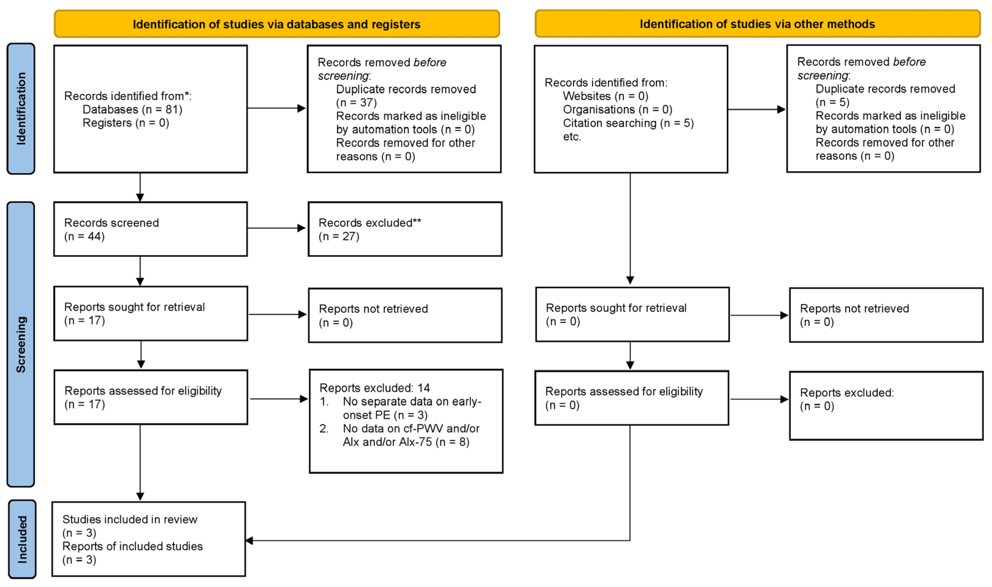
Figure 2.
Absolute numbers and percentages of excluded studies. The numerical order corresponds to different reasons for exclusion as shown in Table 1.
Figure 2.
Absolute numbers and percentages of excluded studies. The numerical order corresponds to different reasons for exclusion as shown in Table 1.
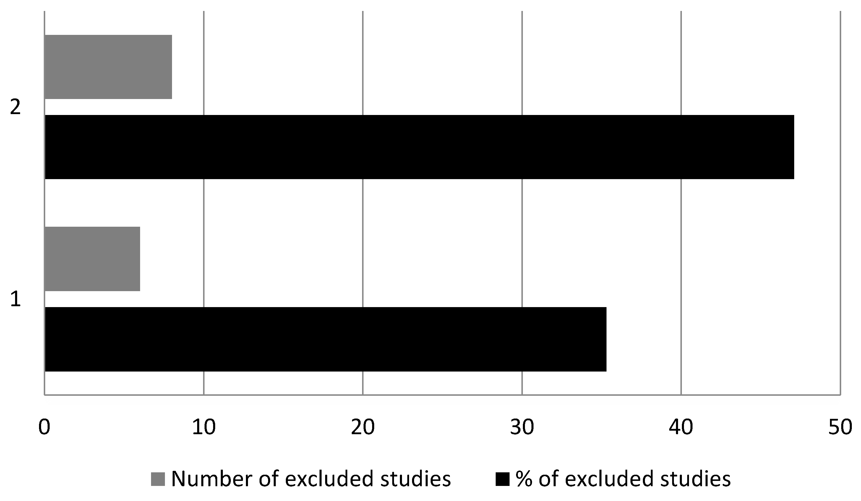
Table 1.
Results of computerized database search and study selection for arterial stiffness and wave reflection measurements in early-onset PE (n=17).
Table 1.
Results of computerized database search and study selection for arterial stiffness and wave reflection measurements in early-onset PE (n=17).
| No. of retrieved references to studies | |
| AS assessment (n = 17) | |
| Excluded | 14 (82.4) |
|
6 (35.3) |
|
8 (47.1) |
| Included | 3 (17.6) |
Note - Numbers in parentheses are percentages Abbreviations: AS, arterial stiffness; PE, preeclampsia; cf-PWV, carotid-femoral pulse wave velocity; Alx, augmentation index; Alx-75, Alx corrected for a heart rate of 75 beats per minute.
Table 2.
Longitudinal studies reporting on ante- and postpartum arterial stiffness and wave reflection measurements.
Table 2.
Longitudinal studies reporting on ante- and postpartum arterial stiffness and wave reflection measurements.
| Antepartum Case (Patient No) | Antepartum Control (Patient No) | Postpartum Case (Patient No) | Postpartum Control (Patient No) | P A vs B |
P A vs C |
P C vs D |
P B vs D |
|
|---|---|---|---|---|---|---|---|---|
| cf-PWV (m/sec) | ||||||||
| Carty et al. (2013) | - | - | - | - | - | - | - | - |
| Franz et al. (2013) | 8.1 ± 0.7 (11) | 7.3 ± 1.5 (10) | 9.9 ± 1.6 (8) | 7.0 ± 1.2 (8) | 0.1280 | 0.0039 | 0.0011 | 0.6522 |
| Robb et al. (2009) | 6.4 ± 0.4 (7)a | 5.0 ± 0.2 (22) | 6.5 ± 0.3 (15)b | 5.5 ± 0.2 (22) | <0.0001 | 0.5195 | <0.0001 | <0.0001 |
| Total | 7.3 ± 0.6 (18) | 6.2 ± 0.9 (32) | 8.2 ± 1(23) | 6.3 ± 0.7(30) | ||||
| Alx (%) | ||||||||
| Carty et al. (2013) | 3.4 ± 10 (17) | 2.4 ± 12 (123) | 18.2 ± 8.5 (23)c | 19.7 ± 10.5 (69) | 0.7410 | <0.0001 | 0.5368 | <0.0001 |
| Franz et al. (2013) | 9.8 ± 18.8 (11) | -72.3 ± 12.9 (10) | -10.7 ± 21.1 (8) | -60.4 ± 10.3 (8) | <0.0001 | 0.0422 | <0.0001 | 0.0500 |
| Robb et al. (2009) | - | - | - | - | - | - | - | - |
| Total | 6.6 ± 14.4 (28) | -35 ± 12.5(133) | 3.8 ± 14.8(31) | -20.4 ± 10.4 (77) | ||||
| Alx-75 (%) | ||||||||
| Carty et al. (2013) | 7.6 ± 10.7 (17) | 7 ± 12 (123) | 15.6 ± 7.3 (23)c | 16.8 ± 10.8 (69) | 0.8401 | 0.0077 | 0.6214 | <0.0001 |
| Franz et al. (2013) | - | - | - | - | - | - | - | - |
| Robb et al. (2009) | 15 ± 5 (7)a | -3.0 ± 2.5 (22) | 16.2 ± 2.5 (15)b | 8.7 ± 1.9 (22) | <0.0001 | 0.6620 | <0.0001 | <0.0001 |
| Total | 11.3 ± 7.9 (24) | 2 ± 7.3 (145) | 15.9 ± 4.9 (38) | 12.8 ± 6.4 (91) | ||||
Abbreviations: No, number; cf-PWV, carotid-femoral pulse wave velocity; Alx, augmentation index; Alx-75, Alx corrected for a heart rate of 75 beats per minute aData derived from bar chart bData including both early-onset and late-onset preeclampsia patients cData including 8 longitudinally followed and 15 non- longitudinally followed patients (women who had been recruited to the original study at booking and had developed preeclampsia, but who had not attended for vascular function studies during pregnancy).
Disclaimer/Publisher’s Note: The statements, opinions and data contained in all publications are solely those of the individual author(s) and contributor(s) and not of MDPI and/or the editor(s). MDPI and/or the editor(s) disclaim responsibility for any injury to people or property resulting from any ideas, methods, instructions or products referred to in the content. |
© 2026 by the authors. Licensee MDPI, Basel, Switzerland. This article is an open access article distributed under the terms and conditions of the Creative Commons Attribution (CC BY) license (http://creativecommons.org/licenses/by/4.0/).
Copyright: This open access article is published under a Creative Commons CC BY 4.0 license, which permit the free download, distribution, and reuse, provided that the author and preprint are cited in any reuse.