
Submitted:
24 August 2026
Posted:
26 August 2026
You are already at the latest version
Abstract
The icon of the Virgin “Hodegetria” (or “Glykophilousa”) from the Church of Panagia Episkopi on Santorini (Thera) represents an important surviving example of Middle Byzantine panel painting in the Aegean. This study investigates its colour palette, painting technology, and chronology through optical microscopy (OM), scanning electron microscopy coupled with energy-dispersive spectroscopy (SEM–EDS), and radiocarbon dating of the wooden support. The analytical results identify a restricted palette of cinnabar, azurite, green earth, lead white, iron-rich ochres, and carbon black. The preparation consists predominantly of gypsum with evidence of a proteinaceous component, while phosphorus detected in most paint layers suggests a proteinaceous binding medium, possibly egg tempera. Distinctive features were identified in the paint stratigraphy and pigment distribution. Homogeneous mixtures of pigments applied as single paint layers occur in three independent colour systems, while Christ’s red garment exhibits an unusual sequence of a thin hematite-rich layer over thicker cinnabar. A later repainting of the Virgin’s tunic was identified through cadmium yellow and lithopone. Radiocarbon dating of the wooden support yielded a calibrated probability distribution centred on AD 1090 ± 60, providing an independent chronological framework for assessing the previously proposed art-historical dating.
Keywords:
Middle Byzantine icon
; Panagia Episkopi
; Santorini
; painting technology
; colour palette
; pigment mixtures
; paint stratigraphy
; radiocarbon dating
1. Introduction
Archaeometric investigations of Byzantine and post-Byzantine panel paintings have expanded considerably in recent decades, providing valuable insights into artists’ materials, painting technology, conservation history, and workshop practices through the application of a range of analytical techniques.
Previous investigations have identified pigments such as cinnabar, azurite, green earth, lead white, iron-rich ochres, and carbon-based blacks in Byzantine and post-Byzantine panel paintings (Afgoranou et al. 1997; Daniilia et al. 2004; Ganitis et al. 2004; Sotiropoulou and Daniilia 2010; Karapanagiotis et al. 2013; Mastrotheodoros 2016; Lazidou et al. 2018; Walczak et al. 2019; Mastrotheodoros et al. 2020, 2021). However, most archaeometric studies concern icons of the post-Byzantine period, while analytical investigations of securely dated Middle Byzantine (10th–12th century) panel icons remain scarce (Lluveras-Tenorio et al. 2017), owing both to the rarity of surviving examples and to the limited opportunities for sampling. Consequently, important aspects of Middle Byzantine painting practice—including paint-layer construction, colour-construction strategies, and working practices—remain insufficiently documented.
The icon of the Virgin preserved in the Church of Panagia Episkopi on Santorini represents one of the most important surviving Middle Byzantine panel paintings in the Cyclades. Although the monument itself has received considerable archaeological and art-historical attention, the icon has not previously been subjected to systematic scientific investigation
As a result, its materials, painting technology, later interventions, and chronology remain largely unexplored.
Historical and Iconographic Background
The Church of Panagia Episkopi in Mesa Gonia, Santorini (Thera), is the island’s most important Byzantine monument and one of the best-preserved examples of Middle Byzantine ecclesiastical architecture in the Cyclades. Traditionally associated with the reign of Emperor Alexios I Komnenos (1081–1118) on the basis of a dedicatory inscription, its foundation has alternatively been attributed to the reign of Alexios II Komnenos (1180–1183) (Tsitouridou 1987). The church has been extensively investigated from archaeological and art-historical perspectives, including its architecture, sculptural decoration, wall paintings, and portable icons (Orlandos 1951; Mendrinos 2000; Barsanti and Pedone 2005; Mitsani 2012; Gkioles and Pallis 2014). The architectural sculpture and surviving painted decoration place the main phase of the monument around the transition from the eleventh to the twelfth century (Orlandos 1951).
Among the church’s most significant works is a panel icon (81 × 106 × 4 cm) depicting the Virgin and Child, flanked by six hierarchs arranged symmetrically along the vertical borders of the panel (Figure 1).
The Virgin carries Christ on her left arm (aristerokratousa), while the Child embraces her neck and presses His face against hers, creating the intimate interaction characteristic of the Glykophilousa (Virgin of Tenderness) iconographic tradition. Nevertheless, the conservation report describes the icon as a Hodegetria (Teliou-Minou 2004), reflecting the iconographic affinity between these types.
The modelling of the faces, characterised by broad brushstrokes and red patches on the cheeks, has been noted to recall the technique of wall painting rather than that of portable icons. On the basis of the rendering of the hierarchs, the decoration of their garments, the lettering of the inscriptions, and comparisons with related representations of the Virgin, a 12th–13th-century date was proposed for the icon (Georgopoulou-Verra 1983). In a later comparative study of the icon of the Virgin in Prayer (Deomene) from Katapoliani on Paros, the Episkopi icon was associated with works of the period around 1200 and described as displaying provincial stylistic characteristics (Mitsani 2002).
2. Research Aim
The present study aims to provide the first scientific investigation of the Middle Byzantine icon of the Virgin from the Church of Panagia Episkopi by integrating optical microscopy (OM), scanning electron microscopy coupled with energy-dispersive spectroscopy (SEM–EDS), and radiocarbon dating of the wooden support.
The objectives are fourfold: (i) to identify the constituent materials of the original painting and the preparation layer; (ii) to investigate paint-layer organisation, pigment combinations, and colour-construction strategies in order to reconstruct aspects of the painter’s working methodology; (iii) to distinguish original Byzantine materials from those associated with later interventions; (iv) to establish an independent absolute chronological framework through radiocarbon dating of the wooden support and assess it in relation to the proposed art-historical chronology of the icon.
By combining material characterisation with technological and chronological evidence, the study seeks to contribute to a better understanding of Middle Byzantine panel-painting practice and to provide a reference dataset for future comparative investigations of securely dated Byzantine icons.
3. Materials and Methods
3.1. Conservation History and Sampling Strategy
According to the conservation report prepared by the late Sofia Teliou-Minou (Teliou-Minou 2004), the icon first underwent conservation treatment in 1975 before being returned to the Church of Panagia Episkopi on Santorini. Following further deterioration, it was transferred to the laboratories of the Directorate of Conservation of Ancient and Modern Monuments (DCAMM) in Athens, where it underwent comprehensive conservation and restoration by Sofia Teliou-Minou, with the support of the then Director of DCAMM, the late Nikos Minos. The present scientific investigation was carried out during this conservation campaign, which enabled the collection of targeted micro-samples.
Five micro-samples were collected in collaboration with the conservator from selected colours and pictorial elements of the icon. Particular attention was given to areas exhibiting distinct stratigraphic sequences, including the garments of the Virgin and Christ, flesh tones, haloes, and the background paint. The samples were initially examined in their original state under a Zeiss stereoscopic microscope to document their morphology and orientation, as well as possible surface deposits.
Polished cross-sections, extending from the external paint surface well into the preparation layer, were subsequently prepared for microscopic and microanalytical examination by embedding the samples in Serifix resin and polishing to 4000 grit. All five samples were analysed and are presented in this study.
3.2. Optical Microscopy
The polished cross-sections were examined by reflected-light optical microscopy (OM) using a Leica metallographic/petrographic optical microscope to document paint stratigraphy, pigment-particle colour and size distribution, and colour differentiation between successive paint layers. Representative micrographs acquired under visible illumination guided the subsequent SEM–EDS investigation.
3.3. Scanning Electron Microscopy and Energy-Dispersive Spectroscopy (SEM–EDS)
For SEM examination and analysis, the polished cross-sections were coated with a thin carbon layer using a Balzers CED 030 coating system to render their surfaces conductive. They were examined using an FEI Quanta scanning electron microscope equipped with an EDAX-Genesis Energy-Dispersive X-ray Spectroscopy (EDS) system, under high vacuum (10⁻⁵ mbar), predominantly using backscattered-electron (BSE) imaging. The electron-beam accelerating voltage was maintained at 25 kV and the beam diameter was set to 3 μm.
EDS analysis was initially performed on multiple rectangular areas, 30–50 μm in horizontal length and 5–30 μm in width, depending on the thickness and uniformity of the paint layer under examination, in order to obtain an average (bulk) analysis of each layer. This was followed by detailed point analyses, using a beam diameter of 3 μm at high magnification, on individual grains within the paint layers in order to identify the pigment particles contributing to the overall colour.
Backscattered-electron imaging was used to distinguish phases of different average atomic number and to investigate paint-layer microstructure. The combined bulk and point analyses were used to identify pigments, preparation materials, alteration products, and materials associated with later interventions.
3.4. Radiocarbon Dating
A small wood sample was collected from the reverse side of the icon, in an area estimated to correspond to the outermost preserved growth rings of the wooden support. Sampling was intentionally kept to a minimum in order to limit intervention on the artwork. The wood was identified as walnut (Juglans regia) in a previous wood anatomical study (Kavouras 2005).
Radiocarbon dating was performed at the Radiocarbon Unit of the Laboratory of Archaeometry, NCSR “Demokritos”. The sample was mechanically and chemically pretreated and dated using the conventional gas proportional counting (GPC) method, based on measurement of 14C activity in purified CO2 gas. Details of the pretreatment and measurement procedures for wood samples have been reported previously (Maniatis et al. 2010).
The conventional radiocarbon age was calibrated using OxCal v4.4.4 (Bronk Ramsey 2021) and the IntCal20 atmospheric calibration dataset (Reimer et al. 2020).
4. Results
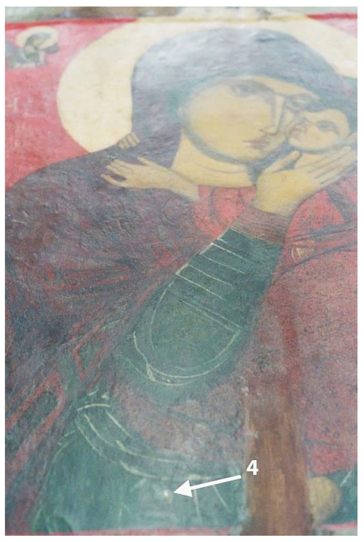
The locations of the analysed samples are presented in Table 1 and shown in Figure 1; the sampling position and macroscopic appearance of Sample 4 are shown in greater detail in Figure 6.
4.1. Sample 1—Dark Red Paint from Christ’s Garment (Figure 1)
Optical microscopy of the polished cross-section revealed two red paint layers over a white preparation layer (Figure 2a); a thinner, dark red surface layer overlying a thicker, brighter red layer. SEM–EDS examination at higher magnification confirmed the presence of the two superimposed red layers resting on the ground layer (Figure 2b).
The outermost paint layer, approximately 5–6 μm thick, appears grey in backscattered electron images and is characterized by high concentrations of iron (Fe) and oxygen (O), together with an appreciable amount of silicon (Si), most likely deriving from quartz, and a small amount of aluminium (Al) (Figure 2c). Its elemental composition is consistent with an iron-rich red earth pigment (red ochre), dominated by hematite (α-Fe2O3) (Mastrotheodoros et al. 2021), a pigment frequently reported in red garments in Byzantine icons (Sotiropoulou and Daniilia 2010). This layer forms a continuous surface coating and corresponds to the dark earthy-red colour observed macroscopically.
Beneath this surface layer, a second red paint layer approximately 12–15 μm thick was identified. SEM–EDS analyses revealed very high concentrations of mercury (Hg) and sulphur (S) (Figure 2d). The elemental composition, combined with the intense bright-red colour of the pigment grains, identifies this layer as cinnabar (HgS) (Eastaugh et al. 2004; Mastrotheodoros et al. 2021). The pigment occurs as relatively fine particles (<15 μm), forming a compact layer. Thus, the brighter cinnabar-rich layer is overlain by a thinner Fe-rich red-earth layer.
Although isolated Hg-rich particles were occasionally observed within the overlying Fe-rich layer (Figure 2b), SEM–EDS analyses indicate that this upper layer is compositionally dominated by the iron-rich earth pigment (Figure 2c). The sporadic occurrence of cinnabar particles within the surface layer is therefore interpreted as mechanical transfer from the underlying layer or minor contamination at the interface, rather than evidence of a deliberate cinnabar–ochre pigment mixture.
Low concentrations of phosphorus (P) were detected, particularly within the upper Fe-rich layer (Figure 2c). Given the stratigraphic position and the absence of phosphorus-bearing black pigments, the detected phosphorus is consistent with the presence of a proteinaceous binding medium, possibly egg tempera. Egg yolk contains phospholipids and phosphoproteins that may leave detectable phosphorus residues in historical paint layers (Phenix 1997; Malletzidou 2021). Nevertheless, secure identification of the binding medium would require molecular analyses, such as FTIR or GC–MS, which were beyond the scope of the present study.
The preparation (ground) layer underlying the paint stratigraphy is characterized by high concentrations of calcium (Ca) and sulphur (S) (Figure 3), indicating the presence of gypsum (CaSO4·2H2O). SEM observations revealed a microstructure composed of characteristic acicular crystals (Figure 3a) whose elemental composition is dominated by Ca and S (Figure 3b), supporting this identification.
In addition, measurable concentrations of phosphorus were detected within the preparation layer (Figure 3d), suggesting the presence of an organic animal-based binder mixed with the gypsum, consistent with traditional gesso preparation techniques employed on wooden panel supports (Mastrotheodoros et al. 2016; Lluveras-Tenorio et al. 2017).
A distinctive feature of the preparation layer in this sample is the occurrence of lead (Pb) within the preparation layer. Lead was detected both near the surface and at depth within the gypsum ground (Figure 3c). This distribution may indicate the deliberate incorporation of lead white into the preparation layer to enhance opacity, although contamination from adjacent lead-containing paint layers cannot be completely excluded.
Overall, the dark red hue observed in this area of Christ’s garment was achieved through the superposition of two red paint layers: a thicker underlying cinnabar layer and a thinner iron-rich red-earth layer applied above it, resulting in an inverted sequence relative to the more commonly expected arrangement of red paint layers.
4.2. Sample 2—Bright Red Paint from the Background (Figure 1)
The bright red paint sampled from the background differs from the red paint of Christ’s garment (Sample 1). Optical microscopy of the polished cross-section revealed a lighter and more saturated red colour (Figure 4a).
SEM examination of the cross-section (Figure 4b) showed a thick homogeneous paint layer, approximately 40–45 μm thick. In BSE images, the layer appears uniformly bright, consistent with the presence of high atomic-number elements.
SEM–EDS analyses detected high concentrations of mercury (Hg) and sulphur (S) (Figure 4c), confirming the identification of the pigment as cinnabar (HgS). As in Sample 1, the pigment occurs as relatively fine particles, generally smaller than 15 μm, distributed throughout the layer.
Unlike Sample 1, where cinnabar forms an underlying layer beneath a thin Fe-rich red-earth paint, the background paint consists of a thick layer dominated by cinnabar.
No measurable phosphorus (P) was detected within the paint layer. Its absence here provides no evidence for the nature of the binding medium, since organic binders lacking detectable phosphorus cannot be excluded by SEM–EDS alone.
The preparation layer shows the same gypsum composition identified in Sample 1. However, unlike Sample 1, neither phosphorus nor lead was detected in the ground layer. The absence of lead indicates that the Pb observed in Sample 1 is not a consistent feature of the preparation layer, whereas the absence of phosphorus may reflect local variability in the distribution or preservation of the organic binder.
Overall, the bright red background paint consists of a thick cinnabar-rich layer applied over a gypsum preparation layer.
4.3. Sample 3—Light Green Paint from Christ’s Halo (Figure 1)
The analysed sample corresponds to the light green paint of Christ’s halo (Figure 5).
Optical microscopy (Figure 5a) and SEM examination (Figure 5b) revealed a homogeneous paint layer, approximately 30 μm thick, directly applied over the preparation layer. No superimposed paint strata were observed within the cross-section.
SEM–EDS analyses of the paint layer (Figure 5c) revealed significant concentrations of Mg, Al, Si, K and Fe, an elemental composition consistent with minerals of the celadonite–glauconite group that constitute the principal components of the historical pigment green earth (terra verde) (Grissom 1986).
Green earth has been widely employed since antiquity and is commonly encountered in Byzantine and post-Byzantine painting (Alexopoulou-Agoranou et al. 1997; Daniilia et al. 2004; Sotiropoulou and Daniilia 2010; Mastrotheodoros et al. 2016).
In addition to the silicate-rich green pigment, elevated concentrations of lead (Pb) were detected and appear to be homogeneously distributed throughout the paint layer. The lead-bearing phase is consistent with the presence of lead white [2PbCO3·Pb(OH)2] (Gliozzo and Ionescu 2021), likely incorporated into the paint mixture. The combination of green earth and lead white accounts for the pale green hue observed both macroscopically (Figure 1) and microscopically (Figure 5a), while also contributing to the opacity and covering power of the paint layer.
Calcium (Ca) and sulphur (S) were also detected within the paint layer. Their presence most likely reflects diffusion or analytical overlap from the underlying gypsum preparation rather than representing primary constituents of the pigment mixture.
The absence of discrete superimposed paint layers and the homogeneous distribution of both the green earth and lead-white components are consistent with application as a pre-mixed paint rather than successive chromatic layers. The paint thus appears to have been applied as a single layer directly over the preparation.
A distinct phosphorus (P) signal was detected within the paint layer. The phosphorus cannot be attributed to bone black pigments, while the underlying preparation contains only minor quantities of P. The detected phosphorus is therefore consistent with the presence of a proteinaceous binding medium, possibly egg tempera, as discussed for Sample 1. Nevertheless, secure identification of the binder would require molecular analyses.
Minor amounts of phosphorus were detected in the preparation layer, consistent with residual animal glue in the ground layer.
Overall, the light green colour of Christ’s halo was produced using a homogeneous mixture of green earth and lead white applied as a single layer over the gypsum preparation.
4.4. Sample 4—Dark Green Paint from the Virgin’s Tunic (Figure 1 and Figure 6)
The sample consisted of two superimposed paint layers that spontaneously separated during sampling and preparation. The detached layers were therefore embedded and examined separately. The outer layer was designated Sample 4a, whereas the underlying layer, in direct contact with the ground, was designated Sample 4b.
Figure 6.
Sampling location and macroscopic appearance of Sample 4 from the Virgin’s tunic.
4.4.1. Sample 4a: Outer Green Layer (Repaint)
The outer paint layer is green in colour and contains yellow, red and white particles under reflected-light microscopy (Figure 7a).
Sample 4a (repaint layer): (a) polished section of the outer green repaint layer under optical microscopy (200x). It appears green-light blue and contains yellow, red and white grains. (b) SEM backscattered electron (BSE) image (1200x). The layer is approximately 65 μm thick. Small and larger grains, appearing brighter in the image, are visible within a dark matrix, possibly consisting of organic material. (c) SEM–EDS spectrum of the paint layer, showing the presence of cadmium (Cd), barium (Ba), zinc (Zn), and sulfur (S), consistent with cadmium yellow and lithopone.
Sample 4b (the original layer): (d) Polished section of the internal original paint layer (dark green/blue) under optical microscopy (200x). It contains intense blue, lighter blue, grey, white and some red/orange and brown grains. The elemental compositions of different grains analysed by SEM–EDS are indicated. Below the paint is the white preparation layer containing several voids. (e) SEM backscattered (BSE) image of the original paint layer, lying directly on the preparation layer (thick darker layer). The paint layer is approximately 30 μm thick and within it small and large fractured grains of crystalline minerals are clearly visible. (f) SEM backscattered (BSE) image of a segment of the paint layer at higher magnification (1372x). The grains are compact, crystalline and fragmented. The composition of some of them, analysed by EDS, is indicated. They consist of either Cu, C, and O (azurite) or Cu and Cl (copper chloride alteration products, possibly atacamite).
SEM examination revealed a heterogeneous paint layer with a thickness of 65 μm, composed of particles with different backscattered electron brightness, indicating components of varying elemental composition (Figure 7b). The bulk EDS spectrum showed the presence of Mg, Al, Si, S, Cd, Ca, K, Ba and Fe, together with a substantial carbon (C) signal (Figure 7c), suggesting the presence of an organic matrix. Based on the uniform light-blue background, this matrix is possibly an organic blue pigment such as indigo (Claes et al. 1999).
Point analyses of individual particles identified Cd and S in yellow grains, Ba and S with minor Zn in white grains, and Fe, Si and Al in reddish particles. These compositions are consistent with cadmium yellow (CdS), lithopone (BaSO4 + ZnS), and iron-based ochres, respectively. Elevated Si concentrations may also indicate the presence of green earth, although this could not be demonstrated unequivocally.
Cadmium yellow was introduced during the nineteenth century and became commercially available after the 1840s (Feller 1986; Pisu et al. 2024), whereas lithopone entered widespread industrial production after approximately 1870 (Eastaugh et al. 2004; Capua 2014). Overpaint layers containing these pigments have previously been reported in Byzantine icons (Daniilia et al. 2002; Brocchieri et al. 2023). The identification of these pigments demonstrates that the outer green paint layer represents a later repainting, dating no earlier than the late nineteenth century. It is possible that this repainting was applied during the first known conservation treatment, carried out in 1975 (Teliou-Minou 2004).
4.4.2. Sample 4b: Original Blue-Green Layer
The underlying paint layer exhibits a darker blue-green colour and lies on the preparation layer. Reflected-light microscopy revealed the presence of intensely blue, green, grey and white particles, together with rare orange and brown grains (Figure 7d).
SEM examination shows that this layer is about 30 μm thick and contains large fragmented crystals. SEM–EDS analysis identified two principal groups of copper-bearing particles (Figure 7e-7f). The first consists of coarse blue particles rich in Cu, C and O, which, based on their colour, composition, grain size (up to approximately 20 μm) and fractured mineral morphology, are consistent with crushed azurite Cu3(CO3)2(OH)2 (Gettens and Fitzhugh 1966, 1993). The second group is composed mainly of Cu and Cl and corresponds to pale blue, grey-green and brownish particles. These particles are interpreted as secondary copper chloride phases most probably of the atacamite group [Cu2Cl(OH)3], formed through alteration of the original copper pigment.
In addition, several particles rich in Si, Al, K, Fe and Ca were identified. Their composition is compatible with green earth pigments, similar to the green earth identified in Sample 3. Significant Pb concentrations were detected throughout the paint layer, consistent with the presence of lead white dispersed within the paint matrix.
Rare orange particles contain As, Fe, Ca and O. Their composition is compatible with an Fe–As secondary mineral tentatively identified as arseniosiderite (Table 2) (Rieck and Rieck 1999), a mineral reported present in the Kamariza mines at Laurion (Figure 8).
Arseniosiderite group [Ca2Fe+3(AsO4)3O2·3H2O] contains approximately 45% As2O5, 31% Fe2O3 and 15% CaO, in good agreement with the oxide composition of the analysed particles (Table 2). These particles occur only sporadically and appear to represent accessory minerals rather than intentionally added pigments. Their presence may provide a clue to the geological source of the azurite.
The combined microscopic and elemental evidence indicates that the original blue-green paint layer is composed primarily of azurite, green earth and lead white. No superimposed paint strata were observed within this layer. Instead, the pigments appear homogeneously distributed within a single paint layer applied directly over the gypsum preparation. A similar application of premixed pigments in a single homogeneous layer was observed in the light green paint of Christ’s halo (Sample 3).
Minor amounts of Cd, Ba and Zn detected in bulk analyses are attributed to contamination from the overlying repainting layer. The detected Ca, S and P originate from the underlying gypsum ground and associated proteinaceous preparation materials.
The preparation layer is the same gypsum-based preparation as in the other samples. However, the presence of voids in the preparation layer (Figure 7d) may indicate local weathering and paint loss, which could have contributed to the need for subsequent repainting.
4.5. Sample 5—Orange Paint from the Preserved Forehead of the Hierarch (Figure 1 and Figure 9a)
The polished cross-section of the small preserved sample, examined by reflected-light microscopy, reveals an orange-brown paint layer containing scattered black particles (Figure 9b).
Figure 9.
Sample 5. Orange paint from the preserved forehead of the Hierarch at the right border of the icon. (a) Sampling position indicated with arrow. (b) Polished section under optical microscopy (200x). (1) the preserved part of the paint layer, showing red, white and black pigment particles and (2) the preparation layer. (c) SEM image of the polished cross section. The darker area on the left of the image corresponds to the preserved orange paint. The purple square, marked “Spectrum 2” is the area (40x40 μm) selected for bulk analysis (Table 3). (d) SEM-BSE image inside the orange paint at higher magnification (1500x), showing the locations of point analyses performed on individual pigment grains (Table 3).
Figure 9.
Sample 5. Orange paint from the preserved forehead of the Hierarch at the right border of the icon. (a) Sampling position indicated with arrow. (b) Polished section under optical microscopy (200x). (1) the preserved part of the paint layer, showing red, white and black pigment particles and (2) the preparation layer. (c) SEM image of the polished cross section. The darker area on the left of the image corresponds to the preserved orange paint. The purple square, marked “Spectrum 2” is the area (40x40 μm) selected for bulk analysis (Table 3). (d) SEM-BSE image inside the orange paint at higher magnification (1500x), showing the locations of point analyses performed on individual pigment grains (Table 3).
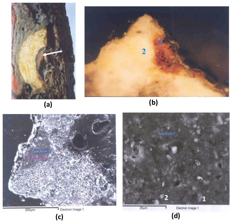
Bulk SEM–EDS analysis of the orange paint layer was performed in the area indicated by the purple rectangle (Spectrum 2) in Figure 9c, while additional point analyses were carried out on selected particles and micro-areas (Figure 9d). Quantitative results are presented in Table 3.
The bulk composition reveals significant concentrations of Fe and Pb, together with appreciable amounts of Al and Si and smaller quantities of S and Ca. Point analyses identified two principal particle populations. The first consists of Fe-rich particles associated with Al and Si, corresponding to iron-rich ochres. The second consists of Pb-rich particles with little contribution from other elements and is consistent with lead white.
The orange-brown colour of the paint layer is therefore best explained by a mixture of iron-based ochres and lead white. In addition, microscopic examination revealed the presence of sparse black particles (Figure 9a), while the paint matrix exhibits elevated carbon, indicating the presence of a carbon-based black pigment in minor quantities.
Furthermore, elemental mapping of Fe and Pb (Figure 10a–c), together with microscopic observations of the carbon particles, indicates a relatively homogeneous distribution of the pigment components throughout the paint layer.
No discrete superimposed paint strata corresponding to separate underpainting and highlighting phases were identified within the examined sample. Instead, the available evidence indicates that the preserved orange flesh tone was produced using a pre-mixed paint composed of iron-rich ochres, lead white and carbon black applied as a single paint layer.
The use of iron-based earth pigments, lead white and carbon black for rendering flesh tones through successive paint layers has been reported in Byzantine and post-Byzantine icons (Daniilia et al. 2004; Ganitis et al. 2004; Mastrotheodoros et al. 2021). In contrast, the present sample shows no evidence of successive modelling layers but instead consists of a homogeneous pre-mixed paint applied as a single layer, similar to Samples 3 and 4b.
Phosphorus detected in this sample within the paint layer (Table 3) is again consistent with the possible use of egg tempera, as discussed above.
Overall, the preserved orange flesh tone consists of a single homogeneous paint layer composed primarily of iron-rich ochres, lead white and minor amounts of carbon black, indicating that the pigments were premixed prior to application.
It should be noted, however, that the sample originates from a small and heavily deteriorated surviving area of the bishop’s face. Consequently, the presence of additional painting phases in areas that have not survived cannot be excluded.
The preparation layer is again the same gypsum-based preparation as in the other samples.
4.6. Radiocarbon Dating of the Wooden Support
The radiocarbon dating results are presented in Table 4.
The wood sample yielded a conventional radiocarbon age of 973 ± 60 BP, with a measured δ13C value of −23.69‰.
The calibrated age ranges are AD 1020–1160 at 68.3% probability and AD 970–1220 at 95.4% probability. The calibrated probability distribution (Figure 11) is approximately normal, with a mean of AD 1090 and a standard deviation of 60 years; the calibrated result can therefore be conveniently summarised as AD 1090 ± 60.
The relatively high uncertainty associated with the radiocarbon age reflects the very small amount of wood sampled in order to minimise intervention on the icon. Despite the relatively broad calibrated age ranges, most of the probability distribution is concentrated in the late eleventh–early twelfth century.
5. Discussion
A summary of the identified pigments, paint stratigraphy and painting technology is presented in Table 5.
5.1. Pigment Palette, Preparation and Binding Medium
The analytical investigation revealed a relatively restricted pigment palette comprising cinnabar, iron-rich ochres, green earth, azurite, lead white and carbon black. All are compatible with Byzantine painting practice, and no chronologically incompatible materials were detected within the original paint layers. Despite the limited palette, the pigments were used in different combinations and stratigraphic arrangements to obtain a range of chromatic effects, from the saturated cinnabar background to the mixed green, blue-green and flesh-tone paints.
The paint layers were consistently applied over a gypsum preparation containing evidence of an animal-derived proteinaceous component, compatible with the traditional use of animal glue in panel grounds. Lead detected within the preparation of Sample 1 may indicate local incorporation of lead white, although its absence from the other analysed ground layers shows that it was not a consistent component of the preparation.
Phosphorus detected within several original paint layers is consistent with the presence of a proteinaceous binding medium, possibly egg tempera. Its absence from the cinnabar background layer indicates some variability between the sampled areas. Since SEM–EDS provides elemental rather than molecular information, however, identification of the binding medium remains tentative and would require confirmation by molecular analytical techniques.
5.2. Painting Technology: Pigment Premixing and Single-Layer Application
One of the most significant technological findings is the recurrent occurrence of pigment mixtures applied as homogeneous single paint layers. This was independently observed in three different colour systems: the light green of Christ’s halo (Sample 3), the original blue-green paint of the Virgin’s tunic (Sample 4b), and the preserved orange flesh tone of the hierarch (Sample 5). The recurrence of this feature in compositionally and chromatically different areas suggests that pigment premixing constituted a deliberate element of the painter’s working methodology rather than an isolated technical solution.
The flesh-tone sample is particularly noteworthy because no stratigraphic separation corresponding to the successive underpainting, modelling and highlighting stages commonly described for Byzantine flesh painting was observed. Instead, iron-rich ochres, lead white and carbon black occur together within the preserved homogeneous paint layer. Although the limited and deteriorated condition of the sampled area prevents exclusion of additional painting phases elsewhere on the face, the surviving stratigraphy provides direct evidence for premixing in the preserved flesh tone.
In this context, earlier observation that the modelling of the faces, with broad brushstrokes and red patches on the cheeks, recalls wall-painting techniques rather than those of portable icons (Georgopoulou-Verra 1983) is particularly interesting. Although this stylistic observation cannot be directly equated with the microstratigraphic evidence, both lines of evidence independently point to distinctive features of the painting practice of the Episkopi icon
5.3. Variation in Paint Construction: The Cinnabar–Hematite Stratigraphy
The recurrent use of premixed single layers did not constitute a uniform painting procedure throughout the icon. Christ’s red garment exhibits a distinctly different approach, consisting of a relatively thick cinnabar layer covered by a thin hematite-rich red earth layer.
As noted in the Results, this sequence is the reverse of stratigraphies more commonly reported for Byzantine red paint systems, in which iron-rich earth pigments form an underlying layer and cinnabar is applied above (Daniilia et al. 2004; Sotiropoulou and Daniilia 2010; Karapanagiotis et al. 2013; Lazidou et al. 2018; Helvacı et al. 2026). In the Episkopi icon, the thin iron-rich surface layer would have moderated the brilliance of the underlying cinnabar, producing the darker earthy-red appearance observed macroscopically.
The coexistence within the same icon of homogeneous premixed paint layers and a deliberately stratified two-layer red system demonstrates considerable flexibility in colour construction. Different pigment combinations and modes of application were therefore employed according to the desired visual effect rather than according to a single uniform technical procedure.
5.4. Later Repainting and Preservation of the Original Paint
Analysis of the Virgin’s tunic clearly distinguished a later repainting from the original paint layer. Cadmium yellow and lithopone establish that the outer layer was applied no earlier than the late nineteenth century. Given that the first documented conservation treatment of the icon took place in 1975, it is possible that the repainting was applied during that intervention, although an earlier undocumented application cannot be excluded.
More importantly for reconstruction of the original painting technology, the underlying blue-green layer remains preserved and consists principally of azurite, green earth and lead white applied as a homogeneous mixture. The presence of secondary copper chloride phases indicates alteration of the original copper-bearing pigment, while the rare Fe–As particles tentatively identified as arseniosiderite may represent accessory minerals associated with the source material rather than deliberately added pigments.
Thus, separation of the later repaint from the original layer provides information not only on the intervention history of the icon but also on the composition and application of the original Byzantine paint.
5.5. Chronology: Radiocarbon and Art-Historical Evidence
Radiocarbon dating of the walnut support provides an independent absolute chronological constraint for the icon. As shown by the approximately normal calibrated probability distribution, the result is centred on AD 1090 ± 60, with most of the probability concentrated in the late eleventh–early twelfth century. Since radiocarbon dating determines the age of the sampled wood rather than the execution of the painting, the result cannot be interpreted as a direct date for the painting event. Nevertheless, the sample was taken from an area estimated to correspond to the outermost preserved growth rings, increasing its chronological relevance to the manufacture of the panel.
The radiocarbon evidence can therefore be assessed independently alongside the art-historical chronology. On stylistic and iconographic grounds, the icon was assigned to the 12th–13th century (Georgopoulou-Verra 1983), while it was subsequently related more specifically to works around 1200 (Mitsani 2002). Although the calibrated distribution extends into the period covered by these stylistic assessments, its highest concentration of probability lies earlier, in the late eleventh–early twelfth century. It is also compatible with the chronology traditionally proposed for the Church of Panagia Episkopi and its decoration (Orlandos 1951).
The radiocarbon result therefore provides an independent absolute chronological framework against which the stylistic and historical assessments of the icon can be evaluated, while recognising the inherent distinction between the age of the wooden support and the execution of the painting.
6. Conclusions
The combined scientific investigation of the icon of the Virgin from the Church of Panagia Episkopi (Mesa Gonia, Thera) provides new evidence concerning the materials, painting technology, conservation history and chronology of an important Byzantine panel painting from the Aegean.
Integration of optical microscopy and SEM–EDS with radiocarbon dating and the available historical and stylistic evidence has enabled both the original painting technique and subsequent interventions to be characterised.
The original palette is relatively restricted, comprising cinnabar, iron-rich ochres, green earth, azurite, lead white and carbon black, applied over a gypsum preparation. Phosphorus detected in most of the analysed paint layers and in the preparation provides evidence for proteinaceous components, although their precise nature cannot be established by SEM–EDS alone.
A particularly significant result is the identification of recurrent pigment premixing in three compositionally different areas of the icon. Together with the unusual sequence of hematite-rich red earth over cinnabar in Christ’s garment, this demonstrates that colour was constructed through more than one technical approach. The analytical evidence therefore reveals greater variation in paint application than would be apparent from identification of the pigment palette alone.
The analyses also differentiate the original paint of the Virgin’s tunic from a later repainting containing cadmium yellow and lithopone, together with a possible organic blue pigment. These materials establish a post-nineteenth-century intervention, possibly associated with the documented conservation treatment of 1975, while the underlying layer preserves evidence of the original blue-green colour and its method of application.
Radiocarbon dating adds an independent chronological dimension to the study. The calibrated probability distribution, centred on AD 1090 ± 60, is concentrated mainly in the late eleventh to early twelfth century. Since radiocarbon dating concerns the sampled wood rather than the execution of the painting, the result does not necessarily date the icon directly. However, it provides an important independent chronological framework within which the art-historical assessment can be evaluated.
Taken together, the results demonstrate the value of examining not only the materials present in Byzantine icons but also their distribution and microstratigraphic relationships. The recurrent premixing of pigments and the unusual red stratigraphy documented at Panagia Episkopi provide useful comparative evidence for future studies of Byzantine panel-painting technology and may help to assess the extent of technological variation between individual painters, workshops and periods.
Acknowledgments
This paper is dedicated to the memory of the late Sofia Teliou-Minou, the conservator responsible for the conservation of the icon, and the late Nikos Minos, former Head of the Directorate of Conservation of Ancient and Modern Monuments of the Hellenic Ministry of Culture. Their commitment to the preservation of Byzantine cultural heritage made both the conservation of this remarkable icon and the present scientific investigation possible. Regrettably, neither had the opportunity to see the results of this study published. Thanks, are also due to Dr. Acheimastou-Potamianou for assisting me in finding the relevant literature regarding the Episkopi church and the icon.
References
- Alexopoulou-Agoranou, A.; Theodoropoulou, O.; Tsairis, G. Μελέτη των υλικών και της τεχνικής κατασκευής της μεταβυζαντινής εικόνας «ο Δείπνος ο Μυστικός» του Μιχαήλ Δαμασκηνού. Δελτίον Της Χριστιανικής Aρχαιολογικής Εταιρείας Περίοδος Δ’ (in Greek). 1997, Τόμος ΙΘ’ 19, 151–166. [Google Scholar] [CrossRef]
- Barsanti, Cl; Pedone, S. Una nota sulla scultura ad incrostazione e il templon della Panaghia Episcopi di Santorini. Traveaux Et. Mémoirs (Mélanges Jean-Pierre Sodini) 2005, 15, 407–425. [Google Scholar]
- Brocchieri, J.; D’Onofrio, A.; Sabbarese, C. Combining XRF, Multispectral Imaging and SEM/EDS to Characterize a Contemporary Painting. Quantum Beam Sci. 2023, 7(2), 13. [Google Scholar] [CrossRef]
- Bronk Ramsey, C. Oxcal v4.4.4 calibration program. 2021. [Google Scholar] [CrossRef]
- Capua, R. The Obscure History of a Ubiquitous Pigment: Phosphorescent Lithopone and its Appearance on Drawings by John La Farge. J. Am. Inst. Conserv. 2014, 53, 75–88. [Google Scholar] [CrossRef]
- Claes, M.; Van Ham, R.; Janssens, K. Micro-analysis of Artists’ Pigments by Grazing-Emission X-ray Fluorescence Spectrometry. Adv. X-Ray Anal. 1999, 41, 262–277. [Google Scholar]
- Daniilia, S.; Bikiaris, D.; Burgio, L. An extensive non-destructive and micro-spectroscopic study of two post-Byzantine overpainted icons of the 16th century. J. Raman Spectrosc. 2002, 33, 807–814. [Google Scholar] [CrossRef]
- Daniilia, S.; Sotiropoulou, S.; Bikiaris, D. Chapter 12 Diagnostic methodology for the examination of Byzantine frescoes and icons. Non-destructive investigation and pigment identification. In Non-Destructive Microanalysis of Cultural Heritage Materials; Elsevier, 2004; pp. 565–604. [Google Scholar]
- Eastaugh, N.; Walsh, T.; Chaplin, T.; Siddall, R. Pigment Compendium: A Dictionary and Optical Properties of Historical Pigments; Elsevier Butterworth-Heinemann: Amsterdam; Boston, 2004. [Google Scholar]
- Feller, R.L. (Ed.) Artists’ Pigments: A Handbook of Their History and Characteristics; National Gallery of Art: Washington, 1986. [Google Scholar]
- Ganitis, V.; Pavlidou, E.; Zorba, F. A post-Byzantine icon of St Nicholas painted on a leather support. Microanalysis and characterisation of technique. J. Cult. Herit. 2004, 5, 349–360. [Google Scholar] [CrossRef]
- Georgopoulou-Verra, M. Βυζαντινά και Μεσαιωνικά Μνημεία Κυκλάδων. Aρχαιολογικόν Δελτίον Μέρος Β’2-Χρονικά (in Greek). 1983, 30, 333–336. [Google Scholar]
- Gettens, R.J.; Fitzhugh, E.W. Azurite and Blue Verditer. Stud. Conserv. 1966, 11, 54–61. [Google Scholar] [CrossRef]
- Gettens, R.J.; Fitzhugh, E.W. Azurite and Blue Verditer in Artist’s Pigments: A handbook of their history and characteristics; Roy, A., Ed.; National Gallery of Art: Washington, 1993; pp. 23–35. [Google Scholar]
- Gkioles, N.; Pallis, G. Atlas of the Christian Monuments of the Aegean from the Early Christian Years to the Fall of Constantinople; General Secretariat of the Aegean and Island Policy: Athens, 2014. [Google Scholar]
- Gliozzo, E.; Ionescu, C. Pigments—Lead-based whites, reds, yellows and oranges and their alteration phases. Archaeol. Anthropol. Sci. 2021, 14, 17. [Google Scholar] [CrossRef]
- Grissom, C.A. Green Earth. In Artists’ Pigments: A Handbook of Their History and Characteristics; Roy, A., Ed.; National Gallery of Art: Washington, D.C., 1986; pp. 141–167. [Google Scholar]
- Helvacı, Y.Z.; Giustetto, R.; Chiari, G. Characterisation of pigments and technical features of Byzantine mural paintings from eastern Crete (Lassithi): a diachronic view of reds between the 8th and 16th centuries. J. Archaeol. Sci. Rep. 2026, 73, 105786. [Google Scholar] [CrossRef]
- Karapanagiotis, I.; Lampakis, D.; Konstanta, A.; Farmakalidis, H. Identification of colourants in icons of the Cretan School of iconography using Raman spectroscopy and liquid chromatography. J. Archaeol. Sci. 2013, 40, 1471–1478. [Google Scholar] [CrossRef]
- Kavouras, P. Ταυτοποίηση ξύλου σανιδιώματος εικόνας: Εικόνα Oδηγήτρια, Ναός Επισκοπής Γωνιάς Θήρας; (in Greek). Report to the Directorate of Conservation of Ancient and Modern Monuments, Ministry of Culture. Laboratory of Anatomy and Technology of Wood, Institute of Mediterranean Forest Ecosystems and Forest Products Technology: Athens, 2005. [Google Scholar]
- Lazidou, D.; Lampakis, D.; Karapanagiotis, I.; Panayiotou, C. Investigation of the Cross-Section Stratifications of Icons Using Micro-Raman and Micro-Fourier Transform Infrared (FT-IR) Spectroscopy. Appl. Spectrosc. 2018, 72, 1258–1271. [Google Scholar] [CrossRef] [PubMed]
- Lluveras-Tenorio, A.; Parlanti, F.; Degano, I. Spectroscopic and mass spectrometric approach to define the Cyprus Orthodox icon tradition - The first known occurrence of Indian lac in Greece/Europe. Microchem. J. 2017, 131, 112–119. [Google Scholar] [CrossRef]
- Malletzidou, L. Contribution to the restoration of paintings: Study of the interaction between binders and pigments and/via characterization of fresh, artificially aged and authentic samples from old paintings. PhD Thesis, Aristotle University of Thessaloniki, Faculty of Sciences, School of Physics, 2021. [Google Scholar]
- Maniatis, Y.; Malamidou, D.; Koukouli-Chryssanthaki, H.; Facorellis, Y. Radiocarbon Dating of the Amphipolis Bridge in Northern Greece, Maintained and Functioned for 2500 Years. Radiocarbon 2010, 52, 41–63. [Google Scholar] [CrossRef]
- Mastrotheodoros, G. Χρωστικές Κονίες και άλλα Υλικά Μεταβυζαντινής Ζωγραφικής. PhD Thesis, University of Ioannina, 2016. [Google Scholar]
- Mastrotheodoros, G.P.; Beltsios, K.G.; Bassiakos, Y. On the blue and green pigments of post-Byzantine Greek icons. Archaeometry 2020, 62, 774–795. [Google Scholar] [CrossRef]
- Mastrotheodoros, G.P.; Beltsios, K.G.; Bassiakos, Y. On the Red and Yellow Pigments of Post-Byzantine Greek Icons. Archaeometry 2021, 63, 753–778. [Google Scholar] [CrossRef]
- Mastrotheodoros, G.P.; Beltsios, K.G.; Bassiakos, Y.; Papadopoulou, V. On the Grounds of Post-Byzantine Greek Icons. Archaeometry 2016, 58, 830–847. [Google Scholar] [CrossRef]
- Mendrinos, M. Panagia Episcope – The Byzantine Church of Santorini. In The Sacred Pilgrimage “Panaghia Episcope”; 2000. [Google Scholar]
- Mitsani, A. Ναός Επισκοπής Θήρας. “Odysseus” Greek Ministry of Culture. 2012. Available online: http://odysseus.culture.gr/h/2/gh251.jsp?obj_id=1696.
- Mitsani, A. Βυζαντινή εικόνα της Παναγίας δεομένης στην Καταπολιανή Πάρου. Δελτίον Της Χριστιανικής Aρχαιολογικής Εταιρείας (in Greek). 2002, 23, 177–198. [Google Scholar] [CrossRef]
- Orlandos, A.K. H ’Πισκοπή της Σαντορίνης (Παναγία της Γωνιάς). In Aρχείον των Βυζαντινών Μνημείων της Ελλάδος - Περιοδικόν Σύγγραμα. Ν. ΕΣΤΙA; Aθήνα, 1951; pp. 178–214. [Google Scholar]
- Phenix, A. (1997) The composition and chemistry of eggs and egg tempera. In Early Italian Paintings: Techniques and Analysis; Bakkenist, T., Hoppenbrouwers, R., Dubois, H., Eds.; Symposium: Maastricht, 9-10 October 1996; pp. 11–20. [Google Scholar]
- Pisu, F.A.; Carbonaro, C.M.; Ricci, P.C. Cadmium Yellow Pigments in Oil Paintings: Optical Degradation Studies Utilizing 3D Fluorescence Mapping Supported by Raman Spectroscopy and Colorimetry. 2024, 7, 2426–2443. [Google Scholar] [CrossRef]
- Reimer, P.J.; Austin, W.E.N.; Bard, E. The IntCal20 Northern Hemisphere Radiocarbon Age Calibration Curve (0–55 cal kBP). Radiocarbon 2020, 62, 725–757. [Google Scholar] [CrossRef]
- Rieck, B.; Rieck, P. Lavrion: Die komplette Mineralliste. Lapis 1999, 24, 61–63. [Google Scholar]
- Sotiropoulou, S.; Daniilia, S. Material Aspects of Icons. A Review on Physicochemical Studies of Greek Icons. Acc. Chem. Res. 2010, 43, 877–887. [Google Scholar] [CrossRef] [PubMed]
- Teliou-Minou, S. Δελτίο Συντήρησης Aντικειμένου: Εικόνα Oδηγήτρια, Ναός Επισκοπής Γωνιάς Θήρας. In Directorate of Conservation of Ancient and Modern Monuments, Ministry of Culture; (In Greek). 2004. [Google Scholar]
- Tsitouridou, A. Επισκοπή Σαντορίνης: Ίδρυμα του Aλεξίου A΄ Κομνηνού ή του Β΄. In Aμητός. Τιμητικός τόμος για τον καθηγητή Μανόλη Aνδρόνικο.; Θεσαλονίκη, 1987; pp. 917–921. [Google Scholar]
- Walczak, M.; Tarsińska-Petruk, D.; Płotek, M. MA-XRF study of 15th–17th century icons from the collection of the National Museum in Krakow, Poland. X-Ray Spectrom. 2019, 48, 303–310. [Google Scholar] [CrossRef]
Figure 1.
The icon of the Virgin Hodegetria (or Glykophilousa type), surrounded by six hierarchs/saints (not all visible in the photograph). Sampling locations: Sample 1, dark red (Christ’s garment); Sample 2, bright red (background); Sample 3, light green (Christ’s halo); Sample 4, dark green (Virgin’s garment); Sample 5, orange (forehead of the hierarch depicted in the right border).
Figure 1.
The icon of the Virgin Hodegetria (or Glykophilousa type), surrounded by six hierarchs/saints (not all visible in the photograph). Sampling locations: Sample 1, dark red (Christ’s garment); Sample 2, bright red (background); Sample 3, light green (Christ’s halo); Sample 4, dark green (Virgin’s garment); Sample 5, orange (forehead of the hierarch depicted in the right border).
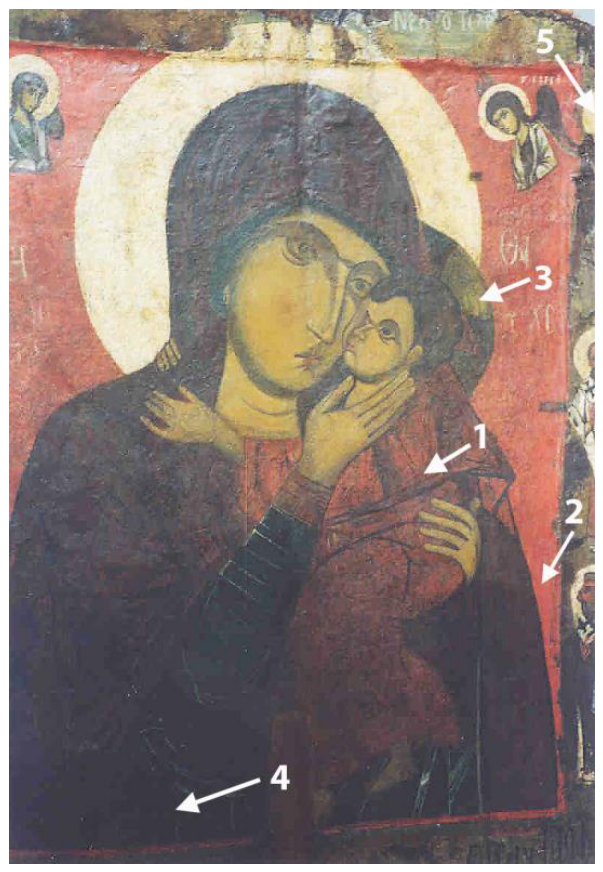
Figure 2.
Sample 1. Red from Christ’s garment. (a) Polished cross-section under optical microscopy (200x). (b) SEM backscattered electron (BSE) image (1200x). Two paint layers are visible at the surface: an upper layer (greyish in the image) and a thicker underlying layer (brighter in the image). The coarse preparation layer is visible below. The higher brightness indicates the presence of elements with higher atomic numbers. (c) SEM–EDS spectrum of the upper surface paint layer. The spectrum is dominated by iron (Fe) and oxygen (O), with minor amounts of silicon (Si), aluminium (Al), potassium (K), calcium (Ca), and other elements associated with aluminosilicate minerals. (d) SEM–EDS spectrum of the lower thicker paint layer. Mercury (Hg) is the dominant element. Sulfur (S) is also present, although its principal peak overlaps with that of Hg.
Figure 2.
Sample 1. Red from Christ’s garment. (a) Polished cross-section under optical microscopy (200x). (b) SEM backscattered electron (BSE) image (1200x). Two paint layers are visible at the surface: an upper layer (greyish in the image) and a thicker underlying layer (brighter in the image). The coarse preparation layer is visible below. The higher brightness indicates the presence of elements with higher atomic numbers. (c) SEM–EDS spectrum of the upper surface paint layer. The spectrum is dominated by iron (Fe) and oxygen (O), with minor amounts of silicon (Si), aluminium (Al), potassium (K), calcium (Ca), and other elements associated with aluminosilicate minerals. (d) SEM–EDS spectrum of the lower thicker paint layer. Mercury (Hg) is the dominant element. Sulfur (S) is also present, although its principal peak overlaps with that of Hg.
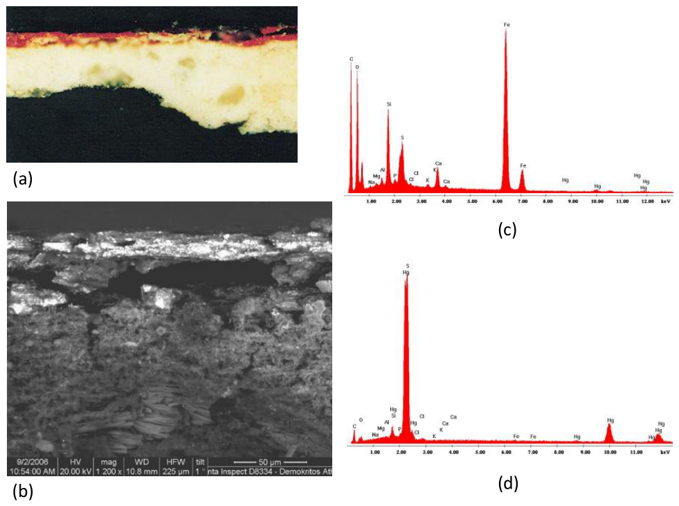
Figure 3.
Sample 1. Preparation layer. (a) SEM backscattered electron (BSE) image of the preparation layer at high magnification (5000x), showing the characteristic acicular gypsum crystals. (b) SEM‒EDS spectrum from point analysis of individual crystals. (c) SEM backscattered electron (BSE) image (277x) of the whole polished cross-section showing the red paint layer and the underlying preparation layer. Lead (Pb)-rich areas reaching deep in the preparation layer are marked. (d) Representative SEM‒EDS spectrum of the preparation layer, showing, in addition to Ca and S, the presence of P.
Figure 3.
Sample 1. Preparation layer. (a) SEM backscattered electron (BSE) image of the preparation layer at high magnification (5000x), showing the characteristic acicular gypsum crystals. (b) SEM‒EDS spectrum from point analysis of individual crystals. (c) SEM backscattered electron (BSE) image (277x) of the whole polished cross-section showing the red paint layer and the underlying preparation layer. Lead (Pb)-rich areas reaching deep in the preparation layer are marked. (d) Representative SEM‒EDS spectrum of the preparation layer, showing, in addition to Ca and S, the presence of P.
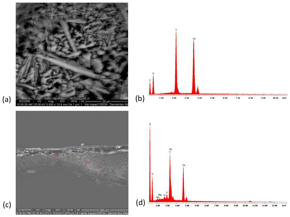
Figure 4.
Sample 2. Red paint from the background. (a) polished cross-section of the red paint under optical microscopy (200x). Numerous cracks are visible within the paint layer. (b) SEM backscattered electron (BSE) image of the section (1200x). The bright surface layer corresponds to the red paint, while the underlying darker layer corresponds to the preparation layer. A major crack is visible within the paint layer. (c) SEM–EDS spectrum of the bright red paint layer from the background. The spectrum is dominated by Hg and S.
Figure 4.
Sample 2. Red paint from the background. (a) polished cross-section of the red paint under optical microscopy (200x). Numerous cracks are visible within the paint layer. (b) SEM backscattered electron (BSE) image of the section (1200x). The bright surface layer corresponds to the red paint, while the underlying darker layer corresponds to the preparation layer. A major crack is visible within the paint layer. (c) SEM–EDS spectrum of the bright red paint layer from the background. The spectrum is dominated by Hg and S.
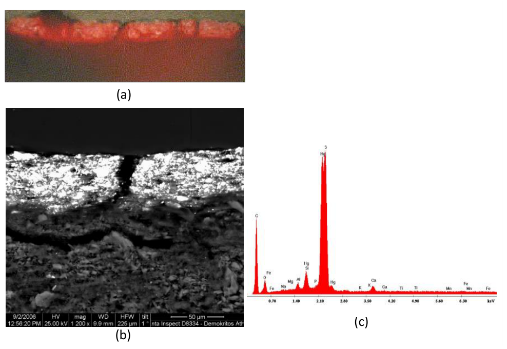
Figure 5.
Sample 3. Light green paint from Christ’s halo. (a) Polished cross-section under optical microscopy (200x). The green paint layer is visible on the white preparation layer. (b) SEM backscattered electron (BSE) image (1200x). A single paint layer approximately 30 μm thick overlies the preparation layer. (c) SEM–EDS spectrum acquired from the green paint layer.
Figure 5.
Sample 3. Light green paint from Christ’s halo. (a) Polished cross-section under optical microscopy (200x). The green paint layer is visible on the white preparation layer. (b) SEM backscattered electron (BSE) image (1200x). A single paint layer approximately 30 μm thick overlies the preparation layer. (c) SEM–EDS spectrum acquired from the green paint layer.
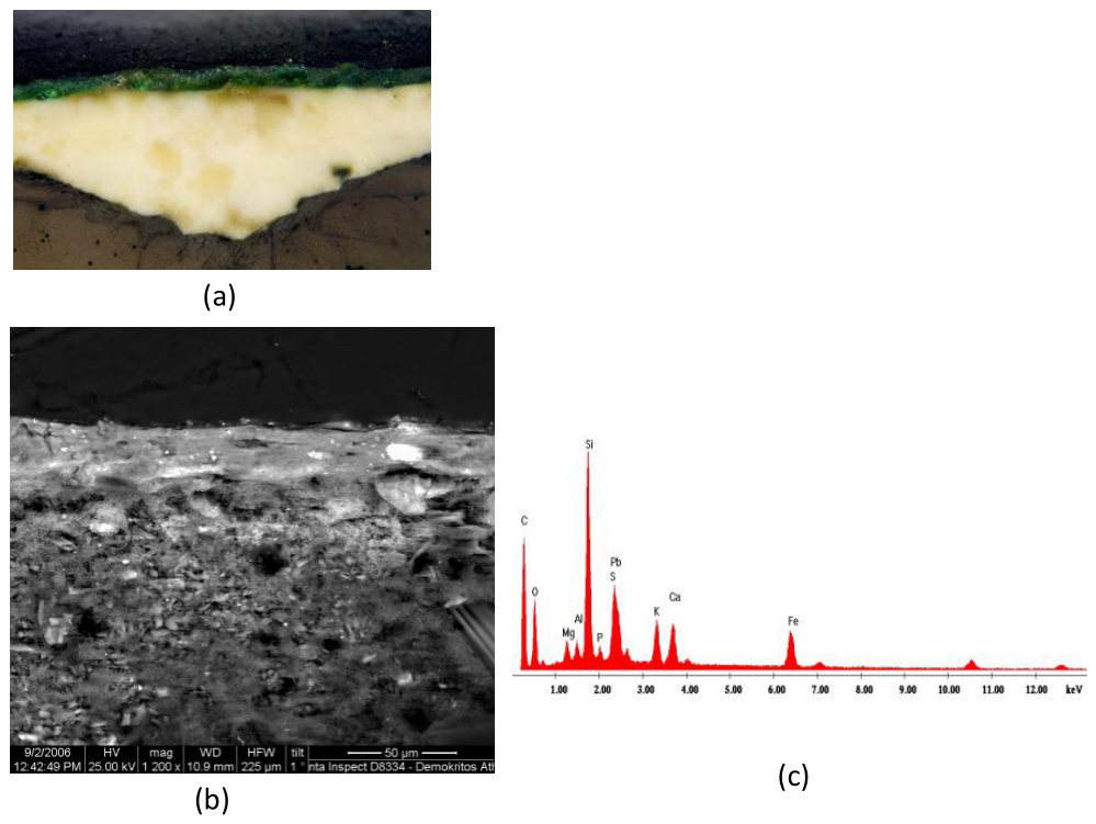
Figure 7.
Sample 4. Dark green paint from the Virgin’s tunic. The sample separated during preparation into two layers: Sample 4a, the outer green-blue repaint layer, and Sample 4b, the lower original dark blue-green layer.
Figure 7.
Sample 4. Dark green paint from the Virgin’s tunic. The sample separated during preparation into two layers: Sample 4a, the outer green-blue repaint layer, and Sample 4b, the lower original dark blue-green layer.
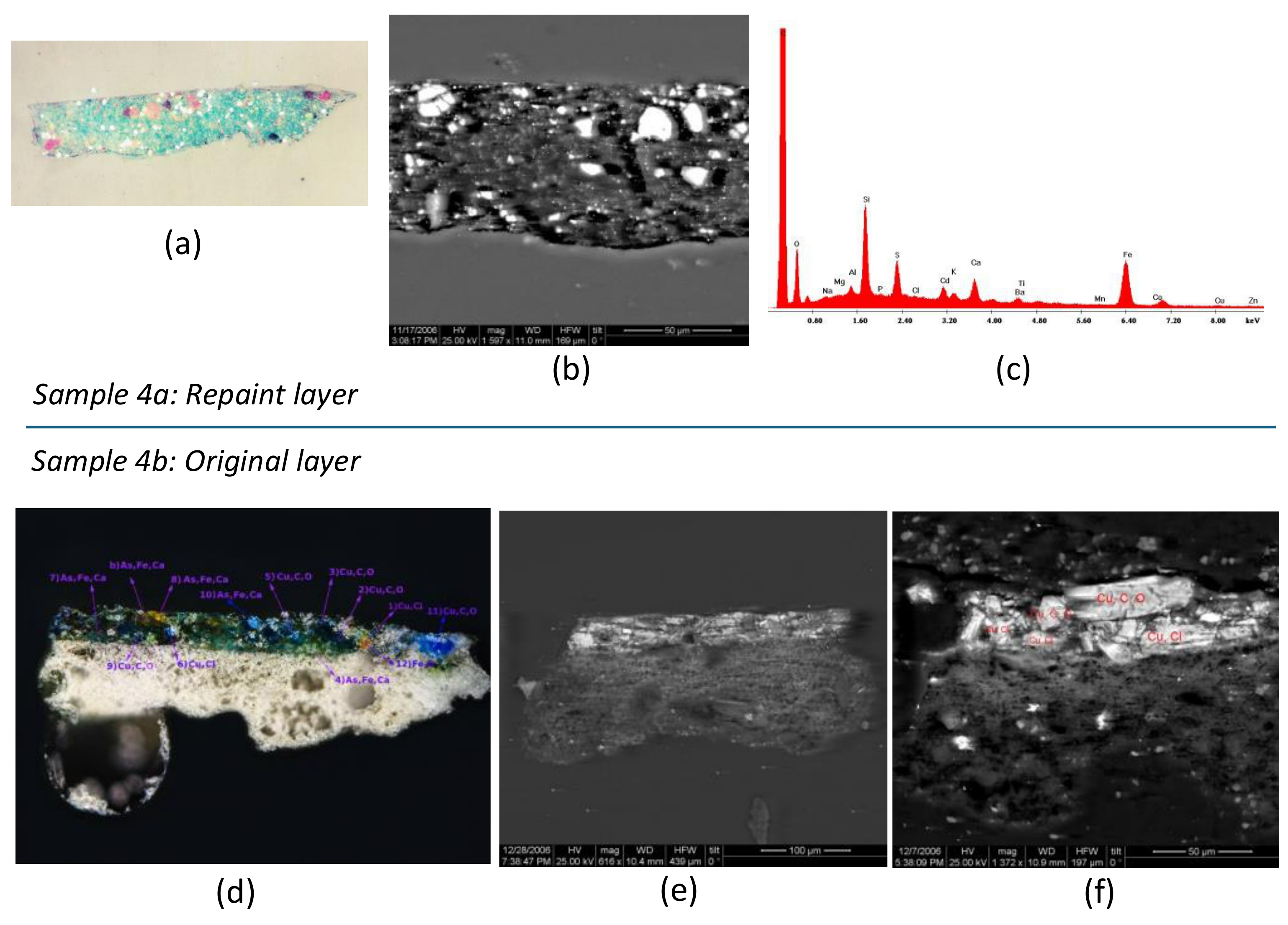
Figure 8.
Reference image of arseniosiderite crystals from the Kamariza mines, Laurion (after Rieck and Rieck (1999)).
Figure 8.
Reference image of arseniosiderite crystals from the Kamariza mines, Laurion (after Rieck and Rieck (1999)).
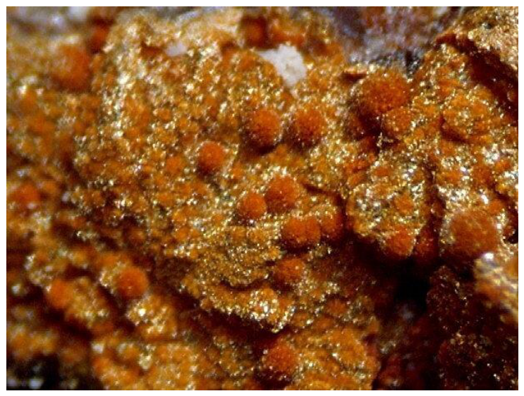
Figure 10.
SEM–EDS elemental maps of Sample 5 showing the distribution of Fe and Pb within the orange paint layer. (a) The area of the paint mapped in Secondary electron (SE) mode. (b) Mapping of the iron Kα1 line. (c) Mapping of the lead Lα1 line. The maps demonstrate the coexistence of iron-rich ochres and lead white within a single homogeneous paint layer.
Figure 10.
SEM–EDS elemental maps of Sample 5 showing the distribution of Fe and Pb within the orange paint layer. (a) The area of the paint mapped in Secondary electron (SE) mode. (b) Mapping of the iron Kα1 line. (c) Mapping of the lead Lα1 line. The maps demonstrate the coexistence of iron-rich ochres and lead white within a single homogeneous paint layer.
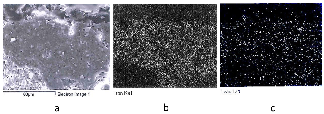
Figure 11.
Calibration plot of the radiocarbon date obtained from the wooden support of the icon (sample DEM-1454), calibrated using OxCal v4.4.4 with the IntCal20 calibration dataset. The calibrated ranges correspond to AD 1020–1160 (68.3% probability) and AD 970–1220 (95.4% probability); the calibrated probability distribution has a mean of AD 1090 and a standard deviation of 60 years.
Figure 11.
Calibration plot of the radiocarbon date obtained from the wooden support of the icon (sample DEM-1454), calibrated using OxCal v4.4.4 with the IntCal20 calibration dataset. The calibrated ranges correspond to AD 1020–1160 (68.3% probability) and AD 970–1220 (95.4% probability); the calibrated probability distribution has a mean of AD 1090 and a standard deviation of 60 years.
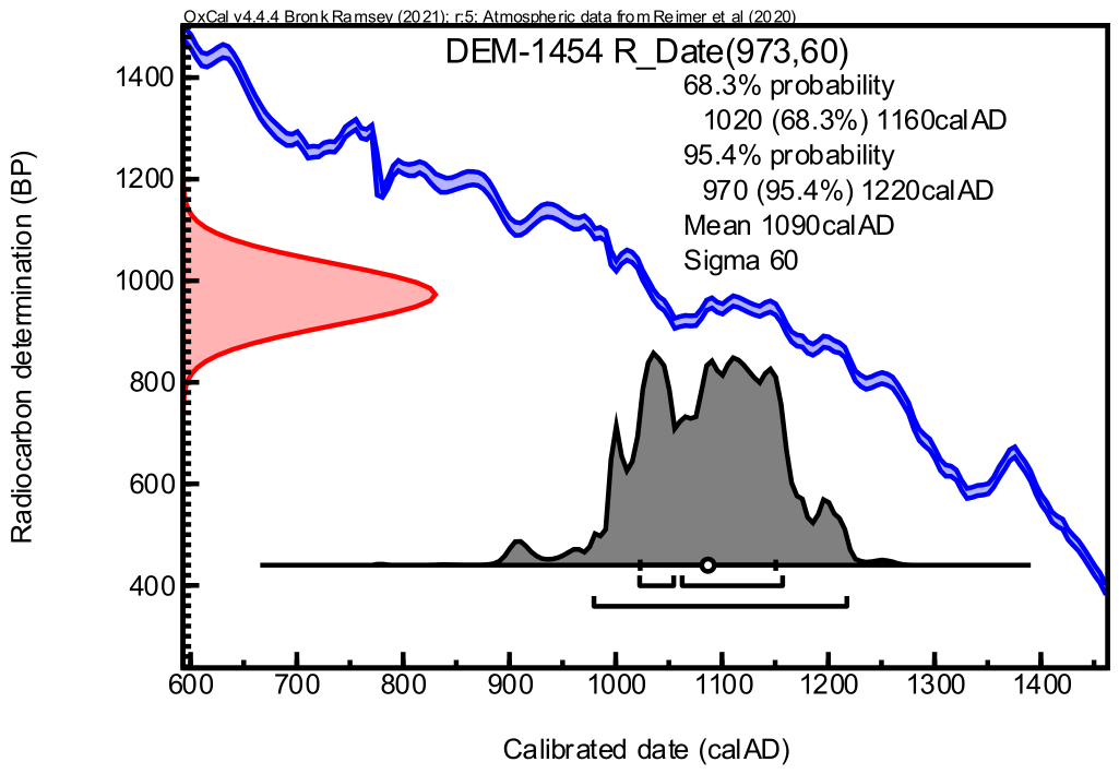

Table 1.
Brief description of the samples, macroscopic colour and pictorial element (see Figure 1).
Table 1.
Brief description of the samples, macroscopic colour and pictorial element (see Figure 1).
| Sample No. | Macroscopic Colour | Pictorial Element from Where the Sample was Taken |
|---|---|---|
| 1 | Dark red | Garment of Christ |
| 2 | Bright red | Background |
| 3 | Light green | Christ’s halo |
| 4 | Dark green | Virgin’s garment |
| 5 | Orange | Face/forehead of a hierarch in the right border |
Table 2.
Sample 4b. (See figures 7d–f): Quantitative bulk SEM–EDS results from three different regions (30 × 80 μm) of the original blue-green paint layer, expressed as oxide wt%. Quantitative analyses of the orange and brown grains, together with qualitative single-grain analyses of characteristic blue and blue-green grains, are also given.
Table 2.
Sample 4b. (See figures 7d–f): Quantitative bulk SEM–EDS results from three different regions (30 × 80 μm) of the original blue-green paint layer, expressed as oxide wt%. Quantitative analyses of the orange and brown grains, together with qualitative single-grain analyses of characteristic blue and blue-green grains, are also given.
| Na2O | MgO | As2O3 | Al2O3 | SiO2 | P2O5 | SO3 | PbO | Cl2O | CdO | K2O | CaO | BaO | TiO2 | Fe2O3 | Cu2O3 | ZnO | |
|---|---|---|---|---|---|---|---|---|---|---|---|---|---|---|---|---|---|
| Bulk analysis (area 1) | 1.77 | 0.93 | 2.37 | 1.28 | 7.01 | 3.34 | 6.15 | 10.83 | 3.07 | 0.86 | 1.52 | 14.65 | 1.58 | 0.00 | 2.88 | 40.97 | 0.77 |
| Bulk analysis (area 2) | 1.66 | 1.77 | 20.07 | 2.98 | 9.74 | 1.49 | 6.67 | 6.70 | 2.60 | 0.44 | 1.20 | 10.20 | 1.53 | 0.18 | 5.65 | 26.43 | 0.69 |
| Bulk analysis (area 3) | 4.07 | 0.99 | 2.62 | 1.83 | 18.70 | 4.00 | 6.04 | 8.59 | 5.92 | 0.58 | 2.79 | 10.88 | 1.52 | 0.00 | 1.91 | 28.84 | 0.71 |
| Orange and brown grains | 1.45 | 2.95 | 41.27 | 2.14 | 1.39 | 0.00 | 1.41 | 4.33 | 0.64 | 0.00 | 0.35 | 12.88 | 0.00 | - | 25.75 | 5.44 | 0.00 |
Table 3.
Bulk and individual grain analyses of Sample 5, the orange paint on the face/forehead of the hierarch in the margin of the icon (see Figure 9c and Figure 9d). Elements expressed as oxides wt%.
| Νa2O | MgO | Al2O3 | SiO2 | P2O5 | SO3 | Cl2O | K2O | CaO | FeO | PbO | |
|---|---|---|---|---|---|---|---|---|---|---|---|
| Bulk | 0.36 | 0.66 | 12.95 | 28.31 | 1.62 | 6.02 | 3.31 | 1.12 | 5.84 | 14.88 | 24.34 |
| Grain 1 | 0.63 | 0.15 | 5.78 | 20.12 | 1.21 | 2.40 | 2.38 | 0.73 | 1.44 | 47.91 | 16.02 |
| Grain 2 | 1.75 | - | - | 2.95 | 2.37 | 4.37 | 6.21 | 1.92 | 9.10 | 1.51 | 57.34 |
Table 4.
Radiocarbon dating results for the wooden support of the icon.
| Laboratory Code | Sample | Material | Radiocarbon Age (BP) | δ13C (‰) | Calibrated Age (AD) | Probability |
|---|---|---|---|---|---|---|
| DEM‒1454 | “Virgin Hodegetria” icon, Church of Episkopi, Thera | Wood from support | 973 ± 60 | -23.69 | 1020‒1160 970‒1220 |
(68.3%) (95.4%) |
| Mean: AD 1090 ± 60 (1σ) | ||||||
|
Sample pretreatment: Kyriaki Gogidou, Marigo-Eirini Kyriazi | ||||||
Table 5.
Summary of identified pigments, paint stratigraphy, binding-media evidence and painting technology of the investigated samples.
Table 5.
Summary of identified pigments, paint stratigraphy, binding-media evidence and painting technology of the investigated samples.
| Sample | Colour Area | Stratigraphy | Identified Pigments | Technological Observations |
|---|---|---|---|---|
| Sample 1 | Dark red garment of Christ | Two superimposed paint layers | Upper layer: iron-rich red ochre (hematite); Lower layer: cinnabar (HgS) | Unusual inverted stratigraphy, with a thin hematite-rich layer covering a thicker cinnabar layer. |
| Sample 2 | Bright red background | Single paint layer | Cinnabar (HgS) | Thick homogeneous cinnabar application. |
| Sample 3 | Light green halo of Christ | Single paint layer | Green earth + lead white | Premixed pigments forming a homogeneous paint layer. |
| Sample 4 | Dark green tunic of the Virgin | Modern repainting layer over original paint layer |
Repainting layer (Sample 4a): cadmium yellow (CdS), lithopone (BaSO4 + ZnS), iron ochre and possibly green earth, associated with an organic blue pigment (possibly indigo). Original layer (Sample 4b): azurite + green earth + lead white. Alteration products: Cu chlorides. Accessory minerals: possibly arseniosiderite and iron ochre. |
Evidence of later repainting (post-nineteenth century) covering the original blue-green paint layer. Original paint composed of premixed pigments applied as a single layer. |
| Sample 5 | Orange flesh tone from the hierarch’s face | Single paint layer | Iron-rich ochres + lead white + carbon black | Homogeneous premixed paint layer. No stratigraphic separation between underpainting and highlights observed. |
| Binding medium (all paints except the red background) | Proteinaceous material (P) | Possibly egg tempera | ||
| Preparation layer | Gypsum + proteinaceous component (consistent with animal glue) |
Disclaimer/Publisher’s Note: The statements, opinions and data contained in all publications are solely those of the individual author(s) and contributor(s) and not of MDPI and/or the editor(s). MDPI and/or the editor(s) disclaim responsibility for any injury to people or property resulting from any ideas, methods, instructions or products referred to in the content. |
© 2026 by the authors. Licensee MDPI, Basel, Switzerland. This article is an open access article distributed under the terms and conditions of the Creative Commons Attribution (CC BY) license (http://creativecommons.org/licenses/by/4.0/).
Copyright: This open access article is published under a Creative Commons CC BY 4.0 license, which permit the free download, distribution, and reuse, provided that the author and preprint are cited in any reuse.