

Submitted:
20 August 2026
Posted:
21 August 2026
You are already at the latest version
Abstract
Background: Outcomes after extracorporeal membrane oxygenation (ECMO) in children vary with
indication, support mode, illness severity, and program context, while data from smaller programs
remain limited. Methods: We conducted a retrospective single-center cohort study of all 30 unique
neonatal and pediatric patients who commenced ECMO at CHRU de Tours from 20 March 2022 to 15
December 2025. The primary outcome was survival to hospital discharge. Continuous variables are
reported as median [interquartile range], categorical variables as n/N (%), and key binary outcomes
with exact 95% confidence intervals. Because only eight patients survived, the primary analysis was
deliberately descriptive; no predictor model or broad unplanned hypothesis-testing panel was retained.
Results: Fourteen patients were neonates and 16 were older children; median age was 4.3 months [0.1-
24.0] and median weight was 6.8 kg [3.0-12.0]. Twenty-seven patients received venoarterial ECMO,
three received venovenous ECMO, and 10/29 with available data received extracorporeal
cardiopulmonary resuscitation. Median annual volume was 7.5 cases (range, 6-9). Successful
decannulation occurred in 9/30 patients (30.0%; 95% CI, 14.7%-49.4%), and 8/30 survived to hospital
discharge (26.7%; 95% CI, 12.3%-45.9%). Fifteen patients had a recorded circuit change. Source-
coded complication flags were frequent, including a field labeled disseminated intravascular
coagulation in 25/30 patients; these fields had no prospective diagnostic criteria, timing, severity
grading, or adjudication. Conclusion: This small, clinically heterogeneous and predominantly
venoarterial cohort provides a local benchmark with substantial statistical uncertainty. The
34
35
36
37
38
39
40
41
42
43
44
45
46
47
48
49
50
51
52
53
54
55
56
57
58
59
60
61
62
63
64
65
66
67
68
69
70
71
72
73
74
75
complication counts should be interpreted as documentation signals rather than validated ECMO-
attributable event rates. Three priorities for Tours are a prospective ELSO-aligned registry, structured
review of every ECMO run and circuit event, and recurring competency-based simulation.
Keywords:
extracorporeal membrane oxygenation
; pediatric critical care
; neonate
; hospital survival
; venoarterial ECMO
; quality improvement
1. Introduction
Extracorporeal membrane oxygenation (ECMO) provides temporary gas-exchange and/or circulatory support when conventional therapy is insufficient. Neonatal and pediatric outcomes differ substantially across respiratory, cardiac, and extracorporeal cardiopulmonary resuscitation (ECPR) populations; they also depend on age, anatomy, reversibility, pre-ECMO injury, and support configuration [1,2,3,4]. Crude survival percentages therefore cannot be compared fairly without attention to case mix and data definitions.
Contemporary pediatric ECMO is not a single clinical entity. Respiratory support, postoperative or nonsurgical cardiac support, and ECPR differ in reversibility, pre-cannulation physiology, technical configuration, and expected outcome [2,3,4,14]. Venoarterial support is also used across a broad range of diagnoses and carries different risks from venovenous support. Consequently, a center-level survival estimate is meaningful only when the underlying age distribution, diagnostic spectrum, support mode, and arrest burden are reported alongside it.
Complications further complicate interpretation. Bleeding, thrombosis, hemolysis, neurological injury, infection, and kidney dysfunction may be present before cannulation, emerge during support, or reflect the severity of the underlying illness [9,10,11,12]. Their reported frequency depends on objective definitions, surveillance intensity, timing, grading, adjudication, and the chosen denominator. Retrospective binary fields are therefore useful for identifying local safety and documentation priorities, but they are not automatically equivalent to standardized registry events.
Observational registry studies have reported associations between higher institutional ECMO volume and survival, but thresholds and populations vary and residual confounding remains likely [5,6,7]. In the present study, the CHRU de Tours program treated 6-9 neonatal and pediatric cases per year. We use the term smaller-volume program descriptively; it is not a universal quality category. The cohort falls below the <15 cases/year stratum used in one pediatric cardiac ECMO analysis [5], whereas other studies have used different cutoffs and case definitions [6,7].
The local workbook contained four calendar years of routine clinical data but had not been converted into a reproducible outcomes report. This study therefore aimed to estimate survival to hospital discharge, describe patient and ECMO characteristics, quantify circuit interventions and source-coded complication fields, and identify a short set of locally actionable quality-improvement priorities. The analysis was intentionally descriptive because the cohort contained only 30 patients and eight survivors.
2. Materials and Methods
2.1. Study Design, Setting, and Participants
This retrospective, observational, single-center cohort included every patient row in the CHRU de Tours neonatal and pediatric ECMO workbook with an ECMO start date from 20 March 2022 through 15 December 2025. The report follows the STROBE framework for cohort studies [15]. The unit of analysis was the ECMO episode represented by one workbook row. All 30 birth dates were unique, supporting 30 unique patients; no row was excluded. A neonate was defined as a patient aged 28 completed days or younger at cannulation; older patients were classified as pediatric.
Ethical approval for this retrospective study was granted by CHRU de Tours. The analysis used pseudonymized data collected during routine care. The study involved no intervention or participant contact and was conducted in accordance with the Declaration of Helsinki, local legislation, and institutional requirements. Direct identifiers and exact patient-level dates were excluded from all analytical outputs.
2.2. Data Source and Variable Handling
The source was one Microsoft Excel workbook with 69 columns, one of which was empty. Available fields covered demographics, diagnoses, pre-ECMO physiology and organ support, ECMO mode and cannulation, duration, circuit interventions, survival, lengths of stay, causes of death, and binary complication indicators. English/French and spelling variants of binary values were normalized without overwriting the source. One lactate value recorded as >20 mmol/L was represented as 20 mmol/L for summaries. One likely shifted cannulation-site entry in the duration column was treated as a site value and the corresponding duration was set to missing. No other clinical value was inferred.
Free-text diagnoses were grouped into congenital heart disease, respiratory/pulmonary disease, myocarditis, and other arrest, shock, or systemic disease. Respiratory and cardiac failure indicators were allowed to overlap. The source did not provide a formal screening log, so the cohort is a census of the supplied workbook rather than proof that every potentially eligible run in the institution was captured.
2.3. Outcomes and Operational Definitions
The primary outcome was survival to hospital discharge. The direct hospital-survival field was populated in 17/30 rows, whereas death status was complete; hospital survival was therefore defined as the complement of death status. There were no discrepancies in the 17 rows with both fields, and the derived outcome agreed with recorded pediatric intensive care unit survival in all 30 rows. Secondary outcomes were successful decannulation, survival to pediatric intensive care unit discharge, ECMO duration, circuit change, and recorded complication fields.
Disseminated intravascular coagulation (DIC) was defined for this analysis solely as a positive value in the source field labeled ‘CIVD.’ The workbook contained no laboratory criteria, International Society on Thrombosis and Haemostasis score, onset date, severity grade, or adjudication. Accordingly, 25/30 denotes 25 positive source flags; it does not establish 25 prospectively validated, incident, or ECMO-attributable DIC diagnoses. The same limitation applies to the other binary complication fields.
2.4. Statistical Analysis
Categorical variables are reported as n/N (%) with the available denominator, and continuous variables as median [25th-75th percentile] because of the small sample, skewed distributions, and extreme values. Exact two-sided 95% Clopper-Pearson confidence intervals were calculated for the primary outcome and other key binary outcomes. No values were imputed; available-case denominators are shown explicitly.
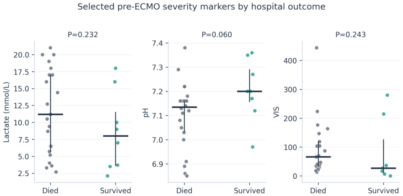
No formal sample-size calculation was performed because all available cases were included. For graphical description only, available pre-ECMO lactate, pH, and vasoactive-inotropic score values were compared by hospital outcome using two-sided Mann-Whitney U tests (Figure 2). The displayed P values are unadjusted and exploratory; they were not used to identify predictors or to support confirmatory inference. No broad multiplicity-adjusted screening, univariable odds-ratio plot, or multivariable model was retained. With only eight survivors, additional non-prespecified comparisons would be unstable, underpowered, and prone to chance findings, while multivariable modeling would be vulnerable to overfitting and separation. This restriction keeps the analysis aligned with its descriptive objective. Analyses were performed in Python using pandas, NumPy, and SciPy; figures were generated with Matplotlib.
3. Results
3.1. Cohort and Pre-ECMO Profile
The cohort contained 14 neonates and 16 older children. Median age was 4.3 months [0.1-24.0], median weight was 6.8 kg [3.0-12.0], and 20/30 patients were male. Congenital heart disease was the largest diagnostic group (12/30), followed by respiratory/pulmonary disease (8/30), myocarditis (5/30), and other arrest, shock, or systemic disease (5/30). Respiratory failure was recorded in 24/30 patients and cardiac failure in 18/30; the indicators were not mutually exclusive. Cardiac arrest before ECMO occurred in 11/30, and ECPR was recorded in 10/29 with available data. Median pre-ECMO lactate was 10.0 mmol/L [5.2-17.0] (n=29), pH 7.16 [7.06-7.20] (n=30), and vasoactive-inotropic score 43.9 [26.9-126.3] (n=28) (Table 1).
The physiological measurements describe marked pre-cannulation illness. Lactate was available in 29 patients, while pH was complete and the vasoactive-inotropic score was available in 28. In descriptive survivor/non-survivor displays, survivors had lower median lactate (8.0 vs 11.2 mmol/L), higher median pH (7.20 vs 7.13), and lower median vasoactive-inotropic score (27.0 vs 66.3) (Figure 2). The individual distributions overlapped substantially, and one lactate value recorded as >20 mmol/L was plotted at 20 mmol/L as prespecified. These visual comparisons are descriptive and were not used to claim prognostic effects.
3.2. ECMO Support and Clinical Outcomes
Seven cases commenced ECMO in 2022, six in 2023, eight in 2024, and nine in 2025; median annual volume was 7.5 cases (range, 6-9). Exactly two patients survived to hospital discharge in each calendar year, corresponding to annual survival proportions of 28.6%, 33.3%, 25.0%, and 22.2%, respectively (Figure 1). With only 6-9 cases per year, these percentages were highly unstable and do not establish a temporal trend. Venoarterial ECMO was used in 27/30 patients and venovenous ECMO in 3/30. Cannulation was classified as peripheral in 17/30 and central or mixed in 13/30. Median ECMO duration was 5.0 days [3.0-13.0] among 29 patients.
Figure 1.
Annual pediatric and neonatal ECMO volume and hospital outcome. Bars show the number of patients who survived to hospital discharge and died in each calendar year. Labels show the observed annual survival percentage. Annual percentages are descriptive and unstable because each year included only 6-9 cases.
Figure 1.
Annual pediatric and neonatal ECMO volume and hospital outcome. Bars show the number of patients who survived to hospital discharge and died in each calendar year. Labels show the observed annual survival percentage. Annual percentages are descriptive and unstable because each year included only 6-9 cases.
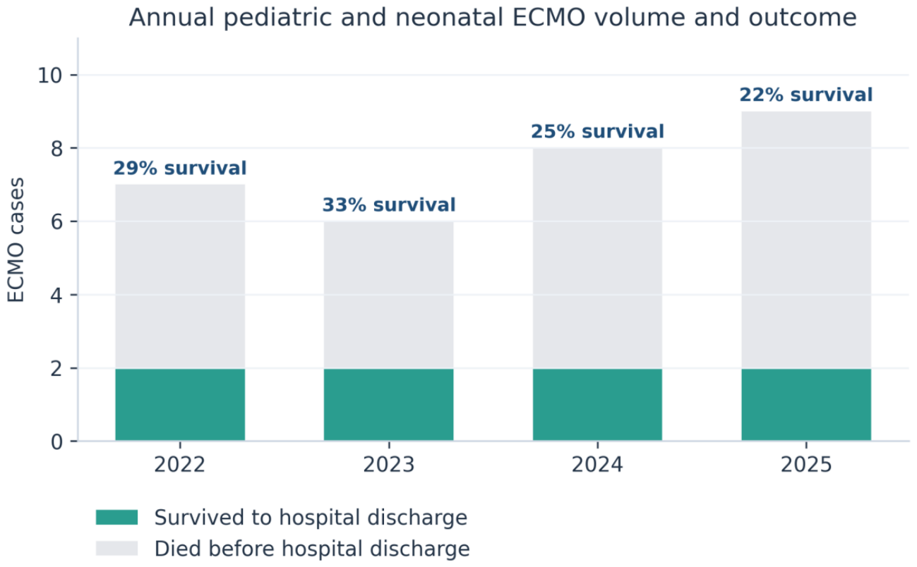
Figure 2.
Selected pre-ECMO severity markers by hospital outcome. Individual points show available lactate, pH, and vasoactive-inotropic score measurements in hospital survivors and non-survivors. Thick horizontal lines indicate medians and vertical lines the interquartile ranges. Displayed P values are exploratory, unadjusted comparisons and should not be interpreted as confirmatory evidence or independent prognostic effects.
Figure 2.
Selected pre-ECMO severity markers by hospital outcome. Individual points show available lactate, pH, and vasoactive-inotropic score measurements in hospital survivors and non-survivors. Thick horizontal lines indicate medians and vertical lines the interquartile ranges. Displayed P values are exploratory, unadjusted comparisons and should not be interpreted as confirmatory evidence or independent prognostic effects.
Successful decannulation occurred in 9/30 patients (30.0%; exact 95% CI, 14.7%-49.4%). Eight patients survived to pediatric intensive care unit and hospital discharge (26.7%; exact 95% CI, 12.3%-45.9%), indicating one death after successful decannulation. Figure 3 displays these nested outcomes and their exact confidence intervals. The identical pediatric intensive care unit and hospital-discharge estimates indicate that all eight patients discharged alive from intensive care also survived to hospital discharge; the wide intervals emphasize the imprecision caused by the small cohort and should not be interpreted as evidence of differences between endpoints. A circuit change was recorded in 15/30 patients (50.0%; exact 95% CI, 31.3%-68.7%) (Table 2). The available free text grouped reasons for circuit change as cannula position, malfunction, or replacement (7/15); thrombus or clot (4/15); bleeding (2/15); failure to wean (1/15); and hemolysis (1/15). Event timing, component-level detail, and the number of changes per patient were unavailable.
3.3. Source-Coded Complication Fields
The most frequent positive source field was CIVD/DIC (25/30), followed by hemolysis (23/30), renal failure (23/30), major bleeding (21/30), and infection (18/30). Thrombosis and stroke/intracranial hemorrhage were each positive in 10/30, seizure in 6/30, the source field labeled CRPT in 5/30, and limb ischemia in 1/30 (Table 3). The source did not define CRPT; it may refer to renal replacement therapy but was retained as a source label. Because onset, diagnostic thresholds, severity, and adjudication were absent, these counts cannot be interpreted as incident ECMO complication rates or compared directly with registry events per 1,000 ECMO hours.
Twenty-two patients died before hospital discharge. The grouped source cause was multiorgan failure or dysfunction in 15/22 deaths, respiratory or pulmonary failure in 3/22, brain death in 2/22, cardiac or cardiopulmonary failure in 1/22, and another cause in 1/22. These categories summarize free text and were not independently adjudicated; multiorgan dysfunction should be understood as a final clinical pathway rather than a single proven cause.

Table 3.
Positive source-coded complication fields.
| Source field | Positive records | Exact 95% CI |
|---|---|---|
| CIVD/DIC | 25/30 (83.3%) | 65.3%-94.4% |
| Hemolysis | 23/30 (76.7%) | 57.7%-90.1% |
| Renal failure | 23/30 (76.7%) | 57.7%-90.1% |
| Major bleeding | 21/30 (70.0%) | 50.6%-85.3% |
| Infection | 18/30 (60.0%) | 40.6%-77.3% |
| Thrombosis | 10/30 (33.3%) | 17.3%-52.8% |
| Stroke/intracranial hemorrhage | 10/30 (33.3%) | 17.3%-52.8% |
| Seizure | 6/30 (20.0%) | 7.7%-38.6% |
| CRPT (source label; undefined) | 5/30 (16.7%) | 5.6%-34.7% |
| Limb ischemia | 1/30 (3.3%) | 0.1%-17.2% |
Note. These are positive binary fields in the retrospective source, not validated incident ECMO-attributable events. DIC was operationalized solely as a positive value in the field labeled CIVD; no laboratory criteria, timing, severity, or adjudication were available. CRPT was not defined in the source. CIVD/DIC, disseminated intravascular coagulation.
4. Discussion
This study provides a transparent local benchmark for 30 neonatal and pediatric ECMO patients treated over 45 months. Hospital survival was 26.7%, but its exact 95% confidence interval ranged from 12.3% to 45.9%, demonstrating substantial statistical uncertainty. The cohort was 90% venoarterial, almost half were neonates, 40% had congenital heart disease, and approximately one-third had ECPR recorded. It is therefore not comparable to a respiratory-dominant ECMO population.
The point estimate is lower than broad ELSO pediatric summaries and some multicenter cohorts [1,4,8], but a crude difference cannot establish inferior program performance. Referral patterns, cardiac anatomy, postoperative states, pre-ECMO injury, ECPR exposure, support mode, and definitions all influence outcome. The absence of patient-level risk adjustment and the wide confidence interval make a league-table comparison inappropriate. ECPR outcomes in particular depend on arrest location, rhythm, low-flow duration, cannulation speed, and neurological injury, several of which were unavailable here [14].
The annual display reinforces the need for restraint (Figure 1). Although case volume increased from seven in 2022 to nine in 2025, the absolute number of survivors remained two in every year. The resulting survival percentages therefore changed mainly because of small denominators. This pattern cannot support a claim of improvement or deterioration over time; year-to-year monitoring will become informative only after more cases accumulate and case mix and process measures are captured consistently.
The cohort entered support with substantial physiological compromise, including a median lactate of 10.0 mmol/L, median pH of 7.16, and median vasoactive-inotropic score of 43.9. Figure 2 shows clinically plausible directions toward lower lactate, higher pH, and lower vasoactive support among survivors, but it also shows wide dispersion and overlap. The pH difference was the closest to a conventional threshold in the exploratory draft, yet the small sample, multiple possible comparisons, timing uncertainty, and missing values make a prognostic interpretation unsafe.
These markers should consequently be treated as descriptors of the state before cannulation, not as independent determinants of outcome. Each value is influenced by diagnosis, age, preceding resuscitation, vasoactive treatment, ventilation, sampling time, and the interval to established ECMO flow. A prospective registry should capture the nearest pre-ECMO and worst pre-ECMO measurements with timestamps, units, and treatments already administered. That design would permit clinically defensible risk-adjusted analysis once the cohort becomes sufficiently large.
The positive complication fields are clinically important documentation signals, not validated event rates. Bleeding, thrombosis, and hemolysis are common and biologically interrelated during pediatric ECMO [9,10,11,12], but comparisons require the same objective thresholds, surveillance intensity, timing, severity grades, and exposure denominator. In this workbook, a positive DIC flag could represent pre-ECMO shock-associated coagulopathy, clinician judgment, laboratory abnormalities, or DIC developing during support. The observation that 25/30 rows were positive is therefore possible as a record of coding, but it cannot be defended as 25 new ECMO-caused DIC cases.
The same caution applies to the 23 hemolysis and 23 renal-failure flags. Their co-occurrence may reflect genuine multiorgan illness, broad local definitions, or documentation practices. The 50% circuit-change flag is more directly actionable: each event can be reconstructed from circuit performance, anticoagulation, imaging, blood-product exposure, and outcome rather than attributed to a generic failure label.
The neurological fields also merit focused follow-up. Stroke or intracranial hemorrhage was positive in 10 patients and seizure in six, but the source did not distinguish events present before ECMO from those detected during support, nor did it document surveillance methods. Survival to discharge is therefore an incomplete endpoint. Future reporting should link acute neurological events to standardized functional and neurodevelopmental outcomes after discharge.
The term smaller-volume program is defined here by the observed 6-9 cases per year and by reference to published strata, not by a universal cutoff. Volume-outcome associations are plausible because frequent exposure may reinforce team readiness, but volume also proxies for case selection, referral timing, surgical capability, and institutional resources [5,6,7]. This dataset cannot isolate a causal effect of volume.
The final statistical strategy is intentionally restrained. Exact confidence intervals quantify what the 30-patient census can support. The three clinically selected survivor/non-survivor comparisons shown in Figure 2 are retained only as exploratory graphical context; their unadjusted P values do not resolve confounding and may be unstable with eight survivors. A broader screening panel and predictor modeling were therefore omitted. The clinically relevant distributions and denominators remain visible, while no independent predictor is claimed.
First, implement a prospective ELSO-aligned registry and data dictionary. Core outcomes, process timestamps, support mode, ECPR details, and objective definitions for bleeding, hemolysis, thrombosis, neurological injury, infection, acute kidney injury, DIC, and renal replacement therapy should be mandatory. A practical target is at least 95% completeness of core fields with quarterly validation.
Second, review every ECMO run, circuit change, and death in a structured, no-blame multidisciplinary conference. The review should capture the component involved, objective trigger, circuit parameters, imaging, anticoagulation and hemostasis data, response, preventability, and assigned follow-up action. Completion within 30 days and closure of assigned actions are measurable process outcomes.
Third, maintain competency between infrequent cases through recurring multidisciplinary simulation. Scenarios should prioritize activation and cannulation, air entrainment, pump or oxygenator failure, major bleeding, emergency circuit exchange, and transport. Individual competency records and team response metrics should be reviewed at least annually, consistent with contemporary ELSO training guidance [13].
Strengths include inclusion of all 30 rows in the supplied workbook, complete primary outcome ascertainment through a reconciled death field, explicit available-case denominators, exact confidence intervals, and conservative interpretation. The analysis also separates source-coded complication flags from validated registry events.
The principal limitations are the small single-center cohort, eight survivors, lack of a screening log, broad diagnostic groups, no external risk adjustment, substantial missingness in ventilation duration and lengths of stay, and absent standardized definitions or timing for complications. One source-column shift and heterogeneous coding create additional misclassification risk. Long-term functional and neurodevelopmental outcomes were unavailable. These limitations prevent causal inference, predictor identification, or a definitive comparison with other centers.
Among 30 neonatal and pediatric ECMO patients treated at CHRU de Tours from 2022 to 2025, eight survived to hospital discharge. The estimate is imprecise and reflects a heterogeneous, predominantly venoarterial cohort. Frequent source-coded complications and circuit changes identify priorities for data validation and structured review but are not validated ECMO-attributable rates. A focused program of prospective standardized data capture, review of every run and circuit event, and recurring competency-based simulation is directly applicable to Tours and will make future outcomes more interpretable and improvable.
Author Contributions
SAS: Conceptualization, Data curation, Formal analysis, Investigation, Methodology, Visualization, Writing—original draft. MH, NR-R, J-MEA, and JC: Clinical interpretation, Validation, Writing—review and editing. All authors contributed to the article, approved the submitted version, and accept responsibility for the work.
Funding
This research received no external funding.
Institutional Review Board Statement
The study involving human participants received institutional ethical approval from CHRU de Tours. The study was conducted in accordance with local legislation, institutional requirements, and the Declaration of Helsinki. The analysis used pseudonymized data collected during routine care and involved no additional intervention or participant contact. No potentially identifiable patient information is included in this article.
Data Availability Statement
The de-identified patient-level dataset is not publicly available because it contains sensitive pediatric health data. Aggregated data supporting the conclusions are included in this article. Requests for access may be directed to the corresponding author and are subject to approval by the CHRU de Tours data controller and applicable French data-protection requirements.
Acknowledgments
The authors acknowledge the multidisciplinary neonatal and pediatric ECMO teams at CHRU de Tours whose clinical work generated the source data.
Conflicts of Interest
The authors declare that the research was conducted in the absence of any commercial or financial relationships that could be construed as a potential conflict of interest.
References
- Tonna JE, Boonstra PS, MacLaren G, Paden M, Brodie D, Anders M, et al. Extracorporeal Life Support Organization Registry International Report 2022: 100,000 Survivors. ASAIO J. 2024;70(2):131-143. [CrossRef]
- Maratta C, Potera RM, van Leeuwen G, Castillo Moya A, Raman L, Annich GM. Extracorporeal Life Support Organization (ELSO): 2020 Pediatric Respiratory ELSO Guideline. ASAIO J. 2020;66(9):975-979. [CrossRef]
- Brown G, Moynihan KM, Deatrick KB, Hoskote A, Sandhu HS, Aganga D, et al. Extracorporeal Life Support Organization (ELSO): Guidelines for Pediatric Cardiac Failure. ASAIO J. 2021;67(5):463-475. [CrossRef]
- O’Neil ER, Guner Y, Anders MM, Priest J, Friedman ML, Raman L, et al. Pediatric Highlights From the Extracorporeal Life Support Organization Registry: 2017-2022. ASAIO J. 2024;70(1):8-13. [CrossRef]
- Karamlou T, Vafaeezadeh M, Parrish AM, Cohen GA, Welke KF, Permut L, et al. Increased extracorporeal membrane oxygenation center case volume is associated with improved extracorporeal membrane oxygenation survival among pediatric patients. J Thorac Cardiovasc Surg. 2013;145(2):470-475. [CrossRef]
- Barbaro RP, Odetola FO, Kidwell KM, Paden ML, Bartlett RH, Davis MM, et al. Association of hospital-level volume of extracorporeal membrane oxygenation cases and mortality: analysis of the Extracorporeal Life Support Organization registry. Am J Respir Crit Care Med. 2015;191(8):894-901. [CrossRef]
- Gonzalez DO, Sebastiao YV, Cooper JN, Minneci PC, Deans KJ. Pediatric extracorporeal membrane oxygenation mortality is related to extracorporeal membrane oxygenation volume in US hospitals. J Surg Res. 2019;236:159-165. [CrossRef]
- Choi YH, Jhang WK, Park SJ, Choi HJ, Oh M, Kwon JE, et al. Pediatric extracorporeal membrane oxygenation in Korea: a multicenter retrospective study on utilization and outcomes spanning over a decade. J Korean Med Sci. 2024;39(3):e33. [CrossRef]
- Dalton HJ, Reeder R, Garcia-Filion P, Holubkov R, Berg RA, Zuppa A, et al. Factors associated with bleeding and thrombosis in children receiving extracorporeal membrane oxygenation. Am J Respir Crit Care Med. 2017;196(6):762-771. [CrossRef]
- Dalton HJ, Cashen K, Reeder RW, Berg RA, Shanley TP, Newth CJL, et al. Hemolysis during pediatric extracorporeal membrane oxygenation: associations with circuitry, complications, and mortality. Pediatr Crit Care Med. 2018;19(11):1067-1076. [CrossRef]
- Gajkowski EF, Herrera G, Hatton L, Velia Antonini M, Vercaemst L, Cooley E. ELSO guidelines for adult and pediatric extracorporeal membrane oxygenation circuits. ASAIO J. 2022;68(2):133-152. [CrossRef]
- McMichael ABV, Ryerson LM, Ratano D, Fan E, Faraoni D, Annich GM. 2021 ELSO adult and pediatric anticoagulation guidelines. ASAIO J. 2022;68(3):303-310. [CrossRef]
- Moore EA, Han P, Shekar K, Ramanathan K, Riera del Brio J, Puslecki M, et al. ELSO 2025 narrative guideline on ECMO training and continuing education. ASAIO J. 2026;72(4):274-283. [CrossRef]
- Guerguerian AM, Sano M, Todd M, Honjo O, Alexander P, Raman L. Pediatric extracorporeal cardiopulmonary resuscitation ELSO guidelines. ASAIO J. 2021;67(3):229-237. [CrossRef]
- von Elm E, Altman DG, Egger M, Pocock SJ, Gotzsche PC, Vandenbroucke JP. Strengthening the Reporting of Observational Studies in Epidemiology (STROBE) statement: guidelines for reporting observational studies. BMJ. 2007;335(7624):806-808. [CrossRef]
Figure 3.
Primary and secondary clinical outcomes. Points show the observed proportions of successful decannulation, survival to pediatric intensive care unit discharge, and survival to hospital discharge; horizontal bars show exact two-sided 95% Clopper-Pearson confidence intervals. The two survival estimates are identical because all eight patients discharged alive from intensive care survived to hospital discharge. These nested endpoints are presented descriptively and were not compared statistically.
Figure 3.
Primary and secondary clinical outcomes. Points show the observed proportions of successful decannulation, survival to pediatric intensive care unit discharge, and survival to hospital discharge; horizontal bars show exact two-sided 95% Clopper-Pearson confidence intervals. The two survival estimates are identical because all eight patients discharged alive from intensive care survived to hospital discharge. These nested endpoints are presented descriptively and were not compared statistically.
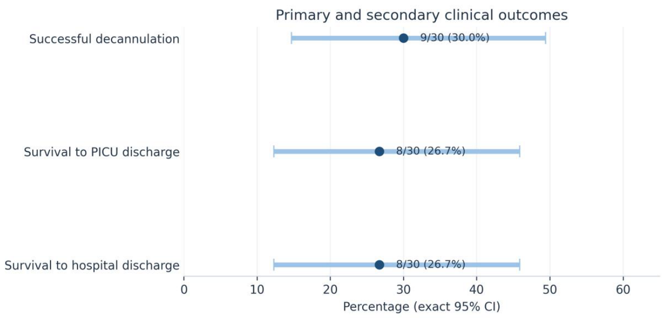
Table 1.
Cohort characteristics and ECMO support.
| Characteristic | Result |
|---|---|
| Age, months | 4.3 [0.1-24.0] (n=30) |
| Neonatal age (<=28 days) | 14/30 (46.7%) |
| Weight, kg | 6.8 [3.0-12.0] (n=30) |
| Male sex | 20/30 (66.7%) |
| Congenital heart disease | 12/30 (40.0%) |
| Respiratory/pulmonary disease | 8/30 (26.7%) |
| Myocarditis | 5/30 (16.7%) |
| Other arrest/shock/systemic disease | 5/30 (16.7%) |
| Cardiac arrest before ECMO | 11/30 (36.7%) |
| ECPR | 10/29 (34.5%) |
| Pre-ECMO lactate, mmol/L | 10.0 [5.2-17.0] (n=29) |
| Pre-ECMO pH | 7.16 [7.06-7.20] (n=30) |
| Pre-ECMO VIS | 43.9 [26.9-126.3] (n=28) |
| Venoarterial ECMO | 27/30 (90.0%) |
| Venovenous ECMO | 3/30 (10.0%) |
| Peripheral cannulation | 17/30 (56.7%) |
| Central or mixed cannulation | 13/30 (43.3%) |
Note. Values are median [interquartile range] or n/N (%). Denominators reflect available data. ECMO, extracorporeal membrane oxygenation; ECPR, extracorporeal cardiopulmonary resuscitation; VIS, vasoactive-inotropic score.
Table 2.
Clinical outcomes and circuit intervention.
| Outcome or measure | Result | Exact 95% CI |
|---|---|---|
| ECMO duration, days | 5.0 [3.0-13.0] (n=29) | - |
| Successful decannulation | 9/30 (30.0%) | 14.7%-49.4% |
| Survival to PICU discharge | 8/30 (26.7%) | 12.3%-45.9% |
| Survival to hospital discharge | 8/30 (26.7%) | 12.3%-45.9% |
| Any recorded circuit change | 15/30 (50.0%) | 31.3%-68.7% |
Note. Hospital survival was derived as the complement of the complete death-status field and agreed with all available direct hospital-survival entries. PICU, pediatric intensive care unit.
Disclaimer/Publisher’s Note: The statements, opinions and data contained in all publications are solely those of the individual author(s) and contributor(s) and not of MDPI and/or the editor(s). MDPI and/or the editor(s) disclaim responsibility for any injury to people or property resulting from any ideas, methods, instructions or products referred to in the content. |
© 2026 by the authors. Licensee MDPI, Basel, Switzerland. This article is an open access article distributed under the terms and conditions of the Creative Commons Attribution (CC BY) license (http://creativecommons.org/licenses/by/4.0/).
Copyright: This open access article is published under a Creative Commons CC BY 4.0 license, which permit the free download, distribution, and reuse, provided that the author and preprint are cited in any reuse.