
Submitted:
20 August 2026
Posted:
21 August 2026
You are already at the latest version
Abstract
Background/Objectives: Point-of-care ultrasound of the antral cross-sectional area (CSA) assesses perioperative aspiration risk. However, the standard 340mm2 cut-off derived from healthy populations is inappropriate for chronic hepatitis B (CHB) patients due to disease-induced gastrointestinal alterations. This study aimed to determine a disease-specific CSA cut-off to confirm an empty stomach in CHB patients and identify factors influencing baseline CSA. Methods: In this prospective cohort study, 223 CHB patients underwent ultrasound CSA measurement in the right lateral decubitus (RLD) position. This was immediately followed by endoscopic gastric fluid aspiration (gold standard), defining an "empty stomach" as < 0.8 mL/kg. Receiver operating characteristic (ROC) curves evaluated diagnostic performance, while multivariate regression identified independent factors affecting the CSA. Results: Among 214 analyzed patients, the area under the ROC curve was 0.763 (95% confidence interval [CI]: 0.685–0.842). The optimal RLD CSA cut-off for an empty stomach was 835 mm2, yielding 86.1% sensitivity, 61.1% specificity, 94.4% positive predictive value (PPV), and 36.3% negative predictive value (NPV). Multivariate analysis identified cholinesterase (CHE) and albumin (ALB) as independent predictors of baseline CSA. Conclusions: RLD antral CSA cut-off of 835 mm2 provides excellent diagnostic confidence (94.4% PPV) for confirming an empty stomach in CHB patients. Utilizing this disease-specific threshold can optimize perioperative airway management and prevent unwarranted defensive anesthetic interventions.
Keywords:
1. Introduction
2. Materials and Methods
Study Design
Study Population and Eligibility Criteria
Sample Size Determination
Ultrasound Protocol
Endoscopic Aspiration (The Reference Standard)
Clinical and Biochemical Data Collection
Statistical Analysis
3. Results
3.1. Patient Enrollment and Baseline Characteristics
3.2. Diagnostic Performance of Antral CSA
3.3. Univariate Analysis of Factors Affecting Baseline CSA
3.4. Multivariate Linear Regression Analysis
4. Discussion
5. Conclusions
Author Contributions
Funding
Institutional Review Board Statement
Informed Consent Statement
Data Availability Statement
Acknowledgments
Conflicts of Interest
Abbreviations
| Abbreviation | Full Term |
| ALB | albumin |
| ALT | alanine aminotransferase |
| ANOVA | analysis of variance |
| Ao | abdominal aorta |
| ASA | American Society of Anesthesiologists |
| AST | aspartate aminotransferase |
| AUC | area under the curve |
| B | unstandardized regression coefficient |
| BMI | body mass index |
| CHB | chronic hepatitis B |
| CHE | cholinesterase |
| CI | confidence interval |
| CSA | cross-sectional area |
| D1 | maximal anteroposterior diameter |
| D2 | longitudinal diameter |
| DBIL | direct bilirubin |
| EGD | esophagogastroduodenoscopy |
| HBV | hepatitis B virus |
| IBIL | indirect bilirubin |
| IQR | interquartile range |
| L | left lobe of the liver |
| NPV | negative predictive value |
| PHG | portal hypertensive gastropathy |
| POCUS | point-of-care ultrasound |
| PPV | positive predictive value |
| PT | prothrombin time |
References
- Okada, Y.; Toyama, H.; Kamata, K.; Yamauchi, M. A clinical study comparing ultrasound-measured pyloric antrum cross-sectional area to computed tomography-measured gastric content volume to detect high-risk stomach in supine patients undergoing emergency abdominal surgery. J. Clin. Monit. Comput. 2020, 34, 875–881. [Google Scholar] [CrossRef]
- Ng, A.; Smith, G. Gastroesophageal reflux and aspiration of gastric contents in anesthetic practice. Anesth. Analg. 2001, 93, 494–513. [Google Scholar] [CrossRef]
- Warner, M.A.; Warner, M.E.; Weber, J.G. Clinical significance of pulmonary aspiration during the perioperative period. Anesthesiology 1993, 78, 56–62. [Google Scholar] [CrossRef]
- Kaydu, A.; Gokcek, E. Preoperative Assessment of Ultrasonographic Measurement of Antral Area for Gastric Content. Med. Sci. 2018, 24, 5542–5548. [Google Scholar] [CrossRef]
- Sebrechts, T.; Perlas, A.; Abbas, S. Serial Gastric Ultrasound to Evaluate Gastric Emptying After Prokinetic Therapy with Domperidone and Erythromycin in a Surgical Patient with a Full Stomach: A Case Report. A A Pract. 2018, 11, 106–108. [Google Scholar] [CrossRef]
- Perlas, A.; Chan, V.W.; Lupu, C.M.; et al. Ultrasound assessment of gastric content and volume. Anesthesiology 2009, 111, 82–89. [Google Scholar] [CrossRef]
- Van de Putte, P.; Perlas, A. Ultrasound assessment of gastric content and volume. Br. J. Anaesth. 2014, 113, 12–22. [Google Scholar] [CrossRef]
- Sharma, V.; Gudivada, D.; Gueret, R.; Bailitz, J. Ultrasound-assessed gastric antral area correlates with aspirated tube feed volume in enterally fed critically ill patients. Nutr. Clin. Pract. 2017, 32, 206–211. [Google Scholar] [CrossRef]
- Castaner, H.V.; Jorda, M.V.; Blanch, X.S.; Valero, R. Preoperative bedside ultrasound assessment of gastric volume and evaluation of predisposing factors for delayed gastric emptying: a case-control observational study. J. Clin. Monit. Comput. 2020, 02. [Google Scholar] [CrossRef]
- Tacken, M.C.T.; van Leest, T.A.J.; van de Putte, P.; Keijzer, C.; Perlas, A. Ultrasound assessment of gastric volumes of thick fluids. Eur. J. Anaesthesiol. 2021, 38, 1223–1229. [Google Scholar] [CrossRef]
- Van de Putte, P.; Perlas, A. Ultrasound assessment of gastric content and volume. Br. J. Anaesth. 2014, 113, 12–22. [Google Scholar] [CrossRef]
- Bouvet, L.; Mazoit, J.X.; Chassard, D.; et al. Clinical assessment of the ultrasonographic measurement of antral area for estimating preoperative gastric content and volume. Anesthesiology 2011, 114, 1086–1092. [Google Scholar] [CrossRef]
- Han, S.H. Extrahepatic manifestations of chronic hepatitis B. Clin. Liver Dis. 2004, 8, 403–418. [Google Scholar] [CrossRef]
- Kalaitzakis, E. Gastrointestinal dysfunction in liver cirrhosis. World J. Gastroenterol. 2014, 20, 14686–14695. [Google Scholar] [CrossRef]
- Vandenbroucke, J.P.; von Elm, E.; Altman, D.G.; et al. Strengthening the Reporting of Observational Studies in Epidemiology (STROBE): explanation and elaboration. PLoS Med. 2007, 4, e297. [Google Scholar] [CrossRef] [PubMed]
- Cook, T.M.; Woodall, N.; Frerk, C. Major complications of airway management in the UK: results of the Fourth National Audit Project of the Royal College of Anaesthetists and the Difficult Airway Society. Part 1: anaesthesia. Br. J. Anaesth. 2011, 106, 617–631. [Google Scholar] [CrossRef]
- Lienhart, A.; Auroy, Y.; Pequignot, F.; et al. Survey of anesthesia-related mortality in France. Anesthesiology 2006, 105, 1087–1097. [Google Scholar] [CrossRef]
- Perlas, A.; Chan, V.W.; Lupu, C.M.; et al. Ultrasound assessment of gastric content and volume. Anesthesiology 2009, 111, 82–89. [Google Scholar] [CrossRef]
- Perlas, A.; Arzola, C.; Van de Putte, P. Point-of-care gastric ultrasound and aspiration risk assessment: a narrative review. Can. J. Anaesth. 2018, 65, 437–448. [Google Scholar] [CrossRef]
- Bouvet, L.; Miquel, A.; Chassard, D.; et al. Could a single standardized ultrasonographic measurement of antral area be of interest for assessing gastric contents? A preliminary report. Eur. J. Anaesthesiol. 2009, 26, 1015–1019. [Google Scholar] [CrossRef]
- Fujigaki, T.; Fukusaki, M.; Nakamura, H.; Shibata, O.; Sumikawa, K. Quantitative evaluation of gastric contents using ultrasound. J. Clin. Anesth. 1993, 5, 451–455. [Google Scholar] [CrossRef]
- Perlas, A.; Mitsakakis, N.; Liu, L.; et al. Validation of a mathematical model for ultrasound assessment of gastric volume by gastroscopic examination. Anesth. Analg. 2013, 116, 357–363. [Google Scholar] [CrossRef]
- Hveem, K.; Hausken, T.; Berstad, A. Ultrasonographic assessment of fasting liquid content in the human stomach. Scand. J. Gastroenterol. 1994, 29, 786–789. [Google Scholar] [CrossRef]
- Tomomasa, T.; Tabata, M.; Nako, Y.; Kaneko, H.; Morikawa, A. Ultrasonographic assessment of intragastric volume in neonates: factors affecting the relationship between intragastric volume and antral cross-sectional area. Pediatr. Radiol. 1996, 26, 815–820. [Google Scholar] [CrossRef]
- Schmitz, A.; Thomas, S.; Melanie, F.; et al. Ultrasonographic gastric antral area and gastric contents volume in children. Paediatr. Anaesth. 2012, 22, 144–149. [Google Scholar] [CrossRef]
- Liu, W.; Zhao, W.; Luo, C.; et al. Expression of HbsAg in the multi-tissues of died fetus delivered by parturients with hepatitis B. Chin. J. Hepatol. 2002, 10, 143–144. [Google Scholar]
- Zhen, Z.; He, Z.; Jin, G.; et al. Influence of HBV infection for gastric mucous membrane in chronic hepatitis B and cirrhosis. Chin. J. Pract. Intern. Med. 2005, 12, 1078–1080. [Google Scholar]
- Yang, L.; Chen, D. The relationship between HBV antigen and HBV DNA detection and histological changes in gastric mucosa of patients with cirrhosis, gastritis and canker. J. Clin. Hepatol. 1996, 12, 206–208. [Google Scholar]
- Mason, A.; Wick, M.; White, H.; et al. Hepatitis B virus replication in diverse cell types during chronic hepatitis B virus infection. Hepatology 1993, 18, 781–789. [Google Scholar] [CrossRef]
- He, Z.; Yuan, W.; Sun, M.; et al. The characteristics of gastric mucosa and pathogenesis in patients with chronic hepatitis B and cirrhosis. Hebei Med. J. 2007, 10, 1072–1074. [Google Scholar]
- Wu, L. Comparison study between clinical diagnosis and pathological diagnosis in patients with chronic hepatitis B. Hainan Med. J. 2013, 24, 560–562. [Google Scholar]
- Dai, G.; Liu, W. Research on the clinical correlations about the findings of gastroscope and B ultrasonography and Child-Pugh’s Score in patients with post-hepatitis B cirrhosis. J. Yanan Univ. (Med. Sci.) 2013, 11, 19–21. [Google Scholar]
- Zhu, J.; Lv, X. Clinical analysis on stomach mucosa disease of patients with chronic hepatitis B. Chin. J. Exp. Clin. Infect. Dis. 2012, 6, 34–36. [Google Scholar]
- Zhu, J.; Rao, J.; Lv, Y.; et al. Clinical effects of H. pylori eradication with 10-day sequential therapy in patients with chronic atrophic gastritis and chronic hepatitis B. Chin. J. Clin. Gastroenterol. 2013, 25, 283–285. [Google Scholar]
- Maini, M.K.; Boni, C.; Lee, C.K.; et al. The role of virus-specific CD8(+) cells in liver damage and viral control during persistent hepatitis B virus infection. J. Exp. Med. 2000, 191, 1269–1280. [Google Scholar] [CrossRef]
- Li, H.; Luo, Y.; Zhang, Q.; et al. Ultrasound-based assessment of impaired gastric emptying in patients with hepatitis B cirrhosis. Clin. Exp. Med. 2025, 25, 121. [Google Scholar] [CrossRef]
- Bouvet; Desgranges; Avbergy; et al. Prevalence and factors predictive of full stomach in elective and emergency surgical patients: a prospective cohort study. Br. J. Anaesth. 2017, 118, 37. [Google Scholar] [CrossRef]
- Kashyap, P.; Farrugia, G. Diabetic gastroparesis: what we have learned and had to unlearn in the past 5 years. Gut 2010, 59, 1716–1726. [Google Scholar] [CrossRef]
- Zubcevic, J.; Richards, E.M.; Yang, T.; et al. Impaired Autonomic Nervous System-Microbiome Circuit in Hypertension. Circ. Res. 2019, 125, 104. [Google Scholar] [CrossRef]
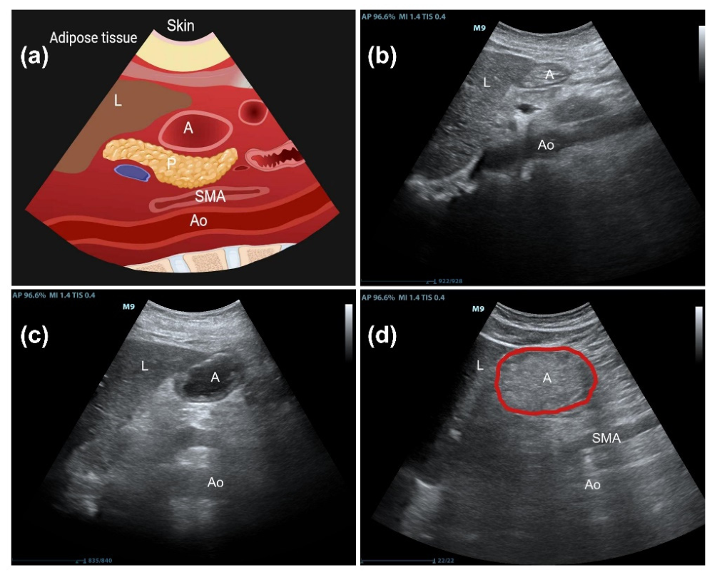
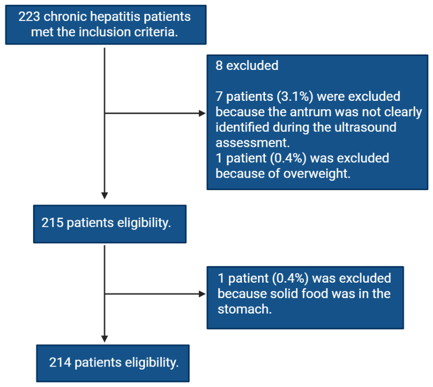
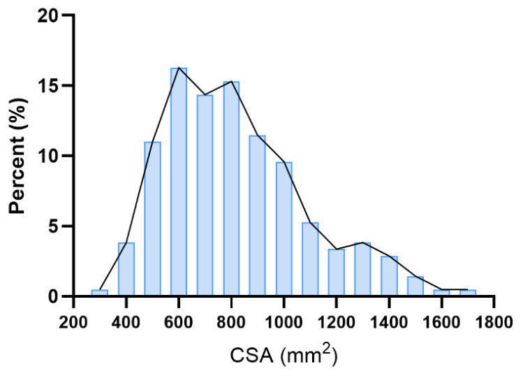
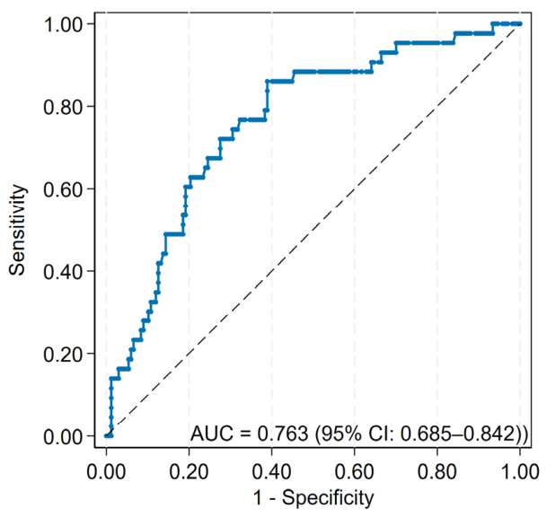

| Characteristics | ±S/n (%) |
| Sex(male/female) | 140/74 |
| Age(year) | 43.69±12.14 |
| Height(cm) | 168.22±8.06 |
| Weight(kg) | 69.90±13.94 |
| Type of hepatitis | |
| Pure hepatitis B | 206(96.3) |
| Hepatitis B complicated other hepatitis | 8(3.7) |
| Complications | |
| Diabetes mellitus | 16(7.5) |
| Hypertension | 32(15.0) |
| Coronary heart disease | 5(2.3) |
| ASA physical status | |
| Ⅰ | 44(20.1) |
| Ⅱ | 135 (63.5) |
| Ⅲ | 35 (16.5) |
| Fasting duration for food (hours) | 14.86±1.66 |
| Indexes | Real gastric fluid volume | |||
| ≤ 0.8mL/kg | > 0.8mL/kg | Total | ||
| CSA | ≤ 835 mm2 | 102 | 6 | 108 |
| > 835 mm2 | 65 | 37 | 102 | |
| Total | 167 | 43 | 210 | |
| Characteristics | Cases | CSA in RLD | z | P |
| Sex | ||||
| Female | 74 | 7.56(6.19, 9.56) | 2.227 | 0.026 |
| Male | 136 | 8.59(7.00, 10.77) | ||
| Type of hepatitis | ||||
| Pure hepatitis B | 203 | 8.31(6.62, 10.42) | 0.724 | 0.469 |
| Hepatitis B complicated other hepatitis | 7 | 7.58(6.00, 8.73) | ||
| Diabetes mellitus | ||||
| No | 192 | 8.30(6.67, 10.29) | -0.281 | 0.779 |
| Yes | 16 | 8.05(6.31, 12.85) | ||
| Hypertension | ||||
| No | 179 | 8.03(6.47, 10.27) | -2.239 | 0.0251 |
| Yes | 31 | 9.49(7.42, 10.98) | ||
| Coronary disease | ||||
| No | 205 | 8.31(6.62, 10.31) | -0.108 | 0.9140 |
| Yes | 5 | 7.42(7.25, 12.51) |
| Index | CSA in RLD | |
| r | P | |
| Age | 0.118 | 0.063 |
| Height | 0.052 | 0.411 |
| Weight | 0.094 | 0.140 |
| Fasting duration time | 0.012 | 0.850 |
| ALT | 0.005 | 0.933 |
| AST | -0.004 | 0.955 |
| CHE | -0.698** | 0.000 |
| TP | -0.092 | 0.147 |
| ALB | -0.163* | 0.010 |
| TBIL | 0.150* | 0.018 |
| DBIL | 0.145* | 0.022 |
| IBIL | 0.058 | 0.360 |
| PT | -0.088 | 0.166 |
| Blood ammonia | 0.148* | 0.020 |
| Index | B | SE | t | P | 95% CI |
| Constant | 9.334 | 0.842 | 11.080 | 0.000 | 7.675~10.994 |
| Sex | 0.166 | 0.179 | 0.924 | 0.356 | -0.188~0.519 |
| CHE | -0.001 | 0.000 | -14.182 | 0.000 | -0.001~0.000 |
| ALB | 0.039 | 0.019 | 2.002 | 0.046 | 0.001~0.077 |
| TBIL | -0.008 | 0.018 | -0.455 | 0.650 | -0.044~0.027 |
| DBIL | 0.011 | 0.020 | 0.569 | 0.570 | -0.028~0.050 |
| Blood ammonia | 0.009 | 0.008 | 1.025 | 0.307 | -0.008~0.025 |
Disclaimer/Publisher’s Note: The statements, opinions and data contained in all publications are solely those of the individual author(s) and contributor(s) and not of MDPI and/or the editor(s). MDPI and/or the editor(s) disclaim responsibility for any injury to people or property resulting from any ideas, methods, instructions or products referred to in the content. |
© 2026 by the authors. Licensee MDPI, Basel, Switzerland. This article is an open access article distributed under the terms and conditions of the Creative Commons Attribution (CC BY) license (http://creativecommons.org/licenses/by/4.0/).