
Submitted:
20 August 2026
Posted:
20 August 2026
You are already at the latest version
Abstract
Autoimmune diseases (AIDs) are a group of disorders caused by immune system dysfunction, in which the immune system mistakenly recognizes and attacks normal tissues and organs of the body. However, no radical cure is available for AIDs. Plant-derived exosome-like nanoparticles (PELNs) are nanoscale membrane structures secreted by plant cells. Recent studies have demonstrated their potential to restore immune homeostasis and retard the progression of AIDs, although the underlying mechanisms remain to be fully elucidated. This review systematically summarizes the biogenesis and functional advantages conferred by the unique composition of PELNs, with a focus on their targeting delivery potential and biological mechanisms in the treatment of various AIDs. Additionally, recent advances in the engineering modification of PELNs are discussed. This article aims to provide a theoretical basis and reference for the development of novel clinical therapeutic strategies for AIDs.
Keywords:
medicinal plant-derived exosome-like nanoparticles
; autoimmune diseases
; biological characteristics of PELNs
; targeted regulation
; engineering
1. Introduction
Autoimmune diseases (AIDs) are chronic inflammatory disorders caused by dysfunction of the immune system, leading to erroneous attacks on self-tissues and organs [1]. Epidemiological data indicate that the global prevalence of AIDs in the general population is approximately 3–5%[2]. These diseases are often insidious in onset, protracted in course, and prone to recurrence, imposing long-term physical and psychological burdens on patients[3,4]. Current evidence suggests that the occurrence and progression of AIDs result from the interplay of genetic susceptibility and environmental triggers, including infections, drugs, smoking, radiation, and hormonal fluctuations. The core pathological mechanisms involve disruption of central and peripheral immune tolerance, imbalance of immune cell subsets, dysregulation of pro-inflammatory and anti-inflammatory cytokine networks, and emerging evidence highlights the role of gut microbiota dysbiosis in systemic immune abnormalities[5].
Currently, the treatment of autoimmune diseases (AIDs) still primarily relies on non-specific immunosuppressants such as glucocorticoids. Although non-specific immunosuppressants like glucocorticoids can rapidly suppress excessive immune responses and alleviate acute symptoms, their long-term use may significantly increase the risk of infections and be accompanied by a range of metabolic and organic side effects. Therefore, the development of novel therapeutic strategies that can both effectively regulate immunity and minimize adverse reactions has become an urgent need to improve the long-term quality of life of AIDs patients[6].
Natural medicinal plants have accumulated thousands of years of experience in regulating immune balance through the synergistic effects of multiple components and multi-target actions[7]. However, the complexity of components, unclear bioactive constituents, and difficulty in elucidating mechanisms have limited their modernization and precise application[8]. In recent years, the discovery of plant-derived exosome-like nanoparticles (PELNs) has provided a novel approach to overcome these bottlenecks. PELNs are nanoscale lipid membrane vesicles secreted by plant cells. Early observations of such nanoparticles date back to 1967, when Halperin and Jensen first described them in carrot cell culture systems[9]. Since 2009, when Laura de la Canal’s team successfully isolated these nanoparticles from sunflower seeds[10], research on PELNs has entered a rapid development phase. Compared with crude plant extracts, PELNs possess unique advantages, including structural stability, well-defined composition, good biocompatibility, and ease of engineering modification[11]. Studies have shown that PELNs can deliver bioactive substances such as proteins, lipids, and non-coding RNAs (ncRNAs) to modulate immune cell function, inhibit key inflammatory signaling pathways, and promote the reconstruction of immune tolerance, demonstrating potential to delay or ameliorate the progression of AIDs in both clinical and preclinical studies.
This review aims to systematically elaborate the biological characteristics, biogenesis, and functional advantages of PELNs, with emphasis on summarizing their regulatory mechanisms and research progress in autoimmune diseases such as rheumatoid arthritis and inflammatory bowel disease. Furthermore, the latest advances in PELN engineering modifications are discussed to prospect their clinical translation, providing theoretical references for the development of next-generation AIDs therapeutics based on PELNs.
2. Biogenesis and Potential Functions of PELNs
The formation of PELNs follows a highly conserved cellular biological process analogous to that of mammalian exosomes (Mammalian-Exos)[12]. The biogenesis of PELNs begins with the invagination of the plasma membrane upon activation by pathogen signals, generating Early Endocytic (EE)[13]. These endocytic vesicles fuse with each other and develop into Early Sorting Endosomes (ESEs). Subsequently, the membrane of ESEs undergoes inward budding to form multiple Intraluminal Vesicles (ILVs), and ILVs mature into Multivesicular Bodies (MVBs)[14]. Ultimately, MVBs have two distinct fates: fusion with the plasma membrane to release the internal ILVs as exosome-like vesicles into the extracellular space[15−17], or degradation via fusion with Lysosomes(Lys)for the clearance and recycling of intracellular components[18] (Figure 1).
During PELNs formation, the sorting mechanisms of the endomembrane system determine the composition of vesicular cargo and their ultimate functions. This process involves the coordinated operation of organelles and endomembrane functional regions, including the Endoplasmic Reticulum (ER), Golgi apparatus, trans-Golgi Network (TGN), EE, Late Endosomes (LE), Multivesicular Bodies (MVBs), and Exocyst-positive organelles (EXPO) .
The endosomal sorting complex required for transport (ESCRT) is one of the major sorting systems for PELNs[14]. The ESCRT system comprises four protein complexes—ESCRT-0, -I, -II, -III—and the VPS4 ATPase[19], which work sequentially and synergistically to recognize ubiquitinated protein substrates, initiate bud formation, remodel LE membrane structures, and mediate ILV release[20]. During the recognition of ubiquitinated substrates, ILVs enriched with ubiquitinated membrane proteins are typically labeled and transported to lysosomes or vacuoles for degradation, thereby enabling the phagocytosis of pathogens and regulation of the endomembrane system[18].
Notably, the ESCRT system plays a bidirectional role in immune regulation: On one hand, its high sensitivity to ubiquitinated substrates enhances plant innate immunogenicity. Under environmental stress, the ESCRT system precisely recognizes pathogen-derived effector proteins and host-derived ubiquitinated molecules generated by damage, promotes vesicle transport containing immune-related receptors and signaling factors, and thus rapidly activates downstream defense responses to strengthen plant resistance to pathogens[21,22].
On the other hand, when immune cells are damaged during inflammatory responses, the ESCRT system initiates a repair mode, slowing the release of reactive oxygen species (ROS), lipid peroxides, and pro-inflammatory contents such as IL-1β, IL-18, HMGB1 and ATP, effectively preventing immune imbalance caused by sudden exposure to inflammatory storms[23]. Recent studies indicate that the ESCRT system rapidly recruits to damaged sites upon excessive Ca+ influx, suggesting its ability to suppress sustained activation of immune receptors and potentially repair cell membrane damage induced by AIDs[24]. Further research shows that CHMP2A, an ESCRT-III subunit, is critical for autophagosome formation. It acts at the stage of phagosome formation to mediate phagosome closure and enhance mitophagy flux. Deficiency of CHMP2A inhibits mitophagy, indicating that ESCRT mechanisms may suppress excessive activation of inflammasomes, correct mitochondrial membrane potential disorders, and restore autophagy function[25].
Another important protein family involved in PELNs transport is the Rab GTPase family, which further refines the PELNs sorting process in conjunction with plant-specific pathways such as EXPO. Rab GTPases are a class of GTP-binding proteins specialized in recognizing PELNs surface membrane proteins and mediating their transport. For example, Rab1/RabD localized to the ER recruits specific effector proteins to direct the transport of newly synthesized secretory vesicles to the Golgi apparatus, while Rab2/RabB localized to the Golgi membrane regulates ESE transport within the Golgi and ILV recycling in the cytoplasm[26]. Additionally, Rab GTPases interact with the trans-Golgi network to exert dual regulatory roles in endocytic and sorting pathways[27], reflecting the high functional integration of the plant TGN[28]: it simultaneously possesses EE functions and governs both PELN generation and sorting[29]. Under the control of VPS9A, a core Rab guanine nucleotide exchange factor (RabGEF), the plant Rab5 homologs RabF2a/RHA1 mediate the delivery of early endosomes to the TGN/EE, while RabF2b/ARA7 governs the maturation of multivesicular bodies (MVBs). The plant-specific RabF1/ARA6, which localizes to MVBs, undergoes N-terminal lipid modification and further mediates the degradation and recycling of PELNs[30]. Studies suggest that this efficient sorting mechanism enhances plant innate immunity against fungal pathogens[31].
In addition, plants possess a unique unconventional secretory pathway mediated by the Exocyst-positive organelle (EXPO). As a plant-specific double-membrane organelle independent of the canonical secretory pathway governed by the TGN/EE, EXPO directly releases defense-related proteins from the cytosol into the apoplast, thereby accelerating the delivery of defense proteins to pathogen infection sites[32]. Moreover, unlike mammalian cells, plant cells are surrounded by a rigid cell wall, which constitutes a physical barrier to vesicle exocytosis. During MVB secretion, plants locally release cell wall remodeling factors such as expansins, pectinases, and hydrolases to transiently loosen the cell wall structure, thereby providing channels for PELNs release[33]. Overall, the biogenesis mechanism of PELNs represents a key link in plant adaptation to environmental stress and maintenance of immune homeostasis, offering novel insights into PELN-mediated immune regulation.
3. Composition of PELNs
PELNs are enriched in lipids, proteins, non-coding RNAs, and secondary metabolites, which constitute the material basis for their immunomodulatory functions (Table 1) .
3.1. Lipids
PELNs contain diverse phospholipid components, mainly including glycerophospholipids (GPLs) such as phosphatidylcholine (PC), phosphatidylethanolamin (PE), phosphatidylinositol (PI), phosphatidylserine (PS), phosphatidic acid (PA), and phytosterols[11]. Compared with animal exosomes, PELNs exhibit greater lipid diversity[34], providing richer functional selectivity for AID therapy. These unique lipid components collectively endow PELNs with superior membrane stability and structural integrity. First, PELNs contain phytosterols[35](e.g., sitosterol, stigmasterol), which reduce membrane fluidity and increase membrane compactness, making PELNs more resistant to degradation during in vivo delivery[36]. Second, PC reinforces the overall structural framework of PELNs through the stable linkage of its “glycerol backbone-fatty acid-choline group”. Additionally, PA, with its negatively charged head group adjacent to the alkyl chain region, exhibits high affinity for divalent cations and regulates membrane fission and fusion processes via intermolecular hydrogen bonding, significantly enhancing PELN stability. This stable membrane system, composed of multiple lipids, not only protects the cargo but also strengthens the retention and functional capacity of PELNs in complex physiological environments, laying the foundation for their application in immune regulation[37].
The phospholipid composition of PELNs is not fixed and varies significantly among plant species[38]. For instance, Curcuma longa-derived exosome-like nanoparticles (CLEs) and Ginger-derived exosome-like nanoparticles (GELNs), both extracted from rhizomes of the Zingiberaceae family, differ in PA content, with GELNs exhibiting higher levels[38,39].
PELNs also act as key lipid messengers in immune regulation and inflammatory responses[40]. This is attributed to their enhanced ability to fuse with and be internalized by recipient cell membranes, thereby augmenting the biological functions of recipient cells. In the intestine, GELNs with high PA content are preferentially taken up by Lactobacillus rhamnosus GG (LGG), triggering the activation of the Aryl Hydrocarbon Receptor (AHR) signaling pathway by GELN-carried miRNAs, which induces intestinal antimicrobial immunity and repairs the intestinal barrier[41]. In the oral microenvironment, GELNs with high PA content are readily recognized and internalized by the HBP35 (Hemin-binding protein 35) protein on the surface of Porphyromonas gingivalis (Pg), a periodontal pathogen. This internalization negatively regulates the Type IX Secretion System (T9SS) in Gram-negative bacteria, inhibiting the secretion of virulence factors such as gingipains and hemagglutinin, thereby alleviating periodontal tissue inflammation and bone loss[42].
3.2. Proteins
Proteins are key functional components of PELNs, governing their biogenesis, intercellular communication, and signal transduction[43]. The protein cargo of PELNs is predominantly cytosolic, including actin, proteases, channel proteins, and annexins, which collectively form the structural scaffold of PELNs and participate in their transport functions[36,44].
In terms of composition, PELNs mainly contain the following protein categories: Channel or transporter proteins such as aquaporins[45]. Tetraspanins serving as vesicular membrane markers[46] and DnaJ homologs involved in protein folding, transport, and degradation[47]. Functionally, proteins enriched in PELNs play diverse roles: Protein kinases and G proteins act as key molecules in signal transduction, mediating PELN-involved intercellular communication; annexins and other intracellular transport proteins are responsible for the transport and delivery of PELN cargo. Ubiquitin, clathrin, and ALIX(ALG-2-interacting protein X)are core sorting factors regulating PELN biogenesis. Integrin family members such as endorepellin mediate the specific binding of PELNs to target cell surface receptors, initiating downstream biological effects[39].
Current research on PELN biomarkers largely draws on knowledge of mammalian exosomal markers[44]. Studies have identified homologous membrane proteins in PELNs that share structural or functional similarities with mammalian exosomal proteins (e.g., TSG101, Alix, HSP70)[48]. Among these, heat shock proteins, aquaporins, clathrin heavy chain, and glyceraldehyde-3-phosphate dehydrogenase exhibit high sequence homology and functional conservation between animals and plants[49]. However, systematic identification of PELN-specific surface marker proteins remains in its infancy, mostly limited to confirmation of membrane localization, with precise membrane topology and post-translational modification status still unclear[50]. Currently recognized markers include Penetration 1 (PEN1), Penetration 3 (PEN3), and Tetraspanin-8 (TET-8)[10,51]. Functionally, PEN1 and PEN3 are primarily involved in plant immune defense[52]. PEN1 is a Mn2+-dependent 5′→3′ exonuclease localized to plastids, capable of specifically cleaving RNA primers, which is essential for plant DNA synthesis[53]. TET-8 shares high homology with the mammalian tetraspanin CD63, both participate in endosomal lipid sorting but via distinct mechanisms[54]. Mammalian CD63 sorts cholesterol into ILVs, forming a storage pool mobilized by NPC1/2 complexes, and exports it to recipient cells via exosomes [55]. In contrast, plant TET-8 recognizes the γ2 subunit of the coat protein complex I (COPI) via its C-terminal tail, thereby mediating the sorting and transport of sphingolipids such as glycosyl inositol phosphoceramide (GIPC) in the Golgi[56].
Furthermore, PELNs can undergo specific, high-affinity interactions with recipient cell membrane proteins via their surface proteins, guiding targeted uptake. For example, Ginger-derived exosome-like nanoparticles (GENs) are efficiently internalized by human Hepatocellular carcinoma cells (HepG2) because the highly expressed transmembrane protein CD98 (also known as 4F2hc or SLC3A2) on HepG2 cells carries glycosylation modifications whose glycan structures are specifically recognized and bound by lectin II on GENs, initiating cellular uptake[57]. This targeting interaction not only reveals the tropism of PELNs for different cells but also provides a potential molecular basis for their precise immunomodulation in AIDs.
3.3. Non-Coding RNAs
Non-coding RNAs (ncRNAs) are RNA molecules that do not encode proteins, including microRNAs (miRNAs)[58]and small interfering RNAs (siRNAs)[59], which primarily function in post-transcriptional regulation. NcRNAs are selectively packaged into ILVs of maturing MVBs via ESCRT-related pathways and ultimately delivered to recipient cells, where they recognize and bind to the 3′ untranslated region (UTR) of target mRNAs, inhibiting translation or mediating mRNA degradation to achieve precise regulation of coding genes[60].
However, differences exist in the biogenesis of animal-derived miRNAs and plant-derived miRNAs (p-miRNAs). Animal miRNA maturation depends on RNA polymerase II (Pol II)-mediated transcription in the nucleus to generate primary miRNA (pri-miRNA) precursors[61], which are then processed by Drosha and Dicer enzymes[62]. The pre-miRNA is subsequently exported to the cytoplasm by Exportin-5[63]. In contrast, p-miRNAs bypass the initial processing step requiring Drosha in the nucleus and the stepwise nuclear-cytoplasmic processing of pre-miRNA. Instead, they are directly sheared into double-stranded forms by the DCL1 complex in the nucleus in a single step. This dicing pattern yields shorter plant miRNAs (p-miRNAs). Unlike animal miRNAs, p-miRNA duplexes undergo immediate 2′-O-methylation at the 3′ terminus catalyzed by the methyltransferase HEN1, which effectively protects them from uridylation and subsequent degradation by nucleases. Thus, nuclear processing and methylation modification represent the main differences between plant and animal miRNA maturation pathways. Compared with animal miRNAs, p-miRNAs exhibit enhanced structural and modification-mediated stability, effectively resisting exonuclease degradation and maintaining higher integrity and half-life in complex biological environments[64].
Research on siRNAs in PELNs is both promising and challenging. It has been confirmed that plants can produce endogenous secondary siRNAs that are fully complementary to target genes[65], mediating efficient and specific gene silencing. This provides a potential biological basis for developing PELNs as natural siRNA delivery vectors[66].
Natural plant siRNAs include subtypes such as trans-acting siRNAs (tasiRNAs) and hepatocyte-targeted cholesterol-conjugated siRNAs (hc-siRNAs)[67]. hc-siRNAs primarily bind to chromatin in the nucleus, guiding DNA methylation and histone modifications to silence aberrant gene transcription. They are core components of the RNA-directed DNA methylation (RdDM) pathway, mainly responsible for establishing and maintaining heterochromatin states in genomic repetitive sequences, transposons, and other regions[68]. Their biogenesis begins with RNA polymerase IV transcribing single-stranded RNA precursors, which are converted into double-stranded RNA (dsRNA) by RNA polymerase II. Dicer-like 3 (DCL3) then recognizes and cleaves dsRNA into 24-nucleotide siRNA duplexes, which are loaded onto ARGONAUTE proteins such as AGO4, AGO6, or AGO9 to form silencing effector complexes[69]. Finally, AGO complexes recruit DNA methyltransferases (e.g., DRM2) and histone-modifying enzymes to catalyze local DNA methylation, thereby establishing and stabilizing heterochromatin and permanently silencing the expression of harmful or redundant genetic elements[70].
TasiRNAs are a subclass of plant siRNAs that act as amplifiers of miRNA silencing signals. Their biosynthesis starts with the precise cleavage of TAS gene transcripts by an Argonaute complex loaded with a specific 22-nucleotide (nt) miRNA[65]. The resulting 3′ cleavage fragment is recognized by RNA-dependent RNA polymerase 6 (RDR6) and converted into a dsRNA molecule[71]. Subsequently, DICER-LIKE4 (DCL4) cuts the dsRNA continuously and equidistantly from the end, generating uniformly 21-nt siRNA duplexes[71,72]. These siRNA duplexes bind to AGO1 to form RNA-induced silencing complexes (RISCs), which silence multiple unrelated target mRNAs at the post-transcriptional level[66]. Because a single tasiRNA array can simultaneously target multiple members of a gene family, it effectively amplifies the regulatory effect of the initiating miRNA, enabling efficient inhibition of entire genetic pathways during development and stress responses.
3.4. Secondary Metabolites
Plant secondary metabolites are organic compounds produced during specific developmental stages or under environmental stress[73]. Similar to parent plants, PELNs selectively encapsulate various anti-inflammatory and antioxidant secondary metabolites during their formation, such as 6-gingerol in ginger, curcumin in turmeric[74], and ginsenoside Rg3 in ginseng[75]. These small molecules are passively accumulated within the vesicular membrane primarily through hydrophobic interactions, with encapsulation efficiency closely related to membrane lipophilicity[76].Secondary metabolites encapsulated in PELNs target key proteins in inflammation, oxidative stress, and apoptosis signaling pathways in animal cells[77,78], and due to their nano-carrier properties, may exhibit superior delivery efficiency and bioavailability compared with direct use of plant extracts[79]. Although accurate studies on the mechanisms of PELNs in animals are lacking, the cross-species action mechanisms of plant-derived secondary metabolites themselves have been well documented, providing references for studying the cross-kingdom regulatory effects of PELN-encapsulated secondary metabolites:
(1) Competitive inhibition: Secondary metabolites can directly enter the active center of target enzymes, competing with endogenous substrates for binding sites. For example, flavonoids compete with ATP for binding to the ATP pocket of JAK kinases, inhibiting ATP-mediated phosphorylation of STAT1, thereby blocking interferon responses and the initiation of downstream pro-inflammatory immune responses[80].
(2) Allosteric regulation: Secondary metabolites can bind to allosteric sites of target proteins, indirectly regulating their activity by inducing conformational changes[81]. For instance, the natural polyphenol resveratrol acts as an allosteric activator of SIRT1, enhancing its deacetylase activity by altering SIRT1 conformation[82]. Activated SIRT1 then deacetylates transcription factors such as NF-κB and p53, alleviating inflammation and apoptosis[83].
(3) Interference with protein-protein interactions: Some secondary metabolites can block interactions between key inflammatory proteins via covalent or non-covalent binding. For example, the diterpenoid oridonin covalently modifies the Cys279 site of the NLRP3 protein, blocking its interaction with NEK7, thereby inhibiting NLRP3 inflammasome assembly and activation[84]. Additionally, flavonoids can inhibit NLRP3 oligomerization, reducing caspase-1 activation and pyroptosis[85].
However, not all plant secondary metabolites can be efficiently encapsulated into PELNs. For example, vitamin C and naringenin are difficult to detect in orange-derived exosome-like nanoparticles (O-ELNs)[86]. Moreover, research on PELNs encapsulation of plant secondary metabolites mostly focuses on component identification and phenotypic validation, and precise molecular mechanisms remain to be further explored.
4. Targeted Delivery Capacity of PELNs
4.1. Advantages of PELN-Mediated Targeted Delivery
Compared with animal cell-derived exosomes, PELNs demonstrate unique delivery advantages in cross-kingdom interventions, attributable to their stable structure, active targeting ability, and flexible delivery kinetics[43].
First, PELNs exhibit excellent structural and cargo stability. For example, Ginger-derived exosome-like nanoparticles (GDNPs), Ginger-derived exosome-like nanoparticles, and ginger-derived exosome-like nanoparticles provide robust physical barriers for internal bioactive substances (e.g., nucleic acids, proteins), effectively resisting degradation by nucleases, digestive enzymes, and other components of the biological environment[87]. Second, the synergistic action of lipids, nucleic acids, and proteins encapsulated in PELNs enhances targeting efficiency. For instance, PC facilitates the recognition and uptake of PELNs by specific microorganisms such as Ruminococcus[88]. MiR159a and miR156c in Ginger-derived exosome-like nanoparticles (NVs) directly bind to the Trfrsf1agene in mouse adipose tissue, inhibiting the TNF-α signaling pathway and improving metabolic abnormalities[89]. The targeting of PELNs is determined by surface transporters via three main mechanisms: 1) direct membrane fusion mediated by SNARE/Rab proteins. 2) clathrin-, lipid raft-, or caveolae-dependent endocytosis. 3) specific internalization triggered by binding of surface ligands (e.g., lectins) to cell surface receptors (e.g., CD98)[90].
Furthermore, PELNs from different sources exhibit unique in vivo delivery kinetics, and administration routes affect their tissue distribution. Studies show that Ginger-derived exosome-like nanoparticles (CAEs) peak in fluorescence intensity 2 h after administration in mice and specifically accumulate in the inflamed colon of colitis models[91], whereas PG-ELNs reach peak uptake in bone marrow mesenchymal stem cells at 12 h[92]. Administration routes determine target organs: oral administration leverages acid resistance for gastrointestinal colonization[93]. Intravenous injection bypasses first-pass effects to target brain ischemic regions or tumor tissues. Nasal administration favors distribution to the brain and lungs[94]. Intramuscular injection results in local enrichment[95].
4.2. Functions and Controversies of PELN-Mediated Targeted Delivery
In immune regulation, PELNs act as efficient cross-kingdom communication vectors, traversing the intestinal barrier via multiple pathways: clathrin-dependent endocytosis, paracellular diffusion, fusion with gut microbiota outer membrane vesicles, and enhancement of membrane permeability by short-chain fatty acids. After entering systemic circulation, PELNs enrich in distal organs such as the liver and lungs[96]. At the regulatory mechanism level, miRNAs carried by PELNs can not only inhibit target mRNA translation via incomplete complementarity but also directly cleave and degrade target mRNAs via complete complementarity, achieving irreversible gene silencing[97,98]. This mechanism is potent, stable, and capable of completely shutting down pathogenic genes, a feature absent in the translation inhibition mode dependent on animal exosomes[58].
However, controversies persist regarding the cross-kingdom regulatory function of PELNs, particularly concerning the biological mechanisms of p-miRNAs and p-siRNAs. Although most studies support that p-miRNAs in PELNs exert biological effects similar to those of animal miRNAs, rigorous detection methods (e.g., RT-qPCR) have failed to stably detect intact p-miRNAs in the plasma and tissues of humans, mice, or bees fed plant-based diets. For example, macaques fed a mixed diet of fruits and soybeans exhibited low plasma p-miRNA levels, mostly non-specifically expressed[99]. Rice miRNAs were undetectable in mice fed rice-containing diets[100]. Further studies indicate that corn miRNAs are present at low levels in the blood and feces of mice orally administered corn, suggesting potential degradation by gastric acid[101]. This contradicts earlier reports by Luo et al. detecting corn miRNAs in pig models fed corn-containing feed[102], highlighting unresolved issues such as methodological differences and interference from false-positive miRNAs[99,103]. Additionally, the amount of free nucleotides in circulating miRNAs accounts for only a fraction of total ingested nucleotides from food-derived genomic DNA and RNA, making it unlikely that free nucleotides exert immunoregulatory effects in mammals[99]. Therefore, whether and how p-miRNAs stably exist and function in animals require further clarification.
Similarly, whether naturally occurring siRNAs in PELNs can cross-kingdom silence human genes remains questionable. The main reasons include: first, no rigorous methods exist to quantify siRNA expression levels in PELNs. Second, no study has confirmed that plant-derived siRNA sequences can achieve full complementarity with human genomes and exert silencing functions[104,105]. Third, even if pairing mechanisms are established, siRNAs may pose potential risks due to off-target effects or induction of non-specific immune responses[106]. Nevertheless, PELN-based cross-kingdom siRNA delivery and gene intervention remain highly attractive future research directions due to their theoretical efficiency and specificity[59].
5. Applications of PELNs in Autoimmune Diseases
5.1. Rheumatoid Arthritis
Rheumatoid arthritis (RA) is a systemic autoimmune disease characterized by chronic inflammation of joints and systemic tissues, pannus formation, and cartilage destruction[107,108]. In susceptible individuals, MHC class II molecules carrying HLA-DRB1 alleles efficiently present citrullinated peptides[109], activating CD4+ T cells and sustaining downstream inflammatory pathways[110]. Activated immune cells further bind to modified self-proteins in the joint, activating inflammatory cells such as macrophages, neutrophils, osteoclasts, and monocytes, releasing pro-inflammatory cytokines, and establishing a chronic inflammatory loop[111].
Current RA-targeted regulation by PELNs primarily focuses on macrophages. Direct evidence shows that folic acid-modified ginger-derived exosome-like nanoparticles (FA-GDEVs) loaded with active components (e.g., gingerol) can reprogram macrophage polarization, promoting their transition from the pro-inflammatory M1 phenotype to the anti-inflammatory M2 phenotype, significantly downregulating the expression levels of key RA inflammatory cytokines and their mRNAs. Specifically, FA-GDEVs markedly inhibit the expression of M1 macrophage markers, including Interleukin-6 (IL-6), Tumor Necrosis Factor-α(TNF-α), Interleukin-1β(IL-1β), Cyclooxygenase-2 (COX-2), and inducible Nitric Oxide Synthase (iNOS), while upregulating the expression of M2 macrophage markers, including Cluster of Differentiation 206 (CD206), Arginase-1 (Arg-1), and Interleukin-10 (IL-10)[74].
Immune cell activation upregulates inflammatory pathways, leading to massive cytokine release and enhanced pro-inflammatory signaling. miRNAs carried by PELNs inhibit pro-inflammatory signals. For example, pgi-miR6135j in GDNPs suppresses KRAS activation in synovial macrophages of collagen-induced arthritis (CIA) mice, blocks RAF kinase (MAPKKK) secretion, and reduces phosphorylation levels of downstream JNK, ERK, and p38 proteins, thereby decreasing inflammatory cytokine expression in macrophages[112]. Studies confirm that GDNPs inhibit IκBα degradation (blocking the NF-κB pathway) and JNK phosphorylation (inhibiting the MAPK/AP-1 pathway), downregulating the transcriptional activity of NFATc1 and c-Fos/AP-1, and reducing the synthesis of functional proteins, such as Tartrate-Resistant Acid Phosphatase (TRAP) and Osteoclast-associated Receptor (OSCAR). This suppresses osteoclast differentiation and bone resorption at the source, ultimately alleviating RA-induced bone erosion[55] (Figure 2).
5.2. Inflammatory Bowel Disease
Inflammatory bowel disease (IBD), mainly comprising ulcerative colitis and Crohn’s disease, is a group of autoimmune intestinal inflammatory disorders caused by disruption of the intestinal barrier and loss of mucosal homeostasis, affecting the ileum, rectum, and colon[113]. Persistent barrier damage activates excessive immune defense, leading to overactivation of pro-inflammatory signals and attenuation of anti-inflammatory signals[114]. Cytokines activate downstream inflammatory signaling cascades, ultimately triggering inflammatory storms, manifested as diffuse transmural inflammatory infiltration, cryptitis, and abscesses in the colon. PELNs can modulate intestinal immune cell balance, inhibit inflammatory signaling pathways, and reverse intestinal inflammation[115] (Figure 3).
5.2.1. Improving the Immune Microenvironment
IBD onset begins with overexpression of the genetic locus C1orf106, leading to abnormal secretion of ADP-ribosylation factor 6 (ARF6) cytokines, increased intestinal antigen permeability, and subsequent immune responses and inflammation[116].In IBD inflammatory infiltrates, neutrophils, dendritic cells, macrophages, and B cells (plasma cells) are recruited to clear pathogens while releasing large amounts of inflammatory signals that activate adaptive immunity[117].However, excessive immune defense contributes to AIDs, whereas PELNs help restore immune balance.
Studies have demonstrated that Coptis chinensis-derived exosome-like nanoparticles (Cc-ELNs) deliver miR-5106, which downregulates the expression of the zinc transporter Slc39a2 in neutrophils. This maintains intracellular Zn2+ homeostasis and consequently reduces the formation of neutrophil extracellular traps (NETs). Concurrently, Cc-ELNs promote the proliferation of intestinal epithelial cells and intestinal stem cells, indicating their potential to enhance cellular regeneration[118].
Dendritic cells are important antigen-presenting cells[119]. PELNs inhibit their excessive differentiation. A study on broccoli-derived exosome-like nanoparticles (B-ELNs) provided direct evidence: after internalization by dendritic cells, B-ELNs activate the cellular energy sensor AMP-activated protein kinase (AMPK). AMPK phosphorylates and inhibits mammalian target of rapamycin complex 1 (mTORC1), downregulating the expression of major histocompatibility complex class II (MHC II) molecules and co-stimulatory molecules (e.g., CD80/CD86)[120].
Furthermore, PELNs alleviate intestinal inflammation by inhibiting the differentiation of naive T cells into pro-inflammatory subsets, such as reprogramming CD4+ T cells. By increasing tryptophan metabolites from Lactobacillus reuteri(e.g., ILA), Portulaca oleracea-derived exosome-like nanoparticles (Po-ELNs) activate the aryl hydrocarbon receptor (AhR) in CD4+ T cells, successfully suppressing Zbtb7b, the master regulator of the CD4+T cell lineage, and promoting the reprogramming of conventional CD4+ T cells into CD4+CD8+ double-positive phenotypes. This regulation suggests that PELNs can indirectly guide the direction of adaptive immune responses in colonic inflammatory regions by altering the intestinal microenvironment[121].
5.2.2. Inhibiting Inflammatory Responses
Macrophages, derived from bone marrow hematopoietic stem cells, are innate immune cells capable of releasing inflammatory factors[122]. Upon IBD onset, macrophages differentiate into subpopulations with distinct phenotypes and functions[123]. M1-type macrophages, induced by IFN-γ and LPS, secrete pro-inflammatory cytokines (e.g., ,IL-1β, IL-6, TNF-α), exacerbating inflammation[124]. M2-type macrophages, induced by IL-4 and IL-13, secrete anti-inflammatory factors (e.g., IL-10), inhibiting M1 pro-inflammatory functions and regulating adaptive immunity[125].
Studies show that GELNs deliver osa-miR164d to target and inhibit TAB1, a key mediator of the TNF signaling pathway, promoting M2 polarization of macrophages and alleviating colitis symptoms[126]. Grape-derived exosome-like nanoparticles (GELEN) upregulate Heme oxygenase-1 (HO-1) and IL-10 expression, enhancing the anti-inflammatory capacity of macrophages and maintaining E-cadherin expression in intestinal epithelial cells, contributing to intestinal barrier homeostasis[127]. Atractylodes macrocephala-derived exosome-like nanoparticles (A-ELNs) upregulate anti-inflammatory IL-10 expression in macrophages while inhibiting pro-inflammatory factors (IL-1β, IL-6, IL-12, TNF-α), promoting intestinal inflammatory repair[128].
Additionally, GDNs can induce macrophage autophagy to influence polarization, improving pathological manifestations in DSS-induced IBD mice. Specifically, GDNs activate macrophage autophagy by inhibiting the AKT/mTOR signaling pathway, promoting M2 polarization, as evidenced by upregulated CD206 expression and increased IL-10 secretion. In co-culture systems of macrophages and intestinal epithelial cells, IL-10 secreted by M2 macrophages significantly reduces nitric oxide (NO) levels and NF-κB protein expression in Caco-2 cell supernatants. In vivo experiments further confirm that GDNs reduce DSS-induced intestinal permeability and mucosal damage, while increasing LC3 and Atg7 protein levels in intestinal tissues, indicating that GDNs exert anti-inflammatory effects by targeting the IKK/IκB/NF-κB signaling pathway to induce autophagy and promote macrophage polarization[75].
Notably, in the IBD microenvironment, NLRP3 inflammasomes activate caspase-1, promoting the maturation and release of IL-1β and IL-18[129]. NLRP3 inflammasomes are intracellular multiprotein complexes of the innate immune system responsible for activating inflammatory responses. Studies show that prune-derived exosome-like nanoparticles (Pr-ELNs) disrupt NEK7-NLRP3 interactions after internalization by macrophages, inhibiting NLRP3 inflammasome formation, caspase-1 autocleavage, and IL-1β secretion, thereby alleviating intestinal mucosal inflammatory infiltration in IBD mice[130].
5.2.3. Repairing the Intestinal Barrier
The Wnt/β-catenin signaling pathway is crucial for regulating intestinal stem cell self-renewal, proliferation, and differentiation[131]. Studies show that GELEN activates Tcf4-mediated Wnt/β-catenin pathway genes (e.g., Lgr5-EGFP), promoting β-catenin accumulation in intestinal epithelial cells, intestinal stem cell differentiation, and repair of inflammatory damage, participating in intestinal tissue remodeling[49].
5.2.4. Regulating Intestinal Oxidative Stress Responses
Oxidative stress arises from imbalance between oxidation and antioxidant systems, and persistent oxidative stress induces intracellular free iron accumulation and lipid peroxidation[132,133]. Preliminary studies have confirmed that PELNs inhibit ferroptosis, alleviate intestinal mucosal inflammation, and enhance mucosal antioxidant capacity. For example, bitter melon-derived exosome-like nanoparticles (MC-ELNs) upregulate the expression of antioxidant enzymes (glutathione peroxidase, GSH; superoxide dismutase, SOD; catalase, CAT) in the intestinal tissues of IBD mice, while reducing lactate dehydrogenase (LDH) and malondialdehyde (MDA) levels. Concurrently, MC-ELNs modulate inflammatory cytokine expression, protecting intestinal barrier function[134].
5.3. Type 1 Diabetes Mellitus
Type 1 diabetes mellitus (T1DM) is a chronic disease characterized by progressive impairment of pancreatic β-cell function due to autoimmunity and severe insulin deficiency[135]. Long-term complications involve blood vessels, nerves, and bone tissue in the feet, ultimately leading to ulceration, infection, and gangrene. PELNs have shown surprising potential in T1DM therapy.
First, PELNs effectively ameliorate T1DM-induced insulin deficiency. For example, intraperitoneal injection of Trifolium pratense-derived exosomes (TPDEs) improves serum biochemical indicators, antioxidant parameters (nitric oxide, NO; total antioxidant capacity, TAC), and pancreatic gene expression (pancreatic and duodenal homeobox 1, PDX1; neurogenin 3, NGN3; sirtuin 1, SIRT1,) in streptozotocin (STZ) -induced diabetic rats[136].
Second, PELNs are rich in functional proteins and metabolites that promote healing of chronic wounds and bone tissue damage induced by diabetes. Portulaca oleracea-derived extracellular vesicles (Po-DENs) contain bioactive components such as heat shock proteins, linoleic acid, ginkgolic acid, and lipoxins, which effectively scavenge intracellular ROS, upregulate ITGB4 and ITGA6 to activate focal adhesion signaling, induce PARP1 expression to initiate DNA repair, inhibit apoptosis[137], and restore skin wound dysfunction in diabetic mice. Mango-derived exosome-like nanoparticles (PDNVs) rescue delayed wound healing in STZ-induced diabetic mice by inducing follistatin-like protein1 (FSTL1) to promote keratinocyte migration[138]. Viola yedoensis-derived extracellular vesicles (VDNPs) promote M2 macrophage polarization by downregulating the NF-κB signaling pathway, balancing the expression of anti-inflammatory genes (Arg-1, IL-10) and pro-inflammatory genes (iNOS, TNF-α), alleviating the inflammatory microenvironment and promoting fracture healing in diabetic mice[139].
Moreover, growing evidence suggests that PELNs possess potent tissue repair and inflammation-regulating capabilities. For instance, Dendrobium catenatum-derived extracellular vesicles (DDNVs) increase eNOS, VEGFR-2, vimentin, fibronectin, and IL-1β gene expression via the Akt/eNOS signaling pathway, while inhibiting inflammatory markers ICAM-1 and IL-1β, providing insights for treating diabetic wounds[140].
5.4. Psoriasis
Psoriasis is an inflammatory skin disease caused by immune dysfunction attacking normal skin structures, typically manifested by abnormal keratinocyte proliferation and immune cell infiltration. PELNs can achieve immunomodulation in psoriatic skin via miRNA-mediated targeting of genes[141]. Studies show that Perilla frutescens leaf-derived extracellular vesicle-like particles (PLEVPs) carry the pab-miR396a-5p gene, which targets and inhibits HSP90 and downstream inflammatory signaling pathway-related genes[142]. This is reflected by reduced levels of pro-inflammatory cytokines (IL-6, IL-1β) and ROS in HaCaT cells, and modulated expression of CD45+T, CD3+CD4+T, and Treg cells.
Although research on PELNs therapy for psoriasis remains in its infancy, their capacity to regulate immunity, exert anti-inflammatory and antioxidant effects in epidermal cells has been fully validated[143]. In chronic inflammatory skin diseases, overexpression of matrix metalloproteinases MMP-3 and MMP-13 degrades collagen, causing chronic inflammation and indirectly generating excess ROS that amplify inflammation, hindering wound healing. Grapefruit-derived exosome-like nanoparticles (Gf-DVLNs) reduce oxidative stress in HaCaT cells, upregulate collagen I and fibronectin expression, and accelerate wound repair[144]. Additionally, Potato-derived exosome-like nanoparticles (ExoPs) inhibit collagen-degrading enzymes (MMP1, MMP2, MMP9) and inflammatory cytokines (IL-6, TNF-α), promote HaCaT cell proliferation, and protect cells from oxidative stress. These studies provide strong references for PELNs research in psoriasis[145]. (Table 2) .
6. Engineering Strategies for PELNs
As efficient bioactive substance delivery systems, PELNs exhibit excellent immunomodulatory functions. Their natural origin and evolutionarily conserved structure confer low immunogenicity, helping active components evade immune surveillance during various delivery routes, while effectively avoiding enzyme degradation and pH fluctuations that compromise drug activity[146]. To further expand therapeutic precision, researchers currently focus on two main aspects of PELN engineering: (1) Encapsulating exogenous drugs or active molecules inside vesicles to improve intervention efficacy. (2) Enhancing targeting via surface modification[147]. Current exogenous loading strategies for PELNs include passive diffusion (co-incubation) and active loading (electroporation, ultrasonication, freeze-thaw cycles, surfactant permeabilization, and extrusion)[148] (Figure 4).
Passive diffusion is a mild loading method based on molecular concentration gradients: it utilizes the chemical potential difference across the membrane to spontaneously drive drugs to migrate into PELN lipid bilayers[149]. In studies loading vancomycin into garlic-derived exosome-like nanoparticles (GEs), highly water-soluble vancomycin spontaneously migrates toward the lower-potential interior of GEs, diffusing and integrating into the vesicular membrane structure. The significant advantage of co-incubation lies in avoiding membrane damage caused by common loading methods such as ultrasonication, extrusion, or electroporation, better maintaining exosomal structural integrity during delivery[150].
Active loading relies on physical intervention to mechanically disturb membrane structures, transiently opening PELN lipid bilayers to allow automatic influx of drugs into the vesicle interior. Compared with passive diffusion, creating transient hydrophilic pores (electroporation), introducing shock waves or liquid jets (ultrasonication), changing aqueous phases (freeze-thaw), adding low-concentration surfactants (surfactant permeabilization), or mechanically reshaping lipid bilayers (extrusion) actively disrupts and rearranges lipid bilayer structures, thereby achieving higher loading efficiency[151]. However, a notable drawback is that excessive energy input (pressure, temperature) beyond the vesicle’s load-bearing capacity can easily lead to vesicle rupture[152], cargo degradation, or protein denaturation[153]. Notably, active loading of small-molecule drugs and active substances into PELNs remains challenging, so most studies combine active and passive loading methods. For example, GELNs treated with ultrasonication plus co-incubation achieved a Doxorubicin (Dox) loading efficiency of 95. 9% ± 0. 26%; loading Dox into GELNs via thin-film hydration-ultrasonic extrusion resulted in 75% of Dox being successfully encapsulated[154].
Surface modification of PELNs is another mainstream strategy to improve disease treatment precision.Through chemical conjugation, hydrophobic embedding, or hybridization, tissue-specific ligands (e.g., peptides, proteins, antibodies) can be precisely modified on the PELNs membrane surface, endowing them with the ability to target specific tissues or penetrate biological barriers[155]. Long et al. conjugated the amino groups of O-ELNs (orange-derived exosome-like nanoparticles) with carboxyl groups of heparin-RGD via amidation, finding that engineered exosomes significantly enhanced penetration in ovarian cancer and improved anticancer efficacy. However, chemical conjugation also has drawbacks: most linking bonds involve toxic substances, requiring cautious application in therapy[156].
In addition to chemical conjugation, electrostatic adsorption is a commonly used PELNs surface modification strategy[148]. PELNs typically carry a negative surface charge under physiological conditions, while mRNA molecules carry a high density of negative charges due to their phosphate backbone and direct mixing results in electrostatic repulsion[157]. Therefore, modifying the membrane potential of PELNs enables loading of oppositely charged mRNA. For example, in vaccine development, Pomatto et al. loaded polycationic substances onto PELN surfaces, allowing negatively charged mRNA encoding the SARS-CoV-2 spike protein RBD to be loaded into positively charged PELNs, providing a novel idea for gene therapy[158]. Another study constructed a composite carrier system combining herbal-derived extracellular vesicles (HDEVs) from neem, mint, and curry leaves with chitosan (CS) and polyethylene glycol-modified graphene oxide (GP), proving efficient delivery of siRNA targeting estrogen receptor α (ERα1) into breast cancer MCF7 cells. In this system, GP serves as the carrier skeleton, adsorbing large amounts of CS and EVs. CS, as a cationic polymer, tightly binds siRNA via electrostatic interactions, compressing it into stable nanocomplexes. Combined with the disease-targeting capability of HDEVs, this enables efficient siRNA delivery into MCF7 cells and blocks target gene function[159].
Furthermore, hybridization of PELNs with chemically modified liposomes enhances PELNs functionality. Recent reports describe the fusion of Hydrangea Macrophyllaleaf EVs (HML-EVs) with liposomal vesicles containing terpinen-4-ol and azelaic acid monoethanolamide, amplifying the anti-inflammatory synergy of both. This significantly inhibits apoptosis and inflammatory factor diffusion, effectively alleviating UV-induced skin irritation[160].
7. Applications of Engineered PELNs in AIDs
With deepening research, the value of PELN engineering in AIDs therapy has become increasingly prominent, with strategiesmainly draws on the principle of the exosome loading method used in mammals. For example, metabolically engineered stem cell exosomes conjugated with dextran sulfate via dibenzocyclooctyne modification targeted macrophages, improving RA therapeutic efficacy[161]. PELN project has engineered to imitate the exosome modification strategies of mammals, achieving precise delivery through the use of composite biomembranes and chemically targeted molecules. In RA research, folic acid-polyethylene glycol 2000-cholesterol (FA-PEG2000-Chol) was conjugated to ginger-derived extracellular vesicles (GELNs). Leveraging the high expression of folate receptors on activated M1 macrophages, this conjugate actively targeted joint inflammatory sites, effectively reducing secretion of IL-6, TNF-α, and IL-1β in inflamed joints[74]. In autoimmune skin disease research, such engineering strategies are more complex: researchers combined grapefruit exosome-like vesicles loaded with the anticancer drug CX5461 (GEV@CX5461) with gingival mesenchymal stem cell membranes expressing the CCR6 receptor. The synthesized FV@CX5461 complex not only maintained CX5461 stability but also utilized the specific recognition between the stem cell membrane CCR6 receptor and the chemokine CCL20 highly expressed at inflammatory sites, precisely guiding vesicle homing to skin inflammatory lesions, providing new ideas for targeted therapy of autoimmune skin diseases[143].
Additionally, engineering modifications significantly enhance the colon-targeting ability of PELNs. For example, red cabbage-derived exosome-like nanovesicles (Rabex) conjugated with hyaluronic acid simultaneously enhanced targeting to intestinal epithelial cells and immune cells. Methotrexate-loaded GDVs reduced the toxic side effects of the drug itself in DSS-induced mouse colitis[162]. Another study developed a more comprehensive delivery system: a biomimetic nanocomposite consisting of large mesoporous silica nanoparticles and GDVs. This composite utilized ultrasonic physical energy input to generate cavitation effects and mechanical forces, deforming the soft GDV membrane, opening it, and rewrapping it around the rigid silica nanoparticle surface, forming a non-covalently bound, tight core-shell structure. Its loading capacity reached 61.3 wt%, improving the gastrointestinal stability, intestinal epithelial permeability, and colon-targeting ability of GDENs[163].
8. Prospects for PELNs in AIDs
The advantages of PELN engineering have been demonstrated in clinical applications. For example, drug loading protects delivered substances from degradation. Studies show that, unlike free mRNA, which is easily degraded by RNase and simulated gastric fluid (SGF), mRNA encapsulated in orange juice-derived exosome-like nanoparticles (oEVs) maintains structural integrity, exhibiting outstanding resistance to degradation[163]. However, bottlenecks remain: First, PELN bioavailability varies significantly among species, and component standardization requires further unification. For instance, EVs and tomato-derived exosome-like nanoparticles (TEVs) exhibit vastly different endocytosis efficiencies after incubation under identical conditions[164]. Second, research on cross-kingdom regulation of mammals by non-coding nucleic acids in PELNs is still exploratory, lacking a dedicated systematic database for plant-mammalian RNA cross-talk. Most studies struggle to clarify specific cross-kingdom regulatory relationships from a bioinformatics perspective. Therefore, future research should focus on: (1) Developing standardized PELN isolation and identification protocols to lay the foundation for subsequent mechanistic studies and preclinical applications.(2)Constructing comprehensive, systematic databases of plant-mammalian cross-kingdom RNA interactions, integrating reported plant-derived non-coding nucleic acids (e.g., miRNAs, siRNAs), their potential target genes in mammals, functional pathways, and experimental evidence. (3) Further exploring the delivery advantages of PELNs as natural nanocarriers and delving into engineering possibilities to promote their translational applications in AIDs.
9. Conclusions
For a long time, AIDs therapy has faced challenges of poor targeting and significant side effects. As a unique green therapeutic strategy derived from plants, PELNs not only inherit the medicinal activities of their parent plants but also achieve cross-kingdom delivery and precise regulation of bioactive components via their unique nano-vesicular structure, demonstrating immense potential in inhibiting inflammatory pathways and remodeling the immune microenvironment. This review systematically elaborates the biogenesis mechanisms, structural stability, and rich bioactive components (proteins, nucleic acids, lipids, secondary metabolites) of PELNs. It also deeply discusses the therapeutic effects of PELNs in disease models such as rheumatoid arthritis, inflammatory bowel disease, autoimmune skin diseases, and type 1 diabetes, mediated through multiple mechanisms: regulating immune cells, inhibiting key inflammatory pathways, modulating gut microbiota balance, and promoting tissue repair. Notably, significant progress has been made in PELNs engineering to enhance targeting and loading capacity. Techniques such as surface modification, membrane fusion, electroporation, and ultrasonic loading can actively endow or strengthen PELNs with recognition ability for specific cells/tissues and drug encapsulation efficiency. In summary, preclinical and clinical studies have confirmed the potential of PELNs as novel therapeutics, providing fresh ideas and a solid theoretical basis for overcoming current bottlenecks in AIDs therapy. Prospectively, PELNs research still faces numerous challenges and opportunities. Their biogenesis, component standardization, large-scale production, specific action targets, and metabolic pathways require further elucidation. Meanwhile, the efficacy and mechanisms of cross-kingdom regulation remain controversial and demand more rigorous investigation. It is believed that with the gradual resolution of these key issues, PELNs will transition from basic research to clinical translation, becoming revolutionary pharmaceutical formulations in AIDs therapeutic strategies.
Author Contributions
L.Y. Zhao was responsible for writing the initial draft, creating the figures and tables. B.Y. Hu and Z.M. Ren contributed to the conceptualization of the study. K. Li and Y. X. Ma performed the investigation. L. Wang and Y. Liu supervised the research, reviewed the manuscript, and handled the funding application. All authors contributed to the editing and revision of the manuscript, read and approved the final version. All authors fully participated in this work and agreed to take responsibility for all aspects of the study. All authors declare that they have no conflicts of interest.
Funding
This research was funded by the Construction project of high-level Traditional Chinese Medicine key discipline of National Administration of Traditional Chinese Medicine (Grant Number: zyyzdxk-2023022). Innovation Project of Shanxi University of Chinese Medicine (Grant Number: 2022TD2003). Key laboratory of rheumatological and immunological diseases treated by integrated Chinese and Western medicine (Grant Number: zyyyjs2024021). Scientific Research Project of Shanxi Administration of Traditional Chinese Medicine in Shanxi Province (Grant Number: 2024ZYYAD008). Excellent graduate tutor team project of Shanxi Province (Grant Number: 2024TD33). Scientific Research Fund for the Doctoral Scholars, Shanxi University of Chinese Medicine (Grant Number: 2026BK10).
Institutional Review Board Statement
Not applicable.
Informed Consent Statement
Not applicable.
Data Availability Statement
The original contributions presented in this study are included in the article. Further inquiries can be directed to the corresponding author(s).
Acknowledgments
We would like to express our sincere gratitude to all individuals who contributed to this article.
Conflicts of Interest
The authors declare no conflicts of interest. The funders participated in all stages of this research, including study design, data collection, data analysis and interpretation, manuscript preparation, and the decision to publish.
References
- A, D. and D. B, Autoimmune diseases. N Engl. J. Med. 2001, 345(5). [CrossRef] [PubMed]
- Lifeng, W.; Fu-Sheng, W.; Eric, G. M. Human autoimmune diseases: a comprehensive update. J. Intern Med. 2015, 278(4). [Google Scholar] [CrossRef] [PubMed]
- Qing-Fang, H.; et al. Exosome biogenesis: machinery, regulation, and therapeutic implications in cancer. Mol. Cancer 2022, 21(1). [Google Scholar] [CrossRef] [PubMed]
- R., M. Science, medicine, and the future: Tolerance and autoimmunity. BMJ 2000, 321. [Google Scholar] [CrossRef]
- Eiji, M.; et al. The impact of the gut microbiome on extra-intestinal autoimmune diseases. Nat. Rev. Immunol. 2022, 23(1). [Google Scholar] [CrossRef] [PubMed]
- Cindy, S.; et al. Defining conditions where long-term glucocorticoid treatment has an acceptably low level of harm to facilitate implementation of existing recommendations: viewpoints from an EULAR task force. Ann. Rheum. Dis. 2016, 75(6). [Google Scholar] [CrossRef] [PubMed]
- Yang, W.; et al. Editorial: Medicinal plants as a source of novel autoimmune-modulating and anti-inflammatory drug products. Front Pharmacol. 2022, 13(0). [Google Scholar] [CrossRef] [PubMed]
- Xiaoliang, L.; et al. Unlocking the Medicinal Potential of Plant-Derived Extracellular Vesicles: current Progress and Future Perspectives. Int. J. Nanomed. 2024, 19(0). [Google Scholar] [CrossRef] [PubMed]
- W., H. and J. W A, Ultrastructural changes during growth and embryogenesis in carrot cell cultures. J. Ultrastruct. Res. 1967, 18(3). [PubMed]
- Mariana, R.; et al. Vesicular fractions of sunflower apoplastic fluids are associated with potential exosome marker proteins. FEBS Lett. 2009, 583(20). [Google Scholar] [CrossRef] [PubMed]
- Qiaoli, Y.; et al. Current understanding of plant-derived exosome-like nanoparticles in regulating the inflammatory response and immune system microenvironment. Pharmacol. Res. 2023, 190(0). [Google Scholar] [CrossRef] [PubMed]
- Sebastian F, M.; Christian, W. Microparticles: protagonists of a novel communication network for intercellular information exchange. Circ. Res. 2010, 107(9). [Google Scholar]
- Viktor, Z.; et al. Exocyst complexes multiple functions in plant cells secretory pathways. Curr. Opin. Plant Biol. 2013, 16(6). [Google Scholar] [CrossRef] [PubMed]
- Robert C, P. K. David J, Biogenesis and function of multivesicular bodies. Annu Rev. Cell Dev. Biol. 2007, 23(0). [Google Scholar]
- Fataneh, T.; et al. Exosomes: Effectual players in rheumatoid arthritis. Autoimmun. Rev. 2020, 19(6). [Google Scholar] [CrossRef] [PubMed]
- Chiara, S.; et al. Small extracellular vesicles released from germinated kiwi pollen (pollensomes) present characteristics similar to mammalian exosomes and carry a plant homolog of ALIX. Front Plant Sci. 2023, 14(0). [Google Scholar] [CrossRef] [PubMed]
- Qianli, A.; Je, v.B. Aart; Ralph, H. Do plant cells secrete exosomes derived from multivesicular bodies? Plant Signal Behav. 2007, 2(1). [Google Scholar] [CrossRef] [PubMed]
- Jean, G. Life in the lumen: The multivesicular endosome. Traffic 2019, 21(1). [Google Scholar] [CrossRef] [PubMed]
- James H, H. ESCRTs are everywhere. EMBO J. 2015, 34(19). [Google Scholar] [CrossRef] [PubMed]
- Oliver, S.; David, T. The ESCRT machinery. Curr. Biol. 2012, 22(4). [Google Scholar] [CrossRef] [PubMed]
- Suraj, S.; et al. ESCRTing proteins in the endocytic pathway. Trends Biochem Sci. 2007, 32(12). [Google Scholar] [CrossRef] [PubMed]
- D J, K.; E. S D, B. M. Ubiquitin-dependent sorting into the multivesicular body pathway requires the function of a conserved endosomal protein sorting complex, ESCRT-I. Cell 2001, 106(2). [Google Scholar] [CrossRef] [PubMed]
- Lohans, P.; et al. Ferroptotic pores induce Ca(2+) fluxes and ESCRT-III activation to modulate cell death kinetics. Cell Death Differ. 2020, 28(5). [Google Scholar] [CrossRef] [PubMed]
- Luana L, S.; et al. Mechanism of Ca2+-triggered ESCRT assembly and regulation of cell membrane repair. Nat. Commun. 2014, 5(0). [Google Scholar] [CrossRef] [PubMed]
- Yan, Z.; et al. ESCRT-mediated phagophore sealing during mitophagy. Autophagy 2019, 16(5). [Google Scholar] [CrossRef] [PubMed]
- Hazel, P.; et al. Genetic evidence that the higher plant Rab-D1 and Rab-D2 GTPases exhibit distinct but overlapping interactions in the early secretory pathway. J. Cell Sci. 2009, 122(0). [Google Scholar] [CrossRef] [PubMed]
- Byung-Ho, K.; et al. Electron tomography of RabA4b- and PI-4Kβ1-labeled trans Golgi network compartments in Arabidopsis. Traffic 2010, 12(3). [Google Scholar] [CrossRef] [PubMed]
- Corrado, V.; et al. Endocytic and secretory traffic in Arabidopsis merge in the trans-Golgi network/early endosome, an independent and highly dynamic organelle. Plant Cell 2010, 22(4). [Google Scholar] [CrossRef] [PubMed]
- Mie, W.; Sean, M. Membrane trafficking. The specificity of vesicle traffic to the Golgi is encoded in the golgin coiled-coil proteins. Science 2014, 346, 6209. [Google Scholar] [CrossRef] [PubMed]
- T, U.; et al. Ara6, a plant-unique novel type Rab GTPase, functions in the endocytic pathway of Arabidopsis thaliana. EMBO J. 2001, 20(17). [Google Scholar] [CrossRef] [PubMed]
- Tatsuaki, G.; et al. VPS9a, the common activator for two distinct types of Rab5 GTPases, is essential for the development of Arabidopsis thaliana. Plant Cell 2007, 19(11). [Google Scholar] [CrossRef] [PubMed]
- Subhash B, A.; C. Samuel, P.; P. Carole, A. The ins-and-outs of exosome biogenesis, secretion, and internalization. Trends Cell Biol. 2023, 34(2). [Google Scholar] [CrossRef] [PubMed]
- Gian-Pietro, D.S.; et al. New insights on plant cell elongation: a role for acetylcholine. Int. J. Mol. Sci. 2014, 15(3). [Google Scholar] [CrossRef] [PubMed]
- Michel, R. Exosome-like nanoparticles from food: protective nanoshuttles for bioactive cargo. Mol. Ther. 2013, 21(7). [Google Scholar] [CrossRef] [PubMed]
- Yanhui, H.; et al. Colon health benefits of plant-derived exosome-like nanoparticles via modulating gut microbiota and immunity. Crit. Rev. Food Sci. Nutr. 2025, 65(31). [Google Scholar] [CrossRef] [PubMed]
- Mingzhen, Z.; et al. Plant derived edible nanoparticles as a new therapeutic approach against diseases. Tissue Barriers 2016, 4(2). [Google Scholar] [CrossRef] [PubMed]
- Rajan, T.; et al. Regulation of Membrane Turnover by Phosphatidic Acid: Cellular Functions and Disease Implications. Front Cell Dev. Biol. 2019, 7(0). [Google Scholar] [CrossRef] [PubMed]
- Juraj, S.; et al. The song of lipids and proteins: dynamic lipid-protein interfaces in the regulation of plant cell polarity at different scales. J. Exp. Bot. 2015, 66(6). [Google Scholar] [CrossRef] [PubMed]
- Xiaofang, W.; et al. Advances in the Therapeutic Applications of Plant-Derived Exosomes in the Treatment of Inflammatory Diseases. Biomedicines 2023, 11(6). [Google Scholar] [CrossRef] [PubMed]
- Zeyu, Z.; et al. The Emerging Role of Plant-Derived Exosomes-Like Nanoparticles in Immune Regulation and Periodontitis Treatment. Front Immunol. 2022, 13(0). [Google Scholar] [CrossRef] [PubMed]
- Yun, T.; et al. Plant-Derived Exosomal MicroRNAs Shape the Gut Microbiota. Cell Host Microbe 2018, 24(5). [Google Scholar] [CrossRef] [PubMed]
- Kumaran, S.; et al. Plant-Derived Exosomal Nanoparticles Inhibit Pathogenicity of Porphyromonas gingivalis. iScience 2019, 21(0). [Google Scholar] [CrossRef] [PubMed]
- Miaolan, Z.; et al. Plant-Derived Exosome-Like Nanoparticles: A Promising Therapeutic for Neurological Disorders and Drug Delivery. Int. J. Nanomed. 2026, 20(0). [Google Scholar] [CrossRef] [PubMed]
- Nai, M.; et al. Plant-Derived Exosome-Like Nanovesicles: Current Progress and Prospects. Int. J. Nanomed. 2023, 18(0). [Google Scholar] [CrossRef] [PubMed]
- Xinya, Z.; et al. Phenolic-Rich Grape Exosome-Like Nanoparticles: Evidence of Multivesicular Body Pathway Secretion and Therapeutic Effect on Oral Inflammation. FASEB J. 2026, 40(1). [Google Scholar] [CrossRef] [PubMed]
- Le, D.; et al. Plant-Derived Exosome-Like Nanovesicles for CNS Drug Delivery and Gut-Brain Axis Modulation: A Narrative Review. Int. J. Nanomed. 2026, 20(0). [Google Scholar] [CrossRef] [PubMed]
- He, Z. H. Wenxi, Ginger: a representative material of herb-derived exosome-like nanoparticles. Front Nutr. 2023, 10(0). [Google Scholar] [CrossRef] [PubMed]
- Daniel, E.-A.-K.; et al. Immunomodulatory Significance of Mast Cell Exosomes (MC-EXOs) in Immune Response Coordination. Clin. Rev. Allergy Immunol. 2025, 68(1). [Google Scholar] [CrossRef] [PubMed]
- Songwen, J.; et al. Grape exosome-like nanoparticles induce intestinal stem cells and protect mice from DSS-induced colitis. Mol. Ther. 2013, 21(7). [Google Scholar] [CrossRef] [PubMed]
- Miriam M., R.d.L., et al., A Compendium of Bona Fide Reference Markers for Genuine Plant Extracellular Vesicles and Their Degree of Phylogenetic Conservation. J. Extracell. Vesicles 2025, 14(9). [PubMed]
- Fatemeh, A.; et al. Molecular mechanisms and therapeutic application of extracellular vesicles from plants. Mol. Biol. Rep. 2024, 51(1). [Google Scholar] [CrossRef] [PubMed]
- Brian, D.R. I. Roger W, Extracellular Vesicles Isolated from the Leaf Apoplast Carry Stress-Response Proteins. Plant Physiol. 2016, 173(1). [Google Scholar]
- Xing, H.; et al. PEN1 catalyses RNA primer removal during plastid DNA replication in maize. Nat. Plants 2025, 11(7). [Google Scholar] [CrossRef] [PubMed]
- Jiayang, G.; et al. Extracellular Vesicle Isolation and Mass Spectrometry-Based Proteomic Analysis in Arabidopsis thaliana. Methods Mol. Biol. 2024, 2841(0). [Google Scholar] [CrossRef] [PubMed]
- Roberta, P.; et al. CD63 sorts cholesterol into endosomes for storage and distribution via exosomes. Nat. Cell Biol. 2024, 26(7). [Google Scholar] [CrossRef] [PubMed]
- Ningjing, L.; et al. Arabidopsis TETRASPANIN8 mediates exosome secretion and glycosyl inositol phosphoceramide sorting and trafficking. Plant Cell 2023, 36(3). [Google Scholar] [CrossRef] [PubMed]
- Heliang, S.; et al. Internalization of Garlic-Derived Nanovesicles on Liver Cells is Triggered by Interaction With CD98. ACS Omega 2020, 5(36). [Google Scholar] [CrossRef] [PubMed]
- Kingsley Miyanda, T.; et al. Exploring the bioactivity of MicroRNAs Originated from Plant-derived Exosome-like Nanoparticles (PELNs): current perspectives. J. Nanobiotechnology 2025, 23(1). [Google Scholar] [CrossRef] [PubMed]
- Mohadeseh, B.; et al. Application of plant-derived exosome-like nanoparticles in drug delivery. Pharm. Dev. Technol. 2023, 28(5). [Google Scholar] [CrossRef] [PubMed]
- Nanxi, C.; et al. Edible plant-derived nanotherapeutics and nanocarriers: recent progress and future directions. Expert Opin. Drug Deliv. 2022, 19(4). [Google Scholar] [CrossRef] [PubMed]
- Behzad, H.; Naser, F. Plant RNA-mediated gene regulatory network. Genomics 2021, 114(1). [Google Scholar] [CrossRef] [PubMed]
- Haedong, K.; Young-Yoon, L.; Narry, K. V. The biogenesis and regulation of animal microRNAs. Nat. Rev. Mol. Cell Biol. 2024, 26(4). [Google Scholar] [CrossRef] [PubMed]
- Ke, W.; et al. The Role of Exportin-5 in MicroRNA Biogenesis and Cancer. Genom. Proteom. Bioinform. 2018, 16(2). [Google Scholar] [CrossRef] [PubMed]
- Filipe, B.; M. Robert, A. The expanding world of small RNAs in plants. Nat. Rev. Mol. Cell Biol. 2015, 16(12). [Google Scholar] [CrossRef] [PubMed]
- Xin, L.; et al. Celery (Apium graveolens L.) Exosome-like Nanovesicles as a New-Generation Chemotherapy Drug Delivery Platform against Tumor Proliferation. J. Agric. Food Chem. 2023, 71(22). [Google Scholar] [CrossRef] [PubMed]
- Keita, M.; et al. Characterization of the miRNA-RISC loading complex and miRNA-RISC formed in the Drosophila miRNA pathway. RNA 2009, 15(7). [Google Scholar] [CrossRef] [PubMed]
- Edwards, A.; et al. microRNA-directed phasing during trans-acting siRNA biogenesis in plants. Cell 2005, 121(2). [Google Scholar] [CrossRef] [PubMed]
- Feng, W.; Seth, P.; Michael J, A. More than meets the eye? Factors that affect target selection by plant miRNAs and heterochromatic siRNAs. Curr. Opin. Plant Biol. 2015, 27(0). [Google Scholar] [CrossRef] [PubMed]
- Ruiqiang, Y.; et al. Cytoplasmic assembly and selective nuclear import of Arabidopsis Argonaute4/siRNA complexes. Mol. Cell 2012, 46(6). [Google Scholar] [CrossRef] [PubMed]
- Marjori, A.; Tatsuo K., M.; M. Antonius, J.M. RNA-Directed DNA Methylation: The Evolution of a Complex Epigenetic Pathway in Flowering Plants. Annu Rev. Plant Biol. 2014, 66(0). [Google Scholar] [CrossRef] [PubMed]
- Rajendran, R.; et al. Sequencing of RDR6-dependent double-stranded RNAs reveals novel features of plant siRNA biogenesis. Nucleic Acids Res. 2012, 40(13). [Google Scholar] [CrossRef] [PubMed]
- Adriana E., C.; et al. Effective Gene Silencing in Plants by Synthetic trans-Acting siRNAs Derived From Minimal Precursors. Bio Protoc. 2025, 15(20). [Google Scholar] [CrossRef] [PubMed]
- Christopher R., H., R. David B., and M. Jonathan, The function of secondary metabolites in plant carnivory. Ann. Bot. 2019, 125(3).
- Ruina, H.; et al. Folic acid-modified ginger-derived extracellular vesicles for targeted treatment of rheumatoid arthritis by remodeling immune microenvironment via the PI3K-AKT pathway. J. Nanobiotechnology 2025, 23(1). [Google Scholar] [CrossRef] [PubMed]
- Song, Y.; et al. Ginseng exosomes modulate M1/M2 polarisation by activating autophagy and target IKK/IкB/NF-кB to alleviate inflammatory bowel disease. J. Nanobiotechnology 2025, 23(1). [Google Scholar] [CrossRef] [PubMed]
- Eric, W.; et al. Plant Extracellular Vesicles and Nanovesicles: Focus on Secondary Metabolites, Proteins and Lipids with Perspectives on Their Potential and Sources. Int. J. Mol. Sci. 2021, 22(7). [Google Scholar] [CrossRef] [PubMed]
- Xiaoying, Z.; et al. Ginger-derived nanoparticles protect against alcohol-induced liver damage. J. Extracell. Vesicles 2015, 4(0). [Google Scholar] [CrossRef] [PubMed]
- Anna, T.; Vojtěch, L.; Karel, K. Recent advances in CE-MS coupling: Instrumentation, methodology, and applications. Electrophoresis 2016, 38(1). [Google Scholar] [CrossRef] [PubMed]
- Xintong, H.; et al. Plant-derived exosomes: Unveiling the similarities and disparities between conventional extract and innovative form. Phytomedicine 2025, 145(0). [Google Scholar] [CrossRef] [PubMed]
- FengQi, Z.; et al. Metabolic effects of quercetin on inflammatory and autoimmune responses in rheumatoid arthritis are mediated through the inhibition of JAK1/STAT3/HIF-1α signaling. Mol. Med. 2024, 30(1). [Google Scholar] [CrossRef] [PubMed]
- J, L.; et al. Negative control of p53 by Sir2alpha promotes cell survival under stress. Cell 2001, 107(2). [Google Scholar] [CrossRef] [PubMed]
- Maria Carla, M.; et al. Mammalian SIRT1 represses forkhead transcription factors. Cell 2004, 116(4). [Google Scholar] [CrossRef] [PubMed]
- H, V.; et al. hSIR2(SIRT1) functions as an NAD-dependent p53 deacetylase. Cell 2001, 107(2). [Google Scholar] [CrossRef] [PubMed]
- Hongbin, H.; et al. Oridonin is a covalent NLRP3 inhibitor with strong anti-inflammasome activity. Nat. Commun. 2018, 9(1). [Google Scholar] [CrossRef] [PubMed]
- Soroush, M.G.; et al. Identification of NLRP3(PYD) Homo-Oligomerization Inhibitors with Anti-Inflammatory Activity. Int. J. Mol. Sci. 2022, 23(3). [Google Scholar] [CrossRef] [PubMed]
- Emmanuelle, B.; et al. Use of Nanovesicles from Orange Juice to Reverse Diet-Induced Gut Modifications in Diet-Induced Obese Mice. Mol. Ther. Methods Clin. Dev. 2020, 18(0). [Google Scholar] [CrossRef] [PubMed]
- Rebecca, L.; B. Palak, B.; Kathryn A, W. Oral delivery of siRNA lipid nanoparticles: Fate in the GI tract. Sci. Rep. 2018, 8(1). [Google Scholar] [CrossRef] [PubMed]
- Ruolan, L.; et al. Plant Derived Exosome-Like Nanoparticles and Their Therapeutic Applications in Glucolipid Metabolism Diseases. J. Agric. Food Chem. 2025, 73(11). [Google Scholar] [CrossRef] [PubMed]
- Katia, A.; et al. Adipocyte metabolism is improved by TNF receptor-targeting small RNAs identified from dried nuts. Commun. Biol. 2019, 2(0). [Google Scholar] [CrossRef] [PubMed]
- Bo, Z.; et al. Exosome-like nanoparticles derived from fruits, vegetables, and herbs: innovative strategies of therapeutic and drug delivery. Theranostics 2024, 14(12). [Google Scholar] [CrossRef] [PubMed]
- Ruipeng, S.; et al. MicroRNA-Enriched Plant-Derived Exosomes Alleviate Colitis by Modulating Systemic Immunity, Metabolic Homeostasis, and Gut Microbiota. Adv. Sci. (Weinh) 2025, 12(42). [Google Scholar] [CrossRef] [PubMed]
- Xue-Han, X.; et al. Plant Exosomes As Novel Nanoplatforms for MicroRNA Transfer Stimulate Neural Differentiation of Stem Cells In Vitro and In Vivo. Nano Lett. 2021, 21(19). [Google Scholar] [CrossRef] [PubMed]
- Hang, N.; et al. Plant-Derived Exosome-Like Nanovesicles: A Novel Strategy for Targeted Oral Therapy in Ulcerative Colitis. Int. J. Nanomed. 2025, 20(0). [Google Scholar] [CrossRef] [PubMed]
- Masakazu, U.; et al. Plant-Derived Nanovesicle Enhanced Microribonucleic Acid (MicroRNA) Transfer from Nasal Cavity to the Brain. Mol. Pharm. 2025, 22(11). [Google Scholar] [CrossRef] [PubMed]
- Ye, W.; et al. Plant Exosome-like Nanoparticles as Biological Shuttles for Transdermal Drug Delivery. Bioengineering 2023, 10(1). [Google Scholar] [CrossRef] [PubMed]
- Tomohiro, U.; et al. Acerola exosome-like nanovesicles to systemically deliver nucleic acid medicine via oral administration. Mol. Ther. Methods Clin. Dev. 2021, 21(0). [Google Scholar] [CrossRef] [PubMed]
- Flaviana, M.; et al. Plant miRNAs Reduce Cancer Cell Proliferation by Targeting MALAT1 and NEAT1: A Beneficial Cross-Kingdom Interaction. Front Genet 2020, 11(0). [Google Scholar] [CrossRef] [PubMed]
- Linpu, Y.; Han, F. Cross-kingdom regulation by plant-derived miRNAs in mammalian systems. Anim. Model Exp. Med. 2023, 6(6). [Google Scholar] [CrossRef] [PubMed]
- Kenneth, W.W.; et al. Real-time quantitative PCR and droplet digital PCR for plant miRNAs in mammalian blood provide little evidence for general uptake of dietary miRNAs: limited evidence for general uptake of dietary plant xenomiRs. RNA Biol. 2013, 10(7). [Google Scholar] [CrossRef] [PubMed]
- Brent, D.; et al. Lack of detectable oral bioavailability of plant microRNAs after feeding in mice. Nat. Biotechnol. 2013, 31(11). [Google Scholar] [CrossRef] [PubMed]
- Haiqiu, H. D. Cindy D., and W. Thomas T Y, Extensive Degradation and Low Bioavailability of Orally Consumed Corn miRNAs in Mice. Nutrients 2018, 10(2). [Google Scholar]
- Yi, L.; et al. Detection of dietetically absorbed maize-derived microRNAs in pigs. Sci. Rep. 2017, 7(1). [Google Scholar] [CrossRef] [PubMed]
- Jonathan, W.S.; et al. Ineffective delivery of diet-derived microRNAs to recipient animal organisms. RNA Biol. 2013, 10(7). [Google Scholar] [CrossRef] [PubMed]
- Manabu, Y.; et al. A pathway for the biogenesis of trans-acting siRNAs in Arabidopsis. Genes Dev. 2005, 19(18). [Google Scholar] [CrossRef] [PubMed]
- Yu, Y.; et al. Plant Noncoding RNAs: Hidden Players in Development and Stress Responses. Annu Rev. Cell Dev. Biol. 2019, 35(0). [Google Scholar] [CrossRef] [PubMed]
- Aimee, L.J.; L. Peter, S. Recognizing and avoiding siRNA off-target effects for target identification and therapeutic application. Nat. Rev. Drug Discov. 2010, 9(1). [Google Scholar] [CrossRef] [PubMed]
- Gary, S.F. Evolving concepts of rheumatoid arthritis. Nature 2003, 423(6937). [Google Scholar] [CrossRef] [PubMed]
- Erika, D.; Felipe, A. Rheumatoid arthritis and citrullination. Curr. Opin. Rheumatol. 2017, 30(1). [Google Scholar] [CrossRef] [PubMed]
- Stephen, W.S.; et al. A molecular basis for the association of the HLA-DRB1 locus, citrullination, and rheumatoid arthritis. J. Exp. Med. 2013, 210(12). [Google Scholar] [CrossRef] [PubMed]
- Yuhang, C.; et al. The Role of Citrullination Modification in CD4(+) T Cells in the Pathogenesis of Immune-Related Diseases. Biomolecules 2024, 14(4). [Google Scholar] [CrossRef] [PubMed]
- Anca, I.C.; et al. Mechanisms leading from systemic autoimmunity to joint-specific disease in rheumatoid arthritis. Nat. Rev. Rheumatol. 2016, 13(2). [Google Scholar] [CrossRef] [PubMed]
- Xing, W.; et al. Ginseng exosomal miRNA ameliorates rheumatoid arthritis by mediating KRAS-MAPK signaling. Int. Immunopharmacol. 2025, 161(0). [Google Scholar] [CrossRef] [PubMed]
- Daniel, C.B.; C. Simon, R. Inflammatory bowel disease: cause and immunobiology. Lancet 2007, 369, 9573. [Google Scholar] [CrossRef] [PubMed]
- Heitor, S.; d.S., P.; Claudio, F. Immunopathogenesis of IBD: current state of the art. Nat. Rev. Gastroenterol. Hepatol. 2015, 13(1). [Google Scholar] [CrossRef] [PubMed]
- Alexander, R.M.; Herbert, T.; Tim, R. IL-12, IL-23 and IL-17 in IBD: immunobiology and therapeutic targeting. Nat. Rev. Gastroenterol. Hepatol. 2018, 16(3). [Google Scholar] [CrossRef] [PubMed]
- Vishnu, M.; et al. C1orf106 is a colitis risk gene that regulates stability of epithelial adherens junctions. Science 2018, 359, 6380. [Google Scholar] [CrossRef] [PubMed]
- Markus, F.N. Cytokines in inflammatory bowel disease. Nat. Rev. Immunol. 2014, 14(5). [Google Scholar] [CrossRef] [PubMed]
- Yi, Y.; et al. Coptis chinensis-derived extracellular vesicle-like nanoparticles delivered miRNA-5106 suppresses NETs by restoring zinc homeostasis to alleviate colitis. J. Nanobiotechnology 2025, 23(1). [Google Scholar] [CrossRef] [PubMed]
- Tuan Hiep, T.; et al. Nanoparticles for dendritic cell-based immunotherapy. Int. J. Pharm. 2018, 542(0). [Google Scholar] [CrossRef] [PubMed]
- Zhongbin, D.; et al. Broccoli-Derived Nanoparticle Inhibits Mouse Colitis by Activating Dendritic Cell AMP-Activated Protein Kinase. Mol. Ther. 2017, 25(7). [Google Scholar] [CrossRef] [PubMed]
- Min-Zheng, Z.; et al. Edible exosome-like nanoparticles from portulaca oleracea L mitigate DSS-induced colitis via facilitating double-positive CD4(+)CD8(+)T cells expansion. J. Nanobiotechnology 2023, 21(1). [Google Scholar] [CrossRef] [PubMed]
- Thomas, A.W.; Ajay, C.; Jeffrey W, P. Macrophage biology in development, homeostasis and disease. Nature 2013, 496, 7446. [Google Scholar] [CrossRef] [PubMed]
- Kun, Z.; et al. Macrophage polarization in inflammatory bowel disease. Cell Commun. Signal 2023, 21(1). [Google Scholar] [CrossRef] [PubMed]
- Yi Rang, N.; et al. Macrophages in intestinal inflammation and resolution: a potential therapeutic target in IBD. Nat. Rev. Gastroenterol. Hepatol. 2019, 16(9). [Google Scholar] [CrossRef] [PubMed]
- Wei, Z.; et al. Disequilibrium of M1 and M2 macrophages correlates with the development of experimental inflammatory bowel diseases. Immunol. Invest 2014, 43(7). [Google Scholar] [CrossRef] [PubMed]
- Ling, Y.; et al. Ginger exosome-like nanoparticle-derived miRNA therapeutics: A strategic inhibitor of intestinal inflammation. J. Adv. Res. 2024, 69(0). [Google Scholar] [CrossRef] [PubMed]
- Baomei, W.; et al. Targeted drug delivery to intestinal macrophages by bioactive nanovesicles released from grapefruit. Mol. Ther. 2013, 22(3). [Google Scholar] [CrossRef] [PubMed]
- Xuejun, T.; et al. Atractylodes macrocephala-derived extracellular vesicles-like particles enhance the recovery of ulcerative colitis by remodeling intestinal microecological balance. J. Nanobiotechnology 2025, 23(1). [Google Scholar] [CrossRef] [PubMed]
- Jian-Hong, L.; et al. Houttuynia cordata-Derived Exosome-Like Nanoparticles Mitigate Colitis in Mice via Inhibition of the NLRP3 Signaling Pathway and Modulation of the Gut Microbiota. Int. J. Nanomed. 2025, 19(0). [Google Scholar] [CrossRef] [PubMed]
- Qi, L.; et al. Prunus mume derived extracellular vesicle-like particles alleviate experimental colitis via disrupting NEK7-NLRP3 interaction and inflammasome activation. J. Nanobiotechnology 2025, 23(1). [Google Scholar] [CrossRef] [PubMed]
- Cui, L.; et al. Oral administration of turmeric-derived exosome-like nanovesicles with anti-inflammatory and pro-resolving bioactions for murine colitis therapy. J. Nanobiotechnology 2022, 20(1). [Google Scholar] [CrossRef] [PubMed]
- Jie, L.; et al. Ferroptosis: past, present and future. Cell Death Dis. 2020, 11(2). [Google Scholar] [CrossRef] [PubMed]
- Y, X.; et al. Ferroptosis: process and function. Cell Death Differ. 2016, 23(3). [Google Scholar] [CrossRef] [PubMed]
- Feng, W.; et al. Momordica charantia-Derived Extracellular Vesicles Provide Antioxidant Protection in Ulcerative Colitis. Molecules 2023, 28(17). [Google Scholar] [CrossRef] [PubMed]
- Anastasia, K.; et al. Type 1 diabetes mellitus. Nat. Rev. Dis. Prim. 2017, 3(0). [Google Scholar]
- Amir Hossein, K.; et al. Trifolium pratense-Derived Exosome Improved Serum Biochemical Parameters and Pancreatic Genes in STZ-Induced Diabetic Rats. Endocrinol. Diabetes Metab. 2025, 8(5). [Google Scholar] [CrossRef] [PubMed]
- Yang, S.; et al. Bioactivity and multi-omics profiling of purslane-derived nanovesicles with therapeutic implications in diabetic wounds. J. Adv. Res. 2025, 0. [Google Scholar] [CrossRef] [PubMed]
- Anaghapriya, S.; et al. Mango ginger-derived exosome-like nanovesicles promotes diabetic wound healing via inducing the promigratory protein, follistatin-like 1. Int. J. Biol. Macromol. 2025, 322(0). [Google Scholar] [CrossRef] [PubMed]
- Jinjunyi, Z.; et al. Viola yedoensis Makino-Derived Exosome-Like Nanovesicles-Loaded Adhesive Promotes Diabetic Fracture Healing via Anti-Inflammatory Properties. Adv. Healthc. Mater. 2025, 14(27). [Google Scholar] [CrossRef] [PubMed]
- Jin, T.; et al. Dendrobium officinale-derived nanovesicles: a natural therapy for comprehensive regulation of angiogenesis, inflammation, and tissue repair to enhance skin wound healing. Bioresour. Bioprocess 2025, 12(1). [Google Scholar] [CrossRef] [PubMed]
- Zhao, B.; et al. ROS-scavenging microneedles loaded with Portulaca oleracea L.-derived exosomes for atopic dermatitis therapy. Nano Res. 2025, 18(12), 94908151. [Google Scholar] [CrossRef]
- Yali, L.; et al. Perilla frutescens Leaf-Derived Extracellular Vesicle-Like Particles Carry Pab-miR-396a-5p to Alleviate Psoriasis by Modulating IL-17 Signaling. Research (Wash D C) 2025. 8, 0. [Google Scholar] [CrossRef] [PubMed]
- Rufan, H.; et al. Plant exosomes fused with engineered mesenchymal stem cell-derived nanovesicles for synergistic therapy of autoimmune skin disorders. J. Extracell. Vesicles 2023, 12(10). [Google Scholar] [CrossRef] [PubMed]
- Rastislav, S.; et al. Zucker diabetic fatty rat: a new model of impaired cutaneous wound repair with type II diabetes mellitus and obesity. Wound Repair Regen. 2011, 19(4). [Google Scholar] [CrossRef] [PubMed]
- Yeji, L.; et al. Preventive and ameliorative effects of potato exosomes on UVB-induced photodamage in keratinocyte HaCaT cells. Mol. Med. Rep. 2023, 28(3). [Google Scholar] [CrossRef] [PubMed]
- Jiyue, Z.; et al. Optimization and Characterization of PEG Extraction Process for Tartary Buckwheat-Derived Nanoparticles. Foods 2024, 13(16). [Google Scholar] [CrossRef] [PubMed]
- Caiyun, W.; et al. Advances in preparation and engineering of plant-derived extracellular vesicles for nutrition intervention. Food Chem. 2024, 457(0). [Google Scholar] [CrossRef] [PubMed]
- Min, C.; et al. Plant exosome nanovesicles (PENs): green delivery platforms. Mater. Horiz. 2023, 10(10). [Google Scholar] [CrossRef] [PubMed]
- Jiayi, Y.; et al. Application of plant-derived extracellular vesicles as novel carriers in drug delivery systems: a review. Expert Opin. Drug Deliv. 2025, 22(6). [Google Scholar] [CrossRef] [PubMed]
- Sisi, Z.; et al. Garlic-Derived Exosome-like Nanovesicles-Based Wound Dressing for Staphylococcus aureus Infection Visualization and Treatment. ACS Appl. Bio Mater. 2024, 7(3). [Google Scholar] [CrossRef] [PubMed]
- Ramila, M.; et al. Protein Biocargo and Anti-Inflammatory Effect of Tomato Fruit-Derived Nanovesicles Separated by Density Gradient Ultracentrifugation and Loaded with Curcumin. Pharmaceutics 2023, 15(2). [Google Scholar] [CrossRef] [PubMed]
- Shoko, I.; et al. Gene knockdown in HaCaT cells by small interfering RNAs entrapped in grapefruit-derived extracellular vesicles using a microfluidic device. Sci. Rep. 2023, 13(1). [Google Scholar] [CrossRef] [PubMed]
- M B., F.; et al. Electroporation of cells in microfluidic devices: a review. Anal. Bioanal. Chem. 2006, 385(3). [Google Scholar] [CrossRef] [PubMed]
- Mingzhen, Z.; et al. Edible Ginger-derived Nano-lipids Loaded with Doxorubicin as a Novel Drug-delivery Approach for Colon Cancer Therapy. Mol. Ther. 2016, 24(10). [Google Scholar] [CrossRef] [PubMed]
- Minghui, C.; et al. Technology insight: Plant-derived vesicles-How far from the clinical biotherapeutics and therapeutic drug carriers? Adv. Drug Deliv. Rev. 2022, 182(0). [Google Scholar] [CrossRef] [PubMed]
- M D., B. Prediction of toxicity from chemical structure. Cell Biol. Toxicol. 2000, 16(1). [Google Scholar] [CrossRef] [PubMed]
- Huang, P.; et al. The roles of polymers in mRNA delivery. Matter 2022, 5(6), 1670–1699. [Google Scholar] [CrossRef]
- Margherita, A.C.P.; et al. Plant-Derived Extracellular Vesicles as a Delivery Platform for RNA-Based Vaccine: Feasibility Study of an Oral and Intranasal SARS-CoV-2 Vaccine. Pharmaceutics 2023, 15(3). [Google Scholar] [CrossRef] [PubMed]
- Saroj, S.; et al. Herb Extracellular Vesicle-Chitosan-PEGylated Graphene Oxide Conjugate Delivers Estrogen Receptor α Targeting siRNA to Breast Cancer Cells. ACS Appl. Bio Mater. 2024, 7(5). [Google Scholar] [CrossRef] [PubMed]
- Ryu, J.S.; et al. Liposomal fusion of plant-based extracellular vesicles to enhance skin anti-inflammation. J. Ind. Eng. Chem. 2025, 144, 443–453. [Google Scholar] [CrossRef]
- Dong Gil, Y.; et al. Metabolically engineered stem cell-derived exosomes to regulate macrophage heterogeneity in rheumatoid arthritis. Sci. Adv. 2021, 7(23). [Google Scholar] [CrossRef] [PubMed]
- Su Jin, K.; Hyun, L. Jeong; Jong, R. Won. Engineered plant-derived extracellular vesicles for targeted regulation and treatment of colitis-associated inflammation. Theranostics 2024, 14(14). [Google Scholar] [CrossRef] [PubMed]
- Yuling, M.; et al. A biomimetic nanocomposite made of a ginger-derived exosome and an inorganic framework for high-performance delivery of oral antibodies. Nanoscale 2021, 13(47). [Google Scholar] [CrossRef] [PubMed]
- Alina, K.; et al. Potential of Plant Exosome Vesicles from Grapefruit (Citrus × paradisi) and Tomato (Solanum lycopersicum) Juices as Functional Ingredients and Targeted Drug Delivery Vehicles. Antioxidants 2023, 12(4). [Google Scholar] [CrossRef] [PubMed]
Figure 1.
The biogenesis pathway of PELNs. A1–A4: This process involves Rab1/Rab2-dependent vesicle trafficking from the Golgi apparatus to endosomes, followed by RHA1, VPS9A and ARA7-mediated maturation of MVBs and subsequent extracellular vesicle release. B1–B4: This process illustrates ESCRT complex-mediated intraluminal vesicle and multivesicular body formation via ubiquitinated receptor recognition, as well as ARA6-dependent targeting of multivesicular bodies to the vacuole/lysosome for degradation. C. This process illustrates EXPO organelles fusing with the plasma membrane to release PELNs.
Figure 1.
The biogenesis pathway of PELNs. A1–A4: This process involves Rab1/Rab2-dependent vesicle trafficking from the Golgi apparatus to endosomes, followed by RHA1, VPS9A and ARA7-mediated maturation of MVBs and subsequent extracellular vesicle release. B1–B4: This process illustrates ESCRT complex-mediated intraluminal vesicle and multivesicular body formation via ubiquitinated receptor recognition, as well as ARA6-dependent targeting of multivesicular bodies to the vacuole/lysosome for degradation. C. This process illustrates EXPO organelles fusing with the plasma membrane to release PELNs.
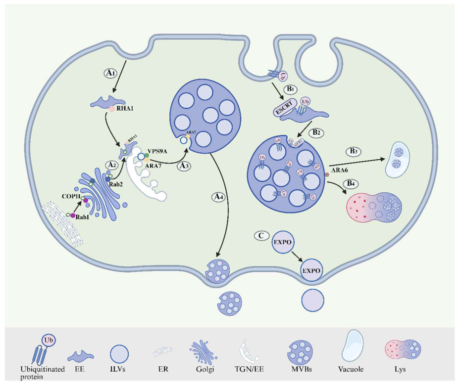
Figure 2.
Therapeutic mechanisms of PELNs in rheumatoid arthritis. A. Folic acid-modified GDEVs activate the PI3K/AKT pathway, regulating the polarization of macrophages from the M1 to the M2 phenotype (CD206/iNOS). B. pgi-miR6135j in GDNPs inhibits KRAS expression and the activation of the p38/JNK/ERK pathway, reducing the expression of inflammatory cytokines IL-6 and TNF-α. C. Rb1/Rg1 in GDNPs suppresses IκBα expression, thereby inhibiting NFATc1 transcription and downregulating the translation of osteoclast markers TRAP and OSCAR.
Figure 2.
Therapeutic mechanisms of PELNs in rheumatoid arthritis. A. Folic acid-modified GDEVs activate the PI3K/AKT pathway, regulating the polarization of macrophages from the M1 to the M2 phenotype (CD206/iNOS). B. pgi-miR6135j in GDNPs inhibits KRAS expression and the activation of the p38/JNK/ERK pathway, reducing the expression of inflammatory cytokines IL-6 and TNF-α. C. Rb1/Rg1 in GDNPs suppresses IκBα expression, thereby inhibiting NFATc1 transcription and downregulating the translation of osteoclast markers TRAP and OSCAR.
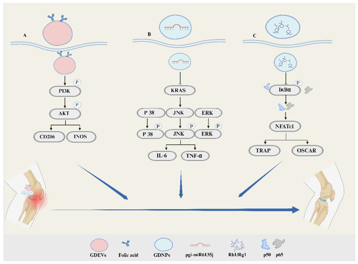
Figure 3.
Therapeutic mechanisms of PELNs in IBD. A. miR-5106 contained in Cc-ELNs inhibits the expression of Slc39A2, reduces Zn²⁺ influx, and thereby decreases mitochondrial ROS production and NET formation. B. SFN in B-ELNs suppresses mTORC1 signaling and upregulates the expression of costimulatory molecules CD80/CD86 on the surface of dendritic cells. C. Po-ELNs elevate the level of ILA to activate AHR, inhibit ZBTB7B expression in CD4⁺ T cells, and induce the reprogramming of CD4⁺ T cells into the CD4⁺CD8⁺ double-positive phenotype. D. miR159 derived from Pr-ELNs inhibits NLRP3 inflammasome assembly and IL-1β maturation in macrophages.
Figure 3.
Therapeutic mechanisms of PELNs in IBD. A. miR-5106 contained in Cc-ELNs inhibits the expression of Slc39A2, reduces Zn²⁺ influx, and thereby decreases mitochondrial ROS production and NET formation. B. SFN in B-ELNs suppresses mTORC1 signaling and upregulates the expression of costimulatory molecules CD80/CD86 on the surface of dendritic cells. C. Po-ELNs elevate the level of ILA to activate AHR, inhibit ZBTB7B expression in CD4⁺ T cells, and induce the reprogramming of CD4⁺ T cells into the CD4⁺CD8⁺ double-positive phenotype. D. miR159 derived from Pr-ELNs inhibits NLRP3 inflammasome assembly and IL-1β maturation in macrophages.
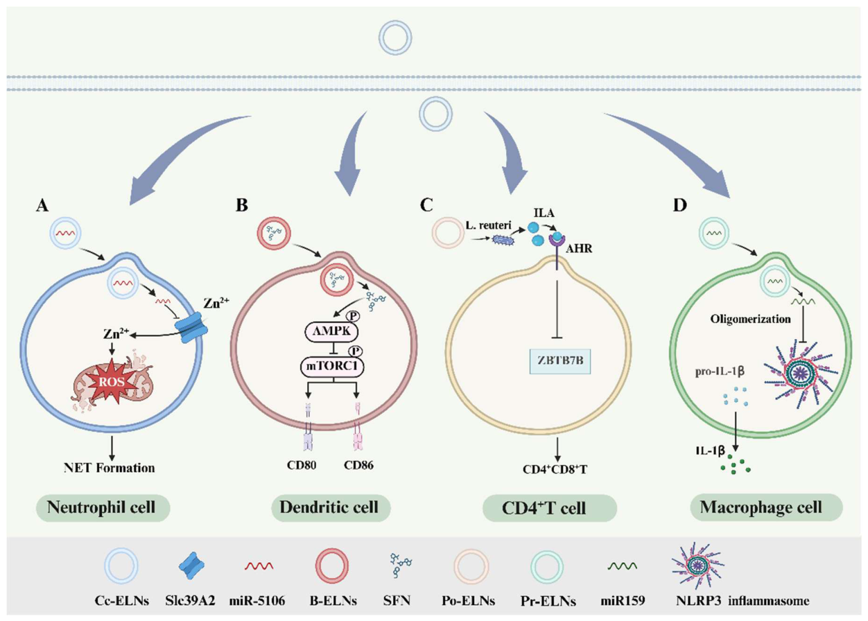
Figure 4.
Engineering strategies of PELNs for enhanced therapeutic efficacy in AIDs. This figure shows that PELNs are engineered through drug loading (co-incubation, electroporation, etc.) and surface modification (conjugation, adsorption, hybridization), targeting inflamed sites in RA, AISD, and IBD to exert anti-inflammatory and reparative effects.
Figure 4.
Engineering strategies of PELNs for enhanced therapeutic efficacy in AIDs. This figure shows that PELNs are engineered through drug loading (co-incubation, electroporation, etc.) and surface modification (conjugation, adsorption, hybridization), targeting inflamed sites in RA, AISD, and IBD to exert anti-inflammatory and reparative effects.
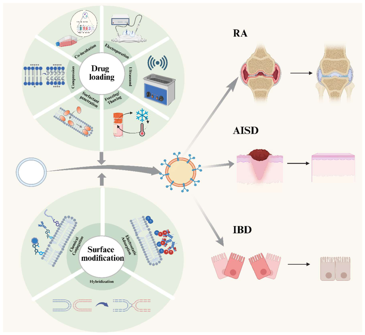

Table 1.
Compositional Comparison Between PELNs and MEVs.
| Category | Aspect of Comparison | PELNs | MEVs | Ref. |
|---|---|---|---|---|
| Lipids | Major Composition | Enriched in diverse phospholipids: PC, PE, PI, PS, PA; contain phytosterols. | Primarily contain cholesterol, sphingomyelin, saturated phosphatidylcholine, etc., with high cholesterol content. | [11,36, 38,49] |
| Structural Function | Higher membrane stability: Phytosterols reduce membrane fluidity and increase rigidity; PA modulates membrane fission/fusion via hydrogen bonding, enhancing stability. | Higher membrane fluidity, primarily regulated by cholesterol. | [37,38] | |
| Function | Lipid mediators: PA can target and regulate specific bacteria, mediating immune modulation. | / | [41,42] | |
| Proteins | Signature Components | Partially homologous/conserved proteins: Heat shock proteins, Annexins, aquaporins, etc. Plant-specific/distinctive proteins: PEN1, PEN3, TET-8. |
Marker proteins: TSG101, Alix, CD9, CD63, CD81, HSP70, etc. | [10,48, 49,51,54] |
| Function | 1. Structural framework and transport 2. Specific targeting 3. Involvement in plant immunity and lipid sorting. |
1.2 Same as PELNs 3. Participate in intercellular signal transduction |
[36,39, 44,53] |
|
| Non-coding RNAs | microRNA | 1. Biogenesis: Nuclear maturation; the DCL1 complex completes processing in the nucleus, no Drosha step required. 2. Modification and Stability: 3′-end methylation enhances its high stability and resistance to degradation. |
1. Biogenesis: Stepwise nuclear-cytoplasmic processing; Drosha cleaves in the nucleus to produce pre-miRNA, Dicer cleaves in the cytoplasm to mature. 2. Modification and Stability: Typically lack 3′ methylation, resulting in lower relative stability. |
[61,64] |
| siRNA | Types: Subtypes such as hc-siRNA and tasiRNA may exist within the vesicles. 2. Cross-kingdom potential: The ability to cross-kingdom silence human genes is not yet confirmed. |
Primarily exogenously introduced or synthetically produced siRNAs for experimental or therapeutic gene silencing. | [65,66, 68,69] |
|
| Mechanism of Action | Competitive inhibition, allosteric regulation, protein interaction interference. | / | [80,82,84] |
Table 2.
Therapeutic Applications and Mechanisms of PELNs in Autoimmune Diseases.
| Disease | PELNs Source |
Therapeutic Application | Mechanism of Action | Ref. | |
|---|---|---|---|---|---|
| RA | Ginger | Targeting inflammatory macrophages | FA-GDEVs drive macrophage polarization from M1 to M2 | [74] | |
| Ginseng | Inhibiting synovial macrophage inflammation | GDNPs-miR6135j targets KRAS to suppress MAPK signaling | [112] | ||
| Ginseng | Inhibiting osteoclast-mediated bone erosion | GDNPs block NF-κB and MAPK/AP-1 pathways | [55] | ||
| IBD | Coptis chinensis | Modulating neutrophil function | Cc-ELNs-miR-5106 downregulates Slc39a2 to suppress NET formation | [118] | |
| Broccoli | Regulating dendritic cell activation | B-ELNs activate AMPK to inhibit mTORC1 | [120] | ||
| Ginger | Promoting anti-inflammatory macrophages | GELNs-miR164d targets TAB1 | [126] | ||
| Grape | Enhancing anti-inflammatory capacity | GELEN upregulates macrophage anti-inflammatory factors | [127] | ||
| Ginseng | Inducing macrophage autophagy | GDNs inhibit AKT/mTOR to induce autophagy | [75] | ||
| Prune | Inhibiting NLRP3 inflammasome | PM-EVLPs disrupt NEK7-NLRP3 interaction | [130] | ||
| Grape | Repairing intestinal barrier | GELEN activates Wnt/β-catenin signaling | [49] | ||
| Bitter melon | Regulating oxidative stress | MC-ELNs enhance cellular antioxidant capacity | [134] | ||
| Type 1 Diabetes | Trifolium pratense | Protecting pancreatic function | TPDEs regulate pancreatic-related genes | [136] | |
| Portulaca oleracea | Promoting wound healing | Po-DENs clear ROS and activate DNA repair | [137] | ||
| Mango | Promoting wound healing | PDNVs induce FSTL1 expression | [138] | ||
| Viola yedoensis | Promoting fracture healing | VDNPs inhibit NF-κB signaling | [139] | ||
| Dendrobium catenatum | Promoting wound healing | DDNVs activate Akt/eNOS signaling | [140] | ||
| Psoriasis | Perilla | Regulating keratinocyte immune response | PLEVPs-miR396a-5p targets HSP90 | [142] | |
| Grapefruit | Reducing oxidative stress and promoting repair | Gf-DVLNs alleviate cellular oxidative stress | [144] | ||
| Potato | Inhibiting inflammation and protecting keratinocytes | ExoPs inhibit MMPs and pro-inflammatory factors | [145] | ||
Disclaimer/Publisher’s Note: The statements, opinions and data contained in all publications are solely those of the individual author(s) and contributor(s) and not of MDPI and/or the editor(s). MDPI and/or the editor(s) disclaim responsibility for any injury to people or property resulting from any ideas, methods, instructions or products referred to in the content. |
© 2026 by the authors. Licensee MDPI, Basel, Switzerland. This article is an open access article distributed under the terms and conditions of the Creative Commons Attribution (CC BY) license (http://creativecommons.org/licenses/by/4.0/).
Copyright: This open access article is published under a Creative Commons CC BY 4.0 license, which permit the free download, distribution, and reuse, provided that the author and preprint are cited in any reuse.