

Submitted:
19 August 2026
Posted:
20 August 2026
You are already at the latest version
Abstract
The crystal structure of benzophenanthridine alkaloid dihydrochelerythrine (1,2-dimethoxy-12-methyl-13H-[1,3]benzodioxolo[5,6-c]phenanthridine), C21H19NO4, was determined by single-crystal X-ray diffraction analysis. The alkaloid was isolated from the stem bark of Zanthoxylum usambarense (Engl.) Kokwaro (Rutaceae) collected from Nairobi, Kenya. In contrast to the earlier reported semisynthetic form which crystallized in the monoclinic space group P21/c, the present natural isolate adopts the orthorhombic Pbca space group. The molecule displays an approximately planar benzophenanthridine framework, but with the dihydro group resulting in a distinctly puckered ring and an out-of-plane methyl group on the tertiary amine. Non-covalent interactions are responsible for the characteristic herringbone packing motif.
Keywords:
Dihydrochelerythrine
; Zanthoxylum usambarense
; X-ray structure
; orthorhombic polymorph
1. Introduction
Benzo[c]phenanthridines are a small subclass of isoquinoline alkaloids widely distributed in higher plant families, notably Papaveraceae and Rutaceae, and shown to possess diverse pharmacological effects including antimicrobial, anti-inflammatory, and antitumour properties [1,2]. Zanthoxylum usambarense (Engl.) Kokwaro (Rutaceae), whose common English name is Usambara prickly ash, is a tree native to East Africa where it is used medicinally among indigenous communities for malaria and other infections, rheumatism, and gastrointestinal ailments [3,4,5]. Extracts of Z. usambarense have demonstrated antiplasmodial activity in experimental models [5].
Previous phytochemical investigations on Z. usambarense have reported several benzo[c]phenanthridine alkaloids including nitidine and chelerythrine [6], as well as norchelerythrine and oxychelerythrine [7]. To the best of our knowledge, dihydrochelerythrine (Figure 1) has not previously been reported from Z. usambarense. However, it has been isolated from the stem bark of Z. nitidum [8,9], the root bark of Z. zanthoxyloides [10], and roots of Z. paracanthum [11] and Z. flavum [12]. Dihydrochelerythrine has demonstrated antiproliferative [10,13] and antifungal activities [14].
While investigating photoactivatable dihydroalkaloids for cancer cell imaging and chemotherapy, Ling et al. [15] semi-synthesized dihydrochelerythrine (1) from chelerythrine and reported its crystal structure in the monoclinic space group P21/c. In contrast, the present study reports the isolation of (1) from the stem bark of Z. usambarense and its single-crystal X-ray structure in the orthorhombic space group Pbca. This represents both the first report of dihydrochelerythrine from Z. usambarense and the first description of this orthorhombic Pbca polymorph. This finding is significant due to potential utility of Z. usambarense as a natural source of dihydrochelerythrine.
2. Results
2.1. Structural Commentary
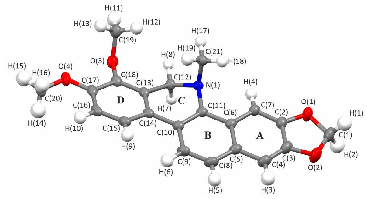
Dihydrochelerythrine (1) crystallized in the orthorhombic space group Pbca with Z = 8. The benzophenanthridine nucleus is fully resolved with all non-hydrogen atoms refined anisotropically and all hydrogen atoms freely refined. Although the fused tetracyclic framework formed by rings A-B-C-D is approximately planar, ring C is not aromatic and is consequently puckered. Least-squares planes fitted through the 10 C and 2 O atoms of rings A and B and through the 6 C and 2 O atoms of ring D show that these are both planar (RMSD values 0.021 and 0.016 Å, respectively); the interplanar angle due to the twist about the C(10)–C(14) bond (Figure 2) in ring C is 12.60°, which is a similar value to two of the torsion angles involving this bond, e.g., ∡ C(9)-C(10)-C(14)-C(15) is 11.70° and ∡ C(11)-C(10)-C(14)-C(13) is 14.32°. In the puckered ring C, the sp3-hybridized C(12) carbon and N(1) nitrogen atoms lie 0.621 and 0.527 Å below / above the mean aromatic planes of rings AB and ring D, respectively. These displacements generate a shallow envelope conformation in ring C, distort π-conjugation, produce a racemic mixture of chiral conformers, and contribute to a herringbone packing motif. This non-planarity contrasts with the more planar iminium framework observed in salts of oxidized benzophenanthridine analogues such as chelerythrine [16].
2.2. Supramolecular Features
The crystal structure exhibits a herringbone packing arrangement (Figure S1). The molecules associate via π–π stacking between adjacent benzophenanthridine ring systems, with centroid–centroid separations consistent with typical aromatic stacking in benzo[c]phenanthridine alkaloids. Additional stabilization arises from weak C-H···O contacts involving both methoxy and methylenedioxy oxygen atoms, which link the molecules into layers extending along the c axis generating the signature three-dimensional herringbone packing framework.
2.3. Database Survey
A search of the Crystallography Open Database using “exact match” command returned one card entry (COD 1558616) for dihydrochelerythrine reported by Ling et al. [15]. A similar search of the Cambridge Structural Database yields the single entry WUZTOD. In contrast to the earlier reported semisynthetic form which has a monoclinic crystal structure with a P21/c space group, the present natural isolate adopts the orthorhombic Pbca space group, implying distinct polymorphic differences between the two crystal forms. Surprisingly, the two crystals contain conformationally-equivalent molecules (Figure S2) with the methyl group on O(4) lying approximately in the fused plane of the A-B-C-D rings but with the methyl groups on O(3) and N(1) pointing out of the plane (Figure 2).
3. Discussion
Polymorphism in organic crystal structures frequently results from flexible organic molecules adopting different conformations, and depending on the crystallization conditions (e.g., speed, choice of solvent), different solid forms may be obtained. We note that fine needles were initially obtained before the final block form was obtained, which may correspond to another polymorphic form. Unusually, the molecules of (1) in the orthorhombic form reported here exhibit a very similar conformation to those found in the monoclinic polymorph reported previously suggesting that this is the most stable conformation for this molecule. In this instance, the two polymorphic forms arise from different packing arrangements of the molecules (cf. Figure S1 and Figure S3). Furthermore, there is likely to be a lack of a distinct minimum in the energy landscape as the crystal structures of both polymorphs are held together solely via weak non-covalent interactions. Thus, it would be interesting to see if there are other forms predicted by crystal structure prediction. The existence of polymorphic forms has implications regarding any pharmaceutical applications of this material.
4. Materials and Methods
4.1. Plant Material
The stem bark of Zanthoxylum usambarense was collected in January 2024 in the outskirts of the Nairobi National Park, Kenya (1.3333° S, 36.7° E). The species was authenticated by comparing the collected specimen with archived herbarium material of Zanthoxylum usambarense preserved at the University of Nairobi (UoN) Herbarium. Authentication was conducted by a senior taxonomist in the Department of Botany, UoN.
4.2. Extraction and Isolation
The dried and ground bark (300 g) was extracted twice by maceration in 1,000 mL of methanol-dichloromethane (1:1) for 72 h with continuous stirring on a magnetic stirrer. The crude extract was filtered and concentrated at 50 ℃ under reduced pressure to afford a dark brown residue (20 g). The extract was adsorbed onto 25 g of silica gel, dry-packed on a column containing 180 g of silica gel, eluted with a hexane-ethyl acetate-methanol gradient and collected in approximately 10 mL fractions. Dihydrochelerythrine was obtained from the hexane-ethyl acetate fractions as colourless needles (227 mg, 1.2%). Recrystallization from acetone afforded colourless blocks, from which crystals suitable for SCXRD were obtained by slow evaporation at room temperature. The identity of the isolated compound was supported by liquid chromatography-mass spectrometric (LC-MS) analysis and single-crystal X-ray diffraction.
4.3. Liquid Chromatography-Mass Spectrometry Analysis
LC-MS analysis was performed using a Shimadzu LCMS-8040 system equipped with an electrospray ionization (ESI) interface. Chromatographic separation was achieved on a Kinetex XB-C18 column (2.6 μm, 150 × 3 mm) fitted with a Phenomenex SecurityGuard Ultra cartridge. The column oven was maintained at 40 °C and the injection volume was 5 μL. The mobile phase consisted of water containing 0.1% formic acid (A) and acetonitrile (B), delivered at a flow rate of 0.2 mL/min. Gradient elution was used: 10% B, 0−0.5 min; 10−50% B, 0.5−5 min; 50−90% B, 5−8 min; 90% B, 8−11 min; 90−10% B, 11−12 min; and 10% B, 12−20 min. Mass spectra were acquired in full-scan mode with polarity switching. The nebulizing and drying gas flow rates were 2.0 and 15 L/min, respectively, while the desolvation-line and heating-block temperatures were 250 and 400 °C, respectively.
4.4. X-Ray Data Collection and Structure Refinement
Single-crystal X-ray diffraction (SCXRD) data were collected on an Agilent Oxford Diffraction SuperNova diffractometer equipped with a microfocus Cu Kα radiation source (λ = 1.54184 Å) and a HyPix-Arc 100 hybrid pixel detector. A piece of (1) was sliced off a much larger block-shaped crystal, mounted on a 20 μm nylon loop (Hampton Research) (Figure S5) and cooled to 150 K using an Oxford Instruments Cryojet5®. A complete sphere of data to a resolution of 0.84 Å was obtained in under 45 min using 0.5° ω-scan frames. Data reduction, including indexing, integration, scaling, and absorption correction, was performed using the CrysAlisPro (Version 1.171.42.60a; Rigaku Oxford Diffraction).
The structure was solved initially using the ‘What is this?’ (WIT) feature in CrysAlisPro [17], with intrinsic phasing in SHELXT [18], followed by full-matrix least-squares refinement on F² using SHELXL [19] within Olex2 [20]. As (1) has conformational chiral forms, atoms in the asymmetric unit were chosen to chiral match the conformation reported in [15]. The positional and anisotropic displacement parameters for the non-hydrogen atoms were refined freely. All hydrogen atoms were located in the difference Fourier maps and were freely refined with isotropic displacement parameters. Molecular and crystal structure illustrations were prepared using Mercury (Cambridge Crystallographic Data Centre, CCDC) [21], with displacement ellipsoids for non-hydrogen atoms shown at the 50% probability level, while hydrogen atoms are shown in their refined positions.
Crystal data for C21H19NO4 (M_r = 349.37): orthorhombic, space group Pbca, a = 14.35758(7) Å, b = 8.45782(4) Å, c = 27.38114(13) Å, V = 3325.00(3) Å3, Z = 8, T = 150 K, ρcalc = 1.396 g cm−3, μ(Cu Kα) = 0.791 mm−1, F(000) = 1472. Crystal size: 0.715 × 0.396 × 0.277 mm3. A total of 72,954 reflections were collected, of which 3567 were independent (Rint = 0.0359, Rsigma = 0.0106). Refinement on F2 converged to R1 = 0.0324 and wR2 = 0.0876 for reflections with I ≥ 2σ(I), and R1 = 0.0341 and wR2 = 0.0890 for all data, with goodness-of-fit = 1.074. The largest residual electron-density peak and hole were +0.24 and −0.16 e Å−3, respectively.
Supplementary Materials
The following supporting information can be downloaded at the website of this paper posted on Preprints.org, Figure S1: Molecular packing of (1) in the orthorhombic polymorph; Figure S2: Overlay of the molecular structures of (1) in the orthorhombic and monoclinic polymorphs; Figure 3: Molecular packing of (1) in the previously reported monoclinic polymorph; Figure S4: Positive-ion LC-MS spectrum of dihydrochelerythrine; Figure S5: Crystal of (1) used for SCXRD; Table S1a: Crystal data and structure refinement; Table S1b: Fractional atomic coordinates and equivalent isotropic displacement parameters; Table S1c: Anisotropic displacement parameters; Table S1d: Selected bod lengths; Table S1e: Selected bond angles; Crystallographic information file (CIF).
Author Contributions
Conceptualization, P.M.N. and G.N.T.; Methodology, B.J.A., P.M.N. and G.N.T.; Analysis, J.K.C. and J.N.L.; Visualization, J.K.C. and J.N.L.; Resources: P.M.N.; Data Curation: P.M.N. and K.O.A.; Writing—original draft preparation, P.M.N. and K.O.A.; Writing—review and editing, J.K.C. All authors have read and agreed to the published version of the manuscript.
Funding
This research received no external funding.
Data Availability Statement
CIF files have been deposited at the Cambridge Crystallographic Data Centre (CCDC) with deposition number 2581015. These data can be obtained free of charge via http://www.ccdc.cam.ac.uk/conts/retrieving.html (or from the CCDC, 12 Union Road, Cambridge CB2 1EZ, UK; Fax: +44-1223-336033; E-mail: deposit@ccdc.cam.ac.uk.) The original contributions presented in this study are included in the article/supplementary material. Further inquiries can be directed to the corresponding author.
Acknowledgments
We gratefully acknowledge the University of Nairobi for the infrastructure that facilitated isolation of (1). We acknowledge infrastructure support from the EPSRC who funded the single-crystal X-ray diffractometer at the Department of Chemistry, University College London, (grant reference EP/K03930X/1) and from the Dean of Mathematical and Physical Sciences, Prof. Ivan P. Parkin, for the instrument hybrid-pixel detector upgrade.
Conflicts of Interest
The authors declare no conflicts of interest.
References
- Peng, R.; Xu, M.; Xie, B.; Min, Q.; Hui, S.; Du, Z.; Liu, Y.; Yu, W.; Wang, S.; Chen, X.; Yang, G.; Bai, Z.; Xiao, X.; Qin, S. Insights on Antitumor Activity and Mechanism of Natural Benzophenanthridine Alkaloids. Molecules 2023, 28, 6588. [CrossRef]
- Slaninová, I.; Pěnčíková, K.; Urbanová, J.; Slanina, J.; Táborská, E. Antitumour activities of sanguinarine and related alkaloids. Phytochem. Rev. 2014, 13, 51–68. [CrossRef]
- Matu, E.N. Zanthoxylum usambarense (Engl.) Kokwaro. [Internet]. In: Schmelzer, G.H.; Gurib-Fakim, A., Eds. PROTA (Plant Resources of Tropical Africa/Ressources végétales de l’Afrique tropicale); PROTA: Wageningen, The Netherlands, 2011. Available online: https://prota.prota4u.org/protav8.asp?p=Zanthoxylum+usambarense (Accessed 11 August 2026).
- Özkan, M.; Mutiso, P.B.C.; Nahar, L.; Liu, P.; Brown, S.; Wang, W.; Sarker, S.D. Zanthoxylum usambarense (Engl.) Kokwaro (Rutaceae) Extracts Inhibit the Growth of the Breast Cancer Cell Lines MDA-MB-231 and MCF-7, But Not the Brain Tumour Cell Line U251 In Vitro. Phytother. Res. 2013, 27, 787–790. [CrossRef]
- Were, P.S.; Kinyanjui, P.; Gicheru, M.M.; Mwangi, E.; Ozwara, H.S. Prophylactic and curative activities of extracts from Warburgia ugandensis Sprague (Canellaceae) and Zanthoxylum usambarense (Engl.) Kokwaro (Rutaceae) against Plasmodium knowlesi and Plasmodium berghei. J. Ethnopharmacol. 2010, 130, 158–162. [CrossRef]
- Kato, A.; Moriyasu, M.; Ichimaru, M.; Nishiyama, Y.; Juma, F.D.; Nganga, J.N.; Mathenge, S.G.; Ogeto, J.O. Isolation of Alkaloidal Constituents of Zanthoxylum usambarense and Zanthoxylum chalybeum Using Ion-Pair HPLC. J. Nat. Prod. 1996, 59, 316–318. [CrossRef]
- He, W.; Van Puyvelde, L.; De Kimpe, N.; Verbruggen, L.; Anthonissen, K.; Van der Flaas, M.; Bosselaers, J.; Mathenge, S.G.; Mudida, F.P. Chemical Constituents and Biological Activities of Zanthoxylum usambarense. Phytother. Res. 2002, 16, 66–70. [CrossRef]
- Yang, C.H.; Cheng, M.J.; Lee, S.J.; Yang, C.W.; Chang, H.S.; Chen, I.S. Secondary Metabolites and Cytotoxic Activities from the Stem Bark of Zanthoxylum nitidum. Chem. Biodivers. 2009, 6, 846–857. [CrossRef]
- Lu, Q.; Ma, R.; Yang, Y.; Mo, Z.; Pu, X.; Li, C. Zanthoxylum nitidum (Roxb.) DC: Traditional uses, phytochemistry, pharmacological activities and toxicology. J. Ethnopharmacol. 2020, 260, 112946. [CrossRef]
- Andima, M.; Coghi, P.; Yang, L.J.; Wong, V.K.W.; Ngule, C.M.; Heydenreich, M.; Ndakala, A.J.; Yenesew, A.; Derese, S. Antiproliferative Activity of Secondary Metabolites from Zanthoxylum zanthoxyloides Lam: In vitro and in silico Studies. Pharmacogn. Commun. 2020, 10, 44–51. [CrossRef]
- Omosa, L.K.; Nchiozem-Ngnitedem, V.A.; Mukavi, J.; Okoko, B.A.; Nyaboke, H.O.; Hashim, I.; Matundura, J.O.; Efferth, T.; Spiteller, M. Cytotoxic alkaloids from the root of Zanthoxylum paracanthum (Mildbr) Kokwaro. Nat. Prod. Res. 2022, 36, 2518–2525. [CrossRef]
- Ross, S.A.; Krishnaven, K.; Radwan, M.M.; Takamatsu, S.; Burandt, C.L. Constituents of Zanthoxylum flavum and their Antioxidant and Antimalarial Activities. Nat. Prod. Commun. 2008, 3, 791–794. [CrossRef]
- Silva, T.C.C.; de Faria Lopes, G.P.; Menezes-Filho, N.J.; de Oliveira, D.M.; Pereira, E.; Pitanga, B.P.S.; Costa, R.S.; Velozo, E.S.; Freire, S.M.; Clarêncio, J.; Borges, H.L.; Rehen, S.K.; Moura-Neto, V.; Costa, S.L. Specific Cytostatic and Cytotoxic Effect of Dihydrochelerythrine in Glioblastoma Cells: Role of NF-κB/β-catenin and STAT3/IL-6 Pathways. Anti-Cancer Agents Med. Chem. 2018, 18, 1386–1393. [CrossRef]
- Yang, H.; Wang, Z.; Guan, Z.; Zhao, M.; Wu, H.; Yang, H. Antifungal Effects and Mechanism of Dihydrochelerythrine Against Fusarium oxysporum. Microorganisms 2025, 13, 2800. [CrossRef]
- Ling, X.; Huang, L.; Li, Y.; Wan, Q.; Wang, Z.; Qin, A.; Gao, M.; Tang, B.Z. Photoactivatable dihydroalkaloids for cancer cell imaging and chemotherapy with high spatiotemporal resolution. Mater. Horiz. 2020, 7, 2696–2701. [CrossRef]
- Li, M.; Li, Z.; Wang, J.-R.; Mei, X. Hydrochromism behaviors of solid forms of chelerythrine hydrochloride. CrystEngComm 2019, 21, 5915–5921. [CrossRef]
- Matsumoto, T.; Yamano, A.; Sato, T.; Ferrara, J. D.; White, F. J.; Meyer, M. “What is This?” A Structure Analysis Tool for Rapid and Automated Solution of Small Molecule Structures. J. Chem. Crystallogr. 2021, 51, 438–450. [CrossRef]
- Sheldrick, G.M. SHELXT—Integrated space-group and crystal-structure determination. Acta Crystallogr. Sect. A 2015, 71, 3–8. [CrossRef]
- Sheldrick, G.M. Crystal structure refinement with SHELXL. Acta Crystallogr. Sect. C 2015, 71, 3–8. [CrossRef]
- Dolomanov, O.V.; Bourhis, L.J.; Gildea, R.J.; Howard, J.A.K.; Puschmann, H. OLEX2: a complete structure solution, refinement and analysis program. J. Appl. Cryst. 2009, 42, 339–341. [CrossRef]
- Macrae, C.F.; Sovago, I.; Cottrell, S.J.; Galek, P.T.A.; McCabe, P.; Pidcock, E.; Platings, M.; Shields, G.P.; Stevens, J.S.; Towler, M.; Wood, P.A. Mercury 4.0: from visualization to analysis, design and prediction. J. Appl. Cryst. 2020, 53, 226–235. [CrossRef]
- Rao, S.W.; Duan, Y.Y.; Pang, H.Q.; Xu, S.H.; Hu, S.Q.; Cheng, K.G.; Liang, D.; Shi, W. Spectrum-Effect Relationship Analysis of Bioactive Compounds in Zanthoxylum nitidum (Roxb.) DC. by Ultra-High Performance Liquid Chromatography Mass Spectrometry Coupled with Comprehensive Filtering Approaches. Front. Pharmacol. 2022, 13, 794277. [CrossRef]
Figure 1.
Chemical structure of dihydrochelerythrine (1).
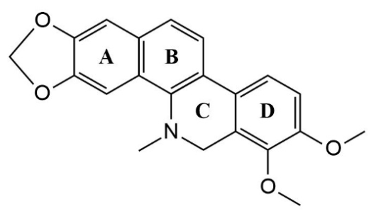
Figure 2.
Refined molecular structure of 1 showing the labelling of the atoms as used for the tables (C= grey, H= white, O= red, and N= blue). For comparative purposes, atoms in the molecule have been labelled identically to those used in the previous study of the monoclinic polymorph by Ling et al. [15]. Displacement ellipsoids are shown at the 50% probability level using Mercury (CCDC).
Figure 2.
Refined molecular structure of 1 showing the labelling of the atoms as used for the tables (C= grey, H= white, O= red, and N= blue). For comparative purposes, atoms in the molecule have been labelled identically to those used in the previous study of the monoclinic polymorph by Ling et al. [15]. Displacement ellipsoids are shown at the 50% probability level using Mercury (CCDC).
Disclaimer/Publisher’s Note: The statements, opinions and data contained in all publications are solely those of the individual author(s) and contributor(s) and not of MDPI and/or the editor(s). MDPI and/or the editor(s) disclaim responsibility for any injury to people or property resulting from any ideas, methods, instructions or products referred to in the content. |
© 2026 by the authors. Licensee MDPI, Basel, Switzerland. This article is an open access article distributed under the terms and conditions of the Creative Commons Attribution (CC BY) license (http://creativecommons.org/licenses/by/4.0/).
Copyright: This open access article is published under a Creative Commons CC BY 4.0 license, which permit the free download, distribution, and reuse, provided that the author and preprint are cited in any reuse.