

Submitted:
19 August 2026
Posted:
20 August 2026
You are already at the latest version
Abstract
The classical Kato-Katz method, the WHO-recommended gold standard for diagnosing intestinal schistosomiasis (Schistosoma mansoni), has limited sensitivity, especially in low-intensity infections, is subject to interference from artifacts, and requires expert microscopists. These limitations lead to underdiagnosis and underestimated disease prevalence. This study evaluated the combination of the Parasimax multicolor® staining method and the Kato-Katz technique to increase its diagnostic sensitivity, while also reducing interference from artifacts. Stool samples were processed in parallel, comparing conventional Kato-Katz and Kato-Katz–Parasimax. The classical Kato-Katz showed a limitation in detecting Schistosoma mansoni eggs in dense areas. In contrast, the Parasimax-associated Kato-Katz significantly increased the visibility of eggs, with greater clarity. Results were obtained in 3–6 minutes (vs. 30–60 minutes for standard Kato-Katz) from sample preparation and observation. The method also allowed the application of an AI segmentation image technique in a few seconds. The classical Kato-Katz has limited sensitivity, but when Parasimax is associated with it, sensitivity is effectively increased, while diagnosis time is reduced to seconds. This novel approach offers a practical, safe, and highly sensitive alternative for field epidemiology and routine diagnosis.
Keywords:
schistosomiasis
; microscopy
; Parasimax multicolor®
; Schistosoma mansoni
; Kato-Katz
; artificial inteligence
1. Introduction
Intestinal schistosomiasis, caused by the blood fluke Schistosoma mansoni, remains a significant public health challenge in sub-Saharan Africa, Latin America, and parts of the Middle East. Approximately 200 million people are infected with schistosomiasis worldwide, with the majority of cases concentrated in resource-limited settings where access to advanced diagnostic tools is scarce [1,2]. The Kato-Katz thick smear technique, developed in the 1970s, has been the gold standard technique for S. mansoni diagnosis for over five decades. Endorsed by the World Health Organization (WHO) as the gold standard for epidemiological surveys and mass drug administration (MDA) programs, the method is valued for its simplicity, low cost, and minimal equipment requirements. The technique involves pressing a measured amount of fresh stool through a sieve, applying it to a glass slide, covering it with a cellophane strip soaked in glycerol–malachite green, and allowing the preparation to clear for 30–60 minutes before microscopic examination. This clearing process renders the fecal debris translucent, enabling the visualization of the characteristic terminal-spined eggs of S. mansoni [2,3]. The limited sensitivity of the Kato-Katz method is particularly problematic in areas with low parasite loads, where many infections remain undetected. This leads to underestimation of disease prevalence, inadequate treatment coverage, and persistence of transmission cycles. Studies have shown that the Kato-Katz method can miss up to 40–60% of light-intensity infections compared to more sensitive techniques such as the formalin–ether concentration method or molecular assays [4,5]. The method demands skilled microscopists to differentiate S. mansoni eggs from artifacts and other helminth eggs—a task that becomes increasingly challenging in low-intensity infections, where egg counts are low [6].
Given these constraints, there is an urgent need for innovative approaches that can enhance the sensitivity of the Kato-Katz method while maintaining its simplicity and cost-effectiveness. The ideal solution would also reduce processing time, eliminate dependency, and improve biosafety. This study evaluates a novel approach: the association of the Parasimax multicolor® staining method with the conventional Kato-Katz technique. We hypothesized that Parasimax, a rapid staining reagent, could increase the diagnostic sensitivity of the Kato-Katz method by enhancing egg visualization, reducing clearing time, and enabling diagnosis from preserved stool samples. By addressing the fundamental limitations of the classical method, this approach aims to provide a practical, safe, and highly sensitive alternative for the diagnosis of intestinal schistosomiasis in endemic regions.
Several previous methods aimed at improving the Kato-Katz technique have failed, mainly because they could not clearly distinguish Schistosoma eggs from artifacts. This problem makes detection difficult for inexperienced professionals. Given that several studies have demonstrated the good efficacy of the method. Therefore, in this study we intended to add only a slight modification to keep the method as close to the original as possible. We simply added Parasimax multicolor® in one of the steps to make the findings more visible. Parasimax multicolor® is registered with the Brazilian Health Regulatory Agency (ANVISA) under number 81649660014, as stated in the manufacturer’s instructions.
2. Materials and Methods
2.1. Study Sites and Sample Collection
Pre-diagnosed positive stool samples for S. mansoni were obtained from a health center in the Marara District of Tete Province, Mozambique (n = 1) and processed according to standard protocols.
2.2. Staining Procedures
2.2.1. Kato-Katz
The conventional Kato-Katz thick smear technique was performed according to the standard WHO protocol. A portion of fresh stool was pressed through a nylon mesh sieve to remove large debris. Approximately 50 mg of the sieved stool was placed on a glass slide and covered with a cellophane strip pre-soaked in a glycerol–malachite green solution. The slide was inverted and pressed firmly to obtain a uniform smear. The preparation was allowed to clear for 30–60 minutes at room temperature before microscopic examination. S. mansoni eggs were identified by their characteristic morphology: a lateral spine and a size of approximately 114–180 µm × 45–70 µm.
2.2.2. Parasimax Modified Kato-Katz
For the Kato-Katz–Parasimax adaptation, the same standard protocol was followed, with a slight modification: approximately 50 µL of Parasimax reagent was added onto the sample in the circular template on the slide and allowed to act for about 1 minute, and then thoroughly mixed with a stylus, ensuring it penetrated the entire sample to stain artifacts and S. mansoni eggs (Figure 1). During this process, Parasimax also blocks the toxicity of malachite green, preserving the viability of larvae inside the eggs in fresh samples.
2.3. Application of Artificial Intelligence for Image Segmentation
The application of artificial intelligence is currently a widely studied method to enable more accurate identification of microscopic findings, thereby increasing the speed of identification. In this work, we adopted Meta AI’s Segment Anything Model (SAM), which performs zero-shot segmentation tasks without reliance on labeled training data or domain-specific priors. The images obtained from the microscope were selected and submitted directly to the Meta AI website, following the described guidelines.
3. Results
In the classical Kato-Katz method, clear egg visualization among fecal debris is nearly impossible, and the use of a lower-magnification lens is therefore not advised (Figure 2a). Malachite green, while useful for egg clarification, often stains both eggs and artifacts without discrimination (Figure 2b). This can be mitigated by gently pressing the cellophane to create a monolayer. Staining intensity, however, varies with each preparation and is generally stronger at the center, with better visibility toward the periphery, where artifact interference is reduced (Figure 2c). Nevertheless, eggs are not always detectable in all areas of the slide.
The adaptation of Parasimax in the Kato-Katz technique was motivated by previous experiments. In this approach, we surprisingly observed that penetration into the sample caused artifacts to stain differently compared to S. mansoni egg, which took on a yellowish color (Figure 2e). The contrast between the yellowish color and the blue increases the chances of visualization, even when using a lower-magnification lens (4x) (Figure 2d). This could not be achieved in classic methodology (Figure 2a).
This latter procedure would drastically reduce response time in epidemiological studies. The most interesting aspect of this adaptation is that the miracidia remain alive for a certain period of time. In other studies, this adaptation also allows the observation of protozoa such as Entamoeba histolytica, Giardia, Cystoisospora, among others. We verified that this combination does not affect sample preservation. This adaptation also allows observation by fluorescence microscopy, which incredibly increases the sensitivity of the method. This particular feature differentiates it from other methods that use toxic and costly chemicals.
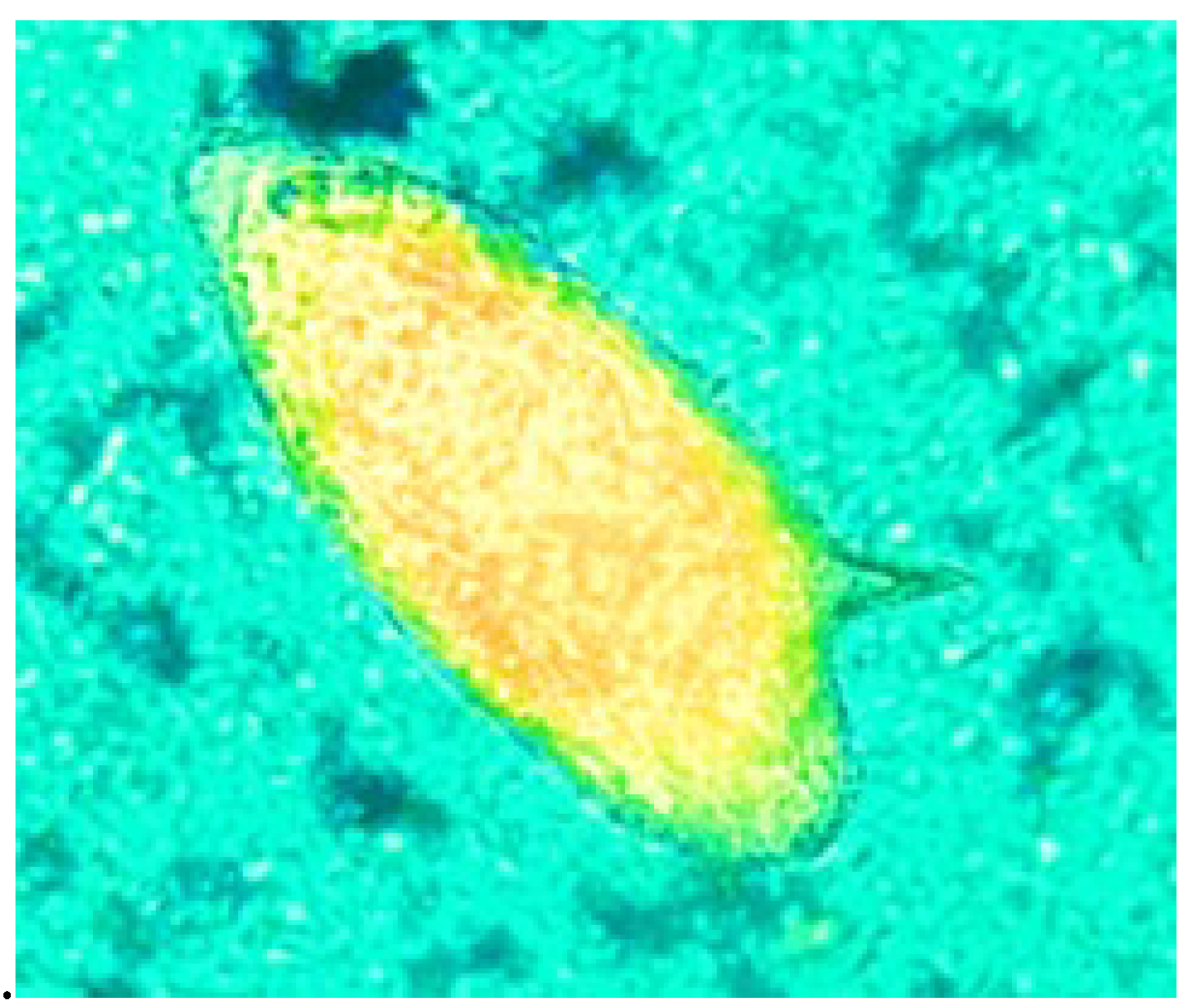
The possibility of applying fluorescence microscopy to the Kato-Katz method adapted with Parasimax is a very important milestone for the advancement in the diagnosis of schistosomiasis caused by S. mansoni, as it allows for a much sharper image due to the fluorescence emission by the Parasimax staining (Figure 3). This is not possible when applying the classical method.
Today, the segmentation of images obtained from a microscope is a very useful procedure for better visualization of structures and their differentiation from other unimportant elements. Several studies have been carried out to facilitate this procedure. However, the main difficulty lies in interference from artifacts, the wide variety of colors, and object sizes, among other factors. Some studies have applied artificial intelligence for segmenting images of samples containing S. mansoni. Many equipment manufacturers are very interested in more economical and simpler differentiation methods to facilitate subsequent steps. Therefore, it is necessary to accumulate thousands of microscopic object data for comparison. With this advancement, new equipment with special cameras can perform automated detection analyses. However, these steps are complicated with current methods, making it still very difficult to apply them to the Kato-Katz method. Nevertheless, the adaptation of Parasimax to the Kato-Katz method could make this task much easier. Some simulations using segmentation programs have shown that this procedure is feasible.
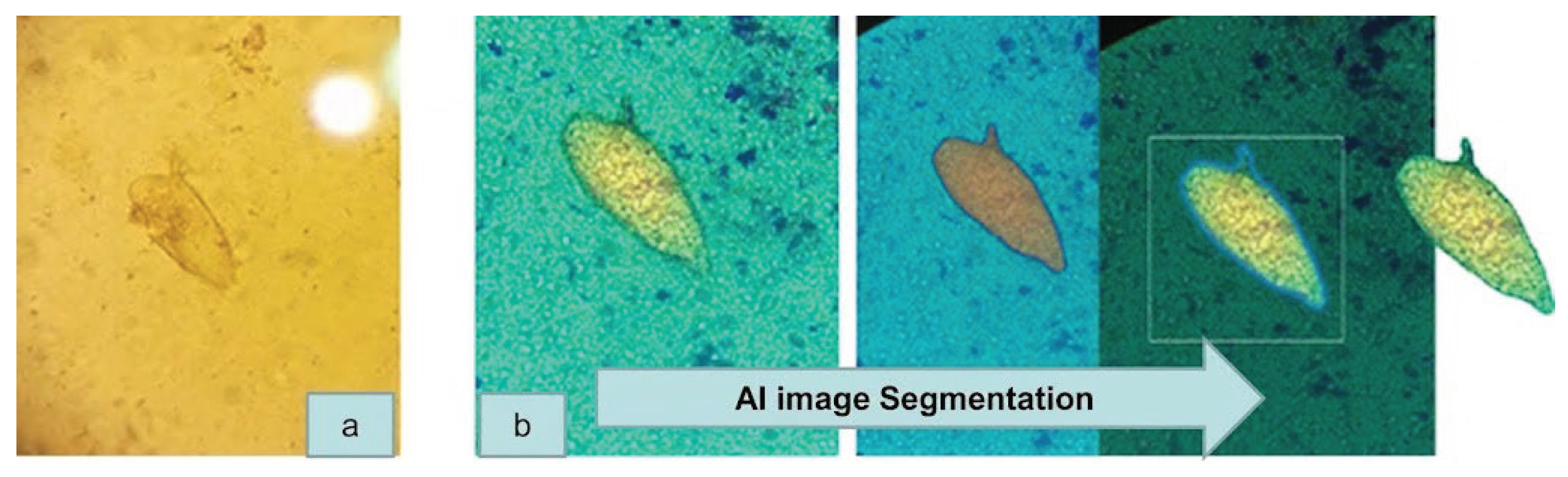
Figure 4.
a) Standard technique; b) Image segmentation by Artificial Inteligence after Parasimax staining.
Figure 4.
a) Standard technique; b) Image segmentation by Artificial Inteligence after Parasimax staining.
4. Discussion
The observation of S. mansoni eggs requires a great deal of experience on the part of the technicians, since artifacts can hinder their visualization. This is because stool samples contain dietary fibers that can resemble the eggs. Although the confirmatory result consists of observing the lateral spines, on certain occasions these can be obscured by artifacts. In some cases, the low excretion of eggs raises doubts about false-negative results, so observing the entire area is essential. Although the use of a nylon filter allows the exclusion of larger particles, this does not prevent artifacts from appearing on the slide. This makes observation difficult in denser areas, especially in the central portion. The Kato-Katz technique is standard for S. mansoni detection; however, some studies have shown some disadvantages due to the interference of artifacts and egg degradation. Several previous studies have shown that a single K–K thick smear preparation is a poor predictor of the “true” prevalence of S. mansoni infections and hence a poor test in assessing the “real” infection status of individuals, particularly when infection intensities are low [7]. Several methods have been proposed to improve the original Kato-Katz method. In some studies, coconut oil and kanag oil were used as clearing agents to replace glycerol. Another study, conducted by Odongo-Aginya et al., replaced malachite green with a solution of eosin and nigrosin [2,8,9,10,11]. The attempt to replace some compounds of the original method is mentioned in several studies due to their potential toxicity. Other authors have also tried to redesign the sample area to further increase the sensitivity of the method [12].
Most studies on replacing malachite green with alternative clarifiers have shown no significant improvement in egg-to-background contrast. However, the adaptation with Parasimax notably enhanced visibility, even when artifacts are closer to the S. mansoni egg, and allowed safe observation at lower magnifications. Although malachite green is widely criticized for its potential toxicity, persistence in food fish tissues, and suspected carcinogenic and genotoxic properties, we retained its use due to its excellent long-term sample preservation, which supports future analyses. Moreover, its impressive fluorescence brightness further accelerated egg detection after Parasimax adaptation.
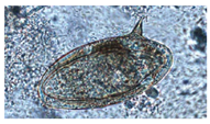
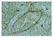
We conducted a comparison of the Kato-Katz methods applied in several studies and encountered some differences, ranging from the color and shape of S. mansoni eggs. In the comparative study with zinc sulfate flotation, the Kato-Katz method proved to be better in terms of preserving egg structure without causing dehydration [13]. However, we observed color changes and a virtual lack of sharpness in the other study [14]. In the method adapted for automation, there was excessive pressure, which caused distortion in the egg shape itself [15]. On the other hand, the other three studies cited show better egg shape and image clarity [16,17,18]. In one of the studies, a slight staining with malachite green could be seen, similar to our study, applying the unmodified Kato-Katz method [16]. Finally, one can clearly see greater egg sharpness when observed using the Kato-Katz method modified with Parasimax. The comparison between images obtained by different authors is listed in the following table.

Table 1.
Comparison of images obtained from S. mansoni applying different methods adapted to Kato-Katz.
Table 1.
Comparison of images obtained from S. mansoni applying different methods adapted to Kato-Katz.
| Title | Technique | References |
| Comparing diagnostic accuracy of Kato-Katz, Koga agar plate, ether-concentration, and FLOTAC for Schistosoma mansoni and soil-transmitted helminths. | 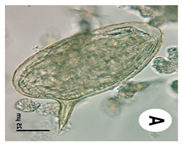 |
[12] |
| Esquistossomose: manejo clínico e epidemiológico na atenção básica | 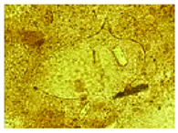 |
[13] |
| Automated diagnosis of schistosomiasis by using faster R-CNN for egg detection in microscopy images prepared by the Kato–Katz technique | 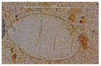 |
[14] |
| Acute schistosomiasis in Brazilian traveler: the importance of tourism in the epidemiology of neglected parasitic diseases | 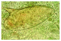 |
[15] |
| A rapid diagnostic test for schistosomiasis mansoni |
 |
[16] |
| Comparison of sensitivity and fecal egg counts of Mini-FLOTAC using fixed stool samples and Kato-Katz technique for the diagnosis of Schistosoma mansoni and soil-transmitted helminths | 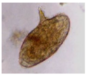 |
[17] |
| First imported case of cerebral Schistosomiasis mansoni (China, May 2025) |
 |
[18] |
| Kato-Katz-adapted Parasimax method | 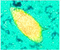 |
This Study |
Schistosomiasis caused by S. mansoni is a major problem in many African countries, especially in Nigeria, as well as in South America, in some states of Brazil [20,21,22,23,24]. However, one of the major limitations of adopting the classical Kato-Katz method is the need for well-trained laboratory professionals, due to several factors, one of which is the presence of artifacts and cells such as erythrocytes that can obscure the eggs [25,26]. This problem, which has occurred for decades, can be solved by applying Parasimax, enabling even less experienced professionals to detect the eggs in samples. Erythrocytes are stained green, creating contrast with the eggs, which appear golden-yellow and bright. It is worth noting that other parasites, mainly helminths, can also be better observed through this new adaptation. The presence of blood in the stool samples is a important signal of S. mansoni infection. Thus the detection by Parasimax stain may increase the chance to avoid false negative results.
A study applying a portable microscope connected to a computer with image processing software was used for the identification of eggs from various helminths, including S. mansoni and S. haematobium eggs. However, it requires several steps until the final identification, due to interference from artifacts in the samples. In this method, no contrast-enhancing substance is used, which makes it somewhat difficult [27,28,29]. An interesting study applying artificial intelligence to detect Schistosoma eggs in histological samples showed promising results. However, the use of hematoxylin staining does not allow differentiation between the adjacent tissue and the eggs embedded in the tissue. During the image segmentation process, aggregated adipose tissue with an oval shape interfered with the analyses [30]. Another type of procedure involves identifying the eggs first, and then eliminating all interfering background, creating a black-and-white contrast. This method is highly dependent on the initial identification. In some cases, the removal of this background may accidentally eliminate important findings, potentially leading to false negatives [31]. In our study, the application of META AI to images obtained using the Parasimax technique facilitates rapid recognition of S. mansoni eggs due to the contrast between the background and these findings. The yellow staining differentiates very easily from the green background, especially in less dense areas. This same procedure cannot be applied with precision when applied to images obtained using the classical technique. Several authors have applied this tool to research in various branches of biology and medicine [32,33,34,35]. The comparison between the application of AI segmentation is shown in the following table.
Table 2.
Comparison between S. mansoni images after Artificial intelligence application.
| Title | Technique | References |
| Development of an automated artificial intelligence-based system for urogenital schistosomiasis diagnosis using digital image analysis techniques and a robotized microscope. | 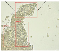 |
[28] |
| Detection of Schistosoma eggs using an AI-based deep learning model on urinary bladder histopathology images. | 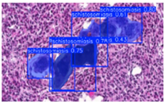 |
[29] |
| Two-stage automated diagnosis framework for urogenital schistosomiasis in microscopy images from low-resource settings. Journal of Medical Imaging | 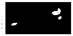 |
[30] |
| Kato-Katz-adapted Parasimax method | 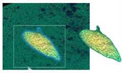 |
This study |
5. Conclusion
In this study, we compared the classical Kato-Katz method and the new adapted method using Parasimax Multicolor. Through the results, we can observe that the new method allows for better visualization of S. mansoni eggs, even in denser areas. The Kato-Katz–Parasimax method was compared with other adaptations found in the literature. It was also possible to adapt Kato-Katz–Parasimax to fluorescence microscopy, which further enhanced the visualization capacity of Schistosoma mansoni eggs, making it a cost-effective and readily acceptable method. Our observations show that eggs can also be observed even with lower magnification objectives, which considerably reduces diagnostic time. This adaptation is suitable for use in endemic areas, where rapid results are often required owing to high demand, and also for epidemiological surveys. An additional key advantage is the capacity to visualize disintegrated eggs, a finding that would be dubious using other methods. Lastly, this new approach enables assessment of egg viability, making it a potentially useful tool for evaluating therapeutic efficacy. Our findings also suggest that this novel method holds strong potential for artificial intelligence applications, enabling more reliable identification of microscopic findings with minimal interference. Our method highlights the ability to visualize disintegrated eggs, identifying a key advantage that remains dubious or unreliable through alternative diagnostic methods.
Competing interests
The authors declare that they have no competing interests.
Abbreviations
| AI | Artificial Intelligence (AI) |
| WHO | World Health Organization (WHO) |
| PCR | Polymerase Chain Reaction |
References
- Ally, O., Kanoi, B. N., Ochola, L., Nyanjom, S. G., Shiluli, C., Misinzo, G., & Gitaka, J. (2024). Schistosomiasis diagnosis: Challenges and opportunities for elimination. PLOS Neglected Tropical Diseases, 18(7), e0012282. [CrossRef]
- Odongo-Aginya, E. I., Narcis, K., Ludwig, S., Wabinga, H., Fenwick, A., & Montresor, A. (2007). Substitution of malachite green with nigrosin-eosin yellow stain in the Kato-Katz method: microscopical appearance of the helminth eggs. African Health Sciences, 7(1). [CrossRef]
- Rifqoh, R., Kustiningsih, Y., & Cahyono, J. A. (2021). Development of modified Kato Katz kit as laboratory-scale prototype for early Detection of soil-transmitted helminths (STHs) infection. Medical Laboratory Technology Journal, 7(2), 201-208. [CrossRef]
- Thi, Q. A. H., Vu, A. N., Thi, T. H. T., Dang Tra, M., Tran, H. T., Tuyet, H. B., ... & Phuong, T. N. (2023). value of the direct, kato-katz, and ether-formalin techniques in diagnosing intestinal helminths. Tạp chí Phòng chống Bệnh sốt rét và các Bệnh ký sinh trùng, 138(6), 10-10.
- Leuenberger, A., Nassoro, T., Said, K., Fenner, L., Sikalengo, G., Letang, E., ... & Knopp, S. (2016). Assessing stool quantities generated by three specific Kato-Katz thick smear templates employed in different settings. Infectious Diseases of Poverty, 5(1), 58. [CrossRef]
- Periago, M. V., Diniz, R. C., Pinto, S. A., Yakovleva, A., Correa-Oliveira, R., Diemert, D. J., & Bethony, J. M. (2015). The right tool for the job: detection of soil-transmitted helminths in areas co-endemic for other helminths. PLoS Neglected Tropical Diseases, 9(8), e0003967. [CrossRef]
- Berhe, N., Medhin, G., Erko, B., Smith, T., Gedamu, S., Bereded, D., ... & Gundersen, S. G. (2004). Variations in helminth faecal egg counts in Kato–Katz thick smears and their implications in assessing infection status with Schistosoma mansoni. Acta Tropica, 92(3), 205-212. [CrossRef]
- Manahan, E. (2014). Virgin coconut oil as an alternative clearing agent used in modified Kato Katz technique. American Journal of Clinical Pathology, 142(suppl_1), A161-A161.
- Maheswari, K. I., Janendra, P. S. A., Widnyana, I., Mahayana, D. G. P., Pramesti, K. K. A., Dwisaputra, I., ... & Giri, M. K. W. (2025). Modification of the Kato-Katz Diagnosis Technique Using “Canang” Flower Waste Extract as a Staining for Ascaris lumbricoides Eggs. [CrossRef]
- Oktavia, S. I., Prabaningtyas, S. A., Nuryati, A., Faruq, Z. H., & Setiawan, B. (2026). Comparative evaluation of beetroot and mangosteen peel extracts as natural alternatives to malachite green in the Kato–Katz method. Journal of Microbiological Methods, 107521. [CrossRef]
- Leuenberger, A., Nassoro, T., Said, K., Fenner, L., Sikalengo, G., Letang, E., ... & Knopp, S. (2016). Assessing stool quantities generated by three specific Kato-Katz thick smear templates employed in different settings. Infectious Diseases of Poverty, 5(1), 58.
- Glinz D, Silué KD, Knopp S, Lohourignon LK, Yao KP, Steinmann P, Rinaldi L, Cringoli G, N’Goran EK, Utzinger J. Comparing diagnostic accuracy of Kato-Katz, Koga agar plate, ether-concentration, and FLOTAC for Schistosoma mansoni and soil-transmitted helminths. PLoS Negl Trop Dis. 2010 Jul 20;4(7):e754. [CrossRef]
- Gomes, Elainne & Domingues, Ana & Barbosa, Constança. (2017). Esquistossomose: manejo clínico e epidemiológico na atenção básica.
- Oliveira, B.A.S., Moreira, J.M.P., Coelho, P.R.S. et al. 2022. Automated diagnosis of schistosomiasis by using faster R-CNN for egg detection in microscopy images prepared by the Kato–Katz technique. Neural Comput & Applic 34, 9025–9042 . [CrossRef]
- Guiguet Leal, D. A., Franco, R. M. B., Neves, M. F., Simoes, L. F., Bastos, L. A. D., Allegretti, S. M., ... & Magalhaes, L. A. (2012). Acute schistosomiasis in Brazilian traveller: the importance of tourism in the epidemiology of neglected parasitic diseases. Case Reports in Infectious Diseases,(1), 650929. [CrossRef]
- Mello-Silva, C. C., João, R. C., Augusto, R. D. C., & Santos, C. P. (2013). A rapid diagnostic test for schistosomiasis mansoni. Memórias do Instituto Oswaldo Cruz, 108, 1078-1080.
- Coulibaly, J. T., Ouattara, M., Becker, S. L., Lo, N. C., Keiser, J., N’Goran, E. K., ... & Utzinger, J. (2016). Comparison of sensitivity and faecal egg counts of Mini-FLOTAC using fixed stool samples and Kato-Katz technique for the diagnosis of Schistosoma mansoni and soil-transmitted helminths. Acta Tropica, 164, 107-116. [CrossRef]
- Chen, W., Zhang, Y., Ma, X., Jiang, T., Zhao, D., & Deng, Y. (2026). First Imported Case of Cerebral Schistosomiasis Mansoni—China, May 2025. China CDC Weekly, 8(5), 131-134.
- Taiwo, O. T., Sam-Wobo, S. O., Ademolu, K. O., Talabi, A. O., & Taiwo, A. M. (2026). Evaluation of Schistosoma mansoni in water sanitation and hygiene-based communities, and associated risk factors in Ogun Central, Nigeria. Discover Public Health, 23(1), 135.
- Abdullahi, M., Dikwa, K. B., Haroun, A. A., & Obimakinde, E. T. (2026). Age-Specific Occurrence of Schistosoma mansoni Infection among Al-majiri Pupils in Rafin Guza and Hayin Dan Bushiya Schools, Kaduna Metropolis, Nigeria. Sahel Journal of Life Sciences FUDMA, 4(1), 702-708. [CrossRef]
- Mogaji, H. O., Omitola, O. O., Bayegun, A. A., Ekpo, U. F., & Taylor-Robinson, A. W. (2023). Livestock reservoir hosts: an obscured threat to control of human schistosomiasis in Nigeria. Zoonotic Diseases, 3(1), 52-67. [CrossRef]
- Ramos, R. E. S., Bezerra, L. P., Lima, M. W. S., Silva, W. S., Nunes, L. K. S., Santos, E. D., ... & Brayner, F. A. (2025). Diagnóstico e fatores de risco associados à infecção por Schistosoma mansoni em uma população de área endêmica do Nordeste do Brasil. Brazilian Journal of Biology, 85, e292478.
- De Moura Fernandes, J. P., Pereira, L. F. C., Monteiro, C. P., Neiva, J. P. D. L. M., de Castro, M. C. Q., Gomes, Y. T., ... & Peixoto, J. A. C. (2024). Esquistossomose no Brasil: Uma análise epidemiológica sobre as repercussões clínicas no aparelho digestivo e nos parâmetros de Saúde Pública. Brazilian Journal of Implantology and Health Sciences, 6(11), 240-267.
- Firmino, F. T., Schmidt, P., de Almeida, M. O., Dolabella, S., Negrão-Corrêa, D., Emelko, M., & Graeff-Teixeira, C. (2026). Effect of variable and low analytical recovery on sensitivity of Kato-Katz, Lutz and Ritchie methods for diagnosis of schistosomiasis. Acta Tropica, 108157. [CrossRef]
- Diehl, J. C., Persaud, M. S. S., & Akella, A. An Upgrade for the Kato Katz Method.
- Ward, P., Dahlberg, P., Lagatie, O., Larsson, J., Tynong, A., Vlaminck, J., ... & Stuyver, L. J. (2022). Affordable artificial intelligence-based digital pathology for neglected tropical diseases: A proof-of-concept for the detection of soil-transmitted helminths and Schistosoma mansoni eggs in Kato-Katz stool thick smears. PLoS Neglected Tropical Diseases, 16(6), e0010500. [CrossRef]
- Meulah, B., Oyibo, P., Hoekstra, P. T., Moure, P. A. N., Maloum, M. N., Laclong-Lontchi, R. A., ... & van Lieshout, L. (2024). Validation of artificial intelligence-based digital microscopy for automated detection of Schistosoma haematobium eggs in urine in Gabon. PLoS Neglected Tropical Diseases, 18(2), e0011967. [CrossRef]
- Rubio Maturana, C., Dantas de Oliveira, A., Zarzuela, F., Ruiz, E., Sulleiro, E., Mediavilla, A., ... & Joseph-Munné, J. (2024). Development of an automated artificial intelligence-based system for urogenital schistosomiasis diagnosis using digital image analysis techniques and a robotized microscope. PLoS Neglected Tropical Diseases, 18(11), e0012614. [CrossRef]
- Shams, M., Bakar, A. A., Zehra, T., Bello, S., Parween, S., & Jafri, A. (2025). Detection of Schistosoma Eggs Using an AI-Based Deep Learning Model on Urinary Bladder Histopathology Images. Training, 476, 1-932. [CrossRef]
- Oyibo, P., Meulah, B., Bengtson, M., van Lieshout, L., Oyibo, W., Diehl, J. C., ... & Agbana, T. (2023). Two-stage automated diagnosis framework for urogenital schistosomiasis in microscopy images from low-resource settings. Journal of Medical Imaging, 10(4), 044005-044005. [CrossRef]
- Khazanchi, R., Govind, S., Congivaram, H. T., Sadagopan, N. S., Jain, R., & Magill, S. T. (2025). Assessment of “Zero-Shot” General Purpose Segmentation Models: An Analysis of the Meta “Segment Anything Model” on Meningioma MRI. American Journal of Neuroradiology, 46(11), 2310-2315. [CrossRef]
- Balamurugan, V., & Sivasankari, K. (2025). Segment anything model-based segmentation with inception-ResNet-v2 classifier for Parkinson’s disease diagnosis and metaverse visualization. Computers in Biology and Medicine, 187, 109732. [CrossRef]
- Kaur, P., Kaushik, A., Singhal, I., Pandey, A., & Singhal, R. (2025). Advancing brain MRI segmentation using segment anything model. Procedia Computer Science, 260, 110-117. [CrossRef]
- Zhang, Y., Shen, Z., & Jiao, R. (2024). Segment anything model for medical image segmentation: Current applications and future directions. Computers in Biology and Medicine, 171, 108238.
- Chauveau, B., & Merville, P. (2023). Segment Anything by Meta as a foundation model for image segmentation: a new era for histopathological images. Pathology, 55(7), 1017-1020. [CrossRef]
Figure 1.
Procedures for Kato-Katz modified by Parasimax multicolor.
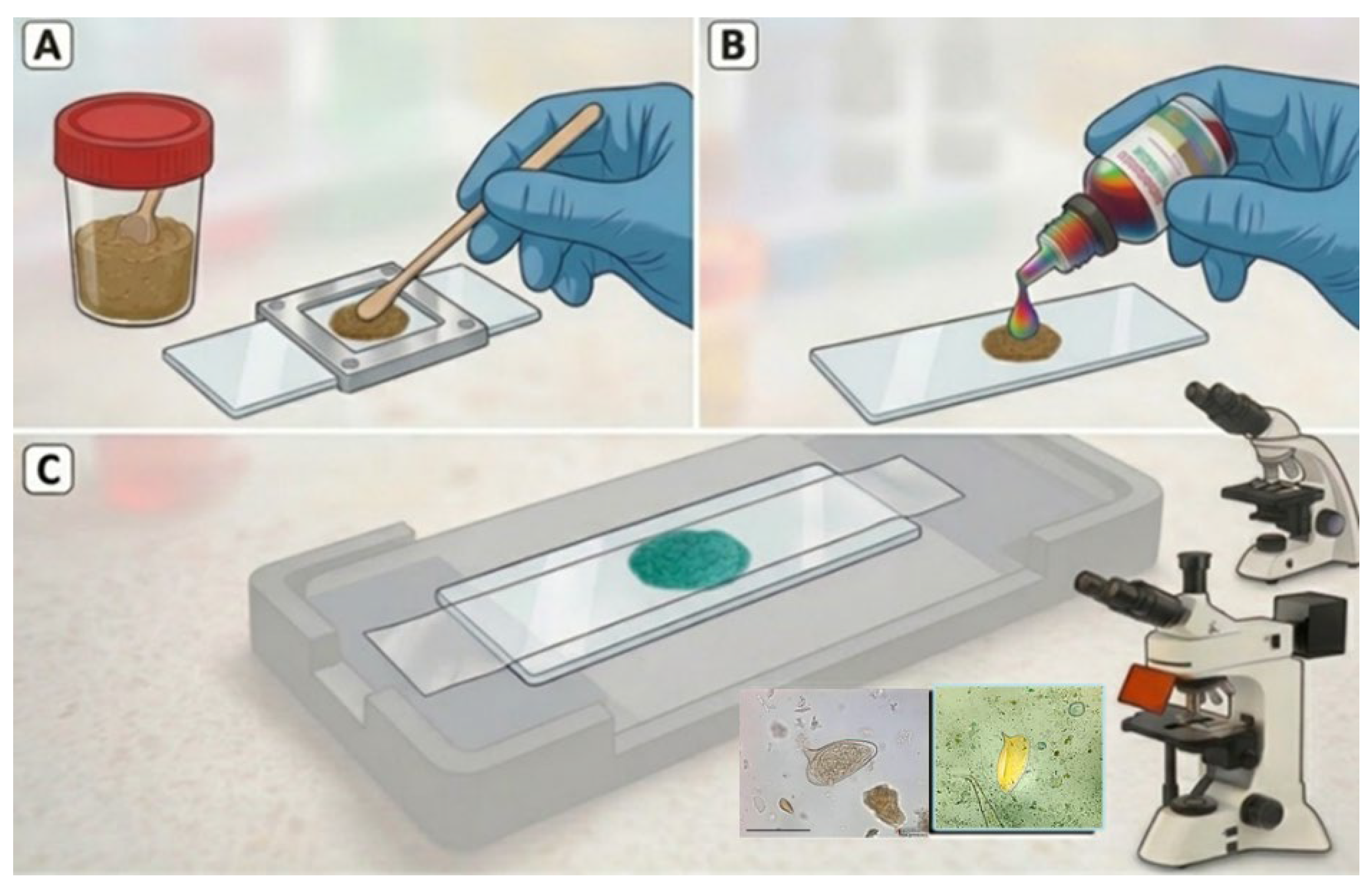
Figure 2.
. Kato-Katz (a, b, c); Kato-Katz-Parasimax (d, e, f).
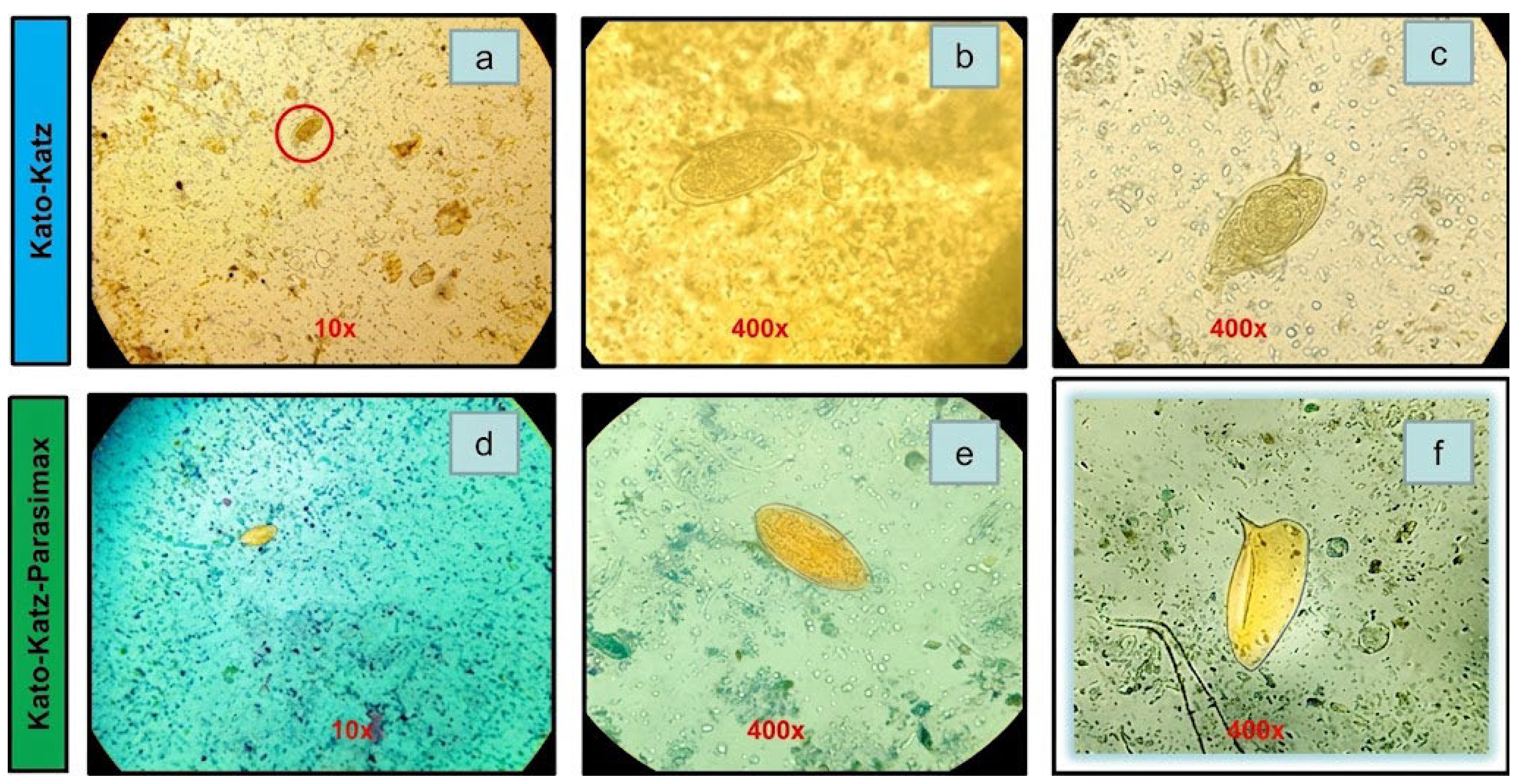
Figure 3.
S. mansoni egg observed by Kato-Katz-adapted Parasimax using a fluorescence microscope.
Disclaimer/Publisher’s Note: The statements, opinions and data contained in all publications are solely those of the individual author(s) and contributor(s) and not of MDPI and/or the editor(s). MDPI and/or the editor(s) disclaim responsibility for any injury to people or property resulting from any ideas, methods, instructions or products referred to in the content. |
© 2026 by the authors. Licensee MDPI, Basel, Switzerland. This article is an open access article distributed under the terms and conditions of the Creative Commons Attribution (CC BY) license (http://creativecommons.org/licenses/by/4.0/).
Copyright: This open access article is published under a Creative Commons CC BY 4.0 license, which permit the free download, distribution, and reuse, provided that the author and preprint are cited in any reuse.