

Submitted:
19 August 2026
Posted:
20 August 2026
You are already at the latest version
Abstract
Pseudomonas aeruginosa is a major opportunistic pathogen associated with nosocomial infections, antimicrobial resistance, and persistent biofilm formation, highlighting the need for alternative therapeutic strategies. This review critically evaluates the current evidence on the phytochemistry, antimicrobial, antibiofilm, anti-inflammatory, and wound-healing properties of Aloe vera (Aloe barbadensis Mill.) against P. aeruginosa. Relevant peer-reviewed literature from major scientific databases was examined to summarize the phytochemical constituents of A. vera, their proposed mechanisms of action, therapeutic applications, safety considerations, and emerging approaches, including nanotechnology and computational drug discovery. The available evidence indicates that anthraquinones, polysaccharides, flavonoids, and related phytochemicals contribute to antibacterial activity, inhibition of biofilm formation, modulation of host inflammatory responses, and promotion of wound healing. However, considerable variability in phytochemical composition, limited pharmacokinetic and toxicological data, lack of standardized formulations, and insufficient high-quality preclinical and clinical studies continue to hinder clinical translation. Overall, A. vera represents a promising source of adjunctive antimicrobial and anti-virulence compounds for the management of biofilm-associated P. aeruginosa infections. Future research should prioritize phytochemical standardization, mechanistic validation, pharmacokinetic evaluation, and well-designed animal and clinical studies to support the development of safe and clinically effective A. vera-based therapeutics.
Keywords:
Aloe vera
; Pseudomonas aeruginosa
; biofilm
; antimicrobial resistance
; phytochemicals
; anthraquinones
; anti-virulence
; wound healing
1. Introduction
Antimicrobial resistance (AMR) is one of the greatest threats to global public health, compromising the effective treatment of bacterial infections and increasing morbidity, mortality, and healthcare costs. Among the priority pathogens identified by the World Health Organization, Pseudomonas aeruginosa is particularly problematic because of its remarkable intrinsic and acquired resistance mechanisms. Its ability to form biofilms, regulate virulence through quorum sensing (QS), and express multidrug efflux pumps enables persistence in both environmental and clinical settings while reducing susceptibility to many conventional antibiotics [1]. Consequently, multidrug-resistant (MDR) P. aeruginosa has become a leading cause of healthcare-associated infections, particularly among immunocompromised patients and individuals with chronic wounds, burns, cystic fibrosis, or indwelling medical devices.
The increasing prevalence of antimicrobial resistance, coupled with the limited development of new antibiotics, has intensified the search for alternative therapeutic strategies. Natural products have emerged as valuable sources of antimicrobial agents because of their structural diversity, broad biological activities, and ability to modulate multiple molecular targets simultaneously [2,3]. Unlike conventional antibiotics that often act on a single target, plant-derived compounds may interfere with bacterial virulence, biofilm formation, quorum sensing, oxidative stress responses, and other pathways, potentially reducing the selective pressure associated with resistance development.
Among medicinal plants, A. vera has attracted considerable attention owing to its long history of use in the treatment of wounds, burns, and skin infections. Its biological activities have been attributed to a diverse range of phytochemicals, including anthraquinones (aloin, aloe-emodin and emodin), polysaccharides such as acemannan, chromones, flavonoids, and phenolic compounds. These constituents possess antimicrobial, anti-inflammatory, antioxidant, and immunomodulatory properties that collectively support both infection control and tissue repair [4,5]. Experimental studies have further demonstrated that A. vera extracts inhibit bacterial growth, impair biofilm formation, reduce virulence factor production, and enhance wound healing, suggesting potential utility against drug-resistant P. aeruginosa infections.
Despite growing interest, current evidence remains fragmented. Most published reviews describe the general antimicrobial properties of A. vera without specifically examining its relevance against drug-resistant P. aeruginosa or critically evaluating the mechanisms by which its phytochemicals interfere with bacterial pathogenicity. In addition, recent advances in nanotechnology, biomaterial-based delivery systems, phytochemical optimisation, and computational approaches have substantially expanded understanding of A. vera as a potential antimicrobial agent, yet these developments have not been comprehensively synthesised within the context of P. aeruginosa. Furthermore, important challenges relating to extract standardization, pharmacokinetics, safety, and clinical translation continue to limit therapeutic development.
This review provides a comprehensive and critical synthesis of current evidence regarding the anti-P. aeruginosa potential of A. vera. We summarise its antimicrobial phytochemistry, evaluate the mechanisms through which its bioactive constituents inhibit bacterial growth and virulence, and discuss emerging strategies including nano-enabled formulations, biomaterial applications, and computational approaches that support therapeutic development. We also compare A. vera with other plant-derived anti-pseudomonal agents and critically examine the safety, pharmacokinetic, and translational challenges that must be addressed before A. vera-derived products can be considered viable adjuncts or alternatives for managing multidrug-resistant P. aeruginosa infections.
2. The Clinical Challenge of Drug-Resistant P. Aeruginosa
2.1. Clinical Importance of Drug-Resistant P. Aeruginosa
P. aeruginosa is one of the most clinically significant opportunistic Gram-negative pathogens and remains a major cause of healthcare-associated infections worldwide. It frequently causes ventilator-associated pneumonia, bloodstream infections, urinary tract infections, surgical site infections, and chronic wound infections, particularly among immunocompromised individuals, patients with cystic fibrosis, burn injuries, or indwelling medical devices [1,6]. The organism possesses remarkable metabolic adaptability and an extensive repertoire of intrinsic and acquired resistance mechanisms, allowing it to survive in diverse environmental and clinical niches while causing persistent infections associated with prolonged hospitalization, increased healthcare costs, and high mortality.
The emergence of multidrug-resistant (MDR), extensively drug-resistant (XDR), and carbapenem-resistant P. aeruginosa (CRPA) has further reduced available treatment options. These strains are associated with increased treatment failure, longer hospital stays, and poorer clinical outcomes, emphasizing the urgent need for alternative therapeutic approaches capable of overcoming conventional resistance mechanisms [7,8].
2.2. Virulence, Resistance Mechanisms and Therapeutic Challenges
The pathogenic success of P. aeruginosa is largely attributable to the coordinated action of multiple virulence and resistance mechanisms (Figure 1). Biofilm formation enables bacterial attachment to tissues and medical devices while providing protection against host immune responses and limiting antibiotic penetration, thereby promoting chronic and recurrent infections [9,10]. The bacterium also secretes numerous virulence factors, including exotoxin A, elastases, pyocyanin, and other extracellular enzymes that damage host tissues and facilitate bacterial dissemination [11]. These processes are tightly regulated by interconnected quorum sensing (QS) systems, principally the Las, Rhl, and Pseudomonas quinolone signal (PQS) networks, which coordinate biofilm development and virulence gene expression in a population density-dependent manner [12].
Treatment of P. aeruginosa infections has traditionally relied on β-lactams, aminoglycosides, fluoroquinolones, carbapenems, and polymyxins. However, their effectiveness has progressively declined owing to multiple resistance mechanisms, including β-lactamase production, porin loss, target-site mutations, enzymatic antibiotic modification, and overexpression of multidrug efflux pumps such as MexAB-OprM [8,13]. Even recently introduced β-lactam/β-lactamase inhibitor combinations, including ceftolozane-tazobactam and ceftazidime-avibactam, have been compromised by the rapid emergence of resistance mediated by novel β-lactamases, efflux pump overexpression, and altered membrane permeability [14]. Likewise, colistin, often considered a last-resort therapy, is limited by nephrotoxicity and the emergence of polymyxin resistance [15].
The continued evolution of antimicrobial resistance has shifted attention toward alternative therapeutic strategies that target bacterial pathogenicity rather than bacterial viability. Approaches aimed at disrupting quorum sensing, inhibiting biofilm formation, attenuating virulence factor production, or enhancing the activity of existing antibiotics are increasingly recognised as promising adjunctive therapies [16,17]. Within this context, medicinal plants such as A. vera, which contain diverse bioactive compounds capable of targeting multiple bacterial pathways, have emerged as attractive candidates for the development of novel anti-pseudomonal therapies.
3. Botanical Overview and Phytochemical Basis of the Antimicrobial Activity of A. Vera
A. vera (Aloe barbadensis Mill.) is a perennial succulent belonging to the family Asphodelaceae that has long been used in traditional medicine for the treatment of burns, skin disorders and wound infections. The leaf consists of three anatomically distinct regions: the outer rind, the yellow latex and the inner mucilaginous gel, each containing different classes of bioactive compounds that contribute to the plant’s pharmacological properties. More than 200 phytochemicals have been identified from A. vera, including polysaccharides, anthraquinones, chromones, flavonoids, phenolic compounds, phytosterols, vitamins, amino acids and organic acids. Collectively, these constituents have been associated with antimicrobial, anti-inflammatory, antioxidant and wound-healing activities [5,19,20].
Interest in A. vera has expanded considerably because of its potential as an adjunctive therapy against multidrug-resistant (MDR) bacterial pathogens, particularly P. aeruginosa. Unlike conventional antibiotics, A. vera appears to exert multiple biological effects simultaneously, combining direct antibacterial activity with anti-inflammatory, immunomodulatory and wound-reparative properties. Such multifunctional activity is particularly attractive for chronic wound infections, where successful treatment requires both bacterial control and restoration of damaged tissue [19,20].
Experimental studies have demonstrated antibacterial activity of A. vera against a range of clinically important Gram-positive and Gram-negative bacteria, including P. aeruginosa and methicillin-resistant Staphylococcus aureus (MRSA). Beyond growth inhibition, several investigations have reported reductions in biofilm biomass, impaired bacterial attachment and disruption of cellular morphology following exposure to A. vera extracts, suggesting that interference with bacterial membranes and biofilm architecture contributes to its antimicrobial activity [5,21]. Although several studies also propose quorum sensing inhibition as an additional mechanism, direct evidence remains limited and requires further experimental validation [20,22].
Despite these promising findings, interpretation of the available literature is complicated by substantial variability in plant material, extraction procedures, phytochemical composition and experimental methodologies. Consequently, antimicrobial activity reported for “A. vera” should always be interpreted in relation to the specific preparation evaluated rather than the plant species as a whole [19,23].
3.1. Bioactive Constituents Associated with Antimicrobial Activity
The biological activity of A. vera depends largely on the phytochemical composition of the preparation used. Different tissues and extraction methods yield distinct chemical profiles that influence antimicrobial potency, mechanisms of action and therapeutic applications. Among the numerous constituents identified, anthraquinones, polysaccharides and phenolic compounds are considered the principal contributors to antimicrobial activity [5,19].
3.1.1. Anthraquinones
Anthraquinones are concentrated primarily in the latex fraction and include aloin (barbaloin), aloe-emodin and emodin [23]. These compounds are regarded as the principal antimicrobial constituents of A. vera because they possess direct antibacterial activity against a broad range of microorganisms. Proposed mechanisms include disruption of bacterial membrane integrity, interference with nucleic acid synthesis, induction of oxidative stress and inhibition of essential metabolic enzymes. Increasing evidence also suggests that anthraquinones contribute to inhibition of biofilm development and attenuation of bacterial virulence, making them attractive candidates for combating drug-resistant P. aeruginosa [21,22,23].
Anthraquinones are also frequently investigated in molecular docking and molecular dynamics studies because their relatively small molecular structures permit favourable interactions with bacterial regulatory proteins involved in biofilm formation and virulence [3]. However, these compounds are also responsible for most of the toxicological concerns associated with A. vera, particularly preparations containing latex or non-decolorized whole-leaf extracts. Consequently, any therapeutic application must carefully balance antimicrobial efficacy with safety considerations [24].
3.1.2. Polysaccharides and Other Supportive Phytochemicals
In contrast to anthraquinones, the inner gel of A. vera is dominated by polysaccharides, particularly acemannan, together with chromones, flavonoids, phenolic acids, phytosterols, saponins and other secondary metabolites. Although these compounds generally possess weaker direct antibacterial activity, they make important contributions to the overall therapeutic potential of A. vera [5,19].
Acemannan is the best-characterised polysaccharide and has demonstrated immunomodulatory, anti-inflammatory and wound-healing properties through stimulation of macrophage activation, fibroblast proliferation, collagen synthesis and angiogenesis [25]. These host-directed effects facilitate tissue repair while creating conditions less favourable for persistent bacterial colonisation [5,25,26]. Similarly, phenolic compounds, flavonoids and chromones contribute antioxidant and anti-inflammatory activities that may complement direct antimicrobial effects through synergistic interactions among multiple phytochemicals [20,23].
The combined presence of antimicrobial anthraquinones and host-supportive polysaccharides distinguishes A. vera from many medicinal plants that rely predominantly on a single class of active compounds.
3.1.3. Extracts and Formulation Approaches
The antimicrobial activity of A. vera is strongly influenced by the extraction method employed. Crude gel preparations largely contain hydrophilic polysaccharides and are most commonly associated with wound-healing, anti-inflammatory and supportive antimicrobial effects. In contrast, organic solvent extracts recover higher concentrations of secondary metabolites, including anthraquinones and phenolic compounds, and generally exhibit greater antibacterial potency [19,27].
Numerous studies have demonstrated that ethanol, methanol, acetone and other organic solvent extracts produce stronger antimicrobial activity against P. aeruginosa than aqueous or crude gel preparations, reflecting differences in phytochemical composition [21,27]. These findings highlight the importance of extraction methodology when comparing antimicrobial efficacy across studies.
More recently, A. vera has also been incorporated into nanotechnology-based delivery systems, including silver and titanium dioxide nanoparticles, where plant metabolites serve as natural reducing and stabilising agents during green synthesis [28]. Such nanoformulations have demonstrated enhanced antimicrobial activity, improved biofilm penetration and sustained release compared with conventional extracts [28,29]. Although these developments remain largely preclinical, they represent an important strategy for improving the therapeutic performance of A. vera-derived antimicrobials and are discussed in greater detail in Section 6.
3.2. Standardisation of A. vera Preparations
Standardisation remains one of the greatest challenges in A. vera research and is essential for ensuring reproducibility, comparability and eventual clinical translation. The phytochemical composition of A. vera varies considerably according to botanical identity, cultivar, geographical origin, environmental conditions, plant age, tissue selection, harvesting practices and post-harvest processing. Furthermore, extraction solvent, temperature, drying method, extraction time and storage conditions all influence the concentration of bioactive constituents recovered [19,23].
Such variability contributes directly to the inconsistent antimicrobial activities reported across the literature and complicates meaningful comparisons between studies. Consequently, future investigations should include rigorous botanical authentication supported by voucher specimens, standardised harvesting and extraction procedures, and comprehensive phytochemical characterisation using analytical techniques such as HPLC, LC-MS or GC-MS. Reporting extraction yields together with quantitative analysis of key marker compounds, particularly anthraquinones and acemannan, would substantially improve reproducibility and facilitate comparison among independent studies [23,27,30].
The establishment of standardised quality-control guidelines for A. vera preparations will be essential for translating promising experimental findings into clinically relevant antimicrobial products [23]. The major A. vera preparations, their principal bioactive constituents and their proposed antimicrobial and biological functions are summarized in Table 1. Collectively, these phytochemical classes provide the molecular basis for the antimicrobial, antibiofilm and wound-healing activities of A. vera. Their proposed mechanisms of action against P. aeruginosa are summarized in Figure 2.
4. Evidence for Anti-P. Aeruginosa Activity of A. Vera
4.1. Evidence for Antibacterial and Antibiofilm Activity
Building upon the phytochemical basis described above, increasing experimental evidence demonstrates that these bioactive constituents exert antibacterial and antibiofilm activity through multiple complementary mechanisms (Figure 2). Experimental studies consistently demonstrate that A. vera possesses antibacterial activity against P. aeruginosa, although the magnitude of activity varies according to the preparation, extraction solvent, phytochemical composition and bacterial strain tested [5,21]. Organic solvent extracts generally exhibit greater activity than crude gel preparations because they concentrate anthraquinones, phenolics and other secondary metabolites with antimicrobial properties [27]. Reported minimum inhibitory concentrations (MICs) for purified anthraquinones are typically lower than those of crude extracts, suggesting that these compounds contribute substantially to antibacterial activity [23].
Beyond growth inhibition, A. vera also interferes with biofilm development, one of the principal determinants of chronic P. aeruginosa infection. Multiple studies have reported significant reductions in biofilm biomass, impaired bacterial attachment and disruption of biofilm maturation following treatment with A. vera extracts [21,22]. These findings suggest that the therapeutic potential of A. vera extends beyond conventional bactericidal activity and includes suppression of bacterial persistence mechanisms.
Overall, current evidence supports A. vera as both an antimicrobial and antibiofilm agent. However, differences in extraction methods, phytochemical composition, bacterial strains and experimental protocols continue to limit direct comparison between studies and highlight the need for standardised methodologies [5,19].
4.2. Mechanisms of Antibacterial Activity
The antimicrobial activity of A. vera appears to result from several complementary mechanisms rather than a single molecular target.
Membrane disruption
Anthraquinones and other lipophilic constituents can disrupt bacterial membrane integrity, increasing membrane permeability and causing leakage of intracellular components. Electron microscopy studies have demonstrated membrane distortion and structural damage following exposure to A. vera extracts, supporting membrane disruption as an early antibacterial event [21].
Oxidative stress
Several anthraquinones promote intracellular reactive oxygen species (ROS) generation, resulting in oxidative damage to proteins, lipids and nucleic acids. Excessive oxidative stress contributes to bacterial growth inhibition and may increase bacterial susceptibility to host immune clearance [23].
DNA and protein interference
Compounds such as aloe-emodin and emodin have been reported to interfere with bacterial DNA replication and protein synthesis, thereby reducing bacterial proliferation. Although these mechanisms have been demonstrated in several bacterial species, the precise molecular targets in P. aeruginosa remain incompletely characterised [23,27].
Quorum sensing and biofilm inhibition
Emerging evidence suggests that A. vera may interfere with quorum sensing-regulated behaviours, including biofilm formation and virulence factor production [22]. However, direct experimental evidence demonstrating inhibition of the Las, Rhl or PQS signalling systems remains limited. At present, the strongest evidence supports inhibition of biofilm development rather than direct quorum sensing blockade, and further mechanistic studies are required to validate these proposed anti-virulence effects [5].
4.3. Synergistic Interactions with Conventional Antibiotics
Several studies have demonstrated enhanced antibacterial activity when A. vera extracts are combined with conventional antibiotics [5,21]. mechanisms include increased bacterial membrane permeability, disruption of biofilm architecture, inhibition of protective bacterial responses and improved antibiotic penetration into biofilms.
Such combinations may restore susceptibility in resistant isolates while allowing lower antibiotic doses, thereby reducing toxicity and potentially slowing the emergence of antimicrobial resistance. Nevertheless, current evidence remains largely limited to in vitro investigations, and the phytochemicals responsible for these synergistic effects have not been fully identified. Standardised combination studies together with mechanistic validation are therefore required before clinical application can be considered.
4.4. Critical Appraisal of Current Evidence
Although the available evidence supports the anti-Pseudomonas potential of A. vera, several important limitations remain.
Most published studies rely on crude or partially characterised extracts with substantial variation in plant source, extraction protocol, phytochemical composition and antimicrobial testing methodology [5,19].). These differences contribute to inconsistent MIC values and make comparisons between studies difficult.
Furthermore, the majority of investigations have been performed using laboratory strains under in vitro conditions. Relatively few studies have evaluated multidrug-resistant clinical isolates, experimentally validated predicted molecular targets or assessed therapeutic efficacy in animal infection models. Clinical evidence remains particularly limited [3,5].
Consequently, while A. vera demonstrates promising antibacterial and antibiofilm activity, the current body of evidence is insufficient to support therapeutic use. Future research should prioritise standardised phytochemically characterised extracts, rigorous mechanistic validation, pharmacokinetic and toxicological characterisation, and well-designed preclinical and clinical studies to determine whether these encouraging experimental findings can be translated into clinically useful anti-P. aeruginosa therapies.
5. Recent Advances in A. Vera-Based Therapeutic Strategies Against P. Aeruginosa
Recent research has expanded the application of A. vera beyond conventional crude extracts towards advanced therapeutic platforms designed to improve antimicrobial efficacy, bioavailability and wound healing. These include green-synthesized nanoparticles, biomaterial-based wound dressings and computational approaches that facilitate the identification of bioactive compounds targeting P. aeruginosa virulence mechanisms [3,5].
5.1. Green-Synthesized Nanoparticles
A. vera extracts are increasingly used as natural reducing and stabilizing agents for the green synthesis of metallic nanoparticles. Phytochemicals including polyphenols, flavonoids and polysaccharides reduce metal ions while simultaneously capping the resulting nanoparticles, eliminating the need for hazardous chemicals while improving nanoparticle stability and biocompatibility [29,31].
Among these systems, silver nanoparticles (AgNPs) have received the greatest attention because of their broad-spectrum antimicrobial activity. Their enhanced efficacy has been attributed to multiple complementary mechanisms, including disruption of bacterial membranes, release of Ag⁺ ions, oxidative stress induction, enzyme inhibition and DNA damage. Compared with crude extracts, AgNPs generally exhibit greater antimicrobial activity because their nanoscale dimensions improve interaction with bacterial cells and facilitate penetration into biofilms [29,32].
Gold nanoparticles (AuNPs) synthesized using A. vera extracts exhibit lower intrinsic antibacterial activity than AgNPs but possess excellent biocompatibility, making them attractive carriers for drug delivery and wound-healing applications [5,33].
A more recent development is the green synthesis of silver-titanium dioxide (Ag/TiO₂) nanocomposites using A. vera extracts. These nanocomposites combine the antimicrobial activity of silver with the photocatalytic properties of titanium dioxide, resulting in enhanced membrane disruption, reactive oxygen species generation and biofilm penetration [34]. In vitro studies have demonstrated potent antibacterial activity against multidrug-resistant pathogens, with reported biofilm inhibition ranging from approximately 79.5% to 96.6%, highlighting their potential as multifunctional antimicrobial platforms [34]. However, evidence remains largely limited to laboratory studies, and efficacy against multidrug-resistant P. aeruginosa requires validation in clinically relevant infection models.
5.2. Biomaterial-Based Wound Healing Platforms
The incorporation of A. vera into advanced biomaterials has created multifunctional wound dressings capable of combining antimicrobial activity with tissue regeneration. Hydrogels, electrospun nanofibres, biodegradable scaffolds and polymeric films provide moist wound environments while serving as sustained-release systems for bioactive phytochemicals [5,19].
Particular attention has focused on acemannan, the principal polysaccharide of A. vera gel, because of its ability to stimulate fibroblast proliferation, collagen deposition, angiogenesis and macrophage-mediated tissue repair. Experimental studies indicate that acemannan activates the AKT/mTOR signalling pathway, promoting re-epithelialisation and accelerating wound healing while modulating inflammatory cytokine production [5,35].
Although clinical evidence remains limited, an acemannan-enriched dressing has been reported to achieve complete healing of a chronic diabetic foot ulcer without systemic antibiotic therapy, illustrating the therapeutic potential of A. vera-based biomaterials for chronic wound management [36]. These properties are particularly relevant to P. aeruginosa wound infections, where successful treatment requires both bacterial control and tissue regeneration. However, robust randomized controlled trials specifically evaluating these biomaterials against biofilm-associated P. aeruginosa infections remain unavailable.
5.3. Computational Approaches for Phytochemical Discovery
Computational methods have become valuable tools for prioritising natural products before experimental evaluation. Molecular docking predicts interactions between phytochemicals and bacterial targets involved in antimicrobial resistance, quorum sensing and biofilm formation, while molecular dynamics (MD) simulations assess the stability of these interactions under physiologically relevant conditions [37,38].
These approaches have identified several A. vera constituents, particularly anthraquinones and flavonoids, as promising candidates capable of interacting with proteins associated with bacterial virulence and biofilm regulation [3]. By reducing the number of compounds requiring experimental screening, computational methods accelerate natural product drug discovery and facilitate the rational selection of lead compounds.
Despite these advantages, computational predictions remain hypothesis-generating rather than confirmatory. Docking scores do not necessarily correlate with biological activity, and MD simulations remain limited by force-field accuracy, conformational sampling and simulation timescales [37,39,40]. Consequently, computational findings must be validated through phytochemical characterisation, antimicrobial susceptibility testing, biofilm assays and mechanistic studies.
The integration of metabolomic profiling, molecular docking, molecular dynamics simulations and experimental validation provides a comprehensive framework for identifying A. vera-derived anti-virulence compounds with therapeutic potential against multidrug-resistant P. aeruginosa [3]. Nevertheless, computational approaches should be regarded as complementary tools that strengthen, but do not replace, rigorous experimental and clinical validation.
6. Safety, Toxicity and Pharmacokinetic Considerations
Although A. vera demonstrates considerable promise as a source of antimicrobial and antibiofilm agents, its therapeutic development requires careful evaluation of safety, toxicity and pharmacokinetic properties. The biological activity of A. vera depends strongly on the composition of the preparation used. While purified inner gel preparations generally exhibit a favourable safety profile, latex-containing and whole-leaf extracts contain anthraquinones associated with dose-dependent toxicity. In addition, many bioactive constituents display poor oral bioavailability and rapid metabolism, presenting significant challenges for systemic therapeutic application [5,19,24].
6.1. Safety and Toxicity of A. Vera Preparations
The safety of A. vera is determined primarily by the plant fraction used. The inner gel is composed largely of water, polysaccharides, particularly acemannan, vitamins and amino acids, and is generally regarded as safe, especially for topical application. In contrast, the yellow latex contains anthraquinones such as aloin, aloe-emodin and emodin, which are responsible for the plant’s laxative effects and most reported adverse reactions [19,24].
Clinical and experimental studies indicate that purified gel preparations are well tolerated, whereas prolonged oral consumption of latex-containing or whole-leaf products has been associated with gastrointestinal irritation, electrolyte imbalance, hepatotoxicity and, in some reports, nephrotoxicity [24,41]. Consequently, removal of the latex fraction substantially improves the safety profile of A. vera-based products.
Among the anthraquinones, aloin exhibits concentration-dependent cytotoxicity and is primarily responsible for the cathartic activity of A. vera. Aloe-emodin possesses antimicrobial activity but may induce oxidative stress, phototoxicity and hepatocellular injury at high concentrations, while emodin has been associated with dose-dependent liver toxicity following prolonged exposure. Since these compounds contribute to both antimicrobial efficacy and toxicity, future therapeutic development should focus on maximizing antimicrobial activity while minimizing anthraquinone exposure through optimized extraction and formulation strategies [24,42].
6.2. Pharmacokinetic Challenges
Despite encouraging antimicrobial activity in vitro, many A. vera phytochemicals exhibit unfavourable pharmacokinetic properties. Anthraquinones and several flavonoids generally possess poor aqueous solubility, limited oral absorption, extensive first-pass metabolism and rapid elimination, resulting in low systemic bioavailability [5,42].
Interestingly, polysaccharides present within A. vera gel may enhance intestinal absorption of certain compounds. Clinical studies have demonstrated improved bioavailability of vitamins C and E following co-administration with A. vera gel, suggesting that gel polysaccharides may protect susceptible molecules from degradation and prolong intestinal residence time [43]. Conversely, these same properties may increase intestinal permeability and alter the absorption of co-administered drugs, introducing additional pharmacokinetic variability.
Following absorption, anthraquinones undergo extensive phase II metabolism, particularly glucuronidation and sulfation, which substantially reduce circulating concentrations of the parent compounds. Emodin is rapidly metabolized in the liver, contributing both to its poor systemic availability and to variability in toxicity among individuals [42].
Collectively, these pharmacokinetic limitations indicate that the promising antimicrobial activity observed under laboratory conditions may not readily translate into systemic therapeutic efficacy without appropriate formulation strategies.
6.3. Implications for Therapeutic Development
The safety and pharmacokinetic characteristics of A. vera strongly favour localized rather than systemic therapeutic applications. Topical formulations are particularly attractive for the management of chronic wounds, burns and diabetic foot ulcers complicated by biofilm-forming P. aeruginosa, as they deliver high concentrations of bioactive compounds directly to the site of infection while minimizing systemic exposure [5,19].
Recent advances in nanoformulations, hydrogels and biomaterial-based dressings may further improve therapeutic efficacy by enhancing compound stability, sustained release, tissue penetration and biofilm penetration while reducing the quantities of potentially toxic anthraquinones required to achieve antimicrobial activity [34,36]. Likewise, selective enrichment of beneficial constituents such as acemannan, combined with reduction of anthraquinone-rich latex components, represents a promising strategy for improving the therapeutic index of A. vera-derived antimicrobials.
6.4. Standardisation and Regulatory Considerations
Successful clinical translation of A. vera-based therapeutics requires standardised formulations supported by rigorous phytochemical characterisation and comprehensive safety evaluation. Considerable variability exists among commercially available preparations owing to differences in cultivar, geographical origin, harvesting conditions, processing methods and extraction procedures, resulting in inconsistent concentrations of active constituents and variable biological activity [5,19].
Future studies should therefore prioritize standardised extraction protocols, routine phytochemical profiling, batch-to-batch quality control and comprehensive pharmacokinetic and toxicological evaluation. In addition, well-designed preclinical studies and randomized clinical trials are needed to establish efficacy, safety and appropriate dosing for the treatment of biofilm-associated P. aeruginosa infections. These measures will improve reproducibility across studies and facilitate regulatory approval of A. vera-derived antimicrobial products [5,24].
Overall, current evidence indicates that purified gel-derived A. vera preparations possess a favourable safety profile, particularly for topical use, whereas anthraquinone-rich preparations require careful toxicological evaluation. Continued development should therefore focus on standardised, phytochemically characterized formulations with validated safety profiles and optimized delivery systems that maximize antimicrobial efficacy while minimizing systemic toxicity.
7. A. Vera in Context: Comparison with Other Plant-Derived Anti-P. Aeruginosa Agents
Several plant-derived compounds have demonstrated antimicrobial, antibiofilm or anti-virulence activity against P. aeruginosa, making them attractive candidates for adjunctive therapy against multidrug-resistant infections. These phytochemicals differ considerably in their mechanisms of action, therapeutic strengths and clinical limitations (Table 2).
Among the best-characterised anti-virulence agents is curcumin, which suppresses quorum sensing and reduces the production of elastase, pyocyanin and other virulence factors while inhibiting biofilm formation [44,45]. However, curcumin exhibits relatively weak direct antibacterial activity and suffers from poor aqueous solubility, rapid metabolism and low oral bioavailability, necessitating advanced delivery systems [45].
Similarly, ajoene, a sulfur-containing compound derived from garlic (Allium sativum), is one of the most potent natural quorum-sensing inhibitors identified against P. aeruginosa. It suppresses quorum sensing-regulated gene expression, impairs biofilm maturation and enhances antibiotic activity without exerting substantial selective pressure for resistance [46].
In contrast, berberine acts primarily through direct antibacterial mechanisms, including disruption of bacterial membranes, inhibition of nucleic acid and protein synthesis, and induction of oxidative stress [47]. Although berberine demonstrates potent antimicrobial activity, its clinical application is similarly limited by poor intestinal absorption and low systemic bioavailability.
Other promising natural products include green tea polyphenols, particularly epigallocatechin gallate (EGCG), and cranberry proanthocyanidins (PACs). EGCG exhibits antibacterial, antibiofilm and anti-inflammatory activity while demonstrating synergistic effects with conventional antibiotics [48]. Cranberry PACs primarily inhibit bacterial adhesion, motility and biofilm formation while attenuating quorum sensing-regulated virulence, making them attractive adjunctive therapies rather than direct bactericidal agents [49].
Compared with these natural products, A. vera occupies a distinct therapeutic niche. Rather than relying on a single dominant mechanism, A. vera combines moderate direct antibacterial activity with antibiofilm, anti-inflammatory and wound-healing properties through the complementary actions of anthraquinones, flavonoids and polysaccharides such as acemannan [5,19]. This multifunctional profile may be particularly advantageous in chronic wound infections, where bacterial eradication must be accompanied by suppression of biofilm formation and promotion of tissue repair.
Overall, A. vera is unlikely to replace more potent plant-derived anti-virulence compounds such as ajoene or curcumin, nor does it exhibit the direct antibacterial potency of berberine. Its principal advantage lies in integrating antimicrobial, antibiofilm, immunomodulatory and wound-healing activities within a single botanical preparation, making it especially suited for topical adjunctive management of biofilm-associated P. aeruginosa infections. Nevertheless, direct comparative studies using standardised extracts, clinically relevant infection models and harmonized experimental protocols remain limited, and further research is needed to establish the relative efficacy of these natural products and identify optimal combination strategies with conventional antibiotics.
8. Translational Challenges and Future Perspectives
Although A. vera demonstrates considerable promise as a source of antibacterial, antibiofilm and wound-healing agents against P. aeruginosa, successful translation from laboratory research to clinical practice remains limited. Most available evidence originates from in vitro investigations, whereas relatively few studies have evaluated efficacy in animal infection models or well-designed clinical trials. Furthermore, variability in phytochemical composition, insufficient pharmacokinetic characterisation, lack of standardised formulations and safety concerns associated with anthraquinone-containing preparations continue to impede therapeutic development [5,19,24]. Addressing these challenges will be essential before A. vera-derived products can be considered reliable adjuncts or alternatives to conventional antimicrobial therapy.
8.1. Current Knowledge Gaps
Despite growing interest in A. vera as a source of anti-pseudomonal agents, several important scientific gaps remain.
One of the major limitations is the incomplete understanding of the molecular mechanisms underlying its antibacterial activity. Although anthraquinones, flavonoids, phenolic compounds and polysaccharides have all been implicated in antimicrobial effects, their individual contributions and potential synergistic interactions remain poorly defined [19,24]. Most published studies evaluate crude or partially characterised extracts containing numerous phytochemicals, making it difficult to identify the compounds primarily responsible for antibacterial, antibiofilm or anti-virulence activity.
Another important limitation is the lack of standardised dose-response investigations. Reported minimum inhibitory concentrations (MICs) and minimum bactericidal concentrations (MBCs) vary considerably among studies owing to differences in plant origin, extraction solvent, phytochemical composition, bacterial strain selection and antimicrobial susceptibility testing protocols [5,21]. Consequently, meaningful comparison across studies remains difficult and clinically relevant dosing regimens cannot yet be established.
Evidence supporting synergistic interactions between A. vera and conventional antibiotics also remains limited. Although several investigations have reported enhanced antibacterial activity when A. vera extracts are combined with antibiotics, the phytochemicals responsible for these interactions and their underlying molecular mechanisms have not been comprehensively characterised [23,50]. Consequently, the potential role of A. vera as an antibiotic adjuvant requires further mechanistic investigation.
Finally, relatively few studies have examined multidrug-resistant (MDR) clinical isolates of P. aeruginosa. Because this pathogen exhibits extensive genetic diversity, strain-specific virulence characteristics and multiple resistance mechanisms, antimicrobial activity demonstrated against laboratory reference strains cannot necessarily be extrapolated to clinically relevant MDR isolates [6,13]. Future investigations should therefore routinely include well-characterised clinical isolates with defined resistance and biofilm-forming phenotypes.
8.2. Challenges to Clinical Translation
Standardisation of plant material
One of the greatest barriers to clinical development is the lack of standardised A. vera preparations. Phytochemical composition varies according to cultivar, geographical origin, environmental conditions, plant age, harvesting practices, tissue selection, extraction solvent, processing methods and storage conditions, all of which influence biological activity [5,19,27].
Future therapeutic development should therefore incorporate rigorous botanical authentication, voucher specimen deposition, standardised cultivation and harvesting procedures, validated extraction protocols and comprehensive phytochemical characterisation using analytical techniques such as HPLC, LC-MS/MS and GC-MS. Without standardised preparations, reproducibility between laboratories and regulatory approval will remain difficult.
Pharmacokinetic limitations
The pharmacokinetic properties of many A. vera phytochemicals remain poorly characterised. Anthraquinones generally exhibit poor aqueous solubility, extensive phase II metabolism and relatively low systemic bioavailability following oral administration, limiting their potential for systemic antimicrobial therapy [24,42].
Conversely, A. vera gel has been reported to enhance intestinal absorption of certain compounds, including vitamins C and E, suggesting that gel polysaccharides may function as natural absorption enhancers and pharmaceutical excipients [43]. Although this property may improve drug delivery, it also raises the possibility of herb-drug interactions and increased variability in the absorption of co-administered medications.
Given these pharmacokinetic limitations, topical administration currently represents the most promising route for antimicrobial application, particularly in chronic wound infections where high local concentrations can be achieved while minimizing systemic exposure [5,19].
Safety considerations
The safety profile of A. vera depends primarily on the plant fraction employed. Purified inner gel preparations are generally regarded as safe for topical application and have demonstrated good tolerability in numerous wound-healing studies [19,41]. In contrast, latex-containing preparations contain anthraquinones such as aloin and aloe-emodin, which have been associated with gastrointestinal irritation, electrolyte imbalance, hepatotoxicity and nephrotoxicity following prolonged oral exposure [24,42].
Accordingly, future antimicrobial formulations should preferentially employ purified gel preparations or standardised extracts with controlled anthraquinone concentrations unless therapeutic benefits clearly justify the associated toxicological risks.
Limited preclinical and clinical evidence
Despite extensive in vitro evidence, relatively few studies have evaluated A. vera in clinically relevant animal infection models. Existing animal investigations primarily focus on wound healing rather than microbiologically confirmed P. aeruginosa infections or biofilm eradication [5].
Clinical evidence is even more limited. Most published human studies investigate burns, chronic wounds, diabetic foot ulcers or dermatological conditions rather than laboratory-confirmed P. aeruginosa infections. Consequently, robust evidence supporting direct antimicrobial efficacy in humans remains insufficient [5,19].
8.3. Future Research Priorities
Future investigations should prioritize the development of standardised A. vera preparations supported by rigorous phytochemical characterisation. Botanical authentication, standardised cultivation conditions, validated extraction procedures and routine chromatographic fingerprinting should become standard practice to improve reproducibility and facilitate comparison among independent studies [5,19].
Greater emphasis should also be placed on identifying the individual phytochemicals responsible for antibacterial and antibiofilm activity. Isolation of active compounds followed by transcriptomic, proteomic and metabolomic analyses, together with molecular target validation, would substantially improve understanding of the mechanisms underlying antimicrobial activity [3].
Comprehensive pharmacokinetic and pharmacodynamic investigations are equally important. Future studies should characterize the absorption, distribution, metabolism and excretion of major bioactive compounds, particularly anthraquinones and polysaccharides, while also evaluating potential herb-drug interactions and optimal dosing regimens [42,43].
Well-designed animal models should precede further clinical evaluation. Infection models involving diabetic wounds, burn wounds, pulmonary infection and catheter-associated biofilm infections caused by MDR P. aeruginosa would provide more clinically relevant evidence of therapeutic efficacy than conventional laboratory assays [6].
Ultimately, randomized controlled clinical trials using standardised, phytochemically characterised A. vera formulations will be required. Such studies should evaluate microbiological clearance, biofilm reduction, wound-healing outcomes, safety, pharmacokinetics and comparison with established antimicrobial therapies [5].
Advances in pharmaceutical formulation also represent an important future direction. Nanoparticles, hydrogels, electrospun nanofibres and tissue-engineered scaffolds incorporating A. vera may improve compound stability, sustained release, biofilm penetration and local tissue retention while reducing systemic toxicity [29,34,36]. Nevertheless, these advanced delivery systems require comprehensive pharmacokinetic, toxicological and biocompatibility evaluation before clinical implementation.
Computational approaches, including molecular docking, molecular dynamics simulations and machine learning-assisted drug discovery, should continue to complement experimental investigations by prioritizing promising phytochemicals and predicting molecular targets [3,37,38]. However, computational predictions should remain hypothesis-generating tools and must always be validated experimentally.
8.4. Concluding Perspective
Current evidence supports the considerable therapeutic potential of A. vera as an antimicrobial, antibiofilm and wound-healing agent against P. aeruginosa, particularly for topical management of chronic wound infections. Its diverse phytochemical composition enables simultaneous antibacterial, antibiofilm, anti-inflammatory, antioxidant, immunomodulatory and tissue-regenerative activities, distinguishing it from many other plant-derived antimicrobials [5,19].
Nevertheless, substantial barriers remain before routine clinical application can be achieved. These include inadequate extract standardization, incomplete mechanistic understanding, limited pharmacokinetic characterisation, insufficient validation in clinically relevant animal models and the absence of high-quality randomized clinical trials. Addressing these limitations will require coordinated multidisciplinary research integrating phytochemistry, microbiology, pharmacology, computational biology, nanotechnology and clinical medicine [3,5].
With continued advances in phytochemical standardization, computational drug discovery, nanotechnology and biomaterial engineering, A. vera has the potential to evolve from a traditional medicinal plant into a scientifically validated adjunctive therapeutic for managing multidrug-resistant and biofilm-associated P. aeruginosa infections. However, this transition will depend upon rigorous experimental validation, standardised manufacturing and robust clinical evaluation before widespread therapeutic adoption can be recommended [5,19,24].
9. Conclusion
This review highlights the growing body of evidence supporting Aloe vera as a promising source of bioactive compounds with potential activity against P. aeruginosa, particularly multidrug-resistant (MDR) and biofilm-forming strains. The available literature demonstrates that A. vera possesses multiple biological properties relevant to infection management, including antibacterial, antibiofilm, antioxidant, anti-inflammatory, immunomodulatory and wound-healing activities [5,19]. Unlike many plant-derived antimicrobials that rely primarily on direct bactericidal effects, A. vera exhibits a multifunctional therapeutic profile capable of simultaneously targeting bacterial growth, biofilm development, host inflammatory responses and tissue repair. This broad spectrum of activity makes it an attractive candidate for adjunctive management of chronic wound infections and other localized infections caused by P. aeruginosa.
Current evidence indicates that the antimicrobial potential of A. vera is largely attributable to its chemically diverse phytochemical composition, particularly anthraquinones, flavonoids, phenolic compounds and polysaccharides such as acemannan, which act through complementary mechanisms including membrane disruption, oxidative stress induction, interference with bacterial metabolism and attenuation of biofilm formation [5,24]. In parallel, advances in computational drug discovery have identified several A. vera-derived metabolites as potential modulators of bacterial virulence-associated proteins, while nanotechnology-based delivery systems and biomaterial platforms have demonstrated improved stability, bioavailability, biofilm penetration and controlled release of phytochemicals in preclinical studies [3,34]. Collectively, these developments provide a strong scientific foundation for the continued investigation of A. vera-based antimicrobial strategies.
Despite these promising findings, important challenges continue to impede clinical translation. The phytochemical composition of A. vera varies considerably according to botanical origin, environmental conditions, harvesting practices, extraction procedures and post-harvest processing, resulting in inconsistent biological activity among studies [19,27]. Furthermore, pharmacokinetic data remain limited, safety concerns associated with anthraquinone-rich preparations require careful consideration, and most available evidence is derived from in vitro investigations, with relatively few studies employing clinically relevant animal models or well-designed clinical trials using standardized formulations. Consequently, the current evidence, although encouraging, remains insufficient to support the routine clinical use of A. vera as an antimicrobial therapy against P. aeruginosa infections.
Future research should therefore prioritize phytochemical standardization, comprehensive pharmacokinetic and toxicological characterization, mechanistic validation of proposed molecular targets and rigorous preclinical and clinical evaluation using clinically relevant MDR and biofilm-forming P. aeruginosa isolates. Integrating metabolomics, advanced computational approaches, molecular docking, molecular dynamics simulations and experimental validation will facilitate the identification of clinically relevant bioactive compounds and accelerate natural product drug discovery. Continued development of advanced delivery systems, including nanoformulations and biomaterial-based wound dressings, may further enhance therapeutic efficacy while minimizing toxicity. Overall, A. vera represents a scientifically credible and biologically versatile medicinal plant with considerable potential as an adjunctive antimicrobial and anti-virulence therapy. However, realizing this potential will require coordinated multidisciplinary research that bridges phytochemistry, microbiology, pharmacology, nanotechnology and clinical medicine to translate promising experimental findings into safe, standardized and clinically effective therapeutic interventions.
Author Contributions
RK: Conceptualization, writing – Original Draft, Investigation, Formal Analysis. MM: Writing – Review and Editing, Supervision, Project Administration. SS: Writing – Review and Editing, Methodology, Software, Validation (specifically for Molecular Docking studies). IR: Writing – Review and Editing, Supervision.
Funding
This research received no external funding.
Institutional Review Board Statement
Not applicable.
Informed Consent Statement
Not applicable.
Data Availability Statement
No new data were created or analyzed in this study. Data sharing is not applicable to this article.
Acknowledgments
The authors would like to thank the University of Limpopo and the Durban University of Technology for supporting this research.
Conflicts of Interest
The authors declare that the research was conducted in the absence of any commercial or financial relationships that could be construed as a potential conflict of interest.
Abbreviations
The following abbreviations are used in this manuscript:
| AMR | Antimicrobial resistance |
| QS | quorum sensing |
| MDR | multidrug-resistant |
| XDR | extensively drug-resistant |
| CRPA | carbapenem-resistant P. aeruginosa |
| PQS | Pseudomonas quinolone signal |
| HPLC | High performance liquid chromatography |
| LC-MS | Liquid chromatography- mass spectrometry |
| GC-MS | Gas chromatography- mass spectrometry |
| ROS | reactive oxygen species |
| AgNPs | silver nanoparticles |
| AuNPs | Gold nanoparticles |
| Ag/TiO₂ | silver-titanium dioxide |
| MD | molecular dynamics |
| EGCG | epigallocatechin gallate |
| PACs | proanthocyanidins |
| MICs | minimum inhibitory concentrations |
| MBCs | minimum bactericidal concentrations |
References
- Oliver, A., Mulet, X., López-Causapé, C. and Juan, C., 2015. The increasing threat of Pseudomonas aeruginosa high-risk clones. Drug Resistance Updates, 21, pp.41-59.
- Newman, D.J. and Cragg, G.M., 2020. Natural products as sources of new drugs over the nearly four decades from 01/1981 to 09/2019. Journal of natural products, 83(3), pp.770-803.
- Atanasov, A.G., Zotchev, S.B., Dirsch, V.M. and Supuran, C.T., 2021. International natural product sciences taskforce. Nat. Rev. Drug Discov, 20(3), pp.200-216.
- Cock, I.E., 2015. The genus Aloe: Phytochemistry and therapeutic uses including treatments for gastrointestinal conditions and chronic inflammation. Novel natural products: therapeutic effects in pain, arthritis and gastro-intestinal diseases, pp.179-235.
- Sánchez Gómez-Serranillos, M., González Burgos, E.M., Iglesias Peinado, I. and Gómez-Serranillos Cuadrado, M.P., 2020. Pharmacological Update Properties of Aloe Vera and its Major Active Constituents.
- Moradali, M.F., Ghods, S. and Rehm, B.H., 2017. Pseudomonas aeruginosa lifestyle: a paradigm for adaptation, survival, and persistence. Frontiers in cellular and infection microbiology, 7, p.39.
- Bassetti, M., Vena, A., Croxatto, A., Righi, E. and Guery, B., 2018. How to manage Pseudomonas aeruginosa infections. Drugs in context, 7, p.212527.
- Botelho, J., Grosso, F. and Peixe, L., 2019. Antibiotic resistance in Pseudomonas aeruginosa–Mechanisms, epidemiology and evolution. Drug resistance updates, 44, p.100640.
- Bjarnsholt, T., 2013. The role of bacterial biofilms in chronic infections. Apmis, 121, pp.1-58.
- Mulcahy, L.R., Isabella, V.M. and Lewis, K., 2014. Pseudomonas aeruginosa Biofilms in Disease: LR Mulcahy et al. Microbial ecology, 68(1), pp.1-12.
- Hauser, A.R. (2011) ‘The type III secretion system of Pseudomonas aeruginosa: infection by injection’, Nature Reviews Microbiology, 7(9), pp. 654–665. [CrossRef]
- Lee, J. and Zhang, L., 2015. The hierarchy quorum sensing network in Pseudomonas aeruginosa. Protein & cell, 6(1), pp.26-41.
- Pang, Z., Raudonis, R., Glick, B.R., Lin, T.J. and Cheng, Z., 2019. Antibiotic resistance in Pseudomonas aeruginosa: mechanisms and alternative therapeutic strategies. Biotechnology advances, 37(1), pp.177-192.
- Papp-Wallace, K.M., Mack, A.R., Taracila, M.A. and Bonomo, R.A., 2020. Resistance to novel β-lactam–β-lactamase inhibitor combinations: the “price of progress”. Infectious disease clinics of North America, 34(4), p.773.
- El-Sayed Ahmed, M.A.E.G., Zhong, L.L., Shen, C., Yang, Y., Doi, Y. and Tian, G.B., 2020. Colistin and its role in the Era of antibiotic resistance: an extended review (2000–2019). Emerging microbes & infections, 9(1), pp.868-885.
- Dickey, S.W., Cheung, G.Y. and Otto, M., 2017. Different drugs for bad bugs: antivirulence strategies in the age of antibiotic resistance. Nature Reviews Drug Discovery, 16(7), pp.457-471.
- Fleitas Martínez, O., Cardoso, M.H., Ribeiro, S.M. and Franco, O.L., 2019. Recent advances in anti-virulence therapeutic strategies with a focus on dismantling bacterial membrane microdomains, toxin neutralization, quorum-sensing interference and biofilm inhibition. Frontiers in cellular and infection microbiology, 9, p.74.
- Bjarnsholt, T., 2013. The role of bacterial biofilms in chronic infections. Apmis, 121, pp.1-58.
- Hamman, J.H., 2008. Composition and applications of Aloe vera leaf gel. Molecules, 13(8), pp.1599-1616.
- Radha, M.H. and Laxmipriya, N.P., 2015. Evaluation of biological properties and clinical effectiveness of Aloeávera: áAásystematic review. Journal of traditional and complementary medicine, 5(1), pp.21-26.
- Cataldi, V., Di Bartolomeo, S., Di Campli, E., Nostro, A., Cellini, L. and Di Giulio, M., 2015. In vitro activity of Aloe vera inner gel against microorganisms grown in planktonic and sessile phases. International journal of immunopathology and pharmacology, 28(4), pp.595-602.
- Rasamiravaka, T., Labtani, Q., Duez, P. and El Jaziri, M., 2015. The formation of biofilms by Pseudomonas aeruginosa: a review of the natural and synthetic compounds interfering with control mechanisms. BioMed research international, 2015(1), p.759348.
- Arbab, S., Ullah, H., Weiwei, W., Wei, X., Ahmad, S.U., Wu, L. and Zhang, J., 2021. Comparative study of antimicrobial action of aloe vera and antibiotics against different bacterial isolates from skin infection. Veterinary medicine and science, 7(5), pp.2061-2067.
- Guo, X. and Mei, N., 2016. Aloe vera: A review of toxicity and adverse clinical effects. Journal of Environmental Science and Health, Part C, 34(2), pp.77-96.
- Maan, A.A., Nazir, A., Khan, M.K.I., Ahmad, T., Zia, R., Murid, M. and Abrar, M., 2018. The therapeutic properties and applications of Aloe vera: A review. Journal of herbal medicine, 12, pp.1-10.
- Goudarzi, M., Fazeli, M., Azad, M., Seyedjavadi, S.S. and Mousavi, R., 2015. Aloe vera gel: effective therapeutic agent against multidrug-resistant Pseudomonas aeruginosa isolates recovered from burn wound infections. Chemotherapy Research and Practice, 2015(1), p.639806.
- Nejatzadeh-Barandozi, F., 2013. Antibacterial activities and antioxidant capacity of Aloe vera. Organic and medicinal chemistry letters, 3(1), p.5.
- Ovais, M., Khalil, A.T., Raza, A., Islam, N.U., Ayaz, M., Saravanan, M., Ali, M., Ahmad, I., Shahid, M. and Shinwari, Z.K., 2018. Multifunctional theranostic applications of biocompatible green-synthesized colloidal nanoparticles. Applied microbiology and biotechnology, 102(10), pp.4393-4408.
- Ahmed, S., Ahmad, M., Swami, B.L. and Ikram, S., 2016. A review on plants extract mediated synthesis of silver nanoparticles for antimicrobial applications: a green expertise. Journal of advanced research, 7(1), pp.17-28.
- Zahra, A.K., Nabila, S., Audya, S., Nurhayati, A. and Syita, A.Z., 2026. Aloe vera (L.) Burm. f.: A Review of Phytochemical Properties and Clinical Applications. Journal Educational Research and Development| E-ISSN: 3063-9158, 2(3), pp.864-867.
- Iravani, S., 2011. Green synthesis of metal nanoparticles using plants. Green chemistry, 13(10), pp.2638-2650.
- Rai, M., Yadav, A. and Gade, A., 2009. Silver nanoparticles as a new generation of antimicrobials. Biotechnology advances, 27(1), pp.76-83.
- Chandran, S.P., Chaudhary, M., Pasricha, R., Ahmad, A. and Sastry, M., 2006. Synthesis of gold nanotriangles and silver nanoparticles using Aloevera plant extract. Biotechnology progress, 22(2), pp.577-583.
- Abouelwafa, M.Z., Saleh, S.E., Hussien, M.S., Yahia, I.S. and Aboshanab, K.M., 2025. Antibacterial, antibiofilm and cytotoxic properties of Aloe vera-synthesized silver titanium nanoparticles. AMB Express, 15(1), p.157.
- Xing, W., Guo, W., Zou, C.H., Fu, T.T., Li, X.Y., Zhu, M., Qi, J.H., Song, J., Dong, C.H., Li, Z. and Xiao, Y., 2015. Acemannan accelerates cell proliferation and skin wound healing through AKT/mTOR signaling pathway. Journal of dermatological science, 79(2), pp.101-109.
- Mei, S.T. and Li, J.J., 2025. Effective application of an innovative acemannan-enriched glycolipid sphere dressing in diabetic foot ulcer wound healing: A case report and review of literature. World Journal of Clinical Cases, 13(28), p.109358.
- Ferreira, L.G., Dos Santos, R.N., Oliva, G. and Andricopulo, A.D., 2015. Molecular docking and structure-based drug design strategies. Molecules, 20(7), pp.13384-13421.
- Hollingsworth, S.A. and Dror, R.O., 2018. Molecular dynamics simulation for all. Neuron, 99(6), pp.1129-1143.
- Warren GL, Andrews CW, Capelli AM, Clarke B, LaLonde J, Lambert MH, Lindvall M, Nevins N, Semus SF, Senger S, Tedesco G. A critical assessment of docking programs and scoring functions. Journal of medicinal chemistry. 2006 Oct 5;49(20):5912-31.
- Lindorff-Larsen, K., Piana, S., Palmo, K., Maragakis, P., Klepeis, J.L., Dror, R.O. and Shaw, D.E., 2010. Improved side-chain torsion potentials for the Amber ff99SB protein force field. Proteins: Structure, Function, and Bioinformatics, 78(8), pp.1950-1958.
- Ulbricht, C., Armstrong, J., Basch, E., Basch, S., Bent, S., Dacey, C., Dalton, S., Foppa, I., Giese, N., Hammerness, P. and Kirkwood, C., 2008. An evidence-based systematic review of Aloe vera by the natural standard research collaboration. Journal of herbal pharmacotherapy, 7(3-4), pp.279-323.
- Dong, X., Fu, J., Yin, X., Cao, S., Li, X., Lin, L., Huyiligeqi and Ni, J., 2016. Emodin: a review of its pharmacology, toxicity and pharmacokinetics. Phytotherapy Research, 30(8), pp.1207-1218.
- Vinson, J.A., Al Kharrat, H. and Andreoli, L., 2005. Effect of Aloe vera preparations on the human bioavailability of vitamins C and E. Phytomedicine, 12(10), pp.760-765.
- Packiavathy, I.A.S.V., Agilandeswari, P., Musthafa, K.S., Pandian, S.K. and Ravi, A.V., 2012. Antibiofilm and quorum sensing inhibitory potential of Cuminum cyminum and its secondary metabolite methyl eugenol against Gram negative bacterial pathogens. Food Research International, 45(1), pp.85-92.
- Teow, S.Y., Liew, K., Ali, S.A., Khoo, A.S.B. and Peh, S.C., 2016. Antibacterial action of curcumin against Staphylococcus aureus: a brief review. Journal of tropical medicine, 2016(1), p.2853045.
- Jakobsen, T.H., van Gennip, M., Phipps, R.K., Shanmugham, M.S., Christensen, L.D., Alhede, M., Skindersoe, M.E., Rasmussen, T.B., Friedrich, K., Uthe, F. and Jensen, P.Ø., 2012. Ajoene, a sulfur-rich molecule from garlic, inhibits genes controlled by quorum sensing. Antimicrobial agents and chemotherapy, 56(5), pp.2314-2325.
- Imenshahidi, M. and Hosseinzadeh, H., 2016. Berberis vulgaris and berberine: an update review. Phytotherapy research, 30(11), pp.1745-1764.
- Steinmann, J.S.J.B.T.P.E., Buer, J., Pietschmann, T. and Steinmann, E., 2013. Anti-infective properties of epigallocatechin-3-gallate (EGCG), a component of green tea. British journal of pharmacology, 168(5), pp.1059-1073.
- Maisuria, V.B., Los Santos, Y.L.D., Tufenkji, N. and Déziel, E., 2016. Cranberry-derived proanthocyanidins impair virulence and inhibit quorum sensing of Pseudomonas aeruginosa. Scientific reports, 6(1), p.30169.
- Kupnik, K., Primožič, M., Knez, Ž. and Leitgeb, M., 2021. Antimicrobial efficiency of Aloe arborescens and Aloe barbadensis natural and commercial products. Plants, 10(1), p.92.
Figure 1.
Major virulence and antimicrobial resistance mechanisms of P. aeruginosa. Biofilm formation protects bacterial cells from host immune responses and antimicrobial agents, while quorum sensing (Las, Rhl, and PQS systems) coordinates virulence factor production and biofilm development. Additional virulence factors, including exotoxin A, elastases, and pyocyanin, contribute to tissue damage and immune evasion. Together with intrinsic and acquired resistance mechanisms such as efflux pumps, porin loss, and β-lactamase production, these processes underpin the persistence and treatment resistance of P. aeruginosa infections [10,11,12,18].
Figure 1.
Major virulence and antimicrobial resistance mechanisms of P. aeruginosa. Biofilm formation protects bacterial cells from host immune responses and antimicrobial agents, while quorum sensing (Las, Rhl, and PQS systems) coordinates virulence factor production and biofilm development. Additional virulence factors, including exotoxin A, elastases, and pyocyanin, contribute to tissue damage and immune evasion. Together with intrinsic and acquired resistance mechanisms such as efflux pumps, porin loss, and β-lactamase production, these processes underpin the persistence and treatment resistance of P. aeruginosa infections [10,11,12,18].
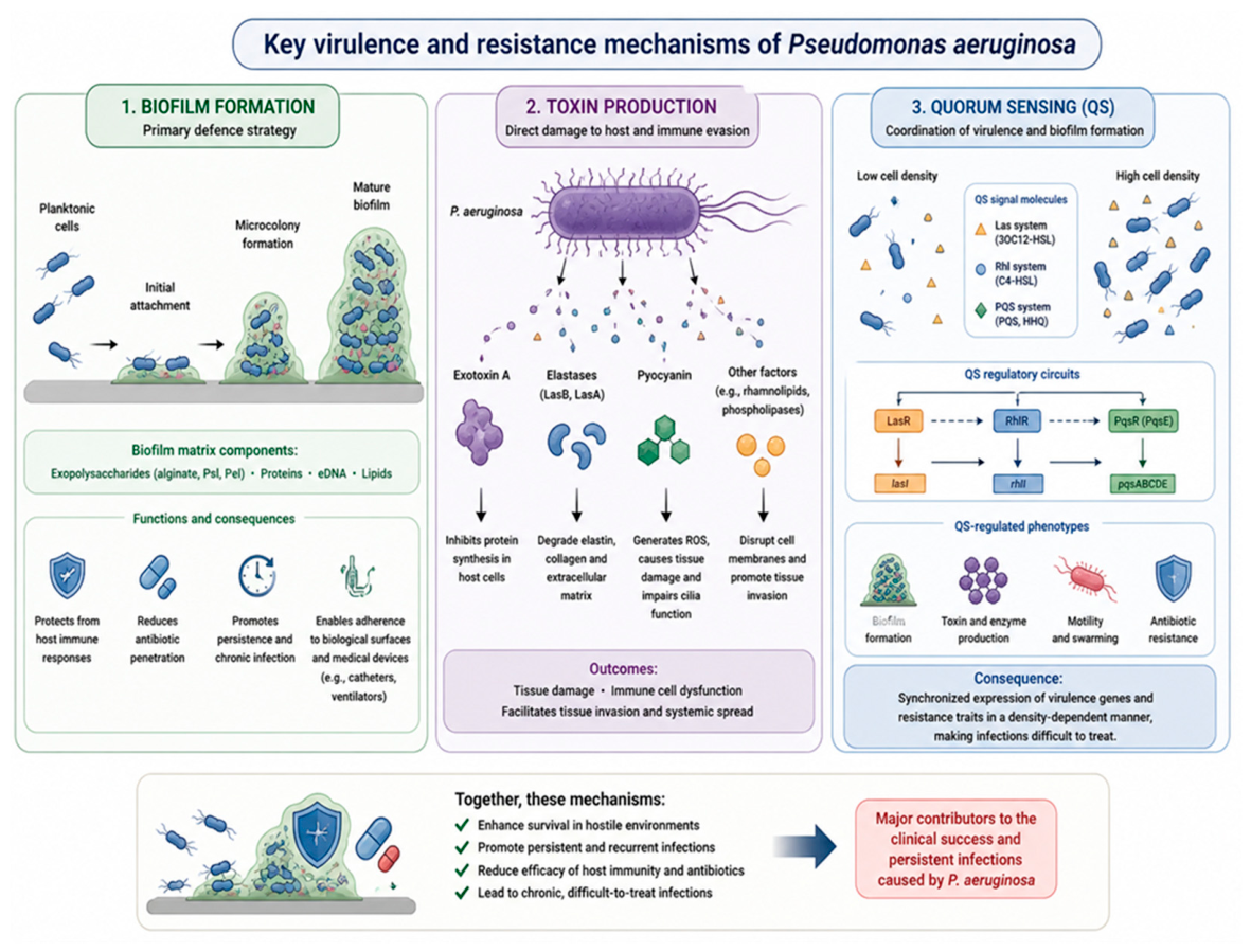
Figure 2.
Proposed mechanisms by which A. vera phytochemicals inhibit P. aeruginosa. Anthraquinones (e.g., aloin, aloe-emodin and emodin) contribute to bacterial membrane disruption, reactive oxygen species (ROS) generation and interference with DNA replication and protein synthesis. Flavonoids exert antioxidant, anti-inflammatory and immunomodulatory effects, whereas the polysaccharide acemannan promotes macrophage activation, wound healing and tissue repair. Collectively, these phytochemicals contribute to inhibition of biofilm formation, attenuation of quorum sensing-regulated virulence and enhanced antibiotic efficacy. The figure represents a conceptual synthesis of published evidence and was created by the authors based on current literature [3,5,19,24].
Figure 2.
Proposed mechanisms by which A. vera phytochemicals inhibit P. aeruginosa. Anthraquinones (e.g., aloin, aloe-emodin and emodin) contribute to bacterial membrane disruption, reactive oxygen species (ROS) generation and interference with DNA replication and protein synthesis. Flavonoids exert antioxidant, anti-inflammatory and immunomodulatory effects, whereas the polysaccharide acemannan promotes macrophage activation, wound healing and tissue repair. Collectively, these phytochemicals contribute to inhibition of biofilm formation, attenuation of quorum sensing-regulated virulence and enhanced antibiotic efficacy. The figure represents a conceptual synthesis of published evidence and was created by the authors based on current literature [3,5,19,24].
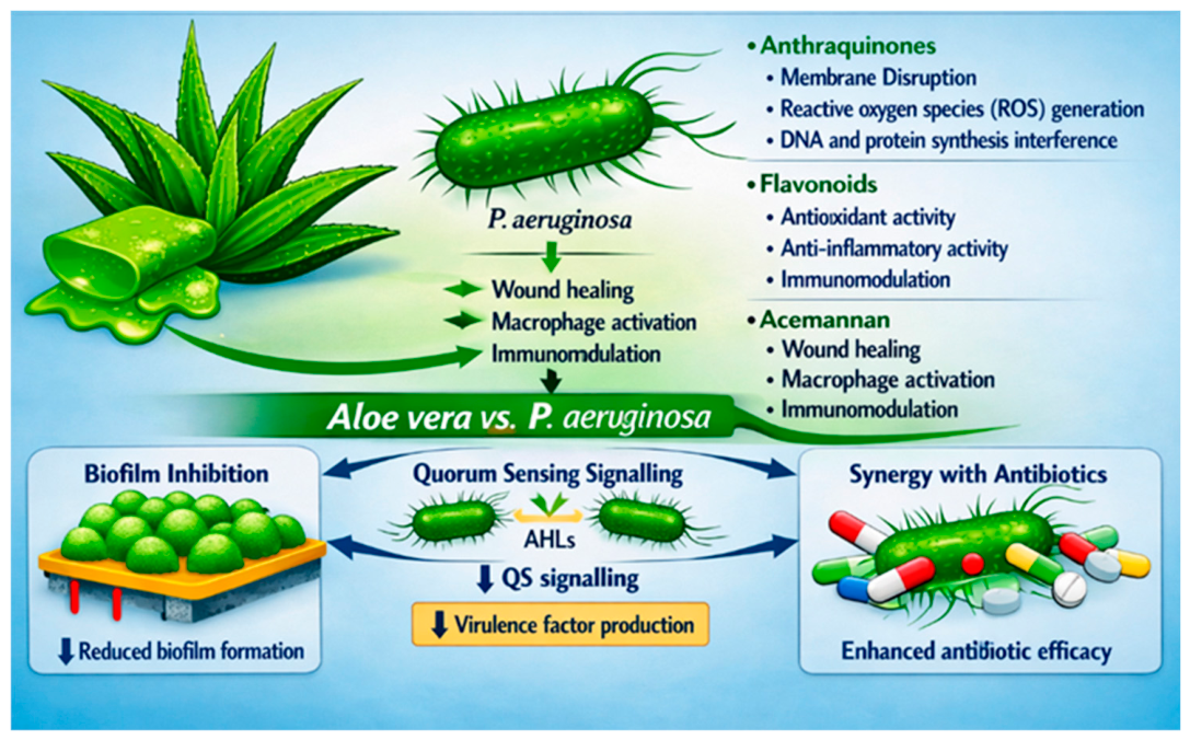

Table 1.
Major A. vera preparations, their principal bioactive constituents and proposed antimicrobial or biological functions.
Table 1.
Major A. vera preparations, their principal bioactive constituents and proposed antimicrobial or biological functions.
| Preparation/Class | Major constituents | Principal antimicrobial/biological functions |
| Crude gel extracts | Water, polysaccharides, vitamins, amino acids, enzymes, minerals, sterols, organic acids | Wound healing, antioxidant activity, immunomodulation, broad-spectrum synergistic antimicrobial effects |
| Solvent extracts | Phenolics, flavonoids, anthraquinones, chromones, sterols | Higher antimicrobial potency due to concentrated secondary metabolites; membrane disruption and metabolic inhibition |
| Anthraquinones | Aloin (barbaloin), aloe-emodin, emodin, chrysophanol, rhein | DNA intercalation, enzyme inhibition, membrane disruption, oxidative stress induction, quorum sensing inhibition, biofilm disruption |
| Polysaccharides | Acemannan, glucomannans, mannose-rich polysaccharides, arabinogalactans | Immunomodulation, macrophage activation, tissue repair, inhibition of bacterial adhesion and early biofilm formation |
| Phenolics and flavonoids | Chromones, phenolic acids, flavonoids | Antioxidant activity, anti-inflammatory effects, synergistic antimicrobial action |
| Saponins | Glycosidic saponins | Membrane permeabilization, antibacterial and antifungal activity |
| Nanoformulations | A. vera-mediated silver, gold and zinc oxide nanoparticles | Enhanced bioavailability, targeted delivery, improved biofilm penetration, increased antimicrobial efficacy against MDR bacteria |
Table 2.
Comparison of plant-derived anti-P. aeruginosa agents.
| Agent | Primary mechanism | Direct antimicrobial potency | Anti-biofilm activity | Anti-virulence effects | Wound-healing properties | Bioavailability |
| A. vera (anthraquinones) | Membrane disruption, DNA intercalation, oxidative stress | High (MIC 200-800 µg/ml) | High (79.5-96.6% inhibition) | Moderate (QS inhibition) | Yes (acemannan) | Poor |
| Curcumin | Type III secretion inhibition | Low (anti-virulence only) | Moderate | High | No | Very poor |
| Ajoene (garlic) | Quorum sensing inhibition | Low (anti-virulence only) | Moderate | High | No | Moderate |
| Berberine | Membrane disruption, DNA damage, protein synthesis inhibition | Very high (MIC 10-100 µg/ml) | Moderate | Moderate | No | Poor |
| Green tea polyphenols | Quorum sensing inhibition, prooxidant H2O2 production | Moderate | Moderate | High | No | Moderate |
| Cranberry PACs | Quorum sensing inhibition, biofilm disruption | Moderate | Very high | High | No | Poor |
Disclaimer/Publisher’s Note: The statements, opinions and data contained in all publications are solely those of the individual author(s) and contributor(s) and not of MDPI and/or the editor(s). MDPI and/or the editor(s) disclaim responsibility for any injury to people or property resulting from any ideas, methods, instructions or products referred to in the content. |
© 2026 by the authors. Licensee MDPI, Basel, Switzerland. This article is an open access article distributed under the terms and conditions of the Creative Commons Attribution (CC BY) license (http://creativecommons.org/licenses/by/4.0/).
Copyright: This open access article is published under a Creative Commons CC BY 4.0 license, which permit the free download, distribution, and reuse, provided that the author and preprint are cited in any reuse.