
Submitted:
10 August 2026
Posted:
20 August 2026
You are already at the latest version
Abstract
Background/Objectives: Panoramic radiography is the first-line examination before mandibular third molar extraction, but two-dimensional imaging may limit assessment of root proximity to the inferior alveolar canal. This study assessed panoramic risk signs for identifying root–inferior alveolar canal (IAC) contact, using cone-beam computed tomography (CBCT) as the reference standard, and evaluated a selective CBCT referral approach. Methods: This retrospective cross-sectional diagnostic study included 86 mandibular third molars from 62 patients. Four examiners with different clinical experience independently assessed panoramic radiographs for established risk signs. CBCT confirmed root–IAC contact and measured the minimum root–IAC distance. Interobserver agreement, regression analyses, ROC-based diagnostic performance and a selective CBCT indication rule were assessed. Results: Root darkening and cortical interruption were the most frequent signs. Agreement was generally low, although higher between the most experienced examiners. Cortical interruption was the only sign significantly associated with CBCT-confirmed root–IAC contact. Panoramic radiography underestimated the root–IAC distance. The selective rule would have recommended CBCT in 33/86 molars (38.4%) and identified 33/38 confirmed contacts (86.8%). Conclusions: Cortical interruption appears to be the most clinically relevant panoramic sign for selecting mandibular third molars requiring three-dimensional assessment. Structured criteria and senior review in borderline cases may support radiation justification and avoid routine CBCT use.
Keywords:
third molar
; cone-beam computed tomography
; panoramic radiography
; inferior alveolar canal
; diagnostic accuracy
; dental imaging
1. Introduction
The extraction of mandibular third molars is one of the most common procedures in oral and maxillofacial surgery and carries a potential risk of inferior alveolar nerve (IAN) injury, with reported incidences reaching up to 8.4% in the literature [1,2,3]. Proper surgical planning requires an accurate assessment of the anatomical relationship between the roots and the inferior alveolar canal (IAC) in order to minimise the risk of complications.
Panoramic radiography (OPG) is typically the initial imaging technique due to its wide availability, low cost, and reduced radiation dose [4,5]. However, being a two-dimensional modality, it may present distortions and superimpositions that hinder interpretation, particularly in cases where the roots lie close to the IAC [4,5,6,7,8]. Cone-beam computed tomography (CBCT) offers precise three-dimensional evaluation, but its indiscriminate use results in increased radiation exposure, unnecessary costs, and the risk of trivialising its indication [9,10,11,12].
In many clinical settings, access to CBCT has increased substantially, including in general dental practices. This broader availability facilitates immediate imaging decisions at the point of care, but may also contribute to variability in prescription patterns and potential overuse when clear and standardised criteria are lacking. Consequently, selective CBCT referral strategies should be applicable to real-world clinical settings, where imaging decisions are often made by clinicians with different levels of experience. Therefore, this study assessed the performance of a pragmatic, guideline-aligned selective CBCT referral algorithm and examined how examiner experience affects its applicability.
The aim of this study was to evaluate interobserver agreement in the identification of panoramic radiographic risk signs, analyse their association with root–IAC contact confirmed by cone-beam computed tomography, and retrospectively assess a selective imaging protocol.
2. Materials and Methods
This retrospective cross-sectional diagnostic study was conducted in a university-based oral surgery setting and reported in accordance with the STARD guidelines. The study protocol received approval from the University’s Research Ethics Committee, and all participants signed informed consent in accordance with the Declaration of Helsinki. Radiographic records obtained between 2021 and 2025 were retrospectively reviewed.
2.1. Selection Criteria
Patients aged 18 to 90 years were included if they presented mandibular third molars with complete root formation and had both preoperative panoramic radiography (OPG) and cone-beam computed tomography (CBCT) available.
Exclusion criteria were: low-quality imaging; local alterations preventing accurate interpretation (cysts, periapical lesions, mandibular fractures, or osteosynthesis hardware); pregnancy; history of head and neck radiotherapy; ongoing oncologic treatment; or refusal to undergo radiographic studies.
2.2. Sample Size and Unit of Analysis
Sample size was calculated at the tooth level, because the primary outcome of the study was CBCT-confirmed root–IAC contact for each mandibular third molar. Assuming a 95% confidence level, a conservative expected proportion of 50% to maximise sample size requirements, and an accepted precision of approximately ±11%, a minimum sample of 80 mandibular third molars was required.
The final sample included 62 patients and 86 mandibular third molars, therefore exceeding the minimum estimated sample size. Each mandibular third molar was independently assessed by four examiners, generating 344 individual radiographic evaluations. Diagnostic accuracy analyses, regression models, ROC curve analysis, and retrospective application of the selective CBCT indication protocol were performed at the tooth level.
Interobserver agreement analyses were based on the individual examiner assessments of each tooth. When both mandibular third molars from the same patient were included, they were analysed as separate anatomical units because each tooth had an independent spatial relationship with the IAC. The potential non-independence of bilateral molars from the same patient was considered when interpreting the findings and is acknowledged as a limitation.
Four examiners with different levels of clinical experience assessed the panoramic radiographs: a first-year postgraduate student, a second-year postgraduate student, a third-year postgraduate student, and a faculty oral surgeon with more than 10 years of clinical experience. Each examiner recorded the presence or absence of the radiographic signs described by Rood and Shehab [1], including cortical interruption, root darkening, curved or deflected roots, root narrowing, canal deviation or narrowing, bifid apices, and “island-shaped” apices (Figure 1). To reduce measurement bias, all panoramic radiographs were independently assessed using predefined radiographic criteria.
CBCT imaging (Planmeca ProMax 3D Classic®) was used as the reference standard to confirm root–IAC contact and to measure the minimum distance (in millimetres) between the root and the IAC.
2.3. Statistical Analysis
Statistical significance was set at P < 0.05. Numerical variables were described using the mean and standard deviation or the median and interquartile range (Q1–Q3), according to their distribution.
Interobserver agreement was assessed using the kappa coefficient (κ) with 95% confidence intervals.
To analyse the association between radiographic signs and root–IAC contact, a two-stage modelling approach was applied:
- Logistic regression to estimate the probability of root–IAC contact.
- Linear regression was used to assess the relationship between radiographic findings and root–IAC distance among mandibular third molars without CBCT-confirmed contact.
The optimal cutoff for the number of panoramic signs predicting complete root–IAC contact was determined using Youden’s index. Diagnostic performance was evaluated through sensitivity, specificity, positive predictive value (PPV), negative predictive value (NPV), and ROC curve analysis.
The association between root position relative to the IAC and the radiographic signs was examined using independent logistic regression models.
Data processing and statistical analyses were performed using R software version 4.3.2 [13].
For the retrospective analysis, CBCT indication was operationally defined as the presence of cortical interruption or ≥2 panoramic risk signs. Examiner experience and medico-legal considerations were incorporated as clinical modifiers in the proposed decision algorithm, but not as additional numerical criteria for the retrospective diagnostic performance analysis.
3. Results
3.1. Sample Characteristics and Imaging Findings
The characteristics of the study sample, imaging assessments, examiner profile, and main CBCT findings are summarized in Table 1.
3.2. Distribution of Radiographic Signs
Among the 86 mandibular third molars evaluated, the most frequent radiographic signs on panoramic imaging were cortical interruption and root darkening (30.6% and 37.5%, respectively). Curved or deflected roots were observed in 18–21% of cases. Root narrowing, canal narrowing, and canal deviation were less common findings (<10%). Bifid apices and “island-shaped” apices were identified only anecdotally. These results are presented in Supplementary Figure S1.
3.3. Interobserver Agreement
Interobserver agreement for the identification of the different radiographic signs was generally low among most examiners, with kappa coefficients close to zero and wide confidence intervals.
The highest agreement was observed between Examiners 3 and 4, particularly for root darkening (κ [95% CI] = 0.64 [0.48–0.79]), curved or deflected roots (κ [95% CI] = 0.59 [0.39–0.80]), and root narrowing (κ [95% CI] = 0.64 [0.33–0.96]), showing moderate to substantial levels of agreement.
Interobserver agreement between the two most experienced examiners is shown in Table 2. The highest agreement was observed for root darkening, curved or deflected roots, and root narrowing, with κ values ranging from 0.59 to 0.64 in the total sample. Agreement was lower for interruption of the cortical outline, canal deviation, and canal narrowing. Kappa values could not be estimated for island-shaped apex because this sign showed no variability between the two examiners.
3.4. Association Between Radiographic Signs and Root–IAC Contact
Cortical interruption was the only sign significantly associated with the presence of root–IAC contact (OR = 3.46; 95% CI: 1.23–10.83).
In contrast, among mandibular third molars without root–IAC contact, cortical interruption was not associated with either greater or lesser distance to the canal (β [95% CI] = 0.10 [–0.78 to 0.99]; p = 0.816). The optimal cutoff for predicting complete root–IAC contact based on the number of radiographic signs was ≥1 sign present. CBCT-confirmed root–IAC contact was more frequent among mandibular third molars presenting at least one panoramic risk sign than among those without radiographic signs. Diagnostic accuracy estimates were calculated using CBCT-confirmed contact as the reference standard. Figure 2 displays the corresponding ROC curve using this cutoff: sensitivity, 0.90 (95% CI: 0.77–0.97); specificity, 0.38 (95% CI: 0.24–0.54); PPV, 0.59 (95% CI: 0.46–0.71); and NPV, 0.80 (95% CI: 0.56–0.94). Figure 3 illustrates the relationship between the cumulative number of panoramic signs and CBCT-confirmed root–IAC contact or distance. Although ≥1 sign maximized sensitivity according to Youden’s index, the final selective protocol prioritized clinical applicability by combining the strongest individual predictor, cortical interruption, with the presence of multiple radiographic signs.
3.5. Comparison Between OPG and CBCT
The distances measured between the root and the IAC were significantly greater on CBCT than on OPG (P < 0.001), with a moderate effect size (Cohen’s d = 0.72). The intraclass correlation coefficient indicated moderate agreement between the two methods (ICC = 0.66). Bland–Altman analysis revealed a systematic tendency for OPG to underestimate the root–IAC distance compared with CBCT.
3.6. Root Position Relative to the IAC
Logistic regression analysis identified curved or deflected roots as the only radiographic feature significantly associated with IAC position. The presence of this sign was associated with an 83% reduction in the odds of a lingual IAC location compared with molars without root curvature. These results are presented in Supplementary Figure S2.
3.7. Application of the Selective Protocol
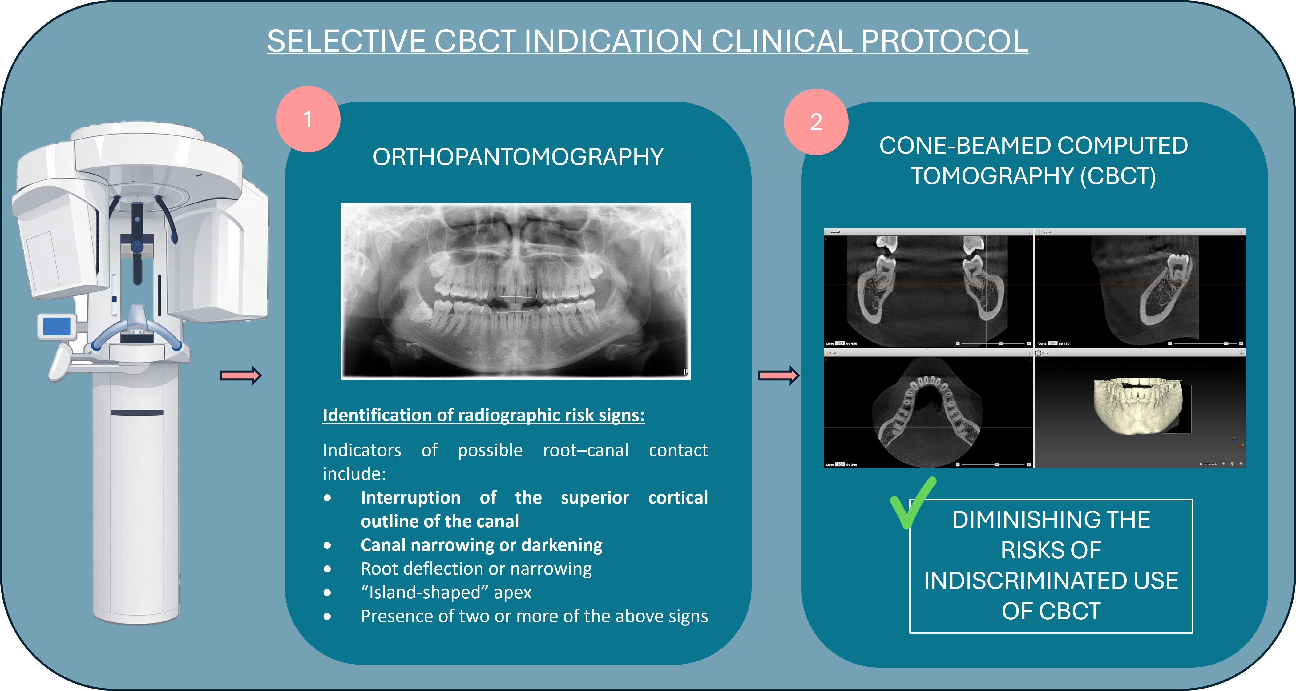
The retrospective application of the selective CBCT indication protocol would have recommended CBCT in 33/86 mandibular third molars (38.4%). Among the 38 molars with CBCT-confirmed root–IAC contact, 33 fulfilled the protocol criteria, whereas five contacts would not have been identified by the protocol. No molars without CBCT-confirmed root–IAC contact fulfilled the protocol criteria. Accordingly, the protocol showed a sensitivity of 86.8%, specificity of 100.0%, PPV of 100.0%, and NPV of 90.6%. These findings should be interpreted as an internal retrospective performance estimate rather than external validation of the protocol. The diagnostic performance of the selective CBCT indication protocol is shown in Table 3. The decision algorithm is shown in Figure 4.
4. Discussion
This study evaluated the reliability of panoramic radiographic signs for predicting contact between mandibular third molar roots and the IAC, using CBCT as the reference standard.
Our results show that cortical interruption was the only panoramic sign significantly associated with CBCT-confirmed root–IAC contact, supporting its diagnostic relevance. However, interobserver agreement varied across signs and was generally higher among more experienced examiners. These findings are consistent with classic studies [1,2,3] as well as more recent investigations [4,5], which also identified this sign as a reliable predictor of surgical risk. In our analysis, the presence of two or more panoramic signs on OPG increased the probability of contact, reinforcing the usefulness of a combined radiographic assessment. Similar results have been reported in studies comparing the diagnostic performance of OPG and CBCT in evaluating root–IAC relationships [10,11,12].
The low kappa values observed among less experienced examiners should not be interpreted as an indication for routine CBCT in all cases. Rather, they reflect the limited reproducibility of subtle panoramic signs when interpreted by junior operators, a finding that may be partly influenced by the low prevalence of several signs and the prevalence-dependence of kappa statistics. Clinically, this supports the need for structured training, use of standardised visual criteria, and senior review in borderline cases before dismissing the need for CBCT.
The higher concordance observed was between Examiners 3 and 4, reflecting that interobserver agreement was influenced by the level of clinical experience. This finding highlights the importance of specialised training and the learning curve in radiographic interpretation, consistent with studies that have shown that diagnostic reliability depends on experience [5,6].
Regarding linear measurements, OPG systematically underestimated the root–IAC distance compared with CBCT. This pattern, previously reported [7,8], confirms that while OPG is useful as an initial examination, it has inherent limitations related to its two-dimensional nature. CBCT provides more precise three-dimensional anatomical information and may improve surgical planning in selected high-risk cases, although its routine use has not consistently been shown to reduce postoperative neurosensory complications [11,12,13,14,15,16,17].
The retrospective application of the selective CBCT protocol identified 33 of 38 CBCT-confirmed root–IAC contacts (86.8%) while recommending CBCT in fewer than 40% of mandibular third molars. The protocol showed high specificity and PPV in this retrospective sample, as no false-positive CBCT indications were observed. However, because the protocol was derived and tested within the same dataset, these estimates may overestimate its true clinical performance. Therefore, the proposed algorithm should be considered a pragmatic referral framework requiring prospective external validation rather than a definitive prediction rule. These findings suggest that a rational use of CBCT based on the presence of panoramic risk signs, modulated by examiner experience and surgical complexity, may optimise the balance between diagnostic confidence and radiation exposure, in accordance with justification and optimisation principles [9,16]. In this regard, Sanmartí-García et al. [18] suggested that CBCT may be useful in selected high-risk cases, although current evidence does not support routine CBCT for all mandibular third molars. Recent evidence also supports a selective rather than indiscriminate use of cone-beam computed tomography in mandibular third molar surgery. Ruiz-Roca et al. reported that the combination of root darkening and interruption of the cortical line on panoramic radiography increased the probability of IAC-related sensory alteration, while Robbins et al. concluded that adding CBCT to panoramic imaging does not routinely reduce nerve injury rates when used indiscriminately [19,20,21,22].
4.1. Strengths and Limitations
The main strengths of this study include the participation of examiners with different levels of clinical experience, the use of CBCT as the reference standard, an a priori sample size calculation, and the application of a clinically oriented selective CBCT indication protocol. In addition, the analysis of interobserver agreement provides relevant information on the influence of examiner experience in the interpretation of panoramic radiographic signs.
However, several limitations should be acknowledged. First, this was a single-centre study with a limited sample size, which may restrict the generalisability of the findings. Second, although the primary analysis was performed at the tooth level, some patients contributed more than one mandibular third molar; therefore, a possible clustering effect of bilateral molars within the same patient cannot be completely excluded. Third, individual anatomical determinants, such as root morphology, canal cortication, depth of impaction, angulation, and buccolingual canal position, were not systematically incorporated into multivariable models. Finally, although clinical follow-up was performed, the study was not designed to establish a standardised longitudinal correlation between radiographic findings and postoperative neurosensory outcomes.
Future multicentre prospective studies with larger samples, external validation cohorts, and more comprehensive anatomical covariates are needed to confirm the clinical applicability of the proposed selective CBCT indication protocol.
4.2. Selective CBCT Indication
Based on these findings, CBCT should be considered when cortical interruption is present or when two or more panoramic risk signs are identified. In cases with a single non-cortical sign, the decision should be individualised according to surgical complexity, examiner experience and the need for medico-legal documentation. In the absence of panoramic risk signs, OPG may be sufficient for initial surgical planning. This approach reserves CBCT for higher-risk cases and aligns with radiological justification and optimisation principles.
4.3. Clinical Implications
Panoramic radiography remains an appropriate first-line imaging modality for the preoperative assessment of mandibular third molars. However, its limitations should be recognised, particularly in cases with suspected proximity between the roots and the IAC. In this study, cortical interruption was the panoramic sign most strongly associated with CBCT-confirmed root–IAC contact, supporting its use as a key criterion for selective CBCT referral.
The findings also highlight the influence of clinical experience on radiographic interpretation. Less-experienced operators may benefit from structured training, standardised visual criteria, and senior review in borderline cases. Therefore, CBCT should be considered when cortical interruption or multiple high-risk panoramic signs are present, especially in complex surgical cases or when medico-legal documentation is required.
This selective approach may improve diagnostic confidence while avoiding routine CBCT use, thereby reducing unnecessary radiation exposure and healthcare costs. It provides a pragmatic framework for balancing patient safety, surgical planning, and radiological justification in mandibular third molar surgery.
5. Conclusion
In conclusion, cortical interruption was the most consistent panoramic indicator of CBCT-confirmed root–IAC contact. However, the generally low interobserver agreement, particularly among less experienced examiners, highlights the limitations of relying solely on panoramic interpretation. A guideline-aligned selective approach, incorporating cortical interruption, multiple signs, operator experience, and clinical complexity, may support more consistent CBCT referral decisions while avoiding routine imaging.
Supplementary Materials
The following supporting information can be downloaded at the website of this paper posted on Preprints.org, Figure S1, distribution of panoramic radiographic signs; Figure S2, association between root position relative to the IAC and panoramic radiographic signs; Table S1, complete interobserver agreement matrix; STARD checklist.
Author Contributions
Conceptualization, J.V.G. and R.G.M.; methodology, R.G.M. and I.C.B.; formal analysis, J.V.G.; investigation, J.V.G.; data curation, J.V.G.; writing and original draft preparation, J.V.G.; writing, review and editing, R.G.M. and I.C.B.; supervision, R.G.M. and I.C.B. All authors have read and agreed to the published version of the manuscript.
Funding
This research received no external funding.
Institutional Review Board Statement
The study was conducted in accordance with the Declaration of Helsinki and approved by the Biomedical Research Ethics Committee of Universidad CEU Cardenal Herrera (protocol code CEEI23/432; date of approval: 15 November 2023).
Informed Consent Statement
Informed consent was obtained from all subjects involved in the study.
Data Availability Statement
The anonymized dataset underlying all figures and statistical analyses reported in this study, together with the R code used for data analysis, will be made available in an anonymized repository upon acceptance.
Acknowledgments
The authors thank the Master’s Program in Oral Surgery and Bucofacial Implantology at CEU Cardenal Herrera University for their support.
Conflicts of Interest
The authors declare no conflicts of interest.
Use of AI-Assisted Technologies
During the preparation of this manuscript, the authors used ChatGPT (OpenAI) for language editing and manuscript refinement. The authors reviewed and edited all AI-assisted output and take full responsibility for the final content of this publication.
References
- Rood, J.P.; Shehab, B.A. The radiological prediction of inferior alveolar nerve injury during third molar surgery. Br. J. Oral Maxillofac. Surg. 1990, 28, 20–25. [Google Scholar] [CrossRef] [PubMed]
- Sedaghatfar, M.; August, M.A.; Dodson, T.B. Panoramic radiographic findings as predictors of inferior alveolar nerve exposure following third molar extraction. J. Oral Maxillofac. Surg. 2005, 63, 3–7. [Google Scholar] [CrossRef] [PubMed]
- Tantanapornkul, W.; Okouchi, K.; Fujiwara, Y.; Yamashiro, M.; Maruoka, Y.; Ohbayashi, N.; Kurabayashi, T. A comparative study of cone-beam computed tomography and conventional panoramic radiography in assessing the topographic relationship between the mandibular canal and impacted third molars. Oral Surg. Oral Med. Oral Pathol. Oral Radiol. Endod. 2007, 103, 253–259. [Google Scholar] [CrossRef] [PubMed]
- Nakamori, K.; Fujiwara, K.; Miyazaki, A.; Tomihara, K.; Tsuji, M.; Nakai, M.; Michifuri, Y.; Suzuki, R.; Komai, K.; Shimanishi, M.; Hiratsuka, H. Clinical assessment of the relationship between the third molar and the inferior alveolar canal using panoramic images and computed tomography. J. Oral Maxillofac. Surg. 2008, 66, 2308–2313. [Google Scholar] [CrossRef] [PubMed]
- Matzen, L.H.; Christensen, J.; Hintze, H.; Schou, S.; Wenzel, A. Influence of cone-beam CT on treatment plan before surgical intervention of mandibular third molars and impact of radiographic factors on deciding on coronectomy versus surgical removal. Dentomaxillofac. Radiol. 2013, 42, 98870341. [Google Scholar] [CrossRef] [PubMed]
- Guerrero, M.E.; Botetano, R.; Beltran, J.; Horner, K.; Jacobs, R. Can preoperative imaging help to predict postoperative outcome after wisdom tooth removal? A randomized controlled trial using panoramic radiography vs. cone-beam CT. Clin. Oral Investig. 2014, 18, 335–342. [Google Scholar] [CrossRef] [PubMed]
- Ghaeminia, H.; Meijer, G.J.; Soehardi, A.; Borstlap, W.A.; Mulder, J.; Bergé, S.J. Position of the impacted third molar in relation to the mandibular canal: Diagnostic accuracy of cone beam computed tomography compared with panoramic radiography. Int. J. Oral Maxillofac. Surg. 2009, 38, 964–971. [Google Scholar] [CrossRef] [PubMed]
- Bell, G.W. Use of dental panoramic tomographs to predict the relation between mandibular third molar teeth and the inferior alveolar nerve: Radiological and surgical findings, and clinical outcome. Br. J. Oral Maxillofac. Surg. 2004, 42, 21–27. [Google Scholar] [CrossRef] [PubMed]
- European Commission. Radiation Protection No. 172: Cone Beam CT for Dental and Maxillofacial Radiology. Evidence-Based Guidelines; Office for Official Publications of the European Communities: Luxembourg, 2012. [Google Scholar] [CrossRef]
- Suomalainen, A.; Vehmas, T.; Kortesniemi, M.; Robinson, S.; Peltola, J. Accuracy of linear measurements using dental cone beam and conventional multislice computed tomography. Dentomaxillofac. Radiol. 2008, 37, 10–17. [Google Scholar] [CrossRef] [PubMed]
- Patel, S.; Durack, C.; Abella, F.; Shemesh, H.; Roig, M.; Lemberg, K. Cone beam computed tomography in endodontics: A review. Int. Endod. J. 2015, 48, 3–15. [Google Scholar] [CrossRef] [PubMed]
- de Toledo Telles-Araújo, G.; Peralta-Mamani, M.; Caminha, R.D.G.; Moraes-da-Silva, A.F.; Rubira, C.M.F.; Honório, H.M.; Rubira-Bullen, I.R.F. CBCT does not reduce neurosensory disturbances after third molar removal compared with panoramic radiography: A systematic review and meta-analysis. Clin. Oral Investig. 2020, 24, 1137–1149. [Google Scholar] [CrossRef] [PubMed]
- R Core Team. R: A Language and Environment for Statistical Computing; R Foundation for Statistical Computing: Vienna, Austria, 2023. [Google Scholar]
- Scarfe, W.C.; Farman, A.G. What is cone-beam CT and how does it work? Dent. Clin. N. Am. 2008, 52, 707–715. [Google Scholar] [CrossRef] [PubMed]
- Pauwels, R.; Araki, K.; Siewerdsen, J.H.; Thongvigitmanee, S.S. Technical aspects of dental CBCT: State of the art. Dentomaxillofac. Radiol. 2015, 44, 20140224. [Google Scholar] [CrossRef] [PubMed]
- Horner, K.; Islam, M.; Flygare, L.; Tsiklakis, K.; Whaites, E. Basic principles for use of dental cone beam computed tomography: Consensus guidelines of the European Academy of Dental and Maxillofacial Radiology. Dentomaxillofac. Radiol. 2009, 38, 187–195. [Google Scholar] [CrossRef] [PubMed]
- Queral-Godoy, E.; Valmaseda-Castellón, E.; Berini-Aytés, L.; Gay-Escoda, C. Incidence and evolution of inferior alveolar nerve lesions following lower third molar extraction. Oral Surg. Oral Med. Oral Pathol. Oral Radiol. Endod. 2005, 99, 259–264. [Google Scholar] [CrossRef] [PubMed]
- Sanmartí-García, G.; Valmaseda-Castellón, E.; Gay-Escoda, C. Does computed tomography prevent inferior alveolar nerve injuries caused by lower third molar removal? J. Oral Maxillofac. Surg. 2012, 70, 5–11. [Google Scholar] [CrossRef] [PubMed]
- Al-Haj Husain, A.; Stadlinger, B.; Winklhofer, S.; Bosshard, F.A.; Schmidt, V.; Valdec, S. Imaging in third molar surgery: A clinical update. J. Clin. Med. 2023, 12, 7688. [Google Scholar] [CrossRef] [PubMed]
- Del Lhano, N.C.; Ribeiro, R.A.; Martins, C.C.; Assis, N.M.S.P.; Devito, K.L. Panoramic versus CBCT used to reduce inferior alveolar nerve paresthesia after third molar extractions: A systematic review and meta-analysis. Dentomaxillofac. Radiol. 2020, 49, 20190265. [Google Scholar] [CrossRef] [PubMed]
- Ruiz-Roca, J.A.; Rodríguez-Molinero, J.A.; Javaloyes-Vicente, P.; Pereira-Lopes, O.; Gay-Escoda, C. Use of CBCT and panoramic radiography in the prediction of alterations in sensitivity of the inferior alveolar nerve in third molars: A retrospective cross-sectional study. Saudi Dent. J. 2024, 36, 1105–1110. [Google Scholar] [CrossRef] [PubMed]
- Robbins, J.; Smalley, K.R.; Ray, P.; Ali, K. Does the addition of cone-beam CT to panoramic imaging reduce inferior dental nerve injuries resulting from third molar surgery? A systematic review. BMC Oral Health 2022, 22, 466. [Google Scholar] [CrossRef] [PubMed]
Figure 1.
Representative panoramic radiographic risk signs assessed in the study. a Root darkening. b Interruption of the superior cortical outline of the mandibular canal. c Canal narrowing. d Canal deviation. e Root narrowing. f Root deflection. g Bifid apex. h Island-shaped apex. Arrows indicate the apical root region of the mandibular third molar where the radiographic feature of interest is observed.
Figure 1.
Representative panoramic radiographic risk signs assessed in the study. a Root darkening. b Interruption of the superior cortical outline of the mandibular canal. c Canal narrowing. d Canal deviation. e Root narrowing. f Root deflection. g Bifid apex. h Island-shaped apex. Arrows indicate the apical root region of the mandibular third molar where the radiographic feature of interest is observed.
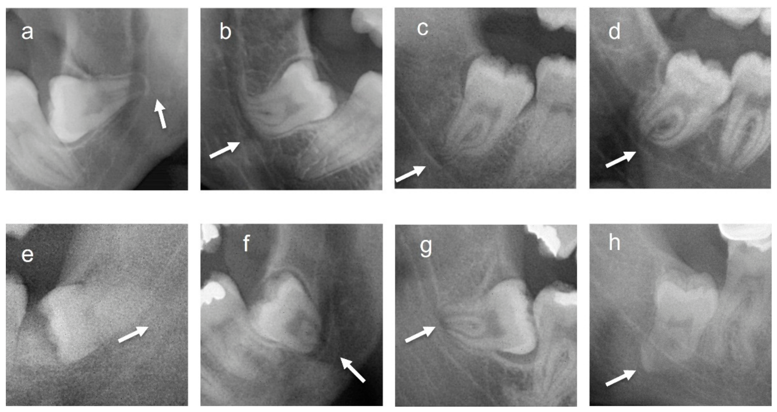
Figure 2.
ROC curve assessing the discriminative ability of the combined count of panoramic signs to predict root–IAC contact confirmed by CBCT.
Figure 2.
ROC curve assessing the discriminative ability of the combined count of panoramic signs to predict root–IAC contact confirmed by CBCT.
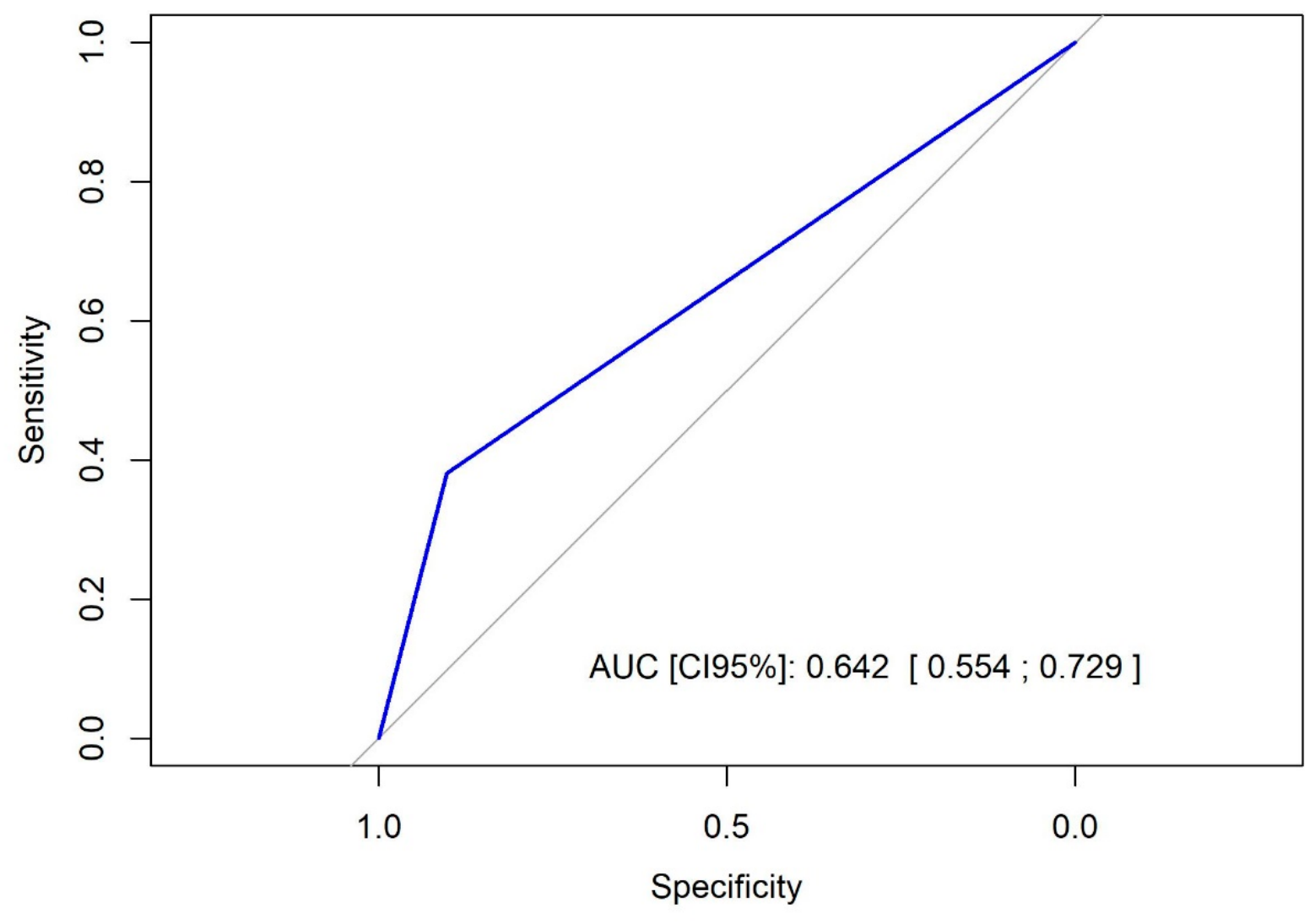
Figure 3.
Relationship between the cumulative number of panoramic radiographic signs and CBCT findings. The left panel shows the percentage of mandibular third molars with CBCT-confirmed root–IAC contact, defined as a root–IAC distance of 0 mm. The right panel shows the mean root–IAC distance on CBCT among mandibular third molars without direct contact.
Figure 3.
Relationship between the cumulative number of panoramic radiographic signs and CBCT findings. The left panel shows the percentage of mandibular third molars with CBCT-confirmed root–IAC contact, defined as a root–IAC distance of 0 mm. The right panel shows the mean root–IAC distance on CBCT among mandibular third molars without direct contact.
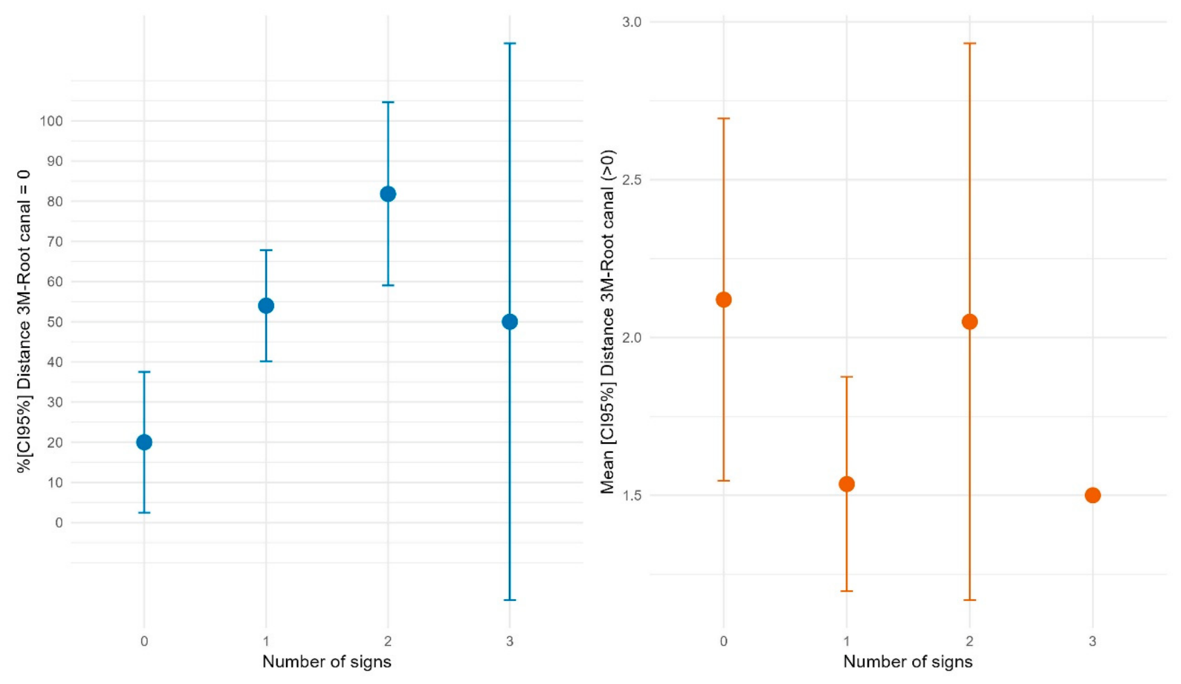
Figure 4.
Proposed clinical protocol for selective CBCT indication in mandibular third molars.
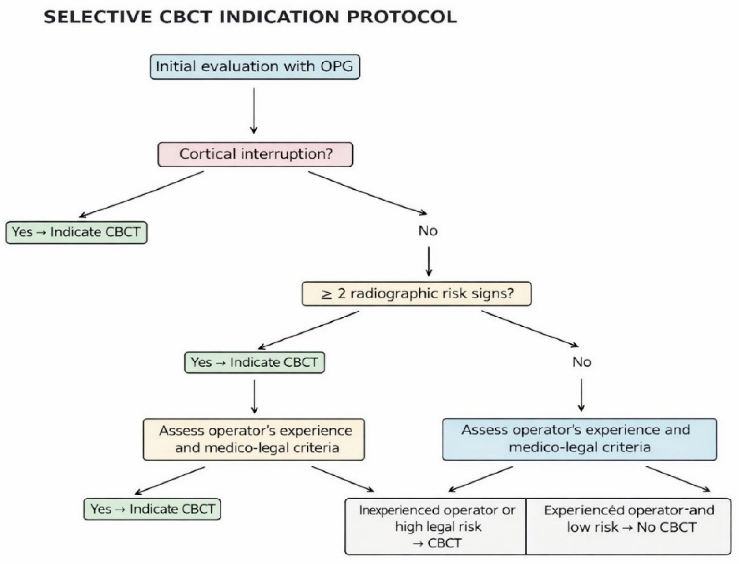

Table 1.
Sample characteristics, imaging assessment, and main radiographic findings.
| Variable | Value |
|---|---|
| Patients, n | 62 |
| Mandibular third molars, n | 86 |
| Individual radiographic evaluations, n | 344 |
| Age, years, mean ± SD | 32.2 ± 13.4 |
| Male patients, n (%) | 29 (46.8) |
| Female patients, n (%) | 33 (53.2) |
| Imaging modalities available | OPG and CBCT |
| Reference standard | CBCT-confirmed root–IAC contact |
| Number of examiners | 4 |
| Examiner 1 | First-year postgraduate student |
| Examiner 2 | Second-year postgraduate student |
| Examiner 3 | Third-year postgraduate student |
| Examiner 4 | Faculty oral surgeon, >10 years’ experience |
| Most frequent panoramic signs | Root darkening and cortical interruption |
| Root darkening, % | 37.5 |
| Cortical interruption, % | 30.6 |
| Curved or deflected roots, % | 18–21 |
| Root narrowing, canal narrowing and canal deviation | <10% each |
| Bifid apices / island-shaped apices | Anecdotal findings |
| CBCT-confirmed root–IAC contact, n (%) | 38 (44.2) |
| No CBCT-confirmed root–IAC contact, n (%) | 48 (55.8) |
OPG, panoramic radiography; CBCT, cone-beam computed tomography; SD, standard deviation.
Table 2.
Interobserver agreement between Examiners 3 and 4 for panoramic radiographic signs.
| Radiographic Sign | Total κ [95% CI] | Total Agreement, % | ORTO 3.8 κ [95% CI] | ORTO 3.8 Agreement, % | ORTO 4.8 κ [95% CI] | ORTO 4.8 Agreement, % |
|---|---|---|---|---|---|---|
| Root darkening | 0.64 [0.48–0.79] | 81.71 | 0.58 [0.35–0.81] | 79.07 | 0.69 [0.48–0.91] | 84.21 |
| Curved or deflected roots | 0.59 [0.39–0.80] | 85.37 | 0.44 [0.15–0.74] | 79.07 | 0.77 [0.53–1.00] | 92.11 |
| Root narrowing | 0.64 [0.33–0.96] | 95.12 | 0.64 [0.20–1.00] | 95.35 | 0.00 [0.00–0.00] | 100.00 |
| Bifid apices over the canal | 0.31 [−0.18–0.80] | 95.12 | 0.64 [0.20–1.00] | 95.35 | Not estimable | 100.00 |
| Interruption of the cortical outline | 0.35 [0.18–0.52] | 67.07 | 0.29 [0.06–0.53] | 62.79 | 0.40 [0.15–0.66] | 71.05 |
| Canal deviation | 0.24 [0.01–0.46] | 81.71 | 0.17 [−0.12–0.45] | 81.40 | 0.30 [−0.03–0.64] | 81.58 |
| Canal narrowing | 0.21 [0.01–0.42] | 79.27 | 0.00 [0.00–0.00] | 79.07 | 0.35 [0.04–0.65] | 78.95 |
| Island-shaped apex | Not estimable | 100.00 | Not estimable | 100.00 | Not estimable | 100.00 |
κ, Cohen’s kappa coefficient; CI, confidence interval. “Not estimable” indicates that κ could not be calculated because one or both examiners showed no variability in the classification of that sign.
Table 3.
Diagnostic performance of the selective CBCT indication protocol.
| Selective CBCT Protocol | CBCT-Confirmed Root–IAC Contact | No CBCT-Confirmed Root–IAC Contact | Total |
|---|---|---|---|
| CBCT indicated | 33 | 0 | 33 |
| CBCT not indicated | 5 | 48 | 53 |
| Total | 38 | 48 | 86 |
Sensitivity: 86.8% (95% CI: 72.7–94.2); specificity: 100.0% (95% CI: 92.6–100.0); PPV: 100.0% (95% CI: 89.6–100.0); NPV: 90.6% (95% CI: 79.7–95.9).
Disclaimer/Publisher’s Note: The statements, opinions and data contained in all publications are solely those of the individual author(s) and contributor(s) and not of MDPI and/or the editor(s). MDPI and/or the editor(s) disclaim responsibility for any injury to people or property resulting from any ideas, methods, instructions or products referred to in the content. |
© 2026 by the authors. Licensee MDPI, Basel, Switzerland. This article is an open access article distributed under the terms and conditions of the Creative Commons Attribution (CC BY) license (http://creativecommons.org/licenses/by/4.0/).
Copyright: This open access article is published under a Creative Commons CC BY 4.0 license, which permit the free download, distribution, and reuse, provided that the author and preprint are cited in any reuse.