

Submitted:
18 August 2026
Posted:
20 August 2026
You are already at the latest version
Abstract
Background and Objectives: Lower urinary tract symptoms (LUTS), such as urgency, frequency, and pelvic discomfort, are often attributed to bladder-centered mechanisms; nonetheless, several patients exhibit overlapping pelvic floor dysfunctions that existing urologic models do not fully explain. Evidence from the musculoskeletal and pain literature demonstrates that myofascial trigger points within the pelvic floor are prevalent in patients with pelvic pain and urinary symptoms, potentially contributing to symptom generation. In this narrative review, we synthesized the literature on pelvic floor myofascial dysfunction, referred pain patterns, and autonomic regulation of bladder function to develop an integrated neuromusculoskeletal framework. Materials and Methods: Relevant studies were identified through targeted database searches, AI-assisted literature screening, and manual reviews and selected based on mechanistic relevance. Results: The reviewed evidence supports a pathway in which active myofascial trigger points generate sustained nociceptive afferent inputs that converge with bladder afferents in the sacral spinal cord, potentially facilitating cross- and central sensitization, lowering the threshold for bladder sensation, and altering micturition reflex processing. The resulting amplification of afferent signaling provides a mechanistic basis for urgency, frequency, and pelvic pain in the absence of primary bladder pathology. Conclusions: This model integrates musculoskeletal and urological perspectives and suggests that in a subset of patients, LUTS may reflect the central processing of peripheral musculoskeletal input. Incorporating pelvic floor assessment into clinical evaluations may improve diagnostic accuracy and provide more targeted management. Further prospective studies are warranted to validate this integrated pathway and its therapeutic implications.
Keywords:
central nervous system sensitization
; lower urinary tract symptoms
; myofascial pain syndromes
; pelvic floor
; trigger points
; urinary bladder
1. Introduction
Lower urinary tract symptoms (LUTS), including urgency, frequency, nocturia, and incontinence, impose a substantial burden on patients and the healthcare system. Overactive bladder syndrome alone affects approximately one in seven adults in the United States, and its prevalence increases with age [1]. Chronic pelvic pain frequently co-occurs with LUTS, with a reported prevalence of 5.7%–26.6% in women worldwide; myofascial pelvic floor pain is identified in 14%–23% of patients presenting with this condition [2,3]. Nevertheless, pelvic pain and urinary dysfunction have traditionally been managed through separate paradigms, with limited mechanistic integration between urological models focused on the bladder and musculoskeletal models focused on pelvic floor structure and tone.
Conventional approaches emphasize urothelial dysfunction, detrusor overactivity, or bladder outlet obstruction as the primary drivers of symptoms [4,5]. Although well supported, these models may be insufficient to provide a full clinical picture, particularly for patients refractory to bladder-directed therapies [5]. Studies of myofascial pelvic floor dysfunction have demonstrated that trigger points in the pelvic musculature are associated with urgency, frequency, pain, and symptoms that overlap extensively with those attributed to primary bladder pathology [3,7,8]. Nonetheless, the mechanisms by which peripheral musculoskeletal pathologies influence nervous system processing and bladder function remain unclear.
This review synthesizes evidence on myofascial trigger points (MTrPs), referred pain patterns, and autonomic bladder regulation to propose an integrated neuromusculoskeletal model tracing a pathway from peripheral musculoskeletal input through spinal afferent processing to alterations in pain perception and urinary function.
2. Materials and Methods
Relevant studies were identified through targeted searches of biomedical databases, including PubMed, supported by Elicit for multi-database querying, and through literature screening and structured data extraction. The retrieved studies were reviewed manually, with inclusion based on relevance to MTrP physiology, pelvic pain syndromes, lower urinary tract function, and mechanisms of central and cross-sensitization. Study selection prioritized mechanistic relevance and contribution to the proposed neuromusculoskeletal model.
3. Neuroanatomy and Neural Control of the Lower Urinary Tract
The lower urinary tract is innervated by parasympathetic, sympathetic, and somatic pathways, converging from S2 to S4 [5]. Parasympathetic fibers via the pelvic nerve promote detrusor contraction during voiding; moreover, sympathetic fibers from T10 to L2 via the hypogastric nerve promote storage by relaxing the detrusor and contracting the outlet, whereas the somatic pudendal nerve maintains sphincter contraction through the guarding reflex [4,5].
Furthermore, bladder afferents travel primarily to the pelvic nerve with additional contributions from the hypogastric and pudendal nerves [9]. Myelinated Aδ-fibers respond to bladder distension and convey filling sensations at physiologic pressures [5,9]. Unmyelinated C-fibers, which are normally silent, activate in response to noxious or inflammatory stimuli and may develop mechanosensitivity in pathological states, contributing to urgency and bladder overactivity [1,5,9].
Afferent terminals contain substance P and calcitonin gene-related peptides (CGRP), which function as sensory transmitters and mediators of neurogenic inflammation [9]. The suburothelial nerve plexus is particularly dense at the bladder neck and trigone. The urothelium itself acts as a mechanosensor, releasing ATP, nitric oxide, and acetylcholine in response to distension, thereby modulating afferent nerve activity and micturition reflex initiation [4,9]. Bladder sensory processing is dynamically modulated by local chemical signaling rather than operating as a fixed system.
The bladder afferent synapses in the sacral spinal cord, where ascending projections reach the pontine micturition center (PMC), coordinate the storage-voiding transitions [5]. The sacral dorsal horn is a convergence site for afferent input from the bladder, bowel, and pelvic floor musculature [1,10], providing a neuroanatomical substrate for the interaction between musculoskeletal input and bladder sensory processing.
4. Myofascial Trigger Points in the Pelvic Floor
4.1. Prevalence and Clinical Significance
MTrPs are hyperirritable foci within the taut bands of the skeletal muscles that produce local and referred pain [8,11]. In the MAPP Research Network, 81% of the participants with urologic chronic pelvic pain syndrome (UCPPS) exhibited pelvic floor tenderness compared with 9% of the controls [12]. Similarly, 78% of patients with interstitial cystitis have at least one identifiable trigger point [7], and MTrPs may be present in up to 85% of urologic pelvic pain patients [8].
These findings extend beyond the pain. In a cohort of patients with pelvic floor myofascial syndrome, 67.6%, 60.3%, and 57.4% reported nocturia, frequency, and urgency, respectively [3]. Additional studies have demonstrated correlations between trigger point pain and overall LUTS severity as well as associations with worse symptom burden and reduced quality of life [2,12].
4.2. Involved Musculature and Biochemistry
The levator ani complex is most commonly affected, with high tenderness rates in patients with chronic pelvic pain [13]. The obturator internus is also frequently involved and has been specifically associated with urinary urgency [2]. Beyond the pelvic floor muscles, trigger points in adjacent structures such as the rectus abdominis and external obliques have been found to reproduce suprapubic, groin, and genital pain, demonstrating that the relevant nociceptive input may originate outside the pelvic floor itself [14]. These findings support a broader view of pelvic dysfunction involving an integrated musculoskeletal network, rather than isolated muscles.
Active trigger points exhibit elevated levels of bradykinin, substance P, CGRP, tumor necrosis factor-α, and serotonin, along with reduced pH [8]. These same mediators are implicated in bladder C-fiber activation and neurogenic inflammation [5,9]. Elevated mediators have also been observed in remote, uninvolved muscles, suggesting biochemical effects beyond local tissue [8].
4.3. Association with Urinary Symptoms
Pelvic floor tenderness was correlated with symptom severity in patients with interstitial cystitis [7]. In some patients, urinary frequency increases even when no primary bladder pathology is identified, reflecting underlying myofascial dysfunction [6]. Manual therapy targeting pelvic floor hypertonicity has produced moderate to marked improvement in urgency-frequency syndromes [15], and combined myofascial release with relaxation training has yielded significant improvements in refractory chronic pelvic pain [16]. Therefore, pelvic floor myofascial dysfunction may be a significant source of afferent inputs capable of influencing nervous system processing.
4.4. Referred Pain and Symptom Misattribution
MTrPs characteristically generate pain at sites that are distant from their origin. In the pelvis, referral patterns follow the predictable trajectories described by Travell and Simons and have been confirmed by subsequent clinical studies [11]. The territories to which pelvic floor trigger points refer pain overlap extensively with regions where patients with urological, gynecological, and colorectal conditions report symptoms; therefore, musculoskeletal pathology may mimic primary organ disease.
Anderson et al. demonstrated that puborectalis and pubococcygeus trigger points reproduced penile pain in over 75% of cases. Conversely, external oblique palpation elicited suprapubic, testicular, and groin pain in at least 80% of cases [14]. Moreover, Weiss [15] reported that the levator ani pressure refers to pain in the suprapubic region, perineum, rectum, glans penis, and labia. Finally, internal obturator tenderness has been associated with vulvar and perineal referrals and urinary urgency [2,13].
The resulting symptoms may be indistinguishable from those of bladder pathology [6,13]. Ackerman and Wise-Fuchs [6] have named myofascial pelvic floor dysfunction a "great pretender," producing frequency, urgency, and sensations of incomplete emptying routinely attributed to bladder disorders. Bassaly et al. [7] observed that pain elicited during the examination of interstitial cystitis patients often originates from structures other than the bladder, and Itza et al. [11] noted that multiple distinct diagnoses, including chronic prostatitis, interstitial cystitis, levator ani syndrome, and vulvodynia, share myofascial pain as a common component.
When pelvic floor examination is omitted, as standard, a musculoskeletal source of symptoms may go undetected, and treatment directed exclusively at the bladder may yield suboptimal results. The finding that myofascia-directed therapies improve urinary symptoms in patients with overlapping presentations [15,16] supports the conclusion that a meaningful portion of the symptom burden is musculoskeletal.
Referred pain also supports the concept of shared spinal processing; nociceptive input from the pelvic floor muscles converges on dorsal horn neurons that also receive bladder afferents from S2 to S4 [1,5,9,10]. Reproducible referral patterns support the view that musculoskeletal inputs access the same spinal circuits that process bladder information.
4.5. Autonomic Regulation and Afferent Vulnerability
Bladder function occurs in two mutually exclusive modes, storage and elimination, governed by reciprocal autonomic activity coordinated through spinal and supraspinal circuits [5]. During storage, sympathetic outflow maintains detrusor relaxation and outlet contraction, whereas parasympathetic input is suppressed. Pudendal nerve activity contracts the external sphincter through the guarding reflex [4,5]. When intravesical volume reaches the micturition threshold, ascending afferent signals activate the PMC, which coordinates parasympathetic-driven detrusor contraction with inhibition of sympathetic and somatic outflow [5]. Any change in afferent signaling reaching the sacral cord may shift this threshold, producing urgency, increased frequency, or involuntary detrusor contractions [1,5,9].
During normal filling, continuous low-level afferent activity ("afferent noise") from bladder mechanoreceptors is transmitted to the sacral cord via Aδ-fibers [1]. Only a fraction reaches conscious awareness; most contributes to subconscious reflexes coordinating sphincter function and detrusor quiescence. This input is filtered by the central nervous system, with nonessential signals suppressed while threshold events that initiate voiding are monitored [1].
These two perturbations may disrupt this filtering. First, bladder-derived input may be increased because of urothelial dysfunction, detrusor micromotion, or altered receptor sensitivity [1,4]. Second, central processing may be altered so that input that is normally subthreshold is facilitated to suprathreshold levels. Reynolds et al. proposed that this central amplification of afferent signaling may underlie bladder hypersensitivity in idiopathic overactive bladders, in which urgency occurs at reduced volumes without a clear structural pathology [1].
Autonomic regulation of the bladder exhibits substantial plasticity in response to sustained changes in afferent input. Yoshimura et al. described how neurotrophic factors, such as nerve growth factors, enhance afferent neuron excitability and modify spinal synaptic transmission, thereby contributing to bladder overactivity [5]. Although this has been demonstrated primarily in bladder-centered conditions, the principle is not restricted to bladder-derived inputs. If sustained nociceptive signaling from pelvic floor trigger points reaches shared sacral circuits, similar neuroplastic mechanisms may be engaged, contributing to altered autonomic regulation independent of intrinsic bladder disease.
Ochodnicky et al. [4] emphasized that autonomic bladder control extends beyond classical efferent regulation. Neurotransmitter receptors on the urothelium, interstitial cells, and afferent terminals form a distributed sensory network that modulates afferent output during filling, underscoring the susceptibility of these pathways to modulation by extrinsic nociceptive inputs.
5. Mechanisms Linking Musculoskeletal Input to Bladder Dysfunction
The preceding sections established three foundational observations: pelvic floor trigger points are a prevalent source of nociceptive afferent input; this input arises from anatomical territories overlapping with bladder-attributed symptoms; and bladder autonomic regulation is fundamentally afferent-dependent and susceptible to perturbations. This section integrates these observations into a mechanistic sequence, tracing how somatic musculoskeletal inputs from the pelvic floor may be processed through the nervous system to produce changes in pain perception and urinary function.
5.1. Peripheral Nociceptive Input
Sustained nociceptive afferent activity is generated by active trigger points through elevated concentrations of bradykinin, substance P, CGRP, and proinflammatory cytokines, with reduced tissue pH [8]. This input is transmitted to the sacral cord via pudendal and pelvic nerve branches [5,13]. Two features are mechanistically significant: the input is sustained, providing the repetitive barrage necessary to induce central changes [1,8]; and the neuropeptides released (substance P and CGRP) are the same mediators involved in bladder C-fiber signaling, where they function as sensory transmitters and mediators of neurogenic inflammation [8,9]. This molecular overlap suggests that pelvic floor-derived nociceptive input engages the same signaling systems that modulate bladder afferent processing.
5.2. Dorsal Horn Convergence
At the sacral dorsal horn, pelvic floor afferents converge with bladder and bowel afferents on shared second-order neurons. The cross-sensitization theory proposes that sustained nociceptive input from one structure increases the excitability of shared neurons, leading to exaggerated responses to input from other structures [10]. In animal studies, dichotomizing sensory fibers and single dorsal root ganglion neurons have been identified, with axons branching to innervate multiple pelvic structures, thereby providing an additional peripheral route for cross-organ influence [10]. Altered afferent processing within the dorsal root ganglia, including changes in electrical coupling and chemical signaling between adjacent cell bodies, has been proposed as a peripheral mechanism of cross-sensitization [10].
A practical implication is that nociceptive input from pelvic floor trigger points does not remain confined to somatic pathways. Instead, it reaches spinal neurons that process bladder signals, creating a route by which musculoskeletal inputs may influence visceral sensory processing.
5.3. Central Sensitization
Sustained peripheral nociceptive input may induce central sensitization, defined as increased responsiveness of central nociceptive neurons to normal or subthreshold afferent input [1]. Reynolds et al. [1] described this process as driven by repetitive activation of C-fiber afferents synapsing in the dorsal horn, resulting in heterosynaptic potentiation: both nociceptive C-fiber signals are amplified, and afferent signals from low-threshold Aδ and Aβ mechanoreceptors, which normally generate nonpainful sensations, are facilitated to suprathreshold levels. Sensitized spinal neurons exhibit reduced firing thresholds, expanded receptive fields, and ongoing, stimulus-independent activity [1].
At the molecular level, NMDA receptor activation and signaling by substance P, CGRP, and nerve growth factor contribute to increased neuronal excitability and synaptic transmission in the dorsal horn [1]. These mediators also overlap with those involved in bladder afferent signaling, supporting a shared biochemical basis for cross-system influence. Because neural circuits converge at the spinal level, the hypersensitivity associated with central sensitization can extend to areas remote from the conditioning stimulus, potentially contributing to symptom spread [1].
Evidence supports the relevance of central sensitization to bladder dysfunction. In a systematic review, Knox et al. found consistent evidence of central sensitization in all 15 studies of bladder pain syndrome, and greater sensitization was associated with more severe symptoms and more comorbidities [17]. Similarly, MAPP Research Network data demonstrated that patients with more widespread pelvic floor tenderness exhibited greater symptom severity and patterns consistent with centralized pain processing [12]. These findings support the hypothesis that peripheral musculoskeletal inputs contribute to the afferent drive that sustains central sensitization.
5.4. Cross-Sensitization Between the Pelvic Floor and the Bladder
Cross-sensitization represents a specific application of central sensitization to interactions among pelvic structures. Experimental studies have demonstrated that pathology in one pelvic organ can alter the sensitivity of afferents innervating adjacent organs via shared spinal pathways [10]. Reynolds et al. [1] extended this concept to clinical observations, suggesting that central sensitization initiated in one organ system may spread to related neural circuits and produce overlapping symptom patterns.
Although the cross-sensitization literature has focused predominantly on viscerovisceral interactions, similar neuroanatomical principles also apply to somatovisceral interactions between the pelvic floor musculature and the bladder. Somatic afferents from the pelvic muscles and visceral afferents from the bladder converge within the sacral spinal segments [5,9,10]. Sustained nociceptive input arising from pelvic floor trigger points may amplify afferent processing of bladder signals and lower the threshold at which filling is perceived as urgent. This somatovisceral cross-sensitization provides a plausible mechanism through which pelvic floor dysfunction may produce urinary symptoms, even without identifiable bladder dysfunction.
5.5. Descending Modulation
Central sensitization is further influenced by descending modulation from supraspinal centers, which can facilitate or inhibit afferent processing. In states of sensitization, descending facilitation may contribute to the amplification and spread of symptoms beyond the initial territory of dysfunction [1]. Because descending projections are typically more diffuse and bilateral than spinal projections, they may explain the clinical observation that pelvic pain and urinary symptoms extend well beyond the territory of the originally involved muscles.
Clinical observations suggest that pelvic floor myofascial dysfunction may precede, or contribute to, more complex pelvic pain syndromes, potentially through sustained afferent input driving progressive neural sensitization [15]. Although unconfirmed by prospective studies, this concept aligns with evidence that greater pelvic floor tenderness is associated with more centralized pain phenotypes [1,12]. The key evidence supporting each domain of this pathway is summarized in Table 1.
5.6. Proposed Neuro-Musculoskeletal Model
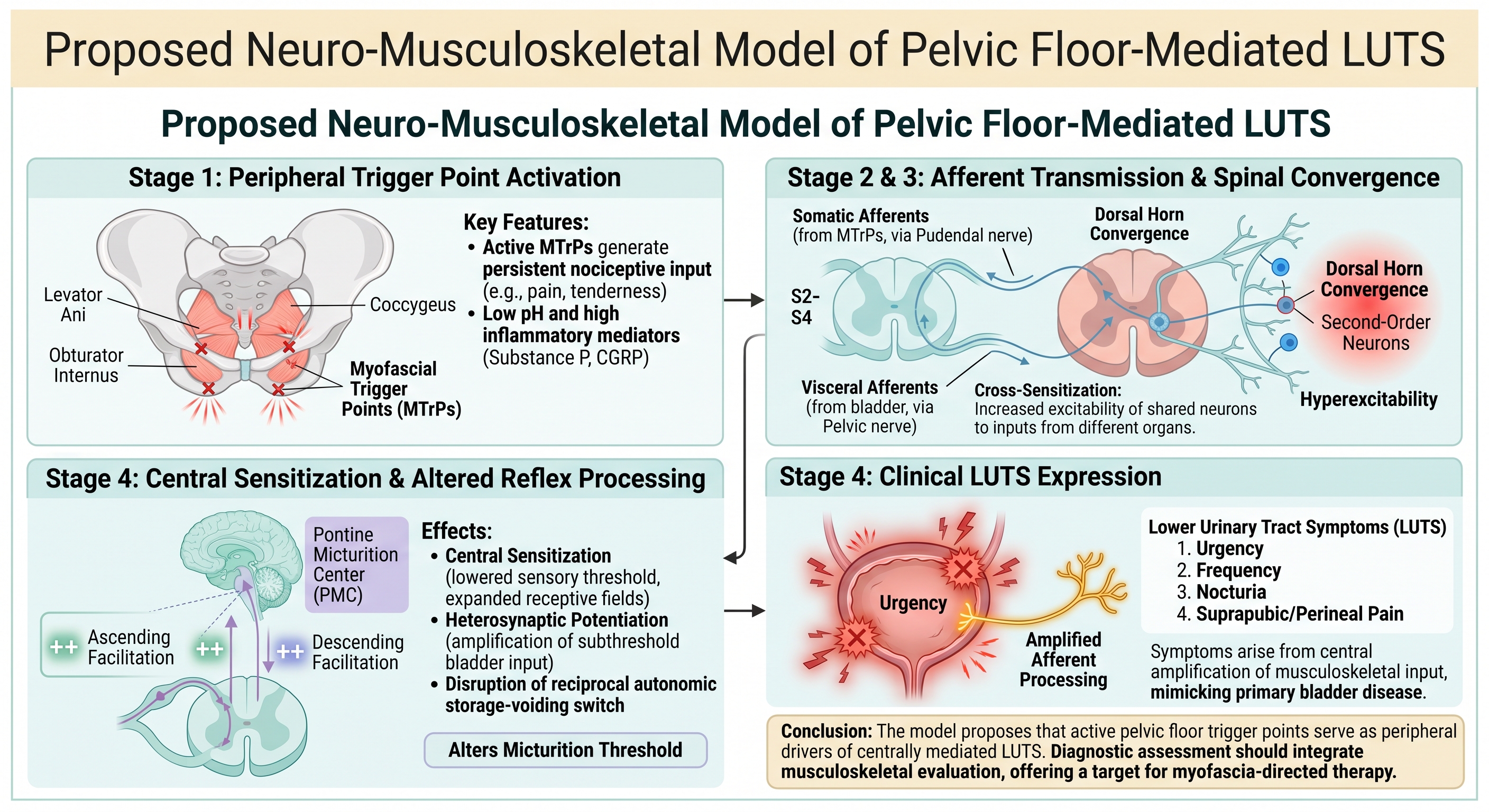
We propose an integrated five-stage model (Figure 1) in which pelvic floor myofascial dysfunction contributes to pain and urinary symptoms through a specified physiological pathway. This model does not replace bladder-centric or musculoskeletal frameworks; instead, it links them. Each component is supported by existing evidence, but the complete pathway remains to be tested prospectively.
5.6.1. Stage 1: Peripheral Trigger Point Activation
5.6.2. Stage 2: Afferent Transmission to the Sacral Spinal Cord
5.6.3. Stage 3: Dorsal Horn Convergence and Cross-Sensitization
Within the sacral dorsal horn, somatic afferents from the pelvic muscles converge with visceral afferents from the bladder on shared second-order neurons [1,10]. Sustained nociceptive input increases neuronal excitability, resulting in cross-sensitization. Normal bladder input may be amplified and perceived as heightened urgency, pressure, or discomfort.
5.6.4. Stage 4: Central Sensitization and Altered Reflex Processing
Persistent convergent inputs may induce central sensitization in spinal and supraspinal circuits. Sensitized neurons exhibit reduced activation thresholds and increased responsiveness [1]. Clinically, this reduces the threshold for bladder sensation and for activation of the micturition reflex, resulting in urgency, frequency, and involuntary detrusor activity. Descending modulatory pathways may further reinforce this sensitized state.
5.6.5. Stage 5: Clinical Expression
The clinical output is a constellation of symptoms, including pelvic pain and lower urinary tract dysfunction. In this model, these symptoms arise from integrated processing of convergent somatic and visceral inputs within a sensitized nervous system. This framework accounts for the prevalence of trigger points in bladder-related syndromes, overlapping referral patterns, and improvements following myofascial interventions [7,8,12,14,15,16].
5.7. Properties of the Model
The model is bidirectional (bladder pathology may also drive pelvic floor dysfunction), graded (severity varies with the intensity and duration of peripheral input) [12], and potentially progressive. It is clinically actionable, and the peripheral trigger point represents a modifiable input to a central process. Evidence that myofascial interventions improve both pain and urinary outcomes [15,16] is consistent with this framework. Nevertheless, prospective studies testing the full mechanistic chain are required.
6. Discussion
A purely urological model cannot account for the high prevalence of pelvic floor trigger points in bladder pain cohorts [7,8,12], reproduction of bladder symptoms by pelvic floor palpation [15,16], or improvement of urinary symptoms following musculoskeletal interventions [15,16]. Conversely, a purely musculoskeletal model cannot explain the autonomic reflex changes, central sensitization, or neuroplastic alterations observed during afferent processing [1,5,17]. These findings support an integrated framework in which peripheral musculoskeletal inputs and visceral afferent processing interact via shared spinal circuits.
This model supports incorporating pelvic floor musculoskeletal assessment into the standard evaluation of patients with LUTS, particularly those refractory to conventional bladder-directed therapy. Given the high prevalence of pelvic floor tenderness in populations [12] with UCPPS and the fact that detailed pelvic floor examinations are not routinely taught in urology or gynecology training [13], a musculoskeletal source of symptoms may go unrecognized when this evaluation is omitted. Patients may then undergo prolonged, ineffective, bladder-focused treatment, at least in part, for peripheral musculoskeletal conditions [6]. Einig et al. demonstrated that a standardized trigger-point examination correlates with LUTS severity, supporting its clinical utility [2].
In this model, urgency may reflect central amplification of normal filling signals within a sensitized spinal cord. Urinary frequency may result from a lowered micturition threshold driven by convergent musculoskeletal input. Suprapubic discomfort may represent referred pain from pelvic floor trigger points [6,13,15]. If pelvic floor trigger points serve as peripheral drivers of centrally mediated urinary symptoms, interventions that reduce trigger-point activity may address both local pain and the afferent input sustaining central sensitization. Manual therapy has produced moderate to marked improvements in urgency-frequency syndromes [15] , and combined myofascial release with relaxation training has yielded clinically significant improvements in refractory chronic pelvic pain [16]. Prospective controlled trials are warranted to determine whether the proposed mechanistic pathway accounts for the observed treatment effects.
6.1. Limitations
The neuromusculoskeletal pathway has not been tested as an integrated sequence within a single prospective study. Although evidence supports each component, studies supporting individual stages involve different populations, methodologies, and outcome measures. Thus, the inference that these stages constitute a connected physiological pathway relies on cross-domain synthesis rather than direct experimental validation.
The evidence linking pelvic floor trigger points to urinary symptoms is predominantly observational and cross-sectional [2,3,7,12]. These designs can establish associations but do not determine causation or directionality. It remains unclear whether trigger points are a primary driver of urinary symptoms, a secondary consequence of bladder pathology, or a co-occurring phenomenon arising from shared underlying mechanisms.
Cross-sensitization evidence has largely been derived from animal models of viscerovisceral interactions, particularly bowel-to-bladder sensitization [10[. The applicability of these findings to somatovisceral interactions between the pelvic floor musculature and the bladder in humans has not been directly investigated. Diagnostic criteria for pelvic floor trigger points have not yet been standardized. Examination methods differ across studies, and the diagnosis has not been confirmed by validated laboratory or imaging modalities [2,13]. This heterogeneity introduces uncertainty regarding both prevalence and clinical significance.
Evidence of central sensitization in an overactive bladder without pain remains indirect. Quantitative sensory testing has not been systematically applied in this population [1], and a systematic review by Knox et al. focused primarily on bladder pain syndrome rather than non-painful urinary dysfunction [17]. Treatment studies supporting this model are limited by small sample sizes, lack of randomization, and absence of blinding [15,16]. Prospective, mechanistically informed trials integrating musculoskeletal assessments, neurophysiological measures, and urodynamic outcomes are needed to test this framework.
7. Conclusions
This review proposes an integrated neuromusculoskeletal model of pelvic floor-mediated LUTS, outlining a pathway from active trigger points to sustained afferent signaling, spinal convergence to central sensitization, and altered bladder function. Each component is supported by published evidence; however, the complete sequence has not yet been tested as an integrated chain.
A principal implication is that, in a subset of patients, LUTS may reflect central processing of peripheral musculoskeletal input rather than solely intrinsic bladder pathology. This study suggests that a comprehensive evaluation should include a pelvic floor assessment. This framework also provides a basis for prospective studies integrating musculoskeletal, neurophysiological, and urodynamic outcomes.
Author Contributions
Conceptualization, J.B.; methodology, J.B.; formal analysis, J.B.; investigation, J.B. and C.B.; data curation, J.B. and C.B.; writing—original draft preparation, J.B.; writing—review and editing, J.B. and C.B.; visualization, J.B.; supervision, J.B.; project administration, J.B. All authors have read and agreed to the published version of the manuscript.
Funding
This research received no external funding.
Institutional Review Board Statement
Not applicable.
Informed Consent Statement
Not applicable.
Data Availability Statement
No new data were generated or analyzed to support this research. All the information presented was derived from previously published studies cited in the manuscript.
Acknowledgments
During the preparation of this manuscript/study, the authors used Elicit for the purposes of assistance with the literature search. The authors have reviewed and edited the output and take full responsibility for the content of this publication.
Conflicts of Interest
The authors declare no conflicts of interest.
Abbreviations
The following abbreviations are used in this manuscript:
| CGRP | Calcitonin gene-related peptide |
| LUTS | Lower urinary tract symptoms |
| MTrPs | Myofascial trigger points |
| PMC | Pontine micturition center |
| UCPPS | Urologic chronic pelvic pain syndrome |
References
- Reynolds, W.S.; Dmochowski, R.; Wein, A.; Bruehl, S. Does central sensitization help explain idiopathic overactive bladder? Nat. Rev. Urol. 2016, 13, 481–491. [Google Scholar] [CrossRef]
- Einig, S.; Ruess, E.; Schoetzau, A.; Ayllon Bartet, K.; Heinzelmann-Schwarz, V.; Vigo, F.; Kavvadias, T. Pelvic pain of myofascial origin in women: correlation with lower urinary tract symptoms. Adv. Urol. 2024, 2024, 1–7. [Google Scholar] [CrossRef]
- Leyva Vazquez, J.I.; Guerrero Reyes, G.; Gutiérrez González, A.; Hernández Velázquez, R.; Loya Maldonado, K.M.; Treviño Cavazos, O.; Reyes Alcaraz, J.E.; Robledo Torres, A.; Saca Cuevas, S.Y.; Herrera Morales, J.C. Female pelvic floor myofascial syndrome and its relationship with lower urinary tract storage symptoms. Int. J. Reprod. Contracept. Obstet. Gynecol. 2024, 13, 826–829. [Google Scholar] [CrossRef]
- Ochodnicky, P.; Uvelius, B.; Andersson, K.E.; Michel, M.C. Autonomic nervous control of the urinary bladder. Acta Physiol. 2013, 207, 16–33. [Google Scholar] [CrossRef]
- Yoshimura, N.; Ogawa, T.; Miyazato, M.; Kitta, T.; Furuta, A.; Chancellor, M.B.; Tyagi, P. Neural mechanisms underlying lower urinary tract dysfunction. Korean J. Urol. 2014, 55, 81–90. [Google Scholar] [CrossRef]
- Ackerman, A.L.; Wise-Fuchs, A. Myofascial pelvic floor dysfunction as a hidden contributor to urinary symptoms. Minerva Obstet. Gynecol. Minerva Obstet. Gynecol. 2026, 78, 250–260. [Google Scholar] [CrossRef]
- Bassaly, R.; Tidwell, N.; Bertolino, S.; Hoyte, L.; Downes, K.; Hart, S. Myofascial pain and pelvic floor dysfunction in patients with interstitial cystitis. Int. Urogynecol J. 2010, 22, 413–418. [Google Scholar] [CrossRef]
- Moldwin, R.M.; Fariello, J.Y. Myofascial trigger points of the pelvic floor: associations with urological pain syndromes and treatment strategies including injection therapy. Curr. Urol. Rep. 2013, 14, 409–417. [Google Scholar] [CrossRef]
- Andersson, K.E. Bladder activation: afferent mechanisms. Urology 2002, 59, 43–50. [Google Scholar] [CrossRef]
- Panicker, J.; Marcelissen, T.; von Gontard, A.; Vrijens, D.; Abrams, P.; Wyndaele, M. Bladder-bowel interactions: do we understand pelvic organ cross-sensitization? International consultation on incontinence research society (ICI-RS). Neurourol. Urodyn. 2019, 38 Suppl 5, S25–S34. [Google Scholar] [CrossRef]
- Itza, F.; Zarza, D.; Serra, L.; Gómez-Sancha, F.; Salinas, J.; Allona-Almagro, A. Síndrome de dolor miofascial del suelo pélvico: una patología urológica muy frecuente. Actas Urol. Esp. 2010, 34, 318–326. [Google Scholar] [CrossRef]
- Gupta, P.; Gallop, R.; Spitznagle, T.; Lai, H.; Tu, F.; Krieger, J.N.; Clemens, J.Q.; Bradley, C.S.; Yang, C.; Sutcliffe, S.; et al. Is pelvic floor muscle tenderness a distinct urologic chronic pelvic pain syndrome phenotype? Findings from the multidisciplinary approach to the study of chronic pelvic pain research network symptom pattern study. J. Urol. 2022, 208, 341–349. [Google Scholar] [CrossRef]
- Bonder, J.H.; Chi, M.; Rispoli, L. Myofascial pelvic pain and related disorders. Phys. Med. Rehabil. Clin. N Am. 2017, 28, 501–515. [Google Scholar] [CrossRef]
- Anderson, R.U.; Sawyer, T.; Wise, D.; Morey, A.; Nathanson, B.H. Painful myofascial trigger points and pain sites in men with chronic prostatitis/chronic pelvic pain syndrome. J. Urol. 2009, 182, 2753–2758. [Google Scholar] [CrossRef]
- Weiss, J.M. Pelvic floor myofascial trigger points: manual therapy for interstitial cystitis and the urgency-frequency syndrome. J. Urol. 2001, 166, 2226–2231. [Google Scholar] [CrossRef]
- Anderson, R.U.; Wise, D.; Sawyer, T.; Chan, C. Integration of myofascial trigger point release and paradoxical relaxation training treatment of chronic pelvic pain in men. J. Urol. 2005, 174, 155–160. [Google Scholar] [CrossRef]
- Knox, S.; Offiah, I.; Hashim, H. Evaluation of central sensitisation in bladder pain syndrome: a systematic review. Int. Urogynecol J. 2024, 35, 1109–1118. [Google Scholar] [CrossRef]
Figure 1.
Five-stage integrated model overview.
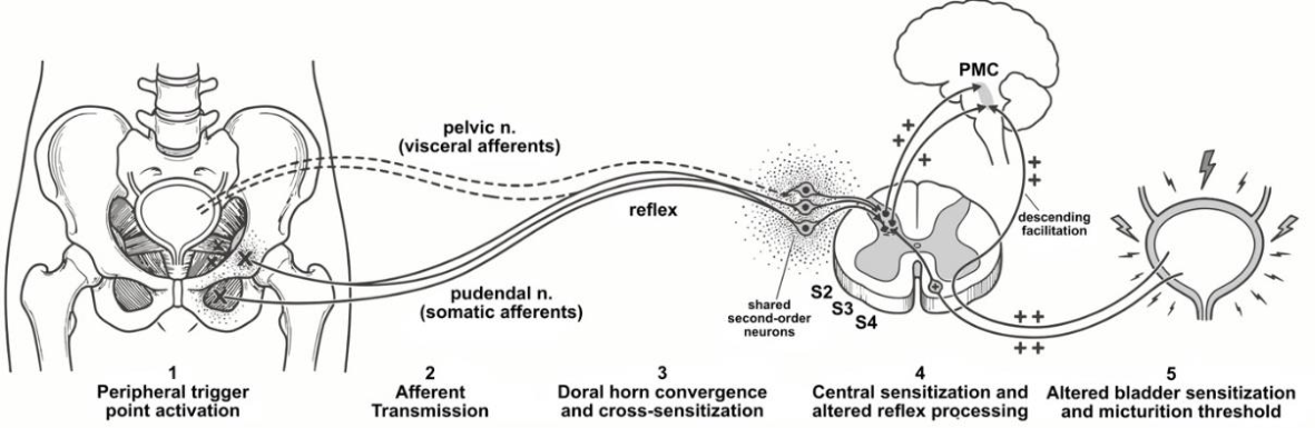

Table 1.
Key evidence by domain.
| Domain | Key Finding | Representative Source |
|---|---|---|
| Trigger Points | 81% of UCPPS participants had pelvic floor tenderness vs 9% of controls; widespread tenderness was associated with worse symptoms and a centralized pain phenotype | Gupta et al (2022) [12] |
| Trigger Points | 78% of IC patients had at least one trigger point; 68% had multiple sites | Bassaly et al, 2010 [7] |
| Trigger Points | Active trigger points contain elevated substance P, CGRP, bradykinin, TNF-α, serotonin, and norepinephrine with reduced pH; elevations also in remote uninvolved muscle | Moldwin and Fariello (2013) [8] |
| Trigger Points | Pelvic floor myofascial syndrome was associated with nocturia (67.6%), frequency (60.3%), urgency (57.4%) | Leyva Vazquez et al (2024) [3] |
| Trigger Points | 5 of 7 pain sites reproduced ≥50% of the time by palpation of designated trigger points | Anderson et al (2009) [14] |
| Trigger Points | Manual therapy produced moderate to marked improvement in 83% of urgency-frequency patients and 70% of IC patients | Weiss (2001) [15] |
| Trigger Points | Combined myofascial release and relaxation training yielded improvement in 72% of men with CP/CPPS refractory to conventional therapy | Anderson et al (2005) [16] |
| Trigger Points | Trigger point pain in the pubococcygeus, iliococcygeus, and obturator internus correlated with LUTS severity and high pelvic floor tone | Einig et al (2024) [2] |
| Trigger Points | MPFD was identified as a hidden contributor to LUTS without primary bladder pathology (myofascial urinary frequency syndrome) | Ackerman and Wise-Fuchs (2026) [6] |
| Autonomic/Afferent Signaling | Bladder Aδ-fibers convey a filling sensation; C-fibers are normally silent but become mechanosensitive in pathological states; the urothelium releases ATP, NO, and ACh as mechanosensory mediators | Andersson (2002) [9] |
| Autonomic/Afferent Signaling | Storage-voiding switch governed by reciprocal autonomic activity; NGF-mediated neuroplasticity alters afferent excitability and spinal reflex processing | Yoshimura et al (2014) [5] |
| Autonomic/Afferent Signaling | Neurotransmitter receptors on urothelium, interstitial cells, and afferent nerves form a distributed sensory network; non-neuronal ACh released during storage increases with age | Ochodnicky et al (2013) [4] |
| Sensitization | Central sensitization produces heterosynaptic potentiation: repetitive C-fiber input amplifies subthreshold Aδ/Aβ signals; proposed mechanism for bladder hypersensitivity in idiopathic OAB | Reynolds et al (2016) [1] |
| Sensitization | Cross-sensitization: afferents from different pelvic structures converge onto shared dorsal horn neurons; disease in one organ increases excitability of neurons processing input from adjacent organs | Panicker et al, 2019 [10] |
| Sensitization | Systematic review found evidence of central sensitization in all 15 BPS studies; greater sensitization correlated with symptom severity and comorbid chronic pain | Knox et al (2024) [17] |
Abbreviations: UCPPS, urologic chronic pelvic pain syndrome; IC, interstitial cystitis; CGRP, calcitonin gene-related peptide; TNF-α, tumor necrosis factor-α; LUTS, lower urinary tract symptoms; CP/CPPS, chronic prostatitis/chronic pelvic pain syndrome; NGF, nerve growth factor; OAB, overactive bladder; MPFD, myofascial pelvic floor dysfunction; BPS, bladder pain syndrome; ACh, acetylcholine.
Disclaimer/Publisher’s Note: The statements, opinions and data contained in all publications are solely those of the individual author(s) and contributor(s) and not of MDPI and/or the editor(s). MDPI and/or the editor(s) disclaim responsibility for any injury to people or property resulting from any ideas, methods, instructions or products referred to in the content. |
© 2026 by the authors. Licensee MDPI, Basel, Switzerland. This article is an open access article distributed under the terms and conditions of the Creative Commons Attribution (CC BY) license.
Copyright: This open access article is published under a Creative Commons CC BY 4.0 license, which permit the free download, distribution, and reuse, provided that the author and preprint are cited in any reuse.