

Submitted:
18 August 2026
Posted:
19 August 2026
You are already at the latest version
Abstract
Mesenteric panniculitis (MP) is an uncommon inflammatory disorder of mesenteric adipose tissue. Its pathophysiology remains unclear. Gut dysbiosis, intestinal barrier dysfunction, metabolic endotoxemia, glycemic variability (GV), and vascular dysfunction are involved in inflammatory and metabolic processes. These may also affect visceral and mesenteric adipose tissue. This narrative review examines the possible relationships among these mechanisms and mesenteric panniculitis and discusses their integration within a proposed gut–mesentery–metabolic axis. The literature was reviewed using PubMed, Scopus, Web of Science, and Google Scholar, including studies published between 2000 and 2026. We focus on recent evidence published between 2021 and 2026. Current data suggest that dysbiosis and impaired intestinal barrier function may facilitate microbial-product translocation and lipopolysaccharide-mediated inflammatory signaling, while GV may contribute to oxidative stress, endothelial dysfunction, and pro-inflammatory responses. Mesenteric vascular anatomy and impaired regional perfusion may represent additional factors influencing local tissue susceptibility. Recent randomized controlled trials of microbiome-targeted interventions in metabolic disorders have shown heterogeneous effects on glycemic, inflammatory, and microbiota-related outcomes. Future investigation of individualized microbiome-directed strategies is needed. Recent evidence demonstrating that these microbial, metabolic, or vascular mechanisms initiate or sustain MP remains limited. The proposed gut–mesentery–metabolic axis should be regarded as a hypothesis for further investigation. Prospective studies integrating microbiome profiling, metabolic biomarkers, glycemic variability assessment, vascular evaluation, and imaging phenotypes are needed to clarify if biological subgroups of MP can be identified.
Keywords:
mesenteric panniculitis
; gut microbiota
; gut dysbiosis
; intestinal barrier dysfunction
; metabolic endotoxemia
; glycemic variability
; mesenteric adipose tissue
; vascular dysfunction
; microbiome modulation
; precision medicine
1. Introduction
Metabolic inflammation is a central feature of obesity [1]. Toll-like receptor 4-mediated innate immune activation contributes to fatty acid-induced insulin resistance [2]. Alterations in the gut microbiota are involved in intestinal and systemic inflammatory processes [3]. Gut-derived low-grade endotoxemia has also been associated with atherothrombosis and cardiovascular disease [4].
Gut dysbiosis and intestinal barrier disruption may contribute to adipose tissue dysfunction in metabolic disorders [5]. Increased intestinal permeability can facilitate the translocation of microbial products into the circulation, thereby promoting systemic inflammatory responses [5].
Mesenteric panniculitis is a rare inflammatory disorder involving the adipose tissue of the mesentery [6]. Its clinical presentation is heterogeneous and may range from an incidental imaging finding to symptomatic abdominal disease [6]. Mesenteric panniculitis has been described in association with inflammatory, autoimmune, surgical, traumatic, and neoplastic conditions [7]. These associations do not establish a direct causal relationship [7].
Actual evidence indicates that gut microbial composition and intestinal barrier integrity are involved in systemic and adipose-tissue inflammation [5]. Dysbiosis-associated increases in intestinal permeability may facilitate microbial-product translocation and metabolic endotoxemia [8]. Gut microbiota, intestinal permeability, and systemic inflammation are interconnected [9]. Enteric-derived lipopolysaccharides may contribute to adipose-tissue and metabolic inflammation [10].
Glycemic variability refers to fluctuations in blood glucose concentrations over time [11]. It has been associated with diabetic microvascular and macrovascular complications [11]. Glucose fluctuations may promote oxidative stress, endothelial dysfunction, and cardiovascular injury [12]. Glycemic variability may exert harmful effects beyond those associated with sustained hyperglycemia alone [12].
Glycemic variability may contribute to oxidative stress, endothelial dysfunction, and inflammatory signaling [12,13].
The intestinal vasculature has specialized structural and functional characteristics that contribute to regional tissue homeostasis [14]. These vascular characteristics may influence local perfusion and tissue homeostasis [14]. Direct evidence demonstrating that mesenteric vascular variants cause mesenteric panniculitis is currently lacking.
Glycemic variability may contribute to vascular dysfunction through oxidative stress and inflammatory signaling [13]. Interactions between the gut microbiota and the host immune system can also promote immune activation and systemic inflammation [15]. The specific contribution of these mechanisms to mesenteric panniculitis remains insufficiently investigated.
This narrative review examines the potential relationships among glycemic variability, gut dysbiosis, intestinal barrier dysfunction, vascular susceptibility, and mesenteric inflammation. It proposes the gut–mesentery–metabolic axis as a hypothesis.
The review also considers the potential role of microbiome-targeted strategies in metabolic and inflammatory disorders [16]. Their relevance to mesenteric panniculitis remains to be established. Recent literature describes development of microbiome-informed approaches for patient and personalized intervention in metabolic disease [17].
2. Materials and Methods
2.1. Review Design and Reporting Framework
This article was designed as a structured narrative review examining the potential interactions among mesenteric panniculitis, gut dysbiosis, intestinal barrier dysfunction, microbial translocation, mesenteric vascular anatomy, regional perfusion, glycemic variability, and metabolic inflammation.
The review was developed to integrate anatomical, mechanistic, experimental, imaging, translational, and clinical evidence relevant to the proposed gut–mesentery–metabolic axis. Attention was given to distinguishing established characteristics of mesenteric panniculitis from biologically plausible mechanisms that have not yet been directly demonstrated in patients with this condition.
The reporting approach was guided by the Scale for the Assessment of Narrative Review Articles (SANRA), with particular attention to the clarity of the review rationale, formulation of the review objective, description of the literature search, appropriate citation of the literature, scientific reasoning, and balanced interpretation of the available evidence [18].
This review was not conducted as a systematic review or scoping review. We did not perform a PRISMA flow diagram, formal risk-of-bias assessment, methodological quality assessment, protocol registration, or quantitative synthesis.
2.2. Search Strategy
We searched PubMed, Scopus, Web of Science, and Google Scholar for articles published mainly between 2000 and 2026. Attention was given to recent publications from 2021 to 2026. The last literature search was performed on 18.08.2026. Only articles published in English were considered.
Earlier publications were also included when they provided important anatomical, histopathological, radiological, or conceptual information regarding mesenteric panniculitis, sclerosing mesenteritis, mesenteric lipodystrophy, intestinal vascular anatomy, microbial translocation, metabolic endotoxemia, or glycemic variability.
The search combined terms related to mesenteric panniculitis and mesenteric inflammation, including “mesenteric panniculitis”, “sclerosing mesenteritis”, “retractile mesenteritis”, “mesenteric lipodystrophy”, “misty mesentery”, “mesenteric inflammation”, and “mesenteric adipose tissue”.
Terms related to the gut microbiota and intestinal barrier included “gut microbiota”, “gut microbiome”, “dysbiosis”, “intestinal permeability”, “intestinal barrier dysfunction”, “leaky gut”, “microbial translocation”, “lipopolysaccharide”, “LPS”, “metabolic endotoxemia”, “short-chain fatty acids”, “butyrate”, “Toll-like receptor 4”, “TLR4”, and “NF-κB, “microbiome modulation”, “microbiota-targeted therapy”, “microbiome-targeted intervention”, “precision microbiome”, “probiotic”, “prebiotic”, “synbiotic”, “postbiotic”, “personalized nutrition”, and “randomized controlled trial”.
Terms related to vascular anatomy and perfusion included “mesenteric vascular anatomy”, “mesenteric arterial variants”, “superior mesenteric artery”, “inferior mesenteric artery”, “celiac trunk”, “mesenteric perfusion”, “mesenteric ischemia”, “chronic mesenteric ischemia”, “ischemia–reperfusion injury”, “endothelial dysfunction”, “microvascular dysfunction”, and “hypoxia-inducible factor 1-alpha”.
Terms related to metabolic dysfunction included “glycemic variability”, “glucose variability”, “glucose fluctuations”, “continuous glucose monitoring”, “oxidative stress”, “metabolic inflammation”, “type 2 diabetes”, “insulin resistance”, “visceral adiposity”, “obesity”, and “metabolic syndrome”.
Terms related to Helicobacter pylori and microbiome–metabolic interactions included “Helicobacter pylori”, “H. pylori”, “H. pylori eradication”, “gastric microbiota”, “dysbiosis”, “gut microbiota”, “insulin resistance”, “metabolic syndrome”, “cardiovascular risk”, “atherosclerosis”, “chronic atrophic gastritis”, and “outer membrane vesicles”.
The search terms were combined using the Boolean operators “AND” and “OR”, according to the requirements of each database. The reference lists of selected publications were also screened to identify additional relevant studies that were not retrieved through the initial database search.
The database-specific search strategies and applied filters are presented in Appendix A, Table A1.
2.3. Selection Criteria
We considered human studies, animal studies, experimental studies, translational investigations, anatomical studies, imaging studies, clinical and observational studies, systematic reviews, meta-analyses, and narrative reviews when they addressed mesenteric panniculitis or mechanisms potentially relevant to mesenteric inflammation.
Studies were considered eligible when they addressed at least one of the principal themes of the review: clinical manifestations of mesenteric panniculitis, radiological characteristics, histopathological findings, mesenteric adipose tissue inflammation, gut dysbiosis, intestinal barrier dysfunction, microbial translocation, metabolic endotoxemia, mesenteric vascular anatomy, vascular anatomical variants, regional perfusion abnormalities, chronic low-grade ischemia, glycemic variability, oxidative stress, endothelial dysfunction, insulin resistance, or metabolic inflammation.
Studies investigating gastrointestinal pathogens, including Helicobacter pylori, were considered when they provided mechanistic evidence regarding dysbiosis, intestinal barrier disruption, microbial-product dissemination, systemic inflammation, or metabolic dysfunction. Because direct evidence connecting individual gastrointestinal pathogens to mesenteric panniculitis is limited, these findings were interpreted as indirect mechanistic evidence.
Studies examining probiotics, prebiotics, postbiotics, short-chain fatty acids, glucose-lowering therapies, vascular interventions, anti-inflammatory therapies, or other microbiota-targeted approaches were considered when they addressed mechanisms potentially relevant to the proposed gut–mesentery–metabolic axis.
Studies on Helicobacter pylori were included when they addressed gut microbiota alterations, dysbiosis, systemic inflammation, metabolic dysfunction, or related microbiome–metabolic mechanisms relevant to the scope of this review.
Randomized controlled trials of microbiome-targeted interventions were considered when they evaluated microbial, metabolic, inflammatory, intestinal barrier, or cardiometabolic outcomes relevant to the proposed gut–mesentery–metabolic axis.
We excluded conference abstracts without accessible full text, letters, comments, and editorials without relevant mechanistic or clinical discussion. We also excluded isolated case reports that did not provide relevant diagnostic, therapeutic, pathological, or mechanistic information.
Non-English publications were excluded. Studies without a clear connection to mesenteric panniculitis, mesenteric inflammation, gut barrier dysfunction, dysbiosis, vascular perfusion, glycemic variability, or metabolic inflammation were also excluded.
The inclusion and exclusion criteria are summarized in Appendix A, Table A2.
2.4. Study Selection and Author Involvement
The literature search and initial organization of the retrieved material were coordinated by S.I., V.M., M.E.T., and A.F.W., with overall scientific supervision from M.R. Titles and abstracts were screened for relevance by S.I., V.M., A.F.W., S.T., S.A.R., B.A.B., I.R.R., and S.M.S., according to the predefined thematic areas of the review.
Articles considered potentially relevant were assessed in full text and subsequently evaluated thematically by authors with complementary expertise in anatomy, diabetes and metabolic diseases, biology, pharmaceutical chemistry, clinical pharmacy and pharmacology, internal medicine, pathology, endocrinology, metabolic disorders, pharmacoeconomics, and translational medicine.
The anatomical and vascular literature was reviewed with particular attention to mesenteric continuity, arterial anatomy, vascular variants, collateral circulation, and regional perfusion. The clinical and imaging literature was evaluated for evidence concerning the diagnosis, differential diagnosis, natural history, and management of mesenteric panniculitis.
The microbiota-related literature was assessed for evidence regarding dysbiosis, intestinal barrier dysfunction, microbial translocation, lipopolysaccharide-mediated signaling, and metabolic endotoxemia. The metabolic literature was evaluated for evidence regarding glycemic variability, oxidative stress, endothelial dysfunction, adipose tissue inflammation, insulin resistance, and glucose-lowering therapies.
A.A., K.H., I.I., and A.P. contributed to the clinical, cardiovascular, endocrine, metabolic, and pharmacoeconomic interpretation of the evidence. Uncertainties regarding article relevance, thematic classification, or interpretation were resolved through discussion among the authors, with final oversight by M.E.T. and M.R.
2.5. Evidence Synthesis
The selected literature was synthesized narratively and organized into thematic sections. The synthesis focused on the clinical, pathological, and imaging characteristics of mesenteric panniculitis; gut dysbiosis and intestinal barrier dysfunction; microbial translocation and lipopolysaccharide-mediated immune activation; the potential systemic effects of gastrointestinal pathogens; mesenteric vascular anatomy and perfusion; glycemic variability; microbiome-targeted interventions and evidence from randomized controlled trials; and their possible relationship within a gut–mesentery–metabolic axis.
Because the included evidence originated from heterogeneous study designs, mechanistic plausibility was distinguished from direct clinical evidence whenever possible. Findings derived from animal models, cellular studies, anatomical investigations, and indirect metabolic evidence were interpreted separately from observations obtained directly in patients with mesenteric panniculitis.
Attention was given to avoiding causal interpretations when the available evidence demonstrated only biological plausibility or epidemiological association. Established diagnostic and pathological features of mesenteric panniculitis were separated from proposed mechanisms involving dysbiosis, vascular susceptibility, chronic low-grade ischemia, and glycemic variability.
The strength of the evidence was interpreted according to its origin. Direct clinical, imaging, or histopathological evidence in patients with mesenteric panniculitis was considered separately from evidence derived from patients with obesity, type 2 diabetes, metabolic syndrome, inflammatory bowel disease, or other disorders involving visceral adipose inflammation.
Selected mechanisms, imaging findings, and representative studies were summarized in tables to improve clarity and facilitate clinical interpretation. The proposed gut–mesentery–metabolic axis was used as an integrative and hypothesis framework. It is not as an established causal model.
3. Mesenteric Panniculitis: Clinical and Pathological Overview
3.1. Epidemiology
Mesenteric panniculitis is an uncommon chronic inflammatory disorder involving the adipose tissue of the mesentery [19]. The mesentery has been redefined recently in anatomical research as a continuous organ with distinct functional characteristics [20]. The condition is frequently identified incidentally during abdominal computed tomography performed for unrelated clinical indications [21].
The true prevalence of mesenteric panniculitis remains uncertain because diagnostic criteria, imaging protocols, study populations, and radiological reporting practices differ considerably among published studies [21]. In a computed tomography study conducted by Daskalogiannaki et al., mesenteric panniculitis was identified in approximately 0.6% of examined patients [22]. More recent reviews have reported prevalence estimates ranging from approximately 0.16% to 7.8% among individuals undergoing abdominal computed tomography [21].
A substantial proportion of patients may remain asymptomatic. When symptoms are present, abdominal pain is the most frequently reported manifestation. Other possible manifestations include abdominal distension, bloating, altered bowel habits, nausea, vomiting, anorexia, fever, fatigue, and weight loss [21].
Mesenteric panniculitis is diagnosed more frequently in men than in women [23]. Published clinical series have reported a male-to-female ratio ranging from approximately 2:1 to 3:1 [23]. The disorder predominantly affects middle-aged and older adults. Most patients are diagnosed in the sixth and seventh decades of life [19]. Nevertheless, cases have also been described outside this age range [24].
Several clinical conditions have been reported in association with mesenteric panniculitis. Previous abdominal surgery or abdominal trauma has been documented in a proportion of affected patients [25]. Autoimmune and inflammatory disorders have also been described in association with the condition [7]. Metabolic syndrome and diabetes mellitus have been reported among patients with mesenteric panniculitis [24]. Vascular disorders have also been reported more frequently in some cohorts of patients with mesenteric panniculitis [24].
An association between mesenteric panniculitis and malignancy has been reported in observational and imaging studies. The strength and clinical significance of this association remain controversial. Selection and imaging-related biases may partly explain the apparent association, because patients with malignancy undergo abdominal imaging more frequently than the general population [24]. Mesenteric panniculitis should not be interpreted as a paraneoplastic condition solely based on its radiological identification [24].
The available epidemiological evidence does not establish a specific causal relationship between mesenteric panniculitis and metabolic, autoimmune, surgical, traumatic, or neoplastic conditions [7]. Mesenteric panniculitis may instead represent a heterogeneous radiological and pathological response to different local or systemic inflammatory stimuli [19].
3.2. Histopathology
Histopathological evaluation of mesenteric panniculitis (MP) reveals a spectrum of changes reflecting the dynamic and evolving nature of the inflammatory process in mesenteric adipose tissue. The spectrum of MP progression is classified into three coexisting stages: mesenteric lipodystrophy (fat necrosis), mesenteric panniculitis (chronic inflammation), and retractile mesenteritis (fibrosis) [23]. Fat necrosis and adipocyte vacuolization, accompanied by minimal inflammation or fibrosis, are typical of the early lipodystrophy stage. The advanced fibrotic form, often referred to as retractile mesenteritis, and it is characterized by extensive collagen deposition and remodeling [23].
A mixed inflammatory infiltrate of plasma cells, histiocytes and lymphocytes can be noticed in the subsequent panniculitis stage [26]. Often this infiltrate is distributed perivascular [27].
While histopathological confirmation is the most definitive, it is rarely obtained unless malignancy is suspected, as diagnosis primarily relies on radiological findings [21,24].
A crucial element that should be considered during tissue evaluation is represented by differential diagnosis from lymphoma and metastases [24], as well as IgG4-related disease [28]. In some cases, fibroblastic proliferation may feature similar to desmoid-type fibromatosis, without the defining features of a true neoplastic fibromatosis [24,29]. Desmoid-type fibromatosis is a rare, locally aggressive fibroblastic neoplasm that does not metastasize but may infiltrate adjacent structures and recur after treatment [30].
3.3. Imaging
MP diagnosis depends on imaging, specifically on Computed Tomography (CT). Elements such as well-defined areas of enhanced fat attenuation, the “fat ring sign” and a pseudocapsule are of utmost importance in this regard [19]. The “fat ring” sign, i.e., normal fat density preserved around mesenteric vessels and nodes, contributes to the differentiation between MP and neoplastic or infectious processes [22].
Although less commonly used than Computed Tomography (CT), magnetic resonance imaging (MRI) serves as an alternative diagnostic tool, with signal characteristics varying according to the relative proportions of inflammation and fibrosis [19].
Positron emission tomography combined with CT (PET/CT) may contribute to the differential diagnosis between MP and malignancy, when mesenteric lesions show absent fluorodeoxyglucose uptake [19].
Occasionally, increased echogenicity of the mesenteric fat and hypoechoic nodules or bands can be detected by ultrasound [24]. Ultrasound has limited diagnostic value compared with CT, for deep mesenteric abnormalities [19].
Imaging facilitates the diagnosis and may contribute to the assessment of disease evolution. Imaging findings must be interpreted clinically, as similar patterns may occur in lymphoma, peritoneal carcinomatosis, or desmoid tumors [24].
3.4. Current Management and Unmet Therapeutic Needs
The absence of standardized treatment protocols, symptom severity guides MP management, hence its empirical nature. For asymptomatic patients, a conservative approach with clinical follow-up may be considered, as the condition may remain stable or regress spontaneously [21]. Pharmacological therapy may be considered in symptomatic patients, in those presenting with abdominal pain, weight loss, or other clinically significant symptoms. Inflammatory markers, including erythrocyte sedimentation rate and C-reactive protein, may be elevated and can be used to monitor the response to treatment. Usually, this therapy starts with the administration of corticosteroids, such as prednisone, to decrease inflammation [19].
Immunomodulators, including azathioprine or methotrexate, and tamoxifen have been used in patients with persistent symptoms or refractory disease [19]. Prednisone combined with tamoxifen is the most frequently reported first-line regimens for symptomatic patients [21].
Surgical intervention is generally reserved for severe complications, recurrent or persistent intestinal obstruction [21]. Surgery is not considered a routine curative treatment for MP [19].
Associated disorders should be identified and managed according to their specific clinical indications [21]. The therapeutic approach to MP should be individualized, balancing symptom severity, comorbidities, and potential treatment risks [23]. Further prospective studies are necessary to validate treatment algorithms and identify prognostic factors that may guide long-term management. The absence of disease-specific, mechanism-based therapeutic strategies also supports investigation of potentially modifiable microbial and metabolic pathways [16].
4. Gut Dysbiosis, Intestinal Barrier Dysfunction, and Mesenteric Inflammation
4.1. Definition and Composition of Dysbiosis
The gut microbiota is a complex microbial ecosystem composed predominantly of bacteria, together with archaea, fungi, protozoa, and viruses. [31] In healthy individuals, the bacterial component is characterized by substantial diversity and is dominated mainly by the phyla Firmicutes, Bacteroidetes, Actinobacteria, and Proteobacteria [3].
Dysbiosis refers to an alteration in the composition, diversity, distribution, or functional activity of the microbiota that disrupts the homeostatic relationship between the host and its microorganisms [32].
Table 1 highlights the transition from a balanced microbial ecosystem, characterized by homeostatic and protective functions, to a dysbiotic ecosystem marked by the loss of beneficial microorganisms, the expansion of pathobionts, reduced microbial diversity, and altered metabolic activity [32]. Dysbiosis may impair intestinal barrier integrity and promote increased intestinal permeability [37]. These changes can activate inflammatory pathways and contribute to systemic metabolic dysfunction [33]. Interactions between gut dysbiosis and mesenteric adipose tissue inflammation have been described in inflammatory bowel disease, although direct evidence in mesenteric panniculitis remains limited [40].
Dysbiosis commonly involves the loss of beneficial microorganisms, the expansion of pathobionts, including Enterobacteriaceae, and a reduction in overall microbial diversity [32].
Western-style diets rich in fat and low in fiber may promote gut dysbiosis, intestinal barrier dysfunction, and metabolic inflammation [34]. Antibiotic exposure may also alter the composition and functional activity of the gut microbiota [32]. Obesity and type 2 diabetes mellitus are frequently associated with dysbiotic microbial profiles [33].
Dysbiosis alters the production and availability of microbial metabolites, including short-chain fatty acids, bile acid derivatives, lipopolysaccharides, and branched-chain amino acids [33]. Microbiota-derived metabolites influence host metabolic homeostasis and immune function [35]. Reduced production of beneficial metabolites, butyrate, may impair epithelial barrier integrity and anti-inflammatory regulation [35].
Intestinal barrier dysfunction associated with dysbiosis facilitates the translocation of lipopolysaccharides into the circulation [4]. Lipopolysaccharides activate Toll-like receptor 4 signaling and promote inflammatory and metabolic responses [2]. Toll-like receptor 4 activation promotes nuclear factor kappa B-dependent inflammatory signaling [2]. This signaling increases the production of pro-inflammatory mediators, including tumour necrosis factor alpha, interleukin-6, and interleukin-1β [4].
Dysbiosis is involved in the pathophysiology of inflammatory bowel disease [38]. Dysbiotic microbial profiles have also been associated with obesity and type 2 diabetes mellitus [33]. Diet-induced dysbiosis and intestinal inflammation contribute to the development of metabolic dysfunction-associated fatty liver disease [34].
Intestinal barrier disruption and microbial translocation may promote inflammatory signaling in tissues adjacent to the gut [37]. E-cadherin dysfunction may further impair epithelial barrier integrity and facilitate bacterial invasion or dissemination [39]. Interactions between gut microbiota and mesenteric adipose tissue have been described in inflammatory intestinal disease [40]. Direct evidence linking these mechanisms to mesenteric panniculitis remains insufficient.
The mesentery is an anatomically continuous and functionally active structure containing vascular, lymphatic, adipose, and immune components [20]. Evidence of interactions between the gut microbiota and mesenteric adipose tissue supports the proposed concept of a gut–mesentery axis [40]. This may have implications for gastrointestinal, inflammatory, and metabolic disorders.
4.2. Intestinal Barrier Dysfunction and Microbial Translocation
An important mechanistic axis related to gut dysbiosis with mesenteric and systemic inflammation is increased intestinal permeability [42]. Increased intestinal permeability facilitates the translocation of microbial products, including lipopolysaccharides, into the circulation [5]. Dysbiosis and microbial-product translocation may contribute to systemic metabolic inflammation [33]. Metabolic endotoxemia induced by circulating lipopolysaccharides may further amplify systemic inflammatory responses [8].
In a healthy intestinal barrier, epithelial cells and the protective mucus layer act synergistically to prevent the passage of microbial antigens into subepithelial tissues [36]. Tight junction proteins, including cingulin, tricellulin, occludin, claudins, zonula occludens-1, and junctional adhesion molecules, regulate paracellular permeability [36]. Akkermansia muciniphila-derived extracellular vesicles may influence intestinal permeability through the regulation of tight junction proteins [43]. Gut barrier dysfunction is also associated with adipose tissue inflammation and metabolic disorders [5].
In the context of dysbiosis, dietary imbalances, or metabolic stress, the intestinal barrier may become compromised [44]. This increase in intestinal permeability is commonly described as “leaky gut” [44].
The proposed sequence from gut dysbiosis to intestinal barrier dysfunction, mesenteric immune activation, and systemic inflammatory effects is illustrated in Figure 1.
Figure 1 illustrates how alterations in gut microbiota composition may disrupt intestinal barrier integrity and disturb mesenteric immune–metabolic homeostasis. This process may initiate a cascade of local and systemic inflammatory. This cascade may induces metabolic responses.
Under physiological conditions, a balanced gut microbiota contributes to epithelial integrity, regulates the production of short-chain fatty acids, and supports immune tolerance [3]. A healthy intestinal barrier also limits the passage of microbial antigens and products into the underlying tissues and circulation [36].
When dysbiosis occurs, beneficial microbial populations may decline and pathobionts may expand [32]. Dysbiosis may also reduce the production of protective microbial metabolites, butyrate [45]. Reduced butyrate availability may impair tight-junction integrity and weaken intestinal barrier function [45].
Intestinal barrier disruption facilitates the translocation of lipopolysaccharides and other microbial products into the circulation [9]. Lipopolysaccharides activate Toll-like receptor 4 signaling in macrophages and adipocytes [2]. Toll-like receptor 4 activation promotes nuclear factor kappa B-dependent inflammatory signaling [2]. This signaling increases the production of pro-inflammatory mediators, including tumour necrosis factor alpha, interleukin-6, and interleukin-1β [4].
Enteric-derived lipopolysaccharides may promote macrophage activation, adipocyte dysfunction, and chronic low-grade inflammation [10]. Direct evidence demonstrating that lipopolysaccharide exposure causes early fibrosis specifically in mesenteric panniculitis remains lacking.
Chronic dysbiosis-associated inflammation may affect mesenteric adipose tissue, in inflammatory intestinal disorders [41]. Interactions among the gut microbiota, intestinal inflammation, and mesenteric adipose tissue have been described in Crohn’s disease [40]. These provide indirect mechanistic support for mesenteric immune activation but do not establish the same pathway in mesenteric panniculitis.
Microbial-product translocation contributes to metabolic endotoxemia and systemic inflammation [8]. Metabolic endotoxemia may promote insulin resistance through inflammatory and innate immune pathways [8]. Diet-induced dysbiosis and intestinal inflammation may also contribute to metabolic dysfunction-associated steatotic liver disease [34].
Lipopolysaccharide is a major structural component of the outer membrane of Gram-negative bacteria and a potent microbe-associated molecular pattern [46]. Persistent exposure to circulating lipopolysaccharides may promote systemic and adipose-tissue inflammation [4]. Enteric-derived lipopolysaccharides have also been implicated in obesity-associated metabolic dysfunction [10]. Their specific causal contribution to mesenteric panniculitis has not been established.
Experimental and translational evidence indicates that metabolic endotoxemia contributes to systemic inflammation and insulin resistance [8]. Dietary factors may alter gut microbiota composition, increase intestinal permeability, and elevate circulating lipopolysaccharide concentrations [47]. In high-fat diet-fed mice, selected lactic acid bacterial strains may improve microbial composition and modulate metabolic and immunological parameters [48]. These experimental findings support microbiota-targeted mechanisms but cannot be directly extrapolated to the treatment of mesenteric panniculitis.
The mesentery is a continuous anatomical structure containing adipose tissue, vessels, lymphatics, connective tissue, and immune components [49]. Its anatomical relationship with the intestine allows it to participate in immune surveillance, lymphatic drainage, and inflammatory responses [49]. In inflammatory bowel disease, mesenteric adipose tissue may become an active source of adipokines and inflammatory mediators [41]. Crosstalk between intestinal microbiota and mesenteric adipose tissue has also been described in Crohn’s disease [40].
Mesenteric panniculitis is histopathologically characterized by varying proportions of fat necrosis, chronic inflammation, and fibrosis [23]. These pathological characteristics should not be attributed directly to lipopolysaccharide exposure without disease-specific experimental or clinical evidence.
Elevated circulating lipopolysaccharide and zonulin concentrations have been reported in patients with type 2 diabetes mellitus [50]. Elevated plasma zonulin concentrations have also been described in obesity-associated fatty liver disease [51]. These findings support an association between intestinal barrier dysfunction and metabolic disease, but they do not demonstrate a correlation between zonulin and radiological signs of mesenteric panniculitis.
Increased intestinal permeability and microbial-product translocation may contribute to systemic metabolic inflammation [9] . These mechanisms may also influence immune responses in tissues adjacent to the intestine [40]. Their role in mesenteric panniculitis remains a biologically plausible, but not an demonstrated hypothesis. The proposed gut–barrier–mesentery pathway should be considered a framework for future investigation.
4.3. Metabolic Endotoxemia and LPS-Mediated Inflammatory Signaling
Once lipopolysaccharide reaches adipose tissue, it may be recognized by Toll-like receptor 4 expressed on macrophages and adipocytes. Toll-like receptor 4 activation initiates intracellular inflammatory signaling, including nuclear factor kappa B-dependent pathways. This signaling promotes the release of pro-inflammatory mediators and contributes to insulin resistance [2]. Gut-derived low-grade endotoxemia is also associated with systemic inflammatory and vascular responses [4].
The proposed LPS–mesentery inflammatory cascade is illustrated in Figure 2.
In Figure 2, the proposed LPS–mesentery axis represents a potential relationship between gut dysbiosis and systemic metabolic inflammation. Dysbiosis may increase the relative abundance of Gram-negative bacteria and alter the production and availability of bacterial lipopolysaccharides [9]. Intestinal barrier dysfunction facilitates the translocation of lipopolysaccharides from the intestinal lumen into the circulation [44]. Gut microbiota alterations, intestinal permeability, and systemic inflammation are interconnected [9]. Circulating lipopolysaccharides activate Toll-like receptor 4-dependent inflammatory pathways in macrophages and adipocytes [2]. Toll-like receptor 4 signaling promotes nuclear factor kappa B activation and contributes to the development of chronic low-grade inflammation [2]. Persistent low-grade endotoxemia has been associated with systemic inflammation, endothelial dysfunction, and atherothrombotic risk [4].
The mesentery may function as a target of gut-derived inflammatory signals because it contains adipose tissue, immune cells, blood vessels, and lymphatic structures [49]. Interactions between intestinal inflammation and mesenteric adipose tissue have been described in inflammatory bowel disease [41]. Crosstalk between the gut microbiota and mesenteric adipose tissue has also been reported in Crohn’s disease [40]. Direct evidence demonstrating that lipopolysaccharide exposure specifically causes mesenteric panniculitis remains lacking.
Lipopolysaccharides may induce adipocyte dysfunction and promote obesity-associated metabolic inflammation [10]. Metabolic endotoxemia contributes to systemic inflammatory responses and insulin resistance [8]. Toll-like receptor 4 activation also interferes with insulin signaling and contributes to fatty acid-induced insulin resistance [2].
Diet-induced dysbiosis and intestinal inflammation may contribute to hepatic steatosis and metabolic dysfunction-associated steatotic liver disease [34]. Gut microbiota alterations, intestinal permeability, and circulating microbial products participate in a metabolic-inflammatory network, These involve the intestine, adipose tissue, liver, and vascular system [9].
Mesenteric panniculitis is characterized histologically by variable degrees of fat necrosis, chronic inflammation, and fibrosis [23]. These pathological changes should not be attributed directly to lipopolysaccharide exposure because disease-specific experimental evidence is currently insufficient.
4.4. Microbiome-Targeted Interventions: Current Evidence and Translational Potential
The mesentery is a continuous anatomical and functional structure containing adipose tissue, immune cells, blood vessels, and lymphatic components [49]. Gut-derived low-grade endotoxemia may promote systemic inflammatory and vascular responses [4].
Elevated circulating lipopolysaccharide concentrations have been reported in patients with type 2 diabetes mellitus [50]. Elevated plasma zonulin concentrations have also been observed in obesity-associated fatty liver disease [51]. These studies did not demonstrate a direct correlation between circulating lipopolysaccharide or zonulin levels and radiological signs of mesenteric panniculitis.
4.4.1. Probiotics, Prebiotics, Synbiotics, and Postbiotics
Probiotics and prebiotics may contribute to the restoration of microbial balance and the modulation of metabolic endotoxemia [47]. Selected lactic acid bacterial strains have improved microbial, metabolic, and immunological parameters in high-fat diet-fed mice [48].
Some randomized controlled trials investigated the patients with metabolic disorders. In a randomized, double-blind, placebo-controlled trial involving 120 patients with type 2 diabetes, a synbiotic containing Bifidobacterium animalis subsp. lactis and galactooligosaccharide reduced fasting blood glucose and modified inflammatory markers, oxidative stress, gut microbiota composition, bile acids, and GLP-1 concentrations [52]. The synbiotic intervention produced greater effects on fasting glucose than the probiotic alone, although between-group differences were not demonstrated for all glycemic outcomes [52].
4.4.2. Short-Chain Fatty Acids and Barrier-Directed Strategies
Butyrate may reinforce intestinal barrier function and reduce inflammatory responses in experimental models [53]. Butyrate has also attenuated lipopolysaccharide-induced intestinal inflammation and epithelial injury in animal studies [54]. These findings support microbiota-targeted mechanisms, but direct therapeutic evidence in mesenteric panniculitis remains lacking.
Short-chain fatty acids represent an important functional link between microbiota composition, intestinal barrier integrity, immune regulation, and metabolic homeostasis. The clinical effects of interventions designed to increase SCFA production depend on baseline microbiota composition, diet, metabolic phenotype, and the specific intervention used [17].
4.4.3. Evidence from Recent Randomized Controlled Trials
A randomized, placebo-controlled study evaluating the synbiotic formulation EDC-HHA01 in adults with prediabetes or type 2 diabetes reported stage-dependent effects prediabetes or type 2 diabetes reported stage-dependent effects, with significant improvements in HbA1c, fasting insulin, insulin resistance indices, and markers of metabolic endotoxemia in participants with prediabetes. Effects were more modest and did not reach statistical significance in participants with established type 2 diabetes [55].
The metabolic response to microbiome-targeted interventions may vary according to disease stage and baseline metabolic status. This heterogeneity is relevant to precision microbiome approaches, in which patient stratification may be required before intervention selection.
Recent randomized controlled trials of microbiome-targeted interventions are summarized in Table 2.
The heterogeneous responses observed across recent randomized controlled trials suggest that the effects of microbiome-targeted interventions may depend on baseline metabolic status, disease stage, intervention composition, and individual microbiome characteristics.
4.4.4. Toward Precision Microbiome Modulation
Recent evidence supports a shift from general microbiome supplementation toward precision strategies based on microbial composition, microbial function, metabolite profiles, and host metabolic characteristics [17]. Multi-omics approaches combining metagenomics, metabolomics, and clinical phenotyping may improve patient stratification and help identify individuals more likely to respond to specific microbiome-directed interventions [59].
For mesenteric panniculitis, this approach remains hypothetical. Future studies could evaluate whether distinct microbial, metabolic, inflammatory, and imaging phenotypes identify subgroups.
No randomized controlled trial has specifically evaluated microbiome-targeted therapy in patients with mesenteric panniculitis.
5. Helicobacter pylori as a Model of Microbiome–Metabolic Interaction
5.1. H. pylori and Gut Microbiota Alterations
Helicobacter pylori is a Gram-negative, spiral-shaped bacterium that colonizes the gastric mucosa [60].
H. pylori infection is a major cause of chronic gastritis, peptic ulcer disease, and gastric malignancy [60]. Chronic atrophic gastritis associated with H. pylori infection contributes to an increased risk of gastric cancer [61]. The infection may also modify the intestinal microbiota, immune responses, and colorectal cancer-related pathways [62].
Beyond its gastric effects, H. pylori infection may influence systemic immune and inflammatory responses [63]. Extra-gastric manifestations and metabolic consequences have also been described [60].
Urease, cytotoxin-associated gene A protein, and vacuolating cytotoxin A are major H. pylori virulence factors involved in bacterial survival, epithelial injury, and immune modulation [60]. Chronic infection promotes immune-cell recruitment and the production of inflammatory mediators [64].
Outer membrane vesicles released by H. pylori can transport bacterial components and virulence factors to host cells [65]. These vesicles may contribute to both gastric and extra-gastric pathological effects [65]. Although localized in the stomach, H. pylori may modulate the distal intestinal microbiome [66]. Its effects on the gastrointestinal microbiota are related to immune regulation and extra-gastric disease [67].
H. pylori may influence the intestinal microbiota through changes in the gastric environment, including gastric acid secretion, and through chronic immune activation [68]. Chronic infection may lead to gastric mucosal atrophy and hypochlorhydria [69]. Hypochlorhydria may reduce the gastric barrier against acid-sensitive microorganisms and thereby facilitate changes in the downstream intestinal microbiota [68].
Patients with H. pylori infection may exhibit reduced microbial diversity, depletion of beneficial commensals, and expansion of potentially pro-inflammatory taxa [70]. The direction and magnitude of changes in specific genera, including Lactobacillus, Bifidobacterium, Clostridium, and Prevotella, vary among studies [70].
The main systemic inflammatory effects of selected gastrointestinal pathogens are summarized in Table 3.
According to Table 3, gastrointestinal pathogens may contribute to systemic inflammatory responses through the release of bacterial toxins and microbial products [8]. Epithelial barrier disruption may facilitate the passage of microbial components into the circulation [39]. Lipopolysaccharides activate Toll-like receptor 4-mediated innate immune signaling [2]. Persistent exposure to circulating microbial products may contribute to metabolic endotoxemia and chronic systemic inflammation [8].
Among these pathogens, Helicobacter pylori acts as both a gastric pathogen and a potential systemic inflammatory modulator [60]. H. pylori-derived outer membrane vesicles may transport bacterial components and virulence factors to distant host tissues [65]. The infection may also alter the intestinal microbiota and promote dysbiosis [66]. Associations between H. pylori infection and metabolic syndrome have also been reported [71]. Direct evidence demonstrating that H. pylori alters gut–mesentery communication or causes mesenteric inflammation remains insufficient.
Experimental evidence suggests that H. pylori infection may impair glucose homeostasis through gut microbiota dysbiosis [80]. Alterations in microbiota composition may influence butyrate-producing bacterial populations and intestinal metabolic signaling [80]. The broader interaction between H. pylori, gut microbiota, epithelial integrity, and immune homeostasis has also been described [81].
These findings indicate the gastrointestinal tract as a potential source of systemic immune activation.
5.2. H. pylori, Metabolic Dysfunction, and Systemic Inflammation
Chronic H. pylori infection induces a sustained immune response characterized by the production of pro-inflammatory mediators, including IL-6, TNF-α, IFN-γ, and C-reactive protein [64].
Persistent systemic inflammation may influence visceral adipose tissue biology and metabolic homeostasis [71]. Associations between H. pylori infection, overweight status, and metabolic dysfunction-associated fatty liver disease have also been reported [82]. H. pylori infection has additionally been discussed in relation to cardiovascular risk [72]. These studies do not directly demonstrate inflammation within mesenteric adipose tissue.
H. pylori-derived outer membrane vesicles can transport bacterial components and virulence factors to distant host cells [65]. Outer membrane vesicles have also been implicated in vascular inflammatory mechanisms and atherosclerosis [83]. Circulating microbial products may activate Toll-like receptor-mediated innate immune pathways [8]. Toll-like receptor 4 activation in adipocytes and macrophages contributes to inflammatory signaling and insulin resistance [2].
An association between H. pylori infection and metabolic syndrome has been reported [71]. Experimental evidence also indicates that H. pylori infection may impair endothelial function through exosome-mediated reactive oxygen species production [84]. An association between H. pylori infection and insulin resistance has been reported in meta-analytic evidence [85]. H. pylori infection has also been associated with an increased risk of diabetes mellitus in observational studies [86]. Direct evidence demonstrating that H. pylori exert specific effects on mesenteric adipose tissue or causes mesenteric panniculitis is currently lacking.
Animal and translational studies further support the capacity of H. pylori to modify gut microbial composition and host metabolism [80]. H. pylori-associated dysbiosis may influence immune regulation and gastric carcinogenesis [81].
H. pylori is not only a gastric pathogen. It is also a potential modulator of intestinal microbial composition, immune regulation, and metabolic homeostasis [81]. Associations with metabolic syndrome have been reported [71]. Direct evidence that these changes alter mesenteric adipose tissue or contribute to mesenteric panniculitis remains insufficient.
The comparative microbiota characteristics of H. pylori-positive and H. pylori-negative individuals are summarized in Table 4.
Table 4 demonstrates that H. pylori colonization may alter the gut microbiota and has been associated with reduced microbial diversity and compositional imbalance [70]. The magnitude and direction of these alterations vary according to the studied population, infection stage, associated gastric pathology, and previous treatment [81].
A reduction in beneficial commensal taxa, including Bifidobacterium, has been reported in selected cohorts [70]. Alterations in the abundance of SCFA-producing bacteria, including Faecalibacterium, have also been described [81]. Expansion of Proteobacteria and other inflammation-associated taxa has been reported in some H. pylori-positive populations [66]. Enrichment of Prevotella has been observed in selected studies, although this finding is not consistent across all populations [68].
These compositional alterations may affect short-chain fatty acid metabolism [81]. H. pylori-associated dysbiosis may also influence intestinal barrier integrity and immune homeostasis [81]. The resulting microbial and immune alterations may contribute to systemic inflammatory and metabolic effects [71]. Direct evidence demonstrating that these changes operate through a specific gut–mesentery axis or contribute to mesenteric panniculitis remains insufficient.
Chronic inflammatory and metabolic disturbances may promote a pro-inflammatory adipose-tissue phenotype characterized by altered adipokine secretion [90]. Increased leptin and resistin levels, together with reduced adiponectin, are features of dysfunctional adipose tissue [90]. Chronic infection may represent an additional inflammatory burden in patients with pre-existing metabolic disease [65].
5.3. Eradication Therapy and Microbiome Modulation
H. pylori eradication therapy may cause short-term disturbances in gastrointestinal microbiota composition [87]. A meta-analysis found that these changes may vary according to the eradication regimen and may differ between short- and long-term follow-up [87]. Eradication therapy can also substantially disrupt gastric microbial communities, which may not fully recover over a short period [88].
Probiotic supplementation may partially mitigate microbiota disruption associated with eradication therapy [88]. In an open-label randomized clinical trial, probiotic supplementation was associated with improvements in the gut microbial environment during H. pylori treatment [89].
Direct evidence showing activation of mesenteric macrophages or adipocytes by H. pylori-derived products in patients with mesenteric panniculitis remains unavailable.
Current evidence does not establish that chronic exposure to H. pylori-derived cytokines or microbial products directly causes mesenteric fat hypertrophy, necrosis, or fibrosis.
Future studies should evaluate whether H. pylori-related dysbiosis, systemic inflammation, endothelial dysfunction, and metabolic impairment are associated with imaging or histopathological features of mesenteric inflammation. The potential effect of H. pylori eradication on mesenteric inflammatory phenotypes also requires direct clinical investigation.
6. Vascular Anatomy Variants and Mesenteric Ischemia
6.1. Normal Anatomy and Variants of the Mesenteric Arteries
The gastrointestinal tract and mesenteric adipose tissue receive their arterial supply through the mesenteric vascular system [91].
The principal unpaired anterior branches of the abdominal aorta supplying the gastrointestinal tract are the celiac trunk, the superior mesenteric artery, and the inferior mesenteric artery [92].
Collateral arcades and arterial anastomoses connect these vascular territories and contribute to the maintenance of intestinal perfusion [92]. The anatomical origins of the celiac trunk, superior mesenteric artery, and inferior mesenteric artery show measurable interindividual variability [93].
The mesentery is currently understood as a continuous anatomical structure. It extends from the mesenteric root and contains arterial, venous, lymphatic, neural, adipose, and connective-tissue components [20].
The celiac trunk usually arises near the T12 level and commonly divides into the left gastric, splenic, and common hepatic arteries. The superior mesenteric artery generally originates near the L1 level and supplies most of the small intestine, the pancreatic head, the right colon, and the proximal transverse colon. The inferior mesenteric artery usually arises near the L3 level and supplies the distal transverse colon, descending colon, sigmoid colon, and upper rectum [94]. Variability in the vertebral levels of origin of these arteries has been demonstrated by computed tomography angiography [92].
Anatomical variants of the mesenteric arterial system are clinically relevant for abdominal surgery, interventional radiology, and the interpretation of collateral perfusion [92].
The celiacomesenteric trunk is a rare anatomical variant in which the celiac trunk and superior mesenteric artery arise from a common aortic origin [95].
The arc of Bühler is a persistent direct anastomosis between the celiac and superior mesenteric arterial territories [96].
The marginal artery of Drummond provides a continuous collateral pathway between superior and inferior mesenteric arterial territories. The pancreaticoduodenal arcades provide important collateral communication between the celiac trunk and superior mesenteric artery territories [92].
6.2. Potential Relationship Between Vascular Dysfunction and Mesenteric Inflammation
Mesenteric arterial variants are important determinants of vascular anatomy and collateral circulation [92]. Current evidence does not establish that these variants directly cause mesenteric panniculitis.
Visceral adipose tissue is highly vascularized and metabolically active [97]. Microvascular dysfunction may contribute to adipose tissue inflammation in obesity and cardiometabolic disease [98]. Hypoxia within expanded adipose tissue can activate inflammatory pathways and contribute to insulin resistance [99]. These mechanisms show biological relationship between impaired tissue perfusion and adipose inflammation.
Encapsulated mesenteric fat necrosis has been described as a distinct benign process involving necrotic mesenteric adipose tissue [100]. This condition should not be treated as direct evidence that anatomical arterial variants cause mesenteric panniculitis.
Chronic mesenteric ischemia is generally caused by significant arterial obstruction and is clinically characterized by postprandial abdominal pain, weight loss, and food avoidance [101]. The term “abdominal angina” describes the postprandial ischemic symptom complex associated with inadequate mesenteric perfusion [102]. Repeated ischemia–reperfusion can generate reactive oxygen species and inflammatory signaling [103].
Histopathological features of mesenteric panniculitis include fat necrosis, chronic inflammatory infiltrates, and fibrosis [23]. These pathological findings do not, by themselves, prove an ischemic origin.
6.3. Imaging and Pathological Evidence
Chronic mesenteric ischemia remains an underexplored possible contributor to mesenteric inflammatory disorders, and direct evidence in mesenteric panniculitis is limited [101].
Contrast-enhanced computed tomography is the principal imaging method for evaluating mesenteric panniculitis [104]. Typical computed tomography findings include increased attenuation of mesenteric fat, commonly described as “misty mesentery,” soft-tissue nodules, a fat-ring sign, and a pseudocapsule [104].
Longitudinal computed tomography studies have evaluated the prevalence, radiological presentation, and clinical evolution of mesenteric panniculitis [105]. Vascular encasement may occur within inflamed mesenteric tissue, but vascular encasement should not be equated with arterial stenosis or ischemia [104].
Computed tomography angiography is the main non-invasive method used to assess suspected mesenteric arterial stenosis or occlusion [101].
Duplex ultrasound can evaluate flow velocity in the celiac and superior mesenteric arteries [101]. A superior mesenteric artery peak systolic velocity above approximately 275 cm/s has traditionally been used as a criterion suggesting significant stenosis [101].
Acute mesenteric ischemia has characteristic computed tomography findings, including arterial occlusion, altered bowel-wall enhancement, bowel-wall thickening or thinning, pneumatosis, and portal venous gas [106]. Multidetector computed tomography has high diagnostic accuracy for acute mesenteric ischemia [107].
Fluorodeoxyglucose (FDG) PET/CT may assist in the differential diagnosis between benign mesenteric panniculitis and metabolically active malignancy [104]. Low or absent FDG uptake may favor a benign process, although inflammatory uptake can occur and PET/CT cannot exclude malignancy with absolute certainty [24].
The comparative clinical and imaging characteristics of acute mesenteric ischemia, chronic mesenteric ischemia, and mesenteric panniculitis are summarized in Table 5.
Available imaging and histopathological evidence support the existence of chronic inflammatory and fibrotic remodeling within mesenteric adipose tissue. Direct evidence demonstrating that chronic low-grade mesenteric ischemia initiates or perpetuates mesenteric panniculitis remains insufficient.
7. Glycemic Variability and Inflammatory Responses
7.1. Definition and Clinical Impact
The fluctuation degree in blood glucose levels, also known as glycemic variability (GV), encompasses short-term intra-day and long-term inter-day variations. It shows the dynamic imbalances of glucose homeostasis, marked by alternating periods of high and low glucose. Huang L. et al. (2023) analyzed short-term GV through continuous glucose monitoring (CGM) metrics, e.g., Mean Amplitude of Glycemic Excursions (MAGE). Long-term GV can be assessed through variability in serial HbA1c or fasting glucose measurements [11].
The proposed role of glycemic variability as an amplifier of oxidative stress, vascular dysfunction, intestinal barrier impairment, and mesenteric inflammatory susceptibility is illustrated in Figure 3.
The figure illustrates the potential relationships among oxidative stress, immune activation, gut dysbiosis and intestinal barrier dysfunction, adipose tissue dysfunction, autonomic dysfunction, and systemic inflammatory and metabolic outcomes.
Clinical evidence identifies GV as a risk marker associated with microvascular and macrovascular complications, including retinopathy, nephropathy, and cardiovascular events [11]. For instance, a 10% decrease in Time in Range (TIR) has been associated with an approximately 64% increase in the risk of retinopathy progression [11].
GV has been shown to impair autonomic regulation and has been associated with diabetic cardiac autonomic neuropathy [111]. Interactions between gut microbiota composition and glucose metabolism have also been described, with short-chain fatty acids representing one potential mechanistic pathway [112]. GV may influence vascular and metabolic tissues and also visceral adipose compartments, including the mesentery. Direct evidence that GV specifically induces localized immune activation or remodeling in mesenteric adipose tissue is currently limited. Hypoxia and inflammatory remodeling have been demonstrated in visceral adipose tissue in obesity and insulin resistance [99].
Clinically, higher GV is associated with adverse diabetes-related outcomes [11]. Given its prognostic relevance, GV is considered a therapeutic target in diabetes management. GLP-1 receptor agonists may improve glycemic control and reduce cardiometabolic risk [113]. Dual GIP/GLP-1 receptor agonists represent an additional therapeutic strategy for improving glycemic control and cardiometabolic outcomes [114]. The role of individual glucose-lowering therapies in reducing glycemic variability should be distinguished from their effects on mean glucose and HbA1c.
7.2. Role in Modulating Inflammation
The pathophysiological impact of GV is primarily driven by its ability to trigger oxidative stress, endothelial dysfunction, and pro-inflammatory signaling. Oscillating glucose levels generate higher levels of reactive oxygen species (ROS) than sustained hyperglycemia, which amplifies oxidative damage in vascular and immune cells [12].
Central transcription factors, including NF-κB, are activated by this oxidative environment, which, in turn, enhances the expression of pro-inflammatory cytokines, including IL-6, IL-1β, and TNF-α [115].
In visceral adipose tissue, metabolic dysfunction is associated with adipocyte hypertrophy, immune-cell infiltration, and a shift toward a pro-inflammatory phenotype [98]. Direct evidence that glycemic variability specifically induces these changes in mesenteric adipose tissue remains limited.
A pro-inflammatory adipose-tissue phenotype may be characterized by altered adipokine signaling, including changes in adiponectin and resistin levels [116].
Glucose fluctuations may contribute to cellular stress through mitochondrial and endoplasmic reticulum-related mechanisms [115]. Endoplasmic reticulum stress is also involved in vascular pathological processes [117]. The potential effects of glycemic variability on vascular, adipose, and mesenteric inflammatory pathways are illustrated in Figure 4.
According to Figure 4, glycemic variability acts as a dynamic pro-inflammatory driver. It is not a passive marker of metabolic imbalance. Repeated glucose oscillations generate oxidative stress and activate molecular pathways involved in cellular stress and inflammation [115]. Glycemic variability is also associated with vascular dysfunction [13].
Adipose-tissue microcirculatory dysfunction may amplify local inflammatory responses [98]. Hypoxia within adipose tissue may further promote inflammatory signaling and insulin resistance [99]. These mechanisms provide biological plausibility for a potential effect on mesenteric adipose tissue, although direct evidence in mesenteric panniculitis is currently insufficient.
The proposed interaction among glycemic variability, vascular dysfunction, adipose-tissue inflammation, and mesenteric susceptibility should be interpreted as a hypothesis framework.
7.3. Links with Dysbiosis and Mesenteric Inflammation
New perspectives on how metabolic dysfunction may contribute to gastrointestinal and immunological disturbances have emerged from recent integrative research examining the interaction between glycemic variability (GV) and gut dysbiosis [112]. The relationship between these processes and mesenteric inflammation remains largely hypothetical. These interconnected processes form the foundation of what can be conceptualized as a gut–mesentery–metabolic axis [49]. Within this proposed axis, glucose variability may initiate inflammatory signaling while also being amplified by ongoing inflammation [115].
Multiple studies have demonstrated that glucose metabolism and gut microbiota composition are interconnected. Alterations in glucose homeostasis may be associated with changes in microbial diversity and in the abundance of taxa involved in metabolic regulation. Short-chain fatty acids (SCFAs), particularly butyrate, are important microbial metabolites involved in intestinal barrier integrity and anti-inflammatory regulation [112].
GV may contribute indirectly to increased intestinal permeability through metabolic and inflammatory stress [13]. Intestinal barrier dysfunction facilitates the translocation of microbial-associated molecular patterns, including lipopolysaccharides, into the circulation [8]. Lipopolysaccharides activate Toll-like receptor 4 signaling. This activation promotes pro-inflammatory signaling and contributes to insulin resistance [2] . Direct evidence that these processes occur specifically in mesenteric immune cells in mesenteric panniculitis remains insufficient.
Mesenteric fat is highly vascular and immunologically active, making it susceptible to immune-metabolic crosstalk [49]. Visceral adipose tissue is also an important site of inflammatory and metabolic signaling [97].
GV serves as a potentiating factor for oxidative stress and vascular dysfunction [13]. These effects may increase tissue susceptibility to inflammatory injury, although direct evidence for a specific pro-inflammatory mesenteric microenvironment induced by GV is currently lacking.
Bidirectional interactions exist between glucose metabolism and the gut microbiota [112]. Microbial metabolites and endotoxins can impair insulin signaling and glucose metabolism [8]. Toll-like receptor 4 activation contributes to insulin resistance [2].
This creates a vicious cycle of dysglycemia and inflammation. Increased circulating lipopolysaccharide levels may contribute to insulin resistance through TLR4-mediated pathways [2]. The extension of this mechanism to persistent mesenteric immune activation remains a biologically plausible but unproven hypothesis.
In patients with type 2 diabetes and metabolic disorders, circulating markers of intestinal permeability, including zonulin, may be elevated [50]. Current evidence does not establish a direct association between elevated glycemic variability, zonulin concentrations, and imaging features of mesenteric panniculitis. Although causality remains to be firmly established, the mechanistic convergence of glycemic variability, dysbiosis, intestinal barrier dysfunction, and systemic inflammation supports further investigation of their potential contribution to mesenteric inflammatory phenotypes.
Understanding and targeting GV can help improve glycemic control [11]. Whether reduction of GV attenuates gut-derived inflammation or mesenteric adipose activation has not yet been demonstrated clinically.
Glycemic variability is a central factor in destabilizing metabolic homeostasis and may act as an amplifier of inflammatory processes [13]. Fluctuations in glucose levels induce oxidative stress and endothelial dysfunction [12]. Glycemic variability is also associated with vascular dysfunction [13]. At the same time, the gut microbiota is related to glucose metabolism, with microbial metabolites such as SCFAs playing important regulatory roles [112].
Alterations in microbial composition may reduce short-chain fatty acid production, particularly butyrate [112]. Reduced butyrate availability may impair intestinal barrier integrity [8]. Increased intestinal permeability facilitates the translocation of microbial products, including lipopolysaccharides, into the circulation [8].
Resident immune cells and adipocytes may respond to microbial and inflammatory signals through innate immune pathways [2]. In mesenteric panniculitis, the established histopathological features include fat necrosis, chronic inflammatory infiltrates, and fibrosis [23]. Current evidence does not demonstrate that these features arise directly from GV-induced dysbiosis or microbial translocation. The proposed convergence of glycemic variability, gut dysbiosis, vascular dysfunction, and mesenteric inflammatory susceptibility is summarized in Figure 5.
Figure 5 summarizes the proposed convergence of glycemic variability, gut dysbiosis, vascular dysfunction, and mesenteric inflammatory susceptibility. This model represents a hypothesis.
8. Discussion
8.1. Integration of Current Evidence
Gut dysbiosis, intestinal barrier dysfunction, and metabolic endotoxemia are recognized as interconnected mechanisms in metabolic inflammation. Altered intestinal permeability may facilitate the translocation of microbial products and contribute to adipose-tissue dysfunction and systemic inflammatory responses [5,8]. The proposed involvement of the mesentery should remain hypothesis-generating, because direct evidence demonstrating that these mechanisms cause mesenteric panniculitis is currently lacking.
8.2. Translational Relevance of Microbiome Modulation
Clinical studies indicate that microbiome-targeted interventions may influence metabolic and inflammatory outcomes, but their effects are not uniform. In type 2 diabetes, synbiotic supplementation has been associated with changes in fasting glucose, inflammatory markers, gut microbiota, bile acids, and GLP-1 [52], whereas another randomized trial found no significant improvement in several glycemic and inflammatory outcomes following probiotic supplementation [57]. These results emphasize the need to identify which patients are most likely to benefit from microbiome-directed interventions.
8.3. Limitations of the Current Evidence
A major limitation is that much of the evidence supporting the proposed gut–mesentery interaction is indirect. Interactions between gut microbiota and mesenteric adipose tissue have been described in inflammatory intestinal disorders such as Crohn’s disease [40], but equivalent evidence in mesenteric panniculitis is currently unavailable. Consequently, findings from other metabolic or inflammatory conditions should not be interpreted as evidence of causality in mesenteric panniculitis.
8.4. Future Directions and Precision-Medicine Perspectives
Future research should determine whether microbiome-related characteristics can be integrated with metabolic, inflammatory, and imaging phenotypes to improve patient stratification. Recent work in precision microbiome medicine emphasizes the integration of microbial profiling, metabolomics, and clinical phenotyping to identify interindividual differences in therapeutic response [17,59]. These approaches may help identify distinct biological phenotypes within mesenteric inflammatory disorders and determine whether microbiome-targeted interventions have clinical relevance in selected patients.
9. Conclusions
The findings of this review highlight the complexity of mesenteric panniculitis (MP), described in the literature as an inflammatory disorder situated at the intersection of metabolic, vascular and microbial pathways. A possible interaction among gut dysbiosis, mesenteric vascular variants, and glycemic variability (GV) may contribute to chronic low-grade inflammation in the mesentery. Our review findings suggest that the mesentery is both a passive anatomical structure and an active immunometabolic organ responsive to systemic metabolic stress and microbial signals.
The potential role of microbiota-targeted interventions, vascular therapies, and glucose-lowering or anti-inflammatory treatments within the proposed gut–mesentery axis requires further clinical investigation. The integration of these strategies into personalized metabolic care requires further investigation, regarding their potential effects on mesenteric inflammation.
The disruption of the epithelial barrier and the translocation of bacterial components are also enhanced by dysbiosis, for example, the activation of TLR4-mediated inflammatory pathways by lipopolysaccharides (LPS), in mesenteric immune and adipose cells. Chronic low-grade ischemia and oxidative stress may be thus fostered by anatomical variants and decreased blood flow, while the increase in cytokine releases contribute to the onset of fibrotic remodeling. This cascade is further shaped by the GV via the induction of endothelial dysfunction, adipocyte stress and metabolic endotoxemia, which is all connected directly to metabolic imbalances and to visceral and mesenteric inflammation.
A gut–mesentery–metabolic axis is emphasized by these interrelated mechanisms. The convergence of dysbiosis and vascular and glycemic factors may contribute to systemic inflammatory phenotypes. The diagnostic and therapeutic developments entailed by the acknowledgement of this axis are evident. A multidimensional strategy that could contribute to the prevention or mitigation of mesenteric and metabolic inflammation involves the focus on gut microbiota composition, vascular perfusion and glycemic equilibrium.
Prospective research objectives should involve the delineation of the corresponding contributions of these interdependent mechanisms, and the identification of biomarkers that distinguish between ischemia-driven, immune-mediated and metabolically driven mesenteric phenotypes. Advanced imaging, on the one hand, and metagenomic and metabolomic approaches, on the other hand, would enable future research to shed more light on the molecular framework of mesenteric inflammation and its systemic connections.
Rigorous clinical assessment necessitates a therapeutic modulation of the gut–mesentery axis by employing microbiota-targeted interventions (i.e., probiotics, prebiotics or postbiotics), vascular therapy and anti-inflammatory or glucose-stabilizing agents (e.g., GIP and GLP-1 receptor agonists or SGLT2 inhibitors). The mesenteric inflammation can be managed by the integration of these strategies into personalized metabolic approaches; this can also contribute to the reduction of the overall burden of metabolic and inflammatory diseases connected with visceral adipose dysfunctions. To manage the mesenteric panniculitis, it is necessary to understand a multisystem and precision-based approach. Integrating metabolic, vascular, and microbial perspectives may help to understand and also to refine the pathophysiological and clinical framework of mesenteric inflammation.
Author Contributions
Conceptualization, S.I., M.E.T., and M.R.; methodology, S.I., V.M., M.E.T., A.F.W., and M.R.; validation, S.I., V.M., I.R.R., S.M.S., A.A., K.H., I.I., and A.P.; formal analysis, S.I., M.E.T., A.F.W., K.H., I.I., and A.P.; investigation, S.I., V.M., M.E.T., A.F.W., S.T., S.A.R., B.A.B., I.R.R., and S.M.S.; resources, A.A., I.I., and A.P.; data curation, S.I., V.M., M.E.T., and A.F.W.; writing—original draft preparation, S.I., V.M., and M.E.T.; writing—review and editing, all authors; visualization, S.I., K.H., and I.I.; supervision, M.R. and M.E.T.; project administration, M.R. and M.E.T. All authors have read and agreed to the submitted version of the manuscript.
Funding
This research received no external funding.
Institutional Review Board Statement
This article does not contain any studies with human participants or animals performed by any of the authors.
Informed Consent Statement
Not applicable.
Data Availability Statement
No new data were generated or analyzed in this review. Data sharing is not applicable to this article.
Acknowledgments
During the preparation of this manuscript, the author(s) used GPT (OpenAI, GPT-5.5 version) for language editing and grammar correction. The authors have reviewed and edited the output and take full responsibility for the content of this publication.
Conflicts of Interest
The authors declare no conflicts of interest.
Abbreviations
The following abbreviations are used in this manuscript:
| Abbreviation | Definition |
| AMI | Acute mesenteric ischemia |
| CagA | Cytotoxin-associated gene A |
| CGM | Continuous glucose monitoring |
| CMI | Chronic mesenteric ischemia |
| CT | Computed tomography |
| FDG | Fluorodeoxyglucose |
| GIP | Glucose-dependent insulinotropic polypeptide |
| GLP-1 | Glucagon-like peptide-1 |
| GOS | Galactooligosaccharides |
| GV | Glycemic variability |
| HbA1c | Glycated hemoglobin A1c |
| HIF-1α | Hypoxia-inducible factor 1-alpha |
| hs-CRP | High-sensitivity C-reactive protein |
| IFN-γ | Interferon gamma |
| IL-1β | Interleukin-1 beta |
| IL-6 | Interleukin-6 |
| IL-17 | Interleukin-17 |
| LDL-C | Low-density lipoprotein cholesterol |
| LPS | Lipopolysaccharide |
| MAFLD | Metabolic dysfunction-associated fatty liver disease |
| MAGE | Mean amplitude of glycemic excursions |
| MAPK | Mitogen-activated protein kinase |
| MASLD | Metabolic dysfunction-associated steatotic liver disease |
| MCP-1 | Monocyte chemoattractant protein-1 |
| MP | Mesenteric panniculitis |
| MRI | Magnetic resonance imaging |
| NF-κB | Nuclear factor kappa B |
| PET/CT | Positron emission tomography/computed tomography |
| ROS | Reactive oxygen species |
| SCFAs | Short-chain fatty acids |
| SGLT2 | Sodium–glucose cotransporter 2 |
| SMA | Superior mesenteric artery |
| T2D | Type 2 diabetes |
| TIR | Time in Range |
| TLR4 | Toll-like receptor 4 |
| TNF-α | Tumor necrosis factor alpha |
| VacA | Vacuolating cytotoxin A |
| VCAM-1 | Vascular cell adhesion molecule-1 |
| XOS | Xylooligosaccharides |
| ZO-1 | Zonula occludens-1 |
Appendix A

Table A1.
PubMed, Scopus, and Web of Science search strategies used for each thematic section of the review.
Table A1.
PubMed, Scopus, and Web of Science search strategies used for each thematic section of the review.
| Strategy/Section | Source / tool | Search approach | Search concepts | Filters / notes |
|---|---|---|---|---|
| A1. Mesenteric Panniculitis / Mesenteric Inflammation | PubMed | MeSH title/abstract [tiab] |
(“mesenteric panniculitis”[tiab] OR “sclerosing mesenteritis”[tiab] OR “retractile mesenteritis”[tiab] OR “mesenteric lipodystrophy”[tiab] OR “misty mesentery”[tiab] OR “mesentery”[tiab] OR “mesenteric inflammation”[tiab] OR “mesenteric adipose tissue”[tiab] OR “Panniculitis, Peritoneal”[MeSH]) | English; 2021–2026; articles and reviews. Earlier seminal studies published before 2021 were additionally included when directly relevant to the conceptual, mechanistic, or clinical framework |
| Scopus | TITLE-ABS-KEY | TITLE-ABS-KEY(“mesenteric panniculitis” OR “sclerosing mesenteritis” OR “retractile mesenteritis” OR “mesenteric lipodystrophy” OR “misty mesentery” OR mesentery OR “mesenteric inflammation” OR “mesenteric adipose tissue” OR “peritoneal panniculitis”) | English; 2021–2026; articles and reviews. Earlier seminal studies published before 2021 were additionally included when directly relevant to the conceptual, mechanistic, or clinical framework | |
| Web of Science Collection | TS= | TS=(“mesenteric panniculitis” OR “sclerosing mesenteritis” OR “retractile mesenteritis” OR “mesenteric lipodystrophy” OR “misty mesentery” OR mesentery OR “mesenteric inflammation” OR “mesenteric adipose tissue” OR “peritoneal panniculitis”) | English; 2021–2026; articles and reviews. Earlier seminal studies published before 2021 were additionally included when directly relevant to the conceptual, mechanistic, or clinical framework | |
| A2. Gut Microbiota / Intestinal Barrier / Metabolic Endotoxemia | PubMed | MeSH; title/abstract [tiab] | (“gut microbiota”[tiab] OR “gut microbiome”[tiab] OR dysbiosis[tiab] OR “intestinal permeability”[tiab] OR “intestinal barrier dysfunction”[tiab] OR “leaky gut”[tiab] OR “microbial translocation”[tiab] OR lipopolysaccharide*[tiab] OR LPS[tiab] OR “metabolic endotoxemia”[tiab] OR “short-chain fatty acid*”[tiab] OR butyrate[tiab] OR “Toll-like receptor 4”[tiab] OR TLR4[tiab] OR “NF-kappa B”[tiab] OR “microbiome modulation”[tiab] OR “microbiota-targeted therapy”[tiab] OR “microbiome-targeted intervention”[tiab] OR “precision microbiome”[tiab] OR probiotic*[tiab] OR prebiotic*[tiab] OR synbiotic*[tiab] OR postbiotic*[tiab] OR “personalized nutrition”[tiab] OR “randomized controlled trial”[tiab]) | English; 2021–2026; articles and reviews. Earlier seminal studies published before 2021 were additionally included when directly relevant to the conceptual, mechanistic, or clinical framework |
| Scopus | TITLE-ABS-KEY | TITLE-ABS-KEY(“gut microbiota” OR “gut microbiome” OR dysbiosis OR “intestinal permeability” OR “intestinal barrier dysfunction” OR “leaky gut” OR “microbial translocation” OR lipopolysaccharide* OR LPS OR “metabolic endotoxemia” OR “short-chain fatty acid*” OR butyrate OR “Toll-like receptor 4” OR TLR4 OR “NF-kappa B” OR “microbiome modulation” OR “microbiota-targeted therapy” OR “microbiome-targeted intervention” OR “precision microbiome” OR probiotic* OR prebiotic* OR synbiotic* OR postbiotic* OR “personalized nutrition” OR “randomized controlled trial”) | English; 2021–2026; articles and reviews. Earlier seminal studies published before 2021 were additionally included when directly relevant to the conceptual, mechanistic, or clinical framework | |
| Web of Science Collection | TS= | TS=(“gut microbiota” OR “gut microbiome” OR dysbiosis OR “intestinal permeability” OR “intestinal barrier dysfunction” OR “leaky gut” OR “microbial translocation” OR lipopolysaccharide* OR LPS OR “metabolic endotoxemia” OR “short-chain fatty acid*” OR butyrate OR “Toll-like receptor 4” OR TLR4 OR “NF-kappa B” OR “microbiome modulation” OR “microbiota-targeted therapy” OR “microbiome-targeted intervention” OR “precision microbiome” OR probiotic* OR prebiotic* OR synbiotic* OR postbiotic* OR “personalized nutrition” OR “randomized controlled trial”) | English; 2021–2026; articles and reviews. Earlier seminal studies published before 2021 were additionally included when directly relevant to the conceptual, mechanistic, or clinical framework | |
| A3. Vascular Anatomy and Perfusion |
PubMed |
MeSH, title/abstract [tiab] | (“mesenteric vascular anatomy”[tiab] OR “mesenteric arterial variants”[tiab] OR “superior mesenteric artery”[tiab] OR “inferior mesenteric artery”[tiab] OR “celiac trunk”[tiab] OR “mesenteric perfusion”[tiab] OR “mesenteric ischemia”[tiab] OR “chronic mesenteric ischemia”[tiab] OR “ischemia-reperfusion injury”[tiab] OR “endothelial dysfunction”[tiab] OR “microvascular dysfunction”[tiab] OR “hypoxia-inducible factor 1-alpha”[tiab]) | English; 2021–2026; articles and reviews. Earlier seminal studies published before 2021 were additionally included when directly relevant to the conceptual, mechanistic, or clinical framework |
| Scopus | TITLE-ABS-KEY | TITLE-ABS-KEY(“mesenteric vascular anatomy” OR “mesenteric arterial variants” OR “superior mesenteric artery” OR “inferior mesenteric artery” OR “celiac trunk” OR “mesenteric perfusion” OR “mesenteric ischemia” OR “chronic mesenteric ischemia” OR “ischemia-reperfusion injury” OR “endothelial dysfunction” OR “microvascular dysfunction” OR “hypoxia-inducible factor 1-alpha”) | English; 2021–2026; articles and reviews. Earlier seminal studies published before 2021 were additionally included when directly relevant to the conceptual, mechanistic, or clinical framework | |
| Web of Science Collection | TS= | TS=(“mesenteric vascular anatomy” OR “mesenteric arterial variants” OR “superior mesenteric artery” OR “inferior mesenteric artery” OR “celiac trunk” OR “mesenteric perfusion” OR “mesenteric ischemia” OR “chronic mesenteric ischemia” OR “ischemia-reperfusion injury” OR “endothelial dysfunction” OR “microvascular dysfunction” OR “hypoxia-inducible factor 1-alpha”) | English; 2021–2026; articles and reviews. Earlier seminal studies published before 2021 were additionally included when directly relevant to the conceptual, mechanistic, or clinical framework | |
| A4. Metabolic Dysfunction / Glycemic Variability | PubMed | MeSH, title/abstract [tiab] | (“glycemic variability”[tiab] OR “glucose variability”[tiab] OR “glucose fluctuations”[tiab] OR “continuous glucose monitoring”[tiab] OR “oxidative stress”[tiab] OR “metabolic inflammation”[tiab] OR “type 2 diabetes”[tiab] OR “insulin resistance”[tiab] OR “visceral adiposity”[tiab] OR obesity[tiab] OR “metabolic syndrome”[tiab] OR “Blood Glucose”[MeSH]) | English; 2021–2026; articles and reviews. Earlier seminal studies published before 2021 were additionally included when directly relevant to the conceptual, mechanistic, or clinical framework |
| Scopus | TITLE-ABS-KEY | TITLE-ABS-KEY(“glycemic variability” OR “glucose variability” OR “glucose fluctuations” OR “continuous glucose monitoring” OR “oxidative stress” OR “metabolic inflammation” OR “type 2 diabetes” OR “insulin resistance” OR “visceral adiposity” OR obesity OR “metabolic syndrome” OR “blood glucose”) | English; 2021–2026; articles and reviews. Earlier seminal studies published before 2021 were additionally included when directly relevant to the conceptual, mechanistic, or clinical framework | |
| Web of Science Collection | TS= | TS=(“glycemic variability” OR “glucose variability” OR “glucose fluctuations” OR “continuous glucose monitoring” OR “oxidative stress” OR “metabolic inflammation” OR “type 2 diabetes” OR “insulin resistance” OR “visceral adiposity” OR obesity OR “metabolic syndrome” OR “blood glucose”) | English; 2021–2026; articles and reviews. Earlier seminal studies published before 2021 were additionally included when directly relevant to the conceptual, mechanistic, or clinical framework | |
| A5. Helicobacter pylori as a Microbiome–Metabolic Modulator | PubMed | MeSH; title/abstract [tiab] | (“Helicobacter pylori”[MeSH] OR “H. pylori”[tiab] OR “H. pylori eradication”[tiab] OR “gastric microbiota”[tiab]) AND (dysbiosis[tiab] OR “gut microbiota”[tiab] OR “insulin resistance”[tiab] OR “metabolic syndrome”[tiab] OR “cardiovascular risk”[tiab] OR atherosclerosis[tiab] OR “chronic atrophic gastritis”[tiab] OR “outer membrane vesicles”[tiab] OR eradication[tiab]) | English; 2021–2026; articles and reviews. Earlier seminal studies published before 2021 were additionally included when directly relevant to the conceptual, mechanistic, or clinical framework |
| Scopus | TITLE-ABS-KEY | TITLE-ABS-KEY(“Helicobacter pylori” OR “H. pylori” OR “H. pylori eradication” OR “gastric microbiota”) AND TITLE-ABS-KEY(dysbiosis OR “gut microbiota” OR “insulin resistance” OR “metabolic syndrome” OR “cardiovascular risk” OR atherosclerosis OR “chronic atrophic gastritis” OR “outer membrane vesicles” OR eradication) | English; 2021–2026; articles and reviews. Earlier seminal studies published before 2021 were additionally included when directly relevant to the conceptual, mechanistic, or clinical framework | |
| Web of Science Collection | TS= | TS=(“Helicobacter pylori” OR “H. pylori” OR “H. pylori eradication” OR “gastric microbiota”) AND TS=(dysbiosis OR “gut microbiota” OR “insulin resistance” OR “metabolic syndrome” OR “cardiovascular risk” OR atherosclerosis OR “chronic atrophic gastritis” OR “outer membrane vesicles” OR eradication) | English; 2021–2026; articles and reviews. Earlier seminal studies published before 2021 were additionally included when directly relevant to the conceptual, mechanistic, or clinical framework |
Table A2.
Inclusion and exclusion criteria.
| Category | Inclusion criteria | Exclusion criteria |
|---|---|---|
| Topic relevance | Studies addressing mesenteric panniculitis, sclerosing mesenteritis, mesenteric inflammation, mesenteric adipose tissue, gut dysbiosis, intestinal barrier dysfunction, microbial translocation, metabolic endotoxemia, glycemic variability, mesenteric vascular anatomy, vascular dysfunction, or regional perfusion. | Studies without a clear relationship to mesenteric panniculitis, mesenteric inflammation, gut microbiota-related mechanisms, vascular perfusion, glycemic variability, or metabolic inflammation. |
| Study type | Human studies, animal studies, experimental and translational studies, anatomical and imaging studies, clinical and observational studies, randomized controlled trials, narrative reviews, systematic reviews, and meta-analyses. | Conference abstracts without accessible full text, letters, comments, editorials without relevant mechanistic or clinical discussion, and opinion pieces. |
| Clinical and pathological relevance | Studies reporting clinical manifestations, radiological findings, histopathological features, differential diagnosis, natural history, or management of mesenteric panniculitis. | Studies without relevant clinical, pathological, imaging, or diagnostic information concerning mesenteric panniculitis or related mesenteric inflammatory disorders. |
| Mechanistic relevance | Studies addressing gut dysbiosis, intestinal permeability, microbial translocation, lipopolysaccharide-mediated signaling, TLR4-related pathways, metabolic endotoxemia, adipose-tissue inflammation, oxidative stress, endothelial dysfunction, ischemia–reperfusion, or glycemic variability. | Studies mentioning these mechanisms only superficially, without mechanistic, translational, or clinical relevance to the proposed gut–mesentery–metabolic axis. |
| Microbiome–metabolic relevance | Studies examining microbiota composition, microbial metabolites, intestinal barrier function, microbiome–host interactions, or systemic metabolic and inflammatory consequences. | Studies focused on microbiota without relevance to metabolic, inflammatory, intestinal barrier, or mesenteric mechanisms. |
| Helicobacter pylori relevance | Studies on Helicobacter pylori were included when they addressed gut microbiota alterations, dysbiosis, systemic inflammation, insulin resistance, metabolic syndrome, endothelial dysfunction, eradication-related microbiota changes, or other microbiome–metabolic mechanisms relevant to the review. | Studies on H. pylori without relevant microbiome, inflammatory, metabolic, vascular, or mechanistic information. |
| Therapeutic relevance | Studies examining probiotics, prebiotics, synbiotics, postbiotics, short-chain fatty acids, microbiome-targeted interventions, glucose-lowering therapies, anti-inflammatory therapies, or vascular interventions relevant to mechanisms discussed in the review. | Studies focused on interventions unrelated to microbiome modulation, metabolic inflammation, vascular dysfunction, glycemic variability, or mesenteric inflammatory mechanisms. |
| Population/model | Studies involving humans, animal models, in vitro models, or experimental systems relevant to mesenteric inflammation, adipose-tissue biology, metabolic disease, vascular dysfunction, or gut microbiota-related mechanisms. | Studies conducted in populations or experimental models without relevance to the clinical or mechanistic framework of the review. |
| Language | Articles published in English. | Articles not published in English. |
| Publication period | Articles published between 2021 and 2026 were prioritized. Earlier seminal studies were included when they provided important anatomical, histopathological, radiological, mechanistic, translational, or clinical information relevant to mesenteric panniculitis and the proposed gut–mesentery–metabolic axis. | Older articles were excluded when they were not foundational, mechanistically relevant, or when the same evidence was adequately represented by more recent publications. |
| Availability | Full-text articles available for assessment. | Articles for which the full text was not available. |
References
- Varra, F.-N.; Varras, M.; Varra, V.-K.; Theodosis-Nobelos, P. Molecular and Pathophysiological Relationship between Obesity and Chronic Inflammation in the Manifestation of Metabolic Dysfunctions and Their Inflammation-mediating Treatment Options (Review). Mol Med Rep 2024, 29, 95. [CrossRef]
- Shi, H.; Kokoeva, M.V.; Inouye, K.; Tzameli, I.; Yin, H.; Flier, J.S. TLR4 Links Innate Immunity and Fatty Acid–Induced Insulin Resistance. J. Clin. Invest. 2006, 116, 3015–3025. [CrossRef]
- Al Bander, Z.; Nitert, M.D.; Mousa, A.; Naderpoor, N. The Gut Microbiota and Inflammation: An Overview. IJERPH 2020, 17, 7618. [CrossRef]
- Violi, F.; Cammisotto, V.; Bartimoccia, S.; Pignatelli, P.; Carnevale, R.; Nocella, C. Gut-Derived Low-Grade Endotoxaemia, Atherothrombosis and Cardiovascular Disease. Nat Rev Cardiol 2023, 20, 24–37. [CrossRef]
- Rosendo-Silva, D.; Viana, S.; Carvalho, E.; Reis, F.; Matafome, P. Are Gut Dysbiosis, Barrier Disruption, and Endotoxemia Related to Adipose Tissue Dysfunction in Metabolic Disorders? Overview of the Mechanisms Involved. Intern Emerg Med 2023, 18, 1287–1302. [CrossRef]
- Yang, N.; Ding, X. Mesenteric Panniculitis: A Comprehensive Description of a Rare Disease. Medicina Clínica 2025, 164, 184–189. [CrossRef]
- Azar, C.; Ibrahim, M.A.; El Kouzi, Z.; El Mokahal, A.; Omran, N.; Muallem, N.; Sharara, A.I. Mesenteric Panniculitis. Inflamm Intest Dis 2024, 9, 157–164. [CrossRef]
- Mohammad, S.; Thiemermann, C. Role of Metabolic Endotoxemia in Systemic Inflammation and Potential Interventions. Front. Immunol. 2021, 11, 594150. [CrossRef]
- Di Vincenzo, F.; Del Gaudio, A.; Petito, V.; Lopetuso, L.R.; Scaldaferri, F. Gut Microbiota, Intestinal Permeability, and Systemic Inflammation: A Narrative Review. Intern Emerg Med 2024, 19, 275–293. [CrossRef]
- Wang, K.; Lai, W.; Min, T.; Wei, J.; Bai, Y.; Cao, H.; Guo, J.; Su, Z. The Effect of Enteric-Derived Lipopolysaccharides on Obesity. IJMS 2024, 25, 4305. [CrossRef]
- Huang, L.; Pan, Y.; Zhou, K.; Liu, H.; Zhong, S. Correlation Between Glycemic Variability and Diabetic Complications: A Narrative Review. IJGM 2023, Volume 16, 3083–3094. [CrossRef]
- Belli, M.; Bellia, A.; Sergi, D.; Barone, L.; Lauro, D.; Barillà, F. Glucose Variability: A New Risk Factor for Cardiovascular Disease. Acta Diabetol 2023, 60, 1291–1299. [CrossRef]
- Offler, L.J.; Wells, L.K.; Palmer, T.M. The Impact of Glycaemic Variability on Vascular Dysfunction in Diabetes. Biomolecules 2025, 15, 1544. [CrossRef]
- Bernier-Latmani, J.; González-Loyola, A.; Petrova, T.V. Mechanisms and Functions of Intestinal Vascular Specialization. Journal of Experimental Medicine 2024, 221, e20222008. [CrossRef]
- Ullah, H.; Arbab, S.; Tian, Y.; Chen, Y.; Liu, C.; Li, Q.; Li, K. Crosstalk between Gut Microbiota and Host Immune System and Its Response to Traumatic Injury. Front. Immunol. 2024, 15, 1413485. [CrossRef]
- Al-Juhani, A.; Desoky, M.S.; Almuhaimid, A.A.; Zaheer, M.; Alhaqbani, H.F.; Abalkhail, E.A.; Alanazi, S.A.; Alzahrani, R.S.; Alrefaai, M.; Desoky, R. Efficacy of Gut Microbiome-Targeted Therapies in Modulating Systemic Inflammation and Low-Grade Chronic Inflammatory States in Adults With Metabolic Disorders: A Systematic Review. Cureus 2025, 17, e92881. [CrossRef]
- Li, Z.; Samui, S.; Liu, J.; Yang, Y.; Liu, X.; Chen, Q.; Li, J.; Gopinath, D.; Luo, P.; Shan, D. Gut Microbiome and Metabolic Health: Mechanisms and Precision Interventions. Gut Microbes 2026, 18, 2644677. [CrossRef]
- Baethge, C.; Goldbeck-Wood, S.; Mertens, S. SANRA—a Scale for the Quality Assessment of Narrative Review Articles. Res Integr Peer Rev 2019, 4, 5. [CrossRef]
- Buragina, G.; Magenta Biasina, A.; Carrafiello, G. Clinical and Radiological Features of Mesenteric Panniculitis: A Critical Overview. Acta Bio Medica Atenei Parmensis 2019, 90, 411–422. [CrossRef]
- Coffey, J.C.; O’Leary, D.P. The Mesentery: Structure, Function, and Role in Disease. The Lancet Gastroenterology & Hepatology 2016, 1, 238–247. [CrossRef]
- Sulbaran, M.; Chen, F.K.; Farraye, F.A.; Hashash, J.G. A Clinical Review of Mesenteric Panniculitis. Gastroenterology & Hepatology 2023, 19, 211–218.
- Daskalogiannaki, M.; Voloudaki, A.; Prassopoulos, P.; Magkanas, E.; Stefanaki, K.; Apostolaki, E.; Gourtsoyiannis, N. CT Evaluation of Mesenteric Panniculitis: Prevalence and Associated Diseases. American Journal of Roentgenology 2000, 174, 427–431. [CrossRef]
- Issa, I.; Baydoun, H. Mesenteric Panniculitis: Various Presentations and Treatment Regimens. WJG 2009, 15, 3827. [CrossRef]
- Eze, V.N.; Halligan, S. Mesenteric Panniculitis: A Clinical Conundrum. BJR 2023, 96, 20211369. [CrossRef]
- Akram, S.; Pardi, D.S.; Schaffner, J.A.; Smyrk, T.C. Sclerosing Mesenteritis: Clinical Features, Treatment, and Outcome in Ninety-Two Patients. Clinical Gastroenterology and Hepatology 2007, 5, 589–596. [CrossRef]
- Emory, T.S.; Monihan, J.M.; Carr, N.J.; Sobin, L.H. Sclerosing Mesenteritis, Mesenteric Panniculitis and Mesenteric Lipodystrophy: A Single Entity?: The American Journal of Surgical Pathology 1997, 21, 392–398. [CrossRef]
- Gu, G.-L.; Wang, S.-L.; Wei, X.-M.; Ren, L.; Li, D.-C.; Zou, F.-X. Sclerosing Mesenteritis as a Rare Cause of Abdominal Pain and Intraabdominal Mass: A Cases Report and Review of the Literature. Cases Journal 2008, 1, 242. [CrossRef]
- Bertoni, M.; Giani, A.; Tozzini, S.; Di Natale, M.E. Sclerosing Mesenteritis as an Uncommon Site of Involvement of IgG4-Related Disease: A Case Report With an Updated Review of the Literature. Cureus 2022, 14, e25041. [CrossRef]
- Zou, Y.; Zhang, Y.; Church, J.; Liu, X. Comparison of β-Catenin and LEF1 Immunohistochemical Stains in Desmoid-Type Fibromatosis and Its Selected Mimickers, With Unexpected Finding of LEF1 Positivity in Scars. Applied Immunohistochemistry & Molecular Morphology 2018, 26, 648–653. [CrossRef]
- Garcia-Ortega, D.Y.; Martín-Tellez, K.S.; Cuellar-Hubbe, M.; Martínez-Said, H.; Álvarez-Cano, A.; Brener-Chaoul, M.; Alegría-Baños, J.A.; Martínez-Tlahuel, J.L. Desmoid-Type Fibromatosis. Cancers 2020, 12, 1851. [CrossRef]
- Zhang, C.; Liu, H.; Sun, L.; Wang, Y.; Chen, X.; Du, J.; Sjöling, Å.; Yao, J.; Wu, S. An Overview of Host-derived Molecules That Interact with Gut Microbiota. iMeta 2023, 2, e88. [CrossRef]
- Alagiakrishnan, K.; Morgadinho, J.; Halverson, T. Approach to the Diagnosis and Management of Dysbiosis. Front. Nutr. 2024, 11, 1330903. [CrossRef]
- Ispas, S.; Tuta, L.A.; Botnarciuc, M.; Ispas, V.; Staicovici, S.; Ali, S.; Nelson-Twakor, A.; Cojocaru, C.; Herlo, A.; Petcu, A. Metabolic Disorders, the Microbiome as an Endocrine Organ, and Their Relations with Obesity: A Literature Review. JPM 2023, 13, 1602. [CrossRef]
- Kang, G.G.; Trevaskis, N.L.; Murphy, A.J.; Febbraio, M.A. Diet-Induced Gut Dysbiosis and Inflammation: Key Drivers of Obesity-Driven NASH. iScience 2023, 26, 105905. [CrossRef]
- Liu, W.; Wang, L.; Ou, J.; Peng, D.; Zhang, Y.; Chen, W.; Wang, Y. Gut Microbiota Metabolites and Chronic Diseases: Interactions, Mechanisms, and Therapeutic Strategies. IJMS 2025, 26, 3752. [CrossRef]
- Barbara, G.; Barbaro, M.R.; Fuschi, D.; Palombo, M.; Falangone, F.; Cremon, C.; Marasco, G.; Stanghellini, V. Inflammatory and Microbiota-Related Regulation of the Intestinal Epithelial Barrier. Front. Nutr. 2021, 8, 718356. [CrossRef]
- Chae, Y.-R.; Lee, Y.R.; Kim, Y.-S.; Park, H.-Y. Diet-Induced Gut Dysbiosis and Leaky Gut Syndrome. J. Microbiol. Biotechnol. 2024, 34, 747–756. [CrossRef]
- Li, C.; Peng, K.; Xiao, S.; Long, Y.; Yu, Q. The Role of Lactobacillus in Inflammatory Bowel Disease: From Actualities to Prospects. Cell Death Discov. 2023, 9, 361. [CrossRef]
- Lialios, P.; Alimperti, S. Role of E-Cadherin in Epithelial Barrier Dysfunction: Implications for Bacterial Infection, Inflammation, and Disease Pathogenesis. Front. Cell. Infect. Microbiol. 2025, 15, 1506636. [CrossRef]
- Hasnaoui, A.; Trigui, R.; Giuffrida, M. Gut Microbiota and Mesenteric Adipose Tissue Interactions in Shaping Phenotypes and Treatment Strategies for Crohn’s Disease. World J Gastroenterol 2024, 30, 4969–4976. [CrossRef]
- Bilski, J.; Mazur-Bialy, A.; Wojcik, D.; Surmiak, M.; Magierowski, M.; Sliwowski, Z.; Pajdo, R.; Kwiecien, S.; Danielak, A.; Ptak-Belowska, A.; et al. Role of Obesity, Mesenteric Adipose Tissue, and Adipokines in Inflammatory Bowel Diseases. Biomolecules 2019, 9, 780. [CrossRef]
- Neurath, M.F.; Artis, D.; Becker, C. The Intestinal Barrier: A Pivotal Role in Health, Inflammation, and Cancer. The Lancet Gastroenterology & Hepatology 2025, 10, 573–592. [CrossRef]
- Chelakkot, C.; Choi, Y.; Kim, D.-K.; Park, H.T.; Ghim, J.; Kwon, Y.; Jeon, J.; Kim, M.-S.; Jee, Y.-K.; Gho, Y.S.; et al. Akkermansia Muciniphila-Derived Extracellular Vesicles Influence Gut Permeability through the Regulation of Tight Junctions. Exp Mol Med 2018, 50, e450–e450. [CrossRef]
- Dmytriv, T.R.; Storey, K.B.; Lushchak, V.I. Intestinal Barrier Permeability: The Influence of Gut Microbiota, Nutrition, and Exercise. Front. Physiol. 2024, 15, 1380713. [CrossRef]
- Chen, J.; Vitetta, L. Gut Microbiota Metabolites in NAFLD Pathogenesis and Therapeutic Implications. IJMS 2020, 21, 5214. [CrossRef]
- Munford, R.S. Sensing Gram-Negative Bacterial Lipopolysaccharides: A Human Disease Determinant? Infect Immun 2008, 76, 454–465. [CrossRef]
- Fuke, N.; Nagata, N.; Suganuma, H.; Ota, T. Regulation of Gut Microbiota and Metabolic Endotoxemia with Dietary Factors. Nutrients 2019, 11, 2277. [CrossRef]
- Fabersani, E.; Marquez, A.; Russo, M.; Ross, R.; Torres, S.; Fontana, C.; Puglisi, E.; Medina, R.; Gauffin-Cano, P. Lactic Acid Bacteria Strains Differently Modulate Gut Microbiota and Metabolic and Immunological Parameters in High-Fat Diet-Fed Mice. Front. Nutr. 2021, 8, 718564. [CrossRef]
- Coffey, J.C.; Byrnes, K.G.; Walsh, D.J.; Cunningham, R.M. Update on the Mesentery: Structure, Function, and Role in Disease. The Lancet Gastroenterology & Hepatology 2022, 7, 96–106. [CrossRef]
- Jayashree, B.; Bibin, Y.S.; Prabhu, D.; Shanthirani, C.S.; Gokulakrishnan, K.; Lakshmi, B.S.; Mohan, V.; Balasubramanyam, M. Increased Circulatory Levels of Lipopolysaccharide (LPS) and Zonulin Signify Novel Biomarkers of Proinflammation in Patients with Type 2 Diabetes. Mol Cell Biochem 2014, 388, 203–210. [CrossRef]
- Kim, A.-S.; Ko, H.-J. Plasma Concentrations of Zonulin Are Elevated in Obese Men with Fatty Liver Disease. DMSO 2018, Volume 11, 149–157. [CrossRef]
- Zhang, C.; Zhang, Q.; Zhang, X.; Du, S.; Zhang, Y.; Wang, X.; Liu, Y.; Fang, B.; Chen, J.; Liu, R.; et al. Effects of Synbiotics Surpass Probiotics Alone in Improving Type 2 Diabetes Mellitus: A Randomized, Double-Blind, Placebo-Controlled Trial. Clinical Nutrition 2025, 44, 248–258. [CrossRef]
- Xia, H.; Guo, J.; Shen, J.; Jiang, S.; Han, S.; Li, L. Butyrate Ameliorated the Intestinal Barrier Dysfunction and Attenuated Acute Pancreatitis in Mice Fed with Ketogenic Diet. Life Sciences 2023, 334, 122188. [CrossRef]
- Han, Y.; Tang, C.; Zhao, Q.; Fan, S.; Yang, P.; Zhang, J. Butyrate Mitigates Lipopolysaccharide-Induced Intestinal Morphological Changes in Weanling Piglets by Regulating the Microbiota and Energy Metabolism, and Alleviating Inflammation and Apoptosis. Microorganisms 2022, 10, 2001. [CrossRef]
- García, G.; Campos, M.D.C.; Soto, J.; Diaz, A.; Buchaca, E.; Martínez, D.; Bernal, M.; Escobar, V.; Rodríguez, L.; Valdés, E.; et al. Effects of the Synbiotic Formulation EDC-HHA01 on Glucose Regulation in Adults with Type 2 Diabetes and Prediabetes: A Randomized, Placebo-Controlled Study. Microorganisms 2026, 14, 749. [CrossRef]
- Niu, X.; Zhang, Q.; Liu, J.; Zhao, Y.; Shang, N.; Li, S.; Liu, Y.; Xiong, W.; Sun, E.; Zhang, Y.; et al. Effect of Synbiotic Supplementation on Obesity and Gut Microbiota in Obese Adults: A Double-Blind Randomized Controlled Trial. Front. Nutr. 2024, 11, 1510318. [CrossRef]
- Cuthill, S.; Muroke, V.; Dubois, A.; Dubé, M.-P.; Guertin, M.-C.; Millette, M.; Tardif, J.-C. Effect of Probiotic Supplementation on Glycemic Control in Patients with Type 2 Diabetes: A Randomized Controlled Trial. Clinical Nutrition ESPEN 2025, 68, 148–152. [CrossRef]
- Zhu, S.; Qiao, Y.; He, W.; Xiao, Y.; Song, G.; Liu, K.; Fang, S. Multi-Strain Probiotics BLa80 , LRa05 , and BBr60 Modulate Inflammation, Bile Acids, and Gut Microbiota in Type 2 Diabetes: A Randomized Controlled Trial. Food Science & Nutrition 2026, 14, e71735. [CrossRef]
- Kumar, A.; Xu, C.; Dakal, T.C. Microbiome Based Precision Medicine through Integrated Multiomics and Machine Learning. Microbiological Research 2026, 303, 128384. [CrossRef]
- Testerman, T.L. Beyond the Stomach: An Updated View of Helicobacter Pylori Pathogenesis, Diagnosis, and Treatment. WJG 2014, 20, 12781. [CrossRef]
- Zhang, Z.; Chen, S.; Li, S.; Zheng, Y.; Mai, L.; Zhang, X. Association of Helicobacter Pylori Related Chronic Atrophic Gastritis and Gastric Cancer Risk: A Literature Review. Front. Med. 2025, 12, 1504749. [CrossRef]
- Engelsberger, V.; Gerhard, M.; Mejías-Luque, R. Effects of Helicobacter Pylori Infection on Intestinal Microbiota, Immunity and Colorectal Cancer Risk. Front. Cell. Infect. Microbiol. 2024, 14, 1339750. [CrossRef]
- Vukovic, J.; Jukic, I. The Controversies in the Relationship Between Helicobacter Pylori Infection and Inflammatory Bowel Disease: Narrative Review. JCM 2025, 14, 6083. [CrossRef]
- Santacroce, L.; Topi, S.; Cafiero, C.; Palmirotta, R.; Jirillo, E. The Role of the Immune Response to Helicobacter Pylori Antigens and Its Relevance in Gastric Disorders. Gastrointestinal Disorders 2025, 7, 6. [CrossRef]
- Wang, C.; Li, W.; Shao, L.; Zhou, A.; Zhao, M.; Li, P.; Zhang, Z.; Wu, J. Both Extracellular Vesicles from Helicobacter Pylori-Infected Cells and Helicobacter Pylori Outer Membrane Vesicles Are Involved in Gastric/Extragastric Diseases. Eur J Med Res 2023, 28, 484. [CrossRef]
- Francisco, A.-J. Helicobacter Pylori Infection Induces Intestinal Dysbiosis That Could Be Related to the Onset of Atherosclerosis. BioMed Research International 2022, 2022, 9943158. [CrossRef]
- Linz, B.; Backert, S. Helicobacter Pylori and the Gut Microbiota. Microbiota in Health and Disease 2021, 3. [CrossRef]
- Tohumcu, E.; Kaitsas, F.; Bricca, L.; Ruggeri, A.; Gasbarrini, A.; Cammarota, G.; Ianiro, G. Helicobacter Pylori and the Human Gastrointestinal Microbiota: A Multifaceted Relationship. Antibiotics 2024, 13, 584. [CrossRef]
- Yao, X.; Smolka, A.J. Gastric Parietal Cell Physiology and Helicobacter Pylori–Induced Disease. Gastroenterology 2019, 156, 2158–2173. [CrossRef]
- Albush, A.; Yassine, F.; Abbas, H.; Hanna, A.; Saba, E.; Bilen, M. The Impact of Helicobacter Pylori Infection and Eradication Therapies on Gut Microbiota: A Systematic Review of Microbial Dysbiosis and Its Implications in Gastric Carcinogenesis. Front. Cell. Infect. Microbiol. 2025, 15, 1592977. [CrossRef]
- Baryshnikova, N.V.; Ermolenko, E.I.; Leontieva, G.F.; Uspenskiy, Y.P.; Suvorov, A.N. Helicobacter Pylori Infection and Metabolic Syndrome. Explor Dig Dis 2024, 414–427. [CrossRef]
- Wernly, S.; Semmler, G.; Völkerer, A.; Flamm, M.; Aigner, E.; Niederseer, D.; Wernly, B.; Datz, C. Helicobacter Pylori and Cardiovascular Risk: Only a Dead Helicobacter Is a Good Helicobacter ? Helicobacter 2022, 27, e12928. [CrossRef]
- Gomez, S.A.; Abrey-Recalde, M.J.; Panek, C.A.; Ferrarotti, N.F.; Repetto, M.G.; Mejías, M.P.; Fernández, G.C.; Vanzulli, S.; Isturiz, M.A.; Palermo, M.S. The Oxidative Stress Induced in Vivo by Shiga Toxin-2 Contributes to the Pathogenicity of Haemolytic Uraemic Syndrome. Clinical and Experimental Immunology 2013, 173, 463–472. [CrossRef]
- Mikołajczyk, M.; Złotkowska, D.; Mikołajczyk, A. Impact on Human Health of Salmonella Spp. and Their Lipopolysaccharides: Possible Therapeutic Role and Asymptomatic Presence Consequences. IJMS 2024, 25, 11868. [CrossRef]
- Gu, J.; Lin, Y.; Wang, Z.; Pan, Q.; Cai, G.; He, Q.; Xu, X.; Cai, X. Campylobacter Jejuni Cytolethal Distending Toxin Induces GSDME-Dependent Pyroptosis in Colonic Epithelial Cells. Front. Cell. Infect. Microbiol. 2022, 12, 853204. [CrossRef]
- Vojdani, A.; Vojdani, E. Reaction of Antibodies to Campylobacter Jejuni and Cytolethal Distending Toxin B with Tissues and Food Antigens. WJG 2019, 25, 1050–1066. [CrossRef]
- Fettucciari, K.; Spaterna, A.; Marconi, P.; Bassotti, G. Pro-Inflammatory Cytokines Enhanced In Vitro Cytotoxic Activity of Clostridioides Difficile Toxin B in Enteric Glial Cells: The Achilles Heel of Clostridioides Difficile Infection? IJMS 2024, 25, 958. [CrossRef]
- Nibbering, B.; Gerding, D.N.; Kuijper, E.J.; Zwittink, R.D.; Smits, W.K. Host Immune Responses to Clostridioides Difficile: Toxins and Beyond. Front. Microbiol. 2021, 12, 804949. [CrossRef]
- Yousefi, F.; Sponseller, B.; Muyyarikkandy, M.S.; Praveen, N.; Greene, M.W.; Mooyottu, S. Clostridioides Difficile Infection Induces a Pro-Inflammatory and pro-Steatotic Metabolic State in Liver. npj Gut Liver 2025, 2, 5. [CrossRef]
- Chen, H.; Wang, Z.; Su, W.; Li, S.; Ye, Q.; Zhang, G.; Zhou, X. Helicobacter Pylori Infection Impairs Glucose Homeostasis through Gut Microbiota Dysbiosis. BMC Microbiol 2025, 25, 663. [CrossRef]
- Fakharian, F.; Asgari, B.; Nabavi-Rad, A.; Sadeghi, A.; Soleimani, N.; Yadegar, A.; Zali, M.R. The Interplay between Helicobacter Pylori and the Gut Microbiota: An Emerging Driver Influencing the Immune System Homeostasis and Gastric Carcinogenesis. Front. Cell. Infect. Microbiol. 2022, 12, 953718. [CrossRef]
- Chen, X.; Fu, J.; Jin, K.; Yang, Z.; Qian, Y.; Mei, K.; Wang, Y.; Min, J.; Du, Y.; Zhu, Z.; et al. Overweight and Helicobacter Pylori Infection: A Correlation in Metabolic Dysfunction-Associated Fatty Liver Disease. Front. Cell. Infect. Microbiol. 2025, 15, 1565298. [CrossRef]
- Wang, N.; Zhou, F.; Chen, C.; Luo, H.; Guo, J.; Wang, W.; Yang, J.; Li, L. Role of Outer Membrane Vesicles From Helicobacter Pylori in Atherosclerosis. Front. Cell Dev. Biol. 2021, 9, 673993. [CrossRef]
- Zhang, L.; Xia, X.; Wu, H.; Liu, X.; Zhu, Q.; Wang, M.; Hao, H.; Cui, Y.; Li, D.-P.; Chen, S.-Y.; et al. Helicobacter Pylori Infection Selectively Attenuates Endothelial Function in Male Mice via Exosomes-Mediated ROS Production. Front. Cell. Infect. Microbiol. 2023, 13, 1142387. [CrossRef]
- Upala, S.; Sanguankeo, A. Association between Helicobacter Pylori Infection and Insulin Resistance: A Meta-analysis. Diabetes Metabolism Res 2016, 32, 176–177. [CrossRef]
- Mansori, K.; Moradi, Y.; Naderpour, S.; Rashti, R.; Moghaddam, A.B.; Saed, L.; Mohammadi, H. Helicobacter Pylori Infection as a Risk Factor for Diabetes: A Meta-Analysis of Case-Control Studies. BMC Gastroenterol 2020, 20, 77. [CrossRef]
- Chen, B.; Li, X.; Cai, T.; Wang, F. Short-Term and Long-Term Alterations of Gastrointestinal Microbiota with Different H. Pylori Eradication Regimens: A Meta-Analysis. Front. Cell. Infect. Microbiol. 2022, 12, 913384. [CrossRef]
- Yuan, Z.; Xiao, S.; Li, S.; Suo, B.; Wang, Y.; Meng, L.; Liu, Z.; Yin, Z.; Xue, Y.; Zhou, L. The Impact of Helicobacter Pylori Infection, Eradication Therapy, and Probiotics Intervention on Gastric Microbiota in Young Adults. Helicobacter 2021, 26, e12848. [CrossRef]
- Chen, L.; Xu, W.; Lee, A.; He, J.; Huang, B.; Zheng, W.; Su, T.; Lai, S.; Long, Y.; Chu, H.; et al. The Impact of Helicobacter Pylori Infection, Eradication Therapy and Probiotic Supplementation on Gut Microenvironment Homeostasis: An Open-Label, Randomized Clinical Trial. EBioMedicine 2018, 35, 87–96. [CrossRef]
- Smitka, K.; Marešová, D. Adipose Tissue as an Endocrine Organ: An Update on Pro-Inflammatory and Anti-Inflammatory Microenvironment. Prague Med. Rep. 2015, 116, 87–111. [CrossRef]
- Bunni, J. Anatomy of the Mesentery. Clin Colon Rectal Surg 2022, 35, 277–280. [CrossRef]
- Walker, T. Mesenteric Vasculature and Collateral Pathways. Semin intervent Radiol 2009, 26, 167–174. [CrossRef]
- Singh, M.K.; Tibrewala, S.; Achhapalia, Y.; Chawda, P. Variations in Origin Level of Superior Mesenteric Artery, Inferior Mesenteric Artery, and Coeliac Trunk in Indian Population. British Journal of Radiology 2024, 97, 1552–1556. [CrossRef]
- Abduwani, A.; Al-Saadi, I.; Al-Hajri, M.; Baawain, S.; Al Riyami, M.S.N.; Al Sidairi, N.; Isma’ili, A.K.A.S.; Jaju, S.; Das, S. Radiological Study of the Normal and Anatomical Variants of the Celiac Trunk and Its Branches: Clinical Implications. Applied Sciences 2024, 14, 12033. [CrossRef]
- Cawich, S.O.; Fortune, M.; Deshpande, R.; Gardner, M.; Pearce, N.; Johnson, P.; Naraynsingh, V. A Comparison of the Celiacomesenteric Trunk in the Caribbean with Global Prevalence Calculated by a Systematic Review. Radiology Research and Practice 2022, 2022, 1–12. [CrossRef]
- Manta, B.A.; Rusu, M.C.; Jianu, A.M.; Ilie, A.C. Hexafurcated Coeliac Trunks, Trifurcated Common Hepatic Artery, and a New Variant of the Arc of Bühler. Folia Morphol 2022, 81, 365–371. [CrossRef]
- Lee, M.-J.; Kim, J. The Pathophysiology of Visceral Adipose Tissues in Cardiometabolic Diseases. Biochemical Pharmacology 2024, 222, 116116. [CrossRef]
- Scalia, R. The Microcirculation in Adipose Tissue Inflammation. Rev Endocr Metab Disord 2013, 14, 69–76. [CrossRef]
- Mirabelli, M.; Misiti, R.; Sicilia, L.; Brunetti, F.S.; Chiefari, E.; Brunetti, A.; Foti, D.P. Hypoxia in Human Obesity: New Insights from Inflammation towards Insulin Resistance—A Narrative Review. IJMS 2024, 25, 9802. [CrossRef]
- De Kock, I.; Delrue, L. Encapsulated Mesenteric Fat Necrosis. Journal of the Belgian Society of Radiology 2016, 100, 53. [CrossRef]
- Hohenwalter, E. Chronic Mesenteric Ischemia: Diagnosis and Treatment. Semin intervent Radiol 2009, 26, 345–351. [CrossRef]
- Biolato, M.; Miele, L.; Gasbarrini, G.; Grieco, A. Abdominal Angina. The American Journal of the Medical Sciences 2009, 338, 389–395. [CrossRef]
- Wang, F.; Huang, H.; Wei, X.; Tan, P.; Wang, Z.; Hu, Z. Targeting Cell Death Pathways in Intestinal Ischemia-Reperfusion Injury: A Comprehensive Review. Cell Death Discov. 2024, 10, 112. [CrossRef]
- Coulier, B. Mesenteric Panniculitis Part 1: Mdct - Pictorial Review. Journal of the Belgian Society of Radiology 2011, 94, 229. [CrossRef]
- Van Putte-Katier, N.; Van Bommel, E.F.H.; Elgersma, O.E.; Hendriksz, T.R. Mesenteric Panniculitis: Prevalence, Clinicoradiological Presentation and 5-Year Follow-Up. The British Journal of Radiology 2014, 87, 20140451. [CrossRef]
- Florim, S.; Almeida, A.; Rocha, D.; Portugal, P. Acute Mesenteric Ischaemia: A Pictorial Review. Insights Imaging 2018, 9, 673–682. [CrossRef]
- Menke, J. Diagnostic Accuracy of Multidetector CT in Acute Mesenteric Ischemia: Systematic Review and Meta-Analysis. Radiology 2010, 256, 93–101. [CrossRef]
- Nyberg, L.; Björk, J.; Björkdahl, P.; Ekberg, O.; Sjöberg, K.; Vigren, L. Sclerosing Mesenteritis and Mesenteric Panniculitis – Clinical Experience and Radiological Features. BMC Gastroenterol 2017, 17, 75. [CrossRef]
- Inoue, A.; Chatani, S.; Uemura, R.; Imai, Y.; Tomozawa, Y.; Murakami, Y.; Sonoda, A.; Roberts, N.; Watanabe, Y. Computed Tomography Imaging of Acute Mesenteric Ischemia for Interventional Radiology. Interventional Radiology 2025, 10, e2024-0013-e2024-0013. [CrossRef]
- Nakamura, Y.; Kondo, S.; Narita, K.; Maeda, S.; Fonseca, D.; Honda, Y.; Tani, C.; Fukumoto, W.; Mitani, H.; Ishibashi, M.; et al. Understanding CT Imaging Findings Based on the Underlying Pathophysiology in Patients with Small Bowel Ischemia. Jpn J Radiol 2023, 41, 353–366. [CrossRef]
- Ponomarev, A.A.; Tsaryk, T.V.; Pavlovskiy, Y.I.; Serhiyenko, V. Glycemic Variability and Diabetic Cardiac Autonomic Neuropathy. Mìžnarodnij endokrinologìčnij žurnal 2024, 20, 225–232. [CrossRef]
- Palmnäs-Bédard, M.S.; Costabile, G.; Vetrani, C.; Åberg, S.; Hjalmarsson, Y.; Dicksved, J.; Riccardi, G.; Landberg, R. The Human Gut Microbiota and Glucose Metabolism: A Scoping Review of Key Bacteria and the Potential Role of SCFAs. The American Journal of Clinical Nutrition 2022, 116, 862–874. [CrossRef]
- Rizzo, M.; Nikolic, D.; Patti, A.M.; Mannina, C.; Montalto, G.; McAdams, B.S.; Rizvi, A.A.; Cosentino, F. GLP-1 Receptor Agonists and Reduction of Cardiometabolic Risk: Potential Underlying Mechanisms. Biochimica et Biophysica Acta (BBA) - Molecular Basis of Disease 2018, 1864, 2814–2821. [CrossRef]
- Rizvi, A.A.; Rizzo, M. The Emerging Role of Dual GLP-1 and GIP Receptor Agonists in Glycemic Management and Cardiovascular Risk Reduction. DMSO 2022, Volume 15, 1023–1030. [CrossRef]
- Zhang, Z.-Y.; Miao, L.-F.; Qian, L.-L.; Wang, N.; Qi, M.-M.; Zhang, Y.-M.; Dang, S.-P.; Wu, Y.; Wang, R.-X. Molecular Mechanisms of Glucose Fluctuations on Diabetic Complications. Front. Endocrinol. 2019, 10, 640. [CrossRef]
- Fajkić, A.; Jahić, R.; Ejubović, M.; Đešević, M.; Ejubović, A.J.; Lepara, O. The Trend of Changes in Adiponectin, Resistin, and Adiponectin–Resistin Index Values in Type 2 Diabetic Patients with the Development of Metabolic Syndrome. Medicina 2024, 60, 1795. [CrossRef]
- Rao, Z.; Zheng, Y.; Xu, L.; Wang, Z.; Zhou, Y.; Chen, M.; Dong, N.; Cai, Z.; Li, F. Endoplasmic Reticulum Stress and Pathogenesis of Vascular Calcification. Front. Cardiovasc. Med. 2022, 9, 918056. [CrossRef]
Figure 1.
Proposed gut dysbiosis–intestinal barrier–mesenteric inflammation axis. The figure illustrates the potential progression from a healthy gut microbiota to dysbiosis, intestinal barrier dysfunction, mesenteric immune activation, and systemic inflammatory and metabolic effects. Created in BioRender. Ispas, S. (2026) https://BioRender.com/po942ks.
Figure 1.
Proposed gut dysbiosis–intestinal barrier–mesenteric inflammation axis. The figure illustrates the potential progression from a healthy gut microbiota to dysbiosis, intestinal barrier dysfunction, mesenteric immune activation, and systemic inflammatory and metabolic effects. Created in BioRender. Ispas, S. (2026) https://BioRender.com/po942ks.
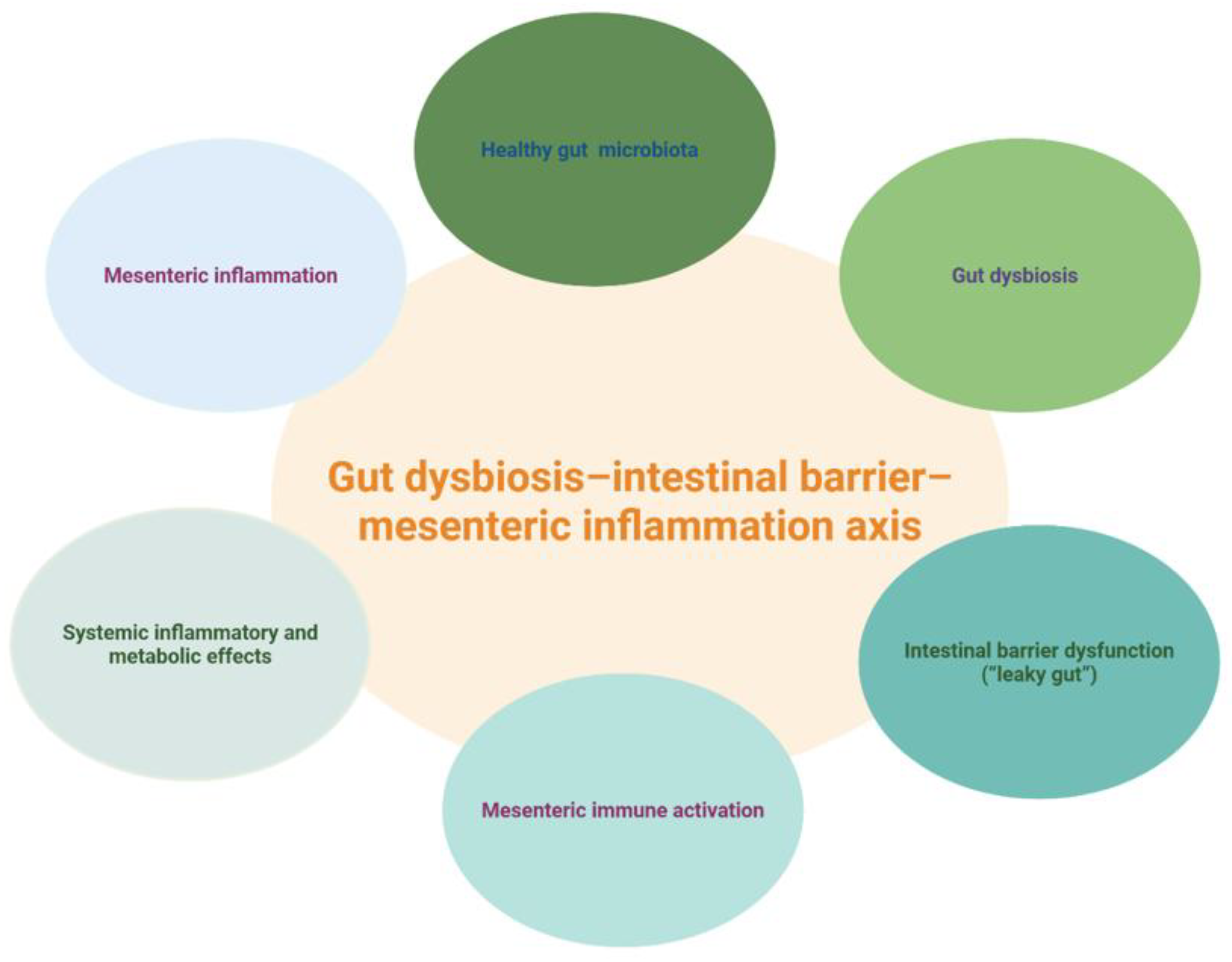
Figure 2.
Proposed LPS–mesentery inflammatory cascade. The figure illustrates a hypothetical sequence in which gut dysbiosis promotes intestinal barrier dysfunction, lipopolysaccharide translocation, mesenteric immune activation, pro-inflammatory cytokine release, and downstream systemic metabolic consequences. Created in BioRender. Ispas, S. (2026) https://BioRender.com/lsyx87c.
Figure 2.
Proposed LPS–mesentery inflammatory cascade. The figure illustrates a hypothetical sequence in which gut dysbiosis promotes intestinal barrier dysfunction, lipopolysaccharide translocation, mesenteric immune activation, pro-inflammatory cytokine release, and downstream systemic metabolic consequences. Created in BioRender. Ispas, S. (2026) https://BioRender.com/lsyx87c.
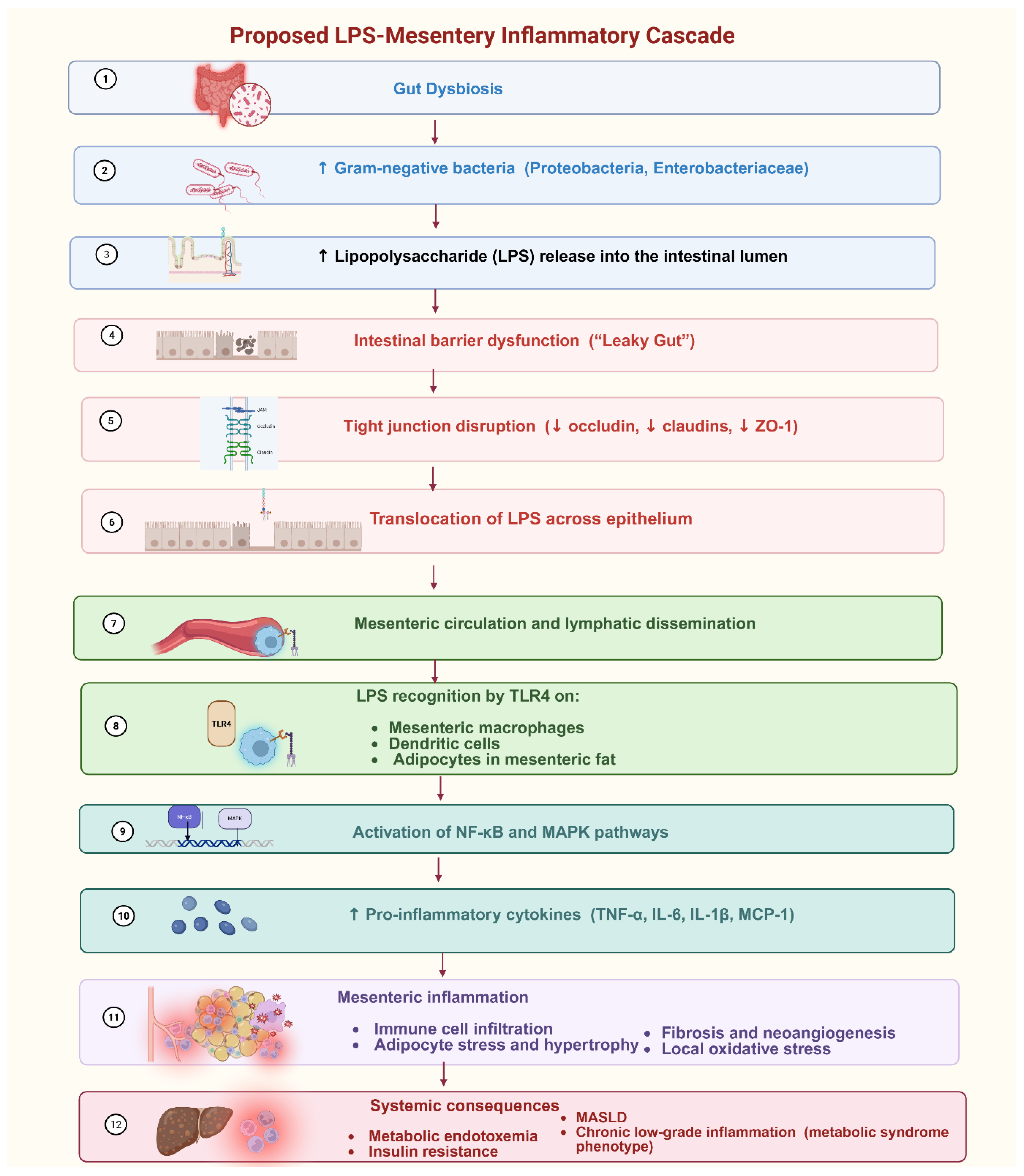
Figure 3.
Glycemic variability and interconnected multisystem inflammatory and metabolic pathways. Created in BioRender. Ispas, S. (2026) https://BioRender.com/ih8vbtf.
Figure 3.
Glycemic variability and interconnected multisystem inflammatory and metabolic pathways. Created in BioRender. Ispas, S. (2026) https://BioRender.com/ih8vbtf.
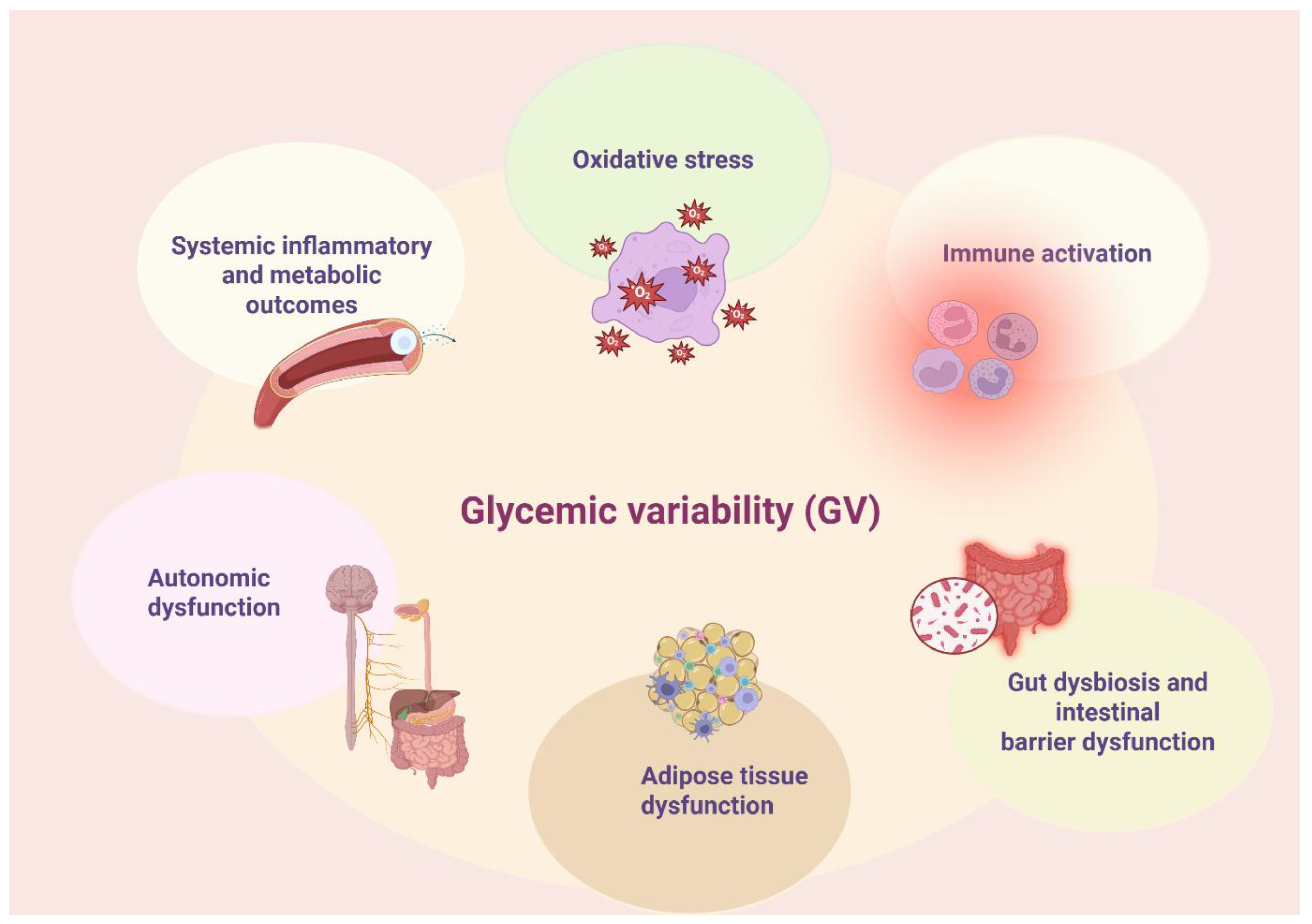
Figure 4.
Glycemic variability as an amplifier of vascular, adipose, and mesenteric inflammatory pathways. Created in BioRender. Ispas, S. (2026) https://BioRender.com/6pkw30h.
Figure 4.
Glycemic variability as an amplifier of vascular, adipose, and mesenteric inflammatory pathways. Created in BioRender. Ispas, S. (2026) https://BioRender.com/6pkw30h.
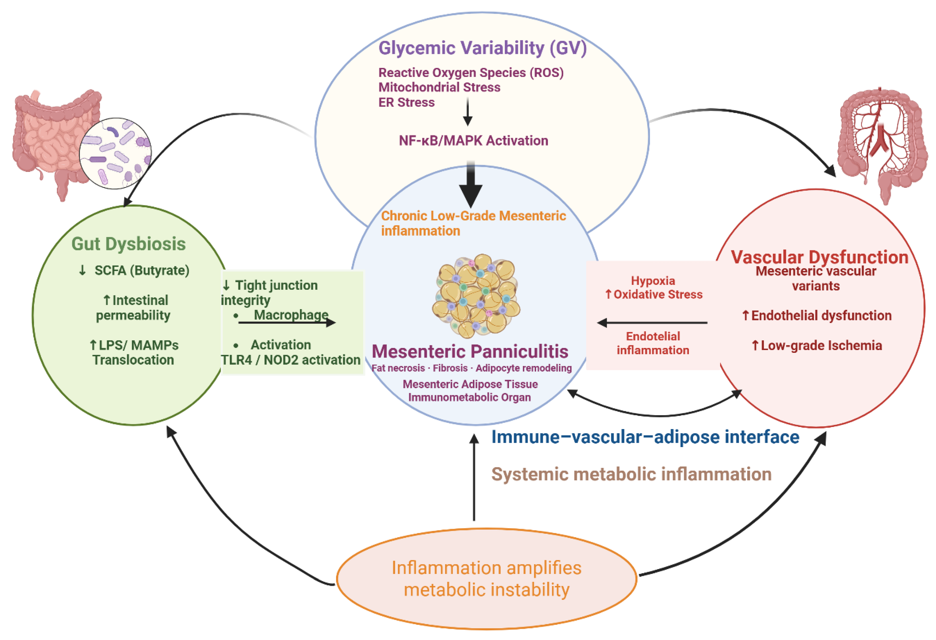
Figure 5.
Proposed integrative gut–vascular–metabolic–mesenteric model of mesenteric panniculitis. Created in BioRender. Ispas, S. (2026) https://BioRender.com/6pkw30h.
Figure 5.
Proposed integrative gut–vascular–metabolic–mesenteric model of mesenteric panniculitis. Created in BioRender. Ispas, S. (2026) https://BioRender.com/6pkw30h.
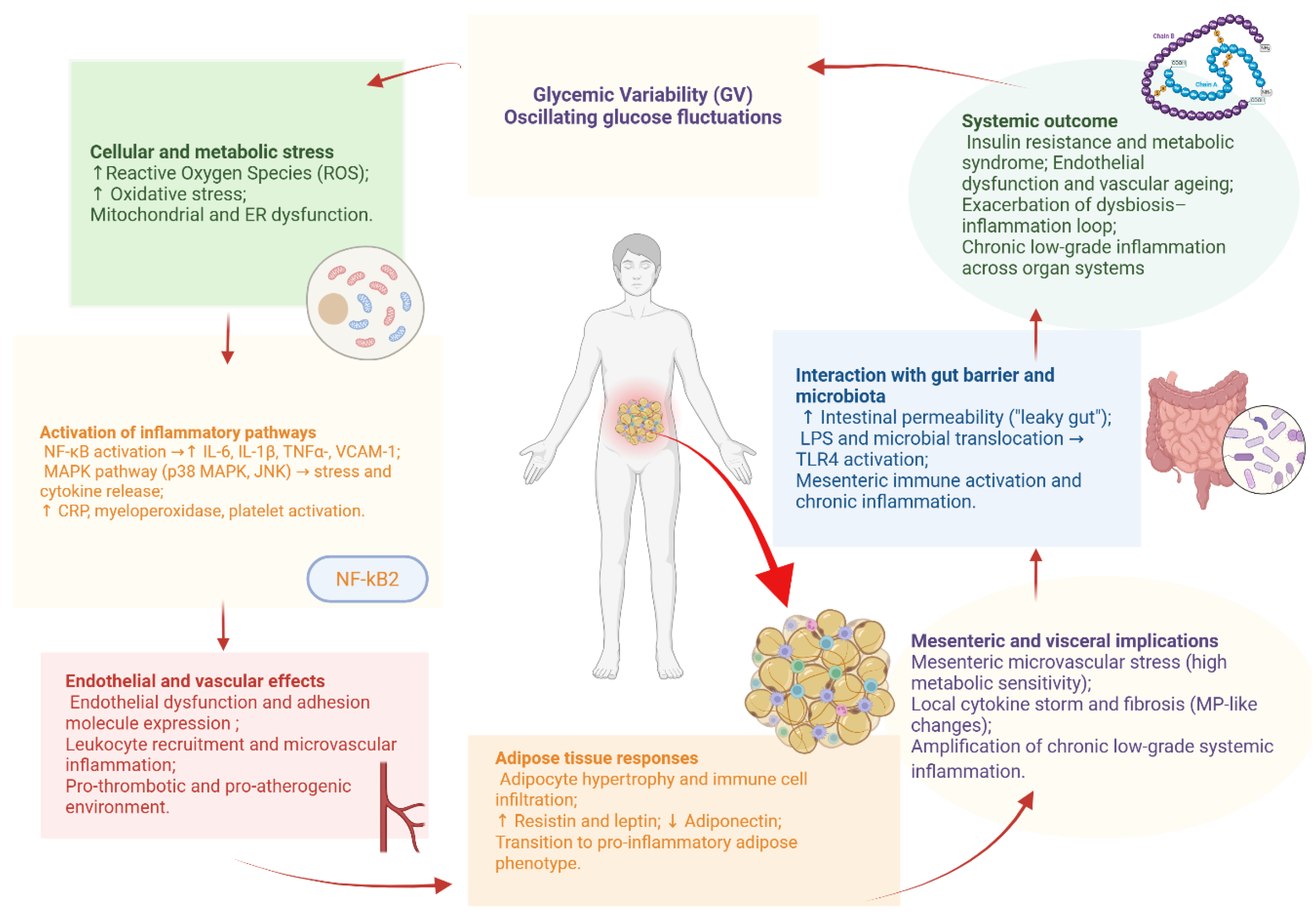
Table 1.
Comparative features of healthy versus dysbiotic gut microbiota and their functional implications.
Table 1.
Comparative features of healthy versus dysbiotic gut microbiota and their functional implications.
| Feature / Function | Healthy Microbiota | Dysbiotic Microbiota | References |
|---|---|---|---|
| Microbial composition | High diversity and stability; dominated by Firmicutes, Bacteroidetes, Actinobacteria, and Proteobacteria | Loss of beneficial taxa; overgrowth of facultative anaerobes and pathobionts (e.g., Enterobacteriaceae, Proteobacteria) | [15,32,33] |
| Metabolic activity | Balanced production of SCFAs (butyrate, acetate, propionate); regulation of bile acids; synthezis of vitamins | Reduced SCFA production (↓ butyrate); altered bile acid metabolism; | [33,34,35] |
| Epithelial barrier function | Reinforces tight junctions, induces mucus layer integrity, maintains selective permeability | Disruption of tight junctions, ↑ intestinal permeability (“leaky gut”) | [34,36,37] |
| Immune modulation | Promotes immune tolerance and an anti-inflammatory immune profile | ↑ Pro-inflammatory signaling; ↑ IL-6, TNF-α, IL-1β; ↓ immunoregulation; | [3,15,38] |
| Systemic metabolic effects | Supports insulin sensitivity, lipid homeostasis, and glucose regulation | Associated with insulin resistance, metabolic syndrome, MAFLD/MASLD, and type 2 diabetes. | [33,34] |
| Interactions with intestinal and adjacent adipose tissues | Maintains local immune homeostasis; no aberrant inflammatory signaling | May promote inflammatory signaling through intestinal barrier disruption, microbial translocation, and interactions between the intestine and adjacent adipose tissues | [37,39,40,41] |
Table 2.
Recent randomized controlled trials of microbiome-targeted interventions in metabolic disorders.
Table 2.
Recent randomized controlled trials of microbiome-targeted interventions in metabolic disorders.
| Study / Population | Intervention | Duration | Main findings | Reference |
|---|---|---|---|---|
| Adults with obesity | Synbiotic: Bifidobacterium animalis subsp. lactis MN-Gup + GOS + XOS vs placebo | 12 weeks | Reduced body fat percentage, waist circumference, and LDL-C; associated changes in gut microbiota, bile acids, and gut hormones | [56] |
| Adults with type 2 diabetes | Synbiotic vs probiotic alone vs placebo | 12 weeks | Synbiotic reduced fasting glucose and modified inflammatory markers, oxidative stress, gut microbiota, bile acids, and GLP-1; not all glycemic outcomes differed significantly between groups | [52] |
| Adults with type 2 diabetes | High-dose probiotic vs placebo | 12 weeks | No significant between-group improvement in HbA1c, glucose, insulin, LDL-C, triglycerides, or hs-CRP | [57] |
| Adults with prediabetes or type 2 diabetes | Synbiotic EDC-HHA01 vs placebo | 6 months | Greater improvements in HbA1c, fasting insulin, insulin-resistance indices, and endotoxemia markers in prediabetes; effects were more modest in established T2D | [55] |
| Adults with type 2 diabetes | Multi-strain probiotic vs placebo | 12 weeks | Reduced IL-17 and TNF-α; altered gut microbiota, bile acids, SCFAs, and selected metabolic pathways | [58] |
Table 3.
Gastrointestinal pathogens and their systemic inflammatory effects.
| Pathogen | Primary site of colonization/infection | Principal virulence factors | Systemic inflammatory effects and mechanisms | Associated systemic/metabolic disorders | References |
|---|---|---|---|---|---|
| Helicobacter pylori | Gastric mucosa | Urease, CagA, VacA, outer membrane vesicles, LPS | Promotes epithelial injury, immune-cell activation, and the production of pro-inflammatory mediators. Outer membrane vesicles may transport bacterial components and virulence factors to distant host tissues. | Insulin resistance, metabolic syndrome, metabolic dysfunction-associated steatotic liver disease, endothelial dysfunction, and cardiovascular risk | [60,61,64,65,71,72] |
| Pathogenic Escherichia coli | Small and large intestine | LPS, Shiga toxin | LPS promotes innate immune activation and systemic inflammation. Shiga toxin induces oxidative stress and endothelial injury and contributes to the pathogenesis of haemolytic uraemic syndrome. | Metabolic endotoxemia, systemic inflammation, haemolytic uraemic syndrome | [8,46,73] |
| Salmonella enterica | Small intestine and Peyer’s patches | LPS and other bacterial virulence mechanisms | Promotes macrophage activation and systemic inflammatory responses. Salmonella-derived LPS contributes to cytokine production and systemic immune activation. | Systemic inflammatory responses and post-infectious complications | [74] |
| Campylobacter jejuni | Distal small intestine and colon | Cytolethal distending toxin | Cytolethal distending toxin causes epithelial injury and inflammatory cell death. Molecular mimicry and antibody cross-reactivity may contribute to extraintestinal autoimmune complications. | Guillain–Barré syndrome and post-infectious inflammatory complications | [75,76] |
| Clostridioides difficile | Colon | Toxin A and toxin B | Disrupts epithelial and neural components of the intestinal barrier, induces apoptosis, and stimulates local and systemic immune responses. Infection may also promote a pro-inflammatory and pro-steatotic hepatic state. | Recurrent colitis, systemic inflammatory responses, and metabolic liver dysfunction | [77,78,79] |
Table 4.
Comparative characteristics of gut microbiota composition in Helicobacter pylori-positive versus H. pylori-negative subjects.
Table 4.
Comparative characteristics of gut microbiota composition in Helicobacter pylori-positive versus H. pylori-negative subjects.
| Parameter | H. pylori-negative individuals | H. pylori-positive individuals | References |
|---|---|---|---|
| Overall microbial diversity | Relatively stable and diverse microbial ecosystem, with preserved compositional balance | Altered microbial diversity and compositional imbalance have been reported, although findings vary according to population characteristics, infection stage, and associated gastric pathology | [70,81] |
| Beneficial commensal taxa | Greater representation of commensal microorganisms involved in epithelial homeostasis and metabolic balance | Reduced abundance of beneficial commensal taxa has been described in selected cohorts | [70,81] |
| Proteobacteria and inflammation-associated taxa | Lower relative abundance of inflammation-associated Gram-negative taxa under homeostatic conditions | Expansion of Proteobacteria and other potentially pro-inflammatory taxa has been reported in association with H. pylori-related dysbiosis | [66,70] |
| Prevotella and related taxa | Variable abundance according to diet, geography, age, and host-related factors | Enrichment of Prevotella and related taxa has been observed in some populations, although findings remain heterogeneous | [68,70] |
| SCFA-producing taxa and microbial metabolites | Preserved microbial metabolic activity and short-chain fatty acid production | Altered abundance of SCFA-producing taxa and changes in butyrate-related metabolic capacity have been reported | [70,80,81] |
| Intestinal barrier and immune regulation | Preserved epithelial barrier integrity and immune homeostasis | Dysbiosis may contribute to epithelial barrier dysfunction, altered immune regulation, and systemic inflammatory signaling | [66,68,81] |
| Effects of eradication therapy | Not applicable | Eradication therapy may induce transient microbiota disruption, followed by partial recovery; probiotic supplementation may attenuate some of these changes | [87,88,89] |
| Associated metabolic effects | Preserved metabolic homeostasis in the absence of other metabolic disorders | Associations with metabolic syndrome and impaired glucose homeostasis have been reported | [71,80] |
Table 5.
Comparative clinical and imaging characteristics of acute mesenteric ischemia, chronic mesenteric ischemia, and mesenteric panniculitis.
Table 5.
Comparative clinical and imaging characteristics of acute mesenteric ischemia, chronic mesenteric ischemia, and mesenteric panniculitis.
| Imaging/clinical feature | Acute mesenteric ischemia (AMI) | Chronic mesenteric ischemia (CMI) | Mesenteric panniculitis (MP) | References |
|---|---|---|---|---|
| Clinical context | Acute or sudden abdominal pain; potentially life-threatening vascular emergency | Chronic or recurrent postprandial abdominal pain, food avoidance, and weight loss | Frequently incidental; when symptomatic, may present with nonspecific abdominal pain or other gastrointestinal symptoms | [24,101,106] |
| CT angiography / arterial findings | May demonstrate arterial embolus, thrombosis, occlusion, or other vascular abnormalities | May demonstrate significant stenosis or occlusion of the celiac or mesenteric arteries | No characteristic arterial occlusion; mesenteric vessels may traverse or become surrounded by inflamed fat | [24,101,104,106] |
| Collateral circulation | Collaterals may be insufficient in acute arterial occlusion | Well-developed collateral vessels may develop in chronic arterial disease | Collateral vascular abnormalities are not a defining imaging feature | [101,104] |
| Bowel-wall findings | Reduced or absent enhancement, wall thickening or thinning, pneumatosis, and portal venous gas may occur depending on severity and mechanism | Bowel-wall abnormalities may be absent unless ischemia becomes advanced or acute-on-chronic | Bowel wall is generally not the primary site of imaging abnormality | [20,101,104,106,108,109] |
| Mesenteric fat | Fat stranding and mesenteric edema may accompany bowel ischemia | No specific mesenteric-fat pattern defines CMI | Increased attenuation (“misty mesentery”), soft-tissue nodules, fat-ring sign, and pseudocapsule are characteristic findings | [104,105,106,110] |
| Mesenteric lymph nodes | Reactive lymphadenopathy is not a defining feature | Not a characteristic diagnostic feature | Multiple small mesenteric lymph nodes may be present within the affected mesentery | [24,104] |
| Duplex ultrasound | Limited role in the emergency assessment of AMI | Elevated flow velocities in the celiac artery or SMA may support hemodynamically significant stenosis | No specific Doppler criterion for MP |
[101] |
| Topographic distribution | Distribution reflects the affected arterial or venous territory | Symptoms and ischemia correspond to compromised mesenteric vascular territories | Usually centered at the root of the small-bowel mesentery | [101,105,106,110] |
| PET/CT | Not routinely used for diagnosis | Not routinely used for diagnosis | Low or absent FDG uptake may favor a benign process; inflammatory uptake may occur | [24,104] |
| Histopathological correlation | Ischemic bowel injury may progress to hemorrhage, necrosis, and infarction | Chronic ischemia is primarily a vascular clinical syndrome; tissue abnormalities depend on severity and duration | Fat necrosis, chronic inflammation, and fibrosis are characteristic pathological components | [23,106] |
| Clinical course | Requires urgent diagnosis and treatment because progression to bowel infarction and death may occur | Chronic progressive disease requiring evaluation and revascularization when clinically significant | Often stable or self-limiting; treatment is generally reserved for symptomatic or complicated disease | [23,24,101,106] |
Disclaimer/Publisher’s Note: The statements, opinions and data contained in all publications are solely those of the individual author(s) and contributor(s) and not of MDPI and/or the editor(s). MDPI and/or the editor(s) disclaim responsibility for any injury to people or property resulting from any ideas, methods, instructions or products referred to in the content. |
© 2026 by the authors. Licensee MDPI, Basel, Switzerland. This article is an open access article distributed under the terms and conditions of the Creative Commons Attribution (CC BY) license.
Copyright: This open access article is published under a Creative Commons CC BY 4.0 license, which permit the free download, distribution, and reuse, provided that the author and preprint are cited in any reuse.