

Submitted:
17 August 2026
Posted:
19 August 2026
You are already at the latest version
Abstract
Three novel compounds with potential antitubercular activity—N-(2-isonicotinoylhydrazine-1-carbonothioyl)benzamide (3), N-(2-nicotinoylhydrazine-1-carbonothioyl)benzamide (4), and N-(2-picolinoylhydrazine-1-carbonothioyl)benzamide (5) were synthesized in good yields via the reaction of benzoyl isothiocyanate with isonicotinic hydrazide, nicotinic hydrazide, and picolinic hydrazide in acetonitrile, respectively. The crystal structures of compounds (3) and (5) were determined by single-crystal X-ray diffraction analysis; both structures exhibit intramolecular C=O···H-N hydrogen bonds, as well as C=S···H-N interactions. Computational pharmacological studies, ADME profiling, and molecular docking were performed for the three synthesized compounds, indicating that they represent promising candidates as alternatives to current tuberculosis treatment regimens, owing to their predicted favorable pharmacokinetic profile and binding affinity for the Mycobacterium tuberculosis InhA enzyme.
Keywords:
aroyl thioureas
; N-(2-isonicotinoylhydrazine-1-carbonothioyl)benzamide
; N-(2-nicotinoylhydrazine-1-carbonothioyl)benzamide
; N-(2-picolinoylhydrazine-1-carbonothionyl)-benzamide
; crystal structure
; molecular docking
; Mycobacterium tuberculosis
1. Introduction
Isoniazid is one of the primary drugs used in current tuberculosis treatment regimens according to WHO recommendations [1]. On the other hand, nicotinic hydrazide, also known as niazid, exhibits anti-inflammatory, analgesic, and anticonvulsant properties [2] with several of its derivatives showing antitubercular activity [3]. Similarly, picolinohydrazide has demonstrated tuberculostatic activity, whereas its derivatives display antibacterial, antifungal, and antioxidant properties [4].
Benzoylthioureas have drawn significant attention in recent years due to their utility as ligands in transition-metal complexation, as starting reagents in the synthesis of heterocyclic compounds, and for exhibiting a broad spectrum of biological activities, particularly antiviral, antimicrobial, and tuberculostatic effects [5,6].
Structural modification of benzoylthioureas aimed at improving their biological activity as antibacterial, anti-inflammatory, and tuberculostatic agents has enabled the synthesis of novel derivatives incorporating both the benzoylthiourea moiety and isoniazid or its analogues niazid and picolinohydrazide, respectively within a single framework. In this work, we report on the general synthesis and characterization of three novel O, N y S donor ligands, along with the crystal structures of the isoniazid- and niazid-derived compounds. Furthermore, while in vitro antitubercular evaluation has not yet been performed, the pharmacokinetic properties of these three novel ligands were validated through ADME and molecular docking studies (analyzing bioavailability and binding affinity for the InhA enzyme), which were conducted using computational tools such as SwissADME, AutoDock Vina, and GROMACS.
2. Results and Discussion
The novel compound N-(2-picolinoylhydrazine-1-carbonothioyl)benzamide (5) was synthesized and characterized according to the literature procedure reported [7] for the synthesis of the ligands N-(2-isonicotinoylhydrazine-1-carbonothioyl)benzamide (3) and N-(2-nicotinoylhydrazine-1-carbonothioyl)benzamide (4). Additionally, the single-crystal X-ray structural analysis of compounds 3 and 5, along with molecular docking studies against Mycobacterium tuberculosis InhA for ligands 3, 4, and 5, are reported.
2.1. Chemistry
Scheme 1 depicts the general synthetic pathway for ligands (3), (4), and (5). The process begins with the preparation of benzoyl isothiocyanate (2) via the reaction of benzoyl chloride (1) with potassium thiocyanate in acetonitrile under reflux for 2 h.
The resulting mixture, containing benzoyl isothiocyanate (2) and the precipitated potassium chloride, is subsequently reacted in separate vessels with isoniazid or its isomers, nicotinic acid hydrazide and picolinic acid hydrazide, under constant stirring for 24 h.
The products were obtained as white solid crystals, soluble in hot DMSO and DMF, and insoluble in water across all compounds. The resulting ligands (3), (4), and (5) were isolated in high yields of 98%, 83%, and 72%, respectively. The pure compounds were characterized by physicochemical analysis (melting point, solubility), elemental analysis, mass spectrometry, Fourier-transform infrared spectroscopy (FTIR), nuclear magnetic resonance (NMR) 1H-NMR, 13C-NMR, and 2D-NMR techniques (for (3) and (4)).
2.1.1. Characterization of Ligands (3), (4), and (5)
Angulo & Tovar [7] reported the synthesis and characterization of compounds (3) and (4) using infrared (IR) spectroscopy, mass spectrometry, 1H and 13C-NMR, and elemental analysis. Structural information regarding ligands (3) and (4) has been adequately detailed in the literature [7]; however, to provide structural references for all the ligands, the 1H-NMR and 13C-NMR spectral data for ligand (4) have been used. Ligands (3) and (5) have been studied via Single Crystal X-ray structural analysis.
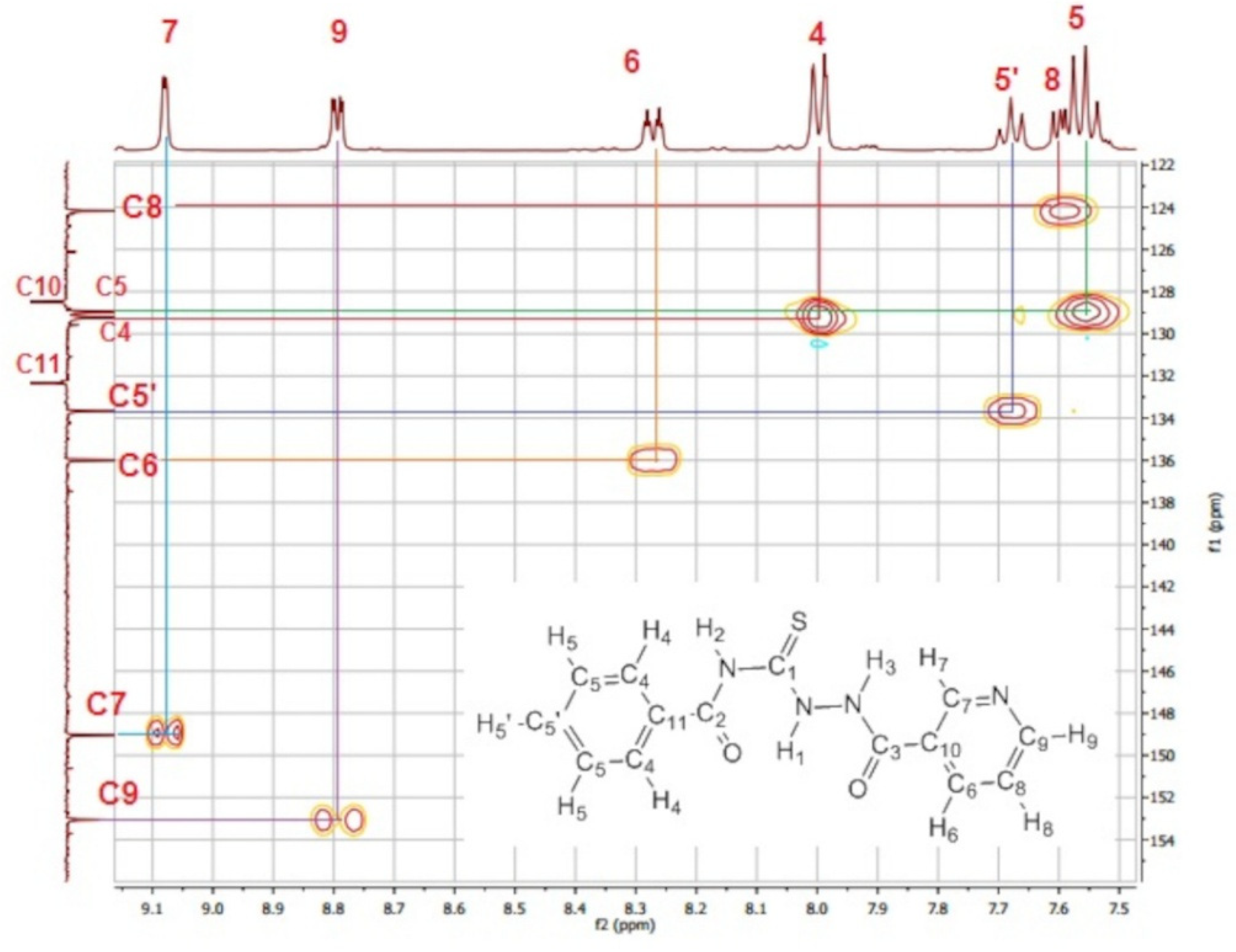
In the 1H-NMR spectrum of ligand (4), three signals corresponding to the N-H1, N-H2, and N-H3 protons were observed at 12.34, 11.78, and 11.34 ppm, respectively. The downfield signal at 12.34 ppm was assigned to the formation of an intramolecular hydrogen bond between the hydrazide N-H1 group and the carbonyl oxygen (N-H···O=C), a characteristic feature of aroyl thiourea derivatives [6]. The other two signals at 11.78 and 11.34 ppm were associated with the N-H2 groups of the hydrazide and the thiourea (N-H3), respectively. The doublet signal at 7.98 ppm and the triplet signals at 7.55 and 7.64 ppm were assigned to the phenyl group. Finally, regarding the signals associated with the pyridine ring, a singlet signal at 9.08 ppm, two doublet signals at 8.79 and 8.27 ppm, and a triplet signal at 7.59 ppm were observed [7].
The 13C-NMR spectrum in DMSO-d6 shows signals at 181.3, 168.2, and 163.7 ppm, corresponding to the thiocarbonyl group (C=S), the carbonyl group (C=O) adjacent to the phenyl group, and the carbonyl group adjacent to the pyridine ring, respectively. The C-H shifts associated with the phenyl group appear in the 133.11–128.5 ppm range, while the C-H shifts related to the pyridine ring were observed between 124 and 154 ppm.
Figure 1.
HSQC-NMR carbon-hydrogen couplings of ligand (4). Elemental analysis of ligand (5) is within the expected range for a pure compound, which is consistent with its melting point.
Figure 1.
HSQC-NMR carbon-hydrogen couplings of ligand (4). Elemental analysis of ligand (5) is within the expected range for a pure compound, which is consistent with its melting point.
In the FT-IR spectrum of compound (5), two signals corresponding to the carbonyl groups (C=O) within the structure were observed: C=O (thiourea) at 1653 cm−1 and C=O (hydrazide) at 1668 cm−1. The stretching frequency of the thiourea C=O group is lower than that of an ordinary carbonyl group; this can be attributed to the proximity of the phenyl ring as well as the formation of an intramolecular hydrogen bond with one of the N-H groups. N-H stretching bands were observed in the 3282–3104 cm−1 region, the C-H (aromatic) stretching signal appeared at 3064 cm−1, and the band associated with the thiocarbonyl group (C=S) was found at 886 cm−1.
2.1.2. X-Ray Structural Analysis of Ligands (3) and (5).
Ligands (3) and (5) were studied by single-crystal X-ray diffraction structural analysis; the crystallographic data are shown below:
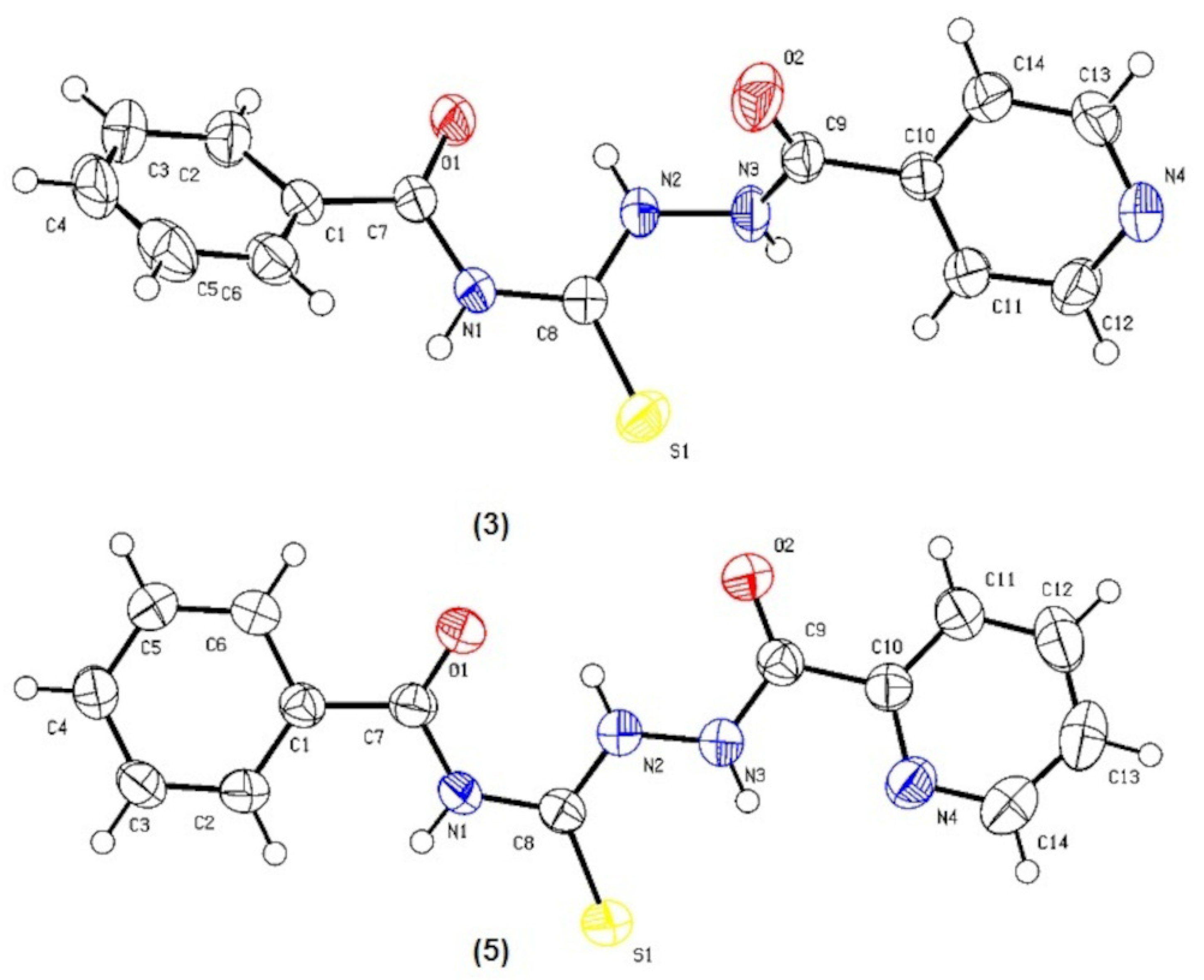
The C=O and C=S bonds are oriented in a trans configuration within a plane stabilized by an intramolecular N2-H···O1 hydrogen bond in both ligands (Figure 2). In compound (5), the plane of the N2-H···O1 hydrogen bond forms dihedral angles of 18.17° and 27.30° with the central phenyl and pyridine rings, respectively, while for compound (3) these angles are 36.93° and 40.36° respectively.
The unit cell contains four asymmetric units for the synthesized compounds (3) and (5). Both molecular crystal cells are centrosymmetric. The crystal packing of ligands (3) and (5) is based on supramolecular hydrogen-bonding interactions. π-π stacking interactions involve the benzene and pyridine rings of different molecules.
Figure 2.
View of the asymmetric unit for ligands (3) and (5) with the atom numbering scheme (50% probability ellipsoid).
Figure 2.
View of the asymmetric unit for ligands (3) and (5) with the atom numbering scheme (50% probability ellipsoid).
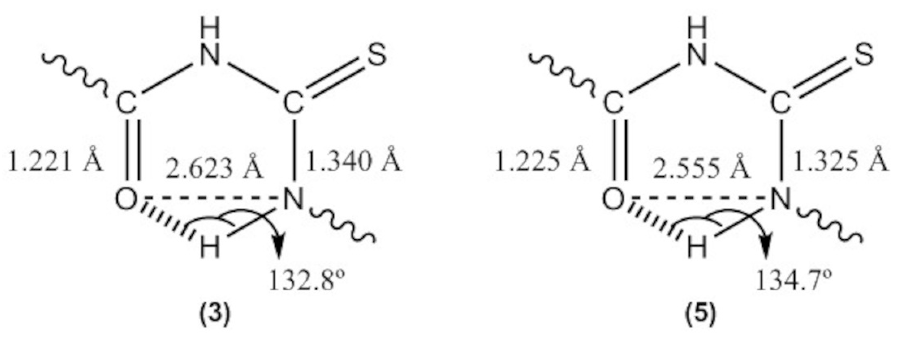
Ligands (3) and (5) exhibit intramolecular hydrogen bonds between the N2-H group and the O1 oxygen (of the C7-O1 carbonyl group). The N2-O1 distances are 2.623 Å and 2.555 Å, while the N2-H-O1 angles are 132.81° and 134.69°, respectively. Electronic delocalization is observed in the central part of the molecules, evidenced by the presence of a short C7-O1 bond—1.221 Å for compound (3) and 1.225 Å for ligand (5) alongside longer C8-N2 bond distances of 1.340 Å and 1.325 Å, respectively; this indicates the π-character of the C7-O1 bond belonging to the carbonyl group attached to the benzene ring in both compounds.
Figure 3.
Hydrogen bonds and C-O and C-N bonds in a fraction of ligands (3) and (5).
2.2. ADME Studies
Pharmacokinetic properties were analyzed using the SwissADME server [8]. The following parameters are essential to consider during the early stages of drug development: molecular weight (MW), volume, number of rotatable bonds (RB), number of hydrogen bond acceptors (HBA) and donors (HBD), topological polar surface area (TPSA), lipophilicity, and water solubility of the compounds.
For the ligands studied (3), (4), and (5), the molecular weight is 300 g/mol, which falls within the optimal range for drug-likeness (150 ≤ MW g/mol ≤ 500).
The RB parameter indicates structural flexibility; this factor is important in drug development, as highly flexible molecules tend to exhibit low selectivity. [9]. The RB value for all synthesized ligands was 7. The numbers of HBA and HBD were 3 for all compounds (HBA ≤ 10; HBD ≤ 5).
The TPSA parameter allows for the estimation of a compound’s overall polarity. This polarity affects the drug’s solubility and availability. High polarity values can lead to high efflux rates. The TPSA value for all compounds was 115.21 (20 < TPSA < 130 Å2 ).
The partition coefficient (logP) is used to evaluate the lipophilicity of a compound. It is typically calculated as the logarithm of the ratio of concentrations measured in a system comprising an aqueous phase and an n-octanol phase. Table A1 (supplementary material) shows the ilogP, XlogP3, WlogP, MlogP, SILICOS-IT, and consensus logPo/w values for ligands (3), (4), and (5).
All synthesized ligands comply with Lipinski’s Rule of 5 [10], which indicates that an oral drug must have an MlogP value < 4.15.
ESOL (Estimated SOLubility in water) values were recorded; for most drug research purposes, an ideal ESOL (logS) value typically falls between 0 and -4. The synthesized ligands exhibit ESOL values ranging from -2.96 to -2.83, which fall within the common range for many commercial drugs [11].
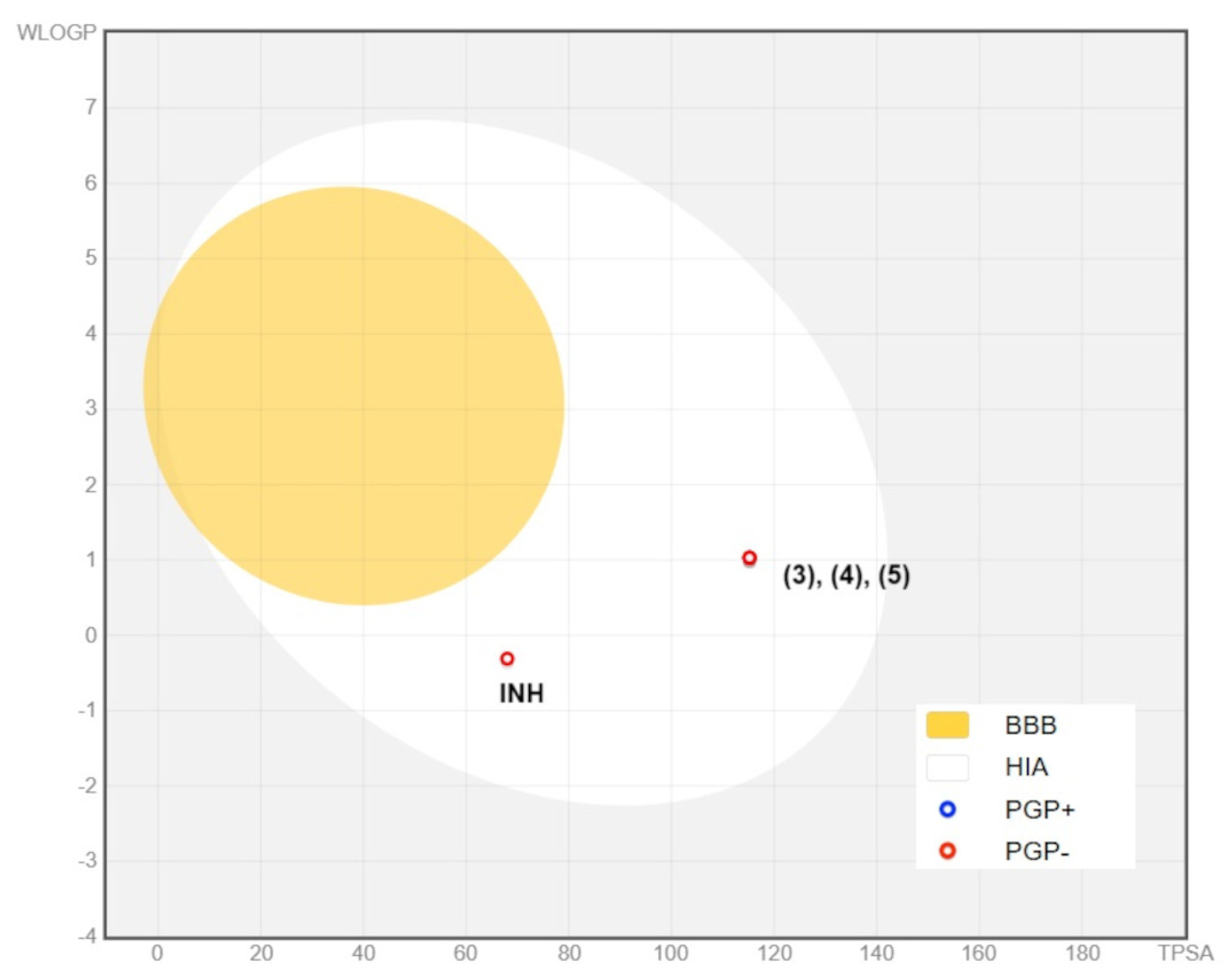
The BOILED-Egg (Brain Or Intestinal EstimateD) model was used to predict human gastrointestinal absorption (HIA) and blood-brain barrier (BBB) penetration; molecules located within the egg exhibit adequate oral bioavailability. The yellow zone represents the area of high probability of blood-brain barrier (BBB) penetration, while the white (white zone) represents the high probability of human intestinal absorption (HIA). [12].
An analysis of the WLOG vs. TPSA plot (Figure 4) shows that compounds (3), (4), and (5) fall within the white region of the egg; therefore, the model predicts that these compounds will most likely be absorbed in the gastrointestinal tract, just like isoniazid (INH).
All the synthesized ligands aligned with other drug-likeness models, such as the Ghose, Veber, Egan, and Muegge indices [13,14,15,16] unlike isoniazid, which did not meet the Ghose and Muegge criteria. ADME results indicated that all synthesized compounds, including isoniazid, exhibit a bioavailability of 55%.
Figure 4.
BOILED-Egg plot based on WLOG and TPSA for the synthesized compounds and isoniazid. The yellow area represents brain penetration, and the white area represents gastrointestinal tract absorption.
Figure 4.
BOILED-Egg plot based on WLOG and TPSA for the synthesized compounds and isoniazid. The yellow area represents brain penetration, and the white area represents gastrointestinal tract absorption.
2.3. Molecular Dynamics and Docking Study
The affinity of the ligands N-(2-nicotinoylhydrazine-1-carbonothioyl)benzamide (3), N-(2-isonicotinoylhydrazine-1-carbonothioyl)benzamide (4), and N-(2-picolinoylhydrazine-1-carbonothioyl)benzamide (5) for Mycobacterium tuberculosis enoyl-acyl carrier protein reductase (InhA) was predicted using molecular docking analysis. Subsequently, molecular dynamics simulations were performed using the best-scoring poses with GROMACS v2022.2 program [17] and the AMBER99SB-ILDN force field. [18].
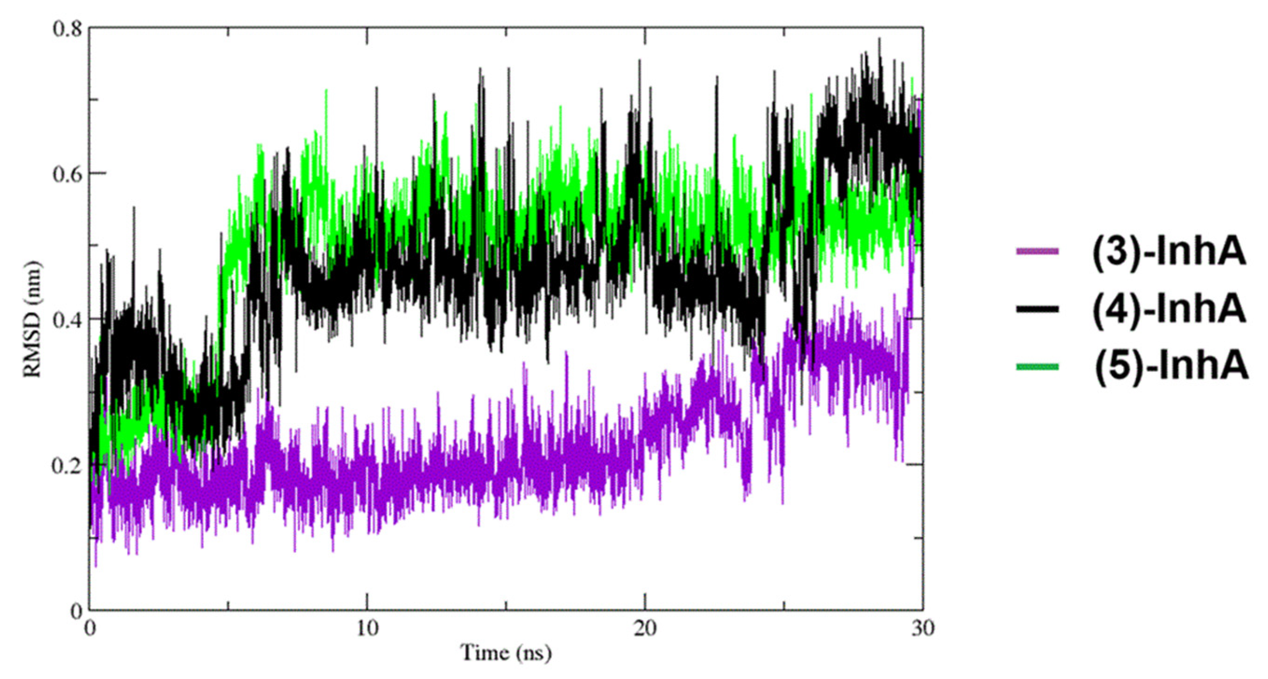
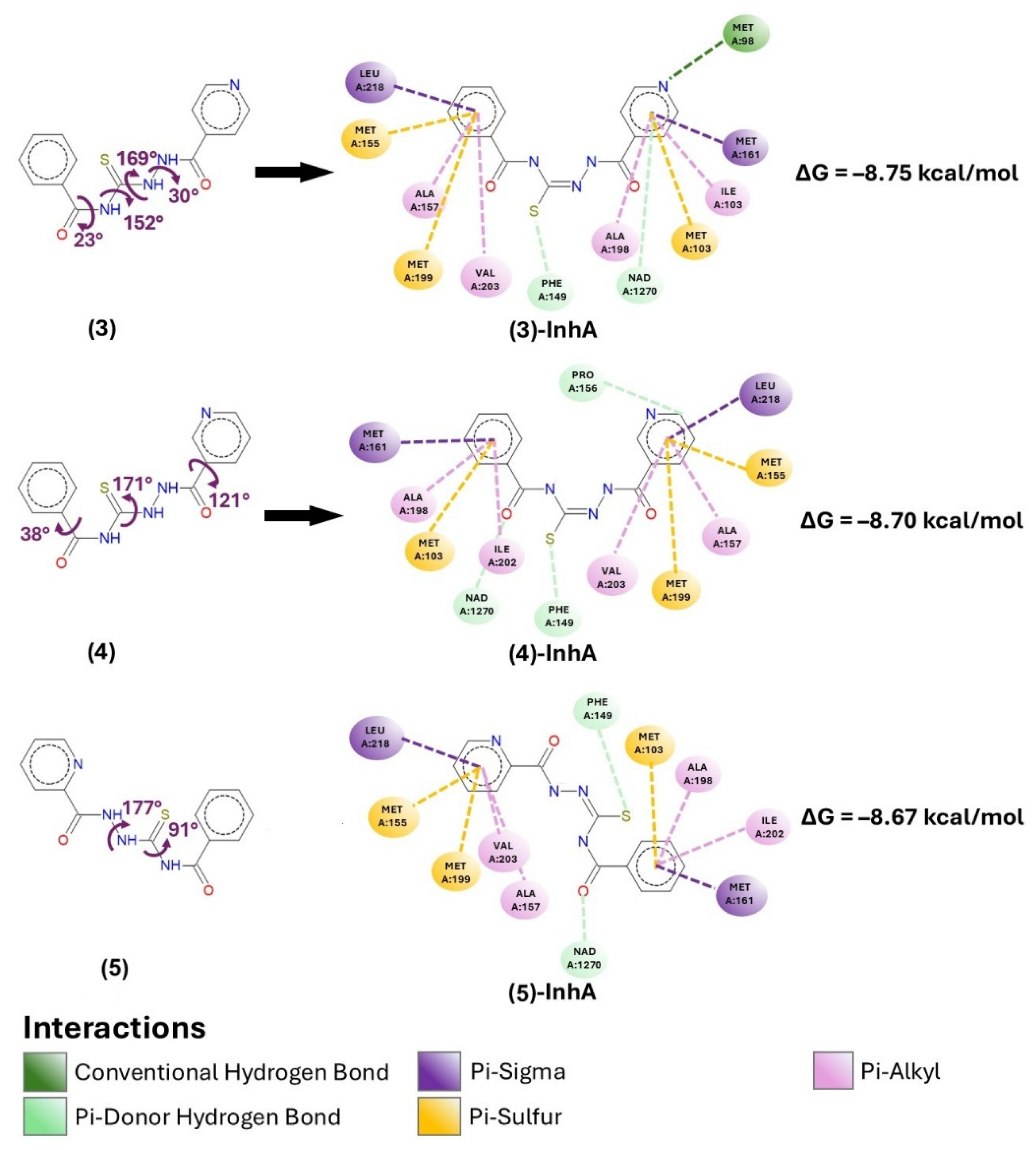
According to these results, at standard temperature and with a simulation time of 30 ns, it was observed that the RMSD of the (3)-InhA complex remained more stable, with values below 0.4 nm. In contrast, the values obtained for the (4)-InhA and (5)-InhA complexes reached up to 0.7 nm (Figure 5). This confirms that the (3)-InhA complex exhibited better results in terms of conformational stability. Based on the docking analyses, the binding poses of the InhA enzyme with the three ligands studied yielded similar reaction free energies. However, more favorable values of ΔG°(–8.75 kcal/mol), Kd(3.83×10⁻⁷ M) and a more negative LE (–0.4167 kcal/mol) are observed for the (3)-InhA complex (Table 1). This is likely because the position of the nitrogen atom within the –C5H5N ring of ligand (3) promotes stronger hydrogen bonding with a methionine residue (MET: A98) in the (3)-InhA complex. No close hydrogen bonds of this type are observed in the other two complexes. This also appears to be influencing the decrease in π-sulfur interactions between the –C5H5N ring of the (3)-InhA complex and the methionine molecules: The C5H5N ring of the complex (3)-InhA exhibits only one π-sulfur interaction with A103. Whereas π-sulfur interactions with A155 and 199 are observed in the (4)-InhA and (5)-InhA complexes at each –C5H5N ring (Figure 5).
Figure 5.
The root mean square deviation (RMSD) value of each ligand-InhA complex during 30 ns.
It is worth emphasizing that, for all three ligands, it was verified that the presence of π-conjugation around the thiouuric sulfur promotes the formation of a π-donor hydrogen bond between the sulfur and phenylalanine (PHE: A149). This may account for the greater spontaneity observed in the interaction of the three ligands with the InhA enzyme (~ –8.7 kcal/mol) compared to the thermodynamic results obtained from the molecular docking of isoniazid with InhA (~ –4.6 kcal/mol) [19]. However, even though the interaction between the InhA gene and the studied ligands exhibits a thermodynamically more favorable trend compared to isoniazid, it is important to note that since all four ligands show similar efficiency values, the expected biological effect would also be comparable. Nevertheless, ligands 3, 4, and 5 could offer an interesting alternative to address the limitations that might arise from potential mutations in the InhA gene that could decrease the efficacy of isoniazid.
Molecular docking and molecular dynamics studies of the three ligands reveal very favorable Gibbs free energies of binding (ΔG°) with the Mycobacterium tuberculosis InhA enzyme (≈ –8.7 kcal/mol) compared to isoniazid (ΔG° ≈ –4.6 kcal/mol). This suggests a higher affinity of the ligands for InhA (the molecular target), which, in practice, would represent an interesting alternative in antitubercular treatments against potential mutations in the InhA gene that could decrease the efficacy of conventional antibiotics such as isoniazid. On the other hand, the sulfur atom in the thiourea group participates in π-donor hydrogen bonding and π-sulfur interactions with aromatic protein residues such as phenylalanine and methionine, increasing the affinity of these ligands for the Mycobacterium tuberculosis InhA enzyme. When analyzing isoniazid, this specific sulfur interaction capability is absent from its structure, giving the synthesized thioureas a greater structural advantage.
Various studies [20,21,22] have demonstrated that ligands structurally similar to (3), (4), and (5) feature intramolecular N-H···O hydrogen bonds. These interactions impart conformational stability to the planar trans arrangement of the C=O and C=S bonds, favorably directing the sulfur atom toward the catalytic site of enoyl-acyl carrier protein reductase de Mycobacterium tuberculosis (InhA).
The intramolecular N–H···O hydrogen bond favors the trans configuration of the C=O and C=S bonds, positioning the sulfur atom directly toward the enzyme and thereby facilitating its approach and interaction with the InhA target.Subsequently, the initial interaction of the π-donor sulfur with the PHEA:149 residue is facilitated by weak intermolecular hydrogen bonds (S···H involving the protein residue) and driven by the molecular recognition between the substrate (π-donor sulfur) and the receptor center (PHEA:149). This induces a pre-organization that stabilizes the interaction through a conformational change, which not only consolidates the formation of an intermolecular bond between the sulfur atom and the PHEA:149 residue but also strengthens the interactions of other ligand moieties such as the aromatic group (ligand phenyl ring) and the pyridine group (para-substituted Py) with the remaining receptor centers of In-hA (Figure 6). For this configuration change, it is observed that the proton involved in the intramolecular bond migrates alongside the sulfur atom to facilitate the conformational inversion.
Figure 6.
Main interactions between ligands (3), (4), (5) and Enoyl-Acyl Carrier Protein Reductase.
3. Materials and Methods
3.1. Chemistry
The reagents used in the synthesis were: benzoyl chloride, Merck (Germany), for synthesis; potassium thiocyanate, Merck (Germany), for synthesis; isoniazid, Sigma-Aldrich (USA), analytical grade, ≥ 99% (TLC); nicotinic hydrazide, Sigma-Aldrich (USA), analytical grade, ≥ 97% (TLC); picolinohydrazide, Sigma-Aldrich (USA), analytical grade, ≥ 98% (TLC). The solvents—acetonitrile (J.T. Baker® ACS), ethanol (J.T. Baker® ACS, USA), and dichloromethane (J.T. Baker® ACS)—were dried prior to use.
3.2. Intrumentation
Infrared spectra were acquired using a Thermo Scientific™ Nicolet™ iS™ 10 FTIR spectrometer (USA). The potassium bromide (KBr) pellet was prepared by mixing 1 mg of the compound with 10 mg of KBr at room temperature (20 °C), and data collection was performed over the wavenumber range of 4000 to 400 cm-1.
The Nuclear Magnetic Resonance (NMR) spectra (1H-RMN, 13C, APT, DEPT, COSY, HSQC and HMBC) of the ligands were recorded on an Agilent Varian Mercury 400 plus (USA) operating at 400 MHz for 1H and 100 MHz for 13C using DMSO-d6 as the solvent at ambient temperature (20 °C). The studied compounds were recrystallized from ethanol and dried at room temperature. These purified samples were sent to Leipzig University to acquire their one-dimensional (13C, 1H) and two-dimensional NMR spectra. Additionally, the purified samples were subjected to elemental analysis using a PerkinElmer® Model 2400 Series II CHNS/O elemental analyzer (USA) at Leipzig University.
Single-crystal X-ray diffraction data for ligands (3) and (5), which crystallized as block-shaped crystals with approximate dimensions of 0.205 × 0.122 × 0.102 mm y 0.333 × 0.139 × 0.027 mm respectively, were collected on a Bruker D8 VENTURE system (PHOTONIII 14 con IμS Diamond) at UNMSM. The crystals were carefully selected and mounted on a nylon loop using Parabar oil.
The system was operated using the APEX6 software package [23] with Mo Kα radiation (λ = 0.71073 Å). Structure refinement was performed using the SHELXL program [24] implemented within APEX6. Crystallographic data for the reported structures were deposited at the Cambridge Crystallographic Data Centre (CCDC) under deposition numbers 2550980 for ligand (3) and 2550977 for ligand (5)..
3.3. Computational Methods
3.3.1. Prediction of ADME Properties
The free web tool SwissADME (https://www.swissadme.ch/) was utilized to calculate physicochemical properties, pharmacokinetics, drug-likeness, and medicinal chemistry friendliness parameters upon input of the molecular structure [8].
3.3.2. Molecular Docking
To predict the binding affinity of ligands 3 (N-(2-nicotinoylhydrazine-1-carbonothioyl)benzamide), 4 (N-(2-isonicotinoylhydrazine-1-carbonothioyl)benzamide), and 5 (N-(2-picolinoylhydrazine-1-carbonothioyl)benzamide) toward Mycobacterium tuberculosis enoyl-acyl carrier protein reductase (InhA), molecular docking studies were performed using AutoDock Vina [25]. Additionally, natural and conformational stability analyses of the ligand-enzyme complexes were performed using the GROMACS v2022.2 software package [17] along with the AMBER99SB-ILDN force field [18]. The solvation of the ligand/enzyme systems was carried out using the explicit TIP3P solvent model [26], and the parameterization of the ligands was performed using the Antechamber module in AmberTools18 [27,28]. The system was equilibrated in two consecutive steps using NVT and NPT ensembles at 300K for periods of 1000ps each. The production simulation was run for a total time of 30 ns employing the V-rescale [29] and Parrinello-Rahma [30,31]. Finally, the binding free energies were calculated via the MM-PBSA method using the gmx_MMPBSA software [32].
Ligand efficiency calculations were performed using the ligand–protein dissociation constant Kd, which reflects the binding affinity between the ligand and the target protein [33,34]:
where ΔG° is the binding energy (kcal/mol) obtained from molecular docking analyses, R is the universal gas constant, and T s the temperature in Kelvin in aqueous solution at a standard temperature of T=298.15 K, neutral pH, and a standard-state concentration of C°=1M. Ligand efficiency allowed the comparison of molecules based on their average binding energies [14] and was determined by the ratio of binding energy per non-hydrogen atom according to equation (2) (see Table 1) [35,36]
where HAC represents the number of non-hydrogen atoms of a ligand.
3.4. Synthesis of Ligands (3), (4), and (5)
3.4.1. Synthesis of N-(2-isonicotinoylhydrazine-1-carbonothioyl)benzamide (3)
The synthesis of ligand (3) was carried out according to the literature [7]. The resulting compound was recrystallized from absolute ethanol to afford white cubic crystals in 98% yield. Melting point: 207–208 °C; molecular mass: 300.34 g/mol. Suitable crystals were selected for structural studies via X-ray diffraction, and the obtained results were compared with those previously reported in the literature [7].
3.4.2. Synthesis of N-(2-nicotinoylhydrazine-1-carbonothioyl)benzamide (4)
The ligand was synthesized following a previously reported procedure [7]. The resulting white crystalline product was filtered, washed with ethanol, and recrystallized from ethanol to obtain the compound in an 83% yield. Melting point: 200-202 °C; Molecular weight: 300.34 g/mol; Soluble in DMSO.
The 1H-NMR and 13C -NMR spectra of ligand 4 exhibit characteristic signals that are in full agreement with the literature data [7].
1H-NMR (400 MHz, DMSO-d6): δ (ppm): 12,34 (s, 1H, 1N-H); 11,78 (s, 1H, 2N-H); 11,34 (s, 1H, 3N-H); 7,64 (J = 4Hz, d, 2H, Ar-H); 7,55 (J = 6Hz t, 3H, Ar-H); 7,98(J = 4Hz, d, 2H, Ar-H); 9,08 (s, 1H, Py-H); 8,79 (J = 4Hz, d, 1H, Py-H); 8,27 (J = 4Hz, d, 1H, Py-H); 7,59 (J = 8Hz, t, 1H, Py-H). 13C{1H}-APT-NMR (100 MHZ, DMSO-d6) δ (ppm): 181,3 (C=S); 168,1 (C=O benzoyl); 163,7 (C=Ohydrazide); 132,2 (ipso-PhC=O): 128,4 (ipso-PyC=O); 153,0 (o-ipso-N-PyC=O); 149,0 (o-ipso-PyC=O); 135,9(o- ipso-PyC=O); 133,6 (p- ipso-PhC=O); 129,2 (2C, o- ipso-PhC=O); 128,9 (2C, m-ipso-PhC=O); 124,1 (m- ipso-PyC=O).
3.4.3. Synthesis of N -(2-picolinoylhydrazine-1-carbonothioyl)benzamide (5)
0.8 mL (7 mmol) of benzoyl chloride was added dropwise with stirring to a suspension of 0.70 g (7 mmol) of KSCN in 20 mL of acetonitrile. The resulting mixture was heated at reflux for 2 h and then allowed to cool to room temperature. The precipitated white solid KCl was removed by filtration. To the filtrate, a solution of 0.77 g (5.6 mmol) of picolinohydrazide in acetonitrile was added dropwise. The reaction mixture was stirred at room temperature for 24 h, yielding a white precipitate that was collected by filtration. The crude product was recrystallized from an ethanol-dichloromethane mixture (1:1 v/v) to afford white crystals. Yield: 72% Melting point: 215-217ºC, soluble in DMSO, Molecular weight: 300.34 g/mol. Anal. Calcd for C14H12N4O2S calculate (found): %C: 55,99 (55,89); %H: 4,03 (4,09); %N: 18,65 (18,55); %S: 10,68 (10,62).
FT-IR (Solid KBr, ν cm−1): 3282-3104 (N-H); 3064 cm-1 (C-H aromatic); 1660 (C=O thiourea); 1671 (C=O hydrazide); 1029 (pyridine ring); 886 (C=S).
Colorless crystals (3) and white crystals of the ligand (5) were selected and analyzed by X-ray diffraction. Data collection was performed on a Bruker D8 VENTURE single-crystal diffractometer (PHOTONIII 14 con IμS Diamond), using monochromatic radiation MoKα λ= 0.71073 A°, The number of measured reflections for ligand 3 was 40,797 and 31,635 for ligand (5). Crystal structure determination and refinement were carried out using the full-matrix least-squares method with the SHELX program suite [24].
Ligand (3), C14H12N4O2S (Mr = 300.34 g/mol), monoclinic, a = 8.7431(3) Å, b = 16.8781(5) Å, c = 9.5996(3) Å, α = 90°, β = 98.2430(10)°, γ = 90°, V = 1401.95(8) Å3, T = 273 K, space group (#14), Z = 4. Density: 1.423 g/cm3, colorless crystals, crystal size: 0.205 × 0.122 × 0.102, linear absorption coefficient: 0.241 mm-1, Range 2Θ (°) 4.71 at 56.606, Number of reflections collected: 40797, Number of independent reflections: 3485 unique (Rint = 0.0986, Rsigma = 0.0423), R1 = 0.0400 (I > 2σ(I)) and wR2 = 0.1030 (all data), with a goodness of fit F2 of 1.057.
Ligand (5), C14H12N4O2S (Mr = 300.34 g/mol), monoclinic, a = 14.920(2) Å, b = 7.5264(11) Å, c = 13.905(2) Å, α = 90°, β = 116.770(5)°, γ = 90°, V = 1394.09(4) Å3, T = 273 K, space group (#14), Z = 4. Density: 1.432 g/cm3, colorless crystals, crystal size: 0.333 × 0.139 × 0.027, linear absorption coefficient: 0,242mm-1, Range 2Θ (°) 5.86 a 52.876, Number of reflections collected: 31635, Number of independent reflections: 2869 unique (Rint = 0.1089, Rsigma = 0.0539), R1 = 0.0439 (I > 2σ(I)) and wR2 = 0.1151 (all data), with a goodness of fit F2 of 1.014.
The tables of bond lengths and bond angles for the two ligands (3) and (5) are shown below.

Table 2.
Bond Lengths for ligand (3).
| Atom | Atom | Length/Å | Atom | Atom | Length/Å | |
|---|---|---|---|---|---|---|
| S1 | C8 | 1.6652(16) | N3 | C9 | 1.347(2) | |
| O1 | C7 | 1.2214(18) | C3 | C4 | 1.368(3) | |
| N1 | C7 | 1.381(2) | N4 | C12 | 1.325(2) | |
| N1 | C8 | 1.3757(19) | N4 | C13 | 1.330(2) | |
| C1 | C2 | 1.382(2) | C4 | C5 | 1.370(3) | |
| C1 | C6 | 1.382(2) | C5 | C6 | 1.389(2) | |
| C1 | C7 | 1.481(2) | C9 | C10 | 1.504(2) | |
| O2 | C9 | 1.2112(19) | C10 | C11 | 1.381(2) | |
| N2 | N3 | 1.3842(17) | C10 | C14 | 1.375(2) | |
| N2 | C8 | 1.340(2) | C11 | C12 | 1.377(2) | |
| C2 | C3 | 1.375(2) | C13 | C14 | 1.382(2) |
Table 3.
Bond Angles for ligand (3).
| Atom | Atom | Atom | Angle/˚ | Atom | Atom | Atom | Angle/˚ | |
|---|---|---|---|---|---|---|---|---|
| C8 | N1 | C7 | 127.52(13) | N1 | C7 | C1 | 115.81(13) | |
| C2 | C1 | C6 | 119.65(15) | N1 | C8 | S1 | 120.47(11) | |
| C2 | C1 | C7 | 118.17(14) | N2 | C8 | S1 | 123.43(12) | |
| C6 | C1 | C7 | 121.95(15) | N2 | C8 | N1 | 116.10(14) | |
| C8 | N2 | N3 | 119.58(14) | O2 | C9 | N3 | 123.19(15) | |
| C3 | C2 | C1 | 120.58(18) | O2 | C9 | C10 | 122.49(16) | |
| C9 | N3 | N2 | 119.54(14) | N3 | C9 | C10 | 114.32(14) | |
| C4 | C3 | C2 | 119.8(2) | C11 | C10 | C9 | 122.47(14) | |
| C12 | N4 | C13 | 116.58(15) | C14 | C10 | C9 | 119.48(15) | |
| C3 | C4 | C5 | 120.33(17) | C14 | C10 | C11 | 117.97(15) | |
| C4 | C5 | C6 | 120.44(17) | C12 | C11 | C10 | 118.99(16) | |
| C1 | C6 | C5 | 119.22(18) | N4 | C12 | C11 | 123.85(17) | |
| O1 | C7 | N1 | 122.12(14) | N4 | C13 | C14 | 123.85(16) | |
| O1 | C7 | C1 | 122.05(14) | C10 | C14 | C13 | 118.73(16) |
Table 4.
Bond Lengths for Bond Lengths for ligand (5).
| Atom | Atom | Length/Å | Atom | Atom | Length/Å | |
|---|---|---|---|---|---|---|
| S1 | C8 | 1.652(2) | C1 | C6 | 1.388(3) | |
| O1 | C7 | 1.225(2) | C1 | C7 | 1.482(3) | |
| O2 | C9 | 1.224(3) | C2 | C3 | 1.381(3) | |
| N1 | C7 | 1.375(3) | C3 | C4 | 1.372(3) | |
| N1 | C8 | 1.388(3) | C4 | C5 | 1.377(3) | |
| N2 | N3 | 1.367(2) | C5 | C6 | 1.377(3) | |
| N2 | C8 | 1.327(3) | C9 | C10 | 1.494(3) | |
| N3 | C9 | 1.334(3) | C10 | C11 | 1.374(3) | |
| N4 | C10 | 1.333(3) | C11 | C12 | 1.376(3) | |
| N4 | C14 | 1.345(3) | C12 | C13 | 1.355(4) | |
| C1 | C2 | 1.382(3) | C13 | C14 | 1.370(4) |
Table 5.
Bond Angles for ligand (5).
| Atom | Atom | Atom | Angle/˚ | Atom | Atom | Atom | Angle/˚ | |
|---|---|---|---|---|---|---|---|---|
| C7 | N1 | C8 | 127.05(18) | N1 | C7 | C1 | 117.12(19) | |
| C8 | N2 | N3 | 120.45(18) | N1 | C8 | S1 | 121.31(16) | |
| C9 | N3 | N2 | 120.73(18) | N2 | C8 | S1 | 123.35(17) | |
| C10 | N4 | C14 | 116.2(2) | N2 | C8 | N1 | 115.34(18) | |
| C2 | C1 | C6 | 119.1(2) | O2 | C9 | N3 | 122.6(2) | |
| C2 | C1 | C7 | 124.17(19) | O2 | C9 | C10 | 124.3(2) | |
| C6 | C1 | C7 | 116.65(19) | N3 | C9 | C10 | 113.13(19) | |
| C3 | C2 | C1 | 120.4(2) | N4 | C10 | C9 | 116.3(2) | |
| C4 | C3 | C2 | 120.3(2) | N4 | C10 | C11 | 123.8(2) | |
| C3 | C4 | C5 | 119.6(2) | C11 | C10 | C9 | 120.0(2) | |
| C6 | C5 | C4 | 120.6(2) | C12 | C11 | C10 | 118.6(2) | |
| C5 | C6 | C1 | 120.0(2) | C13 | C12 | C11 | 118.6(3) | |
| O1 | C7 | N1 | 121.3(2) | C12 | C13 | C14 | 119.7(2) | |
| O1 | C7 | C1 | 121.54(19) | N4 | C14 | C13 | 123.1(3) |
4. Conclusions
A series of isomeric aroyl thiourea ligands (3-5) were synthesized via nucleophilic addition reactions between benzoyl isothiocyanate and isoniazid, nicotinohydrazide, and picolinohydrazide, respectively. All compounds were characterized by FT-IR spectroscopy, nuclear magnetic resonance (1H and 13C-NMR) and elemental analysis. The structures of ligands (3) and (5) were resolved by single-crystal X-ray diffraction analysis, highlighting the presence of stable intra- and intermolecular hydrogen bonds in both structures. The N2-H···O1 interaction in ligand (3) and ligand (5) are particularly prominent, with distances of 2.623 Å and 2.555 Å, respectively. These interactions facilitate the stabilization of the molecular conformation and provide the optimal orientation for biological interactions. The structure of ligand (4) was confirmed by nuclear magnetic resonance (NMR) spectroscopy, which is consistent with that reported in the literature [7]. Furthermore, these results confirm the presence of hydrogen bonds, as observed for ligands (3) and (5). These findings are crucial for understanding the structure-activity relationship and the pharmacological potential of the novel ligands.
From a bioinformatics perspective, ligands (3), (4), and (5) exhibit favorable pharmacokinetic profiles and a higher theoretical binding affinity toward the InhA enzyme compared to isoniazid. These findings position these ligands as promising candidates for the development of novel antitubercular agents. Furthermore, ADME studies predict an excellent oral bioavailability for all synthesized compounds, comparable to that of isoniazid.
Previous experimental evidence confirms that ligands (3) and (4) are active against both Gram-positive and Gram-negative bacteria [7]. Furthermore, considering the well-documented tuberculostatic efficacy of 1,1-diethyl-3-benzoylthiourea [37], advancing to a biological experimental phase—including both in vitro and in vivo evaluations—is fully justified to validate the computationally predicted pharmacological activity. In addition, conducting structural optimization studies of these ligands upon interaction with InhA will help maximize selectivity against multidrug-resistant Mycobacterium tuberculosis strains.
Appendix A
Table A1.
Physicochemical and pharmacokinetic properties of benzoylthioureas 3, 4, 5 and isoniazid, the reference compound.
Table A1.
Physicochemical and pharmacokinetic properties of benzoylthioureas 3, 4, 5 and isoniazid, the reference compound.
| Physicochemical properties | Lipophilicity | Drug likeness | Water Solubility | Pharmacokinetics | ||||||||||||||||||
|---|---|---|---|---|---|---|---|---|---|---|---|---|---|---|---|---|---|---|---|---|---|---|
| Comp. | MW (g/mol) | Fsp3 | RB | HBA | HBD | MR | tPSA (Å2) | ilogP | XlogP3 | WlogP | MlogP | LogS | LogP/w cons. | Lipinski | Ghose | Veber | Egan | Muegge | ESOL | Class | logKp | F |
| 3 | 300.34 | 0.00 | 7 | 3 | 3 | 80.34 | 115.21 | 1.74 | 2.06 | 1.03 | 0.98 | 1.75 | 1.51 | 0 | 0 | 0 | 0 | 0 | -2.96 | Sol. | -6.67 | 0.55 |
| 4 | 300.34 | 0.00 | 7 | 3 | 3 | 80.34 | 115.21 | 1.81 | 2.06 | 1.03 | 0.98 | 1.75 | 1.53 | 0 | 0 | 0 | 0 | 0 | -2.96 | Sol. | -6.67 | 0.55 |
| 5 | 300.34 | 0.00 | 7 | 3 | 3 | 80.34 | 115.21 | 2.20 | 1.85 | 1.03 | 0.98 | 1.75 | 1.56 | 0 | 0 | 0 | 0 | 0 | -2.83 | Sol. | -6.82 | 0.55 |
| INH | 137.14 | 0.00 | 2 | 3 | 2 | 35.13 | 68.01 | 0.03 | -0.70 | -0.31 | -0.47 | -0.27 | -0.35 | 0 | 3 | 0 | 0 | 1 | -0.56 | Vey sol. | -7.63 | 0.55 |
Molecular weight: MW, topological polar surface area: tPSA, Molar Refractivity: MR, fraction of sp3 carbon atoms: Fsp3, HBD: hydrogen bonds donor, HBA: hydrogen bond acceptor, RB: rotatable bonds, LogP(models): indicator of Lipophilicity, ESOL: aqueous solubility parameter, Log Kp: skin permeation, F: Bioavailability Score, INH: Isoniazid.
Table A2.
Crystal data and structure refinement for (3).
| CCDC number | 2550980 |
| Identification code | mo_ISO2025_a1 |
| Empirical formula | C14H12N4O2S |
| Formula weight | 300.34 |
| Temperature/K | 273.15 |
| Crystal system | Monoclinic |
| Space group | P21/c |
| a/Å | 8.7409(4) |
| b/Å | 16.8800(8) |
| c/Å | 9.6013(4) |
| α/° | 90 |
| β/° | 98.273(2) |
| γ/° | 90 |
| Volume/Å3 | 1401.90(11) |
| Z | 4 |
| ρcalcg/cm3 | 1.423 |
| μ/mm-1 | 0.241 |
| F(000) | 624.0 |
| Crystal size/mm3 | 0.205 × 0.122 × 0.102 |
| Radiation | MoKα (λ = 0.71073 Å) |
| 2Θ range for data collection/° | 4.71 to 56.606 |
| Index ranges | -11 ≤ h ≤ 11, -22 ≤ k ≤ 22, -12 ≤ l ≤ 12 |
| Reflections collected | 40797 |
| Independent reflections | 3485 [Rint = 0.0986, Rsigma = 0.0423] |
| Data/restraints/parameters | 3485/0/194 |
| Goodness-of-fit on F2 | 1.057 |
| Final R indexes [I>=2σ (I)] | R1 = 0.0400, wR2 = 0.0929 |
| Final R indexes [all data] | R1 = 0.0711, wR2 = 0.1030 |
| Largest diff. peak/hole / e Å-3 | 0.20/-0.24 |
Table A3.
Crystal data and structure refinement for (5).
| CCDC number | 2550977 |
| Identification code | MR_Pico2-finalcif |
| Empirical formula | C14H12N4O2S |
| Formula weight | 300.34 |
| Temperature/K | 293(2) |
| Crystal system | Monoclinic |
| Space group | P21/c |
| a/Å | 14.920(2) |
| b/Å | 7.5264(11) |
| c/Å | 13.905(2) |
| α/° | 90 |
| β/° | 116.770(5) |
| γ/° | 90 |
| Volume/Å3 | 1394.1(4) |
| Z | 4 |
| ρcalcg/cm3 | 1.431 |
| μ/mm-1 | 0.242 |
| F(000) | 624.0 |
| Crystal size/mm3 | 0.333 × 0.139 × 0.027 |
| Radiation | MoKα (λ = 0.71073 Å) |
| 2Θ range for data collection/° | 5.86 to 52.876 |
| Index ranges | -18 ≤ h ≤ 18, -9 ≤ k ≤ 9, -17 ≤ l ≤ 17 |
| Reflections collected | 31635 |
| Independent reflections | 2869 [Rint = 0.1089, Rsigma = 0.0539] |
| Data/restraints/parameters | 2869/0/190 |
| Goodness-of-fit on F2 | 1.014 |
| Final R indexes [I>=2σ (I)] | R1 = 0.0439, wR2 = 0.0973 |
| Final R indexes [all data] | R1 = 0.0899, wR2 = 0.1151 |
| Largest diff. peak/hole / e Å-3 | 0.16/-0.24 |
References
- van Beek, S. W.; et al. A Model-Informed Method for the Purpose of Precision Dosing of Isoniazid in Pulmonary Tuberculosis. Clin. Pharmacokinet. 2021, vol. 60(no. 7), 943–953. [Google Scholar] [CrossRef] [PubMed]
- Galić, N.; Brodanac, I.; Kontrec, D.; Miljanić, S. Structural investigations of aroylhydrazones derived from nicotinic acid hydrazide in solid state and in solution. Spectrochim. Acta Part A Mol. Biomol. Spectrosc. 2013, vol. 107, 263–270. [Google Scholar] [CrossRef] [PubMed]
- Eldehna, W. M.; Fares, M.; Abdel-Aziz, M. M.; Abdel-Aziz, H. A. Design, Synthesis and Antitubercular Activity of Certain Nicotinic Acid Hydrazides. Molecules 2015, Vol. 20 vol. 20(no. 5), Pages 8800-8815 pp. 8800–8815. [Google Scholar] [CrossRef] [PubMed]
- Kolesnikova, I. N.; Chegodaev, N. A.; Sharanov, P. Y.; Shishkov, I. F. Equilibrium molecular structure and intramolecular interactions of picolinic acid hydrazide. Chem. Phys. Lett. 2022, vol. 793, 139447. [Google Scholar] [CrossRef]
- Zahra, U.; Saeed, A.; Fattah, T. A.; Flörke, U.; Erben, M. F. Recent trends in chemistry, structure, and various applications of 1-acyl-3-substituted thioureas: a detailed review. RSC Adv. 2022, vol. 12(no. 20), 12710. [Google Scholar] [CrossRef] [PubMed]
- Cornejo, J. Angulo. Síntesis de ligandos N, O y S dadores y sus complejos metálicos. Rev. De La Soc. Quím. Del Perú 2007, vol. 73(no. 4), 249–258. [Google Scholar]
- Angulo-Cornejo, J. R.; Tovar-Taboada, C. F. Síntesis de tioureas con isoniacida y niacida estructural: test frente a bacterias Gram + y Gram –. Rev. Colomb. De Quím. 2024, vol. 52(no. 1), 49–54. [Google Scholar] [CrossRef]
- Daina, A.; Michielin, O.; Zoete, V. SwissADME: a free web tool to evaluate pharmacokinetics, drug-likeness and medicinal chemistry friendliness of small molecules. Sci. Rep. 2017, vol. 7, 42717. [Google Scholar] [CrossRef] [PubMed]
- Klebe, G. From In Vitro to In Vivo: Optimization of ADME and Toxicology Properties. In in Drug Design; Springer: Berlin, Heidelberg, 2013; pp. 397–428. [Google Scholar] [CrossRef]
- Lipinski, C. A.; Lombardo, F.; Dominy, B. W.; Feeney, P. J. Experimental and computational approaches to estimate solubility and permeability in drug discovery and development settings1. Adv. Drug Deliv. Rev. 2001, vol. 46(no. 1), 3–26. [Google Scholar] [CrossRef] [PubMed]
- Gupta, T.; et al. Preparation and in vitro evaluation of BBG-250 loaded liposomal formulation for anticancer potential. Futur J. Pharm. Sci. 2024, vol. 10(no. 1), 19. [Google Scholar] [CrossRef]
- Daina, A.; Zoete, V. A BOILED-Egg To Predict Gastrointestinal Absorption and Brain Penetration of Small Molecules. ChemMedChem 2016, vol. 11(no. 11), 1117–1121. [Google Scholar] [CrossRef] [PubMed]
- Ghose, K.; Viswanadhan, V. N.; Wendoloski, J. J. A knowledge-based approach in designing combinatorial or medicinal chemistry libraries for drug discovery. 1. A qualitative and quantitative characterization of known drug databases. J. Comb. Chem. 1999, vol. 1(no. 1), 55–68. [Google Scholar] [CrossRef] [PubMed]
- Veber, D. F.; Johnson, S. R.; Cheng, H.-Y.; Smith, B. R.; Ward, K. W.; Kopple, K. D. Molecular properties that influence the oral bioavailability of drug candidates. J. Med. Chem. 2002, vol. 45(no. 12), 2615–2623. [Google Scholar] [CrossRef] [PubMed]
- Egan, W. J.; Merz, K. M.; Baldwin, J. J. Prediction of drug absorption using multivariate statistics. J. Med. Chem. 2000, vol. 43(no. 21), 3867–3877. [Google Scholar] [CrossRef] [PubMed]
- Muegge, I.; Heald, S. L.; Brittelli, D. Simple selection criteria for drug-like chemical matter. J. Med. Chem. 2001, vol. 44(no. 12), 1841–1846. [Google Scholar] [CrossRef] [PubMed]
- Spoel, D. V. D.; Lindahl, E.; Hess, B.; Groenhof, G.; Mark, A. E.; Berendsen, H. J. C. GROMACS: Fast, flexible, and free. J. Comput. Chem. 2005, vol. 26(no. 16), 1701–1718. [Google Scholar] [CrossRef] [PubMed]
- Lindorff-Larsen, K.; et al. Improved side-chain torsion potentials for the Amber ff99SB protein force field. Proteins Struct. Funct. Bioinform. 2010, vol. 78(no. 8), 1950–1958. [Google Scholar] [CrossRef] [PubMed]
- Mardianingrum, R.; Endah, S. R. N.; Suhardiana, E.; Ruswanto, R.; Siswandono, S. Docking and molecular dynamic study of isoniazid derivatives as anti-tuberculosis drug candidate. Chem. Data Collect. 2021, vol. 32, 100647. [Google Scholar] [CrossRef]
- Haudecoeur, R. Thioureas, thiosemicarbazones, hydroxamic acids and related compounds as copper-chelating tyrosinase inhibitors. Eur. J. Med. Chem. 2026, vol. 315, 119000. [Google Scholar] [CrossRef] [PubMed]
- Agili, F. A. Biological Applications of Thiourea Derivatives: Detailed Review. Chemistry 2024, vol. 6(no. 3), 435–468. [Google Scholar] [CrossRef]
- Ghorab, M. M.; El-Gaby, M. S. A.; Soliman, A. M.; Alsaid, M. S.; Abdel-Aziz, M. M.; Elaasser, M. M. Synthesis, docking study and biological evaluation of some new thiourea derivatives bearing benzenesulfonamide moiety. Chem. Cent. J. 2017, vol. 11(no. 1). [Google Scholar] [CrossRef] [PubMed]
- Bruker AXS Inc. APEX6; Bruker AXS Inc: Madison, Wisconsin USA, 2025. [Google Scholar]
- Sheldrick, G. M. SHELXL; Bruker AXS Inc: Madison, Wisconsin USA, 2025. [Google Scholar] [PubMed]
- Eberhardt, J.; Santos-Martins, D.; Tillack, A. F.; Forli, S. AutoDock Vina 1.2.0: New Docking Methods, Expanded Force Field, and Python Bindings. J. Chem. Inf. Model. 2021, vol. 61(no. 8), 3891–3898. [Google Scholar] [CrossRef] [PubMed]
- Neria, E.; Fischer, S.; Karplus, M. Simulation of activation free energies in molecular systems. J. Chem. Phys. 1996, vol. 105(no. 5), 1902–1921. [Google Scholar] [CrossRef]
- Wang, J.; Wolf, R. M.; Caldwell, J. W.; Kollman, P. A.; Case, D. A. Development and testing of a general amber force field. J. Comput. Chem. 2004, vol. 25(no. 9), 1157–1174. [Google Scholar] [CrossRef] [PubMed]
- Özpınar, G. A.; Peukert, W.; Clark, T. An improved generalized AMBER force field (GAFF) for urea. J. Mol. Model. 2010, vol. 16(no. 9), 1427–1440. [Google Scholar] [CrossRef] [PubMed]
- Bussi, G.; Donadio, D.; Parrinello, M. Canonical sampling through velocity rescaling. J. Chem. Phys. 2007, vol. 126(no. 1), 014101. [Google Scholar] [CrossRef] [PubMed]
- Nosé, S.; Klein, M. L. Constant pressure molecular dynamics for molecular systems. Mol. Phys. 1983, vol. 50(no. 5), 1055–1076. [Google Scholar] [CrossRef]
- Parrinello, M.; Rahman, A. Polymorphic transitions in single crystals: A new molecular dynamics method. J. Appl. Phys. 1981, vol. 52(no. 12), 7182–7190. [Google Scholar] [CrossRef]
- Valdés-Tresanco, M. S.; Valdés-Tresanco, M. E.; Valiente, P. A.; Moreno, E. Gmx_MMPBSA: A New Tool to Perform End-State Free Energy Calculations with GROMACS. J. Chem. Theory Comput. 2021, vol. 17(no. 10), 6281–6291. [Google Scholar] [CrossRef] [PubMed]
- Abad-Zapatero. Ligand efficiency indices for effective drug discovery. Expert Opin. Drug Discov. 2007, vol. 2(no. 4), 469–488. [Google Scholar] [CrossRef] [PubMed]
- Abad-Zapatero, et al. Ligand efficiency indices for an effective mapping of chemico-biological space: the concept of an atlas-like representation. Drug Discov. Today 2010, vol. 15(no. 19), 804–811. [Google Scholar] [CrossRef] [PubMed]
- Reynolds, H.; Tounge, B. A.; Bembenek, S. D. Ligand Binding Efficiency: Trends, Physical Basis, and Implications. J. Med. Chem. 2008, vol. 51(no. 8), 2432–2438. [Google Scholar] [CrossRef] [PubMed]
- Cavalluzzi, M. M.; Mangiatordi, G. F.; Nicolotti, O.; Lentini, G. Ligand efficiency metrics in drug discovery: the pros and cons from a practical perspective. Expert Opin. Drug Discov. 2017, vol. 12(no. 11), 1087–1104. [Google Scholar] [CrossRef] [PubMed]
- Angulo Cornejo, J.; Bazalar de Valdivia, D.; Lino-Pacheco, M.; Sosa-Amaya. Acción tuberculostática y antibacteriana de la 1,1-dietil-3-benzoiltiourea. Rev. De La Soc. Quím. Del Perú 2004, vol. 70(no. 4), 184–188. [Google Scholar]
Scheme 1.
Synthesis route for the derivatives N-(2-isonicotinoylhydrazine-1-carbo-nothioyl)benzamide (3), N-(2-nicotinoylhydrazine-1-carbonothioyl)benzamide (4) and N-(2-picolinoylhydrazine-1-carbonothionyl)benzamide (5) [7].
Scheme 1.
Synthesis route for the derivatives N-(2-isonicotinoylhydrazine-1-carbo-nothioyl)benzamide (3), N-(2-nicotinoylhydrazine-1-carbonothioyl)benzamide (4) and N-(2-picolinoylhydrazine-1-carbonothionyl)benzamide (5) [7].
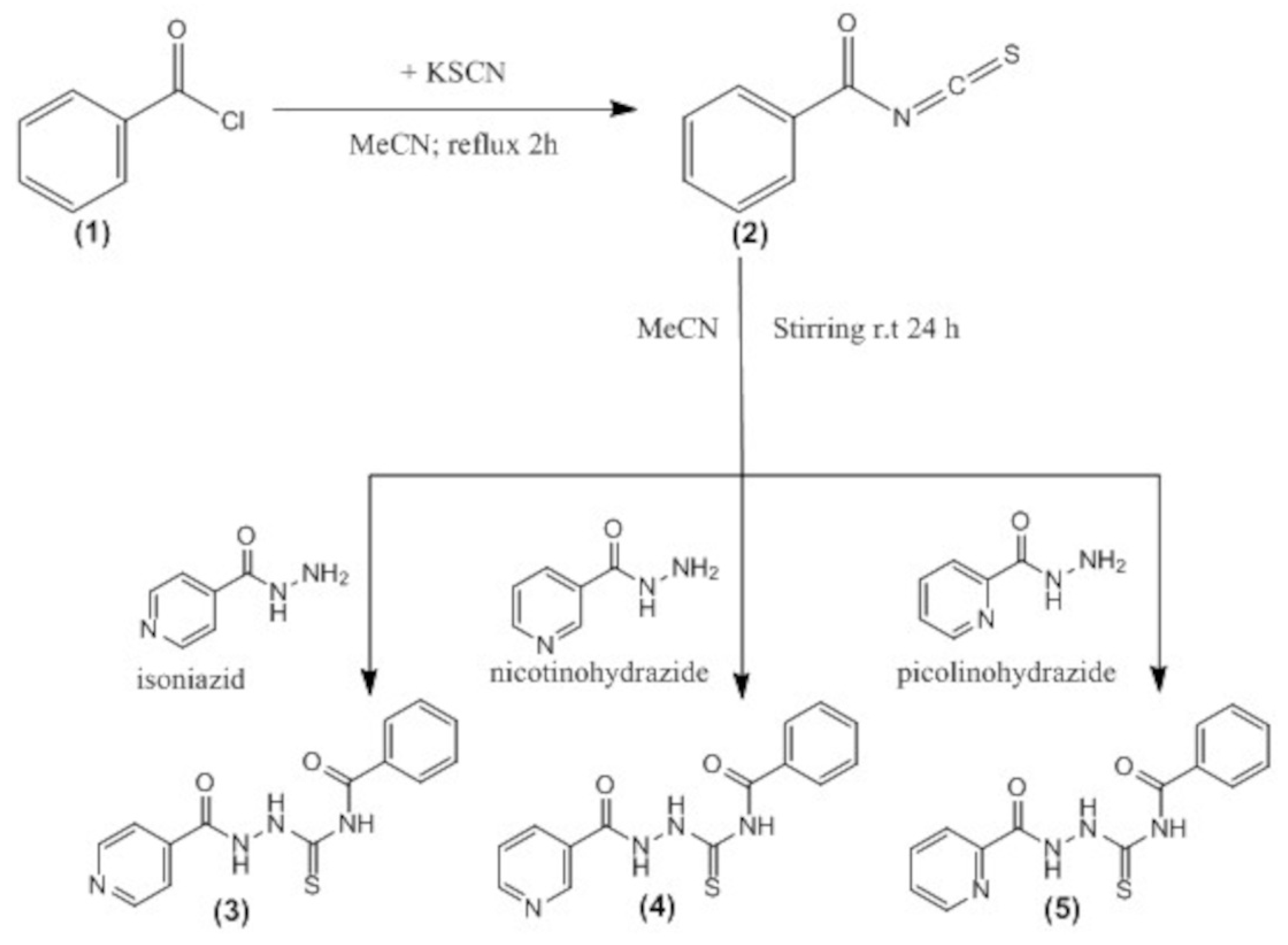
Table 1.
Results of ΔG°, Kd y LE.
| Compound | ΔG° (kcal/mol) | Kd (M) | LE (kcal/mol) |
|---|---|---|---|
| (3)-InhA | –8.75 | 3.83×10⁻⁷ | –0.4167 |
| (4)-InhA | –8.67 | 4.40×10⁻⁷ | –0.4129 |
| (5)-InhA | –8.70 | 4.18×10⁻⁷ | –0.4143 |
Disclaimer/Publisher’s Note: The statements, opinions and data contained in all publications are solely those of the individual author(s) and contributor(s) and not of MDPI and/or the editor(s). MDPI and/or the editor(s) disclaim responsibility for any injury to people or property resulting from any ideas, methods, instructions or products referred to in the content. |
© 2026 by the authors. Licensee MDPI, Basel, Switzerland. This article is an open access article distributed under the terms and conditions of the Creative Commons Attribution (CC BY) license.
Copyright: This open access article is published under a Creative Commons CC BY 4.0 license, which permit the free download, distribution, and reuse, provided that the author and preprint are cited in any reuse.