
Submitted:
17 August 2026
Posted:
19 August 2026
You are already at the latest version
Abstract
Alzheimer’s disease (AD) is one of the most prevalent causes of dementia, with both genetic and environmental factors influencing its progression. The role of phagocytosis and its relationship to AD-associated neuroinflammation has recently garnered considerable attention in the field. For this systematic review, Scopus and PubMed databases were searched to identify studies examining how peripheral immune signaling and systemic metabolic alterations modulate microglial phagocytic function within the context of dementia pathogenesis. Twenty-two full-text original articles, published within the last decade and exclusively in English, were selected for inclusion. Neurodegeneration is a continuous process of barrier breakdown and disruption of cellular homeostasis, during which phagocytosis, a cellular process continuously shaped by peripheral signals, becomes dysregulated, rendering it ineffective and, in certain contexts, neurotoxic. Peripheral stressors such as metabolic stress, systemic infections, and circulating cytokines converge on shared signaling pathways that collectively alter microglial function. The current literature supports a multi-system, multi-hit model in which the central and the peripheral nervous systems engage in continuous bidirectional crosstalk, culminating in the maladaptive remodeling of microglial function upon sustained exposure to peripheral stressors.
Keywords:
phagocytosis
; microglia
; Alzheimer’s disease
; dementia
; neuroinflammation
1. Introduction
Alzheimer’s disease (AD) is a multifactorial neurodegenerative disorder characterized by Amyloid-β (Aβ) plaques, tau pathology, neuroinflammation, metabolic dysfunction, and progressive cognitive decline. While genetic factors contribute to disease risk, most cases are sporadic and influenced by environmental and lifestyle factors [1]. One major mechanism thought to contribute to these neurological effects is chronic neuroinflammation, which involves dysregulated interactions between neurons and glial cells (particularly astrocytes and microglia) and is believed to be an early biomarker of the disease that even precedes Aβ and tau pathology [2,3]. According to the two-hit hypothesis, innate immune dysregulation with increased expression of microglial activation markers, cytokine secretion, and toll-like receptor (TLR) signaling, especially TLR4, alongside Blood-Brain Barrier (BBB) disruption, are the main drivers of inflammation, priming microglia to amplify their inflammatory response to attacks and consequently delaying a return to homeostasis [4,5,6,7,8].
As aforementioned, microglia are central in AD pathology [9]. They are responsible for migrating toward Aβ plaques, engulfing and compacting them to reduce their toxicity [10]. Their function is closely tied to brain homeostasis and cellular metabolism. However, they can exert both protective and detrimental effects depending on their activation state, enabling them to adapt to different environmental stimuli and stressors [11]. Protective microglial phenotypes can clear Aβ through phagocytosis, while excessive activation can release inflammatory cytokines and reactive oxygen species that exacerbate neuronal damage. Microglia appear to exhibit dualistic responses to Aβ oligomers; they tend to remove soluble Aβ oligomers through phagocytosis but amplify neuroinflammation in response to higher-order assemblies [12]. In AD, however, microglia often become dysfunctional and fail to effectively remove Aβ oligomers, which are highly neurotoxic forms of Aβ and are increasingly recognized as major contributors to neuronal toxicity [13]. As a marker of chronic brain inflammation, triggering receptor expressed on myeloid cells 2 (TREM2), which encodes the TREM2 protein, has been linked to the development of AD and Parkinson’s Disease (PD). TREM2 activation determines microglial lipid metabolism, mediating phagocytosis and facilitating plaque-adjacent microglia into the disease-associated microglia (DAM) subtype, thus regulating cellular stress response to damage, such as from alcohol and beta-amyloid [11,14,15,16].
Part of the neuroinflammatory sequelae in AD is the disruption of synaptic plasticity, mediated by over-activation of the complement cascade, resulting in uncontrolled microglial synaptic pruning with subsequent implications for memory function [17,18,19]. Under physiological conditions, one of the main roles of the complement system in the Central Nervous System (CNS) is the preservation of synaptic connections by marking and removing damaged or redundant synapses using astrocytes and microglia [20].
Increasing evidence suggests that immune interactions between the peripheral immune system and the brain play an important role in AD pathology. Although the brain is traditionally considered immune-privileged, neuroinflammation and immune signaling are now recognized as key contributors to neurodegeneration [21]. Systemic infections are emerging as potential triggers that influence neurodegenerative disease progression. Peripheral inflammatory responses can communicate with the central nervous system through circulating cytokines, immune cells, and barrier interfaces such as the Blood-Cerebrospinal Fluid Barrier (BCSF) and BBB. These pathways allow peripheral immune events to alter the brain's immune environment even when pathogens do not directly infect brain tissue [22]. Accordingly, many risk factors for AD, such as age, cardiovascular disease, high-fat diet, and periodontal disease [23], could be the causal link between increased inflammatory damage to the CNS and AD pathology [21,24,25,26,27,28,29].
To further this point, the involvement of the gut–brain axis is being increasingly discussed as a causal factor. Growing evidence suggests that gut microbiota composition can influence brain function, immune response, and disease progression. Several important physiological functions of the gut microbiota are mediated through distant communication via bacterial extracellular vesicles (bEVs), rather than cell-to-cell contact. Both commensal microorganisms, which contribute to immune system maturation and homeostasis, and pathogenic species utilize this pathway to communicate with the immune system, with the latter facilitating pro-inflammatory responses [30,31,32]. Microbial products such as lipopolysaccharides (LPS), amyloids, and short-chain fatty acids (SCFAs) are linked to inflammatory pathways associated with AD pathology by influencing microglial maturation and function in the CNS and affecting the barrier's integrity [33,34,35,36]. Moreover, emerging evidence suggests that age-related gut dysbiosis can increase Trimethylamine N-oxide (TMAO) levels, a gut microbiota–derived metabolite. TMAO is known to promote gastrointestinal inflammation and cytokine production, and is thought to reduce tight junction proteins in the gut, thereby allowing the passage of inflammatory molecules into the circulation and possibly into the CNS [37,38,39]. Interestingly, studies have shown that the absence of host microbiota impairs microglial maturation, differentiation, and immune responses, and compromises brain barrier integrity, collectively indicating that environmental microbes play an active role in shaping CNS immunity [40,41,42,43].
Another key component to neuroinflammation is the adipose tissue–brain axis, where adipocytes act as endocrine cells that release signaling molecules, influencing brain function [44,45]. Disruption of this signaling system is increasingly associated with neurodegeneration, since adipose tissue is the brain's primary lipid reservoir. However, although Apolipoprotein B (ApoB) signaling is known to be disrupted in metabolic diseases, such as obesity and diabetes, its direct effects on glial function remain poorly understood [46].
In an era of pervasive overconsumption, obesity and Type 2 Diabetes (T2D) are increasingly being recognized as risk factors for neurodegenerative diseases [47,48,49,50]. Epidemiological and experimental studies show a bidirectional relationship in which T2D increases the risk and severity of AD. Conversely, T2D is prominent in AD patients, suggesting shared pathological mechanisms and thus linking metabolic disorders to brain dysfunction [51,52,53,54,55,56,57]. Inflammation is central to both T2D and AD pathologies, with pro-inflammatory cytokines found in adipose tissue and the brains of AD patients, leading to subsequent disruption of brain homeostasis [15,58,59]. Chronic hyperglycemia further contributes to neuronal damage through BBB disruption, which together promotes pathological changes in the brain.
Chronic obesity is also associated with systemic inflammation, which can extend to the brain and trigger neuroinflammatory processes that damage neural tissue. Studies have shown that obesity amplifies neuroinflammation and can also impair white matter integrity, particularly in regions such as the corpus callosum and the frontal lobe. However, the molecular mechanisms linking obesity to brain pathology remain poorly understood [60,61,62]. Epidemiological evidence shows that obesity and a high-fat diet (HFD) are significant risk factors for AD, independent of other factors such as diabetes. HFD consumption promotes microglial priming and exaggerated inflammatory responses, which ultimately disrupt synaptic plasticity. As mentioned previously, a key mechanism linking neuroinflammation to synaptic loss is the complement cascade, which normally tags damaged synapses for removal but becomes overactivated in AD, leading to excessive microglial synaptic pruning. In obesity, however, peripheral immune cells and adipose tissue produce complement components, but whether this pathway becomes activated in the brain during obesity is unclear [62,63]. Despite strong associations between HFD and AD, the direct mechanisms remain unclear due to confounding long-term metabolic effects [64].
Following this line of reasoning and in light of the recent surge in research on the topic, the objective of this study was to review the relevant literature published within the last decade and present a comprehensive synopsis of how peripheral immune signaling and metabolic dysfunction modulate phagocytic activity in old age.
2. Materials and Methods
The Preferred Reporting Items for Systematic Reviews and Meta-Analyses (PRISMA) checklist (registration number: CRD420261465060) was used to guide this study. The study’s methods were designed a priori.
2.1. Search Strategy
Scopus and PubMed were the two databases selected to conduct this systematic search, carried out by two investigators (MD and DT). The keywords used to trace all relevant studies published between 1 January 2016 and 18 February 2026 on PubMed were: ((microglia[MeSH Terms] OR microglia*[tiab]) AND (dementia[MeSH Terms] OR alzheimer*[tiab] OR neurodegenerat*[tiab]) AND (phagocyt*[tiab] OR "cell clearance"[tiab] OR "synaptic pruning"[tiab]) AND (systemic[tiab] OR peripheral[tiab] OR metabolic[tiab] OR inflammation[tiab])) NOT (review[pt] OR systematic review[pt]) AND 2017:2026[dp]; and, on Scopus were: (TITLE-ABS-KEY(microglia*) AND TITLE-ABS-KEY(dementia OR alzheimer* OR neurodegenerat*) AND TITLE-ABS-KEY(phagocyt* OR "cell clearance" OR "synaptic pruning") AND TITLE-ABS-KEY(systemic OR peripheral OR metabolic OR inflammatory)) AND NOT DOCTYPE(re) AND NOT DOCTYPE(sh) AND PUBYEAR > 2016, limited to original articles only, in English. All retrieved articles were further hand-searched for any other potentially eligible cited articles. To minimize the risk of bias, the articles chosen were systematically selected peer-reviewed studies based on predefined inclusion criteria. Any disagreement regarding the screening or selection process was resolved by a third investigator (IH) until a consensus was reached.
2.2. Selection Criteria
The sources for this study were original, full-text research articles published in English, excluding secondary analyses, reviews, guidelines, notes, errata, letters, meeting summaries, comments, unpublished abstract-only documents, and retracted articles. There were no exclusionary criteria for study design or other sample characteristics.
2.3. Data Extraction
Data extraction and display (below) were performed independently by one investigator from the team (MD), using a data table format created in Excel. The table headings (see Table 1) include bibliographic information, the type of study, the demographic profiles, the testing method, the dementia stage or type, the peripheral trigger, phagocytic impact, and, finally, the main findings of each study.
2.4. Data Analysis
The statistics found in the table were not analyzed, nor was a meta-analysis performed, as the data have only been descriptively analyzed.
3. Results
3.1. Study Selection
A total of 1391 records were identified through database searching. After removing duplicates and clearly irrelevant studies, the remaining articles were screened based on their titles and abstracts. Full-text assessment was subsequently performed to determine eligibility according to the predefined inclusion and exclusion criteria.
After full-text screening, 22 studies were considered eligible for inclusion in the present review (Figure 1). These studies were selected for their relevance to microglial phagocytosis and for investigating peripheral triggers, mechanistic pathways, and functional outcomes related to neurodegeneration.
3.2. Study Characteristics
The twenty-two publications included were of a wide variety of types. These included 11 preclinical experimental mouse model studies, 3 in vitro experimental studies (1 with a human triculture model; 1 with human multicellular models; 1 with microglial monoculture); 2 preclinical mouse studies with supporting human biopsy analysis; 2 preclinical experimental mouse studies with in vitro microglial assays; 1 human post-mortem observational study, 1 preclinical mouse study with in vitro validation; 1 in vitro mechanistic cell culture study, and; 1 preclinical mechanistic study-Drosophila model.
3.3. Demographic Profile
Across the included studies, a wide range of biological systems and subject characteristics were represented. Those studies that included mice were of the following types: 7 used C57BL/6J mice, 4 used 5xFAD transgenic mice, and 2 used APPNL-G-F mice. Those mouse studies also included 4 with in vitro components. Of the purely in vitro studies, 2 used human cells (brain endothelial cells, gut endothelial cells, microglia, astrocytes, neurons, pluripotent stem cells), 1 used murine-derived cells, and another 1 used both human and murine-derived cells. There was also 1 study that observed human post-mortem brain tissue. The other study used Drosophila flies.
3.4. Testing Methods
The data from these studies were derived using a multitude of methods. The main ones were (various types of) PCR tests (14), behavioral tests (10), Immunohistochemistry (9), RNA sequencing (8), immunofluorescence (7), and phagocytosis assays (6). An assortment of other tests were also used, such as immunostaining, flow cytometry, cell cultures, transcriptomic analysis, live-cell Aβ phagocytosis imaging, spectrometry (LC/MS), Nitric Oxide Measurement, histopathology, lipidomics (LC-MS/MS), NF-κB Luciferase Reporter Assay, Griess Test, ELISA, Seahorse analysis, bacterial stimulation assays, and cytokine arrays.
3.5. Dementia Stage/Type
The included studies encompassed multiple disease states and pathological contexts. The main ones revolved around Alzheimer’s disease states. The Alzheimer’s disease model was used 9 times, including advanced Alzheimer’s disease (with possible co-pathology), one Alzheimer’s disease/Inflammatory polyarthritis model, one Alzheimer’s disease–like pathology, and the other included Cerebral Small Vessel Disease (CSVD).
3.6. Peripheral Triggers
A wide range of peripheral triggers influencing microglial phagocytosis was identified across studies. These included high fat diet (3) whether chronic or short-term, obesity (3) either from a high-sugar diet or a Western diet or genetic predisposition with Western diet induced obesity and type 2 diabetes, ethanol exposure (2) or chronic exposure to inflammation (2) either LPS-induced or chronic/systemic, gut microbiota (3) whether a metabolite (acetate) or bEVs or TMAO, chronic hyperglycemia (insulin deficiency- type 1 diabetes), vacuolating cytotoxin A (VacA) induced H.pylori cytotoxicity, IDE deficiency, microglial insulin resistance, systemic infection or microbial exposure, periodontal disease- associated pathogens, antibiotic treatment- depletion of gut bacteria, infliximab, a tumour necrosis factor αlpha (anti-TNF-a) antibody, and prior systemic SARS-CoV-2 infection (non-fatal).
3.7. Phagocytic Impact
The effects on microglial phagocytosis were heterogeneous across studies. Reported findings included 5 cases of increased microglial phagocytosis, and further cases of increased phagocytosis, whether dependent on Il-17a, or in response to systemic pathogen-associated molecular patterns (PAMPs), or due to increased cluster of differentiation 68 (CD68) expression after ethanol exposure. Lastly, there was an increase in particular types of microglial phagocytosis, such as amyloid-β via macrophage scavenger receptor 1 (MSR1) upregulation, bacterial particles and amyloid-β, and myelin due to complement activation: impairment or a decrease in microglial phagocytosis in three cases. In two further cases, microglial amyloid-β phagocytosis was impaired. In other cases, certain factors affected microglial phagocytosis, such as reduced amyloid-β phagocytosis due to the presence of VacA or TREM2 deficiency. In yet two other cases, there was no significant change in microglial phagocytic activity, whether compared to the control group or in the expression of the CD68 factor. One further case showed no significant change specifically in amyloid-β phagocytosis. Lastly, one case report indicated that the diabetic phenotype decreases microglial clustering around β-amyloid plaques and reduces phagocytic uptake by disrupting TREM2–PI3K–Akt signaling.

Table 1.
Characteristics of the 22 studies included in this review, organized by the primary category of peripheral trigger examined. A hyphen (-) indicates that no dementia stage/type was specified in the study.
Table 1.
Characteristics of the 22 studies included in this review, organized by the primary category of peripheral trigger examined. A hyphen (-) indicates that no dementia stage/type was specified in the study.
| Authors, Year of Publication | Type of Study | Demographic Profile | Testing Method | Dementia Stage/Type | Peripheral Trigger | Phagocytic Impact | Main Finding |
| Effects of Metabolic Syndrome | |||||||
| Zhang et al., 2025 [65] | Preclinical mouse study | 40 7-week-old C57BL/6J mice with PCSK9 overexpression | Immunofluorescence staining, Luxol Fast Blue Staining, Laser Speckle Contrast Imaging, Light Sheet Microscopy, Behavioral Testing | Cerebral small vessel disease (CSVD) | High-fat diet | Increased phagocytic activation (↑CD68) | Combined metabolic stress from genetic (PCSK9 overexpression) and environmental (high-fat diet) factors synergistically drives widespread cerebrovascular dysfunction, vascular rarefaction, neurovascular uncoupling, BBB disruption, white matter injury, neuroinflammation, and subsequent neuronal loss and cognitive impairment. |
| Yang et al., 2025 [11] | Preclinical experimental mouse study with in vitro microglial assays | 8 week-old APPNL−G−F knock-in Alzheimer's disease model mice carrying KM670/671 NL/I716 F mutations; primary microglial cultures treated with oleic acid | Behavioral tests (Y-maze test), Serum Total Cholesterol and Triglyceride Measurements, histopathology, immunofluorescence staining, quantitative analysis of histopathology data, flow cytometry, lipidomics (LC-MS/MS), cell culture, RT-qPCR, Aβ peptide uptake assay | Alzheimer's disease model | Chronic high-fat diet (HFD) | Impaired microglial phagocytosis of amyloid-β | High-fat diet induces cholesterol ester accumulation and lipid droplet formation in microglia, leading to impaired phagocytosis of amyloid-β, reduced plaque clearance, and exacerbation of Alzheimer's disease pathology. |
| Alassaf & Rajan, 2024 [46] | Preclinical mechanistic study-Drosophila model | Adult male Drosophila 7–10 days old | Antennal nerve injury assay, immunostaining, mitochondrial reporters, metabolomics, lipidomics, qPCR, confocal image analysis | - | Obesogenic high-sugar diet (HSD) | Impaired glial phagocytosis and reduced neuronal debris clearance | Adipocyte metabolic dysfunction disrupts ApoB–LpR1 lipid signaling to glia, lowering Draper levels and impairing glial debris clearance |
| Graham et al., 2020 [62] | Preclinical experimental study-mouse model | 12 month-old male and female C57BL/6J mice; C1qa knockout | Behavioral Y-maze cognitive testing, open arenas, grip strength, immunofluorescence, confocal microscopy, image quantification | - | Chronic obesity induced by a Western diet. | Complement activation promotes microglial phagocytosis of myelin, leading to white matter degradation. | Complement component C1q drives obesity-induced neuroinflammation, cerebrovascular damage, microglial phagocytosis of myelin, and white matter loss; deletion of C1qa protects against these pathological changes and prevents cognitive deficits. |
| Mackey-Alfonzo et al., 2024 [64] | Preclinical mouse study | 4-month-old female B6;129-Tg (APPSwe, tauP301L) 1Lfa Psen1tm1Mpm/Mmjax (3xTg-AD) mice | Behavioral testing, tissue processing, RNA extraction, RT-qPCR, ELISA, Western blot, cell culture, phagocytosis assays | Alzheimer's disease model | Short-term high-fat diet (HFD) | Increased microglial phagocytosis | Dietary factors increase cognitive vulnerability very early on in the disease process, highlighting the importance of the diet–neuroinflammation–complement–microglia axis in AD. |
| Wang et al., 2026 [12] | Preclinical experimental study-mouse model | 204 Male C57BL/6J mice 6–8 weeks old with streptozotocin-induced type 1 diabetes; TREM2 conditional knockout mice on C57BL/6J background; murine BV2 microglial cell line | Behavioral tests (MWM, NOR), snRNA sequencing, cell culture, wound scratch and transwell assays, live-cell imaging, flow cytometry, ROS assay, Western blot, immunofluorescence, ELISA, qRT-PCR, RNA sequencing | - | Chronic hyperglycemia caused by insulin deficiency in type 1 diabetes | TREM2 deficiency reduces phagocytosis and increases Aβ accumulation | Hyperglycemia in T1D impairs microglial function and promotes Aβ accumulation; TREM2 supports microglial migration, mitochondrial function, and phagocytosis of Aβ oligomers, protecting against cognitive decline. |
| Natunen et al., 2020 [15] | Preclinical mouse study with supporting human biopsy analysis | 46 female 7-month-old transgenic mice with C57Bl/6J background divided into 3 lines: 1) APPswe/PS1dE9; 2) Tau P301L; 3) APPswe/PS1dE9/Tau P301L; human cortical biopsies from idiopathic normal pressure hydrocephalus (iNPH) patients with β-amyloid pathology | Behavioral tests (spontaneous activity, Morris swim task, passive avoidance test), histochemistry, fluorescent and confocal microscopy, image analysis, human cortical biopsy analysis, immunohistochemistry of human biopsy brain tissue, imaging and analysis of human brain biopsy tissue, cell cultures, phagocytosis assay, Western blot analysis, AlphaLISA assay, ELISA, nitric oxide assays, RT-qPCR, RNA sequencing, FastQC, differential expression analysis, WGCNA, gene set enrichment analysis, TFBS, cell type enrichment analysis | Alzheimer's disease model | Genetic predisposition in combination with Western diet-induced obesity and type 2 diabetes | Diabetic phenotype decreases microglial clustering around β-amyloid plaques and reduces phagocytic uptake via disruption of TREM2–PI3K–Akt signaling. | Type 2 diabetes impairs microglial immune responses to β-amyloid pathology by disrupting TREM2–PI3K–Akt signaling, resulting in fewer plaque-associated microglia, increased neuritic damage, and worsened cognitive impairment. |
| Chen et al., 2024 [9] | Preclinical mouse study and in vitro validation | Microglia-specific insulin receptor knockout (MG-IRKO) mice; primary microglial cultures | Metabolic measurements, behavioral tests, RT-qPCR, immunohistochemistry, TEM, Western blot, phagocytosis assays, ELISA | - | Microglial insulin resistance | Impaired microglial phagocytosis of amyloid-β | Loss of insulin signaling in microglia impairs Aβ phagocytosis, increases neuroinflammation, and exacerbates Alzheimer's disease pathology, linking metabolic dysfunction to neurodegeneration. |
| Huang et al., 2024 [66] | In vitro experimental study | Immortalized Murine Microglial BV2 cells | Western blot, Plasmid DNA Transfection, NF-κB Luciferase Reporter Assay, Griess Test, ELISA, Phagocytosis Assays, fluorogenic probe, TUNEL staining, NOX and SOD measurements | - | Lipopolysaccharide (LPS)-induced inflammation; insulin signaling modulation | Increased microglial phagocytosis of bacterial particles and amyloid-β (Aβ) | Insulin enhances microglial inflammatory activation and microglial phagocytic activity while reducing oxidative stress, suggesting a complex role in regulating neuroimmune responses and amyloid clearance. |
| Corraliza-Gómez et al., 2023 [57] | Preclinical mouse study with in vitro microglial analysis | 12-month-old knockout mice for the IDE gene; primary microglial cultures | Behavioral tests (open field test and habituation, object recognition test), metabolic parameters, molecular biology studies in the olfactory bulb, histological studies in the hippocampus, immunoblotting, RNA-sequencing, qPCR, live/dead viability assay, proliferation assay, ELISA, Luminex assays, phagocytosis assays, FAM-Aβ management experiments | - | IDE deficiency | No significant change in amyloid-β phagocytosis; altered myelin phagocytosis in a sex-dependent manner, increasing internalization in females while decreasing in males | IDE regulates microglial phenotype and neuroimmune responses rather than directly controlling amyloid-β clearance, indicating possible non-enzymatic involvement in Alzheimer's disease pathology. |
| Effects of the Microbiome | |||||||
| Xie et al., 2025 [32] | Preclinical mouse study | 4- and 12-month-old male and female GF AppNL-G-F mice | LAL assay, TEM, immunohistochemistry, glial cell morphology quantification, synaptic imaging, and quantification | Alzheimer's disease model | Microbiota-derived extracellular vesicles (bEVs) | Increased microglial phagocytosis | Gut microbiota and their extracellular vesicles impair microglial phagocytosis and promote amyloid pathology, highlighting the gut–brain axis as a regulator of AD progression. |
| Janeiro et al., 2023 [39] | In vitro mechanistic cell culture study | Multiple cell types: murine adipocytes (3T3-L1), murine macrophages (RAW 264.7), human BBB cells (hCMEC/D3), murine primary neurons, murine primary microglia | Differentiation assay, cell metabolic activity assay, immunochemistry, phagocytosis assay, Western blot, RNA extraction, qPCR | - | Trimethylamine N-oxide (TMAO; gut microbiota-derived metabolite) | Increased microglial phagocytosis | TMAO acts as a key mediator connecting consumption of imbalanced diets, especially high in fat, with peripheral inflammation and central neuroinflammatory processes |
| Tran et al., 2025 [67] | In vitro experimental study with human multicellular models | Multicellular human cell-based models, including gut epithelial cells, brain endothelial cells, microglia, astrocytes, and neurons | Human multicellular co-culture systems, Transwell gut–brain barrier models, microfluidic brain chips, immunofluorescence staining, cytokine arrays, ROS assays, TEER measurements, live-cell Aβ phagocytosis imaging, Immunoprecipitation of HP-VacA, Liquid Chromatography–Mass Spectrometry (LC/MS), ELISA, 3D Tri-culture, Microglial Migration Assay, LRP1-Inhibition Assay, Fluorescent Immunostaining, Nitric Oxide Measurement, Human multiple cytokine assay | - | VacA-induced H. pylori cytotoxicity | VacA inhibits microglial amyloid-β phagocytosis by blocking the LRP1 receptor involved in Aβ clearance. | H. pylori VacA toxin crosses epithelial and brain barriers, activates glial cells, induces neuroinflammation, inhibits microglial Aβ clearance via LRP1, and promotes neurodegenerative pathology markers (p-Tau and p-α-synuclein). |
| Erny et al., 2021 [43] | Preclinical mouse study | Germ-free (GF) mice, specific pathogen-free (SPF) mice controls and 5xFAD mice. | RNA-seq, IPA, ChIP-seq, RT-qPCR, FACS analysis, PCA, ECARs, cytometry, TEM, mass spectrometry, acetate assays | Alzheimer's disease model | Gut microbiota-derived metabolite (acetate) | Decrease microglial phagocytosis in GF mice | Microbiota-derived acetate regulates microglial metabolism and function, influencing phagocytosis and contributing to Alzheimer's disease pathology through modulation of Aβ deposition. |
| Hao et al., 2024 [68] | Preclinical mouse study | Female APP/PS1 transgenic mice; IL-17a deficient AD mice | Tissue collection, 16s rRNA seq, quantification of bacterial DNA, selection of CD11b+ and CD4+ cells, phagocytosis assays, flow cytometry, histological analysis, immunohistochemistry, Western blot, ELISA, qPCR | Alzheimer's disease model | Antibiotic treatment-depletion of gut bacteria | IL-17a-dependent increase in microglial phagocytosis | IL-17a is a key mediator of gut–brain immune communication and supports antibiotic therapy for AD patients |
| Almarhoumi et al., 2023 [23] | Preclinical experimental study in vivo mouse model and in vitro microglial assays | 36 10-12 week-old C57BL/6 Wt mice-experimental periodontal disease model; cultured microglial BV2 cell line | Flow cytometry, T-SNE analysis, isolation and heat-inactivation of mouse microbiome, cell cultures, q-PCR, phagocytosis assays, bacterial stimulation assays | - | Periodontal disease–associated pathogens | Increased microglial phagocytosis of amyloid-β (Aβ42) via MSR1 upregulation | There is a direct mechanistic link between oral infection, microglial activation, and enhanced phagocytic activity, highlighting a possible effect on AD progression. |
| Effects of Alcohol | |||||||
| Le et al., 2025 [69] | Preclinical experimental mouse study | 2.5-month-old male 5xFAD transgenic mice | Behavioral tests (Marble Burying Test, NOR Test), Tissue Harvesting, Microglia Isolation, Bulk RNA Sequencing, Transcriptomic Analysis, Immunofluorescence, Confocal Microscopy, Image Acquisition and Analysis | Alzheimer's disease model | Chronic binge-level ethanol exposure | No significant change in the microglial phagocytic factor CD68 expression | Binge ethanol exposure increases diffuse β-amyloid plaque load and impairs recognition memory in 5xFAD mice, but does not significantly alter microglial activation, morphology, transcriptomic profile, or phagocytic marker expression. |
| Boreland et al., 2026 [16] | In vitro experimental study with a human triculture model | Human-induced pluripotent stem cell (iPSC)-derived neurons, astrocytes, and microglia cultured in a triculture system | Immunohistochemistry, Image acquisition and processing, RNA Purification, RNA sequencing, RT-qPCR | - | Ethanol exposure simulating alcohol consumption | Increased CD68 expression indicates enhanced lysosomal activity and phagocytic microglial activation after ethanol exposure. | Ethanol exposure induces neuroimmune dysregulation, microglial activation, and increased TREM2 expression in a human neuron–astrocyte–microglia model, suggesting alcohol can promote inflammatory brain environments linked to neurodegeneration. |
| Effects of Systemic Inflammation | |||||||
| Bathini et al., 2023 [70] | Preclinical mouse study with supporting human biopsy analysis | PolyI: C mouse model; Frozen Human tissue samples from the entorhinal cortex, including the hippocampal area | Behavioral tests, tissue processing, flow cytometry, bead-based assay, nucleic acid and protein extraction, RNA library and sequencing, bioinformatics analysis, RT-PCR, Western blot, fluorescent immunohistochemistry, image quantification | - | Chronic systemic inflammation | Increased microglial phagocytosis | Chronic systemic inflammation induces long-term microglial dysfunction, tau pathology, and cognitive decline, supporting a link between peripheral immune activation and vascular/AD-like neurodegeneration. |
| Ganz et al., 2022 [8] | Preclinical mouse study | 5xFAD mice with C57Bl/6J background | LPS injections, histopathology, FACS analysis, RT-PCR, immunohistochemistry, endotoxin concentration measurement, Seahorse analysis | Alzheimer's disease model | Systemic infection / microbial exposure | Phagocytosis is enhanced in response to systemic PAMPs | Systemic infections accelerate neurodegeneration in AD via microglial activation; modulation of microglia toward a protective phenotype enhances phagocytosis and reduces neuronal loss. |
| Villareal et al., 2024 [22] | Human post-mortem observational study | Human post-mortem brain tissue from Alzheimer's disease patients (n=35): COVID-19 survivors (n=11), non-COVID AD patients (n=12), and pre-2020 AD controls (n=12) | Immunohistochemistry, QuPath digital analysis, NanoString transcriptomics, RT-qPCR, confocal microscopy across multiple brain regions | Advanced Alzheimer's disease with possible co-pathology in the form of Lewy body disease (LBD), Limbic-predominant age-related TDP-43 proteinopathy (LATE), Aging-related tau astrogliopathy (ARTAG), cerebral amyloid angiopathy (CAA) | Prior systemic SARS-CoV-2 infection (COVID-19) that was not fatal | No significant change in CD68+ microglial phagocytic activity compared with non-COVID AD patients | Surviving COVID-19 infection disrupts neuroimmune homeostasis in AD brains, characterized by increased microglial activation, loss of astrocytes, and dysregulation of oligodendrocyte/myelination pathways that may influence AD progression. |
| Paouri et al., 2017 [71] | Preclinical mouse study | Female 5XFAD/Tg197 double transgenic mice | Tissue processing, Western blot, ELISA, Immunohistochemistry, thioflavine S staining, immunofluorescence, image analysis and quantitation | Alzheimer's disease/Inflammatory polyarthritis model | Infliximab (anti-TNF-a antibody) | Decreased microglial phagocytosis | Peripheral TNF-α reduces amyloid burden by promoting recruitment of blood-derived immune cells and enhancing inflammatory/glial responses; inhibition of TNF-α reverses this effect, indicating a key role for peripheral immune–brain crosstalk rather than microglial phagocytosis alone. |
4. Discussion
This systematic review, utilizing 22 full-text original articles published within the last decade, examines how peripheral immune signaling and systemic metabolic alterations modulate microglial phagocytic function in the context of dementia pathogenesis. The results of these 22 articles could help in the discovery of new preventative or palliative treatments for AD or dementia.
4.1. Effects of Metabolic Syndrome
Reduction in cerebrovascular density has been proven to be an early feature of CSVD, thought to be triggered by multiple metabolic hits that overcome vascular homeostasis, causing endothelial toxicity [65]. Subsequent BBB disruption, itself caused by pro-inflammatory signals at the endothelium, amplifies this effect by allowing the entry of immune cells and lipids from the periphery, thereby enhancing perivascular inflammation, edema, and waste accumulation. The subsequent formation of a lipid-rich environment, combined with Apolipoprotein E (APOE) dysfunction and poor cholesterol efflux, alongside poor lysosomal degradation, leads to the creation of lipid-droplet accumulating microglia (LDAM), representing a metabolically altered and dysfunctional microglial state with impaired Aβ handling, which is linked to hypercholesterolemia alone or in combination with BBB disruption. Although microglia in this model exhibit increased phagocytic activation, as evidenced by elevated CD68 expression, this response appears maladaptive, as amyloid-β accumulates around blood vessels, suggesting impaired clearance capacity. Over the long term, an HFD accelerates AD progression through metabolic disruption of microglia. In pathologic conditions, such as AD, the esterification process, ie., the mechanism involved in storing cholesterol esters (ChE) inside lipid droplets (LDs), is upregulated [11]. It is thought that the pathologic state of AD can augment the sensitivity of ChE metabolism, or conversely that HFD may exacerbate abnormal ChE accumulation in a genetically predisposed individual. HFD was also found to exacerbate Aβ-induced dysregulation of microglial lipid metabolism. LD-accumulating microglia exhibit decreased phagocytic activity and a pro-inflammatory phenotype. Moreover, lipoprotein lipase (LPL) levels were significantly downregulated, indicating that a high-fat environment prevents microglia from acquiring the activated DAM subtype, suggesting that metabolic stress shifts microglia into a dysfunctional state.
In light of these observations, research has shifted toward characterizing how adipose tissue communicates with the brain via metabolic signals that regulate glial phagocytosis [46]. Alassaf & Rajan identify ApoB-containing lipoproteins as a key signal sent from adipocytes to glial cells. These lipoproteins interact with the glial receptor LpR1, thereby maintaining the expression of the engulfment receptor Draper, multiple epidermal growth factor-like domain protein 10 (MEGF10) in mammals, which is essential for clearing neuronal damage. This establishes a previously unrecognized adipocyte–glia signaling pathway. Prolonged high-sugar diet (HSD) causes metabolic adaptations in adipose tissue that resemble starvation, forcing adipocytes to rely on fat metabolism for energy. When ApoB signaling is disrupted, glial cells lose their ability to efficiently clear damaged neurons, highlighting an ApoB-LpR-mediated mechanistic link between obesity and neurodegeneration. The metabolic and lipid changes observed under HSD resemble those seen during aging, leading Alassaf & Rajan to suggest that obesity-induced metabolic dysfunction in adipose tissue may contribute to neurodegeneration. The brain may upregulate endogenous lipid synthesis to compensate for HSD-induced disruption in ApoB-mediated delivery of lipids from adipocytes. This study demonstrates that adipocyte metabolic state regulates brain glial function, that ApoB lipoprotein signaling links peripheral metabolism to glial phagocytosis, and that obesogenic diets disrupt this communication, thereby impairing glial clearance of neuronal debris. Overall, the findings highlight how systemic metabolic dysfunction can transform microglia from protective to pathological states, linking peripheral metabolic disease to cerebrovascular damage and cognitive decline.
Complement components produced by peripheral immune cells and adipose tissue represent another route through which obesity influences CNS function. In this study, activation of the complement cascade, particularly complement component 1q (C1q), was found to contribute to brain pathology associated with obesity [62]. The study showed that mice fed a Western diet- high-fat, high-sugar, high-cholesterol, low-fiber- had increased levels of complement proteins C1q and complement component 3 (C3) in the brain, especially in white matter regions. These changes were associated with microglial activation, cerebrovascular dysfunction, and myelin degradation, indicating that complement activation contributes to obesity-induced neuroinflammation. HFD increases neuroinflammation and enhances microglial phagocytosis of synapses via a complement-dependent mechanism, specifically C1q and C3, leading to excessive synaptic loss and memory impairment early on in the disease process [64].
The TREM2 receptor is another critical regulator of key microglial processes, including migration toward Aβ-containing neurons, phagocytosis of Aβ oligomers, maintenance of mitochondrial integrity, lipid metabolism, immune responses, and cell survival [12]. HFD has been found to reduce TREM2 expression in microglia, disrupting cholesterol transport between intracellular and extracellular compartments and ultimately downregulating microglial migration toward Aβ plaques, decreasing Aβ uptake, and increasing LD accumulation within microglia. While Aβ is thought to drive the DAM response, HFD is also found to trigger genes associated with inflammation and immune responses in genetically predisposed mice [15]. TREM2 signaling activates pathways such as PI3K/Akt/mTOR and extracellular signal-regulated kinase 1 / 2 (ERK1/2), which regulate microglial homeostasis [12]. The study shows that TREM2 expression is increased in microglia in the diabetic brain, particularly in the prefrontal cortex. These findings indicate that TREM2 plays a protective role in diabetic neurodegeneration by enabling microglia to clear toxic Aβ species and maintain cellular energy balance. Western-type diets, however, suppress this immune response by downregulating the expression of PIK3R1, a gene encoding the PI3K regulatory subunit 1, and activating TREM2, hence reducing microglial phagocytosis and inflammatory responses related to amyloid plaques, leading to the metabolic effects and T2D phenotype seen in mice fed a Western-style diet [15]. Disrupted energy metabolism reduces microglia's ability to clear Aβ. At the same time, chronic inflammation further suppresses phagocytic capacity, creating a vicious cycle that promotes disease progression, with TREM2 identified as a key metabolic checkpoint for microglial function [9].
Within this framework of metabolic dysfunction and microglial function, insulin has dual, context-dependent effects on microglial activity [66]. Chen et al. suggest that impaired insulin signaling in the brain, as seen in AD, also known as Type 3 Diabetes (T3D), could contribute to reduced Aβ clearance and thus disease progression. Insulin signaling was also found to be necessary for proper microglial phagocytic function [9]. The study also shows that loss of insulin signaling enhances neuroinflammation, with increased expression of inflammatory cytokines, such as interferon-gamma (IFN-γ), activation markers like ionized calcium-binding adapter molecule 1 (IBA1), and increased Aβ accumulation in the brain, particularly in a genetically predisposed individual. The aforementioned findings were also highlighted, showing that insulin enhances microglial phagocytic activity, including uptake of bacterial particles and fibrillar Aβ, the primary constituent of extracellular amyloid plaques [66]. Insulin-degrading enzyme (IDE) is proposed as a potential molecular link between AD and T2D, capable of binding to and degrading both insulin and Aβ peptides [57]. IDE’s role in AD may be mediated through modulation of microglial responses, in a sex- and genotype- dependent manner, rather than direct control of amyloid clearance. Overall, Corraliza-Gomez et al. conclude that IDE is a key non-enzymatic regulator of microglial phenotype and neuroimmune responses, influencing how microglia react to environmental challenges associated with aging and neurodegeneration.
Collectively, these findings underscore systemic metabolic dysfunction, mediated by complement-dependent mechanisms, disrupted insulin signaling, and TREM2-dependent pathways, as a key driver of the shift from protective to pathological microglial states, perpetuating the cycle of impaired Aβ clearance, neuroinflammation, and synaptic loss that accelerates cognitive decline.
4.2. Effects of the Microbiome
Commensal gut microbiota significantly influence microglial maturation and function, commencing at the embryonal stage [32]. Microbiota-derived bEVs increase BBB permeability and augment microglial activation, which in turn activates the glycogen synthase kinase 3β (GSK-3β) pathway, increasing CNS inflammation, tau hyperphosphorylation, and ultimately cognitive impairment. In this study, it was also determined that gut microbiota is linked to neurogenesis, with dysfunction in this process causing a vicious cycle of Aβ aggregation and neuronal dysregulation.
With respect to microbiota dysfunction, fat- and sugar-rich diets cause a microbiota imbalance that compromises intestinal wall barrier integrity, allowing bacterial LPS to leak into the peripheral circulation [39]. TMAO mediates the effects of an imbalanced HFD, peripheral inflammation (which it enhances), and CNS inflammation. Within the CNS, TMAO induces pro-inflammatory signaling by increasing the expression of interleukin-1β (IL-1β) and the NLR family pyrin domain containing 3 (NLRP3) inflammasome. Microglia exposed to TMAO exhibit increased phagocytic activity, which, while normally protective, may become dysregulated and chronic, potentially leading to excessive myelin phagocytosis and phagoptosis, and promoting neurodegeneration.
Extending this discussion, H. pylori infection, a common gut commensal, when chronic, compromises BBB integrity, allowing peripheral pathogens and immune cells to enter the CNS [67]. H. pylori VacA, the main virulence factor of H. pylori, specifically disrupts BBB integrity and penetrates the CNS via lipoprotein receptor-related protein-1 (LRP1) binding sites, initiating a downstream inflammatory cascade that amplifies microglial activation and further increases cytokine production. By chronically binding to LRP1, VacA impairs microglial clearance of Aβ-amyloid, which may promote amyloid accumulation in the brain, a key pathological feature of Alzheimer’s disease.
On the other hand, the literature on germ-free (GF) mice, which lack microbiota, shows that they display immature and dysfunctional microglia. Microbiota-derived SCFAs are highlighted as critical mediators of gut-brain axis communication [43]. They can translocate from the gut to the blood circulation, cross the BBB, and are known to have immune regulatory effects. This study highlights acetate-enhanced microglial maturation and phagocytic rates of Aβ. This establishes acetate as a possible link between the gut microbiome and brain immune function.
IL-17a is identified as another potential key mediator of gut–brain immune communication that supports antibiotic therapy for AD patients, with IL-17a inhibition achieved by blocking sensitive gut bacteria [68]. Gut bacterial depletion attenuates microglial inflammatory activation and Aβ pathology, whereas IL-17a inhibition has been found to maintain, or even enhance, microglial phagocytic activity. Consequently, it was also found to upregulate LRP1 and ATP-Binding Cassette Subfamily B Member 1 (ABCB1) expression in the BBB, thereby promoting Aβ efflux and clearance in the brain.
Additionally, of equal importance, periodontal disease–associated microbiota directly drive microglial activation and function through specific receptor-mediated mechanisms [23]. Mechanistically, microbial stimulation activates microglia via TLR2 and TLR9 signaling pathways, leading to nuclear factor kappa-light-chain-enhancer of activated B cells (NF-κB) activation and increased production of pro-inflammatory cytokines (TNF-α, IL-1β, IL-6). Importantly, this activation is coupled with a significant increase in microglial phagocytosis of Aβ42, mediated by upregulation of the scavenger receptor MSR1.
In closing, these findings position the gut microbiota as a critical modulator of microglial function via metabolite, structural, and immune-mediated pathways, with disruption of this gut-brain axis consistently shifting microglia toward a pathological phenotype that exacerbates Aβ accumulation and neurodegeneration.
4.3. Effects of Alcohol
Mice exposed to binge-level ethanol showed increased amyloid plaque deposition and impaired recognition memory, without any changes in microglial morphology or in the expression of the phagocytic factor CD68 [69]. As a possible explanation for this finding, it was proposed that microglia were already highly activated by the presence of amyloid plaques in the AD model, leaving little room for further activation by ethanol. Another possibility is that ethanol-induced changes in microglia may return to baseline during the abstinence period but may still contribute to long-term effects overall.
Chronic ethanol exposure, on the other hand, increases inflammatory gene expression, primarily in microglial cells, such as IBA1 and CD68, altering their cellular morphology [16]. More specifically, the upregulation of CD68, a lysosomal-associated membrane protein and microglial phagocytosis marker, suggests that microglia respond to alcohol-induced stress by increasing lysosomal and phagocytic activity. Such effects, however, can vary depending on the duration, dose and frequency of alcohol exposure.
Together, these findings indicate that ethanol’s impact on microglial activation and phagocytosis is contingent on exposure pattern and existing pathological context, ranging from negligible effects under conditions of pre-existing amyloid-driven activation to pronounced morphological and phagocytic marker changes with chronic exposure.
4.4. Effects of Systemic Inflammation
Systemic chronic inflammation, often caused by infections or metabolic dysfunction, significantly disrupts brain homeostasis and may contribute to the development of neurodegenerative diseases [70]. Evidence shows that peripheral immune activation can influence the brain through immune signaling, promoting neuroinflammation, microglial dysfunction, and vascular damage. Microglia are highlighted as central regulators of these processes, with aging and genetic risk factors impairing their normal functions, including phagocytosis and immune responses. Microglia undergo age-dependent morphological and functional changes, transitioning toward a more activated, phagocytic-like state, a function associated with dysfunction rather than effective neuroprotection, reflecting features of aging- and disease-associated microglia. Chronic systemic inflammation can drive progressive neurodegenerative changes resembling a vascular AD phenotype, primarily through long-term alterations with accumulation of neuroinflammation, protein aggregates, increased vascular permeability, and microglial dysfunction.
Recent findings indicate that BBB disruption occurs early in AD pathogenesis, thus allowing penetration of endotoxins into the CNS [8]. A major finding of Ganz et al. is that AD brains are uniquely vulnerable to infection-induced damage. Chronic asymptomatic exposure to endotoxin-producing pathogens and recurrent subclinical infections accelerate neurodegeneration in AD mice, but not in healthy controls. Literature suggests a bidirectional relationship within the gut-brain axis. This study highlights that microglia are central mediators of infection-induced neurotoxicity by producing inflammatory mediators that damage neurons. However, microglia are functionally diverse and can also adopt neuroprotective phenotypes.
As a case in point, AD patients who survived COVID-19 show significant disruption of neuroimmune homeostasis [22]. At the same time, a reduction in microglial cells expressing homeostatic markers such as purinergic receptor P2RY12 has been observed in these patients, indicating a shift toward activated or disease-associated microglial phenotypes. However, markers associated with microglial phagocytic activity (such as CD68) were not significantly altered.
Similarly, Paouri et al highlight peripheral TNF-α as a critical regulator of intracerebral inflammation and, thus, AD pathology by affecting Aβ amyloid deposition and neuronal integrity [71]. During peripheral inflammation, TNF-α enhances the recruitment of blood-derived mononuclear cells into the brain vasculature and activates microglia and astrocytes, thereby promoting a coordinated immune response that may enhance amyloid clearance. An increased concentration of the phagocytic marker CD68 was also indicative of increased phagocytic activity in both microglia and peripheral-derived macrophages during the aforementioned conditions.
Altogether, these findings highlight systemic inflammation and peripheral immune signaling as critical accelerators of BBB breakdown and microglial dysfunction in AD, with microglia acting as context-dependent mediators that shape the balance between neurotoxic and neuroprotective outcomes.
4.5. Emerging Therapeutic Targets and Future Directions
Based on the aforementioned information, microglia appear to function as key regulators of neuroinflammation and neurodegeneration, exerting protective effects against neuronal damage; however, when dysregulated, they can become detrimental to brain cell homeostasis. These incompletely understood mechanisms underscore the potential of microglia as a therapeutic target for patients with AD. In this light, selective retinoic acid receptor alpha (RARα) agonists could be an attractive therapy, since they regulate the neurotoxic activation of microglial cells, help maintain their basic immune functions, and support their phagocytic activity. This model has been tested on transgenic AD mouse models [8].
Complement components are another important contributor to neurodegeneration. As mentioned previously, they appear to be produced by both CNS-resident microglia and peripheral monocytes in obese mice and are associated with white matter loss and cerebrovascular damage. HFD consumption exacerbates neuroinflammation, augmenting complement cascade activation, which in turn further exacerbates neurodegeneration [64]. In this study, complement inhibition prevented the aforementioned effects without altering mouse metabolic profiles, suggesting complement inhibition as a potential therapeutic strategy for preserving brain health throughout aging [62].
As mentioned previously, the TREM2 receptor is also a key regulator of microglial function, controlling microglial migration, Aβ phagocytosis, and mitochondrial integrity under high-glucose conditions, thus playing a protective role against diabetic neurodegeneration and highlighting its therapeutic potential in this context [12]. HFD, however, suppresses this response, impairing downstream signaling by Trem2 and the PI3K-Akt pathway. Monoclonal antibodies against the TREM2 receptor are being tested [15]. They could mitigate this impairment, given their dual mechanism of action: stabilizing TREM2 on the cell surface to reduce its inactivation and concomitantly activating downstream signaling.
The importance of gut microbiome homeostasis and the role of bacteria and bacterial metabolites, such as TMAO, in BBB breakdown, neuroinflammation, and subsequent neurodegeneration have been previously discussed. Building on this premise, it may be postulated that the restoration of the gut microbiota via probiotic treatment, fecal microbiota transplantation, or TMAO-reducing compounds represents a promising therapeutic strategy for neurodegenerative diseases [39].
Lastly, it is important to consider the effects on neurodegeneration of medication that is widely being used for the treatment of systemic inflammatory conditions such as RA. Paouri et al. focused on the unexpected effects of peripheral TNF-a in modulating amyloid pathology and neuronal injury by regulating cerebral inflammation. This suggests that future therapeutic approaches in AD should also involve peripheral TNF-a [71].
Despite the novelty and inherent complexity of the neuroinflammation and neurodegeneration field, the previously- mentioned therapeutic strategies point to a multifactorial therapeutic landscape that, while requiring further elucidation and development, appears highly promising.
5. Limitations
This review does not come without its limitations. Its biggest constraint is the heavy reliance on animal models, which may not capture the complexity and heterogeneity of the human nervous system. Fundamental differences between species can limit the translatability of the findings to humans. Moreover, many studies investigate signal pathways and stimuli in isolation from other systemic changes that happen in the body, possibly oversimplifying the dynamic and multifactorial nature of microglial cells in vivo. The sometimes-contradictory findings of some studies also highlight the difficulty of accurately defining microglial function. This review focuses on microglial phagocytosis as a central mechanism. However, it is important to acknowledge the complex interactions among microglia, other CNS and Peripheral Nervous System (PNS) cell types, and immune cells. Therefore, focusing predominantly on a single cell type might overlook the critical contributions of other cellular components. Lastly, many investigations focus either on early or late disease stages, rather than on the evolution of microglial responses, the effects of early-life exposure to stressors, or disease progression. In this regard, longitudinal studies are needed better to define causal relationships and the sequence of pathological events.
6. Conclusions
This review highlights microglial phagocytosis as a process not limited to the CNS but shaped by continuous input from peripheral systems. Microglia receive signals from the periphery, translating them into metabolic and immune responses that determine neuronal fate. Neurodegeneration reflects a complete breakdown in brain-cell homeostasis, where phagocytosis becomes dysregulated, rendering it ineffective and at times neurotoxic. Regarding CNS disease progression, there is a growing emphasis on peripheral systems as active drivers rather than passive onlookers, as previously thought. Metabolic stress, microbiome-derived vesicles and metabolites, systemic infections, and circulating cytokines converge on shared signaling pathways, such as TREM2, PI3K/Akt/mTOR, and complement cascades, that alter microglial function. At the same time, BBB and BCSF disruption allow peripheral signals to pass into the CNS, amplifying neuroinflammation and neurodegeneration. These findings challenge the traditional amyloid-centric view of neurodegeneration, and position microglial phagocytic dysfunction as a key determinant of disease progression. Within this context, pathologic protein accumulation, synaptic loss, and white matter degradation can be viewed as downstream effects of impaired neuroimmune regulation rather than as initiating events. Current evidence thus suggests a multi-system, multi-hit model in which lifelong exposure to peripheral stressors progressively reshapes microglial function into a maladaptive state characterized by metabolic insufficiency, lipid dysregulation, and increased inflammatory signaling. Future directions for research should prioritize approaches that address the systemic nature of neurodegeneration, such as the role of the gut-brain axis and vascular integrity, as well as the role of microglial regulatory checkpoints, perhaps paving the way for new, promising therapeutic strategies.
Author Contributions
M.D. and D.T. reviewed the literature, screened the abstracts of the reference list, deleted duplicates and citations not meeting the inclusion criteria, and assessed the articles; I.I. solved any disagreements regarding screening or the selection process; M.D. wrote the first manuscript; N.G. reviewed the tables, the presentation of the data, and the methodology. The corrected version was discussed collegially. M.D. All authors have read and agreed to the published version of the manuscript.
Funding
This research received no external funding.
Institutional Review Board Statement
Not applicable.
Informed Consent Statement
Not applicable.
Data Availability Statement
All data discussed within this manuscript are available on PubMed.
Conflicts of Interest
The authors declare no conflict of interest.
Abbreviations
| Abbreviation | Full Term |
| ABCB1 | ATP-Binding Cassette Subfamily B Member 1 |
| AD | Alzheimer’s Disease |
| Akt | Alpha serine/threonine-protein kinase or Protein Kinase B |
| ApoB | Apolipoprotein B |
| APOE | Apolipoprotein E |
| Aβ | Amyloid-β |
| BBB | Blood-Brain Barrier |
| BCSF | Blood-Cerebrospinal Fluid Barrier |
| bEVs | Bacterial extracellular vesicles |
| C1q | Complement Component 1q |
| C3 | Complement Component 3 |
| CD68 | Cluster of Differentiation 68 |
| ChE | Cholesterol esters |
| CNS | Central Nervous System |
| CSVD | Cerebral Small Vessel Disease |
| DAM | Disease-associated microglia |
| ERK | Extracellular signal-regulated kinase |
| GF | Germ-free |
| GSK-3β | Glycogen synthase kinase 3β |
| HFD | High-fat diet |
| HSD | High-sugar diet |
| IBA1 | Ionized calcium-binding adapter molecule 1 |
| IDE | Insulin-degrading enzyme |
| IFN-γ | Interferon-gamma |
| IL-x | Interleukin-x |
| LDAM | Lipid-droplet accumulating microglia |
| LDs | Lipid droplets |
| LPL | Lipoprotein lipase |
| LRP1 | Lipoprotein receptor-related protein-1 |
| LPS | Lipopolysaccharides |
| MEGF10 | Multiple epidermal growth factor-like domain protein 10 |
| MSR1 | Macrophage scavenger receptor 1 |
| mTOR | Mammalian target of Rapamycin |
| NF-ΚB | Nuclear factor kappa-light-chain-enhancer of activated B cells |
| NLRP3 | NLR family pyrin domain containing 3 |
| P2RY12 | Purinergic receptor P2Y12 |
| PAMPs | Pathogen-Associated Molecular Patterns |
| PD | Parkinson’s Disease |
| PI3K | Phosphoinositide 3-kinase |
| PNS | Peripheral Nervous System |
| RA | Rheumatoid arthritis |
| RARα | Retinoic acid receptor alpha |
| SCFAs | Short-chain fatty acids |
| T2D | Type 2 Diabetes |
| T3D | Type 3 Diabetes |
| TLR | Toll-like receptor |
| TMAO | Trimethylamine N-oxide |
| TNF-a | Tumour necrosis factor α |
| TREM2 | Triggering receptor expressed on myeloid cell 2 |
| VacA | Vacuolating cytotoxin A |
References
- Irie, F.; Fitzpatrick, A.L.; Lopez, O.L.; Kuller, L.H.; Peila, R.; Newman, A.B.; et al. Enhanced Risk for Alzheimer Disease in Persons with Type 2 Diabetes and APOE Epsilon4: The Cardiovascular Health Study Cognition Study. Arch. Neurol. 2008, 65, 89–93. [Google Scholar] [PubMed]
- Iulita, M.F.; et al. Identification and Preliminary Validation of a Plasma Profile Associated with Cognitive Decline in Dementia and At-Risk Individuals: A Retrospective Cohort Analysis. J. Alzheimers Dis. 2019, 67, 327–341. [Google Scholar] [CrossRef] [PubMed]
- Cuello, A.C. Early and Late CNS Inflammation in Alzheimer's Disease: Two Extremes of a Continuum? Trends Pharmacol. Sci. 2017, 38, 956–966. [Google Scholar] [CrossRef] [PubMed]
- Bivona, G.; et al. Microglial Activation and Priming in Alzheimer's Disease: State of the Art and Future Perspectives. Int. J. Mol. Sci. 2023, 24, 884. [Google Scholar] [CrossRef] [PubMed]
- Gambuzza, M.E.; et al. Toll-Like Receptors in Alzheimer's Disease: A Therapeutic Perspective. CNS Neurol. Disord. Drug Targets 2014, 13, 1542–1558. [Google Scholar] [CrossRef] [PubMed]
- Lehnardt, S.; et al. Activation of Innate Immunity in the CNS Triggers Neurodegeneration through a Toll-Like Receptor 4-Dependent Pathway. Proc. Natl. Acad. Sci. USA 2003, 100, 8514–8519. [Google Scholar] [CrossRef] [PubMed]
- Hughes, C.; et al. Beta Amyloid Aggregates Induce Sensitised TLR4 Signalling Causing Long-Term Potentiation Deficit and Rat Neuronal Cell Death. Commun. Biol. 2020, 3, 79. [Google Scholar] [CrossRef] [PubMed]
- Ganz, T.; Fainstein, N.; Elad, A.; Lachish, M.; Goldfarb, S.; Einstein, O.; Ben-Hur, T. Microbial Pathogens Induce Neurodegeneration in Alzheimer's Disease Mice: Protection by Microglial Regulation. J. Neuroinflammation 2022, 19, 5. [Google Scholar] [CrossRef] [PubMed]
- Chen, W.; Liu, X.; Munoz, V.R.; Kahn, C.R. Loss of Insulin Signaling in Microglia Impairs Cellular Uptake of Aβ and Neuroinflammatory Response Exacerbating Alzheimer-like Neuropathology. bioRxiv 2024, 2024.08.22.609112. [Google Scholar] [CrossRef] [PubMed]
- Bolmont, T.; Haiss, F.; Eicke, D.; Radde, R.; Mathis, C.; Klunk, W.; Kohsaka, S.; Jucker, M.; et al. Dynamics of the Microglial/Amyloid Interaction Indicate a Role in Plaque Maintenance. J. Neurosci. 2008, 28, 4283–4292. [Google Scholar] [CrossRef] [PubMed]
- Yang, S.; Miyazaki, H.; Wannakul, T.; Amo, E.; Saido, T.; Saito, T.; Sasaguri, H.; Maekawa, M.; et al. High-Fat Diet-Induced Excessive Accumulation of Cerebral Cholesterol Esters and Microglial Dysfunction Exacerbate Alzheimer's Disease Pathology in APPNL−G−F Mice. Mol. Neurobiol. 2025, 62, 12231–12251. [Google Scholar] [CrossRef] [PubMed]
- Wang, Y.; Wang, R.; Liu, Y.; Wang, Z.; Ding, H.; Wei, X.; Aihemaitijiang, A.; Sun, M.; et al. Cognitive Dysfunction in Type 1 Diabetes: Role of TREM2 in Microglial Activation and Aβ Pathology. J. Neuroinflammation 2026, 23, 15. [Google Scholar] [CrossRef] [PubMed]
- Borutaite, V.; Morkuniene, R.; Valincius, G. Beta-Amyloid Oligomers: Recent Developments. Biomol. Concepts 2011, 2, 211–222. [Google Scholar] [CrossRef] [PubMed]
- Ulland, T.K.; Song, W.M.; Huang, S.C.C.; Ulrich, J.D.; Sergushichev, A.; Beatty, W.L.; et al. TREM2 Maintains Microglial Metabolic Fitness in Alzheimer's Disease. Cell 2017, 170, 649–663.e13. [Google Scholar] [CrossRef] [PubMed]
- Natunen, T.; Martiskainen, H.; Marttinen, M.; Gabbouj, S.; Koivisto, H.; et al. Diabetic Phenotype in Mouse and Humans Reduces the Number of Microglia around β-Amyloid Plaques. Mol. Neurodegener. 2020, 15, 66. [Google Scholar] [CrossRef] [PubMed]
- Boreland, A.J.; Abbo, Y.; Li, X.; Stillitano, A.C.; Zhang, S.; Duan, J.; Pang, Z.P.; Hart, R.P. Ethanol Induces Neuroimmune Dysregulation and Soluble TREM2 Generation in a Human iPSC Neuron, Astrocyte, Microglia Triculture Model. Alcohol 2026, 130, 57–72. [Google Scholar] [CrossRef] [PubMed]
- Yuan, H.; Du, L.; Ge, P. Complement Receptor 1 Genetic Polymorphism Contributes to Sporadic Alzheimer's Disease Susceptibility in Caucasians: A Meta-Analysis. Biosci. Rep. 2020, 40, BSR20200321. [Google Scholar] [CrossRef] [PubMed]
- Liddelow, S.A.; et al. Neurotoxic Reactive Astrocytes Are Induced by Activated Microglia. Nature 2017, 541, 481–487. [Google Scholar] [CrossRef] [PubMed]
- Stephan, A.H.; Barres, B.A.; Stevens, B. The Complement System: An Unexpected Role in Synaptic Pruning during Development and Disease. Annu. Rev. Neurosci. 2012, 35, 369–389. [Google Scholar] [CrossRef] [PubMed]
- Gomez-Arboledas, A.; Acharya, M.M.; Tenner, A.J. The Role of Complement in Synaptic Pruning and Neurodegeneration. ImmunoTargets Ther. 2021, 10, 373. [Google Scholar] [CrossRef] [PubMed]
- Barrientos, R.M.; Kitt, M.M.; Watkins, L.R.; Maier, S.F. Neuroinflammation in the Normal Aging Hippocampus. Neuroscience 2015, 309, 84–99. [Google Scholar] [CrossRef] [PubMed]
- Villareal, J.A.B.; Bathe, T.; Hery, G.P.; Phillips, J.L.; Tsering, W.; et al. Deterioration of Neuroimmune Homeostasis in Alzheimer's Disease Patients Who Survive a COVID-19 Infection. J. Neuroinflammation 2024, 21, 202. [Google Scholar] [CrossRef] [PubMed]
- Almarhoumi, R.; Alvarez, C.; Harris, T.; Tognoni, C.M.; Paster, B.J.; Carreras, I.; Dedeoglu, A.; Kantarci, A. Microglial Cell Response to Experimental Periodontal Disease. J. Neuroinflammation 2023, 20, 142. [Google Scholar] [CrossRef] [PubMed]
- Ownby, R.L. Neuroinflammation and Cognitive Aging. Curr. Psychiatry Rep. 2010, 12, 39–45. [Google Scholar] [CrossRef] [PubMed]
- Li, X.; Kiprowska, M.; Kansara, T.; Kansara, P.; Li, P. Neuroinflammation: A Distal Consequence of Periodontitis. J. Dent. Res. 2022, 101, 1441–1449. [Google Scholar] [CrossRef] [PubMed]
- Spencer, S.J.; et al. High-Fat Diet and Aging Interact to Produce Neuroinflammation and Impair Hippocampal- and Amygdalar-Dependent Memory. Neurobiol. Aging 2017, 58, 88–101. [Google Scholar] [CrossRef] [PubMed]
- Butler, M.J.; Eckel, L.A. Eating as a Motivated Behavior: Modulatory Effect of High Fat Diets on Energy Homeostasis, Reward Processing and Neuroinflammation. Integr. Zool. 2018, 13, 673–686. [Google Scholar] [CrossRef] [PubMed]
- Hao, S.; Dey, A.; Yu, X.; Stranahan, A.M. Dietary Obesity Reversibly Induces Synaptic Stripping by Microglia and Impairs Hippocampal Plasticity. Brain Behav. Immun. 2016, 51, 230–239. [Google Scholar] [CrossRef] [PubMed]
- Buckman, L.B.; et al. Obesity Induced by a High-Fat Diet Is Associated with Increased Immune Cell Entry into the Central Nervous System. Brain Behav. Immun. 2014, 35, 33–42. [Google Scholar] [CrossRef] [PubMed]
- Bittel, M.; Reichert, P.; Sarfati, I.; Dressel, A.; Leikam, S.; Uderhardt, S.; Stolzer, I.; Phu, T.A.; Ng, M.; Vu, N.K.; et al. Visualizing Transfer of Microbial Biomolecules by Outer Membrane Vesicles in Microbe-Host-Communication In Vivo. J. Extracell. Vesicles 2021, 10, e12159. [Google Scholar] [CrossRef] [PubMed]
- Xie, J.; Cools, L.; Van Imschoot, G.; Van Wonterghem, E.; Pauwels, M.J.; Vlaeminck, I.; De Witte, C.; EL Andaloussi, S.; Wierda, K.; De Groef, L.; et al. Helicobacter pylori-Derived Outer Membrane Vesicles Contribute to Alzheimer's Disease Pathogenesis via C3-C3aR Signalling. J. Extracell. Vesicles 2023, 12, e12306. [Google Scholar] [CrossRef] [PubMed]
- Xie, J.; Van Hoecke, L.; Van Wonterghem, E.; Van Imschoot, G.; Andries, V.; Vereecke, L.; Vandenbroucke, R.E. The Gut–Brain Axis in Alzheimer's Disease Is Shaped by Commensal Gut Microbiota Derived Extracellular Vesicles. Gut Microbes 2025, 17, 2501193. [Google Scholar] [CrossRef] [PubMed]
- Tejera, D.; Mercan, D.; Sanchez-Caro, J.M.; Hanan, M.; Greenberg, D.; Soreq, H.; Latz, E.; Golenbock, D.; Heneka, M.T. Systemic Inflammation Impairs Microglial Aβ Clearance through NLRP3 Inflammasome. EMBO J. 2019, 38, e101064. [Google Scholar] [CrossRef] [PubMed]
- Xie, J.; Gorle, N.; Vandendriessche, C.; Van Imschoot, G.; Van Wonterghem, E.; Van Cauwenberghe, C.; Parthoens, E.; Van Hamme, E.; Lippens, S.; Van Hoecke, L.; et al. Low-Grade Peripheral Inflammation Affects Brain Pathology in the AppNL-G-F Mouse Model of Alzheimer's Disease. Acta Neuropathol. Commun. 2021, 9, 163. [Google Scholar] [CrossRef] [PubMed]
- Xie, J.; Van Hoecke, L.; Vandenbroucke, R.E. The Impact of Systemic Inflammation on Alzheimer's Disease Pathology. Front. Immunol. 2022, 12, 796867. [Google Scholar] [CrossRef] [PubMed]
- Colombo, A.V.; Sadler, R.K.; Llovera, G.; Singh, V.; Roth, S.; Heindl, S.; Sebastian Monasor, L.; Verhoeven, A.; Peters, F.; Parhizkar, S.; et al. Microbiota-Derived Short Chain Fatty Acids Modulate Microglia and Promote Aβ Plaque Deposition. eLife 2021, 10, e59826. [Google Scholar] [CrossRef] [PubMed]
- Janeiro, M.H.; Ramírez, M.J.; Milagro, F.I.; Martínez, J.A.; Solas, M. Implication of Trimethylamine N-Oxide (TMAO) in Disease: Potential Biomarker or New Therapeutic Target. Nutrients 2018, 10, 1398. [Google Scholar] [CrossRef] [PubMed]
- Brunt, V.E.; LaRocca, T.J.; Bazzoni, A.E.; Sapinsley, Z.J.; Miyamoto-Ditmon, J.; Gioscia-Ryan, R.A.; Neilson, A.P.; Link, C.D.; Seals, D.R. The Gut Microbiome–Derived Metabolite Trimethylamine N-Oxide Modulates Neuroinflammation and Cognitive Function with Aging. GeroScience 2020, 43, 377–394. [Google Scholar] [CrossRef] [PubMed]
- Janeiro, M.H.; Solas, M.; Orbe, J.; Rodríguez, J.A.; Sanchez de Muniain, L.; Escalada, P.; Yip, P.K.; Ramirez, M.J. Trimethylamine N-Oxide as a Mediator Linking Peripheral to Central Inflammation: An In Vitro Study. Int. J. Mol. Sci. 2023, 24, 17557. [Google Scholar] [CrossRef] [PubMed]
- Erny, D.; Prinz, M. How Microbiota Shape Microglial Phenotypes and Epigenetics. Glia 2020, 68, 1655–1672. [Google Scholar] [CrossRef] [PubMed]
- Erny, D.; Hrabě de Angelis, A.L.; Jaitin, D.; Wieghofer, P.; Staszewski, O.; David, E.; Keren-Shaul, H.; Mahlakoiv, T.; Jakobshagen, K.; Buch, T.; et al. Host Microbiota Constantly Control Maturation and Function of Microglia in the CNS. Nat. Neurosci. 2015, 18, 965–977. [Google Scholar] [CrossRef] [PubMed]
- Xie, J.; Bruggeman, A.; De Nolf, C.; Vandendriessche, C.; Van Imschoot, G.; Van Wonterghem, E.; Vereecke, L.; Vandenbroucke, R.E. Gut Microbiota Regulates Blood-Cerebrospinal Fluid Barrier Function and Aβ Pathology. EMBO J. 2023, 42, e111515. [Google Scholar] [CrossRef] [PubMed]
- Erny, D.; Dokalis, N.; Mezö, C.; Castoldi, A.; Mossad, O.; Staszewski, O.; Frosch, M.; Villa, M.; Fuchs, V.; Mayer, A.; et al. Microbiota-Derived Acetate Enables the Metabolic Fitness of the Brain Innate Immune System during Health and Disease. Cell Metab. 2021, 33, 2260–2276. [Google Scholar] [CrossRef] [PubMed]
- Kim, S.; Yi, H.A.; Won, K.S.; Lee, J.S.; Kim, H.W. Association between Visceral Adipose Tissue Metabolism and Alzheimer's Disease Pathology. Metabolites 2022, 12, 258. [Google Scholar] [CrossRef] [PubMed]
- Oliveras-Canellas, N.; Castells-Nobau, A.; de la Vega-Correa, L.; Latorre-Luque, J.; Motger-Alberti, A.; Arnoriaga-Rodriguez, M.; Garre-Olmo, J.; Zapata-Tona, C.; Coll-Martinez, C.; Ramio-Torrenta, L.; et al. Adipose Tissue Coregulates Cognitive Function. Sci. Adv. 2023, 9, eadg4017. [Google Scholar] [CrossRef] [PubMed]
- Alassaf, M.; Rajan, A. Adipocyte Metabolic State Regulates Glial Phagocytic Function. bioRxiv 2024, 2024, 09.24.614765. [Google Scholar] [CrossRef] [PubMed]
- Xu, W.L.; Atti, A.R.; Gatz, M.; Pedersen, N.L.; Johansson, B.; Fratiglioni, L. Midlife Overweight and Obesity Increase Late-Life Dementia Risk: A Population-Based Twin Study. Neurology 2011, 76, 1568–1574. [Google Scholar] [CrossRef] [PubMed]
- Hassing, L.B.; Dahl, A.K.; Thorvaldsson, V.; Berg, S.; Gatz, M.; Pedersen, N.L.; Johansson, B. Overweight in Midlife and Risk of Dementia: A 40-Year Follow-Up Study. Int. J. Obes. 2009, 33, 893–898. [Google Scholar] [CrossRef] [PubMed]
- Verdile, G.; Fuller, S.J.; Martins, R.N. The Role of Type 2 Diabetes in Neurodegeneration. Neurobiol. Dis. 2015, 84, 22–38. [Google Scholar] [CrossRef] [PubMed]
- Biessels, G.J.; Staekenborg, S.; Brunner, E.; Brayne, C.; Scheltens, P. Risk of Dementia in Diabetes Mellitus: A Systematic Review. Lancet Neurol. 2006, 5, 64–74. [Google Scholar] [CrossRef] [PubMed]
- Ott, A.; Stolk, R.P.; Hofman, A.; van Harskamp, F.; Grobbee, D.E.; Breteler, M.M.B. Association of Diabetes Mellitus and Dementia: The Rotterdam Study. Diabetologia 1996, 39, 1392–1397. [Google Scholar] [CrossRef] [PubMed]
- Ott, A.; Stolk, R.P.; van Harskamp, F.; Pols, H.A.P.; Hofman, A.; Breteler, M.M.B. Diabetes Mellitus and the Risk of Dementia: The Rotterdam Study. Neurology 1999, 53, 1937. [Google Scholar] [PubMed]
- Kuusisto, J.; Koivisto, K.; Mykkanen, L.; Helkala, E.-L.; Vanhanen, M.; Hanninen, T.; et al. Association between Features of the Insulin Resistance Syndrome and Alzheimer's Disease Independently of Apolipoprotein E4 Phenotype: Cross Sectional Population Based Study. BMJ 1997, 315, 1045–1049. [Google Scholar] [CrossRef] [PubMed]
- Stewart, R.; Liolitsa, D. Type 2 Diabetes Mellitus, Cognitive Impairment and Dementia. Diabet. Med. 1999, 16, 93–112. [Google Scholar] [CrossRef] [PubMed]
- Leibson, C.L.; Rocca, W.A.; Hanson, V.A.; Cha, R.; Kokmen, E.; O'Brien, P.C.; et al. Risk of Dementia among Persons with Diabetes Mellitus: A Population-Based Cohort Study. Am. J. Epidemiol. 1997, 145, 301–308. [Google Scholar] [CrossRef] [PubMed]
- Xu, W.L.; Qiu, C.X.; Wahlin, A.; Winblad, B.; Fratiglioni, L. Diabetes Mellitus and Risk of Dementia in the Kungsholmen Project: A 6-Year Follow-Up Study. Neurology 2004, 63, 1181–1186. [Google Scholar] [PubMed]
- Corraliza-Gomez, M.; Bermejo, T.; Lilue, J.; Rodriguez-Iglesias, N.; Valero, J.; Cozar-Castellano, I.; Arranz, E.; Sanchez, D.; Ganfornina, M.D. Insulin-Degrading Enzyme (IDE) as a Modulator of Microglial Phenotypes in the Context of Alzheimer's Disease and Brain Aging. J. Neuroinflammation 2023, 20, 233. [Google Scholar] [CrossRef] [PubMed]
- Swardfager, W.; Lanctôt, K.; Rothenburg, L.; Wong, A.; Cappell, J.; Herrmann, N. A Meta-Analysis of Cytokines in Alzheimer's Disease. Biol. Psychiatry 2010, 68, 930–941. [Google Scholar] [CrossRef] [PubMed]
- Sjöholm, Å.; Nyström, T. Inflammation and the Etiology of Type 2 Diabetes. Diabetes Metab. Res. Rev. 2006, 22, 4–10. [Google Scholar] [CrossRef] [PubMed]
- Graham, L.C.; Harder, J.M.; Soto, I.; de Vries, W.N.; John, S.W.M.; Howell, G.R. Chronic Consumption of a Western Diet Induces Robust Glial Activation in Aging Mice and in a Mouse Model of Alzheimer's Disease. Sci. Rep. 2016, 6, 21568. [Google Scholar] [CrossRef] [PubMed]
- Graham, L.C.; Grabowska, W.A.; Chun, Y.; Risacher, S.L.; Philip, V.M.; Saykin, A.J.; Sukoff Rizzo, S.J.; Howell, G.R. Alzheimer's Disease Neuroimaging Initiative (ADNI). Exercise Prevents Obesity-Induced Cognitive Decline and White Matter Damage in Mice. Neurobiol. Aging 2019, 80, 154–172. [Google Scholar] [PubMed]
- Graham, L.C.; Kocalis, H.E.; Soto, I.; Howell, G.R. Deficiency of Complement Component C1Q Prevents Cerebrovascular Damage and White Matter Loss in a Mouse Model of Chronic Obesity. eNeuro 2020, 7, ENEURO.0057–20. [Google Scholar] [CrossRef] [PubMed]
- Zhang, J.; Wright, W.; Bernlohr, D.A.; Cushman, S.W.; Chen, X. Alterations of the Classic Pathway of Complement in Adipose Tissue of Obesity and Insulin Resistance. Am. J. Physiol. Endocrinol. Metab. 2007, 292, E1433–E1440. [Google Scholar] [CrossRef] [PubMed]
- Mackey-Alfonso, S.E.; Butler, M.J.; Taylor, A.M.; Williams-Medina, A.R.; Muscat, S.M.; Fu, H.; Barrientos, R.M. Short-Term High Fat Diet Impairs Memory, Exacerbates the Neuroimmune Response, and Evokes Synaptic Degradation via a Complement-Dependent Mechanism in a Mouse Model of Alzheimer's Disease. Brain Behav. Immun. 2024, 121, 56–69. [Google Scholar] [CrossRef] [PubMed]
- Zhang, M.; Letian, A.; Mohl, G.A.; Kroke, B.; Arkelius, K.; Clelland, C.D.; Goldberg, E.L.; Singhal, N.S. PCSK9 and High-Fat Diet Synergistically Induce Neurovascular Dysfunction and Neuroinflammation. bioRxiv 2025, 2025.10.29.685380. [Google Scholar] [CrossRef] [PubMed]
- Huang, C.-C.; Tsai, S.-F.; Liu, S.-C.; Yeh, M.-C.; Hung, H.-C.; Lee, C.-W.; Cheng, C.-L.; Hsu, P.-L.; Kuo, Y.-M. Insulin Mediates Lipopolysaccharide-Induced Inflammatory Responses and Oxidative Stress in BV2 Microglia. J. Inflamm. Res. 2024, 17, 7993–8008. [Google Scholar] [CrossRef] [PubMed]
- Tran, V.T.A.; Etemadi, N.; Tran, M.; Lee, J.; Ko, H.S.; Jo, D.; Lee, L.P.; Cho, H. Helicobacter pylori-Vacuolating Cytotoxin A Induces Neurotoxic Neuroinflammation in Human Multicellular Models. BioChip J. 2025, 19, 556–576. [Google Scholar] [CrossRef]
- Hao, W.; Luo, Q.; Tomic, I.; Quan, W.; Hartmann, T.; Menger, M.D.; Fassbender, K.; Liu, Y. Modulation of Alzheimer's Disease Brain Pathology in Mice by Gut Bacterial Depletion: The Role of IL-17a. Gut Microbes 2024, 16, 2363014. [Google Scholar] [CrossRef] [PubMed]
- Le, L.; Lowery, R.L.; Arya, N.; Varodayan, F.P.; O'Banion, M.K.; Majewska, A.K. Ethanol Increases Diffuse Amyloid Plaque Load and Impairs Memory in the 5xFAD Mouse Model of Alzheimer's Disease. Eur. J. Neurosci. 2025, 62, e70218. [Google Scholar] [CrossRef] [PubMed]
- Bathini, P.; Dupanloup, I.; Zenaro, E.; Terrabuio, E.; Fischer, A.; Ballabani, E.; Doucey, M.-A.; Alberi, L. Systemic Inflammation Causes Microglial Dysfunction with a Vascular AD Phenotype. Brain Behav. Immun. Health 2023, 28, 100568. [Google Scholar] [CrossRef] [PubMed]
- Paouri, E.; Tzara, O.; Kartalou, G.-I.; Zenelak, S.; Georgopoulos, S. Peripheral Tumor Necrosis Factor-Alpha (TNF-α) Modulates Amyloid Pathology by Regulating Blood-Derived Immune Cells and Glial Response in the Brain of AD/TNF Transgenic Mice. J. Neurosci. 2017, 37, 5155–5171. [Google Scholar] [CrossRef] [PubMed]
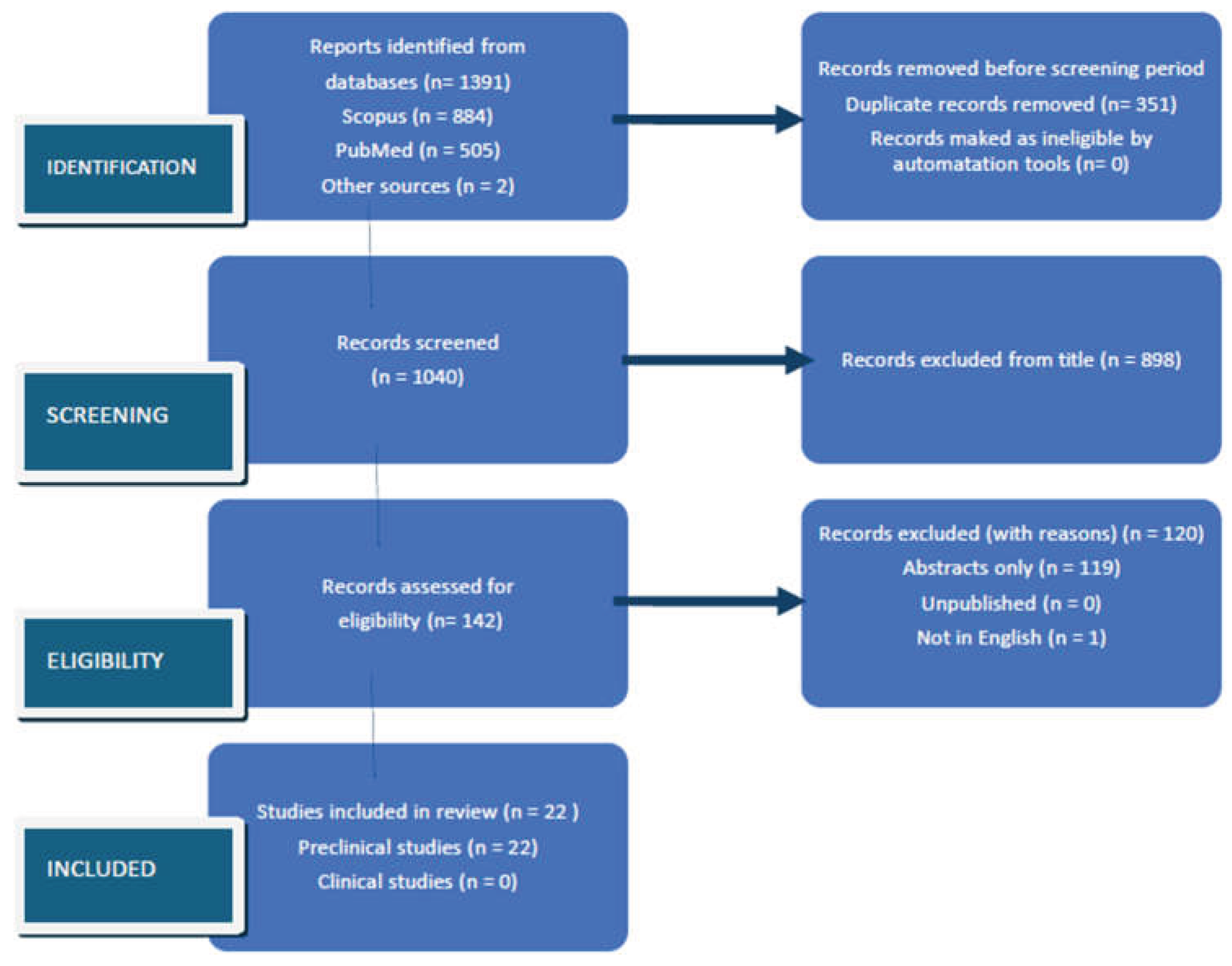
Disclaimer/Publisher’s Note: The statements, opinions and data contained in all publications are solely those of the individual author(s) and contributor(s) and not of MDPI and/or the editor(s). MDPI and/or the editor(s) disclaim responsibility for any injury to people or property resulting from any ideas, methods, instructions or products referred to in the content. |
© 2026 by the authors. Licensee MDPI, Basel, Switzerland. This article is an open access article distributed under the terms and conditions of the Creative Commons Attribution (CC BY) license (http://creativecommons.org/licenses/by/4.0/).
Copyright: This open access article is published under a Creative Commons CC BY 4.0 license, which permit the free download, distribution, and reuse, provided that the author and preprint are cited in any reuse.